KDOQI US Commentary on the 2012 KDIGO Clinical … · KDOQI US Commentary on the 2012 KDIGO...
24
KDOQI Commentary KDOQI US Commentary on the 2012 KDIGO Clinical Practice Guideline for Acute Kidney Injury Paul M. Palevsky, MD, 1,2 Kathleen D. Liu, MD, PhD, 3 Patrick D. Brophy, MD, 4 Lakhmir S. Chawla, MD, 5 Chirag R. Parikh, MD, PhD, 6,7 Charuhas V. Thakar, MD, 8,9 Ashita J. Tolwani, MD, 10 Sushrut S. Waikar, MD, 11 and Steven D. Weisbord, MD 1,2 In response to the recently released 2012 KDIGO (Kidney Disease: Improving Global Outcomes) clinical practice guideline for acute kidney injury (AKI), the National Kidney Foundation organized a group of US experts in adult and pediatric AKI and critical care nephrology to review the recommendations and comment on their relevancy in the context of current US clinical practice and concerns. The first portion of the KDIGO guideline attempts to harmonize earlier consensus definitions and staging criteria for AKI. While the expert panel thought that the KDIGO definition and staging criteria are appropriate for defining the epidemiology of AKI and in the design of clinical trials, the panel concluded that there is insufficient evidence to support their widespread application to clinical care in the United States. The panel generally concurred with the remainder of the KDIGO guidelines that are focused on the prevention and pharmacologic and dialytic management of AKI, although noting the dearth of clinical trial evidence to provide strong evidence-based recommendations and the continued absence of effective therapies beyond hemodynamic optimization and avoidance of nephrotoxins for the prevention and treatment of AKI. Am J Kidney Dis. 61(5):649-672. Published by Elsevier Inc. on behalf of the National Kidney Foundation, Inc. This is a US Government Work. There are no restrictions on its use. Editorial, p. 686 INTRODUCTION KDIGO (Kidney Disease: Improving Global Out- comes) is an international initiative to develop and implement clinical practice guidelines for patients with kidney disease. In March 2012, KDIGO published its guideline for the evaluation and management of acute kidney injury (AKI). 1 This guideline covers numerous topics, including the definition and classification of AKI, the prevention and treatment of AKI in general with specific recommendations for the prevention of contrast- induced AKI, and the management of renal replacement therapy (RRT) in patients with AKI. Because interna- tional guidelines need to be adapted for the United States, the National Kidney Foundation–Kidney Disease Outcomes Quality Initiative (NKF-KDOQI) convened a multidisciplinary work group with expertise in adult and pediatric nephrology and critical care medicine to com- ment on the applicability and implementation of the KDIGO AKI guideline in the United States. This com- mentary provides a summary of the KDIGO recommen- dation statements along with the supporting rationales and comments on their applicability to clinical practice in the United States. The KDOQI Work Group congratu- lates KDIGO, the members of the AKI Guideline Work Group, and the evidence review team for producing such a comprehensive document and believes that this guide- line will be of great value to health professionals and will advance both current clinical care of patients with AKI and future clinical research. AKI represents the sudden loss of kidney function, generally occurring over the course of hours to days and resulting in the retention of metabolic waste products and dysregulation of fluid, electrolyte, and acid-base homeostasis. During the past decade, this acute loss of kidney function, previously referred to as acute renal failure, has been the subject of significant re-examination, with increased recognition of the impor- Originally published online March 18, 2013 From the 1 Renal Section, VA Pittsburgh Healthcare System; 2 Renal-Electrolyte Division, Department of Medicine, University of Pittsburgh School of Medicine, Pittsburgh, PA; 3 Nephrology Division, Department of Medicine, University of California, San Francisco, CA; 4 Nephrology Division, Department of Pediatrics, University of Iowa Carver College of Medicine, Iowa City, IA; 5 Department of Anesthesiology and Critical Care Medicine and Division of Renal Diseases and Hypertension, Department of Medicine, George Washington University, Washington, DC; 6 Re- nal Section, VA Connecticut Healthcare System, West Haven; 7 Section of Nephrology, Department of Medicine, Yale University School of Medicine, New Haven, CT; 8 Renal Section, Cincinnati VA Medical Center; 9 Division of Nephrology and Hypertension, Department of Internal Medicine, University of Cincinnati, Cincin- nati, OH; 10 Division of Nephrology, Department of Medicine, University of Alabama at Birmingham, Birmingham, AL; and 11 Renal Division, Department of Medicine, Brigham and Women’s Hospital, Boston, MA. Address correspondence to Paul M. Palevsky, MD, Rm 7E123 (111F-U), VAPittsburgh Healthcare System, University Dr, Pitts- burgh, PA 14240. E-mail: [email protected] Published by Elsevier Inc. on behalf of the National Kidney Foundation, Inc. This is a US Government Work. There are no restrictions on its use. 0272-6386/$0.00 http://dx.doi.org/10.1053/j.ajkd.2013.02.349 Am J Kidney Dis. 2013;61(5):649-672 649
Transcript of KDOQI US Commentary on the 2012 KDIGO Clinical … · KDOQI US Commentary on the 2012 KDIGO...

KDOQI Commentary
KDOQI US Commentary on the 2012 KDIGO Clinical PracticeGuideline for Acute Kidney Injury
Paul M. Palevsky, MD,1,2 Kathleen D. Liu, MD, PhD,3 Patrick D. Brophy, MD,4
Lakhmir S. Chawla, MD,5 Chirag R. Parikh, MD, PhD,6,7 Charuhas V. Thakar, MD,8,9
Ashita J. Tolwani, MD,10Sushrut S. Waikar, MD,11 and Steven D. Weisbord, MD1,2
In response to the recently released 2012 KDIGO (Kidney Disease: Improving Global Outcomes) clinicalpractice guideline for acute kidney injury (AKI), the National Kidney Foundation organized a group of USexperts in adult and pediatric AKI and critical care nephrology to review the recommendations and comment ontheir relevancy in the context of current US clinical practice and concerns. The first portion of the KDIGOguideline attempts to harmonize earlier consensus definitions and staging criteria for AKI. While the expertpanel thought that the KDIGO definition and staging criteria are appropriate for defining the epidemiology ofAKI and in the design of clinical trials, the panel concluded that there is insufficient evidence to support theirwidespread application to clinical care in the United States. The panel generally concurred with the remainderof the KDIGO guidelines that are focused on the prevention and pharmacologic and dialytic management ofAKI, although noting the dearth of clinical trial evidence to provide strong evidence-based recommendationsand the continued absence of effective therapies beyond hemodynamic optimization and avoidance ofnephrotoxins for the prevention and treatment of AKI.Am J Kidney Dis. 61(5):649-672. Published by Elsevier Inc. on behalf of the National Kidney Foundation, Inc.This is a US Government Work. There are no restrictions on its use.
Editorial, p. 686
INTRODUCTION
KDIGO (Kidney Disease: Improving Global Out-comes) is an international initiative to develop andimplement clinical practice guidelines for patients withkidney disease. In March 2012, KDIGO published itsguideline for the evaluation and management of acutekidney injury (AKI).1 This guideline covers numeroustopics, including the definition and classification of AKI,the prevention and treatment of AKI in general withspecific recommendations for the prevention of contrast-induced AKI, and the management of renal replacementtherapy (RRT) in patients with AKI. Because interna-tional guidelines need to be adapted for the UnitedStates, the National Kidney Foundation–Kidney DiseaseOutcomes Quality Initiative (NKF-KDOQI) convened amultidisciplinary work group with expertise in adult andpediatric nephrology and critical care medicine to com-ment on the applicability and implementation of theKDIGO AKI guideline in the United States. This com-mentary provides a summary of the KDIGO recommen-dation statements along with the supporting rationalesand comments on their applicability to clinical practicein the United States. The KDOQI Work Group congratu-lates KDIGO, the members of the AKI Guideline WorkGroup, and the evidence review team for producing sucha comprehensive document and believes that this guide-line will be of great value to health professionals and willadvance both current clinical care of patients with AKI
and future clinical research.Am J Kidney Dis. 2013;61(5):649-672
AKI represents the sudden loss of kidney function,generally occurring over the course of hours to daysand resulting in the retention of metabolic wasteproducts and dysregulation of fluid, electrolyte, andacid-base homeostasis. During the past decade, thisacute loss of kidney function, previously referred to asacute renal failure, has been the subject of significantre-examination, with increased recognition of the impor-
Originally published online March 18, 2013From the 1Renal Section, VA Pittsburgh Healthcare System;
2Renal-Electrolyte Division, Department of Medicine, Universityof Pittsburgh School of Medicine, Pittsburgh, PA; 3NephrologyDivision, Department of Medicine, University of California, SanFrancisco, CA; 4Nephrology Division, Department of Pediatrics,University of Iowa Carver College of Medicine, Iowa City, IA;5Department of Anesthesiology and Critical Care Medicine andDivision of Renal Diseases and Hypertension, Department ofMedicine, George Washington University, Washington, DC; 6Re-nal Section, VA Connecticut Healthcare System, West Haven;7Section of Nephrology, Department of Medicine, Yale UniversitySchool of Medicine, New Haven, CT; 8Renal Section, CincinnatiVA Medical Center; 9Division of Nephrology and Hypertension,Department of Internal Medicine, University of Cincinnati, Cincin-nati, OH; 10Division of Nephrology, Department of Medicine,University of Alabama at Birmingham, Birmingham, AL; and11Renal Division, Department of Medicine, Brigham and Women’sHospital, Boston, MA.
Address correspondence to Paul M. Palevsky, MD, Rm 7E123(111F-U), VA Pittsburgh Healthcare System, University Dr, Pitts-burgh, PA 14240. E-mail: [email protected]
Published by Elsevier Inc. on behalf of the National KidneyFoundation, Inc. This is a US Government Work. There are norestrictions on its use.
0272-6386/$0.00
http://dx.doi.org/10.1053/j.ajkd.2013.02.349649

s.
Palevsky et al
tance of relatively small changes in kidney function onboth short- and longer term clinical outcomes.2-6 Thishas resulted in the change in terminology from acuterenal failure, for which the focus generally was limitedto the most severe episodes with complete or near-complete loss of kidney function, to the current terminol-ogy of AKI, with increased focus on smaller decrementsin kidney function.7-9
AKI may develop in a wide variety of settings, includ-ing in ambulatory outpatients, hospitalized patients, and,in particular, critically ill patients, for whom AKI repre-sents a common complication of both underlying diseaseand treatment.AKI is associated with substantial morbid-ity and mortality. For example, severe AKI occurs in�5% of critically ill patients and is associated withmortality rates of 40%-70%.10-12 Although recovery ofkidney function occurs in the majority of patients surviv-ing an episode of AKI, many patients remain dialysisdependent or are left with severe renal impairment. Morerecently, it has been recognized that even patients whohave complete or near-complete recovery of kidneyfunction are at increased risk of progressive chronickidney disease (CKD) and that superimposition of AKI
Table 1. RIFLE and AKIN Criteria
RIFLE
Class SCra
Risk Increased SCr to �1.5� baseline Urinefor
Injury Increased SCr to �2� baseline Urinefor
Failure Increased SCr to �3� baseline; or anincrease of �0.5 mg/dL to a valueof �4 mg/dL
Urinefor�1
Loss Need for RRT for �4 wk
End Stage Need for RRT for �3 mo
Abbreviations: AKI, acute kidney injury; AKIN, Acute Kidney Injrenal replacement therapy; SCr, serum creatinine.
aFor RIFLE, the increase in SCr should be both abrupt (within 1bFor AKIN, the increase in SCr must occur in less than 48 hour
Table 2. Pediatric Modified RIFLE (pRIFLE) Crit
Class eCCr
Risk eCCr decrease by �25%
Injury eCCr decrease by �50%
Failure eCCr decrease by �75%; or eCCr �35 mL/min/1
Loss Persistent failure for �4 wk
End Stage Persistent failure for �3 mo
Abbreviations and definitions: AKI, acute kidney injury; eCCr:risk, injury, failure, loss, end-stage disease.
Adapted and reproduced from Akcan-Arikan et al,20 with permission
650
on CKD is associated with acceleration in the rate ofprogression to end-stage disease.13-17
Our understanding of the epidemiology of AKI andinterpretation of results across clinical trials has beenhindered by the prior absence of a broadly acceptedclinical definition, with more than 30 operationaldefinitions of AKI used in published studies.18 Duringthe past decade, there has been a considerable effort toforge a consensus definition. The first attempt atdeveloping a consensus definition, known as theRIFLE criteria, was developed by the Acute DialysisQuality Initiative (ADQI) in 2002 (Table 1).19 Thisdefinition considered 3 strata of severity (risk, injury,and failure) based on the magnitude of increase inserum creatinine level and/or the duration of oliguria,as well as 2 outcome stages (loss of kidney functionand end-stage kidney disease). The risk, injury, andfailure categories were constructed to provide grada-tions in severity of kidney dysfunction, with greatersensitivity associated with risk and greater specificitywith failure. The RIFLE criteria subsequently weremodified by the AKI Network (AKIN) by the additionof an absolute increase in serum creatinine level �0.3
iagnosis and Classification of AKI
ne Outputmon to both)
AKIN
Stage SCrb
ut �0.5 mg/kg/h 1 Increase in SCr �0.3 mg/dL orincrease in SCr to �150%-200% of baseline
ut �0.5 mg/kg/hh
2 Increase in SCr to �200%-300% of baseline
ut �0.3 mg/kg/hh or anuria for
3 Increase in SCr to �300% ofbaseline; or to �4 mg/dLwith an acute increase of�0.5 mg/dL; or on RRT
etwork; RIFLE, risk, injury, failure, loss, end-stage disease; RRT,
ys) and sustained (�24 hours).
r Diagnosis and Classification of AKI in Children
Urine Output
Urine output �0.5 mL/kg/h for �8 h
Urine output �0.5 mL/kg/h for �16 h2 Urine output �0.3 mL/kg/h for �12 h; or anuria for �12 h
ated creatinine clearance using the Schwartz formula; RIFLE,
for D
Uri(com
outp�6 h
outp�12
outp�122 h
ury N
-7 da
eria fo
.73 m
estim
of MacMillan Publishers Ltd.
Am J Kidney Dis. 2013;61(5):649-672

KDOQI Commentary
mg/dL to the definition of AKI, a shortening of thetime for the increase in serum creatinine level from 7days to no more than 48 hours, and elimination of the2 outcome criteria (Table 1).7,8 A modification of theRIFLE criteria for use in pediatric patients (pRIFLE)has also been developed (Table 2).20 Validation stud-ies using these definitions have demonstrated in-creased mortality risk associated with progressivelymore severe stages of AKI. The KDIGO AKI guide-line builds upon these earlier efforts in defining AKI,with a full section of the guideline devoted to thedefinition of AKI.
The development of successful therapeutic strate-gies for the prevention and treatment of most forms ofAKI has been disappointing. Although numerousagents have shown promise in experimental models,none has demonstrated utility in clinical care. Aparticular area of focus has been in the prevention ofcontrast-induced AKI. This common cause of AKI hasbeen the focus of multiple preventative interventionsbecause individuals at high risk of contrast-inducedAKI can be readily identified and the timing ofexposure can be predetermined, allowing an opportu-nity for intervention. In the absence of effective phar-macologic therapy, the management of establishedAKI is predominantly supportive care, with the use ofRRT in severe AKI. There has been tremendousadvancement in the technology and modalities avail-able for providing RRT; however, the optimal ap-proach to management of RRT in this setting remainscontroversial. The KDIGO AKI guideline providesspecific recommendations, based on the current litera-ture, for best practices in the prevention and manage-ment of AKI. In this KDOQI commentary, we haveattempted to place this international guideline in thecontext of care practices in the United States. How-ever, our commentary should be viewed in conjunc-tion with the full KDIGO document when makingclinical decisions.
REVIEW AND APPROVAL PROCESS FORTHIS COMMENTARY
This commentary was developed by a Work Groupconvened by the NKF-KDOQI, beginning with se-lecting Co-Chairs by the KDOQI steering commit-tee and individual members selected based on theirclinical expertise and interest in the guideline pro-cess. Teleconferences took place during 2011 and2012 to determine the specific areas for focus forthis commentary. Individual sections focusing oneach of the topical areas of the KDIGO AKI guide-line were drafted by groups of coauthors based ondetailed review of the particular KDIGO chaptersupplemented by additional literature review, as
needed. Because this was a commentary, no specificAm J Kidney Dis. 2013;61(5):649-672
voting occurred; rather, consensus among coau-thors was achieved through discussion. The docu-ment was reviewed and approved by all coauthorsand by the KDOQI leadership.
ASSESSMENT OF GUIDELINE QUALITY
The KDIGO AKI guideline contains 87 individualrecommendations, of which 26 (30%) are ungraded,39 (45%) are level 2 recommendations, and only 22(25%) are level 1 recommendations, reflecting therelative paucity of high-level data guiding the manage-ment of AKI (Fig 1). In addition, many of the level 1recommendations advise against the use of specificagents or therapeutic interventions.
All members of the Work Group completed theAGREE (Appraisal of Guideline for Research andEvaluation) II instrument to assess the quality of theKDIGO AKI guideline.21-23 Mean Work Group scoresfor each of the 23 elements and scaled domain scoresfor the 6 domains are provided in Table 3, along withthe scoring range for domain and overall scores. Thedomain scores ranged from 0.53 for applicability to0.85 for editorial independence. The mean overallscore provided was 5.3 � 1.0 on a 7-point scale. AllWork Group members recommended the KDIGOAKI guideline for use, although the majority sug-gested that modifications could improve the guide-
Figure 1. Strength of recommendation and level of evi-dence of the KDIGO Clinical Practice Guideline for AcuteKidney Injury recommendations. Level 1 corresponds to arecommendation statement of “we recommend”; Level 2, to astatement of “we suggest”; Not Graded was used to provideguidance based on common sense or when the topic does notallow adequate application of evidence. The quality of support-ing evidence is graded from A to D, with letter grades corre-sponding to high, moderate, low, and very low quality ofevidence, respectively.
line.
651
Palevsky et al
REVIEW OF KDIGO AKI RECOMMENDATIONS
Definition andClassificationofAKI
Commentary
Overview. The first group of recommendation state-ments in the KDIGO AKI Guideline addresses thedefinition of AKI (Box 1). Of these 13 recommenda-tion statements, 12 are not graded. The KDIGO WorkGroup began by defining AKI by harmonizing the
Table 3. KDOQI Work Group’s Appraisal of the
Domain Ele
Scope and purpose 1. The overall objective(s) of the gu
2. The health question(s) covered bdescribed.
3. The population (patients, public,to apply is specifically described.
Stakeholder involvement 4. The guideline development grouprelevant professional groups.
5. The views and preferences of theetc) have been sought.
6. The target users of the guideline
Rigor of development 7. Systematic methods were used t
8. The criteria for selecting the evid
9. The strengths and limitations of tdescribed.
10. The methods for formulating thedescribed.
11. The health benefits, side effectsformulating the recommendations
12. There is an explicit link betweensupporting evidence.
13. The guideline has been externapublication.
14. A procedure for updating the gu
Clarity of presentation 15. The recommendations are spec
16. The different options for managissue are clearly presented.
17. Key recommendations are easi
Applicability 18. The guideline describes facilitat
19. The guideline provides advice arecommendations can be put into
20. The potential resource implicatirecommendations have been con
21. The guideline presents monitori
Editorial independence 22. The views of the funding body hguideline.
23. Competing interests of guidelinebeen recorded and addressed.
Abbreviation: AGREE, Appraisal of Guideline for Research andaGiven as mean � standard deviation. Individual element st
disagree” and 7 represents “strongly agree.”bDomain scores are reported on a scale from 0-1, where a sco
score for each element in the domain and a score of 1 indicating thdomain.
prior RIFLE and AKIN criteria.7,19 In the RIFLE
652
criteria (Table 1), AKI was defined based on a �50%increase in serum creatinine level occurring over 1-7days or the presence of oliguria for more than 6 hours.The AKIN criteria added an absolute increase inserum creatinine level of 0.3 mg/dL and reduced thetimeframe for the increase in serum creatinine level to48 hours (Table 1). The KDIGO Work Group harmo-nized these 2 definitions, keeping the absolute in-crease in serum creatinine level of �0.3 mg/dL within
O AKI Guideline Using the AGREE II Instrument
ScoreaScaled Domain
Scoreb
e is (are) specifically described. 6.0 � 0.9 0.83
guideline is (are) specifically 6.1 � 0.8
o whom the guideline is meant 5.8 � 1.3
udes individuals from all 5.7 � 1.6 0.58
et population (patients, public, 2.9 � 1.8
learly defined. 4.9 � 1.1
rch for evidence. 6.0 � 1.0 0.76
are clearly described. 6.1 � 0.9
dy of evidence are clearly 6.1 � 0.9
mmendations are clearly 5.9 � 0.9
risks have been considered in 5.6 � 1.5
ecommendations and the 5.4 � 0.9
viewed by experts prior to its 4.9 � 2.1
e is provided. 4.4 � 2.2
nd unambiguous. 4.9 � 0.9 0.73
t of the condition or health 5.1 � 1.5
ntifiable. 6.1 � 0.8
nd barriers to its application. 4.6 � 1.8 0.53
tools on how thetice.
4.1 � 1.5
f applying theed.
4.4 � 1.3
d/or auditing criteria. 3.7 � 1.9
ot influenced the content of the 6.1 � 0.9 0.85
elopment group members have 6.1 � 0.9
luation.ent is scored on a 7-point scale where 1 represents “strongly
0 would represent all reviewers providing the minimum possiblel reviewers gave the highest possible score for all elements in the
KDIG
ment
idelin
y the
etc) t
incl
targ
are c
o sea
ence
he bo
reco
, and.
the r
lly re
idelin
ific a
emen
ly ide
ors a
nd/orprac
ons osider
ng an
ave n
dev
Evaatem
re ofat al
48 hours from the AKIN definition, but returning to
Am J Kidney Dis. 2013;61(5):649-672

he gu
KDOQI Commentary
the 7-day timeframe for the �50% increase in serumcreatinine level. With regard to pediatric AKI, theKDIGO definition did not adopt the smaller changesin estimated creatinine clearance proposed in thepRIFLE criteria. Rather, the KDIGO Work Groupsuggested that the absolute change in creatinine levelof �0.3 mg/dL that is part of the KDIGO AKIdefinition would capture most pediatric AKI. TheKDIGO definition considers a decrease in estimatedglomerular filtration rate (eGFR) to �35 mL/min/1.73 m2 to be stage 3 AKI in pediatric patients, asinitially proposed in the pRIFLE criteria.20
Overall, while our KDOQI Work Group concurredwith the efforts on the part of KDIGO to meld the RIFLEand AKIN criteria into a uniform definition of AKI, wehave significant reservations regarding the applicabilityof this definition and staging system to clinical practice.The RIFLE and AKIN definitions and staging systemshave substantially enhanced the ability to conduct epide-miologic studies, and we support the use of the KDIGOAKI guideline definition and staging strategy for future
Box 1. Summary of KDIGO Recomm
2.1.1: AKI is defined as any of the following (Not Graded):● Increase in SCr by �0.3 mg/dL (�26.5 �mol/L) within 4● Increase in SCr to �1.5 times baseline, which is known● Urine volume �0.5 mL/kg/h for 6 hours.
2.1.2: AKI is staged for severity according to the following criteriStage 1: Increase in SCr by 1.5-1.9 times baseline; OR
Increase in sSCr by �0.3 mg/dL (�26.5 �mol/L)Urine output �0.5 mL/kg/h for 6-12 hours
Stage 2: Increase in SCr by 2.0-2.9 times baseline; ORUrine output �0.5 mL/kg/h for �12 hours
Stage 3: Increase in SCr by 3.0 times baseline; ORIncrease in SCr to 4.0 mg/dL (353.6 �mol/L); ORInitiation of renal replacement therapy; ORIn patients �18 years, decrease in eGFR to 35 mUrine output �0.3 mL/kg/h for �24 hours; ORAnuria for �12 hours
2.1.3: The cause of AKI should be determined whenever possib2.2.1: We recommend that patients be stratified for risk of AKI a2.2.2: Manage patients according to their susceptibilities and e
(Not Graded)2.2.3: Test patients at increased risk for AKI with measuremen
frequency and duration of monitoring based on patient ris2.3.1: Evaluate patients with AKI promptly to determine the caus2.3.2: Monitor patients with AKI with measurements of SCr an
2.1.2. (Not Graded)2.3.3: Manage patients with AKI according to the stage (Fig 2) a2.3.4: Evaluate patients 3 months after AKI for resolution, new o
● If patients have CKD, manage these patients as detaile● If patients do not have CKD, consider them to be at incr
Guideline 3 for patients at increased risk for CKD. (Not
Note: Conversion factor for SCr mg/dL to �mol/L, �88.4.Abbreviations: AKI, acute kidney injury; CKD, chronic kidne
creatinine.Reproduced with permission of KDIGO from the KDIGO ClinicaaThe staging information is adapted from a table presented in t
epidemiologic research until novel biomarkers facilitate
Am J Kidney Dis. 2013;61(5):649-672
the development of better criteria. We concur that therigorous application of a standard definition will permitepidemiologic comparisons across populations and overtime. In addition, the definitions as described have valuein standardization of entry criteria and endpoints inclinical trials of AKI. However, we question whetherthese criteria are currently appropriate to guide clinicalmanagement of adult patients.
The successive stages of a serum creatinine–basedAKI definition reflect a tradeoff between sensitivity andspecificity. At low stages, sensitivity is high (ie, almostevery patient with true AKI is identified), but specificityis low (ie, many patients without true AKI are misidenti-fied as having AKI). As part of the decision to recom-mend a cutoff value based on a biomarker such as serumcreatinine or physiologic variable such as urine output,the implications of the cutoff need to be carefully consid-ered. We thought that particularly in the United States,where defensive medicine is common due to the highlylitigious environment in which many physicians prac-tice, the KDIGO definition may result in a dramatic
ation Statements: Definition of AKI.
rs; oresumed to have occurred within the prior 7 days; or
ot Graded)
in/1.73 m2; OR
ot Graded)ing to their susceptibilities and exposures. (1B)res to reduce the risk of AKI (see relevant guideline sections).
SCr and urine output to detect AKI. (Not Graded) Individualizeclinical course. (Not Graded)
th special attention to reversible causes. (Not Graded)e output to stage the severity, according to Recommendation
use. (Not Graded)or worsening of pre-existing CKD. (Not Graded)e KDOQI CKD Guideline (Guidelines 7-15). (Not Graded)
d risk for CKD and care for them as detailed in the KDOQI CKDed)
ease; eGFR, estimated glomerular filtration rate; SCr, serum
ctice Guideline for Acute Kidney Injury.1
ideline.
end
8 houor pr
a.a (N
; OR
L/m
le. (Nccordxposu
ts ofk ande, wi
d urin
nd canset,d in theaseGrad
y dis
l Pra
increase in nephrology consultations and have the unin-
653

Palevsky et al
tended adverse consequence of diverting limited re-sources toward a large number of patients for whomspecialty referral may have uncertain benefit.
However, we recognize that the clinical applicationof the KDIGO AKI definition could increase the earlyrecognition of AKI. Given the current lack of effec-tive pharmacologic interventions for AKI, early recog-nition based on small changes in serum creatininelevel or urine output could allow implementation ofsimple interventions—such as avoiding potentiallynephrotoxic medications or diagnostic or therapeuticprocedures that are associated with increased risk ofAKI unless clearly clinically indicated, or judiciousprotocol-driven volume resuscitation—that could re-duce the risk of more severe kidney injury.
In addition to these more global concerns, our WorkGroup identified several specific issues regarding thedefinition and staging criteria that we believe requirespecific comment, as discussed next.
Inclusion of urine output into the definition of AKI.Both RIFLE and AKIN included duration of oliguriain their definitions and staging criteria (Table 1).Several criticisms have been offered of these urineoutput criteria, many of which are acknowledged bythe KDIGO AKI Guideline Work Group but deservefurther mention. Only a small number of studies haveexamined the urine output criteria for AKI and corre-lated them with adverse clinical outcomes, in contrastto the numerous studies that have focused on theserum creatinine criteria. The studies that have evalu-ated the urine output component of the criteria havefound poor calibration between these criteria and theserum creatinine criteria.24 Specifically, when as-sessed, there was poor prognostic correlation betweenthe briefer durations of oliguria and small changes inserum creatinine level. Second, oliguria may be anappropriate response to volume depletion and hencereflect under-resuscitation rather than injury to thekidney, which is implicit in the term “AKI.” There-fore, even demonstrating an association between oli-guria and adverse clinical outcomes is insufficient tojustify its incorporation into the definition of AKI.Third, the use of a weight-based definition for AKIlimits its use in the obese due to the nonlinear relation-ship between body weight and urine output. Under thecurrent definition, urine output of 40 mL/h in a 90-kgpatient for 12 hours would lead to classification asstage 2 AKI. Fourth, diuretic administration has beenshown to change RIFLE classification by urine outputcriteria,24 as would be expected when a definition isbased on a physiologic variable that can be manipu-lated pharmacologically. Finally, the use of oliguria ina definition of AKI may promote its use in clinicalpractice as a surrogate end point. We know from
several studies that pharmacologic agents that can654
increase urine output independent of augmentation ofkidney function, such as loop diuretics or dopamine,may not be helpful and may even be harmful in somesettings.25,26 While the inclusion of these criteria inthe AKI guideline may have the beneficial effect ofencouraging more rigorous documentation of urineoutput and overall fluid balance in settings outside theoperating room and critical care units, there are alsorisks of unintended consequences with defining tran-sient oliguria as AKI. In particular, we believe thatcurrent data are inadequate to support the reliance onoliguria as a surrogate end point in clinical trials or inperformance metrics. We note that pediatric, and inparticular neonatal, providers rely heavily on urineoutput and weight-based fluid balance and thereforeinclusion of these parameters in the pediatric AKIcriteria is reasonable; furthermore, neonates in particu-lar are unlikely to have morbid obesity and thereforethe methodological issues with weight-based urineoutput are somewhat less problematic. Nonetheless,further research is needed in the pediatric populationto determine what metrics are best used to define AKI,and practitioners should recognize that many of thesame limitations to the use of urine output criteriaapply in the pediatric population.
Use of small changes in serum creatinine to defineAKI. The inclusion of a small absolute change inserum creatinine level in the AKIN definition of AKIwas based on the demonstration by Chertow et al4 thatminor fluctuations in serum creatinine concentrationwere strongly associated with adverse outcomes in aretrospective analysis of data from individuals hospi-talized at a single medical center. Although the asso-ciation remained, albeit attenuated, after multivari-able adjustment, only administrative data wereavailable for construction of the multivariable mod-els, and residual confounding may have been present.Small changes in serum creatinine level have alsobeen identified to be of prognostic importance insubsequent studies. Even a 0.1-mg/dL increase inserum creatinine level appears to be associated withincreased risk compared to no change in serum creati-nine level.27 Whether these small changes in serumcreatinine level reflect clinically meaningful fluctua-tions in kidney function that are causally linked toadverse outcomes or are merely a marker of underly-ing vascular disease or diminished renal reserve, whichcould be the actual mediators of the adverse out-comes, remains unresolved. Furthermore, an implicitassumption underlying the incorporation of smallchanges in serum creatinine concentration into thedefinition of AKI is that GFR in an individual isconstant over time and not subject to physiologicfluctuation. In addition, biological fluctuation in se-
rum creatinine level may also result from variations inAm J Kidney Dis. 2013;61(5):649-672
KDOQI Commentary
diet, creatinine generation, tubular secretion, medica-tions, and variability in sodium and volume homeosta-sis that are within the physiologic range. Such fluctua-tion in serum creatinine concentration is moreprominent in individuals with decreased renal reserveor overt CKD. For all these reasons, an absolutechange in serum creatinine level of 0.3 mg/dL mayrepresent a relatively inconsequential change in GFRin patients with underlying CKD and not reflect super-imposed acute pathology. Despite these concerns inadult patients, in neonatal/pediatric patients, such smallchanges in serum creatinine level may represent rela-tively large changes in actual GFR that should not bedisregarded. Whether novel biomarkers of tubularinjury may enhance risk stratification of patients withsmall changes in serum creatinine level is a questionthat requires further investigation and may lead tofurther refinement of AKI staging.
The independent role of duration of AKI. Severalrecent studies have suggested that duration of AKImay be a more important predictor of outcomes thanthe magnitude of change in serum creatinine level.For example, in one study, even a transient elevationin creatinine level (�3 days) was associated withincreased risk of death, and the risk of mortality wasgreater in patients with prolonged duration of AKI.28
After adjustment for duration of AKI, staging was nolonger predictive of adverse outcomes. Unfortunately,this important dimension can only be assessed retro-spectively and cannot be included in prospectivestaging criteria; however, consideration should begiven to including an assessment of duration of AKIin future criteria designed for epidemiologic studies.
Pediatric-specific concerns. The KDOQI WorkGroup also had several concerns regarding the appli-cation the KDIGO definition and staging of AKI to thepediatric population. One of the major concerns withboth the RIFLE and AKIN criteria is that they areproblematic for smaller (pediatric) patients with lowmuscle mass. The pRIFLE criteria were developed toaddress the issues of applying the adult criteria to thepediatric population.20 For example, because abnor-mally elevated serum creatinine measurements areoften overlooked in pediatric patients (due to adultnormative data in laboratory readouts), utilization ofthe pRIFLE criteria might improve identification ofthose with true AKI. The pRIFLE criteria have beenused widely in pediatric clinical research and effortsare now being made to apply these criteria in clinicalpractice. However, there are significant differencesbetween the KDIGO and pRIFLE criteria. Conse-quently, further validation will be required prior to theadoption of the KDIGO criteria into research andpractice. Specifically, pediatric nephrologists will want
to better understand whether the differences in the 2Am J Kidney Dis. 2013;61(5):649-672
definitions result in the identification of differentpatient groups, and if so, what systematic biases maybe introduced by these differences. Thus, it was un-clear to the KDOQI Work Group whether pediatricnephrologists would widely embrace the new KDIGOAKI definition or continue to apply the pRIFLEcriteria.
In addition, most pediatric patients will not neces-sarily have had previous serum creatinine measure-ments and therefore baseline renal function determina-tion is often assumed to be normal, which may beproblematic. In addition, the neonatal population re-mains an enigma in terms of renal function determina-tion, making both the definition and identification ofAKI in this population difficult. Many neonates havenot attained full renal mass development at birth andare obligatorily born with low GFR, which improveswith growth and development. AKI in neonates isoften associated with high urine output, which may gounrecognized given the paucity of serum creatininemeasurements in this patient cohort. Finally, immedi-ately after birth, serum creatinine level in neonatesoften reflects maternal creatinine levels, which mustbe considered when evaluating renal function. How-ever, the recommendation of a 0.3-mg/dL increase inserum creatinine level (even in the context of baselinematernal creatinine level) should be sufficient to trig-ger concern, and this component of the definition isapplicable in the neonatal population.
In addition to defining and staging AKI, the KDIGOguideline introduces the new term acute kidney dis-ease (AKD), defined as AKI, or GFR �60 mL/min/1.73 m2 for less than 3 months, or a decrease in GFRby �35% or an increase in serum creatinine level by�50% for less than 3 months, or structural kidneydamage of less than 3 months’ duration. While weunderstand the nosological rationale for developingterminology to describe patients with kidney diseasethat does not meet the criteria for either AKI or CKD,we have concern that the introduction of this terminol-ogy may confuse clinicians and inappropriately divertattention away from diagnostic considerations. WhileCKD encompasses a number of pathophysiologicallyand histopathologically distinct diseases, arguably themajority of patients with CKD have diseases such ashypertensive nephrosclerosis and diabetic nephroscle-rosis, for which treatment approaches are similar.However, in the acute setting, distinctions betweencauses of disease have greater consequence. To lumpthe broad range of conditions that result in acute andsubacute kidney disease, ranging from acute tubularnecrosis to obstructive uropathy, atheroembolic dis-ease, and rapidly progressive glomerulonephritis, intothe single umbrella term of AKD runs the risk of
promoting diagnostic laziness among clinicians who655
tensi
Palevsky et al
would have a convenient name to apply rather than anabnormal laboratory value to investigate. We wouldhave welcomed a more in-depth discussion of whatspecific kidney diseases are likely to segregate toAKD as opposed to AKI or CKD and how the intro-duction of a new term was thought by the authors tohelp in clinical practice. While AKD may be a usefulconstruct in epidemiologic studies, we believe the useof this term should be discouraged in clinical practice.
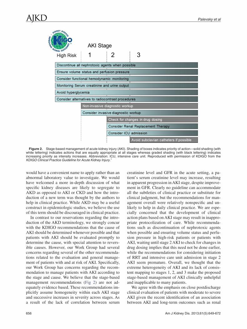
In contrast to our reservations regarding the intro-duction of the AKD terminology, we strongly concurwith the KDIGO recommendations that the cause ofAKI should be determined whenever possible and thatpatients with AKI should be evaluated promptly todetermine the cause, with special attention to revers-ible causes. However, our Work Group had severalconcerns regarding several of the other recommenda-tions related to the evaluation and general manage-ment of patients with and at risk of AKI. Specifically,our Work Group has concerns regarding the recom-mendation to manage patients with AKI according tothe stage and cause. We believe that the stage-basedmanagement recommendations (Fig 2) are not ad-equately evidence based. These recommendations im-plicitly assume homogeneity within each AKI stageand successive increases in severity across stages. As
Figure 2. Stage-based management of acute kidney injury (Awhite lettering) indicates actions that are equally appropriate aincreasing priority as intensity increases. Abbreviation: ICU, inKDIGO Clinical Practice Guideline for Acute Kidney Injury.1
a result of the lack of correlation between serum
656
creatinine level and GFR in the acute setting, a pa-tient’s serum creatinine level may increase, resultingin apparent progression in AKI stage, despite improve-ment in GFR. Clearly no guideline can accommodateall the subtleties of clinical practice or substitute forclinical judgment, but the recommendations for man-agement overall were relatively nonspecific and un-likely to help in daily clinical practice. We are espe-cially concerned that the development of clinicalaction plans based on AKI stage may result in inappro-priate protocolization of care. While recommenda-tions such as discontinuation of nephrotoxic agentswhen possible and ensuring volume status and perfu-sion pressure in high-risk patients or patients withAKI, waiting until stage 2 AKI to check for changes indrug dosing implies that this need not be done earlier,while the recommendations for considering initiationof RRT and intensive care unit admission in stage 2AKI seem premature. Overall, we thought that theextreme heterogeneity of AKI and its lack of consis-tent mapping to stages 1, 2, and 3 make the proposedstage-based management of AKI clinically unhelpfuland inapplicable to many patients.
We agree with the emphasis on close postdischargeclinical evaluation of patients with moderate to severeAKI given the recent identification of an association
hading of boxes indicates priority of action—solid shading (withtages whereas graded shading (with black lettering) indicates
ve care unit. Reproduced with permission of KDIGO from the
KI). St all s
between AKI and long-term outcomes such as renal
Am J Kidney Dis. 2013;61(5):649-672

KDOQI Commentary
function decline and mortality. However, we are con-cerned that clinicians may be overwhelmed by 3-monthfollow-up of many patients with stage 1 AKI, whowill constitute the majority of individuals identifiedby the AKI definition due to transient oliguria or smallchanges in serum creatinine level. The risk of progres-sive CKD after AKI is related to the severity ofAKI,16,29,30 suggesting that risk stratification on thebasis of AKI severity may be useful in guiding thetiming of outpatient follow-up. Those with stage 3AKI, for example, will likely require far earlier post-discharge follow-up. Furthermore, patients with AKIin the setting of pre-existing CKD or those whodevelop worsening CKD as a consequence of anepisode of AKI could represent a particularly high-risk group.
In the pediatric population, the 3-month time tofollow-up may be reasonable. The CKiD (ChronicKidney Disease in Children) national trial has identi-fied that a large number of the patients available forthat patient cohort were at-risk patients in the neonatalpopulation.31 Although the evidence for transitionfrom AKI to CKD in the pediatric population is alsoobservational and further epidemiologic research isrequired, the number of pediatric patients with AKI ismuch smaller than in the adult population and thestakes of missing nascent CKD may be greater giventhe potential longer duration of follow-up than in theadult population. Thus, we would suggest that earlyfollow-up among pediatric patients with AKI is advis-able.
ImplicationsWithinUSHealthCare
1. The KDIGO definition and staging system forAKI provides an important tool for conducting epide-miologic studies and for the design of clinical trialsand helps increase awareness of the importance ofsmall changes in kidney function in the acute setting.However, there is insufficient validation of this defini-tion and staging system for its use in the diagnosis andclinical management of patients. In particular, we donot believe that there are sufficient data to support useof the stage-based management approach proposed inthe KDIGO guideline. Rather, management of pa-tients with AKI should be based on assessment ofoverall clinical status, including specific cause ofAKI, trends in kidney function over time, comorbidconditions, assessment of volume status, and concomi-tant acid-base and electrolyte disturbances.
2. While AKI is an important risk factor for thedevelopment and progression of CKD, the majority ofpatients with mild, readily reversible AKI (eg, thepatient with no baseline CKD who presents withreversible AKI in the setting of volume depletion) are
at relatively low risk of progressive CKD. From aAm J Kidney Dis. 2013;61(5):649-672
public health standpoint in the United States, fol-low-up of kidney function after an episode of AKIshould be targeted to the highest risk populations,including neonatal/pediatric patients, individuals withbaseline CKD, and patients with severe AKI or whohave incomplete recovery of kidney function at hospi-tal discharge.
3. The KDIGO recommendations for definition andstaging of adult AKI should not be used for thedevelopment of clinical performance measures.
4. Administrative coding for AKI should not bebased solely on this definition and staging system.
Prevention andTreatment ofAKI
Commentary
The section of the KDIGO guideline on preventionand treatment of AKI includes 7 level 1 recommenda-tions and 22 level 2 recommendations (Box 2). OurKDOQI Work Group agreed with the 7 level 1 recom-mendations and thought that they were generallyapplicable in the United States. These focused on theuse of vasopressors and fluids to treat patients inshock; avoidance of diuretics, dopamine, and recom-binant human IGF-1 (insulin-like growth factor 1) toprevent or treat AKI; therapeutic drug monitoringduring the use of aminoglycosides; and avoidance ofnephrotoxic medications when possible. Importantly,avoidance of nephrotoxins is not always possible. Forexample, some pathogens are not effectively treatedby azoles or echinocandins: Candida krusei is intrinsi-cally resistant to azoles, and Candida parapsilosis isfrequently resistant to echinocandins.
The KDOQI Work Group largely agreed with therecommendation to use isotonic crystalloids ratherthan colloids for volume expansion in patients at riskof AKI or with AKI. In addition, we noted that thereappears to be harm with starch-containing fluids.32-36
Therefore, these solutions should be avoided in pa-tients at risk of AKI or with AKI. Along the samelines, albumin resuscitation has been associated withharm in patients with traumatic brain injury andshould be avoided in that setting.37,38
There are also specific settings in which albumin isappropriate for initial management of expansion ofintravascular volume. Specifically, in patients withliver disease, intravenous albumin administration ap-pears to be beneficial for the prevention of renalfailure and death in patients with spontaneous bacte-rial peritonitis, as well as for the prevention of renalfailure in those undergoing large-volume paracente-sis.39,40 Along the same lines, the most recent diagnos-tic criteria for hepatorenal syndrome include a lack ofimprovement in renal function after volume expan-sion with albumin (1 g/kg/d up to 100 g/d) for at least
2 days and withdrawal of diuretic therapy.41 Data657

l Pra
Palevsky et al
published since the KDIGOAKI guideline first appearedsuggest that there may be harm associated with theadministration of hyperchloremic intravenous solutions,including crystalloid42,43; however, these studies haveall been observational and this premise needs to be testedin a randomized clinical trial given the enormous effectsize observed and the inability to control for othertemporal trends in care in the prior studies.
With regard to the use of vasopressors in addition tointravenous fluids in patients with vasomotor shock,we noted that fluid resuscitation is typically insuffi-cient to fully restore blood pressure to normal levels,which is critical for the prevention and managementof AKI. However, whether one vasopressor is moreeffective for patients with or at risk of AKI is un-known. In a large multicenter clinical trial of patientswith shock comparing dopamine and norepinephrine
Box 2. Summary of KDIGO Recommendati
3.1.1: In the absence of hemorrhagic shock, we suggest using ismanagement for expansion of intravascular volume in pat
3.1.2: We recommend the use of vasopressors in conjunction wi3.1.3: We suggest using protocol-based management of hem
worsening of AKI in high-risk patients in the perioperative3.3.1: In critically ill patients, we suggest insulin therapy targetin3.3.2: We suggest achieving a total energy intake of 20-30 kcal/3.3.3: We suggest to avoid restriction of protein intake with the a3.3.4: We suggest administering 0.8-1.0 g/kg/d of protein in non
patients with AKI on RRT (2D), and up to a maximum of 1.and in hypercatabolic patients. (2D)
3.3.5: We suggest providing nutrition preferentially via the enter3.4.1: We recommend not using diuretics to prevent AKI. (1B)3.4.2: We suggest not using diuretics to treat AKI, except in the3.5.1: We recommend not using low-dose dopamine to prevent3.5.2: We suggest not using fenoldopam to prevent or treat AKI.3.5.3: We suggest not using atrial natriuretic peptide (ANP) to p3.6.1: We recommend not using recombinant human (rh)IGF-13.7.1: We suggest that a single dose of theophylline may be giv
AKI. (2B)3.8.1: We suggest not using aminoglycosides for the treatm
alternatives are available. (2A)3.8.2: We suggest that, in patients with normal kidney function
daily rather than multiple-dose daily treatment regimens.3.8.3: We recommend monitoring aminoglycoside drug levels
hours. (1A)3.8.4: We suggest monitoring aminoglycoside drug levels when
(2C)3.8.5: We suggest using topical or local applications of aminogl
than i.v. application, when feasible and suitable. (2B)3.8.6: We suggest using lipid formulations of amphotericin B rat3.8.7: In the treatment of systemic mycoses or parasitic infe
echinocandins rather than conventional amphotericin B, i3.9.1: We suggest that off-pump coronary artery bypass graft su
AKI or need for RRT. (2C)3.9.2: We suggest not using NAC to prevent AKI in critically ill pa3.9.3: We recommend not using oral or i.v. NAC for prevention o
Abbreviations: AKI, acute kidney injury; IGF-1, insulin-like greplacement therapy.
Reproduced with permission of KDIGO from the KDIGO Clinica
for first-line vasopressor support, the use of dopamine
658
was associated with more adverse events in those withsepsis and with an increased risk of death in those withcardiogenic shock.44 In a subsequent meta-analysis andsystematic review of patients with septic shock, dopa-mine was associated with an increased risk of death andarrhythmias.45,46 Thus, we believe that dopamine shouldbe used with caution as the first-line agent of choice inpatients with shock at present.
Our Work Group did not fully agree with therecommendation to use protocol-based managementof hemodynamic and oxygenation parameters to pre-vent the development or worsening of AKI in high-risk patients in the perioperative setting or in patientswith septic shock. The group noted that the recommen-dation for early goal-directed therapy in patients withseptic shock is based primarily on single-center ran-domized clinical trials47,48 and observational stud-
atements: Prevention and Treatment of AKI
ic crystalloids rather than colloids (albumin or starches) as initialat risk for AKI or with AKI. (2B)
ids in patients with vasomotor shock with, or at risk for, AKI. (1C)amic and oxygenation parameters to prevent development org (2C) or in patients with septic shock (2C).
sma glucose 110-149 mg/dL (6.1-8.3 mmol/L). (2C)in patients with any stage of AKI. (2C)preventing or delaying initiation of RRT. (2D)olic AKI patients without need for dialysis (2D), 1.0-1.5 g/kg/d ing/d in patients on continuous renal replacement therapy (CRRT)
te in patients with AKI. (2C)
gement of volume overload. (2C)at AKI. (1A)
t (2C) or treat (2B) AKI.vent or treat AKI. (1B)neonates with severe perinatal asphyxia, who are at high risk of
f infections unless no suitable, less nephrotoxic, therapeutic
ady state, aminoglycosides are administered as a single dose
treatment with multiple daily dosing is used for more than 24
tment with single-daily dosing is used for more than 48 hours.
des (e.g., respiratory aerosols, instilled antibiotic beads), rather
an conventional formulations of amphotericin B. (2A)s, we recommend using azole antifungal agents and/or thel therapeutic efficacy can be assumed. (1A)not be selected solely for the purpose of reducing perioperative
s with hypotension. (2D)tsurgical AKI. (1A)
factor 1; i.v., intravenous; NAC, N-acetylcysteine; RRT, renal
ctice Guideline for Acute Kidney Injury.1
on St
otonientsth fluodynsetting plakg/dim ofcatab7 g/k
al rou
manaor tre(2C)
revento preen in
ent o
in ste(2B)when
trea
ycosi
her thction
f equargery
tientf pos
rowth
ies.49-52 There was no assessment of renal outcomes
Am J Kidney Dis. 2013;61(5):649-672

KDOQI Commentary
in the original randomized clinical trial, so it is un-clear if the intervention had any impact on renaloutcomes. Similarly, perioperative protocols have simi-larly shown aggregate improvement when assessed ina meta-analysis, but individual studies have beenlimited by small numbers and single-center design.53
Furthermore, our Work Group thought that the proto-col must be specified. That is, any protocol that isinstituted based on this recommendation should beone that has been previously studied and validated.For example, previous studies of increasing oxygentransport have demonstrated that the use of dobut-amine to increase oxygen transport can worsen mortal-ity,54 although the Rivers et al47 study of early goal-directed therapy suggests that dobutamine in anappropriate clinical context may not be harmful andmay have benefit. Thus, medical centers adoptingprotocolized care should only adopt protocols thathave been previously shown to be helpful and demon-strated no harm. If de novo protocols are developedfor use, they should be used in the confines of aclinical trial to ensure no harm.
With regard to glycemic control, we agreed withthe recommended target of 110-149 mg/dL, but notedthat emerging evidence suggests that rapid and sus-tained correction of glycemia in diabetic patients withprevious poor glycemic control may have worse out-comes.55 Our Work Group agreed with the recommen-dations for nutritional support in AKI, but noted thatthe evidence base for these recommendations is lim-ited. Specifically, the guideline recommends total en-ergy intake of 20-30 kcal/kg/d in patients with anystage of AKI with administration of 0.8-1.0 g/kg/d ofprotein in noncatabolic patients with AKI withoutneed for dialysis, 1.0-1.5 g/kg/d in patients with AKIon RRT, and up to a maximum of 1.7 g/kg/d inpatients on continuous RRT (CRRT) and in hypercata-bolic patients. The provision of protein to patientswith AKI is intended to avoid marked net negativenitrogen balance. Patients with AKI on RRT will haveadditional protein loss as amino acids are removed.Therefore, these patients require additional proteincompared with patients with AKI who are not on RRT.Similarly, patients who are hypercatabolic will re-quire additional protein to avoid net negative nitrogenbalance. Along the same lines, the KDOQI WorkGroup agreed with the recommendation to avoidrestriction of protein intake with the aim of preventingor delaying initiation of RRT. We also agreed with therecommendation to provide nutrition preferentially bythe enteral route in patients with AKI. The WorkGroup noted that although evidence is more limited inthe setting of AKI,56 in a recent large randomizedclinical trial of critically ill patients, early parenteral
nutrition was associated with a higher rate of compli-Am J Kidney Dis. 2013;61(5):649-672
cations, primarily infectious complications.57 Alongthe same lines, recent trials suggest that enteral feed-ing is tolerated by either the nasogastric or nasojejunalroutes.58,59
Nutritional support in pediatric patients must in-clude the recognition that these patients are generallyin a developmental growth phase; thus, their require-ments may be increased compared with adult patients.Recent estimates have been published in terms ofestimated protein/amino acid supplementation in criti-cally ill children and suggest that protein require-ments are on the order of 2-3 g/kg/d for children aged0-2 years, 1.5-2.0 g/kg/d for children aged 2-13 years,and 1.5 g/kg/d for adolescents aged 13-18 years.60
Children requiring RRT appear to need supplementa-tion beyond this.61
There are no pharmacotherapies available for theprevention or treatment of AKI in adults, so recommen-dations to avoid these are reasonable. Specifically, weagreed that although there is theoretical benefit to theuse of loop diuretics to prevent AKI, there are no datato support the use of diuretics to prevent AKI. Recentstudies in patients undergoing cardiac surgery or con-trast studies suggest that the use of diuretics does notprevent AKI62 and, in the case of contrast administra-tion, increases the risk of AKI.63,64 The KDOQI WorkGroup thought that it was important to comment onthe use of diuretics in patients with AKI and rhabdo-myolysis. With regard to osmotic diuretics, one retro-spective study has suggested that the use of mannitoladministration may be of benefit only in patients withmarked elevations in creatinine kinase level (�30,000U/L).65 However, even in these patients with severerhabdomyolysis, the true benefit associated with man-nitol administration remains undefined. Furthermore,mannitol should be administered carefully and iscontraindicated in patients with oligoanuria. Diureticsmay be used for the treatment of volume overload inAKI.66 We agree that low-dose dopamine, fenoldo-pam, IGF-1, and N-acetylcysteine (NAC) should notbe used for the treatment or prevention of AKI.
We concur with the recommendation to administera single dose of theophylline to neonates with severeperinatal asphyxia who are at high risk of AKI.Several clinical trials in the neonatal population sug-gest that although mortality outcomes are not af-fected, improved fluid control and higher GFR areassociated with theophylline administration in thissetting.67-69 A similar renal-selective response hasbeen demonstrated in asphyxiated term newbornsreceiving a single theophylline dose in the first 60minutes of life.70 Given the generally physiologichyperactive state of the renal autoregulatory systemapparent at transition from in utero to ex utero environ-
ments and the role that adenosine plays in these659
Palevsky et al
processes, the use of theophylline in the aforemen-tioned settings is reasonable.
A substantial number of recommendations weremade with regard to the use of aminoglycosides andantifungal agents. Our Work Group agreed with theserecommendations but noted that there are situations inwhich aminoglycosides or multidose regimens arepreferable (eg, multiple dose regimens remain thestandard of care for enterococcal endocarditis). Wealso noted that while local instillation of aminoglyco-sides is reasonable, it is unknown what the incidenceof nephrotoxicity is with local administration of ami-noglycosides; for example, there are case reports ofnephrotoxicity associated with inhaled tobramycintherapy.71-73
With regard to the use of lipid formulations ofamphotericin B rather than conventional formulationsof amphotericin B, we note that it has been suggestedthat the decreased incidence of AKI and the associateddecrease in hospitalization costs makes the lipid for-mulations cost-effective compared to conventionalformulations of amphotericin B.74,75 Furthermore,other simple strategies that may reduce the risk ofnephrotoxicity include discontinuation of diuretics,sodium loading and volume repletion, and potassiumand magnesium supplementation, particularly whenusing non-liposomal preparations of amphotericin B.76
The 2 most significant risk factors for the develop-ment of AKI following cardiac surgery are cardiopul-monary bypass time and a history of CKD.77,78 As aresult, there has been considerable interest in thepotential for off-pump bypass to decrease the risk ofAKI. The largest retrospective study focused on pa-tients with CKD was published after the KDIGO AKIguideline.79 This study examined nonemergent iso-lated coronary artery bypass grafting (CABG) casesin the Society of Thoracic Surgery Database from2004 through 2009 (N � 742,909). Among patientswith eGFR of 15-29 mL/min/1.73 m2, off-pumpCABG was associated with a decreased risk of postop-erative RRT (risk difference, 2.79; 95% confidenceinterval, 1.37-4.20) in the propensity-adjusted analy-sis (P � 0.01). There was no difference in those withless advanced CKD, although those with eGFR of30-59 mL/min/1.73 m2 were examined in aggregaterather than as separately as CKD stages 3a and 3b.This potential benefit needs to be balanced against theconcern of higher graft occlusion rates using off-pump techniques.80,81
ImplicationsWithinUSHealthCare
1. In the United States, starch-containing intrave-nous fluids should be avoided. The use of albumin forresuscitation should be limited to specific situations in
which it is clear that albumin is of benefit, given the660
increased costs of these solutions compared to crystal-loid and the intermittent nationwide shortages of thesesolutions. Recent studies have suggested that theremay be harm with hyperchloremic intravenous solu-tions (eg, isotonic saline), but this needs to be furthertested prior to the widespread use of balanced electro-lyte solutions with lower chloride content (eg, Plasma-Lyte, Hartmann’s and lactated Ringer’s) in the UnitedStates.
2. With regard to vasopressor support in patientswith shock, dopamine is associated with an increasedrate of complications in patients with septic andcardiogenic shock compared to norepinephrine. Al-though this needs to be further tested and a number ofguidelines still recommend the use of dopamine as afirst-line agent, norepinephrine should be used as afirst-line agent over dopamine unless there are spe-cific contraindications to the use of norepinephrine.
3. Protocol-based management of perioperative pa-tients and patients with sepsis is a reasonable ap-proach, but the protocols for such management needto be further developed and tested. We note thatseveral large randomized clinical trials will test proto-col-based management of sepsis (ClinicalTrials.gov identi-fiers NCT00975793 and NCT00510835 and Interna-tional Standard Randomised Controlled Trial NumberISRCTN36307479). Until then, care should not bebenchmarked based on the implementation of suchprotocols within a given time frame; it is reasonable touse time to antibiotics and time to fluid administrationas benchmarks for care in sepsis.
4. A target blood glucose level of 110-149 mg/dL incritically ill patients may be associated with decreasedrisk of AKI and other morbidity and mortality. How-ever, emerging evidence suggests that rapid and sus-tained correction of hyperglycemia in diabetic pa-tients with previous poor glycemic control may haveworse outcomes.
5. Nutritional support in patients with AKI shouldbe provided by the enteral route when possible. Pro-tein supplementation should not be withheld to delaythe initiation of RRT. Patients with AKI are oftenhighly catabolic and protein requirements typicallyincrease with RRT due to amino acid loss, so nutri-tional prescriptions for patients with AKI need toreflect these considerations.
6. No agents are recommended for the treatment orprevention of AKI in adults; a single dose of theophyl-line can be provided to neonates at risk of AKI.
7. The cost-effectiveness of the widespread use ofliposomal amphotericin B should be studied. Therapeu-tic drug monitoring for aminoglycosides should re-main standard of care.
8. Off-pump CABG may be of benefit to reduce the
risk of RRT in those with advanced CKD, but furtherAm J Kidney Dis. 2013;61(5):649-672

l Pra
KDOQI Commentary
studies are warranted. Off-pump CABG should not beconsidered standard of care in those with CKD.
Contrast-InducedAKI
Commentary
The KDIGO guideline contains 12 recommenda-tions on prevention of contrast-induced AKI; 5 are notgraded, 4 are level 1 recommendations, and the remain-ing 3 are level 2 recommendations (Box 3). The firstof these recommendations is that contrast-inducedAKI be defined and staged using the KDIGO defini-tion and staging criteria. This recommendation makessense from the standpoint of consistency and is gener-ally applicable in the United States, albeit with thelimitations noted in the previous comments on defini-tion and staging. However, we note that most clinicalstudies related to contrast-induced AKI have usedalternative definitions based on increments in serumcreatinine level of �25% or �50% relative to base-line and/or an absolute change in serum creatininelevel �0.5 mg/dL within 2-5 days following theadministration of iodinated contrast media. Althougha multitude of largely observational studies have dem-onstrated associations of contrast-induced AKI, de-fined by these changes in serum creatinine level, withserious adverse events, the causal nature of suchassociations remains unproved, leaving unansweredthe question of how to best define “clinically signifi-cant” contrast-induced AKI. Thus, the recommenda-tion to operationalize the definition of contrast-induced AKI within the KDIGO framework is justified.Furthermore, applicability of the urine output criteriato the diagnosis and staging for this form of AKI isuncertain. Most episodes of contrast-induced AKI are
Box 3. Summary of KDIGO Recomme
4.1: Define and stage AKI after administration of intravascular c4.1.1: In individuals who develop changes in kidney function aft
as well as for other possible causes of AKI. (Not Graded)4.2.1: Assess the risk for CI-AKI and, in particular, screen for
considered for a procedure that requires intravascular (i.v4.2.2: Consider alternative imaging methods in patients at incre4.3.1: Use the lowest possible dose of contrast medium in patie4.3.2: We recommend using either iso-osmolar or low-osmolar i
media in patients at increased risk of CI-AKI. (1B)4.4.1: We recommend i.v. volume expansion with either isotonic
volume expansion, in patients at increased risk for CI-AKI4.4.2: We recommend not using oral fluids alone in patients at in4.4.3: We suggest using oral NAC, together with i.v. isotonic cry4.4.4: We suggest not using theophylline to prevent CI-AKI. (2C4.4.5: We recommend not using fenoldopam to prevent CI-AKI.4.5.1: We suggest not using prophylactic intermittent hemodialy
at increased risk for CI-AKI. (2C)
Abbreviations: AKI, acute kidney injury; CI-AKI, contrast-indN-acetylcysteine.
Reproduced with permission of KDIGO from the KDIGO Clinica
nonoliguric. In addition, most contrast-enhanced radio-
Am J Kidney Dis. 2013;61(5):649-672
graphic procedures are performed in the outpatientsetting, where monitoring of urine volume is notperformed and is impractical. Furthermore, recogniz-ing that the majority of clinicians performing contrast-enhanced procedures are not nephrologists, it is impor-tant that future efforts to refine the definition ofcontrast-induced AKI include input from national andinternational radiology and cardiology societies.
Underlying impairment in renal function is theprincipal risk factor for contrast-induced AKI. There-fore, identification of patients with decreased kidneyfunction is essential in order to optimize the benefit ofpreventive care. However, measuring serum creati-nine prior to all contrast-enhanced procedures is nei-ther practical nor feasible. Simple questionnaires havebeen shown to be effective for the identification ofpatients at higher risk of abnormal underlying renalfunction.82 In patients without a recent serum creati-nine measurement, it is reasonable in the UnitedStates to use such questionnaires to identify patientswho should have serum creatinine measured prior tocontrast administration. The use of dipstick testing ofurine for protein is also suggested as a means ofidentifying pre-existing kidney disease. Our WorkGroup had reservations regarding this approach andsuggests that further validation of this approach isneeded before widespread adoption of this strategy isconsidered. Additional risk factors for contrast-induced AKI, including diabetes in the setting of renalimpairment, heart failure, repeated contrast exposureover short periods, and concomitant nephrotoxin ad-ministration (eg, nonsteroidal anti-inflammatory drugsand aminoglycosides) are important to recognize.Equipoise on the impact of discontinuing angiotensin-
on Statements: Contrast-Induced AKI
st media as per Recommendations 2.1.1-2.1.2. (Not Graded)ministration of intravascular contrast media, evaluate for CI-AKI
existing impairment of kidney function in all patients who area.) administration of iodinated contrast medium. (Not Graded)risk for CI-AKI. (Not Graded)risk for CI-AKI. (Not Graded)ted contrast media, rather than high-osmolar iodinated contrast
ium chloride or sodium bicarbonate solutions, rather than no i.v.)sed risk of CI-AKI. (1C)ids, in patients at increased risk of CI-AKI. (2D)
D) or hemofiltration (HF) for contrast-media removal in patients
acute kidney injury; i.a., intra-arterial; i.v., intravenous; NAC,
ctice Guideline for Acute Kidney Injury.1
ndati
ontraer ad
pre-. or i.asednts atodina
sod. (1Acreastallo)(1B)sis (IH
uced
converting enzyme–inhibitor and angiotensin recep-
661

Palevsky et al
tor blocker therapy prior to contrast administrationpersists and the KDIGO AKI guideline acknowledgesthe inadequacy of the evidence to support the discon-tinuation of these agents prior to contrast-enhancedprocedures. While the administration of loop diureticsfor the purpose of preventing contrast-induced AKI isineffective and potentially harmful, there are no datato support the recommendation to routinely discon-tinue these agents in all patients prior to contrastadministration.
In patients at increased risk of contrast-inducedAKI, it is appropriate to discuss the risk to benefitratio of the planned procedure with the practitionerperforming the study (ie, radiologist or interventional-ist), as well as with the patient. In considering alterna-tive procedures that do not involve the administrationof iodinated contrast, the risk of nephrogenic systemicfibrosis associated with the administration of gadolin-ium-based contrast agents is important given the po-tential morbidity associated with this condition. Pa-tients with eGFR �30 mL/min/1.73 m2 and patientswith AKI are at risk of nephrogenic systemic fibrosisand appropriate cautionary measures should be takenregarding gadolinium-based contrast agents in thisgroup. While the KDOQI Work Group agrees with theimportance of avoiding unnecessary radiocontrast ad-ministration in high-risk patients, a cautionary note isrequired because several studies have suggested thatconcern over contrast-induced AKI leads to underuti-lization of imaging techniques, particularly coronaryangiography, in high-risk patients with CKD.83
Studies of the association of dose of contrast me-dium, considered as overall volume and grams ofiodine per eGFR ratio, with risk of contrast-inducedAKI suggest that higher doses are associated withgreater risk.84-86 Recommendations to consider notonly the total volume of contrast, but also the grams ofiodine per eGFR ratio, are reasonable based on avail-able evidence, yet are most likely to be applicable toradiologists and interventionalists performing contrast-enhanced procedures. Furthermore, routine calcula-tion of the grams of iodine per eGFR ratio adds a layerof complexity to the conduct of contrast-enhancedprocedures that may not be readily acceptable to allclinicians.
The specific contrast agent used also has an effecton the risk of contrast-induced AKI. High-osmolalcontrast media are associated with higher rates ofcontrast-induced AKI compared with low-osmolalagents in at-risk patients and should not be used in thispatient group.87 Clinical trials and meta-analyses com-paring the nephrotoxicity of low-osmolal contrastagents with iso-osmolal contrast have yielded conflict-ing results.88,89 Guidelines issued by the American
Heart Association/American College of Cardiology in662
2009 recommended the use of iso-osmolal or low-osmolal contrast exclusive of iohexol and ioxaglate inpatients with non–dialysis-dependent CKD undergo-ing angiography based on data demonstrating higherrates of contrast-induced AKI among patients whoreceived these 2 agents compared with iso-osmolalcontrast and other low-osmolal agents.90 The recom-mendation of our Work Group is to follow the guide-lines outlined by the American Heart Association/American College of Cardiology in 2009. It isimportant to note that iodixanol (Visipaque) is theonly iso-osmolal contrast agent clinically available inthe United States; there is a substantially higher costfor brand-name Visipaque than the low-osmolal con-trast agents, which may be important to consider indecisions related to choice of contrast.
The provision of intravenous fluids prior to andafter the administration of iodinated contrast media isthe primary intervention with demonstrated effective-ness for the prevention of contrast-induced AKI inhigh-risk patients. We concur with the recommenda-tion to not use oral fluids alone in patients at increasedrisk of contrast-induced AKI. Controversy exists re-garding the benefit of isotonic sodium bicarbonateadministration compared to isotonic saline solution,with divergent results reported from the multiplesmall- to medium-sized clinical trials91-99 and meta-analyses.100-110 Given the divergent results of studiesto date and the recognition that none of the random-ized clinical trials to date have demonstrated thatisotonic bicarbonate is less effective than isotonicsaline solution, the KDIGO guideline recommenda-tion to use either isotonic fluid in high-risk patients isappropriate. The guideline makes an important men-tion of the potential for compounding errors withsodium bicarbonate administration because no com-mercially available isotonic bicarbonate solutions areavailable, and the attendant risk for the administrationof hypertonic solutions. This risk is not associatedwith the provision of premixed saline solution. It isalso important to note that the additional time neededto prepare isotonic sodium bicarbonate solution is notrequired when administering standard saline solu-tions. Therefore, the administration of isotonic salinesolution may be preferable in situations in whichemergent contrast procedures are indicated.
Acute administration of both mannitol and furo-semide has been associated with increased risk ofcontrast-induced AKI.63,64 However, a series of recenttrials examined the benefit of generating high urineflow rates using furosemide and saline solution infu-sions combined with a device that matches intrave-nous fluid administration with urine output.111,112
These trials, which randomly assigned patients to this
strategy or to conventional periprocedural intrave-Am J Kidney Dis. 2013;61(5):649-672

KDOQI Commentary
nous fluids, reported lower rates of contrast-inducedAKI among patients randomly assigned to this device.However, these studies were relatively small and didnot confirm whether sodium intake in the form ofintravenous fluids was matched to urinary sodiumexcretion in patients randomly assigned to the device.Recognizing that urine composition following furo-semide administration is likely to be hypotonic rela-tive to saline, patients randomly assigned to the device-based protocol may have experienced net positivesodium balance, which could confound the interpreta-tion of the benefits of this device.
A multitude of clinical trials have investigatedNAC for the prevention of contrast-induced AKI andgenerated, much like the studies comparing isotonicbicarbonate with isotonic saline solution, highly con-flicting findings.113-138 These studies have been lim-ited by small study populations, clinically implausibleeffect sizes, and a paucity of data for the effect ofNAC on “hard” patient outcomes. Meta-analyses basedon these trials also documented conflicting findingson the efficacy of NAC and underscore the limitationsin the primary clinical trials.139-142 In 2011, a largerandomized controlled trial of 2,308 patients reportedno reduction in the rate of contrast-induced AKI with1,200 mg of oral NAC dosed daily for 2 days ascompared to placebo.143 While this study is largerthan all prior published trials of NAC, several limita-tions to its design have been raised, including the factthat only 15.7% of study participants had a serumcreatinine level �1.5 mg/dL and the baseline serumcreatinine level may have been obtained up to 90 daysprior to the angiographic procedure. Intravenous NACis associated with potentially serious adverse effectsand, in the absence of sound data supporting itseffectiveness, should not be routinely administeredfor the prevention of contrast-induced AKI. However,oral NAC is inexpensive and largely devoid of ad-verse side effects in the doses employed to preventcontrast-induced AKI. Therefore, the recommenda-tion to administer oral NAC together with intravenousisotonic crystalloid is not inappropriate despite thequestions of its efficacy. However, oral NAC shouldnot be used in lieu of intravenous isotonic crystalloidin high-risk patients. While the KDIGO guidelinedoes not recommend a specific dose or duration oftherapy, data suggesting a possible dose-dependenteffect justify the administration of 1,200 mg by mouthtwice daily for 2 days.
Our Work Group concurred with the KDIGO recom-mendations against the use of fenoldopam and theoph-ylline for prevention of contrast-induced AKI. Al-though preliminary uncontrolled trials of fenoldopamsuggested a benefit with regard to prevention of con-
trast-induced AKI, this was not confirmed in a random-Am J Kidney Dis. 2013;61(5):649-672
ized controlled trial.144 While studies have suggesteda potential benefit to the adenosine antagonist theoph-ylline for the prevention of contrast-induced AKI, thedata to date are not conclusive.145 Moreover, unlikeNAC, theophylline is associated with potential cardio-vascular side effects and has numerous drug interac-tions. Our KDOQI Work Group also concurred withthe recommendations not to use prophylactic hemodi-alysis or hemofiltration for contrast media removaland prevention of contrast-induced AKI. Althoughextracorporeal therapies are able to remove contrastmedia from the circulation, the rate of removal isunlikely to prevent the kidney damage, which devel-ops within minutes of contrast administration. Meta-analysis of clinical trials of hemodialysis and hemofil-tration have demonstrated an absence of benefit andpotential risk of harm with prophylactic hemodialy-sis.146
ImplicationsWithinUSHealthCare
1. All patients undergoing contrast-enhanced imag-ing procedures should be evaluated for risk of AKI.Screening can most appropriately be performed usingstandardized questionnaires. Routine measurement ofserum creatinine in patients identified as low risk byquestionnaire is not indicated; however, if there is anyquestion regarding risk of kidney disease, serum creat-inine level should be obtained prior to contrast admin-istration.
2. The risks and benefits of contrast administrationneed to be carefully evaluated in patients at high riskof contrast-induced AKI. The risk of contrast-inducedAKI should not preclude the performance of neededdiagnostic imaging and therapeutic procedures in high-risk patients.
3. Iso-osmolal or selected low-osmolal contrast inthe lowest possible dose should be used in high-riskpatients.
4. The only intervention that has consistently beendemonstrated to decrease the risk of contrast-inducedAKI is periprocedural intravenous volume administra-tion using isotonic crystalloid. Thus, all patients atincreased risk of contrast-induced AKI should receiveperiprocedural intravenous isotonic crystalloid. Theoptimal rate of fluid administration remains uncertain,and whether bicarbonate administration is associatedwith greater benefit at risk reduction than saline re-mains unresolved.
5. The benefit associated with NAC administrationremains uncertain. Given the minimal risk and costassociated with this agent, we do not recommendagainst its use, although it should not be used in lieuof more effective interventions, particularly periproce-
dural administration of isotonic crystalloid.663

Palevsky et al
Box 4. Summary of KDIGO Recommendation Statements: Dialysis Interventions for Treatment of AKI
5.1.1: Initiate RRT emergently when life-threatening changes in fluid, electrolyte, and acid-base balance exist. (Not Graded)5.1.2: Consider the broader clinical context, the presence of conditions that can be modified with RRT, and trends of laboratory
tests—rather than single BUN and creatinine thresholds alone—when making the decision to start RRT. (Not Graded)5.2.1: Discontinue RRT when it is no longer required, either because intrinsic kidney function has recovered to the point that it is
adequate to meet patient needs, or because RRT is no longer consistent with the goals of care. (Not Graded)5.2.2: We suggest not using diuretics to enhance kidney function recovery, or to reduce the duration or frequency of RRT. (2B)5.3.1: In a patient with AKI requiring RRT, base the decision to use anticoagulation for RRT on assessment of the patient’s potential
risks and benefits from anticoagulation (see Figure 17). (Not Graded)5.3.1.1: We recommend using anticoagulation during RRT in AKI if a patient does not have an increased bleeding risk or
impaired coagulation and is not already receiving systemic anticoagulation. (1B)5.3.2: For patients without an increased bleeding risk or impaired coagulation and not already receiving effective systemic
anticoagulation, we suggest the following:5.3.2.1: For anticoagulation in intermittent RRT, we recommend using either unfractionated or low-molecular-weight heparin,
rather than other anticoagulants. (1C)5.3.2.2: For anticoagulation in CRRT, we suggest using regional citrate anticoagulation rather than heparin in patients who do
not have contraindications for citrate. (2B)5.3.2.3: For anticoagulation during CRRT in patients who have contraindications for citrate, we suggest using either
unfractionated or low-molecular-weight heparin, rather than other anticoagulants. (2C)5.3.3: For patients with increased bleeding risk who are not receiving anticoagulation, we suggest the following for anticoagulation
during RRT:5.3.3.1: We suggest using regional citrate anticoagulation, rather than no anticoagulation, during CRRT in a patient without
contraindications for citrate. (2C)5.3.3.2: We suggest avoiding regional heparinization during CRRT in a patient with increased risk of bleeding. (2C)
5.3.4: In a patient with heparin-induced thrombocytopenia (HIT), all heparin must be stopped and we recommend using directthrombin inhibitors (such as argatroban) or Factor Xa inhibitors (such as danaparoid or fondaparinux) rather than other or noanticoagulation during RRT. (1A)5.3.4.1: In a patient with HIT who does not have severe liver failure, we suggest using argatroban rather than other thrombin
or Factor Xa inhibitors during RRT. (2C)5.4.1: We suggest initiating RRT in patients with AKI via an uncuffed nontunneled dialysis catheter, rather than a tunneled catheter. (2D)5.4.2: When choosing a vein for insertion of a dialysis catheter in patients with AKI, consider these preferences (Not Graded):
● First choice: right jugular vein;● Second choice: femoral vein;● Third choice: left jugular vein;● Last choice: subclavian vein with preference for the dominant side.
5.4.3: We recommend using ultrasound guidance for dialysis catheter insertion. (1A)5.4.4: We recommend obtaining a chest radiograph promptly after placement and before first use of an internal jugular or subclavian
dialysis catheter. (1B)5.4.5: We suggest not using topical antibiotics over the skin insertion site of a nontunneled dialysis catheter in ICU patients with AKI
requiring RRT. (2C)5.4.6: We suggest not using antibiotic locks for prevention of catheter-related infections of nontunneled dialysis catheters in AKI
requiring RRT. (2C)5.5.1: We suggest to use dialyzers with a biocompatible membrane for IHD and CRRT in patients with AKI. (2C)5.6.1: Use continuous and intermittent RRT as complementary therapies in AKI patients. (Not Graded)5.6.2: We suggest using CRRT, rather than standard intermittent RRT, for hemodynamically unstable patients. (2B)5.6.3: We suggest using CRRT, rather than intermittent RRT, for AKI patients with acute brain injury or other causes of increased
intracranial pressure or generalized brain edema. (2B)5.7.1: We suggest using bicarbonate, rather than lactate, as a buffer in dialysate and replacement fluid for RRT in patients with AKI. (2C)5.7.2: We recommend using bicarbonate, rather than lactate, as a buffer in dialysate and replacement fluid for RRT in patients with
AKI and circulatory shock. (1B)5.7.3: We suggest using bicarbonate, rather than lactate, as a buffer in dialysate and replacement fluid for RRT in patients with AKI
and liver failure and/or lactic acidemia. (2B)5.7.4: We recommend that dialysis fluids and replacement fluids in patients with AKI, at a minimum, comply with American
Association of Medical Instrumentation (AAMI) standards regarding contamination with bacteria and endotoxins. (1B)5.8.1: The dose of RRT to be delivered should be prescribed before starting each session of RRT. (Not Graded) We recommend
frequent assessment of the actual delivered dose in order to adjust the prescription. (1B)5.8.2: Provide RRT to achieve the goals of electrolyte, acid-base, solute, and fluid balance that will meet the patient’s needs. (Not
Graded)5.8.3: We recommend delivering a Kt/V of 3.9 per week when using intermittent or extended RRT in AKI. (1A)5.8.4: We recommend delivering an effluent volume of 20-25 mL/kg/h for CRRT in AKI (1A). This will usually require a higher
prescription of effluent volume. (Not Graded)
Abbreviations: AKI, acute kidney injury; BUN, blood urea nitrogen; CRRT, continuous renal replacement therapy; ICU, intensivecare unit; RRT, renal replacement therapy.
Reproduced with permission of KDIGO from the KDIGO Clinical Practice Guideline for Acute Kidney Injury.1
Am J Kidney Dis. 2013;61(5):649-672664

KDOQI Commentary
6. Fenoldopam, theophylline, and prophylactic he-modialysis and hemofiltration should not be used todecrease the risk of contrast-induced AKI.
Dialysis Interventions for Treatment ofAKI
Commentary
In the KDIGO AKI guideline, there are 33 recom-mendation statements on dialysis interventions fortreatment of AKI; 9 are not graded, 10 are level 1recommendations, and 14 are level 2 recommenda-tions (Box 4). The 9 recommendations that are notgraded are largely practical suggestions with the excep-tion of the recommendation for dialysis catheter-siteselection. There are data to suggest that the risk ofsubclavian stenosis is greater with large, nontunneledcatheters and data for the association of catheter sitewith catheter function and risk of infection from theCathedia trial.147 Our Work Group recommends thatin addition to site selection, dialysis catheters shouldbe of an adequate length to minimize the risks ofaccess recirculation and catheter malfunction. Theremaining recommendations are largely practicalstatements about the initiation and termination ofdialysis, as well as the use of anticoagulation. TheWork Group thought that it was important to notethe paucity of evidence to support a recommenda-tion regarding the timing of initiation of RRT inAKI and believes that this is an area in need ofwell-designed clinical trials.
The KDOQI Work Group also agreed with the 10level 1 recommendations. These included using anti-coagulation for patients who are receiving RRT whoare not at increased risk of bleeding, using heparin foranticoagulation during intermittent hemodialysis,avoiding heparin products in patients with heparin-induced thrombocytopenia, using ultrasound for dialy-sis catheter insertion and chest radiography to confirmline placement, and appropriate dosing of bothintermittent and continuous RRTs. With regard tothe level 1 recommendation to use bicarbonate-containing dialysate and replacement fluid in pa-tients with shock, the KDOQI Work Group notedthat bicarbonate has replaced lactate and acetate asthe dialysate buffer of choice for intermittent RRT.The evidence of the benefit of bicarbonate-basedover lactate-based solutions for CRRT is inconsis-tent.148-150 Nevertheless, the increased availabilityof commercially prepared bicarbonate-based CRRTfluids in the United States seems to support theiruse as the buffer of choice.
The recommendations regarding dose of RRT inAKI are based upon some of the most rigorous evi-dence for the entire guideline. The RENAL (Random-ized Evaluation of Normal Versus Augmented Level)
Replacement Therapy study, conducted in AustraliaAm J Kidney Dis. 2013;61(5):649-672
and New Zealand, randomly assigned 1,508 patie-nts to continuous venovenous hemodiafiltration(CVVHDF) at an effluent flow rate of either 40 or 25mL/kg/h.12 The VA/NIH Acute Renal Failure TrialNetwork (ATN) study randomly assigned 1,124 pa-tients in the United States to a strategy of moreintensive (CVVHDF at 35 mL/kg/h or intermittenthemodialysis or SLED [sustained low-efficiency dialy-sis] on a 6-day-per-week schedule) or less intensive(CVVHDF at 20 mL/kg/h or intermittent hemodialy-sis or SLED on a 3-day-per-week schedule) RRT.11 Inthe ATN study, patients moved between modality oftherapy as their hemodynamic status changed. In bothstudies, there was no added benefit with regard tosurvival or recovery of kidney function associatedwith more intensive therapy. Although the KDIGOGuideline bases their target doses of therapy for bothintermittent and continuous therapy on the results ofthese studies, it should be recognized that neitherstudy was designed to identify the minimum adequatedose of therapy. In addition, it should also be notedthat the KDIGO recommended weekly delivered Kt/Vof 3.9 is based upon the arithmetic sum of the dose perindividual treatment. Kinetic modeling suggests thatthis is erroneous and that prescription of intermittenthemodialysis to provide a Kt/V of 1.3 three times perweek or a Kt/V of 0.65 six times per week are notequivalent.151 Available evidence suggests that deliv-ery of RRT often falls short of the prescribed dose,152-155
supporting the need for frequent assessment of theactual delivered dose.
With regard to the level 2 recommendations, ourWork Group agreed that diuretics should not be usedspecifically for improving kidney function or reduc-ing the need for RRT. However, with regard to the useof anticoagulation for CRRT, the Work Group be-lieved that there is no consensus on which anticoagu-lant should be first choice for CRRT. Rather, thechoice of anticoagulant for CRRT should be deter-mined by patient characteristics, local expertise, nurs-ing comfort, ease of monitoring, and pharmacy issues.Monitoring should include evaluation of anticoagu-lant effect, filter efficacy, and circuit life and compli-cations. Although regional citrate anticoagulation isgaining wide acceptance with the development ofsimpler and safer protocols,156,157 citrate is not ap-proved by the US Food and Drug Administration(FDA) as an anticoagulant for CRRT. As a result, thecommercially available citrate solutions in the UnitedStates are used for blood banking and are hypertonic,increasing the risk of metabolic complications. Be-cause citrate is not universally available in the UnitedStates, it cannot be recommended over heparin. Alongthe same lines, citrate cannot be recommended over
no anticoagulation during CRRT at present. Addi-665
Palevsky et al
tional work is needed to create standardized citrateadministration and monitoring protocols. There is alsoa need for development and approval of commerciallyavailable citrate solutions before this approach can beroutinely recommended.
With regard to alternative anticoagulation strate-gies, our Work Group noted that danaparoid, fondipa-rinux, and hirudin are all renally excreted with ex-tended half-lives in AKI and have no specific antidote.Therefore, these agents must be used with caution inpatients with AKI on CRRT. Danaparoid is not avail-able in the United States. Argatroban has been used atdoses ranging between 0.7 and 1.7 �g/kg/min asanticoagulation for CRRT.158,159
Our Work Group thought that there was insufficientevidence to support the recommendation for use ofnontunneled dialysis catheters for initiation of RRT inpatients with AKI. In a small, single-center, random-ized, controlled trial, tunneled femoral catheters werefound to give better flow characteristics, fewer postin-sertion complications, greater longevity, and less like-lihood of inadequate prescription as compared tonontunneled femoral catheters.160 Furthermore, pa-tients with AKI in the setting of advanced CKD (andsuspected long duration of dialysis-dependent AKI ordevelopment of end-stage renal disease) may benefitfrom a tunneled catheter to avoid increased risk ofvessel stenosis with multiple catheter insertions.
Our Work Group noted that no high-quality datacurrently exist to support the routine use of topicalantibiotics or antibiotic locks for short-term dialysiscatheters. The KDOQI Work Group suggests reserv-ing the catheter exclusively for extracorporeal treat-ment only to prevent unnecessary manipulation andrisk of infection of the catheter.
Our Work Group concurred with the KDIGO recom-mendation that the continuous and intermittent modali-ties of RRT are complementary. While we also agreedwith the general recommendation for the use of CRRTrather than standard intermittent hemodialysis for themanagement of hemodynamically unstable patients,we noted a paucity of discussion of the variousmodalities of prolonged intermittent RRT (PIRRT,also known as SLED; and extended duration dialysis[EDD]). We believe that these modalities can besafely used in hemodynamically unstable adult pa-tients and that additional clinical research is requiredto compare outcomes with these therapies to the othermodalities of RRT. Experience with these modalitiesin pediatric patients is extremely limited, precludingthe ability of the Work Group to make recommenda-tions. With regard to the recommendation to useCRRT, rather than intermittent RRT, for patients withAKI with acute brain injury or other causes of in-
creased intracranial pressure or generalized brain666
edema, the KDOQI Work Group noted that althoughpublished data in support of this approach are limited,the available data support this approach.161-164 Further-more, based on the lower amount of solute flux perunit time with CRRT versus intermittent RRT, the rateof solute shifts should be slower with CRRT andtherefore better tolerated by the patient with increasedintracranial pressure or brain edema.
ImplicationsWithinUSHealthCare
1. In the United States, given the lack of FDA-approved citrate solutions for regional anticoagula-tion during CRRT, this approach cannot be recom-mended at present.
2. Given the increased risk of AKI with underlyingCKD and the high prevalence of CKD in the UnitedStates, the selection of a tunneled versus nontunneleddialysis catheter should take into account the likeli-hood of end-stage renal disease.
3. Given the widespread availability of ultrasoundin the United States, ultrasound should be used fordialysis catheter insertion to minimize the risk ofcomplications. Along the same lines, internal jugularcatheter placement should be confirmed with a chestradiograph.
4. It is reasonable to use bicarbonate-based dialy-sate and replacement fluid for intermittent hemodialy-sis and CRRT given the widespread availability ofthese solutions in the United States.
5. Along the same lines, given the relative availabil-ity of CRRT compared with developing countries,CRRT is preferable in hemodynamically unstablepatients and those with acute brain injury with in-creased intracranial pressure or generalized edema.
6. The modalities of PIRRT are reasonable alterna-tives to CRRT in hemodynamically unstable adultpatients. Additional research is required to compareboth the effectiveness and cost of these modalities ofRRT. In addition, there is a need for assessment of therole of peritoneal dialysis in the acute setting, particu-larly in the neonatal/pediatric population in whomhemodialysis/hemofiltration-based techniques be-yond CRRT are not readily available or utilized atmost centers.
7. Optimal timing of initiation of RRT remainsuncertain and is a topic that requires additional re-search.
8. There is a need to implement quality improve-ment strategies to ensure that the desired dose of RRTis actually delivered.
RESEARCH
Throughout the guideline, the KDIGO Work Groupmakes multiple recommendations of areas that require
further research. Our KDOQI Work Group concursAm J Kidney Dis. 2013;61(5):649-672

KDOQI Commentary
with the critical importance for further research inthese areas. In particular, we note that the majority ofthe KDIGO recommendation statements are based onrelatively low levels of evidence. Research in theseareas is needed to develop the evidence upon whichfuture practice guidelines can be based.
CONCLUSION
The KDIGO Clinical Practice Guideline for AKIrepresents a landmark effort in improving the care ofpatients with AKI. The guideline is broad in scope,spanning the spectrum from definition and stagingthrough prevention and nondialytic management andthe management of short-term RRT. While our KDOQIWork Group concurred with many of the KDIGOrecommendations, we had reservations regarding sev-eral aspects of the guideline. Our Work Group recog-nizes the need for a consensus definition of AKI andcommends KDIGO for harmonizing the prior defini-tions and staging criteria. While we recognized theutility of the consensus definition in the epidemiologyand clinical research arenas, we do not believe that itis sufficiently validated for use in the diagnosis andclinical management of patients. In particular, we donot believe that there are sufficient data to support useof the stage-based management approach proposed inthe KDIGO guidelines. Rather, management of pa-tients with AKI should be based on assessment ofoverall clinical status, including specific cause ofAKI, trends in kidney function over time, comorbidconditions, assessment of volume status, and concomi-tant acid-base and electrolyte disturbances. While wegenerally agreed with the recommendations for preven-tion and nondialytic management of AKI, includingthe prevention of contrast-induced AKI, we note thepaucity of high-level evidence and the absence ofhighly effective therapies. While we can support theuse of protocol-based management of perioperativepatients and patients with sepsis to decrease the risk ofAKI, we note that such protocols need to be furtherdeveloped and validated and do not believe that thereare sufficient data to benchmark care based on theimplementation of such protocols. With regard to themanagement of RRT in AKI, while we are in generalagreement with the KDIGO recommendations, wehave some reservations regarding the application ofseveral of these to care in the United States. Given thelack of FDA-approved citrate solutions for regionalanticoagulation during CRRT, we do not believe thatthis approach can be currently recommended; how-ever, we believe that this will need to be reconsideredif approved citrate solutions become available. Wealso note that the KDIGO recommendations provide
little discussion of the modalities of PIRRT and be-Am J Kidney Dis. 2013;61(5):649-672
lieve that these modalities are a reasonable alternative toCRRT in hemodynamically unstable patients.
ACKNOWLEDGEMENTSGuideline recommendations included in this article originally
were published in Kidney International Supplements and werereproduced with permission from KDIGO.
We thank Drs Jeffrey Berns, Michael Choi, Holly Kramer,Michael Rocco, and Joseph Vassalotti for careful review of thismanuscript; and Kerry Willis and Emily Howell from the NationalKidney Foundation for their help in coordinating the work of thegroup and preparing the manuscript.
Financial Disclosure: Dr Chawla has consulted for AbbottMedical, Affymax Medical, Alere Medical, AM Pharma, AstuteMedical, Covidien Medical, Gambro Medical, and Sanofi; hasreceived grant support from Eli Lilly, Medical University of SouthCarolina and UPMC; has lectured for Nxstage Medical and Gam-bro; and holds MAKO Corp stock. Dr Liu has received researchsupport from Abbott and CMIC; has served on the ScientificAdvisory board for Complexa, the Data and Safety MonitoringBoard for Cytopherx, and the Clinical Events Adjudication Com-mittee for Astute; and holds Amgen stock. Dr Palevsky hasresearch support from Spectral Diagnostics and is a consultant forSanofi, Complexa and Cytopherx. Dr Thakar is a member of theClinical Events Committee of Cytopherx; is Co-Editor of SpringerNephrology; and has offered clinical trial support to Reata. DrTolwani has consulted for Gambro Renal Products and served asan expert panelist for Gambro Renal Products. Dr Waikar isconsultant for CVS Caremark, Bio Trends Research Group, andSalix; and has served as expert witness for GE Healthcare, NorthStar Rx, and Salix. Drs Brophy, Parikh, and Weisbord have nofinancial relationships with any commercial entity producing healthcare-related products and/or services.
REFERENCES1. Kidney Disease: Improving Global Outcomes (KDIGO) Acute
Kidney Injury Work Group. KDIGO Clinical Practice Guidelinefor Acute Kidney Injury. Kidney Int Suppl. 2012;2:1-138.
2. Levy EM, Viscoli CM, Horwitz RI. The effect of acute renalfailure on mortality. A cohort analysis. JAMA. 1996;275(19):1489-1494.
3. Lassnigg A, Schmidlin D, Mouhieddine M, et al. Minimalchanges of serum creatinine predict prognosis in patients aftercardiothoracic surgery: a prospective cohort study. J Am SocNephrol. 2004;15(6):1597-1605.
4. Chertow GM, Burdick E, Honour M, Bonventre JV, BatesDW. Acute kidney injury, mortality, length of stay, and costs inhospitalized patients. J Am Soc Nephrol. 2005;16(11):3365-3370.
5. Uchino S, Bellomo R, Goldsmith D, Bates S, Ronco C. Anassessment of the RIFLE criteria for acute renal failure in hospital-ized patients. Crit Care Med. 2006;34(7):1913-1917.
6. Hoste EA, Kellum JA. Acute renal failure in the critically ill:impact on morbidity and mortality. Contrib Nephrol. 2004;144:1-11.
7. Mehta RL, Kellum JA, Shah SV, et al. Acute Kidney InjuryNetwork (AKIN): report of an initiative to improve outcomes inacute kidney injury. Crit Care. 2007;11(2):R31.
8. Levin A, Warnock DG, Mehta RL, et al. Improving outcomesfrom acute kidney injury: report of an initiative. Am J Kidney Dis.2007;50(1):1-4.
9. Kellum JA, Bellomo R, Ronco C. The concept of acutekidney injury and the RIFLE criteria. Contrib Nephrol. 2007;156:10-16.
10. Uchino S. The epidemiology of acute renal failure in the
world. Curr Opin Crit Care. 2006;12(6):538-543.667

Palevsky et al
11. Palevsky PM, Zhang JH, O’Connor TZ, et al. Intensity ofrenal support in critically ill patients with acute kidney injury.N Engl J Med. 2008;359(1):7-20.
12. Bellomo R, Cass A, Cole L, et al. Intensity of continuousrenal-replacement therapy in critically ill patients. N Engl J Med.2009;361(17):1627-1638.
13. Lo LJ, Go AS, Chertow GM, et al. Dialysis-requiring acuterenal failure increases the risk of progressive chronic kidneydisease. Kidney Int. 2009;76(8):893-899.
14. Ishani A, Xue JL, Himmelfarb J, et al. Acute kidney injuryincreases risk of ESRD among elderly. J Am Soc Nephrol. 2009;20(1):223-228.
15. Wald R, Quinn RR, Luo J, et al. Chronic dialysis and deathamong survivors of acute kidney injury requiring dialysis. JAMA.2009;302(11):1179-1185.
16. Bucaloiu ID, Kirchner HL, Norfolk ER, Hartle JE II,Perkins RM. Increased risk of death and de novo chronic kidneydisease following reversible acute kidney injury. Kidney Int. 2012;81(5):477-485.
17. Wald R, Quinn RR, Adhikari NK, et al. Risk of chronicdialysis and death following acute kidney injury. Am J Med.2012;125(6):585-593.
18. Mehta RL, Chertow GM. Acute renal failure definitions andclassification: time for change? J Am Soc Nephrol. 2003;14(8):2178-2187.
19. Bellomo R, Ronco C, Kellum JA, Mehta RL, Palevsky P.Acute renal failure—definition, outcome measures, animal mod-els, fluid therapy and information technology needs: the SecondInternational Consensus Conference of the Acute Dialysis QualityInitiative (ADQI) Group. Crit Care. 2004;8(4):R204-R212.
20. Akcan-Arikan A, Zappitelli M, Loftis LL, Washburn KK,Jefferson LS, Goldstein SL. Modified RIFLE criteria in criticallyill children with acute kidney injury. Kidney Int. 2007;71(10):1028-1035.
21. AGREE Collaboration. Development and validation of aninternational appraisal instrument for assessing the quality ofclinical practice guidelines: the AGREE project. Qual Saf HealthCare. 2003;12(1):18-23.
22. Brouwers MC, Kho ME, Browman GP, et al. AGREE II:advancing guideline development, reporting and evaluation inhealth care. J Clin Epidemiol. 2010;63(12):1308-1311.
23. AGREE Next Steps Consortium. The AGREE II Instru-ment. 2009. http://www.agreetrust.org. Accessed October 2, 2012.
24. Ricci Z, Cruz D, Ronco C. The RIFLE criteria and mortalityin acute kidney injury: a systematic review. Kidney Int. 2008;73(5):538-546.
25. Friedrich JO, Adhikari N, Herridge MS, Beyene J. Meta-analysis: low-dose dopamine increases urine output but does notprevent renal dysfunction or death. Ann Intern Med. 2005;142(7):510-524.
26. Mehta RL, Pascual MT, Soroko S, Chertow GM. Diuretics,mortality, and nonrecovery of renal function in acute renal failure.JAMA. 2002;288(20):2547-2553.
27. Newsome BB, Warnock DG, McClellan WM, et al. Long-term risk of mortality and end-stage renal disease among theelderly after small increases in serum creatinine level duringhospitalization for acute myocardial infarction. Arch Intern Med.2008;168(6):609-616.
28. Coca SG, King JT Jr, Rosenthal RA, Perkal MF, Parikh CR.The duration of postoperative acute kidney injury is an additionalparameter predicting long-term survival in diabetic veterans. Kid-ney Int. 2010;78(9):926-933.
29. Chawla LS, Amdur RL, Amodeo S, Kimmel PL, Palant CE.The severity of acute kidney injury predicts progression to chronic
kidney disease. Kidney Int. 2011;79(12):1361-1369.668
30. James MT, Ghali WA, Knudtson ML, et al. Associationsbetween acute kidney injury and cardiovascular and renal out-comes after coronary angiography. Circulation. 2011;123(4):409-416.
31. Greenbaum LA, Munoz A, Schneider MF, et al. The associa-tion between abnormal birth history and growth in children withCKD. Clin J Am Soc Nephrol. 2011;6(1):14-21.
32. Brunkhorst FM, Engel C, Bloos F, et al. Intensive insulintherapy and pentastarch resuscitation in severe sepsis. N EnglJ Med. 2008;358(2):125-139.
33. Schortgen F, Lacherade JC, Bruneel F, et al. Effects ofhydroxyethylstarch and gelatin on renal function in severe sepsis: amulticentre randomised study. Lancet. 2001;357(9260):911-916.
34. Myburgh JA, Finfer S, Bellomo R, et al. Hydroxyethylstarch or saline for fluid resuscitation in intensive care. N EnglJ Med. 2012;367(20):1901-1911.
35. Perner A, Haase N, Guttormsen AB, et al. Hydroxyethylstarch 130/0.42 versus Ringer’s acetate in severe sepsis. N EnglJ Med. 2012; 367(2):124-134.
36. Zarychanski R, Abou-Setta AM, Turgeon AF, et al. Associa-tion of hydroxyethyl starch administration with mortality andacute kidney injury in critically ill patients requiring volumeresuscitation. A systematic review and meta-analysis. JAMA. 2013;309(7):678-688.
37. Finfer S, Bellomo R, Boyce N, French J, Myburgh J,Norton R. A comparison of albumin and saline for fluid resuscita-tion in the intensive care unit. N Engl J Med. 2004;350(22):2247-2256.
38. Myburgh J, Cooper DJ, Finfer S, et al. Saline or albumin forfluid resuscitation in patients with traumatic brain injury. N EnglJ Med. 2007;357(9):874-884.
39. Gines P, Tito L, Arroyo V, et al. Randomized comparativestudy of therapeutic paracentesis with and without intravenousalbumin in cirrhosis. Gastroenterology. 1988;94(6):1493-1502.
40. Sort P, Navasa M, Arroyo V, et al. Effect of intravenousalbumin on renal impairment and mortality in patients with cirrho-sis and spontaneous bacterial peritonitis. N Engl J Med. 1999;341(6):403-409.
41. Salerno F, Gerbes A, Gines P, Wong F, Arroyo V. Diagnosis,prevention and treatment of hepatorenal syndrome in cirrhosis.Gut. 2007;56(9):1310-1318.
42. Yunos NM, Bellomo R, Hegarty C, Story D, Ho L, BaileyM. Association between a chloride-liberal vs chloride-restrictiveintravenous fluid administration strategy and kidney injury incritically ill adults. JAMA. 2012;308(15):1566-1572.
43. Shaw AD, Bagshaw SM, Goldstein SL, et al. Major compli-cations, mortality, and resource utilization after open abdominalsurgery: 0.9% saline compared to Plasma-Lyte. Ann Surg. 2012;255(5):821-829.
44. De Backer D, Biston P, Devriendt J, et al. Comparison ofdopamine and norepinephrine in the treatment of shock. N EnglJ Med. 2010;362(9):779-789.
45. De Backer D, Aldecoa C, Njimi H, Vincent JL. Dopamineversus norepinephrine in the treatment of septic shock: a meta-analysis. Crit Care Med. 2012;40(3):725-730.
46. Vasu TS, Cavallazzi R, Hirani A, Kaplan G, Leiby B, MarikPE. Norepinephrine or dopamine for septic shock: systematicreview of randomized clinical trials. J Intensive Care Med. 2012;27(3):172-178.
47. Rivers E, Nguyen B, Havstad S, et al. Early goal-directedtherapy in the treatment of severe sepsis and septic shock. N EnglJ Med. 2001;345(19):1368-1377.
48. Lin SM, Huang CD, Lin HC, Liu CY, Wang CH, Kuo HP. Amodified goal-directed protocol improves clinical outcomes inintensive care unit patients with septic shock: a randomized
controlled trial. Shock. 2006;26(6):551-557.Am J Kidney Dis. 2013;61(5):649-672
KDOQI Commentary
49. Jones AE, Focht A, Horton JM, Kline JA. Prospectiveexternal validation of the clinical effectiveness of an emergencydepartment-based early goal-directed therapy protocol for severesepsis and septic shock. Chest. 2007;132(2):425-432.
50. Nguyen HB, Corbett SW, Menes K, et al. Early goal-directed therapy, corticosteroid, and recombinant human activatedprotein C for the treatment of severe sepsis and septic shock in theemergency department. Acad Emerg Med. 2006;13(1):109-113.
51. Rhodes A, Bennett ED. Early goal-directed therapy: anevidence-based review. Crit Care Med. 2004;32(11)(suppl):S448-S450.
52. Crowe CA, Mistry CD, Rzechula K, Kulstad CE. Evalua-tion of a modified early goal-directed therapy protocol. Am JEmerg Med. 2010;28(6):689-693.
53. Brienza N, Giglio MT, Marucci M, Fiore T. Does perioper-ative hemodynamic optimization protect renal function in surgicalpatients? A meta-analytic study. Crit Care Med. 2009;37(6):2079-2090.
54. Rudis MI, Basha MA, Zarowitz BJ. Is it time to repositionvasopressors and inotropes in sepsis? Crit Care Med. 1996;24(3):525-537.
55. Egi M, Bellomo R, Stachowski E, et al. The interaction ofchronic and acute glycemia with mortality in critically ill patientswith diabetes. Crit Care Med. 2011;39(1):105-111.
56. Fiaccadori E, Maggiore U, Giacosa R, et al. Enteral nutri-tion in patients with acute renal failure. Kidney Int. 2004;65(3):999-1008.
57. Casaer MP, Mesotten D, Hermans G, et al. Early versus lateparenteral nutrition in critically ill adults. N Engl J Med. 2011;365(6):506-517.
58. Rice TW, Wheeler AP, Thompson BT, et al. Initial trophicvs full enteral feeding in patients with acute lung injury: the EDENrandomized trial. JAMA. 2012;307(8):795-803.
59. Davies AR, Morrison SS, Bailey MJ, et al. A multicenter,randomized controlled trial comparing early nasojejunal withnasogastric nutrition in critical illness. Crit Care Med. 2012;40(8):2342-2348.
60. Mehta NM, Compher C. A.S.P.E.N. Clinical Guidelines:nutrition support of the critically ill child. JPEN J Parenter EnteralNutr. 2009;33(3):260-276.
61. Zappitelli M, Goldstein SL, Symons JM, et al. Protein andcalorie prescription for children and young adults receiving continu-ous renal replacement therapy: a report from the ProspectivePediatric Continuous Renal Replacement Therapy Registry Group.Crit Care Med. 2008;36(12):3239-3245.
62. Schetz M, Bove T, Morelli A, Mankad S, Ronco C, KellumJA. Prevention of cardiac surgery-associated acute kidney injury.Int J Artif Organs. 2008;31(2):179-189.
63. Solomon R, Werner C, Mann D, D’Elia J, Silva P. Effects ofsaline, mannitol, and furosemide to prevent acute decreases inrenal function induced by radiocontrast agents. N Engl J Med.1994;331(21):1416-1420.
64. Majumdar SR, Kjellstrand CM, Tymchak WJ, Hervas-MaloM, Taylor DA, Teo KK. Forced euvolemic diuresis with mannitoland furosemide for prevention of contrast-induced nephropathy inpatients with CKD undergoing coronary angiography: a random-ized controlled trial. Am J Kidney Dis. 2009;54(4):602-609.
65. Brown CV, Rhee P, Chan L, Evans K, Demetriades D,Velmahos GC. Preventing renal failure in patients with rhabdomy-olysis: do bicarbonate and mannitol make a difference? J Trauma.2004;56(6):1191-1196.
66. Grams ME, Estrella MM, Coresh J, Brower RG, Liu KD.Fluid balance, diuretic use, and mortality in acute kidney injury.Clin J Am Soc Nephrol. 2011;6(5):966-973.
67. Jenik AG, Ceriani Cernadas JM, Gorenstein A, et al. A
randomized, double-blind, placebo-controlled trial of the effects ofAm J Kidney Dis. 2013;61(5):649-672
prophylactic theophylline on renal function in term neonates withperinatal asphyxia. Pediatrics. 2000;105(4):E45.
68. Bakr AF. Prophylactic theophylline to prevent renal dysfunc-tion in newborns exposed to perinatal asphyxia—a study in adeveloping country. Pediatr Nephrol. 2005;20(9):1249-1252.
69. Bhat MA, Shah ZA, Makhdoomi MS, Mufti MH. Theophyl-line for renal function in term neonates with perinatal asphyxia: arandomized, placebo-controlled trial. J Pediatr. 2006;149(2):180-184.
70. Eslami Z, Shajari A, Kheirandish M, Heidary A. Theophyl-line for prevention of kidney dysfunction in neonates with severeasphyxia. Iran J Kidney Dis. 2009;3(4):222-226.
71. Izquierdo MJ, Gomez-Alamillo C, Ortiz F, et al. Acute renalfailure associated with use of inhaled tobramycin for treatment ofchronic airway colonization with Pseudomonas aeruginosa. ClinNephrol. 2006;66(6):464-467.
72. Cannella CA, Wilkinson ST. Acute renal failure associatedwith inhaled tobramycin. Am J Health Syst Pharm. 2006;63(19):1858-1861.
73. Laporta R, Ussetti P, Carreno MC. Renal toxicity due toinhaled tobramycin in lung transplant recipients. J Heart LungTransplant. 2006;25(5):608.
74. Kleinberg M. What is the current and future status ofconventional amphotericin B? Int J Antimicrob Agents. 2006;27(suppl 1):12-16.
75. Ullmann AJ, Sanz MA, Tramarin A, et al. Prospective studyof amphotericin B formulations in immunocompromised patientsin 4 European countries. Clin Infect Dis. 2006;43(4):e29-e38.
76. Branch RA. Prevention of amphotericin B-induced renalimpairment: a review of the use of sodium supplementation. ArchInt Med. 1988;148(11):2389-2394.
77. Thakar CV, Arrigain S, Worley S, Yared JP, Paganini EP. Aclinical score to predict acute renal failure after cardiac surgery.J Am Soc Nephrol. 2005;16(1):162-168.
78. Wijeysundera DN, Karkouti K, Dupuis JY, et al. Derivationand validation of a simplified predictive index for renal replace-ment therapy after cardiac surgery. JAMA. 2007;297(16):1801-1809.
79. Chawla LS, Zhao Y, Lough FC, Schroeder E, Seneff MG,Brennan JM. Off-pump versus on-pump coronary artery bypassgrafting outcomes stratified by preoperative renal function. J AmSoc Nephrol. 2012;23(8):1389-1397.
80. Shroyer AL, Grover FL, Hattler B, et al. On-pump versusoff-pump coronary-artery bypass surgery. N Engl J Med. 2009;361(19):1827-1837.
81. Lamy A, Devereaux PJ, Prabhakaran D, et al. Off-pump oron-pump coronary-artery bypass grafting at 30 days. N EnglJ Med. 2012;366(16):1489-1497.
82. Choyke PL, Cady J, DePollar SL, Austin H. Determinationof serum creatinine prior to iodinated contrast media: is it neces-sary in all patients? Tech Urol. 1998;4(2):65-69.
83. Chertow GM, Normand SL, McNeil BJ. “Renalism”: inap-propriately low rates of coronary angiography in elderly individu-als with renal insufficiency. J Am Soc Nephrol. 2004;15(9):2462-2468.
84. Nyman U, Almen T, Aspelin P, Hellstrom M, KristianssonM, Sterner G. Contrast-medium-induced nephropathy correlated tothe ratio between dose in gram iodine and estimated GFR inml/min. Acta Radiol. 2005;46(8):830-842.
85. Laskey WK, Jenkins C, Selzer F, et al. Volume-to-creatinineclearance ratio: a pharmacokinetically based risk factor for predic-tion of early creatinine increase after percutaneous coronary inter-vention. J Am Coll Cardiol. 2007;50(7):584-590.
86. Nyman U, Bjork J, Aspelin P, Marenzi G. Contrast medium
dose-to-GFR ratio: a measure of systemic exposure to predict669
Palevsky et al
contrast-induced nephropathy after percutaneous coronaryintervention. Acta Radiol. 2008;49(6):658-667.
87. Barrett BJ, Carlisle EJ. Metaanalysis of the relative nephro-toxicity of high- and low-osmolality iodinated contrast media.Radiology. 1993;188(1):171-178.
88. Aspelin P, Aubry P, Fransson SG, Strasser R, WillenbrockR, Berg KJ. Nephrotoxic effects in high-risk patients undergoingangiography. N Engl J Med. 2003;348(6):491-499.
89. Heinrich MC, Haberle L, Muller V, Bautz W, Uder M.Nephrotoxicity of iso-osmolar iodixanol compared with nonioniclow-osmolar contrast media: meta-analysis of randomized con-trolled trials. Radiology. 2009;250(1):68-86.
90. Kushner FG, Hand M, Smith SC Jr, et al. 2009 FocusedUpdates: ACC/AHA Guidelines for the Management of PatientsWith ST-Elevation Myocardial Infarction (updating the 2004 Guide-line and 2007 Focused Update) and ACC/AHA/SCAI Guidelineson Percutaneous Coronary Intervention (updating the 2005 Guide-line and 2007 Focused Update): a report of the American Collegeof Cardiology Foundation/American Heart Association Task Forceon Practice Guidelines. Circulation. 2009;120(22):2271-2306.
91. Merten GJ, Burgess WP, Gray LV, et al. Prevention ofcontrast-induced nephropathy with sodium bicarbonate: a random-ized controlled trial. JAMA. 2004;291(19):2328-2334.
92. Briguori C, Airoldi F, D’Andrea D, et al. Renal Insuffi-ciency Following Contrast Media Administration Trial (REME-DIAL): a randomized comparison of 3 preventive strategies.Circulation. 2007;115(10):1211-1217.
93. Masuda M, Yamada T, Mine T, et al. Comparison ofusefulness of sodium bicarbonate versus sodium chloride to pre-vent contrast-induced nephropathy in patients undergoing an emer-gent coronary procedure. Am J Cardiol. 2007;100(5):781-786.
94. Ozcan EE, Guneri S, Akdeniz B, et al. Sodium bicarbonate,N-acetylcysteine, and saline for prevention of radiocontrast-induced nephropathy. A comparison of 3 regimens for protectingcontrast-induced nephropathy in patients undergoing coronaryprocedures. A single-center prospective controlled trial. Am Heart J.2007;154(3):539-544.
95. Pakfetrat M, Nikoo MH, Malekmakan L, et al. A compari-son of sodium bicarbonate infusion versus normal saline infusionand its combination with oral acetazolamide for prevention ofcontrast-induced nephropathy: a randomized, double-blind trial.Int Urol Nephrol. 2009;41(3):629-634.
96. Recio-Mayoral A, Chaparro M, Prado B, et al. The reno-protective effect of hydration with sodium bicarbonate plus N-ace-tylcysteine in patients undergoing emergency percutaneous coro-nary intervention: the RENO Study. J Am Coll Cardiol. 2007;49(12):1283-1288.
97. Brar SS, Shen AY, Jorgensen MB, et al. Sodium bicarbonatevs sodium chloride for the prevention of contrast medium-inducednephropathy in patients undergoing coronary angiography: a ran-domized trial. JAMA. 2008;300(9):1038-1046.
98. Maioli M, Toso A, Leoncini M, et al. Sodium bicarbonateversus saline for the prevention of contrast-induced nephropathy inpatients with renal dysfunction undergoing coronary angiographyor intervention. J Am Coll Cardiol. 2008;52(8):599-604.
99. Vasheghani-Farahani A, Sadigh G, Kassaian SE, et al.Sodium bicarbonate plus isotonic saline versus saline for preven-tion of contrast-induced nephropathy in patients undergoing coro-nary angiography: a randomized controlled trial. Am J Kidney Dis.2009;54(4):610-618.
100. Brown JR, Block CA, Malenka DJ, O’Connor GT, School-werth AC, Thompson CA. Sodium bicarbonate plus N-acetylcyste-ine prophylaxis: a meta-analysis. JACC Cardiovasc Interv. 2009;2(11):1116-1124.
101. Zoungas S, Ninomiya T, Huxley R, et al. Systematic
review: sodium bicarbonate treatment regimens for the prevention670
of contrast-induced nephropathy. Ann Intern Med. 2009;151(9):631-638.
102. Navaneethan SD, Singh S, Appasamy S, Wing RE, SehgalAR. Sodium bicarbonate therapy for prevention of contrast-induced nephropathy: a systematic review and meta-analysis. Am JKidney Dis. 2009;53(4):617-627.
103. Meier P, Ko DT, Tamura A, Tamhane U, Gurm HS.Sodium bicarbonate-based hydration prevents contrast-inducednephropathy: a meta-analysis. BMC Med. 2009;7:23.
104. Hoste EA, De Waele JJ, Gevaert SA, Uchino S, KellumJA. Sodium bicarbonate for prevention of contrast-induced acutekidney injury: a systematic review and meta-analysis. NephrolDial Transplant. 2010;25(3):747-758.
105. Joannidis M, Schmid M, Wiedermann CJ. Prevention ofcontrast media-induced nephropathy by isotonic sodium bicarbon-ate: a meta-analysis. Wien Klin Wochenschr. 2008;120(23-24):742-748.
106. Hogan SE, L’Allier P, Chetcuti S, et al. Current role ofsodium bicarbonate-based preprocedural hydration for the preven-tion of contrast-induced acute kidney injury: a meta-analysis. AmHeart J. 2008;156(3):414-421.
107. Ho KM, Morgan DJ. Use of isotonic sodium bicarbonateto prevent radiocontrast nephropathy in patients with mild pre-existing renal impairment: a meta-analysis. Anaesth IntensiveCare. 2008;36(5):646-653.
108. Kanbay M, Covic A, Coca SG, Turgut F, Akcay A, ParikhCR. Sodium bicarbonate for the prevention of contrast-inducednephropathy: a meta-analysis of 17 randomized trials. Int UrolNephrol. 2009;41(3):617-627.
109. Kunadian V, Zaman A, Spyridopoulos I, Qiu W. Sodiumbicarbonate for the prevention of contrast induced nephropathy: ameta-analysis of published clinical trials. Eur J Radiol. 2011;79(1):48-55.
110. Brar SS, Hiremath S, Dangas G, Mehran R, Brar SK, LeonMB. Sodium bicarbonate for the prevention of contrast induced-acute kidney injury: a systematic review and meta-analysis. ClinJ Am Soc Nephrol. 2009;4(10):1584-1592.
111. Briguori C, Visconti G, Focaccio A, et al. Renal Insuffi-ciency After Contrast Media Administration Trial II (REMEDIALII): RenalGuard System in high-risk patients for contrast-inducedacute kidney injury. Circulation. 2011;124(11):1260-1269.
112. Marenzi G, Ferrari C, Marana I, et al. Prevention ofcontrast nephropathy by furosemide with matched hydration: theMYTHOS (Induced Diuresis With Matched Hydration Comparedto Standard Hydration for Contrast Induced Nephropathy Preven-tion) trial. JACC Cardiovasc Interv. 2012;5(1):90-97.
113. Tepel M, van der Giet M, Schwarzfeld C, Laufer U,Liermann D, Zidek W. Prevention of radiographic-contrast-agent-induced reductions in renal function by acetylcysteine. N EnglJ Med. 2000;343(3):180-184.
114. Baker CS, Wragg A, Kumar S, De Palma R, Baker LR,Knight CJ. A rapid protocol for the prevention of contrast-inducedrenal dysfunction: the RAPPID study. J Am Coll Cardiol. 2003;41(12):2114-2118.
115. Balderramo DC, Verdu MB, Ramacciotti CF, et al. Reno-protective effect of high periprocedural doses of oral N-acetylcys-teine in patients scheduled to undergo a same-day angiography.Rev Fac Cien Med Univ Nac Cordoba. 2004;61(2):13-19.
116. Diaz-Sandoval LJ, Kosowsky BD, Losordo DW. Acetylcys-teine to Prevent Angiography-Related Renal Tissue Injury (theAPART trial). Am J Cardiol. 2002;89(3):356-358.
117. Drager LF, Andrade L, Barros de Toledo JF, Laurindo FR,Machado Cesar LA, Seguro AC. Renal effects of N-acetylcysteinein patients at risk for contrast nephropathy: decrease in oxidantstress-mediated renal tubular injury. Nephrol Dial Transplant.
2004;19(7):1803-1807.Am J Kidney Dis. 2013;61(5):649-672

KDOQI Commentary
118. Kay J, Chow WH, Chan TM, et al. Acetylcysteine forprevention of acute deterioration of renal function following elec-tive coronary angiography and intervention: a randomized con-trolled trial. JAMA. 2003;289(5):553-558.
119. MacNeill BD, Harding SA, Bazari H, et al. Prophylaxis ofcontrast-induced nephropathy in patients undergoing coronaryangiography. Catheter Cardiovasc Interv. 2003;60(4):458-461.
120. Marenzi G, Assanelli E, Marana I, et al. N-Acetylcysteineand contrast-induced nephropathy in primary angioplasty. N EnglJ Med. 2006;354(26):2773-2782.
121. Miner SE, Dzavik V, Nguyen-Ho P, et al. N-Acetylcys-teine reduces contrast-associated nephropathy but not clinicalevents during long-term follow-up. Am Heart J. 2004;148(4):690-695.
122. Ochoa A, Pellizzon G, Addala S, et al. Abbreviated dosingof N-acetylcysteine prevents contrast-induced nephropathy afterelective and urgent coronary angiography and intervention. J In-terv Cardiol. 2004;17(3):159-165.
123. Shyu KG, Cheng JJ, Kuan P. Acetylcysteine protectsagainst acute renal damage in patients with abnormal renal func-tion undergoing a coronary procedure. J Am Coll Cardiol. 2002;40(8):1383-1388.
124. Allaqaband S, Tumuluri R, Malik AM, et al. Prospectiverandomized study of N-acetylcysteine, fenoldopam, and saline forprevention of radiocontrast-induced nephropathy. Catheter Cardio-vasc Interv. 2002;57(3):279-283.
125. Amini M, Salarifar M, Amirbaigloo A, Masoudkabir F,Esfahani F. N-Acetylcysteine does not prevent contrast-inducednephropathy after cardiac catheterization in patients with diabetesmellitus and chronic kidney disease: a randomized clinical trial.Trials. 2009;10:45.
126. Azmus AD, Gottschall C, Manica A, et al. Effectiveness ofacetylcysteine in prevention of contrast nephropathy. J InvasiveCardiol. 2005;17(2):80-84.
127. Briguori C, Manganelli F, Scarpato P, et al. Acetylcysteineand contrast agent-associated nephrotoxicity. J Am Coll Cardiol.2002;40(2):298-303.
128. Carbonell N, Blasco M, Sanjuan R, et al. IntravenousN-acetylcysteine for preventing contrast-induced nephropathy: arandomised trial. Int J Cardiol. 2007;115(1):57-62.
129. Coyle LC, Rodriguez A, Jeschke RE, Simon-Lee A, Ab-bott KC, Taylor AJ. Acetylcysteine In Diabetes (AID): a random-ized study of acetylcysteine for the prevention of contrast nephrop-athy in diabetics. Am Heart J. 2006;151(5):1032.e9-1032.e12.
130. Durham JD, Caputo C, Dokko J, et al. A randomizedcontrolled trial of N-acetylcysteine to prevent contrast nephropa-thy in cardiac angiography. Kidney Int. 2002;62(6):2202-2207.
131. Fung JW, Szeto CC, Chan WW, et al. Effect of N-acetylcys-teine for prevention of contrast nephropathy in patients withmoderate to severe renal insufficiency: a randomized trial. Am JKidney Dis. 2004;43(5):801-808.
132. Goldenberg I, Shechter M, Matetzky S, et al. Oral acetyl-cysteine as an adjunct to saline hydration for the prevention ofcontrast-induced nephropathy following coronary angiography. Arandomized controlled trial and review of the current literature.Eur Heart J. 2004;25(3):212-218.
133. Gomes VO, Poli de Figueredo CE, Caramori P, et al.N-Acetylcysteine does not prevent contrast induced nephropathyafter cardiac catheterisation with an ionic low osmolality contrastmedium: a multicentre clinical trial. Heart. 2005;91(6):774-778.
134. Kefer JM, Hanet CE, Boitte S, Wilmotte L, De Kock M.Acetylcysteine, coronary procedure and prevention of contrast-induced worsening of renal function: which benefit for whichpatient? Acta Cardiol. 2003;58(6):555-560.
135. Oldemeyer JB, Biddle WP, Wurdeman RL, Mooss AN,
Cichowski E, Hilleman DE. Acetylcysteine in the prevention ofAm J Kidney Dis. 2013;61(5):649-672
contrast-induced nephropathy after coronary angiography. AmHeart J. 2003;146(6):E23.
136. Rashid ST, Salman M, Myint F, et al. Prevention ofcontrast-induced nephropathy in vascular patients undergoing an-giography: a randomized controlled trial of intravenous N-acetyl-cysteine. J Vasc Surg. 2004;40(6):1136-1141.
137. Sandhu C, Belli AM, Oliveira DB. The role of N-acetylcys-teine in the prevention of contrast-induced nephrotoxicity. Cardio-vasc Intervent Radiol. 2006;29(3):344-347.
138. Webb JG, Pate GE, Humphries KH, et al. A randomizedcontrolled trial of intravenous N-acetylcysteine for the preventionof contrast-induced nephropathy after cardiac catheterization: lackof effect. Am Heart J. 2004;148(3):422-429.
139. Kelly AM, Dwamena B, Cronin P, Bernstein SJ, CarlosRC. Meta-analysis: effectiveness of drugs for preventing contrast-induced nephropathy. Ann Intern Med. 2008;148(4):284-294.
140. Gonzales DA, Norsworthy KJ, Kern SJ, et al. A meta-analysis of N-acetylcysteine in contrast-induced nephrotoxicity:unsupervised clustering to resolve heterogeneity. BMC Med. 2007;5:32.
141. Vaitkus PT, Brar C. N-Acetylcysteine in the prevention ofcontrast-induced nephropathy: publication bias perpetuated bymeta-analyses. Am Heart J. 2007;153(2):275-280.
142. Trivedi H, Daram S, Szabo A, Bartorelli AL, Marenzi G.High-dose N-acetylcysteine for the prevention of contrast-inducednephropathy. Am J Med. 2009;122(9):874 e879-815.
143. ACT Investigators. Acetylcysteine for prevention of renaloutcomes in patients undergoing coronary and peripheral vascularangiography: main results from the randomized Acetylcysteine forContrast-induced nephropathy Trial (ACT). Circulation. 2011;124(11):1250-1259.
144. Stone GW, McCullough PA, Tumlin JA, et al. Fenoldopammesylate for the prevention of contrast-induced nephropathy: arandomized controlled trial. JAMA. 2003;290(17):2284-2291.
145. Dai B, Liu Y, Fu L, Li Y, Zhang J, Mei C. Effect oftheophylline on prevention of contrast-induced acute kidney in-jury: a meta-analysis of randomized controlled trials. Am J KidneyDis. 2012;60(3):360-370.
146. Cruz DN, Goh CY, Marenzi G, Corradi V, Ronco C,Perazella MA. Renal replacement therapies for prevention ofradiocontrast-induced nephropathy: a systematic review. Am JMed. 2012;125(1):66-78 e63.
147. Parienti JJ, Thirion M, Megarbane B, et al. Femoral vsjugular venous catheterization and risk of nosocomial events inadults requiring acute renal replacement therapy: a randomizedcontrolled trial. JAMA. 2008;299(20):2413-2422.
148. Barenbrock M, Hausberg M, Matzkies F, de la Motte S,Schaefer RM. Effects of bicarbonate- and lactate-buffered replace-ment fluids on cardiovascular outcome in CVVH patients. KidneyInt. 2000;58(4):1751-1757.
149. McLean AG, Davenport A, Cox D, Sweny P. Effects oflactate-buffered and lactate-free dialysate in CAVHD patients withand without liver dysfunction. Kidney Int. 2000;58(4):1765-1772.
150. Thomas AN, Guy JM, Kishen R, Geraghty IF, Bowles BJ,Vadgama P. Comparison of lactate and bicarbonate buffered haemo-filtration fluids: use in critically ill patients. Nephrol Dial Trans-plant. 1997;12(6):1212-1217.
151. Gotch FA, Sargent JA, Keen ML. Whither goest Kt/V?Kidney Int Suppl. 2000;76:S3-S18.
152. Evanson JA, Himmelfarb J, Wingard R, et al. Prescribedversus delivered dialysis in acute renal failure patients. Am JKidney Dis. 1998;32(5):731-738.
153. Venkataraman R, Kellum JA, Palevsky P. Dosing patternsfor continuous renal replacement therapy at a large academic medical
center in the United States. J Crit Care. 2002;17(4):246-250.671

Palevsky et al
154. Overberger P, Pesacreta M, Palevsky PM. Management ofrenal replacement therapy in acute kidney injury: a survey ofpractitioner prescribing practices. Clin J Am Soc Nephrol. 2007;2(4):623-630.
155. Vesconi S, Cruz DN, Fumagalli R, et al. Delivered dose ofrenal replacement therapy and mortality in critically ill patientswith acute kidney injury. Crit Care. 2009;13(2):R57.
156. Tolwani AJ, Prendergast MB, Speer RR, Stofan BS, WilleKM. A practical citrate anticoagulation continuous venovenoushemodiafiltration protocol for metabolic control and high soluteclearance. Clin J Am Soc Nephrol. 2006;1(1):79-87.
157. Tolwani A, Wille KM. Regional citrate anticoagulation forcontinuous renal replacement therapy: the better alternative? Am JKidney Dis. 2012;59(6):745-747.
158. Link A, Girndt M, Selejan S, Mathes A, Bohm M, RensingH. Argatroban for anticoagulation in continuous renal replacementtherapy. Crit Care Med. 2009;37(1):105-110.
159. Tolwani AJ, Wille KM. Anticoagulation for continuous
renal replacement therapy. Semin Dial. 2009;22(2):141-145.672
160. Klouche K, Amigues L, Deleuze S, Beraud JJ, Canaud B.Complications, effects on dialysis dose, and survival of tunneledfemoral dialysis catheters in acute renal failure. Am J Kidney Dis.2007;49(1):99-108.
161. Davenport A, Will EJ, Losowsky MS, Swindells S. Con-tinuous arteriovenous haemofiltration in patients with hepaticencephalopathy and renal failure. Br Med J. 1987;295(6605):1028.
162. Davenport A, Goldsmith HJ. Haemofiltration in manage-ment of patients with acute renal failure complicated by raisedintracranial pressure. Lancet. 1987;1(8526):216.
163. Davenport A, Will EJ, Davison AM. Early changes inintracranial pressure during haemofiltration treatment in patientswith grade 4 hepatic encephalopathy and acute oliguric renalfailure. Nephrol Dial Transplant. 1990;5(3):192-198.
164. Davenport A, Will EJ, Davison AM. Continuous vs. inter-mittent forms of haemofiltration and/or dialysis in the managementof acute renal failure in patients with defective cerebral autoregula-tion at risk of cerebral oedema. Contrib Nephrol. 1991;93:225-
233.Am J Kidney Dis. 2013;61(5):649-672