JVI Accepts, published online ahead of print on 7 July...
41
1 HIV infection upregulates Caveolin 1 (Cav-1) expression to restrict virus production 1 2 3 4 5 Shanshan Lin, Xiao Mei Wang, Peter E. Nadeau, and Ayalew Mergia* 6 Department of Infectious Disease and Pathology, College of Veterinary Medicine, University 7 of Florida, Gainesville, FL 32611, USA 8 9 Keywords: HIV, Caveolin 1, macrophage 10 Running title: HIV infection upregulates Cav-1 expression 11 12 13 14 15 *Corresponding Author 16 Tel. (352)-294-4139 17 Fax (352)-392-9704 18 E-mail: [email protected] 19 20 Copyright © 2010, American Society for Microbiology and/or the Listed Authors/Institutions. All Rights Reserved. J. Virol. doi:10.1128/JVI.00763-10 JVI Accepts, published online ahead of print on 7 July 2010 on June 24, 2018 by guest http://jvi.asm.org/ Downloaded from
Transcript of JVI Accepts, published online ahead of print on 7 July...

1
HIV infection upregulates Caveolin 1 (Cav-1) expression to restrict virus production 1
2
3
4
5
Shanshan Lin, Xiao Mei Wang, Peter E. Nadeau, and Ayalew Mergia* 6
Department of Infectious Disease and Pathology, College of Veterinary Medicine, University 7
of Florida, Gainesville, FL 32611, USA 8
9
Keywords: HIV, Caveolin 1, macrophage 10
Running title: HIV infection upregulates Cav-1 expression 11
12
13
14
15
*Corresponding Author 16
Tel. (352)-294-4139 17
Fax (352)-392-9704 18
E-mail: [email protected] 19
20
Copyright © 2010, American Society for Microbiology and/or the Listed Authors/Institutions. All Rights Reserved.J. Virol. doi:10.1128/JVI.00763-10 JVI Accepts, published online ahead of print on 7 July 2010
on June 24, 2018 by guesthttp://jvi.asm
.org/D
ownloaded from

2
ABSTRACT 21
Caveolin-1 (Cav-1) is a major protein of a specific membrane lipid raft known as caveolae. 22
Cav-1 interacts with the gp41 of the human immunodeficiency virus (HIV) envelope, but the 23
role of Cav-1 in HIV replication and pathogenesis is not known. In this report, we 24
demonstrate that HIV infection in primary human monocyte derived macrophages (MDMs), 25
THP-1 macrophages, and U87-CD4 cells results in a dramatic upregulation of Cav-1 26
expression mediated by HIV Tat. The activity of p53 is essential for Tat induced Cav-1 27
expression as our findings show enhanced phosphorylation of serine residues at amino acid 28
positions 15 and 46 in the presence of Tat with a resulting Cav-1 upregulation. Furthermore, 29
inhibition of p38 mitogen-activated protein kinase (MAPK) blocked phosphorylation of p53 in 30
the presence of Tat. Infection studies in Cav-1 overexpressing cells reveal a significant 31
reduction of HIV production. Taken together these results suggest that HIV infection 32
enhances the expression of Cav-1 which subsequently causes virus reduction suggesting 33
that Cav-1 may contribute to persistent infection in macrophages. 34
35
on June 24, 2018 by guesthttp://jvi.asm
.org/D
ownloaded from
3
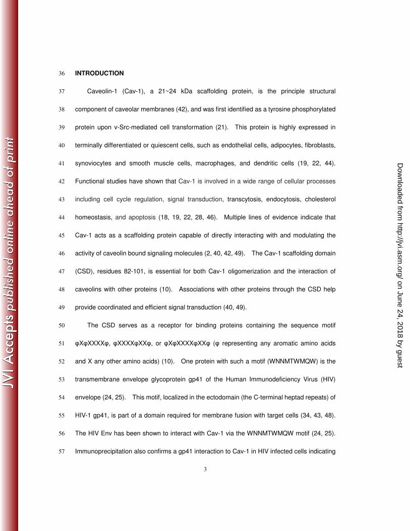
INTRODUCTION 36
Caveolin-1 (Cav-1), a 21~24 kDa scaffolding protein, is the principle structural 37
component of caveolar membranes (42), and was first identified as a tyrosine phosphorylated 38
protein upon v-Src-mediated cell transformation (21). This protein is highly expressed in 39
terminally differentiated or quiescent cells, such as endothelial cells, adipocytes, fibroblasts, 40
synoviocytes and smooth muscle cells, macrophages, and dendritic cells (19, 22, 44). 41
Functional studies have shown that Cav-1 is involved in a wide range of cellular processes 42
including cell cycle regulation, signal transduction, transcytosis, endocytosis, cholesterol 43
homeostasis, and apoptosis (18, 19, 22, 28, 46). Multiple lines of evidence indicate that 44
Cav-1 acts as a scaffolding protein capable of directly interacting with and modulating the 45
activity of caveolin bound signaling molecules (2, 40, 42, 49). The Cav-1 scaffolding domain 46
(CSD), residues 82-101, is essential for both Cav-1 oligomerization and the interaction of 47
caveolins with other proteins (10). Associations with other proteins through the CSD help 48
provide coordinated and efficient signal transduction (40, 49). 49
The CSD serves as a receptor for binding proteins containing the sequence motif 50
φXφXXXXφ, φXXXXφXXφ, or φXφXXXXφXXφ (φ representing any aromatic amino acids 51
and X any other amino acids) (10). One protein with such a motif (WNNMTWMQW) is the 52
transmembrane envelope glycoprotein gp41 of the Human Immunodeficiency Virus (HIV) 53
envelope (24, 25). This motif, localized in the ectodomain (the C-terminal heptad repeats) of 54
HIV-1 gp41, is part of a domain required for membrane fusion with target cells (34, 43, 48). 55
The HIV Env has been shown to interact with Cav-1 via the WNNMTWMQW motif (24, 25). 56
Immunoprecipitation also confirms a gp41 interaction to Cav-1 in HIV infected cells indicating 57
on June 24, 2018 by guesthttp://jvi.asm
.org/D
ownloaded from

4
binding under physiological conditions. The strong association of Cav-1 with the HIV Env, 58
which is mediated by the gp41 region, suggests a role for Cav-1 in HIV infection and 59
pathogenesis. In support of this, inhibition of virus production is observed in fibroblast cells 60
transfected with HIV provirus DNA in cells where Cav-1 is over expressing (31). 61
Furthermore, we recently reported Cav-1 can block HIV Env induced apoptosis of innocent 62
bystander cells as well as inhibition of Env mediated effector/target cell fusion implicating the 63
role of Cav-1 in inhibiting virus production by interaction with Env (47). 64
The cav-1 gene promoter contains cis-acting sequences for binding to Sp1 and E2F 65
transcription factors (4, 5, 15). The promoter also contains three G/C rich box consensus for 66
sterol response elements. Deletion analysis of the cis acting sequences for the sterol 67
response element revealed the requirement of these sites for cholesterol dependent 68
regulation of gene expression. The sterol response element binding protein 1 (SREBP-1) 69
binds to the promoter for cholesterol dependent regulation of cav-1 gene expression which 70
mediates its effect through the E2F/Sp1 cis-elements. Further studies of the cav-1 gene 71
promoter reveal that direct binding of p53 and an interaction with E2F and Sp1 form a 72
complex that stimulate cav-1 gene expression (4, 5, 9). There are also cellular oncogenes 73
which can modulate Cav-1 expression through transcriptional mechanisms (14, 27, 35, 45). 74
Upregulation of Cav-1 was found in macrophages in response to oxidized low-density 75
lipoprotein (oxLDL) or simvastatin (20, 50). Similarly, treatment of mouse macrophage cell 76
lines with lipopolysaccharides has been shown to increase Cav-1 expression at the mRNA 77
level (29, 30). It was also observed that oxidative stress activates Cav-1 expression (11). 78
Since macrophages express Cav-1 and are one of the major target cells for HIV infection 79
on June 24, 2018 by guesthttp://jvi.asm
.org/D
ownloaded from

5
the expression of Cav-1 may be influenced by HIV infected macrophages. In this report, for 80
the first time we demonstrate an upregulation of Cav-1 expression in HIV infected cells. 81
This upregulation is mediated by Tat and involves the p53 protein, and very significantly 82
results in a reduction of virus production. Because Cav-1 is involved in many important 83
cellular processes including lipid trafficking, cell-cycle regulation, apoptosis, signal 84
transduction and translation, its upregulation during HIV-1 infection has potentially important 85
implications in HIV pathogenesis and persistence. 86
87
MATERIALS AND METHODS 88
Plasmids and Reagents. The HIV-1 proviral construct pNL4-3 (T-tropic) and pNL-AD8 89
(M-tropic) were kindly provided by NIH AIDS Research and Reference Reagent Program (1, 90
17). Constructs for expressing gag and pol (psPAX2), vif (pcDNA-Hvif), vpr (pEGFP-Vpr), 91
vpu (pcDNA-Vphu), rev (pCMV-rev) and nef (pcDNASF2Nef) were also kindly provided by the 92
NIH AIDS Research and Reference Reagent Program. The plasmid pTatz expressing HIV-1 93
Tat was constructed by inserting the HIV-1 Tat coding sequence downstream of a CMV 94
promoter (36). The envelope expressing plasmid (pCI-NL4-3-Env) was also generated by 95
inserting the envelope coding sequence from NL4-3 provirus downstream of a CMV promoter 96
(47). A Tat defective provirus was generated by changing the initiator ATG codon into a stop 97
TGA codon using an oligonucleotide 98
AATTGGGTGTCGACATAGCAGAATAGGCGTTACTCGACAGAGGAGAGCAAGAATGAGA99
GCCAGTAGA (TatMf) and amplification with primers TatMf and 100
CTCTTAATTTGCTAGCTATCTGT (TatMr). The amplified product was digested with 101
on June 24, 2018 by guesthttp://jvi.asm
.org/D
ownloaded from

6
restriction enzymes SalI and NheI and replaced the wild type sequence in the pNL4-3 102
provirus construct generating pNL4-3-dTat. A Cav-1 expressing plasmid, pCZ-cav-1, was 103
generated by inserting the cav-1 cDNA fragment (Origene, Rockville, MD) into the SacI and 104
BamHI restriction enzyme sites of pCZ vector. pCZ vector was derived by cloning the 105
zeocin expression cassette into the pCI plasmid (Invitrogen, Carlsbad, CA). pGL3-cavFL (9) 106
was kindly provided by Dr. Vijay Shah of the Mayo Clinic College of Medicine (Rochester, 107
MN). This construct contains the luciferase coding sequence downstream of the cav-1 108
promoter region (-737 to +37). pEFp53 plasmid was constructed by first placing the human 109
p53 gene coding sequence into pSP73 (Promega, Madison, WI) at the XbaI restriction 110
enzyme site. Then the EF1 promoter was cloned upstream p53 into the ClaI and BamHI 111
restriction enzyme sites generating pEFp53. The p38 MAPK (mitogen-activated protein 112
kinase) inhibitor SB203580 was purchased from Calbiochem (San Diego, CA). HIV-1 Tat 113
protein was kindly provided by NIH AIDS Research and Reference Reagent Program (7, 26). 114
Transfections. DNA transfections were carried out using Fugene 6 (Roche Diagnostics, 115
Indianapolis, IN) according to the manufacturer’s protocol. Specific siRNAs targeting Sp1, 116
p53, and control siRNAs (Santa Cruz Biotechnology, Santa Cruz, CA) were transfected 117
according to the manufacturer’s protocol. 118
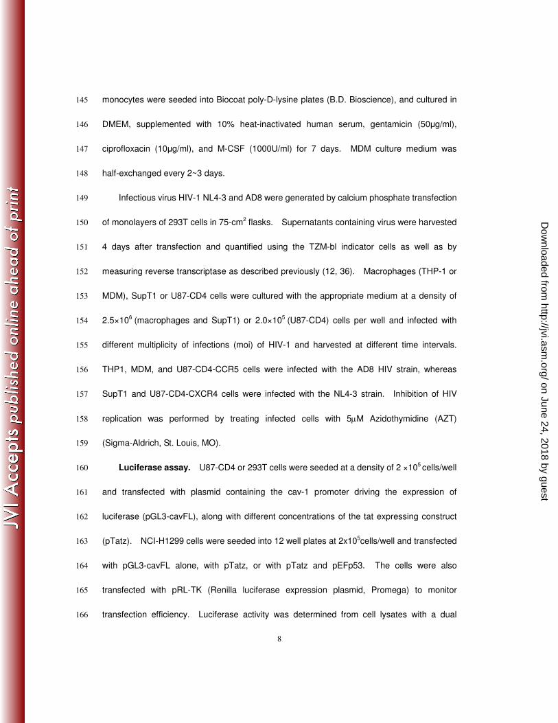
Virus and Cell Cultures. Human U87MG-CD4 cells stably transfected with CXCR4 119
(U87-CD4-CXCR4) or CCR5 (U87-CD4-CCR5), human acute monocytic leukemia (THP-1), 120
an indicator cell line for titering HIV (TZM-bl) and SupT1 cell lines were kindly provided by the 121
NIH AIDS Research and Reference Reagent Program. Human embryonic kidney 293T and 122
on June 24, 2018 by guesthttp://jvi.asm
.org/D
ownloaded from

7
non-small-cell-lung carcinoma (NCI H1299) cell lines were obtained from American Type 123
Culture Collection (Rockville, Md.). U87-CD4-CXCR4 and U87-CD4-CCR5 were 124
maintained in DMEM containing 15% FBS, penicillin-streptomycin (100 µg/mL), glutamine, 125
puromycin (1µg/ml; Sigma Chemical), and neomycin (G418; 300µg/ml; Sigma) (6). THP-1 126
cells were grown in RPMI-1640 containing 10% FBS, 1.0mM sodium pyruvate, and 0.05 mM 127
2-mercaptoethanol (51). For differentiation into macrophages, 2.5×10⁶ THP-1 cells were 128
seeded into 12 well plates and treated with 50 ng/ml of phorbol 12-myristate 13-acetate (PMA, 129
Sigma Chemical) for 5 days until the cells adhered and exhibited macrophage-like 130
morphology. SupT1 cells were cultured in RPMI-1640 containing 10% FBS and 131
penicillin-streptomycin (100 µg/mL). TZM-bl and 293T cells were grown in DMEM medium 132
supplemented with 10% FBS and penicillin-streptomycin (100 µg/mL). U87-CD4-CCR5 133
stably expressing Cav-1 (U87-CD4-CCR5-Cav-1) was established by transfecting with 134
pCZ-cav-1 and selecting for stable transformants in medium containing 100 µg/ml Zeocin 135
(Invivogen, San Diego, CA). Control cell line U87-CD4-CCR5-vect was generated by 136
transfecting the parental lines with vector construct lacking the cav-1 sequence (pCZ). 137
Stable cell lines were maintained in 50 µg/ml Zeocin. 138
Peripheral blood mononuclear cells (PBMCs) were isolated from buffy coat prepared 139
from healthy donors by centrifugation through a Ficoll gradient (Sigma-Aldrich, St. Louis, MO). 140
Monocytes were isolated by positive selection with CD14+ magnetic beads according to the 141
manufacturer’s instructions (EasySep® Human Monocyte Enrichment Kit, Stemcell 142
Technologies). The monocyte preparations contained 98% CD14+ cells, as determined by 143
flow cytometry. For differentiation of monocytes into macrophages (MDM), 2.5×10⁶ 144
on June 24, 2018 by guesthttp://jvi.asm
.org/D
ownloaded from
8
monocytes were seeded into Biocoat poly-D-lysine plates (B.D. Bioscience), and cultured in 145
DMEM, supplemented with 10% heat-inactivated human serum, gentamicin (50µg/ml), 146
ciprofloxacin (10µg/ml), and M-CSF (1000U/ml) for 7 days. MDM culture medium was 147
half-exchanged every 2~3 days. 148
Infectious virus HIV-1 NL4-3 and AD8 were generated by calcium phosphate transfection 149
of monolayers of 293T cells in 75-cm2 flasks. Supernatants containing virus were harvested 150
4 days after transfection and quantified using the TZM-bl indicator cells as well as by 151
measuring reverse transcriptase as described previously (12, 36). Macrophages (THP-1 or 152
MDM), SupT1 or U87-CD4 cells were cultured with the appropriate medium at a density of 153
2.5×10⁶ (macrophages and SupT1) or 2.0×10⁵ (U87-CD4) cells per well and infected with 154
different multiplicity of infections (moi) of HIV-1 and harvested at different time intervals. 155
THP1, MDM, and U87-CD4-CCR5 cells were infected with the AD8 HIV strain, whereas 156
SupT1 and U87-CD4-CXCR4 cells were infected with the NL4-3 strain. Inhibition of HIV 157
replication was performed by treating infected cells with 5µM Azidothymidine (AZT) 158
(Sigma-Aldrich, St. Louis, MO). 159
Luciferase assay. U87-CD4 or 293T cells were seeded at a density of 2 ×10⁵ cells/well 160
and transfected with plasmid containing the cav-1 promoter driving the expression of 161
luciferase (pGL3-cavFL), along with different concentrations of the tat expressing construct 162
(pTatz). NCI-H1299 cells were seeded into 12 well plates at 2x10⁵cells/well and transfected 163
with pGL3-cavFL alone, with pTatz, or with pTatz and pEFp53. The cells were also 164
transfected with pRL-TK (Renilla luciferase expression plasmid, Promega) to monitor 165
transfection efficiency. Luciferase activity was determined from cell lysates with a dual 166
on June 24, 2018 by guesthttp://jvi.asm
.org/D
ownloaded from

9
luciferase assay system as described by the manufacturer (Promega, Madison, WI). The 167
results are reported as normalized means±S.D. 168
Reverse transcription-polymerase chain reaction. Total RNA was isolated from 169
U87-CD4 cells transfected with pTatz or vector lacking tat (pCZ) using the RNeasy kit 170
(Qiagen, Valencia, CA). One microgram of RNA was reverse transcribed using Moloney 171
Murine Leukemia virus reverse transcriptase and random primers according to the 172
manufacturer’s instructions. The PCR was performed with forward (5’ 173
TCAACCGCGACCCTAAACACC 3’) and reverse (5’ TGAAATAGCTCAGAAGAGACAT 3’) 174
primers for 30 cycles of denaturing at 95°C for 40 seconds, annealing at 60°C for 40 seconds, 175
and extension at 72°C for 1 minute, with a 5 minute final extension cycle. To monitor for 176
recovery of mRNA the housekeeping gene Glyceraldhyde-3-phosphate dehydrogenase 177
(GAPDH) was reverse transcribed and amplified for 30 cycles of denaturing at 95° for 40 178
seconds, annealing 58°C for 40 seconds, extension at 72°C for 1 minute using forward 179
5´-TGGTATCGTGGAAGGACTCATGAC-3´ and reverse 180
5´-AGTCCAGTGAGCTTCCCGTTCAGC-3´ primers. 181
Western blot analysis. Total cellular proteins were extracted in lysis buffer (50 mM 182
Tris pH 7.5,100 mM NaCl, 1 mM EDTA, 0.1% (v/v) Triton X-100, 10 mM NaF, 1 mM 183
phenylmethyl sulfonyl fluoride, and 1 mmol/L vanadate) and protein concentration was 184
determined by the Lowry method (BioRad Protein Assay). Extracted protein was separated 185
by 10-15% SDS-PAGE gel electrophoresis and transferred onto a nitrocellulose membrane 186
(Roche). The membrane was blocked with Tris-Buffered Saline Tween 20 containing 5% 187
non-fat milk and then incubated with antibodies specific to Cav-1, p53, Sp1 (Santa Cruz 188
on June 24, 2018 by guesthttp://jvi.asm
.org/D
ownloaded from
10
Biotechnology, Inc, Santa Cruz, CA), Gag, Pol, Vif, Vpr, Vpu, Env, Tat, Rev, Nef (NIH AIDS 189
Research and Reference Reagent Program), p38, p38T180/Y182P, p53S15P, p53S46P 190
(Cell Signaling Technology, Inc., Danvers, MA), or β-actin protein (Sigma, St. Louis, MO) for 191
overnight at 4°C. The membrane was then incubated with horse radish peroxidase (HRP) 192
linked-anti-rabbit, anti-human IgG (Abcam, Inc., Cambrige, MA), or anti-mouse secondary 193
antibody (Cell Signaling Technology, Inc., Danvers, MA) for 1 hour at room temperature. 194
Blots were analyzed by densitometry using NIH image software. 195
196
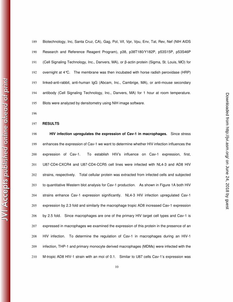
RESULTS 197
HIV infection upregulates the expression of Cav-1 in macrophages. Since stress 198
enhances the expression of Cav-1 we want to determine whether HIV infection influences the 199
expression of Cav-1. To establish HIV’s influence on Cav-1 expression, first, 200
U87-CD4-CXCR4 and U87-CD4-CCR5 cell lines were infected with NL4-3 and AD8 HIV 201
strains, respectively. Total cellular protein was extracted from infected cells and subjected 202
to quantitative Western blot analysis for Cav-1 production. As shown in Figure 1A both HIV 203
strains enhance Cav-1 expression significantly. NL4-3 HIV infection upregulated Cav-1 204
expression by 2.3 fold and similarly the macrophage tropic AD8 increased Cav-1 expression 205
by 2.5 fold. Since macrophages are one of the primary HIV target cell types and Cav-1 is 206
expressed in macrophages we examined the expression of this protein in the presence of an 207
HIV infection. To determine the regulation of Cav-1 in macrophages during an HIV-1 208
infection, THP-1 and primary monocyte derived macrophages (MDMs) were infected with the 209
M-tropic AD8 HIV-1 strain with an moi of 0.1. Similar to U87 cells Cav-1’s expression was 210
on June 24, 2018 by guesthttp://jvi.asm
.org/D
ownloaded from

11
significantly enhanced by HIV infection in macrophages (2.7 fold in MDMs and 2.3 fold in 211
THP-1) (Figure 1B,C). As a negative control Cav-1 expression was monitored in SupT1 212
cells infected with the NL4-3 HIV strain; with no Cav-1 expression, as monitored by Western 213
blot analysis, being seen in either infected or uninfected cells (Figure 1D). 214
To further confirm the upregulation of Cav-1 by HIV infection time course and dose 215
dependent experiments were performed using both U87-CD4-CXCR4 and U87-CD4-CCR5 216
cell lines by infection with NL4-3 and AD8 HIV strains, respectively. Time course 217
experiments were performed by harvesting samples for Western analysis at days 1, 3, 5, and 218
7 days post-infection. Uninfected cells show increased amounts of Cav-1 expression with 219
increased time in culture (Figure 2A,B). In infected cells the amount of Cav-1 is significantly 220
higher than that of the uninfected cells in both U87-CD4-CXCR4 and U87-CD4-CCR5 cells at 221
each time point. The increase in NL4-3 infected cells reached 2.8 fold (day 7) and 2.5 fold 222
(day 7). Dose dependent infection studies were conducted by infecting U87 cells with 223
different multiplicity of infections (mois). Cav-1 expression increased with increasing HIV 224
moi reaching a 2.7 fold enhancement (Figure 2C,D). Similar experiments were carried out 225
using THP-1 macrophages. As seen in the U87 cells the level of Cav-1 protein increased 226
with increasing moi in the THP-1 macrophages reaching 2.5 fold (Figure 2E). To determine 227
whether Cav-1 upregulation can occur in the absence of de novo viral gene expression 228
infected THP-1 and U87 cells were treated with AZT and the level of Cav-1 expression was 229
examined. No enhancement of Cav-1 expression was observed when infected cells were 230
treated with AZT (Figure 2F), establishing that Cav-1 upregulation does not take place in the 231
absence of de novo viral gene expression. As expected we observed no Cav-1 expression 232
on June 24, 2018 by guesthttp://jvi.asm
.org/D
ownloaded from

12
with different mois of infected or uninfected SupT1 cells by Western Blotting (Figure 2G). 233
These results establish that Cav-1 expression is upregulated by HIV infection in 234
macrophages. 235
Tat is responsible for HIV mediated Cav-1 upregulation. Since the cav-1 gene 236
promoter contains cis acting elements for binding factors that potentially cross-talk to Tat (4, 5, 237
15) we focused on the role of Tat in influencing Cav-1 expression. In order to elucidate the 238
mechanism of upregulation of Cav-1 by HIV infection we examined the role of Tat in Cav-1 239
expression. U87-CD4-CXCR4 cells were transfected in a dose dependent manner with a 240
plasmid construct expressing Tat (pTatz). As a control U87-CD4-CXCR4 cells were also 241
transfected with expression vector lacking the tat sequence (pCZ). The Cav-1 expression 242
increased in a Tat dose dependent manner reaching a 3.1 fold enhancement with 1.0µg of 243
pTatz transfection (Figure 3A). These results establish that the upregulation of Cav-1 by 244
HIV infection is mediated by Tat. In an effort to determine whether the changes in Cav-1 245
protein levels are associated with differences in mRNA levels, RT-PCR was performed using 246
primers based on the cav-1 sequence. RNA was extracted from U87-CD4-CXCR4 cells that 247
were transfected with pTatz for RT-PCR analysis and the results were compared to that of 248
cells receiving the expression construct devoid of the tat sequence. The cav-1 mRNA 249
expression in the presence of Tat was 2.5-fold higher than that of the cells lacking Tat (Figure 250
3B). To further confirm this finding the expression of Luciferase under the control of the 251
cav-1 gene promoter (pGL3-cavFL) was examined in U87-CD4-CXCR4 or 293T cells 252
transfected with different doses of pTatz. In both cell lines the luciferase expression was 253
significantly higher, 2.5 fold for U87-CD4 and 2.0 fold for 293T cells, as compared to cells 254
on June 24, 2018 by guesthttp://jvi.asm
.org/D
ownloaded from

13
receiving no Tat (Figure 3C,D). This level of transcriptional enhancement is comparable to 255
that of Cav-1 protein expression. In addition, Tat’s upregulation of the cav-1 gene promoter 256
occurred in a dose dependent manner. These results reveal that the upregulation of Cav-1 257
by HIV infection is mediated by Tat and that the induction takes place at the transcriptional 258
level. 259
To determine whether Cav-1 upregulation is only Tat dependent, we first tested each of 260
the HIV proteins for their ability to upregulate Cav-1. Expression constructs for each of the 261
HIV genes were transfected into U87-CD4-CXCR4 cells and the level of Cav-1 expression 262
was determined by Western blot (Figure 4A,B). As shown in Figure 4B, none of the other 263
HIV proteins enhanced the expression of Cav-1. Second, we generated a Tat defective 264
provirus (pNL4-3-dTat), by changing the tat initiator codon ATG to a stop codon TGA, and 265
examined the influence on Cav-1 expression. Cells transfected with the pNL4-3-dTat 266
showed no difference in Cav-1 expression as compared to mock transfection while 267
cotransfection with pTatz showed an uprgulation in Cav-1 (Figure 4C). Supernatant 268
harvested from cells co-transfected with pNL4-3-dTat and pTatz were used to infect fresh 269
U87-CD4-CXCR4 cells to determine whether virus infection enhances cav-1 expression non 270
specifically, in the absence of Tat, as an innate response. These viral particles can undergo 271
one round of infection but due to the defect in Tat viral gene expression is restricted for 272
subsequent rounds of virus replication. The level of Cav-1 expression in these infected cells 273
by the Tat defective HIV was similar to that of uninfected cells (Figure 4D), thus further 274
strengthening the evidence that Cav-1 upregulation is only dependent on Tat. 275
p53 activity is essential for HIV Tat-mediated caveolin-1 upregulation. 276
on June 24, 2018 by guesthttp://jvi.asm
.org/D
ownloaded from

14
Transcription factors known to regulate cav-1 gene expression include p53 and Sp1 (4, 9, 11). 277
Several studies also describe cooperation of Tat with p53 or Sp1 (3, 32). The transcriptional 278
upregulation of Cav-1 could, therefore, be mediated through p53 and Sp1. To address this 279
possibility we first examined Sp1’s possible role in Tat mediated Cav-1 upregulation. 280
U87-CD4-CXCR4 cells were pretreated with siRNA to knockdown the expression of Sp1 281
which was followed by transfection of the tat expressing construct. The siRNA treatment 282
reduced the expression of Sp1 by 78%; however, this decrease in Sp1 has no impact on 283
Cav-1 expression (Figure 5A). This, therefore, suggests that Sp1 does not play a role in Tat 284
mediated upregulation of Cav-1. Using a similar experimental approach we next examined 285
whether p53 activation is involved in the regulation of Cav-1 in HIV Tat-expressing cells. 286
The p53 specific siRNA treatment resulted in a knock down of p53 expression by 72%, which 287
then leads to a significant reduction in Cav-1 expression (Figure 5B). In cells that were 288
treated with no siRNA Tat enhanced the expression of Cav-1 as expected. Similarly, in cells 289
receiving nonspecific siRNA comparable levels of Cav-1 upregulation by Tat was evident. 290
The role of Tat in the upregulation of Cav-1 expression was examined in the absence of p53 291
by cotransfecting the tat expression plasmid (pTatz) and a construct with luciferase under the 292
control of the cav-1 gene promoter (pGL3-cavFL) into the p53 null NCI-H1299 cell line. 293
Tat-induced Cav-1 promoter activation was completely blocked in the p53 null NCI-H1299 294
cell line (Figure 5C). Transfection of a p53 expression plasmid (pEFp53) along with the 295
pTatz and pGL3-cavFL constructs resulted in the upregulation of luciferase expression 296
reaching 2.4 fold when 0.5 ug of the pEFp53 expression construct was transfected. These 297
results show that p53 is essential for Tat mediated upregulation of Cav-1 in HIV infected cells 298
on June 24, 2018 by guesthttp://jvi.asm
.org/D
ownloaded from
15
while Sp1 has no role. 299
Further analysis revealed that the level of p53 expression did not increase in cells 300
transfected with the tat construct as compared to cells receiving the expression vector devoid 301
of the tat sequence (Figure 5B). These results suggest that the role of p53 in Tat mediated 302
Cav-1 upregulation is not correlated to the amount of p53, but rather to the modification of 303
p53 for its activity or another factor that interacts with p53 is limiting. Phosphorylation at 304
specific residues of the N-terminal domain is crucial for p53 activity (13, 33, 39). We 305
therefore tested whether phosphorylation of p53 is stimulated during an HIV-1 infection. To 306
this end, U87-CD4-CXCR4 cells were infected with HIV-1 NL4-3 at an moi of 0.1 for 1, 3, 5, or 307
6 hours and p53 phosphorylation at serine residues 15 (Ser15) and 46 (Ser46) were 308
determined. Phosphorylation of Ser46 and Ser15 was detected at 3 and 5 hours, 309
respectively, post infection by Western blot analysis and increasing thereafter, whereas the 310
HIV infection has no effect on total p53 levels (Figure 6A). U87-CD4-CXCR4 cells were 311
treated with recombinant Tat protein (0.5 ug/ml) for 6 hours to assess whether it can induce 312
the phosphorylation of p53. Similar to an HIV infection Tat increased the p53 313
phosphorylation of ser15 and ser46 by 1.8- and 2.3-fold, respectively, compared to control 314
cells that were not cultured with Tat protein (Figure 6B). The total p53 level remains the 315
same whether cells receive Tat or not, suggesting that the p53 activation during an HIV 316
infection is essential for Tat mediated upregulation of Cav-1. 317
The p38 mitogen-activated protein kinase (MAPK) is involved in the phosphorylation of 318
p53 residues ser15 and ser46 and p38 MAPK activation of p53 has been observed in HIV 319
infected cells (8, 37, 38). We, therefore, next examined the phosphorylated forms of p38 320
on June 24, 2018 by guesthttp://jvi.asm
.org/D
ownloaded from

16
MAPK by immunoblotting. U87-CD4-CXCR4 cells were serum starved overnight, and then 321
infected with HIV NL4-3 at an moi of 0.1 for 3, 6, 24 and 48 hours. Western blot analysis 322
reveals that the HIV infection stimulated phosphorylation of p38 at 3 and 6 hours post 323
infection with the amount of phosphorylated p38 decreasing at 24 and 48 hours (Figure 6C). 324
The phosphorylation of ser15 and ser46 of p53 also followed a similar pattern. Cav-1 325
upregulation was observed at 24 and 48 hours post infection which is after the optimum p38 326
MAPK and p53 phosphorylation (2-6 hour time point). Addition of p38 MAPK 327
phosphorylation specific inhibitor SB203580 (10 µM), beginning 2 hours prior to infection and 328
maintained during HIV infection, significantly reduced the phosphorylation of p38 MAPK as 329
well as p53 at ser15 and ser46. The p38 MAPK phosphorylation inhibitor also reduced the 330
Cav-1 upregulation by HIV infection. Similar experiments were performed using 331
recombinant Tat protein (0.5 ug/ml) instead of virus infection (Figure 6D). As observed with 332
viral infection Tat also induced the phosphorylation of p38 MAPK and p53 at ser15 and ser46 333
at 2 hours, significantly increasing at 5 hours and decreasing at 24 and 48 hours after the 334
addition of Tat. Similar to the results with the HIV infection Cav-1 upregulation was also 335
observed at 24 and 48 hours after Tat treatment was initiated. The inhibitor of p38 MAPK 336
phosphorylation (SB203580) also reduced p53 phosphorylation and Cav-1 upregulation as 337
observed with the viral infection. Taken together these results suggest that the activation of 338
p38 MAPK in HIV infected cells mediated by Tat leads to the phosphorylation of p53 which 339
subsequently upregulates Cav-1 expression. 340
Enhanced Cav-1 expression results in a reduction of HIV replication. To examine 341
the role of the upregulation of Cav-1 in an HIV infection, we first examined virus production by 342
on June 24, 2018 by guesthttp://jvi.asm
.org/D
ownloaded from

17
transient overexpression of Cav-1 in 293T cells where Cav-1 normally cannot be detected by 343
Western Blot analysis (Figure 7A). Cells were transfected with a constant amount of 344
provirus DNA along with various concentrations of cav-1 expression construct. As shown in 345
Figure 7A and 7B Cav-1 inhibits HIV production in a dose dependent manner. Second, we 346
established a cell line overexpressing Cav-1 (U87-CD4-CCR5-Cav-1) as well as a cell line 347
containing the expression vector minus the cav-1 sequence (U87.CD4.CCR5-vect). These 348
established cells were infected with AD8 (CCR5 expressing cells) at an moi of 0.1 and HIV 349
growth kinetics followed. Supernatants from the infected cells were harvested at intervals 350
for 12 days to determine the levels of virus production by monitoring reverse transcriptase 351
activity. The level of virus release monitored by reverse transcriptase assay was adjusted 352
for cell growth normalizing to protein concentration. As shown in Figure 7C, at all the time 353
points, virus production was dramatically reduced in cells overexpressing Cav-1 as compared 354
to cells expressing only endogenous Cav-1. The inhibition of virus replication reached 72%. 355
These results along with the observation of Tat mediated upregulation suggests that Cav-1 356
contributes in the reduction of HIV replication in a negative feedback loop. 357
358
DISCUSSION 359
In this study we have investigated the status of Cav-1 expression in HIV-1 infected cells 360
and the molecular mechanisms involved in HIV-mediated Cav-1 regulation. Cav-1 protein 361
expression is significantly enhanced in HIV infected cells and its upregulation is mediated by 362
the HIV Tat protein. Induction of Cav-1 expression by Tat is at the transcriptional level and 363
involves the p53 protein. The expression of p53 is not affected by HIV infection or Tat; 364
on June 24, 2018 by guesthttp://jvi.asm
.org/D
ownloaded from
18
however phosphorylation of p53 at ser15 and ser46 are enhanced suggesting the level of p53 365
activity is important for Tat-mediated upregulation of Cav-1. Furthermore, p38 MAPK 366
inhibitor blocked phosphorylation of p53, subsequently negatively influencing Cav-1 367
upregulation. As p38 mediates p53 phosphorylation, this confirms the concept of p53 368
activity being important to Cav-1 upregulation. More importantly, HIV infection induces 369
Cav-1 expression in macrophages. Since overexpression of Cav-1 restricts HIV production, 370
we propose that its upregulation in macrophages plays a role in persistent infection and 371
pathogenesis. 372
Studies of the cav-1 gene promoter reveals a direct binding of p53 at positions –292 to 373
–283 and –273 to –264 (5). Bound p53 interacts with E2F and Sp1 to form a complex that 374
stimulates cav-1 gene expression (4). HIV Tat has been shown to cooperate with either Sp1 375
(32) or p53 (3) and influence gene expression. Our observation that Cav-1 expression does 376
not change when Sp1 expression is knocked down by siRNA treatment suggests that Tat 377
mediated Cav-1 upregulation does not involve Sp1. Previous studies reveal that p53 is a 378
positive regulator of Cav-1 expression and inactivation of p53 by viral oncoproteins results in 379
decreased Cav-1 expression (41). Consistent with these studies our data shows that p53 380
activity is essential for Tat mediated upregulation of Cav-1. The knockdown of p53 381
expression by siRNA results in the blocking of Tat’s mediated upregulation of Cav-1. Our 382
results further show that the upregulation of Cav-1 is not dependent on the amount of p53, 383
but rather that the level of p53 phosphorylation is critical, potentially mediated by p38 MAPK. 384
The exact mechanism of cross-talk between Tat and p53 is not clearly defined. In the 385
current study we demonstrate that the upregulation of Cav-1 occurs at the transcriptional 386
on June 24, 2018 by guesthttp://jvi.asm
.org/D
ownloaded from
19
level. When our results showing that the increased phosphorylation of p53 in the presence 387
of Tat is combined with the knowledge that the binding of p53 to the cav-1 gene promoter 388
enhances cav-1 expression (4, 5) reveals a possible mechanism for the cross communication 389
between Tat and p53 in the upregulation of Cav-1 expression. 390
The presence of Cav-1 has been investigated in different cells of the immune system and 391
its expression and distribution might be dependent on the activation and/or maturation state 392
of the cells (22). As an important target for HIV infection, macrophages have minimal 393
cytopathology in response to HIV infection, and are able to support sustained virus replication, 394
acting as a major viral reservoir. Interestingly, we found that Cav-1 expression is enhanced 395
in both HIV-1 infected primary monocyte derived macrophages (MDMs) as well as 396
established THP-1 cells stimulated to differentiate into macrophages. The upregulation was 397
also observed in U87-CD4 cells infected with HIV-1 T-tropic or M-tropic viruses. However, 398
Cav-1 expression is not detected in either HIV infected or uninfected SupT1 cells, which is 399
consistent with previous studies that Cav-1 expression has not been observed in human and 400
murine T lymphocytes (16, 23). Compared to T cells, infected macrophages are relatively 401
resistant to cytopathic effect and consequently play an essential role in viral dissemination to 402
host tissues and organs. The strong Cav-1 interaction with gp41 (24, 25, 47), the lack of 403
Cav-1 in T cells, enhanced Cav-1 expression in macrophages during an HIV infection, and 404
reduced viral production in cells with an overexpression of Cav-1 all lead to the implication 405
that Cav-1 plays a role in the persistent infection of macrophages by HIV. Based on this 406
study, our data shows that Cav-1 adds to the body of knowledge being used in building a 407
model for the persistent infection of macrophages in which HIV Tat-mediates the upregulation 408
on June 24, 2018 by guesthttp://jvi.asm
.org/D
ownloaded from

20
of Cav-1, creating an environment where excess Cav-1 binds the gp41 of Env, thereby 409
blocking/reducing virus production. 410
on June 24, 2018 by guesthttp://jvi.asm
.org/D
ownloaded from

21
ACKNOWLEDGMENTS 411
This research was supported by a grant from the National Institute of Health (AI39126) 412
to A. Mergia. We thank Dr. David Allred for reading the manuscript and helpful suggestions. 413
414
on June 24, 2018 by guesthttp://jvi.asm
.org/D
ownloaded from

22
REFERENCES 415
416
1. Adachi, A., H. E. Gendelman, S. Koenig, T. Folks, R. Willey, A. Rabson, and M. A. 417
Martin. 1986. Production of acquired immunodeficiency syndrome-associated 418
retrovirus in human and nonhuman cells transfected with an infectious molecular 419
clone. J Virol 59:284-91. 420
2. Anderson, R. G. 1993. Caveolae: where incoming and outgoing messengers meet. 421
Proc Natl Acad Sci U S A 90:10909-13. 422
3. Ariumi, Y., A. Kaida, M. Hatanaka, and K. Shimotohno. 2001. Functional 423
Cross-Talk of HIV-1 Tat with p53 through Its C-Terminal Domain. Biochemical and 424
Biophysical Research Communications 287:556–561. 425
4. Bist, A., C. J. Fielding, and P. E. Fielding. 2000. p53 regulates caveolin gene 426
transcription, cell cholesterol, and growth by a novel mechanism. Biochemistry 427
39:1966-72. 428
5. Bist, A., P. E. Fielding, and C. J. Fielding. 1997. Two sterol regulatory element-like 429
sequences mediate up-regulation of caveolin gene transcription in response to low 430
density lipoprotein free cholesterol. Proc. Natl. Acad. Sci. USA 94:10693-10698. 431
6. Bjorndal, A., H. Deng, M. Jansson, J. R. Fiore, C. Colognesi, A. Karlsson, J. 432
Albert, G. Scarlatti, D. R. Littman, and E. M. Fenyo. 1997. Coreceptor usage of 433
primary human immunodeficiency virus type 1 isolates varies according to biological 434
phenotype. J Virol 71:7478-87. 435
7. Bohan, C. A., F. Kashanchi, B. Ensoli, L. Buonaguro, K. A. Boris-Lawrie, and J. N. 436
on June 24, 2018 by guesthttp://jvi.asm
.org/D
ownloaded from

23
Brady. 1992. Analysis of Tat transactivation of human immunodeficiency virus 437
transcription in vitro. Gene Expr 2:391-407. 438
8. Bulavin, D. V., S. Saito, M. C. Hollander, K. Sakaguchi, C. W. Anderson, E. 439
Appella, and A. J. Fornace, Jr. 1999. Phosphorylation of human p53 by p38 kinase 440
coordinates N-terminal phosphorylation and apoptosis in response to UV radiation. 441
Embo J 18:6845-54. 442
9. Cao, S., M. E. Fernandez-Zapico, D. Jin, V. Puri, T. A. Cook, L. O. Lerman, X. Y. 443
Zhu, R. Urrutia, and V. Shah. 2005. KLF11-mediated repression antagonizes 444
Sp1/sterol-responsive element-binding protein-induced transcriptional activation of 445
caveolin-1 in response to cholesterol signaling. J Biol Chem 280:1901-10. 446
10. Couet, J., S. Li, T. Okamoto, T. Ikezu, and M. P. Lisanti. 1997. Identification of 447
peptide and protein ligands for the caveolin-scaffolding domain. Implications for the 448
interaction of caveolin with caveolae-associated proteins. J Biol Chem 449
272:6525-6533. 450
11. Dasari, A., J. N. Bartholomew, D. Volonte, and F. Galbiati. 2006. Oxidative stress 451
induces premature senescence by stimulating caveolin-1 gene transcription through 452
p38 mitogen-activated protein kinase/Sp1-mediated activation of two GC-rich 453
promoter elements. Cancer Res 66:10805-14. 454
12. Derdeyn, C. A., J. M. Decker, J. N. Sfakianos, X. Wu, W. A. O'Brien, L. Ratner, J. C. 455
Kappes, G. M. Shaw, and E. Hunter. 2000. Sensitivity of human immunodeficiency 456
virus type 1 to the fusion inhibitor T-20 is modulated by coreceptor specificity defined 457
by the V3 loop of gp120. J Virol 74:8358-67. 458
on June 24, 2018 by guesthttp://jvi.asm
.org/D
ownloaded from
24
13. Dumaz, N., and D. W. Meek. 1999. Serine 15 phosphorylation stimulates p53 459
transactivation but does not directly influence interaction with HDM2. EMBO J. 460
18:7002-7010. 461
14. Engelman, J. A., R. J. Lee, A. Karnezis, D. J. Bearss, M. Webster, P. Siegel, W. J. 462
Muller, J. J. Windle, R. G. Pestell, and M. P. Lisanti. 1998. Reciprocal regulation of 463
neu tyrosine kinase activity and caveolin-1 protein expression in vitro and in vivo. 464
Implications for human breast cancer. J Biol Chem 273:20448-55. 465
15. Fielding, C. J., A. Bist, and P. E. Fielding. 1999. Intracellular cholesterol transport in 466
synchronized human skin fibroblasts. Biochemistry 38:2506–2513. 467
16. Fra, A. M., E. Williamson, K. Simons, and R. G. Parton. 1995. De novo formation of 468
caveolae in lymphocytes by expression of VIP21-caveolin. Proc Natl Acad Sci U S A 469
92:8655-9. 470
17. Freed, E. O., G. Englund, and M. A. Martin. 1995. Role of the basic domain of 471
human immunodeficiency virus type 1 matrix in macrophage infection. J Virol 472
69:3949-54. 473
18. Galbiati, F., D. Volonte, J. Liu, F. Capozza, P. G. Frank, L. Zhu, R. G. Pestell, and M. 474
P. Lisanti. 2001. Caveolin-1 expression negatively regulates cell cycle progression by 475
inducing G(0)/G(1) arrest via a p53/p21(WAF1/Cip1)-dependent mechanism. Mol Biol 476
Cell 12:2229-44. 477
19. Gargalovic, P., and L. Dory. 2003. Caveolins and macrophage lipid metabolism. J 478
Lipid Res 44:11-21. 479
20. Gargalovic, P., and L. Dory. 2003. Cellular apoptosis is associated with increased 480
on June 24, 2018 by guesthttp://jvi.asm
.org/D
ownloaded from

25
caveolin-1 expression in macrophages. J Lipid Res 44:1622-32. 481
21. Glenney, J. R., Jr. 1989. Tyrosine phosphorylation of a 22-kDa protein is correlated 482
with transformation by Rous sarcoma virus. J Biol Chem 264:20163-6. 483
22. Harris, J., D. Werling, J. C. Hope, G. Taylor, and C. J. Howard. 2002. Caveolae and 484
caveolin in immune cells: distribution and functions. Trends Immunol 23:158-64. 485
23. Hatanaka, M., T. Maeda, T. Ikemoto, H. Mori, T. Seya, and A. Shimizu. 1998. 486
Expression of caveolin-1 in human T cell leukemia cell lines. Biochem Biophys Res 487
Commun 253:382-7. 488
24. Hovanessian, A. G., J. P. Briand, A. S. Said, J. Svab, S. Ferris, H. Dali, S. Muller, 489
C. Desgranges, and B. Krust. 2004. The caveolin-1 binding domain of HIV-1 490
glycoprotein gp41 is an efficient B-cell epitope vaccine candidate against virus 491
infection. Immunity 21:617-627. 492
25. Huang, J. H., L. Lu, L. Hong, X. Chen, S. Jiang, and Y.-H. Chen. 2007. 493
Identification of the HIV-1 gp41 Core-binding Motif in the Scaffolding Domain of 494
Caveolin-1. J. Bio. Chem. 282:6143-6152. 495
26. Kashanchi, F., J. F. Duvall, and J. N. Brady. 1992. Electroporation of viral 496
transactivator proteins into lymphocyte suspension cells. Nucleic Acids Res 497
20:4673-4. 498
27. Koleske, A. J., D. Baltimore, and M. P. Lisanti. 1995. Reduction of caveolin and 499
caveolae in oncogenically transformed cells. Proc Natl Acad Sci U S A 92:1381-5. 500
28. Lee, S. W., C. L. Reimer, P. Oh, D. B. Campbell, and J. E. Schnitzer. 1998. Tumor 501
cell growth inhibition by caveolin re-expression in human breast cancer cells. 502
on June 24, 2018 by guesthttp://jvi.asm
.org/D
ownloaded from
26
Oncogene 16:1391-7. 503
29. Lei, M. G., and D. C. Morrison. 2000. Differential expression of caveolin-1 in 504
lipopolysaccharide-activated murine macrophages. Infect Immun 68:5084-9. 505
30. Lei, M. G., X. Tan, N. Qureshi, and D. C. Morrison. 2005. Regulation of cellular 506
caveolin-1 protein expression in murine macrophages by microbial products. Infect 507
Immun 73:8136-43. 508
31. Llano, M., T. Kelly, M. Vanegas, M. Peretz, T. E. Peterson, R. D. Simari, and E. M. 509
Poeschla. 2002. Blockade of Human Immunodeficiency Virus Type 1 Expression by 510
Caveolin-1. J. Virol. 76:9152–9164. 511
32. Loregian, A., K. Bortolozzo, S. Boso, B. Sapino, M. Betti, M. A. Biasolo, A. 512
Caputo, and G. Palú. 2003. The Sp1 transcription factor does not directly interact 513
with the HIV-1 Tat protein. J Cell Physiol. 196:252-257. 514
33. Meek, D. W. 1999. Mechanisms of switching on p53: a role for covalent modification? 515
Oncogene 18:7666-7675. 516
34. Melikyan, G. B., R. M. Markosyan, H. Hemmati, M. K. Delmedico, D. M. Lambert, 517
and F. S. Cohen. 2000. Evidence that the transition of HIV-1 gp41 into a six-helix 518
bundle, not the bundle configuration, induces membrane fusion. J. Cell Biol. 519
151:413-423. 520
35. Park, D. S., B. Razani, A. Lasorella, N. Schreiber-Agus, R. G. Pestell, A. Iavarone, 521
and M. P. Lisanti. 2001. Evidence that Myc isoforms transcriptionally repress 522
caveolin-1 gene expression via an INR-dependent mechanism. Biochemistry 523
40:3354-62. 524
on June 24, 2018 by guesthttp://jvi.asm
.org/D
ownloaded from
27
36. Park, J., P. E. Nadeau, and A. Mergia. 2009. Activity of TAR in inducible inhibition of 525
HIV replication by foamy virus vector expressing siRNAs under the control of HIV LTR. 526
Virus Res. 140:112-120. 527
37. Perfettini, J. L., M. Castedo, R. Nardacci, F. Ciccosanti, P. Boya, T. Roumier, N. 528
Larochette, M. Piacentini, and G. Kroemer. 2005. Essential role of p53 529
phosphorylation by p38 MAPK in apoptosis induction by the HIV-1 envelope. J Exp 530
Med 201:279-89. 531
38. Perfettini, J. L., R. Nardacci, M. Bourouba, F. Subra, L. Gros, C. Seror, G. Manic, 532
F. Rosselli, A. Amendola, P. Masdehors, L. Chessa, G. Novelli, D. M. Ojcius, J. K. 533
Siwicki, M. Chechlinska, C. Auclair, J. R. Regueiro, H. de The, M. L. Gougeon, M. 534
Piacentini, and G. Kroemer. 2008. Critical involvement of the ATM-dependent DNA 535
damage response in the apoptotic demise of HIV-1-elicited syncytia. PLoS One 536
3:e2458. 537
39. Puca, R., L. Nardinocchi, D. Givol, A. Sacchi, G. Rechavi, and G. D'Orazi. 2009. 538
HIPK2 modulates p53 activity towards pro-apoptotic transcription. . Molecular cancer 539
8:85. 540
40. Quest, A. F. G., L. Leyton, and M. Párraga. 2004. Caveolins, caveolae, and lipid 541
rafts in cellular transport, signaling, and disease. Biochem. Cell Biol. 82:129-144. 542
41. Razani, B., Y. Altschuler, L. Zhu, R. G. Pestell, K. E. Mostov, and M. P. Lisanti. 543
2000. Caveolin-1 expression is down-regulated in cells transformed by the human 544
papilloma virus in a p53-dependent manner. Replacement of caveolin-1 expression 545
suppresses HPV-mediated cell transformation. Biochemistry 39:13916-24. 546
on June 24, 2018 by guesthttp://jvi.asm
.org/D
ownloaded from

28
42. Rothberg, K. G., J. E. Heuser, W. C. Donzell, Y. S. Ying, J. R. Glenney, and R. G. 547
Anderson. 1992. Caveolin, a protein component of caveolae membrane coats. Cell 548
68:673-82. 549
43. Shu, W., J. Hong, and M. Lu. 2000. Interactions between HIV-1 gp41 core and 550
determinants and implications for membrane fusion. J. Biol. Chem. 275:1839-1845. 551
44. Smart, E. J., G. A. Graf, M. A. McNiven, W. C. Sessa, J. A. Engelman, P. E. 552
Scherer, T. Okamoto, and M. P. Lisanti. 1999. Caveolins, liquid-ordered domains, 553
and signal transduction. Mol Cell Biol 19:7289-304. 554
45. Timme, T. L., A. Goltsov, S. Tahir, L. Li, J. Wang, C. Ren, R. N. Johnston, and T. C. 555
Thompson. 2000. Caveolin-1 is regulated by c-myc and suppresses c-myc-induced 556
apoptosis. Oncogene 19:3256-65. 557
46. Uittenbogaard, A., W. V. Everson, S. V. Matveev, and E. J. Smart. 2002. 558
Cholesteryl ester is transported from caveolae to internal membranes as part of a 559
caveolin-annexin II lipid-protein complex. J Biol Chem 277:4925-31. 560
47. Wang, X. M., P. E. Nadeau, Y.-T. Lo, and A. Mergia. 2010. Caveolin-1 modulates 561
HIV-1 envelope induced bystander apoptosis through gp41. J. Virol. 84:6515-6526. 562
48. Wild, C., J. W. Dubay, T. Greenwell, J. T. Baird, T. G. Oas, C. McDanal, E. Hunter, 563
and T. Matthews. 1994. Propensity for a leucine zipper-like domain of human 564
immunodeficiency virus type 1 gp41 to form oligomers correlates with a role in 565
virus-induced fusion rather than assembly of the glycoprotein complex. Proc.Natl. 566
Acad. Sci. USA 91:12676–12680. 567
49. Williams, T. M., and M. P. Lisanti. 2005. Caveolin-1 in oncogenic transformation, 568
on June 24, 2018 by guesthttp://jvi.asm
.org/D
ownloaded from

29
cancer, and metastasis. Am J Physiol Cell Physiol 288:C494-506. 569
50. Wu, C. C., S. H. Wang, Kuan, II, W. K. Tseng, M. F. Chen, J. C. Wu, and Y. L. Chen. 570
2009. OxLDL upregulates caveolin-1 expression in macrophages: Role for caveolin-1 571
in the adhesion of oxLDL-treated macrophages to endothelium. J Cell Biochem 572
107:460-72. 573
51. Wu, L., T. D. Martin, M. Carrington, and V. N. KewalRamani. 2004. Raji B cells, 574
misidentified as THP-1 cells, stimulate DC-SIGN-mediated HIV transmission. Virology 575
318:17-23. 576
577
578
on June 24, 2018 by guesthttp://jvi.asm
.org/D
ownloaded from

30
FIGURE LEGENDS 579
580
Figure1: HIV infection upregulates the expression of caveolin 1 (Cav-1) in 581
macrophages and U87 cells. (A) U87 cells expressing CD4 and CXCR4 or CCR5 were 582
infected with NL4-3 or AD8 HIV cloned virus, respectively. (B) Primary monocyte derived 583
macrophages infected with AD8 HIV. (C). Human acute monocytic leukemia cells (THP-1), 584
which were allowed to differentiate into macrophages by treatment with phorbol 12-myristate 585
13-acetate (PMA), infected with HIV-1 AD8 strain. (D) SupT1 cells infected with NL4-3 HIV. 586
A multiplicity of infection (moi) of 0.1 was used for infection for all cell types. Cell lysates 587
were harvested 3 days after infection and the expression levels of Cav-1 were detected by 588
Western blotting. Mock denotes uninfected cells. The Cav-1 expression levels shown by 589
Western blot were quantified using densitometric analysis. β-actin was used as a loading 590
control. Blots were quantified by densitometry and normalized to the corresponding β-actin 591
band. The relative values of Cav-1 protein expression are shown at the bottom. * denotes 592
P value <0.05 compared to the control. 593
594
Figure 2: HIV infection increases Cav-1 expression in a time and dose-dependent 595
manner. (A) U87-CD4-CXCR4 and (B) U87-CD4-CCR5 cells were infected with HIV-1 596
NL4-3 and AD8, respectively, at an moi of 0.005 and cell lysates were collected at 1, 3, 5 and 597
7 days post-infection. The numbers at the bottom of each blot are the relative values of 598
Cav-1 upregulation in infected (In) cells compared to uninfected (Mock) cells. (C) and (D) 599
represent Cav-1 expression in a dose-dependant infection of U87-CD4-CXCR4 or 600
U87-CD4-CCR5 cells by NL4-3 or AD8, respectively. (E) Cav-1 expression in THP-1 601
on June 24, 2018 by guesthttp://jvi.asm
.org/D
ownloaded from

31
macrophages infected with HIV AD8. (F) Cav-1 expression in HIV infected THP-1 and 602
U87-CD4-CXCR4 cells in the presence of viral inhibitor AZT. THP-1 cells were infected with 603
the AD8 HIV strain and U87-CD4-CXCR4 with NL4-3. (G) SupT1 cells infected with NL4-3 604
HIV. Mock is uninfected cells. The expression of Cav-1 was normalized as a ratio of Cav-1 605
to β-actin. 606
607
Figure 3: Cav-1 upregulation by HIV infection is mediated by Tat. (A) U87-CD4-CXCR4 608
cells were transiently transfected with different doses of Tat expression vector (pTatz) or 609
vector lacking the tat coding sequence (Vect). The Western blot is representative of 3 610
independent experiments. (B) Cav-1 expression as mRNA level in the presence of Tat. 611
U87-CD4-CXCR4 cells were transfected with pTatz or vector lacking tat and total RNA was 612
extracted 24 hours post transfection then subjected to RT-PCR using cav-1 specific primers. 613
(C) 293T or (D) U87-CD4-CXCR4 cells were transfected with plasmid construct (pGL3-cavFL) 614
containing luciferase under the control of the cav-1 gene promoter along with different doses 615
of pTatz. Cells were harvested 48 hours post transfection and luciferase activities were 616
measured. * denotes P<0.05 and ** denotes P<0.01 compared to control. 617
618
Figure 4: Upregulation of Cav-1 by HIV is dependent on the Tat protein alone. (A) 619
Constructs for expressing gag and pol (psPAX2), vif (pcDNA-Hvif), vpr (pEGFP-Vpr), vpu 620
(pcDNA-Vphu), env (pCI-NL4-3-Env) rev (pCMV-rev), and nef (pcDNASF2Nef) were used to 621
transfect U87-CD4-CXCR4 and expression of each of these proteins was analyzed by 622
on June 24, 2018 by guesthttp://jvi.asm
.org/D
ownloaded from

32
Western blot using specific antibodies. (B). The influence of each of the HIV proteins on 623
the level of Cav-1 expression. Each of the indicated viral gene expression cassettes were 624
transfected into U87-CD4-CXCR4 and the level of Cav-1 was examined by Western blot 72 625
hours post-transfection. g-p refers to gag/pol. (C) Cav-1 expression in cells transfected 626
with a tat defective provirus DNA. Tat defective provirus (pNL4-3-dTat) was transfected with 627
Tat expressing plasmid (pTatZ) or vector without tat (Vect) into U87-CD4-CXCR4 cells. The 628
expressions of Cav-1 and viral protein Gag were determined in the presence or absence of 629
Tat. (D) Cav-1 expression in U87-CD4-CXCR4 cells infected with virus harvested from 293T 630
cells cotransfected with pNL4-3-dTat and pTatZ (pNL4-3-dTat+Tat) or pNL4-3-dTat and vector 631
devoid of tat (pNL4-3-dTat). Virus production was assayed by reverse transcriptase and 632
supernatants (Sup) were used to infect U87-CD4-CXCR4. The levels of Tat and Cav-1 633
expression were examined by Western blot analysis. 634
635
Figure 5: p53 is necessary for HIV induced Cav-1 expression. (A) Knock down of Sp1 636
expression with specific siRNA has no impact on Cav-1 upregulation by Tat. 637
U87-CD4-CXCR4 cells were pretreated with siRNA targeting Sp1 (lane 4) or with non-specific 638
siRNA (lane 3) overnight. Cells were then transfected with vector lacking tat (lane1) or 639
Tat-expressing vector (lanes 2, 3, 4) and cultured for an additional 48 hours. (B) Knock 640
down of p53 expression with siRNA reduces Cav-1 upregulation by Tat. The experiments 641
were performed as described for Sp1 in (A). (C) cav-1 gene promoter driven luciferase 642
expression in cells lacking p53 in the presence or absence of Tat. The NCI-H1299 p53 null 643
cell line was transfected with pGL3-cavFL alone or with pTatz or with pTatz and pEFp53. 644
on June 24, 2018 by guesthttp://jvi.asm
.org/D
ownloaded from
33
Cells were harvested 48 hours post transfection and luciferase activity was measured. * 645
represents P<0.05 compared to control. 646
647
Figure 6: p38 MAPK mediated p53 phosphorylation is required for Cav-1 upregulation 648
by HIV infection or recombinant Tat. (A) HIV infection induces p53 phosphorylation. 649
U87-CD4-CXCR4 cells were infected with HIV-1 NL4-3 at an moi of 0.1 for 1, 3, 5 and 6 hours. 650
Cell lysates were collected and subjected to Western blotting to demonstrate the level of 651
phosphorylation of p53 at ser residue 15 [phospho-p53(ser15)] and 46 [phospho-p53(ser46)] 652
as well as detection of total p53. (B) Tat enhances p53 phosphorylation. 653
U87-CD4-CXCR4 cells were treated with 0.5 ug/ml recombinant Tat protein for 6 hours and 654
cell lysates were examined for phosphorylated as well as total p53 protein. Mock denotes 655
samples from untreated cells. (C) p53 phosphorylation and Cav-1 expression in HIV 656
infected cells in the presence or absence of p38 MAPK activation inhibitor. 657
U87-CD4-CXCR4 cells were treated with SB203580 (10µM) and infected with NL4-3 while 658
maintaining the SB203580 (10µM) treatment during infection for 3, 6, 24 and 48 hours. 659
Control cells were also infected with NL4-3 in the absence of SB203580 for the same time 660
points. Cell lysates were prepared at the indicated time points and subjected to Western 661
blotting to monitor for the expression levels of p38, p53, Cav-1, phosphorylated p38, and 662
phosphorylated p53. (D) The same experiments were performed as in (C) with the 663
exception that recombinant Tat (0.5 ug/ml) was used rather than HIV infection. 664
665
Figure 7: HIV replication in cells where Cav-1 is overexpressed or downregulated by 666
on June 24, 2018 by guesthttp://jvi.asm
.org/D
ownloaded from

34
siRNA treatment. (A) 293T cells, lacking Cav-1 expression as seen by Western Blotting, 667
were transfected with plasmid expressing Cav-1 (pCZ-cav-1) or vector without cav-1 668
sequence (pCZ) along with HIV provirus DNA (NL4-3). Cell supernatants for RT assays and 669
cell lysates for Western blots were harvested 3 days post transfection to monitor Cav-1 670
expression and inhibition of virus production by Cav-1. (B) Dose dependent inhibition of HIV 671
replication by Cav-1. 293T cells were transfected with 0.5, 1 and 2 µg of pCZ-cav-1 with 672
supernatants being harvested 3 days post transfection and levels of virus production assayed 673
by infecting TZM-bl indicator cells. Vector alone and a GFP expressing construct were used 674
as controls. (C) A stable cell line overexpressing Cav-1, U87-CD4-CCR5-Cav-1, was 675
infected with AD8 at an moi of 0.1. Supernatants were harvested at the indicated time 676
intervals in triplicate and assayed for reverse transcriptase. The reverse transcriptase 677
activity is shown in counts per minute (cpm)/mg protein concentration. A stable cell line 678
containing vector without cav-1 sequence (U87-CD4-CCR5-vect) was used as a control. * 679
denotes P<0.05 and ** denotes P<0.01 compared to control. 680
681
on June 24, 2018 by guesthttp://jvi.asm
.org/D
ownloaded from

Figure 1
Primary MonocyteDerived Macrophage (MDM)
Cav-1
ββββ-actin
Mock InfectedB.
0
1
2
3
4
1 2
Ca
v-1
Ex
pres
sion
(Fo
ld O
ver
Co
ntr
ol)
*
Mock Infected
THP-1 Macrophage
Cav-1
ββββ-actin
C.
0
0.5
1
1.5
2
2.5
3
1 2
Cav
-1 E
xpre
ssio
n
(Fol
d O
ver
Con
trol
)
*
SupT1 cells
ββββ-actin
Cav-1
Mock InfectedD.
A.
U87-CD4-
CXCR4
U87-CD4-
CXCR4
U87-CD4-
CCR5
U87-CD4-
CCR5
Mock
Infected
NL4-3 AD8Mock
ββββ-actin
Cav-1
0
0.5
1
1.5
2
2.5
3
1 2 3 4
Ca
v-1
Ex
pre
ssio
n
(Fo
ld O
ver
Co
ntr
ol) * *
on June 24, 2018 by guesthttp://jvi.asm
.org/D
ownloaded from

Figure 2
B.
U87-CD4-CCR5
Cav-1
Time (day) 1 3 5 7
AD8 Mock In Mock In Mock In Mock In
ββββ-
actin
1 1.2 1 2.0 1 2.3 1 2.5
A.Time (day) 1 3 5 7
Cav-1
NL4-3 Mock In Mock In Mock In Mock In
U87-CD4-CXCR4
ββββ-
actin
1 1.3 1 2.2 1 2.6 1 2.8
C.NL4-3 (MOI) Mock 0.005 0.01 0.05 0.1
Cav-1ββββ-actin
U87-CD4-CXCR4
1 1.2 1.5 1.8 2.7
D.
U87-CD4-CCR5
AD8 (MOI) Mock 0.005 0.01 0.05 0.1
Cav-1ββββ-actin
1 1.2 1.8 2.4 2.6
E.
THP-1 Macrophage
AD8 (MOI) Mock 0.005 0.01 0.05 0.1
Cav-1ββββ-
actin
1 1.3 1.5 2.2 2.5
G.NL4-3 (MOI) Mock 0.005 0.05 0.1
Cav-1ββββ-actin
SupT1
ββββ-actin
THP-1 U87-CD4-CXCR4
Cav-1
Mock AD8 Mock NL4-3
AZT
1.0 0.98 1.0 1.04
F.
on June 24, 2018 by guesthttp://jvi.asm
.org/D
ownloaded from

Figure 3
A.Cav-1
pTatz (μμμμg ) Vect 0.1 0.5 1.0
0
1
2
3
4
1 2 3 4
Cav-1
Exp
ress
ion
(Fold
Ov
er C
on
trol)
*
**
B.Caveolin-1
GAPDH
Vect Tat
0
1
2
3
4
1 2C
av
-1
Ex
pressio
n(F
old
Ov
er C
on
trol)
*
0
0.5
1
1.5
2
2.5
3
Con 0.1ug 0.5ug 1.0ugRela
tiv
e L
ucif
era
se A
cti
vit
y
(Fo
ld O
ver C
on
tro
l)
U87.CD4
**
**
pTatz Vect 0.1μμμμg 0.5 μμμμg 1.0 μμμμg
D.
0
0.5
1
1.5
2
2.5
con 0.1ug 0.5ug 1.0ugRela
tiv
e L
ucif
era
se A
cti
vit
y
(Fo
ld O
ver C
on
tro
l)
HEK293
*
**
**
pTatz Vect 0.1μμμμg 0.5 μμμμg 1.0 μμμμg
C.
ββββ-actin
Tat
on June 24, 2018 by guesthttp://jvi.asm
.org/D
ownloaded from

Mock Nef
Mock Vif
Mock Vpu
Mock rev
Mock Vpr Mock Env
gp41
Mock gag/pol
* 51pol
* 55
* 24
gag
* 66
0
200
400
600
800
1000
1200
1400
1600
1800
pNL4-3dTat pNL4-3dTat+Tat
HIV
-1 R
T A
cti
vit
y (
CP
M)
pNL4-3 dTat - + + -
pTat Z - - + -
Vect + - - -
pNL4-3 - - - +
55*
24*
Tat
Cav-1ββββ-actin
gag
Transfections
U87-CD4-CXCR4
C.
A.B.
D.
Sup 1
Sup 2
Figure 4
Mock g-p Nef Vif Vpu Env Vpr Rev
Cav-1ββββ-actin
Tat
Cav-1ββββ-actin
Infections
Mock Sup 1 Sup 2
U87-CD4-CXCR4
on June 24, 2018 by guesthttp://jvi.asm
.org/D
ownloaded from

Figure 5
siRNA-Sp1 - - - +
non-siRNA - - + -
pTatz - + + +
Cav-1
Sp1
Tat
ββββ-actin
1 2 3 4
A.
Cav-1
Tat
ββββ-actin
p53
siRNA-p53 - - - +
non-siRNA - - + -
pTatz - + + +
1 2 3 4
B.
C.
0
0.5
1
1.5
2
2.5
3
Rel
ati
ve
Lu
cife
rase
Act
ivit
y
(Fo
ld O
ver
Co
ntr
ol)
**
*
on June 24, 2018 by guesthttp://jvi.asm
.org/D
ownloaded from

Figure 6
B.
phospho-p53(ser15)
phospho-p53(ser46)
p53
ββββ-actin
Mock TatA.
phospho-p53(ser15)
phospho-p53(ser46)
p53
ββββ-actin
Time (hour) 0 1 3 5 6
HIV infection
phospho-p53(ser15)
phospho-p38
p38
p53
Cav-1ββββ-actin
phospho-p53(ser46)
Mock 3h 6h 24h 48h 3h 6h 24h 48h
Medium SB203580
1 2 3 4 5 6 7 8 9
HIV Infection
C.
Mock 2h 5h 24h 48h 2h 5h 24h 48h
Medium SB203580
1 2 3 4 5 6 7 8 9
phospho-p38
phospho-p53(ser46)
phospho-p53(ser15)
p38
p53
Cav-1ββββ-actin
Recombinant Tat
D.
on June 24, 2018 by guesthttp://jvi.asm
.org/D
ownloaded from

Figure 7
0.00E+00
1.00E+04
2.00E+04
3.00E+04
4.00E+04
Con GFP 0.5ug 1.0ug 2.0ug
HIV
-1 V
ira
l P
arti
cles/
ml
Cav-1
*
** **
B.
* * * *
*
0
500
1000
1500
2000
2500
3000
3500
4000
4500
5000
2 4 6 8 10 12
CP
M / m
g p
rote
in
Days
vector
cav-1
Coomassie
staining
Cav-1
U87-CD4-CCR5
C.
0
1000
2000
3000
4000
5000
Con Cav-1
HIV
-1 R
T A
cti
vit
y(C
PM
)
*
A.
Gag
24KD *
55KD *
Con Cav-1
Cav-1
on June 24, 2018 by guesthttp://jvi.asm
.org/D
ownloaded from