
JVI Accepted Manuscript Posted Online 14 October 2015 J. Virol. doi ...

1
PB1-F2 expression by the 2009 pandemic H1N1 influenza virus has minimal impact on 1
virulence in animal models 2
3
4
Rong Hai1, Mirco Schmolke1, Zsuzsanna T. Varga1, Balaji Manicassamy1, Taia T. Wang1, 5
Jessica A. Belser4, Melissa B. Pearce4, Adolfo García-Sastre1,2,3, Terrence M. Tumpey4 and 6
Peter Palese1,3, * 7
8
9
1 Department of Microbiology, 2 Institute of Global Health and Emerging Pathogens, 3 10
Department of Medicine, Division of Infectious Diseases, Mount Sinai School of 11
Medicine, New York, New York, 4 Immunology and Pathogenesis Branch, Influenza 12
Division, CCID, NCIRD, Centers for Disease Control and Prevention, Atlanta, Georgia 13
* Corresponding author 14
15
16
Running title: PB1-F2’s impact on the virulence of current pandemic H1N1 influenza 17
virus 18
19
Copyright © 2010, American Society for Microbiology and/or the Listed Authors/Institutions. All Rights Reserved.J. Virol. doi:10.1128/JVI.02717-09 JVI Accepts, published online ahead of print on 24 February 2010
on June 21, 2018 by guesthttp://jvi.asm
.org/D
ownloaded from

2
Abstract: 1
2
Unlike previous pandemic viruses, the 2009 H1N1 pandemic influenza virus does not 3
code for the virulence factor PB1-F2. The genome of the 2009 H1N1 virus contains 4
three stop codons preventing PB1-F2 expression; however, PB1-F2 production could 5
occur following genetic mutation or reassortment. Thus, it is of great interest to 6
understand the impact that expression of the PB1-F2 protein might have in the context 7
of the 2009 pandemic influenza virus, A/California/04/2009 (Cal/09). We have 8
addressed this question by generating two Cal/09 viruses with productive PB1-F2 open 9
reading frames containing either an asparagine at position 66 of PB1-F2 (66N) or a 10
serine at position 66 (66S): this N66S change has previously been shown to be 11
associated with increased virulence in mice. We used these viruses to investigate the 12
effect on virulence conferred by expression of the 66N or 66S PB1-F2 proteins in both in 13
vitro and in vivo systems. Our results show enhanced replication of the 66S virus in 14
A549 cells, while studies in BALB/c and DBA/2 mice and in ferrets revealed no significant 15
differences in symptoms of infection with the wild-type Cal/09, the 66N or the 66S virus 16
variants. Also co-infection of mice with Streptococcus pneumoniae and the different 17
viruses (rWT Cal/09, 66N and 66S) did not result in significant differences in mortality. 18
Mice infected with either PB1-F2-expressing virus did demonstrate altered protein levels 19
of pro-inflammatory cytokines; differences were observed to be greater in infection 20
caused by the 66S virus. In summary, our study demonstrates that PB1-F2 expression by 21
on June 21, 2018 by guesthttp://jvi.asm
.org/D
ownloaded from

3
the Cal/09 virus modulates the immune response to infection while having minimal 1
effect on virus virulence in two mammalian models. 2
on June 21, 2018 by guesthttp://jvi.asm
.org/D
ownloaded from

4
Introduction: 1
2
Influenza A viruses, members of the Orthomyxoviridae family, cause recurrent 3
epidemics and global pandemics (21). Early in 2009, a new H1N1 quadruple reassortant 4
influenza virus of swine origin emerged and spread globally (22). The pandemic 2009 5
H1N1 virus is generally associated with mild disease and a relatively low mortality rate. 6
In contrast, influenza viruses responsible for the three pandemics of the last century, in 7
1918 (H1N1), 1957 (H2N2) and 1968 (H3N2), caused millions of deaths worldwide (11, 8
20). It is speculated that the absence of specific virulence factors, such as expression of 9
the PB1-F2 protein, is responsible for the low virulence associated with this 2009 H1N1 10
virus. Influenza viruses can acquire virulence factors either through mutation due to the 11
low fidelity of the viral RNA polymerase or through genetic reassortment with other 12
circulating influenza viruses (21). Recent studies have addressed the virulence, 13
pathogenicity, and transmissibility of a prototypic strain of the 2009 H1N1 virus, 14
influenza A/California/04/2009 (Cal/09), in different mammalian models (12, 14, 18), 15
however, a crucial question remains: how might acquisition of additional virulence 16
factors affect the disease caused by the 2009 H1N1 influenza virus? In this study we 17
have sought to address the contribution of PB1-F2 protein production on virulence to 18
better understand the potential consequences of genetic changes resulting in 19
acquisition of a functional PB1-F2 gene in this pandemic virus. 20
21
on June 21, 2018 by guesthttp://jvi.asm
.org/D
ownloaded from
5
PB1-F2 is a short viral protein of approximately 90 amino acids expressed from a +1 1
reading frame in the PB1 gene segment (4). Previous studies have shown that the PB1-2
F2 protein plays an important role in determining the degree of virulence seen in both 3
primary influenza virus infection (5, 27) and secondary bacterial infection (16). In 4
addition, a serine at position 66 in PB1-F2 is associated with increased disease pathology 5
in a mouse model (5). The underlying molecular mechanisms behind the increased 6
virulence associated with PB1-F2 expression are still a subject of discussion and 7
research. PB1-F2 is known to have a pro-apoptotic function in immune cells resulting 8
from interaction with the mitochondrial membrane-associated proteins VDAC-1 and 9
ANT-3 (4, 26-27). Additionally, PB1-F2 has been shown to increase influenza virus 10
polymerase activity in vitro through binding to the viral polymerase subunit PB1 (15). 11
12
The pandemic H1N1 virus encodes a truncated 11 amino acid form of PB1-F2; the open-13
reading frame for PB1-F2 in Cal/09 contains three stop codons preventing expression of 14
the full length protein. Since PB1-F2 is under the selection pressure of the PB1 segment, 15
the amino acid sequence of the Cal/09 virus PB1-F2 that would be generated if these 16
three stop codons will be mutated is quite different from the PB1-F2 sequences found in 17
Puerto Rico/8/34 (PR8) or the 1918 virus, the prototype viruses used in prior studies on 18
PB1-F2. The predicted full-length Cal/09 PB1-F2 is most closely related to the PB1-F2 19
proteins from H3 viruses and contains the basic amphipathic helix in the C-terminus. 20
This region has been shown to be responsible for localization to the inner-mitochondrial 21
on June 21, 2018 by guesthttp://jvi.asm
.org/D
ownloaded from

6
membrane (3, 25) and to mediate the formation of nonspecific pores in synthetic lipid 1
bilayers (3). 2
3
In this study we used reverse genetics to rescue Cal/09 viruses coding for both the 66N 4
and 66S forms of the full-length PB1-F2 protein as models of acquisition via genetic 5
mutation. We then evaluated the effect of the introduced PB1-F2 on viral virulence in 6
both in vitro and in vivo systems. We found that viruses possessing a functional PB1-F2 7
protein displayed altered expression of pro-inflammatory genes, with enhanced 8
replication in human cells associated with the 66S mutation. However, the PB1-F2 9
protein did not significantly enhance virus virulence in either the mouse or the ferret 10
model. 11
12
on June 21, 2018 by guesthttp://jvi.asm
.org/D
ownloaded from

7
Materials and Methods: 1
2
Cells, bacteria and viruses. A549, 293T and MDCK cells were obtained from the 3
American Type Culture Collection (ATCC, Manassas, VA) and were maintained in either 4
DMEM or MEM culture media (Gibco, Invitrogen) supplemented with 10% fetal calf 5
serum (Hyclone, Thermo Scientific) and penicillin/streptomycin (Gibco, Invitrogen). 6
Streptococcus pneumoniae ATCC 6303, a strain of serotype 3, was obtained from the 7
American Type Culture Collection (ATCC, Manassass, VA, USA), and was grown to mid-8
logarithmic phase at 37°C in Todd-Hewitt broth (Difco, Detroit, Mich.) in a 5% CO2 9
chamber. 10
All Influenza A/California/04/2009 (Cal/09) recombinant viruses were propagated in 11
MDCK cells for 3 days at 35°C. All experiments involving 2009 H1N1 viruses were 12
conducted under biosafety level 3 (BSL3) conditions for both in vitro work and in vivo 13
animal work, in accordance with guidelines of the World Health Organization 14
(https://www.who.int/csr/resources/publications/swineflu/Laboratorybioriskmanagem15
ent.pdf). 16
17
Construction of plasmids. The eight reverse genetics plasmids used for rescue of 18
recombinant influenza A/California/04/2009 (Cal/09) virus, pDZ-Cal04-PB2, pDZ-Cal04-19
PB1, pDZ-Cal04-PA, pDZ-Cal04-NP, pDZ-Cal04-HA, pDZ-Cal04-NA, pDZ-Cal04-M, and 20
pDZ-Cal04-NS, were constructed by methods previously described (8-9, 19). A genetic 21
tag was inserted into the NS-encoding plasmid by site-specific mutagenesis, using the 22
on June 21, 2018 by guesthttp://jvi.asm
.org/D
ownloaded from

8
QuikChange XL site-directed mutagenesis kit (Stratagene, La Jolla, CA). Nucleotides 263 1
to 268 (5’-GCTACC-3’) were replaced with 5’-GGTACC-3’, resulting in one silent mutation 2
and the creation of a novel KpnI restriction enzyme site. The mutation was confirmed 3
by sequencing. 4
5
The PB1 plasmids coding for complete PB1-F2 proteins, pDZ-Cal04-66N-PB1 and pDZ-6
Cal04-66S-PB1 were derivatives of the WT PB1 segment. Briefly, the pDZ-Cal04-PB1 7
plasmid was subjected to site-directed mutagenesis using the Stratagene Quick-Change 8
mutagenesis kit (Stratagene). Stop codons in position 12, 58 and 88 were changed to 9
serine, tryptophan and tryptophan, respectively. Sequences of each construct were 10
confirmed by automated sequencing performed at the Mount Sinai School of Medicine 11
sequencing core facility. 12
13
Rescue of recombinant influenza A viruses. The rescue of influenza A Cal/04 viruses 14
was performed as described previously (9). 293T cells were transfected with eight pDZ 15
vectors containing viral genomic RNA segments. At 12 h post transfection, the 293T 16
cells were co-cultured with MDCK cells. The rescued viruses were further isolated by 17
plaque purification on MDCK cells. The presence of the introduced mutations in the PB1 18
segments was confirmed by sequencing the RT-PCR product of vRNA. 19
20
Growth curves of recombinant viruses. To analyze viral replication, confluent MDCK 21
and A549 cells were infected at an MOI of 0.05. After incubation at 33°C in minimal 22
on June 21, 2018 by guesthttp://jvi.asm
.org/D
ownloaded from

9
essential medium containing 0.3% bovine albumin and 1 µg of TPCK-treated trypsin/ml, 1
viral titers in supernatants were determined by plaque assay in MDCK cells at selected 2
time points post infection. 3
4
Infections in mice. Eight-week-old female BALB/c mice and DBA/2 mice (Jackson 5
Laboratory, Bar Harbor, ME) were anesthetized with a mixture of ketamine and xylazine, 6
administered intra-peritoneally, and infected intranasally with different doses of viruses 7
in a volume of 50 µl. To evaluate pathogenicity of the viruses, mice were inoculated 8
with indicated doses and were monitored daily for weight loss up to 14 days post 9
infection. All mice showing more than 25% body weight loss were considered to have 10
reached experimental endpoint and were euthanized humanely. For the determination 11
of lung virus titers, histology and cytokines, mice were euthanized at the indicated day 12
postinfection. Lungs were homogenized and resuspended in 1 ml of sterile PBS, and 13
viral titers were evaluated on MDCK cells in the presence of 1.5 µg of TPCK-treated 14
trypsin/ml. For viral and bacterial co-infection experiments, recombinant influenza 15
viruses were given intranasally to groups of ten mice at a dose of 5X10⁵ PFU per mouse. 16
At day 7 post infection, the animals were challenged with 20 CFU of S. pneumoniae. 17
Body weights were monitored daily. The bacterial lung titer, cytokine/chemokine 18
expression, and lung histopathology were evaluated at day 4 post bacterial challenge. 19
20
Cytokine/chemokine Quantitation. The expression of cytokines/chemokines in the 21
lung was determined through Multiplex ELISA. 22
on June 21, 2018 by guesthttp://jvi.asm
.org/D
ownloaded from

10
Concentrations of six different cytokines/chemokines (IFNγ, IL-1β, MCP-1, MIP-1β, 1
Rantes and TNFα) were investigated in the supernatants of the lung homogenates. A 2
Beadlyte Human Multiplex ELISA analysis (Millipore) was used based on the 3
manufacturer’s instructions. Briefly, 100 µl from supernatant samples per well were 4
incubated in a 96-well filter polyvinylidene difluoride 1.2-µm plate specially designed to 5
retain cytokines/chemokines, with a mixture of anti-cytokine IgG-conjugated beads for 6
the different cytokines/chemokines assayed. After incubation over night at 4°C, the 7
plate was washed three times with assay solution (PBS, pH 7.4 containing 1% BSA, 0.05% 8
Tween 20, and 0.05% sodium azide). The washes were followed by a 1.5-hour 9
incubation with biotin-conjugated anti-cytokine IgG at room temperature. After 10
washing, streptavidin-phycoerythrin (streptavidin-PE) was added followed by addition 11
after 30 min of stop solution (0.2% (v/v) formaldehyde in PBS (pH 7.4). The plate was 12
then filtered and each well was resuspended in 125 µl of assay buffer and read in a 13
Luminex 100 machine. 14
15
Histopathologic Examination. The mouse lungs were removed immediately folowing 16
euthanasia, inflated, and fixed with 10% neutral buffered formalin overnight at 4°C. 17
Subsequently, they were embedded in paraffin, sectioned at 4 µm, stained with 18
hematoxylin and eosin (H&E) and examined under light microscopy for histopathologic 19
changes. The images were obtained on a Zeiss Axioplan2IE epi-fluorescence microscope 20
with 20X magnification. 21
22
on June 21, 2018 by guesthttp://jvi.asm
.org/D
ownloaded from
11
Ferret pathogenesis experiment. Male Fitch ferrets (Triple F Farms, Sayre, PA), 6 to 1
12 months of age and serologically negative by hemagglutination inhibition (HI) assay 2
for currently circulating influenza viruses, were used in this study. Ferrets were housed 3
for the duration of each experiment in a Duo-Flow Bioclean mobile clean room (Lab 4
Products, Inc., Seaford, DE). 3-5 ferrets were inoculated i.n. with 106 PFU of each virus. 5
Ferrets were monitered daily for clinical signs as previously described (1). Nasal washes 6
were collected on alternate days p.i. and titrated by standard plaque assay for 7
determination of viral titer. Statistical significance for all experiments was determined 8
using Student’s t test. 9
10
Ferret hematologic analyses. On days 0, 3, 7, and 19 p.i., blood was collected in EDTA 11
vacutainer tubes (BD, Franklin Lakes, NJ) from 3-5 inoculated ferrets from each virus 12
group. Complete blood counts from peripheral blood were quantified using a HEMAVET 13
HV950FS instrument per manufacturer’s instructions (Drew Scientific, Inc, Oxford, CT). 14
on June 21, 2018 by guesthttp://jvi.asm
.org/D
ownloaded from

12
Results: 1
2
Generation of PB1-F2-expressing A/California/04/2009 (Cal/09) influenza viruses. 3
4
All known human pandemic influenza viruses express the virulence factor PB1-F2 with 5
the exception of the current pandemic virus, Cal/09. With stop codons removed, all 6
2009 pandemic H1 viruses would code for the same PB1-F2 protein (based on available 7
sequences) which is 40% divergent from the 1918 H1N1 PB1-F2 and close to 30% 8
divergent from the 1957 H2 and 1968 H3 virus PB1-F2s (Fig. 1). 9
10
In order to restore PB1-F2 expression by Cal/09, we first established a reverse genetics 11
system for this virus. All eight viral gene segments were cloned into the pDZ rescue 12
plasmid through reverse transcriptase PCR of viral RNA purified from supernatants of 13
MDCK cells infected with A/California/04/2009 (8-9). Viruses were rescued following 14
previously described methods (8, 19). The presence of the genetic marker KpnI, located 15
in the viral NS segment, was verified in rescued viruses (data not shown). 16
17
The PB1 segment of the Cal/09 virus contains three stop codons in the PB1-F2 +1 open 18
reading frame (ORF) at amino acid positions 12, 58 and 88 that preclude expression of 19
the full-length protein (Fig 1). To mimic a potential genetic change in Cal/09 that would 20
enable PB1-F2 expression, we used our reverse genetics system to generate 21
recombinant viruses with desired genetic modifications. We first generated a PB1 22
on June 21, 2018 by guesthttp://jvi.asm
.org/D
ownloaded from
13
segment with a productive PB1-F2 open reading frame by changing the three stop 1
codons to the corresponding conserved codons enabling wild-type PB1-F2 production. 2
This was done without changing the amino acid sequence of the PB1 gene. Additionally, 3
we generated a recombinant Cal/09 virus coding for a previously described point 4
mutation at position 66 in the PB1-F2 ORF that changes the asparagine (66N) to serine 5
(66S) without altering the amino acid sequence of the PB1 gene (Fig. 1). This mutation is 6
associated with enhanced virulence in mice in the 1918 virus (5). The two Cal/09 viruses 7
(Cal/09 66N and Cal/09 66S) were rescued in the same manner as the rWT Cal/09 virus. 8
We could not obtain direct evidence for the expression of the two forms of PB1-F2 9
under in vivo infection condition due to the lack of Cal/09 PB1-F2 specific antibody. 10
However, the Cal/09 PB1 segment has all the critical sequences for PB1-F2 translation as 11
proposed by Chen W. et al. (4) and the flag tagged 66S/66N Cal/09 PB1-F2 protein is 12
detected in Western-blot assay under in vitro expression conditions (data not shown). 13
This suggests that the PB1-F2 is likely expressed in our PB1-F2 mutant viruses. All 14
recombinant viruses were subsequently amplified in MDCK cells. The sequences of the 15
PB1 segments were confirmed through sequencing. 16
17
The PB1-F2 66S mutant enhances viral replication in A549 cells. 18
19
To study the impact of the restored PB1-F2 ORF in the Cal/09 virus, we examined the 20
replication of both the 66N and 66S PB1-F2-expressing viruses compared with the rWT 21
Cal/09 virus in both MDCK and A549 cells. All viruses grew to comparable titers in 22
on June 21, 2018 by guesthttp://jvi.asm
.org/D
ownloaded from

14
MDCK cells (Fig. 2A). Furthermore, the plaque size of the different viruses in MDCK cells 1
was similar (Fig. 2C). In contrast, when we grew these viruses in A549 cells, a human 2
alveolar basal epithelial cell line, the Cal/09 66S virus exhibited higher peak titers than 3
the Cal/09 66N and rWT viruses (Fig. 2B). Following this in vitro work, we utilized 4
animal systems to more closely evaluate the effect of PB1-F2 expression on disease. 5
6
Expression of PB1-F2s does not have a significant impact on virulence in BALB/c mice. 7
8
To ascertain if differences observed in in vitro growth kinetics affected virus 9
pathogenicity in vivo, we inoculated BALB/c mice with either PB1-F2-expressing or wild-10
type Cal/09 viruses to evaluate weight loss, viral replication in the lungs, pro-11
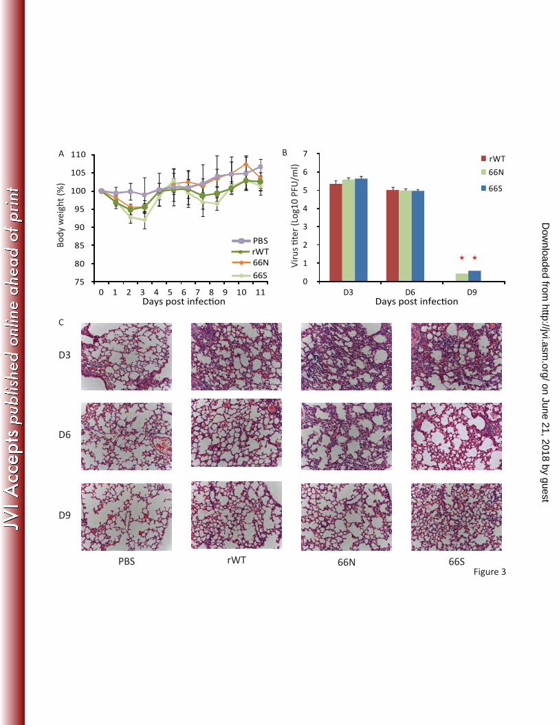
inflammatory gene expression and lung pathology following infection. In general, mice 12
infected with 5X10⁵ PFU of any virus displayed a comparable and transient morbidity (as 13
measured by weight loss) (Fig. 3A). Mice infected with the Cal/09 66S virus experienced 14
slightly more weight loss than other infected animals on days 2 and 3 post challenge. 15
Similar results were obtained following virus challenge with 5X10³ PFU or 5X10² PFU 16
(data not shown). 17
18
To better understand the differences in virulence of disease caused by infection with 19
these viruses, we next examined the kinetics of virus replication in the lungs. Mice were 20
infected intranasally with 5X10³ PFU of different Cal/09 viruses indicated in Fig. 3B. On 21
days 3, 6 and 9 post-infection, three mice per group were sacrificed to determine viral 22
on June 21, 2018 by guesthttp://jvi.asm
.org/D
ownloaded from
15
titers in lung homogenates (Fig. 3B). On both day 3 and 6 p.i., all three groups showed 1
similar viral lung titers. On day 9 p.i., there remained virus in the homogenate of only 2
one out the three animals infected with either PB1-F2 expressing Cal/09 viruses. 3
4
It has been shown that PB1-F2 affects the immune response to influenza virus infection 5
by enhancing the production of pro-inflammatory cytokines/chemokines and the 6
recruitment of immune cells (5, 16). We therefore proceeded to evaluate pathologic 7
changes (Fig. 3C) in the lungs of mice infected with the Cal/09 viruses and the induction 8
of pro-inflammatory genes (Fig. 3D). Histologic examination of lungs taken at 3, 6 and 9 9
days p.i. revealed degenerative changes with varying degrees of neutrophilic infiltrates 10
and diffuse alveolar damage with edema in all infected mice, especially on day 3 p.i. In 11
accordance with the recovery of body weight and decreased pro-inflammatory gene 12
expression, the degenerative changes were diminished on day 9 post infection. The 13
production of cytokines IFNγ, IL-1β and TNFα was similar in all infected animals. 14
However, levels of MCP-1, MIP-1β and RANTES were significantly higher in mice infected 15
with the Cal/09 66S virus compared with mice infected with Cal/09 rWT virus at day 6 16
p.i. (Fig. 3D). . Together, this data indicates that infection with the Cal/09 66S virus 17
induces both higher expression levels of selected cytokines and chemokines at early 18
time points over the other two viruses (Fig. 3C,D). 19
20
PB1-F2s do not have a significant impact on virulence in DBA/2 mice. 21
22
on June 21, 2018 by guesthttp://jvi.asm
.org/D
ownloaded from
16
It was recently shown that DBA/2 mice are more susceptible to influenza A virus 1
infection compared with BALB/c mice (2). To better discriminate between potential 2
differences in viral virulence conferred by PB1-F2 expression on the Cal/09 background, 3
we assessed the virulence of these viruses in the DBA/2 model. DBA/2 mice were 4
infected intranasally with the different Cal/09 viruses with either 5X10⁵ PFU, 5X10⁴ PFU 5
or 5X10³ PFU (Fig. 4). Pronounced morbidity and mortality was observed following virus 6
challenge as low as 5X10³ PFU, with all animals succumbing to infection by day 7 p.i. 7
(Fig. 4A). To evaluate viral replication in the lungs of the DBA/2 mice, animals were 8
infected intranasally with recombinant viruses at a dose of 5X10³ PFU and were 9
sacrificed on either day 2, 5 or 7 p.i. to determine viral titers (Fig. 4B). Similar viral titers 10
were recovered from all three groups of infected mice on days 2 and 5 p.i. On day 7 p.i., 11
animals infected with either the Cal/09 66N or Cal/09 66S virus had a 5-fold increase in 12
titer compared to mice infected with the rWT Cal/09 virus. In summary, all three viruses 13
demonstrated similar virulence in DBA/2 mice with respect to both morbidity and 14
mortality. It should be noted, however, that small differences in virulence may not have 15
been detectable under the conditions used in the experiments. 16
17
We next performed an analysis of cytokine/chemokine expression in the lungs during 18
infection (Fig. 4C). Infection with the Cal/09 66S virus was associated with enhanced 19
expression of particular pro-inflammatory genes but with a different profile as 20
compared to the BABL/c model. On day 2 and 5 post infection IL-1β protein could only 21
be detected in samples from mice infected with the Cal/09 66S virus. Protein levels of 22
on June 21, 2018 by guesthttp://jvi.asm
.org/D
ownloaded from
17
MIP1β and RANTES were also significantly increased in the lung samples from mice 1
infected with the Cal/09 66S on day 5 post infection. 2
3
PB1-F2 expression does not exacerbate secondary bacterial infection with 4
Streptococcus pneumoniae. 5
6
It has been shown that the PB1-F2 protein promotes bacterial infection secondary to 7
influenza virus infection in mice (16). To determine the impact of the Cal/09 PB1-F2 on 8
priming the host for secondary bacterial infection, we conducted co-infection studies in 9
BALB/c mice (Fig. 5). Animals infected with the different Cal/09 viruses were challenged 10
with a sub-lethal dose of S. pneumoniae 7 days post viral infection. Viral lung titers 11
were determined at the time of bacterial infection. Infection with the Cal/09 66S virus 12
lead to a 5-fold increase in viral lung titer over infection with the rWT Cal/09 virus and a 13
2-fold increase in titer over Cal/09 66N virus infection (Fig. 5A). By day 5 post bacterial 14
challenge, two out of ten mice in the Cal/09 66S virus infection group compared with 15
one out of ten mice in the other two infection groups had succumbed to infection (Fig. 16
5B). These data indicate that expression of PB1-F2 in the context of the novel Cal/09 17
virus does not significantly alter the mortality associated with secondary bacterial 18
infection in mice. 19
20
Expression of PB1-F2 does not significantly alter disease caused by infection with 21
Cal/09 in ferrets. 22
on June 21, 2018 by guesthttp://jvi.asm
.org/D
ownloaded from

18
1
Ferrets are an established model in influenza virology for studies of disease pathology 2
and transmission (23, 28). We therefore investigated the impact of PB1-F2 expression in 3
virulence of the Cal/09 virus in the Fitch ferret model. Animals were inoculated with 106 4
pfu of the Cal/09 wild-type or a variant Cal/09 virus by intranasal administration and 5
observed for 14 days p.i. for clinical signs of infection (Fig. 6A). Ferrets infected with 6
the 66S virus experienced slightly more weight loss than did those infected with the 66N 7
or wild-type viruses, but as observed in the mouse models, disease associated with 66S 8
infection was minimally exacerbated over disease caused by wild-type Cal/09 infection. 9
Nasal wash titers on days 1, 3 and 5 p.i. revealed no significant difference in upper 10
respiratory tract growth between any of the Cal/09 viruses (Fig. 6B). 11
12
Next we performed a leukocyte analysis on peripheral blood taken 3, 7 and 19 days post 13
infection. Interestingly, infection with any PB1-F2-expressing virus but not with wild-14
type virus was correlated with significantly dysregulated white cell counts on days 3 and 15
7 post infection (p<0.05); PB1-F2 expression was associated with both lymphopenia and 16
elevated neutrophil counts(Fig. 7). Lymphopenia in all ferrets was transient and levels 17
of leukocytes returned to baseline levels by day 19 p.i 18
19
Since PB1-F2 expression in the Cal/09 virus could occur by either genetic mutation (as 20
has been explored in this study) or by genetic reassortment, we generated an additional 21
virus expressing the PB1-F2 from a prototype H1 virus, PR/8/34. This model of a Cal/09 22
on June 21, 2018 by guesthttp://jvi.asm
.org/D
ownloaded from
19
reassortant virus, PR8-PB1-F2 Cal/09, was associated with a slightly exacerbated disease 1
phenotype in ferrets compared with viruses expressing PB1-F2 as a result of genetic 2
mutation in the Cal/09 PB1 gene (66S and 66N viruses) (Fig. 6 and 7). 3
on June 21, 2018 by guesthttp://jvi.asm
.org/D
ownloaded from

20
Discussion 1
2
The severity of influenza A virus infections is modulated by the expression of virulence 3
factors such as PB1-F2. In contrast to the PB1-F2-encoding viruses that caused the three 4
pandemics of the 20th century, the H1N1 virus responsible for the 2009 pandemic does 5
not express PB1-F2 and is associated with a relatively mild disease phenotype. In order 6
to better understand the consequences of potential genetic change in the pandemic 7
Cal/09 influenza virus, we conducted experiments designed to characterize the disease 8
phenotype associated with a PB1-F2-expressing version of Cal/09. 9
10
By developing a reverse genetics system for the Cal/09 virus, we were able to rescue 11
three Cal/09 viruses that have identical PB1 protein sequences: the wild-type virus, 12
along with variants that encode either the 66N or 66S PB1-F2 protein (Fig.1). We 13
examined the growth kinetics for all three Cal/09 viruses and observed enhanced 14
replication by the 66S virus in the human epithelial cell line A549, leading to increased 15
peak titers of roughly one log. This effect was not observed in the canine MDCK cell 16
line. Since influenza viruses replicate very efficiently in MDCK cells, it is likely that the 17
rather mild contribution to virulence by the PB1-F2 protein in the Cal/09 background is 18
simply not detectable in this cell system. 19
20
Our in vivo experiments were conducted in two different mouse strains. We first tested 21
the effect of PB1-F2 on the virulence of Cal/09 infection in BALB/c mice. Expression of 22
on June 21, 2018 by guesthttp://jvi.asm
.org/D
ownloaded from
21
PB1-F2 in the Cal/09 virus had no significant effect on disease pathogenesis (Fig. 3). We 1
only observed transient morbidity, with no mortality, following infection with any of the 2
Cal/09 viruses. In addition, peak lung viral titers were similar between the Cal/04 rWT, 3
66N and 66S virus groups. Analysis of cytokine/chemokine levels in the supernatants of 4
lung homogenates revealed increased expression of pro-inflammatory genes (e.g. MCP-5
1, MIP-1β and RANTES) which are essential for recruitment and activation of immune 6
cells in infected tissues (6 , 7, 13, 24). These pro-inflammatory proteins have also been 7
shown to prevent apoptosis of alveolar macrophages in the context of influenza virus 8
infection in vivo (10). Accordingly, we detected more severe histopathology in lung 9
tissues of mice infected with the PB1-F2-expressing viruses, especially in infection with 10
the 66S mutant virus. In summary, the introduction of a functional PB1-F2 ORF did not 11
enhance the mortality associated with infection with the Cal/09 virus in the BALB/c 12
mouse system. PB1-F2 expression did, however, result in a modulated host immune 13
response characterized by increased expression of pro-inflammatory genes. 14
15
The data obtained in our BALB/c mouse study suggested that in a more susceptible 16
mouse strain, such as the DBA/2 mouse (2), we would observe more pronounced 17
differences in disease phenotype caused by the PB1-F2-expressing Cal/09 viruses 18
compared with the wildtype virus. However, under the experimental conditions the 19
results of our studies in DBA/2 mice were very similar to the results from our BALB/c 20
mouse studies. Expression of the 66S PB1-F2 was correlated with enhanced expression 21
of the pro-inflammatory genes IL-1β and RANTES. Additionally, we observed that 22
on June 21, 2018 by guesthttp://jvi.asm
.org/D
ownloaded from
22
expression of PB1-F2 did not affect the susceptibility of BALB/c mice to secondary 1
bacterial infections. 2
3
Taken together, our mouse studies suggest that a genetic change resulting in the 4
presence of a functional PB1-F2 ORF would not significantly increase the virulence of 5
primary infection caused by the Cal/09 virus or the susceptibility of a host for secondary 6
bacterial infection with Streptococcus pneumoniae. 7
8
The fact that the expression of the PB1-F2 protein in the Cal/09 virus did not have a 9
significant impact on primary viral infection or secondary bacterial infection in mice is 10
intriguing as the prototype PB1-F2 proteins have been shown to affect both viral and 11
bacterial infections (5, 16, 27). As previously mentioned, the sequence of the Cal/09 12
PB1-F2 is quite unique. Even though the Cal/09 PB1-F2 is predicted to contain known 13
functional regions such as the mitochondrial targeting sequence, the protein seems to 14
have a diminished effect on viral virulence relative to protype PB1-F2 proteins, at least 15
in the context of the Cal/09 virus. This supports findings that enhanced viral virulence 16
conferred by PB1-F2 expression is a strain-specific phenomenon (17). 17
18
Our studies on weight loss and viral replication in ferrets paralleled the results from our 19
mouse studies. Expression of PB1-F2 was not associated with enhanced virulence to 20
statistical significance, but ferrets infected with the 66S variant did demonstrate a 21
slightly increased weight loss (without mortality) over those infected with the 66N or 22
on June 21, 2018 by guesthttp://jvi.asm
.org/D
ownloaded from
23
the wild-type Cal/09 viruses. PB1-F2 expression did cause a significant dysregulation in 1
peripheral leukocyte counts including lymphopenia and elevated neutrophil levels. 2
Infection with the reassortant PR8-PB1-F2 Cal/09 virus caused the most severe disease 3
phenotype of all viruses studied in ferrets; importantly, however, the PR8 PB1 protein 4
may contribute to the observed phenotype. 5
6
In summary, we found that mutations enabling the production of PB1-F2 in the Cal/09 7
influenza virus do not have a significant impact on virus virulence in mice or in ferrets. 8
These preliminary observations, however, invite further studies into the virulence of 9
other PB1-F2 mutant viruses and of possible reassortants of Cal/09 withcurrent viruses. 10
The present findings enhance our understanding of PB1-F2 as a virulence factor and 11
provide new insights into the impact genetic changes that may have on the virulence of 12
the 2009 pandemic virus. 13
14
on June 21, 2018 by guesthttp://jvi.asm
.org/D
ownloaded from

24
Acknowledgments: 1
We thank Dmitriy Zamarin for helpful discussions and critical reviewing. We also thank 2
Rafael A. Medina and Randy Albrecht for their help with the BSL3 training process and 3
we express our appreciation to Lily Ngai for excellent technical assistance. 4
5
This work was supported by CRIP (Center for Research on Influenza Pathogenesis, NIAID 6
contract HHSN266200700010C) and by grants from the NIH, P01 AI 058113, and U19 7
AI62623 (Center for Investigating Viral Immunity and Antagonism). TTW was supported 8
by NIH training grant T32 AI007647 and Mount Sinai Medical Scientists Training Grant 9
T32 GM007280. 10
11
on June 21, 2018 by guesthttp://jvi.asm
.org/D
ownloaded from

25
Figure legends: 1
Fig1. Alignment of the PB1-F2 open reading frames of pandemic influenza A virus 2
strains. Alignment of the PB1-F2 coding regions encoded by the amino acid sequences 3
of the wild type Cal/09 virus and of other pandemic influenza viruses (1918 H1N1, 1957 4
H2N2 and 1968 H3N2). The mutations of stop codons necessary for restoration of the 5
PB1-F2 open reading frame (rWT) coding for the 66N or the 66S versions of the protein 6
are indicated. 7
8
Fig2. Characterization of the recombinant Cal/09 viruses in vitro. MDCK cells (A) and 9
A549 cells (B) were infected with the rWT and PB1-F2 expressing viruses (MOI of 0.05). 10
At the indicated time points after infection, virus titers in the supernatants were 11
determined by plaque assay on MDCK cells. Average titers ± SD are indicated. (C) 12
Plaque phenotypes of the recombinant Cal/09 viruses in MDCK cells. 13
14
Fig3. Disease caused by infection with the wild type or PB1-F2-expressing Cal/09 viruses 15
in BALB/c mice. Eight-week-old female BALB/c mice, five animals per group, were 16
infected intranasally with 5X10⁵ PFU of the indicated rescued rWT or PB1-F2 expressing 17
mutant Cal/09 viruses. (A) Following viral infection, mice were weighed daily, and the 18
average body weights ± SD of surviving animals in each group up to day 11 post 19
infection are indicated as percentages of the original body weights. (B) Three mice per 20
group were infected intranasally with 5X10³ PFU of the same set of viruses. On days 3, 6 21
and 9 post- infection, three mice per group were sacrificed ( indicates that viruses 22
on June 21, 2018 by guesthttp://jvi.asm
.org/D
ownloaded from

26
were only recovered from one out of three animals.). Both the virus titers (B) in the 1
lungs and the level of the cytokines/chemokines (D) were determined from the lung 2
homogenate supernatants. Average lung titer ± SD are depicted. The limit of detection 3
was 5 PFU. (C) Six mice per group were infected intranasally with 5X10³ PFU of the 4
recombinant Cal/09 viruses. On days 3, 6 and 9 post-infection, two mice per group were 5
euthanized. The lungs were extracted for histologic examination. Representative 6
pictures are shown. (D) Cytokine/chemokine levels in supernatants of lung 7
homogenates were detected by multiplex ELISA at 3, 6 and 9 days post infection. 8
Average cytokine levels ± SD are depicted. (*P< 0.05, **P<0.01 vs. rWT Cal/09 virus-9
infected mice) 10
11
Fig4. Disease caused by infection with wild type or PB1-F2-expressing Cal/09 viruses in 12
DBA/2 mice. Eight-week-old female DBA/2 mice, five animals per group, were infected 13
intranasally with 5X10³ PFU of the indicated rescued rWT and PB1-F2 expressing mutant 14
Cal/09 viruses. (A) Following viral infection, the body weights of mice were monitored 15
daily, and the average body weights ± SD of surviving animals in each group up to day 8 16
post-infection are indicated as percentages of the original body weights. (B) 9 mice per 17
group were infected intranasally with 5X10³ PFU of the same set of viruses. On days 2, 5 18
and 7 post-infection, three mice were sacrificed and virus titers in the lungs were 19
determined. Average lung titer ± SD are depicted. The limit of detection was 5 PFU. (C) 20
Changes in cytokine/chemokine expression were detected by multiplex ELISA at 2, 5 and 21
on June 21, 2018 by guesthttp://jvi.asm
.org/D
ownloaded from

27
7 days post infection. Average cytokine/chemokine levels ± SD are depicted. (*P< 0.05, 1
**P<0.01 vs. rWT Cal/09 virus-infected mice) 2
3
Fig5. Characterization of the impact of Cal/09 PB1-F2 on secondary bacterial infection. 4
Groups of 15 eight-week-old female BALB/c mice were infected intranasally with 5X10⁵ 5
PFU of the indicated rescued rWT and PB1-F2 expressing mutant Cal/09 viruses or PBS, 6
then challenged with a sub-lethal dose of 20 CFU of S. pneumoniae, serotype 3, 7 days 7
later. (A) On day 7 post infection, three mice per infection group were sacrificed and 8
virus titers in the lungs were determined. The limit of detection was 5 PFU. (B) Survival 9
is plotted until day 7 post bacterial challenge. 10
11
Fig6. Weight change of H1N1-infected ferrets and viral replication in the upper 12
respiratory tract of infected ferrets. (A) Ferrets were inoculated i.n. with 1X10⁶ PFU of 13
each virus (rWT Cal/09, 66S, 66N and PR8-PB1 reassortant viruses). The percent weight 14
change was determined daily by comparing the mean weight of animals infected with 15
each virus to the mean preinfection weight. The mean percentage weight change for 16
each virus is shown. (B) Ferrets were inoculated i.n. with 1X10⁶ PFU/ml of each virus 17
shown. Viral titers were measured from nasal washes collected on indicated days p.i. 18
and are expressed as mean log10 PFU/ml plus standard deviation. The limit of virus 19
detection was 10 PFU. Titers on day 7 p.i. were below the limit of detection. 20
21
on June 21, 2018 by guesthttp://jvi.asm
.org/D
ownloaded from

28
Fig 7. Analysis of circulating lymphocytes following influenza virus infection. 3-5 ferrets 1
were inoculated i.n. with 1X10⁶ PFU of each virus. Blood was collected on days 3 (A), 7 2
(B), and 19 p.i. (C) in EDTA vacutainer tubes and analyzed with a hematology scanner. 3
Blood collected immediately prior to inoculation was included as a baseline control 4
(naïve). The average percentage of lymphocytes (LY), neutrophils (NE), monocytes (MO), 5
eosinophils (EO), and basophils (BA) in whole blood is shown. 6
on June 21, 2018 by guesthttp://jvi.asm
.org/D
ownloaded from

29
REFERENCES 1
2
1. Belser, J. A., X. Lu, T. R. Maines, C. Smith, Y. Li, R. O. Donis, J. M. Katz, 3 and T. M. Tumpey. 2007. Pathogenesis of avian influenza (H7) virus infection in 4 mice and ferrets: enhanced virulence of Eurasian H7N7 viruses isolated from 5 humans. J Virol 81:11139-47. 6
2. Boon, A. C., J. deBeauchamp, A. Hollmann, J. Luke, M. Kotb, S. Rowe, D. 7 Finkelstein, G. Neale, L. Lu, R. W. Williams, and R. J. Webby. 2009. Host 8 genetic variation affects resistance to infection with a highly pathogenic H5N1 9 influenza A virus in mice. J Virol 83:10417-26. 10
3. Chanturiya, A. N., G. Basanez, U. Schubert, P. Henklein, J. W. Yewdell, and 11 J. Zimmerberg. 2004. PB1-F2, an influenza A virus-encoded proapoptotic 12 mitochondrial protein, creates variably sized pores in planar lipid membranes. J 13 Virol 78:6304-12. 14
4. Chen, W., P. A. Calvo, D. Malide, J. Gibbs, U. Schubert, I. Bacik, S. Basta, 15 R. O'Neill, J. Schickli, P. Palese, P. Henklein, J. R. Bennink, and J. W. 16 Yewdell. 2001. A novel influenza A virus mitochondrial protein that induces cell 17 death. Nat Med 7:1306-12. 18
5. Conenello, G. M., D. Zamarin, L. A. Perrone, T. Tumpey, and P. Palese. 19 2007. A single mutation in the PB1-F2 of H5N1 (HK/97) and 1918 influenza A 20 viruses contributes to increased virulence. PLoS Pathog 3:1414-21. 21
6. Dawson, T. C., M. A. Beck, W. A. Kuziel, F. Henderson, and N. Maeda. 2000. 22 Contrasting effects of CCR5 and CCR2 deficiency in the pulmonary 23 inflammatory response to influenza A virus. Am J Pathol 156:1951-9. 24
7. Dessing, M. C., K. F. van der Sluijs, S. Florquin, and T. van der Poll. 2007. 25 Monocyte chemoattractant protein 1 contributes to an adequate immune response 26 in influenza pneumonia. Clin Immunol 125:328-36. 27
8. Fodor, E., L. Devenish, O. G. Engelhardt, P. Palese, G. G. Brownlee, and A. 28 Garcia-Sastre. 1999. Rescue of influenza A virus from recombinant DNA. J 29 Virol 73:9679-82. 30
9. Hai, R., L. Martinez-Sobrido, K. A. Fraser, J. Ayllon, A. Garcia-Sastre, and 31 P. Palese. 2008. Influenza B virus NS1-truncated mutants: live-attenuated vaccine 32 approach. J Virol 82:10580-90. 33
10. Herold, S., M. Steinmueller, W. von Wulffen, L. Cakarova, R. Pinto, S. 34 Pleschka, M. Mack, W. A. Kuziel, N. Corazza, T. Brunner, W. Seeger, and J. 35 Lohmeyer. 2008. Lung epithelial apoptosis in influenza virus pneumonia: the role 36 of macrophage-expressed TNF-related apoptosis-inducing ligand. J Exp Med 37 205:3065-77. 38
11. Hilleman, M. R. 2002. Realities and enigmas of human viral influenza: 39 pathogenesis, epidemiology and control. Vaccine 20:3068-87. 40
12. Itoh, Y., K. Shinya, M. Kiso, T. Watanabe, Y. Sakoda, M. Hatta, Y. 41 Muramoto, D. Tamura, Y. Sakai-Tagawa, T. Noda, S. Sakabe, M. Imai, Y. 42 Hatta, S. Watanabe, C. Li, S. Yamada, K. Fujii, S. Murakami, H. Imai, S. 43 Kakugawa, M. Ito, R. Takano, K. Iwatsuki-Horimoto, M. Shimojima, T. 44
on June 21, 2018 by guesthttp://jvi.asm
.org/D
ownloaded from

30
Horimoto, H. Goto, K. Takahashi, A. Makino, H. Ishigaki, M. Nakayama, M. 1 Okamatsu, D. Warshauer, P. A. Shult, R. Saito, H. Suzuki, Y. Furuta, M. 2 Yamashita, K. Mitamura, K. Nakano, M. Nakamura, R. Brockman-3 Schneider, H. Mitamura, M. Yamazaki, N. Sugaya, M. Suresh, M. Ozawa, G. 4 Neumann, J. Gern, H. Kida, K. Ogasawara, and Y. Kawaoka. 2009. In vitro 5 and in vivo characterization of new swine-origin H1N1 influenza viruses. Nature 6 460:1021-5. 7
13. Lin, K. L., Y. Suzuki, H. Nakano, E. Ramsburg, and M. D. Gunn. 2008. 8 CCR2+ monocyte-derived dendritic cells and exudate macrophages produce 9 influenza-induced pulmonary immune pathology and mortality. J Immunol 10 180:2562-72. 11
14. Maines, T. R., A. Jayaraman, J. A. Belser, D. A. Wadford, C. Pappas, H. 12 Zeng, K. M. Gustin, M. B. Pearce, K. Viswanathan, Z. H. Shriver, R. Raman, 13 N. J. Cox, R. Sasisekharan, J. M. Katz, and T. M. Tumpey. 2009. 14 Transmission and pathogenesis of swine-origin 2009 A(H1N1) influenza viruses 15 in ferrets and mice. Science 325:484-7. 16
15. Mazur, I., D. Anhlan, D. Mitzner, L. Wixler, U. Schubert, and S. Ludwig. 17 2008. The proapoptotic influenza A virus protein PB1-F2 regulates viral 18 polymerase activity by interaction with the PB1 protein. Cell Microbiol 10:1140-19 52. 20
16. McAuley, J. L., F. Hornung, K. L. Boyd, A. M. Smith, R. McKeon, J. 21 Bennink, J. W. Yewdell, and J. A. McCullers. 2007. Expression of the 1918 22 influenza A virus PB1-F2 enhances the pathogenesis of viral and secondary 23 bacterial pneumonia. Cell Host Microbe 2:240-9. 24
17. McAuley, J. L., K. Zhang, and J. A. McCullers. 2010. The Effects of Influenza 25 A Virus PB1-F2 Protein on Polymerase Activity Are Strain Specific and Do Not 26 Impact Pathogenesis. J Virol 84:558-64. 27
18. Munster, V. J., E. de Wit, J. M. van den Brand, S. Herfst, E. J. Schrauwen, 28 T. M. Bestebroer, D. van de Vijver, C. A. Boucher, M. Koopmans, G. F. 29 Rimmelzwaan, T. Kuiken, A. D. Osterhaus, and R. A. Fouchier. 2009. 30 Pathogenesis and transmission of swine-origin 2009 A(H1N1) influenza virus in 31 ferrets. Science 325:481-3. 32
19. Neumann, G., T. Watanabe, H. Ito, S. Watanabe, H. Goto, P. Gao, M. 33 Hughes, D. R. Perez, R. Donis, E. Hoffmann, G. Hobom, and Y. Kawaoka. 34 1999. Generation of influenza A viruses entirely from cloned cDNAs. Proc Natl 35 Acad Sci U S A 96:9345-50. 36
20. Palese, P. 2004. Influenza: old and new threats. Nat Med 10:S82-7. 37 21. Palese, P. a. S., M.L. 2007. Orthomyxoviridae: The Viruses and their 38
Replication. In D. M. Knipe, Howley, P.M. (ed.), Fields Virology, 5th ed, vol. 1. 39 Lippincott-Raven Press, Philadelphia. 40
22. Smith, G. J., D. Vijaykrishna, J. Bahl, S. J. Lycett, M. Worobey, O. G. 41 Pybus, S. K. Ma, C. L. Cheung, J. Raghwani, S. Bhatt, J. S. Peiris, Y. Guan, 42 and A. Rambaut. 2009. Origins and evolutionary genomics of the 2009 swine-43 origin H1N1 influenza A epidemic. Nature 459:1122-5. 44
23. Smith, H., and C. Sweet. 1988. Lessons for human influenza from pathogenicity 45 studies with ferrets. Rev Infect Dis 10:56-75. 46
on June 21, 2018 by guesthttp://jvi.asm
.org/D
ownloaded from

31
24. Wareing, M. D., A. B. Lyon, B. Lu, C. Gerard, and S. R. Sarawar. 2004. 1 Chemokine expression during the development and resolution of a pulmonary 2 leukocyte response to influenza A virus infection in mice. J Leukoc Biol 76:886-3 95. 4
25. Yamada, H., R. Chounan, Y. Higashi, N. Kurihara, and H. Kido. 2004. 5 Mitochondrial targeting sequence of the influenza A virus PB1-F2 protein and its 6 function in mitochondria. FEBS Lett 578:331-6. 7
26. Zamarin, D., A. Garcia-Sastre, X. Xiao, R. Wang, and P. Palese. 2005. 8 Influenza virus PB1-F2 protein induces cell death through mitochondrial ANT3 9 and VDAC1. PLoS Pathog 1:e4. 10
27. Zamarin, D., M. B. Ortigoza, and P. Palese. 2006. Influenza A virus PB1-F2 11 protein contributes to viral pathogenesis in mice. J Virol 80:7976-83. 12
28. Zitzow, L. A., T. Rowe, T. Morken, W. J. Shieh, S. Zaki, and J. M. Katz. 13 2002. Pathogenesis of avian influenza A (H5N1) viruses in ferrets. J Virol 14 76:4420-9. 15
16 17
on June 21, 2018 by guesthttp://jvi.asm
.org/D
ownloaded from

12 50....|....| ....|....| ....|....| ....|....| ....|....|
1918 MGQEQDTPWI LSTGHISTQK REDGQQTPRL EHHNSTRLMD HCQKTMNQVV 501957 .E.......T Q..E..NI.. .GS....RK. .RP.L.Q... .YLR.....D 501968 .E.......T Q..E..NI.. KGS....RK. .RP.L.Q... .YLRI.S..D 50rWT .E.......T Q..E.TN... ..S.R..Q.. V.PS...... .YLRI....G 49
58 66 88....|....| ....|....| ....|....| ....|....| .
1918 MPKQIVYWKQ WLSLRSPTPV SLKTRVLKRW RLFSKHEWTS . 901957 .H..TAS... ....KN..QE .......... K..N.Q...N . 901968 .H..T.S... ....KN..QG .......... K..N.QG..D . 90rWT .H..T.F.RL ....KN..QE Y.RIHA..Q. K..N.QG.IN . 87
C terminal region.Stop codon amino acid 66
rWT: A/California/04/2009(H1N1)
1918: A/Brevig Mission/18(H1N1) 1957: A/Guiyang/1/1957(H2N2) 1968: A/Hong Kong/1/1968(H3N2)
Minimal mitochondrial targeting sequence.
Figure 1
on June 21, 2018 by guesthttp://jvi.asm
.org/D
ownloaded from

0
1
2
3
4
5
6
7
8
9
0 24 48 72
A
B
A
Figure 2
C
66S66NrWT
66S66NrWT
0
1
2
3
4
5
6
7
8
9
0 24 48 72
Viru
stit
er(lo
g10
PFU
/ML)
Hours post infection
rWT 66S66N
on June 21, 2018 by guesthttp://jvi.asm
.org/D
ownloaded from
0
1
2
3
4
5
6
7
D3 D6 D9
66N
66S
rWT
Viru
ste
r(L
og10
PFU
/ml)
B
C
A
75
80
85
90
95
100
105
110
0 1 2 3 4 5 6 7 8 9 10 11
66N
Days post infection Days post infection
66S
rWTPBSBo
dy w
eigh
t (%
)
66N 66SrWT
D3
D6
D9
PBSFigure 3
on June 21, 2018 by guesthttp://jvi.asm
.org/D
ownloaded from

rWT
D366
N D3
66S
D3
rWT
D666
N D6
66S
D6
rWT
D966
N D9
66S
D9
rWT
D366
N D3
66S
D3
rWT
D666
N D6
66S
D6
rWT
D966
N D9
66S
D9
rWT
D366
N D3
66S
D3
rWT
D666
N D6
66S
D6
rWT
D966
N D9
66S
D9
rWT
D366
N D3
66S
D3
rWT
D666
N D6
66S
D6
rWT
D966
N D9
66S
D9
rWT
D366
N D3
66S
D3
rWT
D666
N D6
66S
D6
rWT
D966
N D9
66S
D9
rWT
D366
N D3
66S
D3
rWT
D666
N D6
66S
D6
rWT
D966
N D9
66S
D9
IFNγ IL-1β MCP-1
MIP-1β RANTES TNFα
Prot
ein
Conc
entr
ation
(pg/
ml)
D
Figure 3 - Continued
0
400
800
1200
1600
0
100
200
300
400450
0
10000
20000
30000
40000
0
10000
20000
30000
40000
50000
0
20000
40000
60000
70000
0
20
40 on June 21, 2018 by guesthttp://jvi.asm
.org/D
ownloaded from
0
1
2
3
4
5
6
7
D2 D5 D7Vi
rus
ter(
Log1
0PF
U/m
l)
B
C
Figure 4
A
60
70
80
90
100
0 1 2 3 4 5 6 7 8
Bod
y w
eigh
t (%
)
Days post infection Days post infection
rWT
D266
N D2
66S
D2
rWT
D566
N D5
66S
D5rW
T D7
66N
D766
S D7
rWT
D266
N D2
66S
D2
rWT
D566
N D5
66S
D5rW
T D7
66N
D766
S D7
rWT
D266
N D2
66S
D2
rWT
D566
N D5
66S
D5rW
T D7
66N
D766
S D7
IFNγ IL-1β MCP-1
MIP-1β RANTES TNFα
Pro
tein
Con
cent
ratio
n (p
g/m
l)
66N66S
rWTPBS
66N66S
rWT
rWT
D266
N D2
66S
D2rW
T D5
66N
D566
S D5
rWT
D766
N D7
66S
D7
rWT
D266
N D2
66S
D2rW
T D5
66N
D566
S D5
rWT
D766
N D7
66S
D7
rWT
D266
N D2
66S
D2
rWT
D566
N D5
66S
D5rW
T D7
66N
D766
S D7
0
4000
8000
12000
1600018000
0
40
80
120
0
10000
20000
25000
0
10000
20000
30000
0
1000
2000
30003500
0
20
40
60
80
100
on June 21, 2018 by guesthttp://jvi.asm
.org/D
ownloaded from

0
1
2
3
4
5
D7
Days post bacterial challenge
A
B
Figure 5
66N
66S
rWTVirustiter
(log1
0PF
U/m
l)Su
rvival(%
)
50
60
70
80
90
100
0 1 2 3 4 5 6 7
66N 66S
rWTPBS
on June 21, 2018 by guesthttp://jvi.asm
.org/D
ownloaded from

75
80
85
90
95
100
105
0 1 2 3 4 5
Days post infection
Bod
y w
eigh
t (%
)
6 7 8 9 10 11 12 13 14
rWT
PR8 PB1
66S
66N
rWT PR8 PB166S
987654321
D1 D3 D5
66N
Viru
s tit
er (L
og10
PFU
/ml)
Figure 6
A
B on June 21, 2018 by guesthttp://jvi.asm
.org/D
ownloaded from

0%
20%
40%
60%
80%
100%
Days post infection 3
BA EO MO NE LY
Days post infect 7
0%
20%
40%
60%
80%
100%
0%
20%
40%
60%
80%
100%
% W
hite
Blo
od C
ells
baseline rWT PR8/PB1 66S 66N
Days post infection 19
Figure 7
A
B
C
on June 21, 2018 by guesthttp://jvi.asm
.org/D
ownloaded from