Juvenile Nephronophthisis. Part II A Histologic and ...
10
Acta PEdiatrica 49: 480-487. July 1960 From the Department of Pediatric Pathology at the Department of Pathology (Head: A. Wilton), Karolinska sjukhuset, Stockholm Juvenile Nephronophthisis. Part I1 A Histologic and Microangiographic Study by BIORN I. IVEMARK, ARNE LJUNGQVIST and ALEXANDER BARRY1 The renal changes in fatal cases of fa- milial juvenile nephronophthisis have been described previously (Fanconi et al., 1951, Hackzell & Lundmark, 1958, and Hooft et al., 1959). The changes were re- garded as being due to a diffuse renal con- traction. No morphologic evidence of con- genital defects in the kidney was given by any of these authors. In the present paper, the renal pathology of two fatal cases of this disease will be described. The clinical features have been presented in the report by Broberger, Winberg & Zetterstrom in this issue (Cases 2 and 3). Evidence will be presented to show that there is a characteristic lesion of the descending and ascending limbs of the loop of Henle. This defect is associated with a cortical atrophy which is considered to be secondary to the tubular lesions. In view of the tendency of this disease to occur as a familial trait, it seems probable that the tubular defect has a congenital basis. Material and Methods The kidneys from two patients (both 121", years old) with t>he clinical picture of familial juvenile nephronophthisis with terminal azo- On leave from the Department of Anatomy, University of Michigan Medical School, Ann Arbor, Mich., U.S.A. temia were available for stndy (Cases 2 and 3 of Broberger etal.). Autopsy permission was restricted to an examination of the kidneys. A partial autopsy was performed 2 hours (Case 2) and 3 hours (Case 3) post nzortem, respectively. One kidney from each case was kept intact in it's capsule and immediately stored at -20°C. The other kidney was re- moved from its capsule, split open, and pre- served in 10 76 neutral formalin for histologic examination. The formalin-fixed left kidney from Case 2 weighed 40 g, and the fixed right kidney from Case 3 weighed 60 g (normal, 95 g). The gross appearance (Figs. 1 and 2) of both specimens was essentially similar. The fibrous capsule stripped off easily, the subcapsular surface was slightly granular and pink. In both specimens a few pin- point-sized, thin-walled cysts were seen at the surface of the cortex. On sectioning, the cort>ex was found to measure 2-3 mm in thickness (normal 5 to 6 mm). The medulla measured 10-12 mm in thickness which is within the normal size range. Cysts were also found in the medulla, close to t,he cortico- medullary junction as well as in the center of the pyramids (Figs. 1 and 2). Cortical cysts measuring 0.2-1.8 mm in diameter were present in both specimens (Fig. 2). The calyces, pelves, and ureters appeared normal. No malformations of the major renal vessels were present. The renal tissue taken from both specimens for histologic study was dehydrated, embedded in paraffin and stai- ned with hematoxylin-eosin, van Gieson's connect'ive tissue stain, Verhoeff's elastic tissue stain (counterstained with van Giesons
Transcript of Juvenile Nephronophthisis. Part II A Histologic and ...
Acta PEdiatrica 49: 480-487. July 1960
From the Department of Pediatric Pathology a t the Department of Pathology (Head: A. Wilton), Karolinska sjukhuset, Stockholm
Juvenile Nephronophthisis. Part I1
A Histologic and Microangiographic Study
by BIORN I. IVEMARK, ARNE LJUNGQVIST and ALEXANDER BARRY1
The renal changes in fatal cases of fa- milial juvenile nephronophthisis have been described previously (Fanconi et al., 1951, Hackzell & Lundmark, 1958, and Hooft et al., 1959). The changes were re- garded as being due to a diffuse renal con- traction. No morphologic evidence of con- genital defects in the kidney was given by any of these authors.
I n the present paper, the renal pathology of two fatal cases of this disease will be described. The clinical features have been presented in the report by Broberger, Winberg & Zetterstrom in this issue (Cases 2 and 3). Evidence will be presented to show that there is a characteristic lesion of the descending and ascending limbs of the loop of Henle. This defect is associated with a cortical atrophy which is considered t o be secondary to the tubular lesions. I n view of the tendency of this disease to occur as a familial trait, it seems probable that the tubular defect has a congenital basis.
Material and Methods
The kidneys from two patients (both 121", years old) with t>he clinical picture of familial juvenile nephronophthisis with terminal azo-
On leave from the Department of Anatomy, University of Michigan Medical School, Ann Arbor, Mich., U.S.A.
temia were available for stndy (Cases 2 and 3 of Broberger e ta l . ) . Autopsy permission was restricted to an examination of the kidneys. A partial autopsy was performed 2 hours (Case 2) and 3 hours (Case 3) post nzortem, respectively. One kidney from each case was kept intact in it's capsule and immediately stored a t -20°C. The other kidney was re- moved from its capsule, split open, and pre- served in 1 0 76 neutral formalin for histologic examination. The formalin-fixed left kidney from Case 2 weighed 40 g, and the fixed right kidney from Case 3 weighed 60 g (normal, 95 g). The gross appearance (Figs. 1 and 2) of both specimens was essentially similar. The fibrous capsule stripped off easily, the subcapsular surface was slightly granular and pink. In both specimens a few pin- point-sized, thin-walled cysts were seen at the surface of the cortex. On sectioning, the cort>ex was found to measure 2-3 mm in thickness (normal 5 to 6 mm). The medulla measured 10-12 mm in thickness which is within the normal size range. Cysts were also found in the medulla, close to t,he cortico- medullary junction as well as in the center of the pyramids (Figs. 1 and 2 ) . Cortical cysts measuring 0.2-1.8 mm in diameter were present in both specimens (Fig. 2). The calyces, pelves, and ureters appeared normal. No malformations of the major renal vessels were present. The renal tissue taken from both specimens for histologic study was dehydrated, embedded in paraffin and stai- ned with hematoxylin-eosin, van Gieson's connect'ive tissue stain, Verhoeff's elastic tissue stain (counterstained with van Giesons
JUVENILE NEPHRONOPHTHISIS. PART I1 48 1
stain), Lendrnm’s stain for reticdin, and alcian blue-PAS according to McManus & Cuerrero.
The two frozen kidneys were used for microangiography (Bellman) employing a techniqne modified for the infantile kidney (Ljimgqvist). This procedure was carried out after 18-31 months of storage, which un- fortunately resulted in impairment of the histologic detail. The ureter was injected with a 35 % solution of UrografinC3, using slight pressure, and roentgenograms were taken. Following this the contrast medium was removed, the pelvis washed with saline, and the renal artery injected with a 10 % aqueous mixture of MicropaqueC3 for fonr hours. An initial pressure of 110 mm Hg was increased to a final injection pressure of 175 mm Hg. After stereo-X-ray photo- graphs had been taken, the gross specimens were sliced, embedded in paraffin, and pre- pared for stereo-microangiography. After this procedure samples were sectioned se- rially at 5 p. The sections were stained with Verhoeff’s elastic tissue stain counterstained with van Gieson’s stain. A few sections were stained with alcian blue.
Histologic Findings
The histologic findings differed in degree but were essentially similar in the two cases. In the kidneys from Case 3 the cortex was thinner and appeared to contain a larger number of hyalinized glomeruli, while the cortex from Case 3 displayed more numerous cortical cysts.
Rend corpuscles. Two basically different types of degeneration were noted, (a) hyalinization, and (b) cystic dilatation. Varying degrees of hyaline change were present in most of the glomeruli, although a small number were apparently normal. As was noted by previous observers, in those renal corpuscles which showed minimal change, a thickened basement membrane was seen only in the parietal 32 - 603605 Acta Pzdiatrica Vol. 49
layer, while the glomerular tuft appeared to be unaffected. In some instances the thickend parietal layer of Bowman’s capsule stained a deep magenta with the alcian blue-PAS, denoting the presence of neutral mucopolysaccharides. In more severely affected corpuscles, the thickened basement membrane involved the glome- rular tuft as well, and many extreme cases were found in which the lumen was com- pletely obliterated. It was interesting to note that only rarely did the completely scarred glomeruli stain magenta with the alcian blue-PAS.
The cystic glomerular change was pre- sent in the subcapsular layer. In serial sections all the cortical cysts were found to contain glomerular tufts of varying degrees of atrophy. For this reason they were considered to be dilated renal cor- puscles. The parietal layer cf these cysts varied in thickness, some cysts having thick fibro-elastic walls, others displaying thin reticulin sheaths. None of them, however, stained positively with the alcian blue- PAS. No correlation could be found be- tween the structure of the parietal wall of these cysts and either their size or the degree of atrophy of the glomerular tuft. These cysts were regularly spherical and did not communicate with any part of the uriniferous tubules, although atrophic nephric tubules could be identified in their immediate vicinity (Fig. 3). Proximal convoluted tubules. In addition
to the previously mentioned atrophic proximal convuluted tubules, hypertro- phic tubules were found arising from the glomeruli which showed minimal changes. Occasional groups of proximal tubular cells displayed hyaline droplet change. The granules were PAS-positive. The al-

483 B. I . IVEMARK, A. LJUNCQVIST AND A. BARRY
cian blue-PAS technique also brought out occasional PAS-positive thickening of the basement membrane, especially in the neck portion of the tubule.
Descending limbs of the loop of Henle. This portion of the nephron exhibited the most characteristic and marked change in these specimens, in that they were modera- tely enlarged and strikingly convoluted (Fig. 4). There was considerable variability between different nephrons with respect to the degree of this convolution (see below). The size and configuration of the individual cells appeared to be within the normal range. The basement membrane was slightly thickened, and PAS-positive. The thin segments had a moderately thicke- ned basement membrane. Except for this and an occasionally tortuous course, they appeared essentially normal.
In occasional instances the flexure of Henle’s loop was dilated. In extreme cases this had the appearance of a small ovoid medullary cyst of approximately 2 mm in diameter. A study of serial sections con- firmed the fact that one descending and one ascending limb of Henle’s loop opened into the cortical aspect of each cyst. As is seen in Pig. 5, the descending limb with its thickened basement membrane may project nozzle-like into the cyst. The as- cending limb of Henle’s loop sometimes leaves the cyst in a tangential manner. This configuration might have acted as a valve, and prevented free passage of fluid from the cyst further along the nephron.
Ascending limbs of the loop of Henle. The ascending limbs-whether or not they were connected with a medullary cyst-showed a varying degree of coiling with thickening of the basement mem- brane. This membrane was massive, homo-
geneous and PAS-positive (Fig. 6). In some cases the segment consisted of a thin cel- lular cord surrounded by a PAS-positive sheath, while in other instances no rem- nants of the parenchymal cells could be seen, and the segment was represented by a coiled strand of PAS-positive material. It seems reasonable to assume that these findings represented stages in the pro- gressive obliteration of this segment of the nephron.
Distal convoluted tubules. This portion of the nephron displayed the same range of morphologic characteristics as were seen in the ascending limb of Henle’s loop.
Collecting ducts. From the arched col- lecting tubules to the papillary ducts, the collecting duct system showed no apparent abnormalities. The papillary ducts, how- ever, frequently exhibited a variable de- gree of dilatation. The lumen of such an ectatic portion was in continuity with a minor calyx. These dilatations were fusi- form and characteristically lined with a tall, columnar, stratified epithelium. Other similarly lined spaces in the papillary zone of the medulla were essentially spherical and measured up to 8 mm in diameter. From a study of serial sections these were found to have no connection with either straight collecting tubules or with minor calyces. In view of the essential identity in histologic structure it seems reasonable to assume that these cysts were derived from papillary ducts. In contrast to the medullary sponge kidney (Ekstrom et al.) these cysts did not contain calculi.
Pelvis and calyces. The pyelograms of the specimens showed no abnormality in gross morphology of pelvis or calyces. There was no filling of the medullary cysts or ectasies. The histologic structure of the

JUVENILE NEPHRONOPHTHISIS. PART I1 483
wall of the pelvis and calyces was normal. Stroma. The increased interstitial fibrous
tissue contained foci of plasma cells and lymphocytes as previously described for this condition. The arteries had moderately thickened media and occasionally there was a slight intimal thickening. The elastic laminae appeared normal. The adventitia of a few archiform arteries contained thick sheaths of circularly arranged smooth muscle. This was occasionally in conti- nuity with the calyceal musculature. Very occasional interlobular arteries exhibited a, similar but thinner muscular adventitia. Within this adventitial smooth muscle were strikingly developed venous sinu- soids.
Microangiographic Findings
Cortex. The vascular pattern is seen in Figs. 7-8. The thinning of the cortex is represented by a reduced vasculature, a tortuosity of the interlobular arteries and afferent glomerular arterioles (Fig. 8). The tortuosity was most apparent in the long juxtamedullary afferent glomerular vessels. The cystically enlarged renal cor- puscles were never filled. Nor could these cysts be localized on the basis of the vas- cular pattern. As a rule, the peritubular capillaries were not filled.
Medulla. The efferent arterioles from the juxtamedullary glomeruli and the vasa rectae could easily be visualized. Usually, the vasa rectae were straight, although occasionally they were sinuous, only rarely were they markedly tortuous. No aneurysms were demonstrated, nor was there any direct filling of the medul- lary cysts.
Occasionally, glomerular capillaries rup-
tured during injection. This allowed the radioopaque material to fill the nephron, although only rarely did this injection ex- tend beyond the loop of Henle. Thus it was possible to study from stereo-microangio- grams the relationship of the vasculature to the nephron especially in the medullary region. The unexpected observation was made that an uncoiled blood vessel was intimately associated with a markedly coiled descending limb of Henle’s loop, so that in the microangiographic projec- tion they have the appearance of the Esculapian serpent (Fig. 10). Occasionally this filling of the nephron resulted in the filling of a small medullary cyst confirm- ing the fact that such cysts are dilatations of the flexure of Henle’s loop (Fig. 9).
Measurements of the Descending Limb of the Loop of Henle
In view of the striking convolution of the descending limb of Henle’s loop, and the fact that the medulla was normal in thickness, some approximate measure- ments seemed warranted. Tracings of such convoluted segments as visualized in the microangiograms were made by means of a camera lucida a t a magnifica- tion of x 67. From such tracings the over- all length of the descending limb was mea- sured. This length was compared to the length of the straight line between the end points of the descending limb. This ratio in a markedly convoluted segment was 3.3. Thus the coiling involves an actual increase in length and is not a result of mechanical distortion. The diameter of the lumen and of the cross- sectional dia- meter inside the basement membrane were measured for a series of abnormal descend-
481 B. I. IVEMARK, A. LJUNCQVIST AND A. BARRY
ing limbs. Similar measurements were made from a normal kidney from a 12- year-old boy. Since in both instances the segment is roughly circular in cross sec- tion, the area of the parenchyma could be calculated. A comparison of such cross- sectional areas indicated that the abnor- mal segment had a parenchymatous cross-sectional area 3.5 times greater than the normal. Thus, it appears that the volume of parenchyma of a convoluted descending limb of Henle may be as much as 11 times greater than normal. Since the size of the parenchymatous cells were of the same order in the normal and abnormal specimens, it would appear that in this condition there is a hyperplasia of the descending limb of the loop of Henle.
Discussion
The morphologic findings in kidneys from patients displaying the clinical fea-
tures of familial juvenile nephronophthi- sis as have been reported previously may be summarized as follows. A diffuse con- traction of the kidney causing coiling of the nephrons, an ensheathing of the nephrons by a characteristically thickened base- ment membrane, an initial thickening of the basement membrane of the parietal layer of the renal corpuscles eventually progressing to complete hyaline degenera- tion of the entire corpuscle.
In both cases described in this report the kidneys were reduced in size and weight. However, this reduction of the renal parenchyma appeared to be restricted to the cortex, which was markedly thinned. The thicliness of the medulla, on the other hand, was within the normal range.
Within the cortex both the interlobular arteries and the afferent glomerular ar- terioles were markedly distorted and con- voluted. This would seem to have been
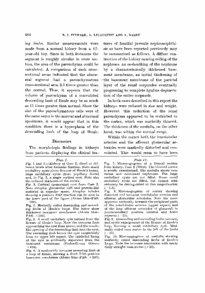
Plate I . Fig. 1 and 3.-Kidney of Case 2, sliced at dif- ferent levels after formalin fixation. Note small medullary cysts (from flexures of Henle’s loops), large medullary cysts (from papillary ducts) and, in Fig. 3, a single cortical cyst. Note also the reduced thickness of the cortex. Fig. 3. Cortical (gomerular) cyst from Case 3. Note atrophic glomerular tuft and protein-like material in capsular space. Atrophic tubules showing a positive PAR reaction can be seen in the lower part of the figure (Alcian blue-PAS:
Fig. 4. Markedly coiled descending and ascend- ing limbs of Henle’s loops. The latter show thickened basement membranes (Alcian blue- PAS: > 100). Fig. 5. A small medullary cyst formed from the flexure of Henle’s loop. Note the tortuous limb approaching the cyst from above, and the nozzle- like opening of the descending limb into the cyst. The ascending limb leaves the cyst tangentially from its upper left aspect. The epithelial lining of the cyst is artificially separated from the basement membrane (Verhoeff-van Gieson:
A 100). Fig. 6. A moderately tortuous ascending limb of a loop of Henle, showing a thick PAS-positive basement membrane (Alcian blue-PAS: x 240).
,’ 100).
Plate I I . Fig. 7 . Microangiogram of a frontal section from kidney, Case 2 ( 7 0 0 ~ ) . The thinned cortex is poorly vascularized. The medulla shows vasa rectae and occasional nephrones. The large medullari cysts are not filled. Some small medullary cysts are filled, but cannot with certainty be distinguished a t this magnification ( I’ 1.5). Fig. 8. Microangiogram of cortex showing distorted and tortuous interlobular arteries and afferent glomerular arterioles. Note the more apparent tortuosity towards the peripheral parts of the int,erlobular arteries (upper aspect) and of the long afferent arterioles of glomeruli in juxtamedullary position (central and lower aspects) ( 1’ 2 5 ) . Fig. 9. Descending and ascending limbs (arrows), and cystic enlargement of the flexure of Henle’s loop, forming a small medullary cyst. Mini- mally coiled vasa rectae to the left of the limbs
Fig. 10. Microangiogram of medulla showing markedly coiled descending limbs of Henle’s loops. Note the intimate association with essen- tially straight vasa rectae ( x 25).
( ’; 25).
JUVENlLE NEPHRONOPHTHISIS. PART I1 485
the inevitable result of a progressive thin- ning of the cortex. Many stages of hyaline degeneration of renal corpuscles were found. In this respect these two cases con- form to the characteristics described in previous reports, but they differ, however, in that some renal corpuscles showed cystic enlargement with atrophy of the glomerular tuft. None of these glomerular tufts or cysts whether studied in micro- angiograms or in histologic sections, could lie demonstrated to contain contrast me- dium. It seems reasonable to assume, therefore, that at the time of death they were essentially avascular.
Although there was a characteristic thickening of the basement membrane of all segments of the nephrons, it was rela- tively more massive in association with the ascending limbs of Henle’s loops and the distal convoluted tubules. In extreme cases the lumen of the ascending limb of Henle’s loop was obliterated. This more or less complete blockage may have resul- ted in sufficient back-pressure to have initiated the cystic dilatation of the flexure of the loop which was found in these two specimens. The characteristic tangential relationship of an ascending limb of Henle’s loop to such a cyst would act as a valve and further interfere with free passage of fluid along the nephron, causing further enlargement of the cyst. This cystic feature has not been described previously in cases of familial juvenile nephronophthi- sis. The occurrence of these renal cysts in the two patients of the present study may be explained by the fact that they were older than those of previous studies.
The striking tortuosity of the limbs of Henle’s loops cannot have been due to a diffuse contraction of the renal paren-
chyma in these two cases. In the first place, the medulla did not exhibit any shrinkage. Moreover, it seems that any generalized change in the dimensions of the medulla would affect equally all struc- tures lying in it.
If the coiling of the descending limbs of Henle’s loops is not the result of compres- sion of the medulla, then it must be due to an actual increase in their length. Quantitative estimations made on such nephric segments showed that this was actually the case. The length of a mar- kedly convoluted descending limb of the loop of Henle was three times greater than normal, while the volume of its paren- chyma exceeded that of a normal control by tenfold. The convolutions shown by the vessels in the cortex seem to be se- condary to the thinning of the cortical layer, and not to reflect a hyperplasia.
The histologic structure of the walls of the large cysts in the renal papillae was indistinguishable from that of the ectatic papillary ducts. In fact these two struc- tures could be distinguished with cer- tainty only from a study of serial sections. It seems probable that the cysts represent ectatic areas of papillary ducts nhich secondarily lost their connection with the collecting duct system. It would be un- profitable to speculate, from the material available, concerning the mechanisms in- volved in this sequence of events.
The high incidence of this rare disease in certain families has led previous in- vestigators to regard i t as being geneti- cally determined. The two cases reported here throw no light on this aspect of the problem. Their clinical history is conipa- tible with the assumption that the pri- mary lesion was a slowly progressing dis-
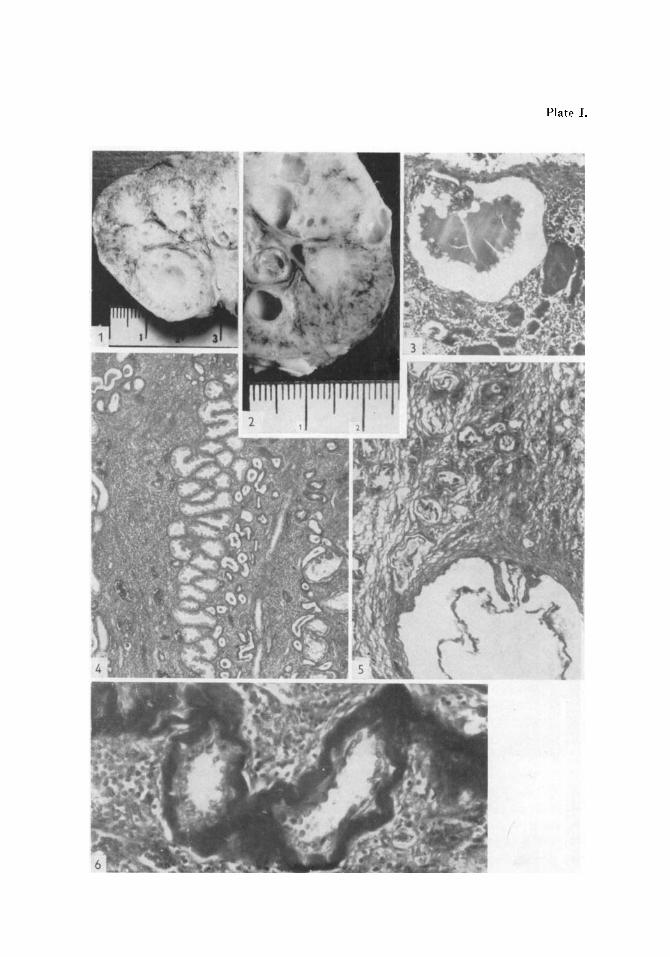
Plate I.

Plate 11.
486 B. I. IVEMARK, A. LJUNGQVIST AND A. BARRY
turbance of the function of the more distal part of the nephron. This is supported by the fact that the distal half of the nephron was more severely affected than was the proximal half. It seems probable that the medullary cysts found in these cases as well as the changes in the renal corpuscles were secondary to the thickened base- ment membranes. It is not possible to determine whether the latter is a primary stromal defect, or is secondary to a bio- chemical abnormality of the parenchyma of the nephrons themselves. It does seem clear, however, that the distal half of the nephron was initially hypertrophic prior to the massive increase in its basement membrane, since even the completely atrophic ascending limbs of the loops of Henle showed a convoluted course analo- gous to that followed by the convoluted descending limbs of Henle’s loops.
Summary
The kidneys from two cases of familial juvenile nephronophthisis were studied grossly, in histologic sections and by means of stereo-microangiography. The clinical history of these same cases is reported in the preceding paper in this issue (Broberger, Winberg & Zetterstrom).
The kidneys from these two individuals showed essentially the same characteris- tics:
1. Their weight was significantly less than normal.
3. Their cortex was thinned with dis- tortion and convolution of the interlobular arteries as well as of the afferent glomeru- lar arterioles.
3. Most of the renal copuscles showed varying degrees of hyaline degeneration, although some were cystically enlarged and their glomeruli were atrophic. 4. The nephric tubules were ensheathed
in a thickened, PAS-positive basement membrane. This condition was more mar- ked in the distal half of the nephron.
5. The medulla showed no decrease in thickness and contained characteristically convoluted limbs of Henle’s loops. The descending limbs were shown to be hyper- trophic. The convoluted character of the ascending limbs suggests that they too may have undergone hyperplasia prior to the massive thickening of their basement membranes.
6. The flexures of Henle’s loops were oc- casionally cystically dilatated.
7. The papillary ducts exhibited occa- sional ectatic portions, which in some in- stances had progressed to form cysts.
References
ALLEN, A. (2.: The kidney. Medical and Sur- gical Diseases, 1951.
BELLMAN, S.: Microangiography. Acta radiol., Stockh., Suppl. 102, 1953.
BROBERGER, O., WINBERG, J. and ZETTER- STROM, R.: Juvenile nephronophthisis. Acta pzdiat . , (Ups.) 49: 470, 1960.
EKSTROM, T., ENGFELDT, B., LAGERGREN, C. and LINDVALL, N.: Medullary Sponge
Kidney. Almqvist & Wicksell, Stockholm, 1959.
FANCONI, G. et al.: Die familiare juvenile Ne- phronophthise. (Die idiopathische parenchy- matose Schrumpfniere.) Helu. paediat. acta,
HACKZELL, G. and LUNDMARK, (2.: Familial juvenile nephronophthisis. Acta pzdiat.,
6: 1-49, 1951.
Stockh., 47: 428-440, 1958.
JUVENILE NEPHRONOPHTHISIS. PART I1 487
HOOFT, C., ROELS, H. and HERPOL, J.: A case of MCMANUS, J. F. A. and GUERRERO, M. A.: Histochemical procedures in the study of heart disease. I n Gould: Pathology of the
LJUNGQVIST, A.: Presented at the Swedish As- Heart, 2nd ed., p. 1070. Thomas, Spring- field, Ill., U.S.A., 1960.
Fanconi's familial juvenile nephronophthisis. Helit. pafdiat . acta, 14: 217-233, 1959.
sociation of Pathologists, May 7, 1960. Ab- stract in press, Nord. Med.
Department of Pediatric Pathology Karolinska s j ukhuset Stockholm 60 Sweden