Worldwide biogeography of Symbiodinium in tropical octocorals

Universidade Federal do Rio Grande – FURG
Instituto de Ciências Biológicas – ICB
Programa de Pós-Graduação em Ciências Fisiológicas
Dissertação de Mestrado
EFEITOS INTERATIVOS DO AUMENTO DE
TEMPERATURA E DA EXPOSIÇÃO AO COBRE EM
PARÂMETROS FISIOLÓGICOS E BIOQUÍMICOS NO
CORAL Mussismilia harttii
Dissertação apresentada ao Programa de
Pós-graduação em Ciências Fisiológicas da
Universidade Federal do Rio Grande -
FURG, como requisito para obtenção do
título de MESTRE.
JULIANA DA SILVA FONSECA
Orientador: Dr. Adalto Bianchini
RIO GRANDE
Fevereiro/2017

2
Agradecimentos 1
2
3
Agradeço a minha mãe Leda e ao meu saudoso pai pelos ensinamentos, amor e esforço 4
que me permitiram realizar este sonho! 5
6
Ao meu orientador, Dr. Adalto Bianchini, pela confiança para a realização deste trabalho 7
e os grandes conhecimentos propiciados durante essa jornada acadêmica. 8
9
À FURG e ao Instituto de Ciências Biológicas por toda a infraestrutura que permitiu a 10
realização deste trabalho e em especial ao Programa de Pós- Graduação em Ciências 11
Fisiológicas pela bolsa concedida. 12
13
Ao projeto Coral Vivo e seus patrocinadores pela infraestrutura e financiamento para a 14
realização deste projeto. 15
16
A todos os membros do grupo de pesquisa, principalmente as minhas amigas Laura e 17
Jose, por todo o esforço para o desenvolvimento deste trabalho. 18
19
Aos meus colegas e principalmente amigos Pelotenses: Viviane, Juliano Barreto, 20
Silvana, Amanda, Débora e Yuri Zebral pelas grandes oportunidades de aprendizagem e 21
também diversão que tornaram esse trajeto muito mais leve. Agradeço também a todos 22
os membros da “salinha 1” pelos momentos de descontração e reflexão a mim 23
concedidos! 24
25
Ao meu amor, Gabriel Ramos por toda a “paciência”, compreensão, tolerância e 26
carinho! 27
28
Aos professores Carlos Eduardo da Rosa e Clovis Barreira e Castro por terem aceitado 29
compor a banca avaliadora. 30
31
A todos os professores que participaram da minha formação durante esses anos e 32
certamente contribuíram para o meu amor pela ciência! 33

4
Sumário 34
35
36
37
Resumo geral .................................................................................................................... 5 38
Introdução geral ................................................................................................................ 6 39
Objetivo .......................................................................................................................... 14 40
Objetivo geral .............................................................................................................. 14 41
Objetivos específicos ................................................................................................... 14 42
Referências bibliográficas: ............................................................................................. 15 43
Manuscrito a ser submetido à revista "Aquatic Toxicology" .......................................... 22 44
Abstract ........................................................................................................................... 23 45
1. Introduction ................................................................................................................ 24 46
2. Materials and methods ................................................................................................ 27 47
2.1. Coral collection and maintenance ....................................................................... 27 48
2.2. Temperature treatments and combination with copper exposure ......................... 28 49
2.3. Seawater collection and analysis ......................................................................... 29 50
2.4. Sample preparation for biomarkers analyses ...................................................... 30 51
2.5. Carbonic anhydrase (CA) activity........................................................................ 30 52
2.6. (Ca2+
, Mg2+
)-ATPase activity .............................................................................. 31 53
2.7. Maximum photosynthetic capacity of photosystem II (Fv/Fm) ............................ 32 54
2.8. Chlorophyll a ........................................................................................................ 32 55
2.9. ATP concentration ................................................................................................ 33 56
2.10. Total antioxidant capacity against peroxyl radicals (ACAP) ............................. 33 57
2.11. Lipid peroxidation (LPO) ................................................................................... 34 58
2.12. Data presentation and statistical analyses ......................................................... 34 59
3. Results ........................................................................................................................ 35 60
3.1. Water physicochemical parameters...................................................................... 35 61
3.2. Enzyme activities .................................................................................................. 35 62
3.3. Metabolic parameters .......................................................................................... 36 63
3.4. Oxidative status parameters ................................................................................. 37 64
4. Discussion ................................................................................................................... 38 65
5. Conclusions ................................................................................................................ 48 66
6. Acknowledgments ...................................................................................................... 49 67
7. References .................................................................................................................. 49 68
Figure legends ................................................................................................................ 61 69
Discussão geral: .............................................................................................................. 85 70
71

5
Resumo geral 72
O efeito do aumento da temperatura da água e da combinação desse estressor com a 73
exposição ao cobre (Cu) foi avaliado no coral Mussismilia harttii. Os parâmetros 74
avaliados incluem aqueles envolvidos no processo de calcificação [anidrase carbônica 75
(AC) e (Ca2+
, Mg2+
)-ATPase], metabolismo energético [capacidade fotossintética 76
máxima (Fv/Fm) e concentrações de clorofila a e ATP] e estado oxidativo [capacidade 77
antioxidante contra radicais peroxil (ACAP) e peroxidação lipídica (LPO)]. Pólipos do 78
coral foram coletados, aclimatados em mesocosmo marinho e expostos a três 79
tratamentos térmicos (temperatura ambiente da água do mar e acréscimos de 1ºC e 2ºC 80
nesta temperatura) de forma isolada e combinada a diferentes concentrações de Cu 81
dissolvido [1.9 (sem adição de Cu), 3.8, 5.4 e 8.6 μg L-1
] por 4, 8 e 12 dias. A Fv/Fm 82
diminuiu ao longo do tempo de exposição ao aumento de temperatura. A interação deste 83
estressor com a exposição ao Cu aumentou este efeito. As atividades da AC e (Ca2+
, 84
Mg2+
)-ATPase aumentaram até 8 dias de exposição, mas não foram afetadas após 12 85
dias de exposição. A exposição aos estressores causou uma redução da LPO, sugerindo 86
a ativação de mecanismos para remodelamento de lipídeos nas membranas biológicas. 87
Porém, após a exposição prolongada (12 dias) ao aumento de temperatura foi observado 88
aumento da LPO, sugerindo uma menor produção de energia pela redução na 89
transferência de fotossintatos. Por sua vez, a ACAP, a clorofila a e o ATP não foram 90
alterados após a exposição aos estressores. Estes resultados indicam que os estressores 91
avaliados reduzem a fotossíntese e possivelmente a transferência de fotossintatos 92
utilizados como fonte de energia para os corais. Portanto, o aumento de temperatura e a 93
exposição ao Cu podem reduzir a disponibilidade de energia, afetar o crescimento e 94
consequentemente aumentar a susceptibilidade do coral M. harttii ao branqueamento. 95
Palavras-chave: calcificação, cobre, coral, estresse oxidativo, fotossíntese, temperatura. 96

6
Introdução geral 97
Os recifes de coral são ambientes muito produtivos e apresentam grande 98
biodiversidade (Hoegh-Guldberg , 1999). Além disso, são importantes fontes de renda 99
(Carte 1996), possuindo um alto valor econômico, devido à atividade pesqueira 100
dependente desses ecossistemas. De fato, os recifes de coral proporcionam um ambiente 101
rico para peixes e outros organismos marinhos, sendo que 6 milhões de toneladas do 102
pescado capturado anualmente estão associadas a esses ecossistemas (James and 103
Crabbe, 2008). 104
Os corais são organismos que vivem em ambientes tropicais com temperaturas 105
entre 18 e 30 ºC (van Dam et al., 2011). A formação e manutenção dos recifes de coral 106
de águas rasas dependem principalmente dos corais escleractíneos (Classe Anthozoa, 107
Subclasse Hexacorallia, Ordem Scleractinia), conhecidos como corais pétreos ou 108
verdadeiros. Existem cerca de 600 espécies de corais calcificadores (escleractíneos) que 109
contribuem diretamente para formação dos recifes de corais, um habitat característico 110
para peixes, algas e invertebrados (Hoegh-Guldberg, 1999). Os corais escleractíneos 111
realizam o processo de calcificação e possuem simbiose com zooxantelas, 112
dinoflagelados endossimbiontes do gênero Symbiodinium spp., localizadas nas células 113
do tecido oral dos corais (Al-Sofyani & Floos, 2013). 114
As zooxantelas podem fornecer até 90% do carbono que é direcionado a 115
alimentação dos corais (Papina et al. 2003) e da energia para o crescimento (Goreau 116
1959). Porém, a simbiose entre corais e zooxantelas é vulnerável a variações nas 117
condições ambientais, tais como alterações na temperatura, poluição e intensidade de 118
luz, fatores que podem induzir o fenômeno de branqueamento em corais (Fabricius et 119
al., 2005; Hoegh-Guldberg, 1999). Este fenômeno se caracteriza pela expulsão das 120
zooxantelas pelo coral e/ou degradação dos pigmentos fotossintetizantes dessas 121

7
microalgas (Downs et al., 2002). Isto afeta os corais, uma vez que sem as zooxantelas, 122
eles são privados dos nutrientes provenientes da fotossíntese (Ferrier-Pagès et al., 123
1998). Neste contexto, cabe ressaltar que o aumento da temperatura e a poluição 124
química são causas reconhecidas de branqueamento em corais (Nystron et al., 2001), 125
sendo que estas são as principais causas propostas para explicar a crescente degradação 126
dos recifes de coral em escala mundial (Anthony et al., 2007; Fabricius et al., 2005). 127
No que concerne à fauna de corais esclarectíneos do Brasil, essa se restringe a 18 128
espécies (Leão et al., 2003), o que caracteriza uma baixa diversidade quando comparada 129
àquela dos recifes de outras partes do mundo. Por exemplo, os recifes do Caribe 130
apresentam 918 espécies (Miloslavich et al., 2010). Em contrapartida, os recifes 131
brasileiros apresentam um alto grau de endemismo. Dentre as 18 espécies de corais 132
esclarectíneos recorrentes, 6 espécies são exclusivas do Brasil (Leão et al., 2003). 133
Dentre as espécies endêmicas, destaca-se Mussismilia harttii (Verrill, 1868), um coral 134
zooxantelado que ocorre desde o Rio Grande do Norte até a Bahia. Este desempenha 135
importante papel ecológico na formação dos recifes (Soares, 2011). Porém, segundo o 136
Instituto Chico Mendes da Biodiversidade (ICMBio, 2014), M. harttii está listada como 137
sendo uma espécie ameaçada de extinção. 138
Estudos têm mostrado que M. harttii é uma espécie sensível ao aumento da 139
temperatura da água, e que pode sofrer estresse térmico após um longo período de 140
exposição à 28ºC, que é a temperatura média máxima de verão na região de Abrolhos 141
(Soares, 2011). Além disso, a ação de estressores locais, como a contaminação por 142
cobre, pode alterar a atividade de enzimas de calcificação e causar estresse oxidativo em 143
M. harttii, aumentando a susceptibilidade desta espécie de coral ao branqueamento 144
(Marangoni et al., 2017). Apesar disso, pouco se sabe a respeito das respostas 145
bioquímicas de M. harttii ao efeito da exposição a múltiplos estressores, tais como o 146
8
aumento de temperatura da água do mar associada às mudanças climáticas e a 147
contaminação da água pelo cobre devida a ações antrópicas. 148
As mudanças climáticas globais tornou-se uma questão proeminente no final do 149
século passado, sendo que as perspectivas futuras apontam um sério transtorno do 150
potencial impacto dessas mudanças para o bem-estar das gerações futuras (Sokolova et 151
al., 2008). Modelagens recentes preveem um aumento de 0,2ºC na média da 152
temperatura global a cada década, podendo atingir um aumento de 2,6 a 4,8ºC até o ano 153
de 2100, fazendo com que o nível dos oceanos aumente entre 45 e 82 cm (IPCC 2007; 154
IPCC 2014). Este cenário é resultante de um conjunto de atividades humanas, tais como 155
desmatamento, erosão do solo, queima de combustíveis fósseis, entre outras, que 156
colaboram para elevar os níveis atmosféricos dos gases de efeito estufa, principalmente 157
do dióxido de carbono (CO2) (Freitas et al., 2012). 158
A elevação na temperatura global pode acarretar sérias consequências, tais como 159
aumento na incidência de doenças (Marengo et al.,2008; Martens et al., 1999), declínio 160
da abundância de zooplâncton (Reid et al., 1998), branqueamento em massa de corais 161
(Suzuki et al., 2007), mudanças nos habitats marinhos (Beaugrand 2004), introdução de 162
espécies invasivas (Lewis et al., 2003), extinção local de populações (Helmuth et al., 163
2002), ameaça ao fornecimento de água (Carere et al., 2011), derretimento de geleiras 164
(Marengo et al.,2008) e aumento de eventos extremos, como tempestades e furacões 165
(Freitas et al., 2012), aumentando assim a hostilidade do ambiente para a vida dos 166
organismos. 167
Os ecossistemas aquáticos são mais vulneráveis aos efeitos do aumento da 168
temperatura, pois mais de 95% das espécies aquáticas são ectotérmicas (Willmer et al., 169
2000). O aumento da temperatura tem um papel chave na fisiologia de ectotérmicos, 170
pois pode resultar em alterações na estabilidade de biomoléculas, nas reações 171

9
bioquímicas e taxas fisiológicas (Hochachka & Somero 2002). Dentre todos os 172
ecossistemas marinhos, os recifes de coral são os mais vulneráveis a mudanças 173
climáticas (Madin et al., 2012), sendo que o estresse térmico pode causar mudanças na 174
estrutura, dinâmica, produtividade e diversidade de recifes de coral (Calderon-Aguilera 175
et al., 2012). Estima-se que entre 50 a 70% dos recifes de coral estão sobre ameaça 176
direta e imediata de mudanças climáticas e poluição química (Wilkinson, 2004). 177
Em geral, as áreas costeiras e estuarinas são mais impactadas do que as áreas 178
oceânicas, em virtude do desenvolvimento industrial e agrícola (Clark, 2001). Na 179
presença dos impactos antrópicos locais, os organismos têm que lidar com a influência 180
de múltiplos estressores, tais como poluição, hipoxia, sobrepesca, eutrofização, entre 181
outros, além daquela associada às mudanças climáticas. O estresse causado pela 182
contaminação química pode aumentar a susceptibilidade dos organismos aos impactos 183
de mudanças climáticas (van Dam et al., 2012). Portanto, o entendimento sobre o efeito 184
interativo de múltiplos estressores, tais como o aumento da temperatura e a poluição por 185
metais, é necessário para compreender os mecanismos envolvidos nas respostas dos 186
organismos a esses estressores e prever os limites de tolerância e sobrevivência dos 187
organismos (Sokolova et al., 2008). 188
Muitos estudos têm investigado o efeito do aumento da temperatura na 189
toxicidade de metais em ectotérmicos (Heugens et al., 2006; Martínez-Jerónimo et al., 190
2006; Yang and Chen, 1996). O aumento da temperatura pode elevar as taxas 191
metabólicas, que por sua vez favorece a captação e acumulação dos metais nos 192
organismos (Brown et al., 2004; Heugens et al., 2003; Muyssen et al., 2010; Wang et 193
al., 2014). Além disso, a biodisponibilidade dos metais no ambiente aquático pode ser 194
afetada pelo aumento da temperatura (Rainbow, 2007), uma vez que o aumento da 195
temperatura pode reduzir a solubilidade de compostos que funcionam como 196
10
complexantes dos metais na água. Compostos com agrupamentos carboxílicos e 197
fenólicos são exemplos dos quais os metais têm alta afinidade. Desta forma, o aumento 198
da temperatura pode influenciar na especiação de metais no ambiente, aumentando a 199
biodisponibilidade dos metais nas suas formas livres (Bianchini et al., 2009). Além 200
disso, o aumento da temperatura altera a permeabilidade das membranas biológicas, o 201
que também pode influenciar no grau de exposição ao metal no ambiente (Kerswell & 202
Jones, 2003, Wood et al., 1999). 203
Os metais são poluentes persistentes no ambiente aquático, estando entre as 20 204
substâncias mais perigosas do mundo (EPA, 2005). Sabe-se que a incorporação de 205
metais no ambiente ocorre principalmente através de processos naturais, tais como, 206
vento, erosão continental, erupções vulcânicas, entre outros. Entretanto, sabe-se que 207
atividades antrópicas colaboram cada vez mais para elevar os níveis desses 208
contaminantes no ambiente (Bianchini et al., 2009). De fato, as atividades industriais, 209
como mineração e fundição, bem como o despejo de dejetos e extração de petróleo são 210
importantes fontes antrópicas de metais no ambiente marinho (Loya & Rinkevich, 211
1980). Dentre os impactos locais que afetam a saúde do ambiente recifal, destaca-se a 212
contaminação por metais, como o cobre (van Dam et al., 2011). A incorporação do 213
cobre é feita pelo uso em materiais condutores (fios e cabos), tubos de encanamento, 214
motores elétricos e interruptores. Além disso, o cobre é utilizado como algicida em 215
reservatórios de abastecimento de água e unidades de aquicultura para o controle de 216
microorganismos (Bianchini et al., 2009). O cobre se caracteriza por ser um metal 217
essencial para a manutenção das funções fisiológicas (Morgan, 2000). Porém, em 218
concentrações excessivas pode ser tóxico aos organismos aquáticos (Heath, 1995), 219
podendo inclusive induzir o branqueamento em corais (Bielmyer et al., 2010; Jones, 220
2004, 1997). 221

11
Com relação aos efeitos do aumento da temperatura e exposição ao cobre, sabe-222
se que ambos estressores interferem sobre o metabolismo dos corais (Nystron et al., 223
2001). O aumento da temperatura reduz a capacidade fotossintética (Nystron et al., 224
2001) e aumenta a taxa respiratória dos corais (Coles and Jokiel 1977; Porter, 1999; 225
Rivest & Hofmann 2014), devido ao aumento do metabolismo para a síntese de 226
biomoléculas protetoras ao estresse térmico (proteínas de choque térmico, 227
metalotioneínas, glutationa, etc.), causando o desequilíbrio em outros sistemas 228
fisiológicos, como neste caso, a fotossíntese (Gates & Edmunds 1999). Por sua vez, 229
altas concentrações de cobre reduzem a taxa de respiração (Nystron et al., 2001) e 230
inibem o transporte de elétrons para o fotossistema II das zooxantelas (Kuzminov et al., 231
2013; Samson et al., 1988). 232
Dentre as estratégias para se avaliar o efeito biológico de estressores, destaca-se 233
o uso de biomarcadores. Essas ferramentas biotecnológicas podem ser definidas como 234
alterações bioquímicas, celulares e fisiológicas, que fornecem evidências sobre a 235
exposição e/ou efeitos causados por contaminantes (Depledge, 1995) ou variações 236
ambientais. A vantagem da utilização dos biomarcadores consiste no seu potencial 237
preventivo contra os possíveis efeitos biológicos e ecológicos de estressores (Heath, 238
1995). 239
A calcificação realizada pelos corais é uma característica fisiológica que se 240
revela como um interessante biomarcador da saúde do ambiente recifal (Marangoni et 241
al., 2017; Marques et al., 2017). Este processo está intimamente ligado à relação 242
simbiótica entre os corais e as zooxantelas (Freitas et al., 2012), uma vez que o 243
branqueamento pode resultar na redução das taxas de calcificação (Bachok et al., 2006). 244
A calcificação dos corais resulta na formação da aragonita, um polimorfo de carbonato 245
de cálcio (CaCO3). Estes cristais formam-se a partir da obtenção de cálcio (Ca2+
) por 246
12
transporte ativo e do carbono inorgânico, principalmente sob a forma de bicarbonato 247
(HCO3-), advindos da água do mar ou do metabolismo do coral, e do transporte desses 248
compostos para o fluído de calcificação extracelular (FCE). No FCE, que se localiza 249
entre o tecido orgânico e o esqueleto mineral, forma-se a aragonita (Allemand et al., 250
2004). Assim, visto que o processo de calcificação tem papel central na fisiologia dos 251
corais, ferramentas para avaliação deste processo podem indicar quão saudável 252
encontra-se o recife ou o quanto este está se desenvolvendo (De‟ath et al., 2009; Hoegh-253
Guldberg, 1999). 254
A anidrase carbônica (AC) e a (Ca2+
, Mg2+
)-ATPase são enzimas-chave no 255
processo de calcificação realizado pelos corais. A AC catalisa a reação reversível de 256
hidratação do CO₂ em HCO3- e prótons (H
+), desempenhando assim um papel 257
fundamental no suprimento de carbono inorgânico dissolvido (CID) para a calcificação 258
dos corais e a fotossíntese realizada pelos simbiontes (Bielmyer et al., 2010). Com 259
relação a Ca2+
-ATPase, esta possui um sistema acoplado ao transporte de Ca2+
em 260
corais, pois ao mesmo tempo em que direciona Ca2+
para o sítio de calcificação, remove 261
2H+ do mesmo. Desta forma, esta enzima estimula a formação de CaCO3 (Ca
2+ + CO2 + 262
H2O ↔ CaCO3 + 2H+) (Al-Horani et al., 2003) e auxilia na manutenção do estado de 263
saturação da aragonita (Marangoni et al., 2016). Por sua vez, sugere-se que a Mg2+
-264
ATPase é a enzima responsável pelo controle temporal e espacial da correta liberação do 265
Mg2+
nas superfícies mineralizantes. Portanto, ela desempenha um papel importante na 266
formação de esqueletos de coral (Meibom et al., 2004). 267
Com relação ao efeito de estressores, sabe-se que a exposição ao cobre pode 268
interferir no funcionamento das enzimas mencionadas acima em corais, incluindo M. 269
harttii (Bielmyer et al., 2010; Marangoni et al., 2017). Reduções nas atividades da Ca2+
-270
ATPase e AC, devido à exposição ao cobre, podem portanto afetar o processo de 271

13
calcificação em M. harttii. Neste contexto, cabe ressaltar que o cobre é um metal 272
envolvido na geração de estresse oxidativo via reação de Fenton (Hermes-Lima, 2004; 273
Wardman & Cadeias, 1996). Considerando a fisiologia de corais zooxantelados, é 274
importante salientar que o estresse oxidativo afeta a associação coral-simbionte, na qual 275
o oxigênio produzido na fotossíntese é passado para as células dos corais (Rotchell & 276
Ostrander, 2011), podendo aumentar os danos oxidativo neste último. Assim, sugere-se 277
que o estresse oxidativo esteja relacionado ao processo de branqueamento em corais. De 278
fato, sob determinado grau de estresse, os corais eliminam a fonte dominante de 279
produção de espécies reativas de oxigênio (ERO), expulsando assim as zooxantelas 280
(Downs et al., 2002). 281
O branqueamento é um fenômeno que afeta gravemente os corais (Freitas et al., 282
2012). Na ausência das zooxantelas, os corais perdem acesso aos combustíveis 283
energéticos responsáveis pela produção de ATP, tais como oxigênio, aminoácidos, 284
glicose e alguns peptídeos, que são importantes para o aumento das taxas de 285
calcificação e manutenção de outras funções fisiológicas (Teece et al., 2011). Além 286
disso, as zooxantelas provêm ao coral acesso à luz, nutrientes inorgânicos (CO2) e 287
proteção (Marangoni et al., 2016; van Dam et al., 2011). Portanto, parâmetros para 288
avaliação da fotossíntese, podem ser indicadores importantes para avaliação da saúde da 289
relação entre o coral e as algas simbiontes. Por sua vez, cabe salientar que fatores 290
ambientais como o aumento de temperatura e a exposição ao cobre podem desencadear 291
o aumento de ERO (Halliwell & Gutteridge, 1999) e resultar na oxidação de lipídios, 292
proteínas e ácidos nucleicos (Monserrat et al., 2007). Portanto, biomarcadores de 293
estresse oxidativo são potenciais ferramentas para a avaliação da saúde dos corais, uma 294
vez que danos oxidativos possuem correlação direta com o branqueamento em 295
organismos de ambientes recifais (Prazeres et al., 2012). 296

14
Como mencionado anteriormente, um cenário de múltiplos estressores é 297
composto pelos impactos gerados pelo homem em escala local, como é o caso da 298
poluição por metais, associados às potenciais mudanças climáticas em escala global. 299
Apesar da crescente preocupação com o impacto biológico destes estressores 300
ambientais, existem poucas informações sobre os efeitos interativos de múltiplos 301
estressores ambientais em corais. Esse fato, aliado a carência de estudos relacionados à 302
fisiologia de espécies coralinas endêmicas no Brasil, faz com que o uso de 303
biomarcadores no coral M. harttii para avaliar os efeitos isolados e combinados do 304
aumento da temperatura e contaminação pelo cobre se torne uma importante ferramenta 305
biológica para um maior entendimento das respostas desses organismos aos estressores 306
em questão, bem como para o subsídio na proposição de ações visando à conservação 307
dos recifes de coral no Brasil, que atualmente encontram-se sujeitos tanto aos impactos 308
locais quanto às mudanças climáticas globais. 309
310
Objetivo 311
Objetivo geral 312
313
Avaliar o efeito isolado do aumento da temperatura e da combinação deste 314
estressor com a exposição ao cobre sobre parâmetros fisiológicos e bioquímicos no 315
coral escleractínio Mussismilia harttii. 316
317
Objetivos específicos 318
319
- Avaliar o efeito isolado do aumento da temperatura e da combinação deste estressor 320
com diferentes concentrações ambientalmente relevantes de cobre sobre os seguintes 321
parâmetros envolvidos no processo de calcificação: atividades específicas da 322
anidrase carbônica e da (Ca2+
, Mg2+
)-ATPase. 323
15
- Determinar o efeito isolado do aumento da temperatura e da combinação deste 324
estressor com diferentes concentrações ambientalmente relevantes de cobre sobre os 325
seguintes parâmetros envolvidos no estresse oxidativo: capacidade antioxidante 326
contra radicais peroxil e peroxidação lipídica. 327
- Analisar o efeito isolado do aumento da temperatura e da combinação deste estressor 328
com diferentes concentrações ambientalmente relevantes de cobre sobre a capacidade 329
fotossintética máxima das zooxantelas (Fv/Fm) e a densidade de endossimbiontes 330
(concentração de clorofila a). 331
- Avaliar o efeito isolado do aumento da temperatura e da combinação deste estressor 332
com diferentes concentrações ambientalmente relevantes de cobre sobre a 333
concentração de ATP do holobionte. 334
335
Referências bibliográficas: 336
337
Al-Horani, F.A., Al-Moghrabi, S.M., De Beer, D. 2003. The mechanism of calcification 338
and its relation to photosynthesis and respiration in the scleractinian coral Galaxea 339
fascicularis. Mar. Biol. 142: 419-426. 340
341
Allemand, D., Ferrier-Pagès, C., Furla, P., Houlbrèque, F., Puverel, S., Reynaud, S., 342
Tambutté, É.,Tambutté, S., Zoccola., D. 2004. Biomineralisation in reef-building corals: 343
from molecular mechanisms to environmental control. Gen. Palaeont. 3: 453-467. 344
345
Al-Sofyani, A.A., Floos, Y.A.M. 2013. Effect of temperature on two reef-building 346
corals Pocillopora damicornis and P. verrucosa in the Red Sea. Oceanologia 55: 917-347
935. 348
349
Anthony, K.R.N., Connolly, S.R., Hoegh-Guldberg, O. 2007. Bleaching, energetics and 350
coral mortality risk: effects of temperature, light, and sediment regime. Limnol. 351
Oceanogr. 52: 716-726. 352
353
Bachok, Z., Mfilinge, P., Tsuchiya, M. 2006. Characterization of fatty acid composition 354
in healthy and bleached corals from Okinawa, Japan. Coral Reefs 25: 545- 554. 355
356
Beaugrand, G. 2004. The North Sea regime shift: evidence, causes, mechanisms and 357
consequences. Prog. Oceanogr. 60: 245-262. 358
359
16
Bianchini, A., Martins, S.E., Jorge, M.B. 2009. O Modelo do Ligante Biótico e suas 360
Aplicações em Ecotoxicologia. Universidade Federal do Rio Grande - FURG, Rio 361
Grande, RS, Brazil. 34p. 362
363
Bielmyer, G.K., Grosell, M., Bhagooli, R., Baker, A.C., Langdon, C., Gillette, P., Capo, 364
T.R. 2010. Differential effects of copper on three species of scleractinian corals and 365
their algal symbionts (Symbiodinium spp.). Aquat.Toxicol. 97: 125-133. 366
367
Brown, J. H., Gillooly, J.F., Allen, A.P., Savage, V.M., West, G.B. 2004. Toward a 368
metabolic theory of ecology. Ecology 85: 1771-1789. 369
370
Calderon-Aguilera, L.E., Rivera-Monroy, V.H., Porter-Bolland, L., Martínez-Yrízar, A., 371
Ladah, L.B., Martínez-Ramos, M., Alcocer, J., Santiago-Pérez, A.L., Hernandez-Arana, 372
H.A., Reyes-Gómez, V.M., Pérez-Salicrup, D.R., Díaz-Nuñez, V., Sosa-Ramírez, J., 373
Herrera-Silveira, J., Búrquez, A. 2012. An assessment of natural and human disturbance 374
effects on Mexican ecosystems: current trends and research gaps. Biodiver. Conserv. 21: 375
589-617. 376
377
Carere, M., Miniero, R., Cicero, M.R. 2011. Potencial effects of climate change on the 378
chemical quality of aquatic biota. Trends Analyt. Chem. 30: 1011-1021. 379
380
Carte, B. K. 1996. Biomedical potential of marine natural products. BioScience 46: 381
271-86. 382
383
Clark, R.B. 2001. Marine Pollution. Oxford University Press, Oxford. 384
385
Coles, S.L., Jokiel, P.L. 1977. Effects of temperature on photosynthesis and respiration 386
in hermatypic corals. Mar. Biol. 43: 209-216. 387
388
De'ath, G., Lough, J.M., Fabricius, K.E. 2009. Declining coral calcification on the Great 389
Barrier Reef. Science 323: 116-119. 390
391
Depledge, M.H., Aagaard , A., Gyorkos, P. 1995. Assessment of trace metal toxicity 392
using molecular, physiological and behavioural biomarkers. Mar. Pollut. Bull. 31: 19-393
27. 394
395
Downs, C.A., Fouth, J.E., Halas, J.C., Dustan, P., Bemiss, J., Woodley, C.M. 2002. 396
Oxidative stress and seasonal coral bleaching. Free. Rad. Biol. Med. 33: 533-543. 397
398
EPA. 2005. United States Environmental Protection Agency. Handbook for Developing 399
Watershed Plans to Restore and Protect our Waters. EPA 841-B-05-005: Washington. 400
Disponível em http://www.epa.gov/owow/nps/watershed_handbook/pdf/handbook.pdf. 401
Acesso em 30 de dezembro de 2016. 402
403
Fabricius, K.E. 2005. Effects of terrestrial runoff on the ecology of corals and coral 404
reefs: review and synthesis. Mar. Pollut. Bull. 50: 125-146. 405
406
Ferrier-Pagès, C., Allemand, D., Gattuso, J. P., Jaubert, J. 1998. Microheterotrophy in 407
the zooxanthellate coral Stylophora pistillata: effect of light and prey density. Limnol. 408
Oceanogr. 43: 1639-1648. 409

17
410
Freitas, L.M., Oliveira, M.D.M., Kikuchi, R.K.P. 2012. Os mecanismos de 411
sobrevivência dos corais diante do impacto das mudanças climáticas sobre o 412
ecossistema de recifes. Cadern. Geociênc. 9: 2. 413
414
Gates, R.D., Edmunds, P.J. 1999. The physiological mechanisms of acclimatization in 415
tropical coral. Am. Zool. 39: 30-43. 416
417
Goreau, T. F. 1959. The physiology of skeleton formation in corals. I. A method for 418
measuring the rate of calcium deposition by corals under different conditions. Biol. Bull. 419
116: 59-75. 420
421
Halliwell, B., Gutteridge, J.M.C. 1999. Free Radicals in Biology and Medicine. Oxford 422
University Press, Oxford. 423
424
Heath, A.G. 1995. Water Pollution and Fish Physiology. CRC Press, Florida. 384p. 425
426
Helmuth, B., Harley, C.D.G., Halpin, P.M., O‟Donnell, M., Hofmann, G.E., Blanchette, 427
C.A. 2002. Climate change and latitudinal patterns of intertidal thermal stress. Science 428
298: 1015-1017. 429
430
Hermes-Lima, M. 2004. Oxygen in biology and biochemistry: Role of free radicals. In: 431
Storey, K.B. (ed.) Functional Metabolism: Regulation and Adaptation. Hobocken, New 432
Jersey, pp. 319-368. 433
434
Heugens, E.H., Jager, T., Creyghton, R., Kraak, M.H., Hendriks, A.J., Van Straalen, 435
N.M., Admiraal, W. 2003. Temperature-dependent effects of cadmium on Daphnia 436
magna: accumulation versus sensitivity. Environ. Sci. Technol. 37: 2145-2151. 437
438
Heugens, E.H., Tokkie, L.T., Kraak, M.H., Hendriks, A.J., van Straalen, N.M., 439
Admiraal, W. 2006. Population growth of Daphnia magna under multiple stress 440
conditions: joint effects of temperature, food, and cadmium. Environ. Toxicol. Chem. 441
25: 1399-1407. 442
443
Hochachka, P.W., Somero, G.N. 2002. Biochemical Adaptation: Mechanism and 444
Process in Physiological Evolution. Oxford University Press, Oxford. 445
446
Hoegh-Guldberg, O. 1999. Climate change, coral bleaching and the future of the 447
world.s coral reefs. Mar. Freshwater Res. 50: 839-866. 448
449
ICMBio. 2014. Lista de espécies ameaçadas de extinção. Disponível em: 450
www.icmbio.gov.br/portal/biodiversidade/fauna-brasileira/lista-de especies.html. 451
452
IPCC, 2007. The Fourth Assessment Report of the Intergovernmental Panel on Climate 453
Change (IPCC). Cambridge University Press, Cambridge, UK. 454
455
IPCC, 2014. The Fifth Assessment Report of the Intergovernmental Panel on Climate 456
Change (IPCC). Cambridge University Press, Cambridge, UK. 457
458

18
James, M., Crabbe, C. 2008. Climate change, global warming and coral reefs: Modeling 459
the effects of temperature. Comput. Biol. Chem. 32: 311-314. 460
461
Jones, R.J. 1997. Zooxanthellae loss as a bioassay for assessing stress in corals. Mar. 462
Ecol. Prog. Ser. 149: 163-171. 463
464
Jones, R.J. 2004. Testing the „photoinhibition‟ model of coral bleaching using chemical 465
inhibitors. Mar. Ecol. Prog. Ser. 284: 133-145. 466
467
Kerswell, A.P., Jones, R.J. 2003. Effects of hypo-osmosis on the coral Stylophora 468
pistillata: nature and cause of low-salinity bleaching. Mar. Ecol. Prog. Ser. 253: 145-469
154. 470
471
Kuzminov, F.I., Brown, C.M., Fadeev, V.V., Gorbunov, M.Y. 2013. Effects of metal 472
toxicity on photosynthetic processes in coral symbionts, Symbiodinium spp. J. Exp. 473
Mar. Biol. Ecol. 446: 216-227. 474
475
Leão, Z. M. A. N., Kikuchi, R. K. P., Testa, V. 2003. Corals and coral reefs of Brazil. In: 476
Cortés, J. (ed.). Latin America Coral Reefs. Elsevier Science, New York. pp. 9-52. 477
478
Lewis, P.N., Hewitt, C.L., Riddle, M., McMinn, A. 2003. Marine introductions in the 479
Southern Ocean: an unrecognised hazard to biodiversity. Mar. Pollut. Bull. 46: 213-23. 480
481
Loya, Y., Rinkevich, B. 1980. Effects of oil pollution on coral reef communities. Mar. 482
Ecol. Prog. Ser. 3: 167-180. 483
484
Madin, J.S., Hughes, T.P., Connolly, S.R. 2012. Calcification, storm damage and 485
population resilience of tabular corals under climate change. PLoS One 7: 1-10. 486
487
Marangoni, L.F.B., Marques, J.A., Bianchini, A. 2016. Fisiologia de corais: a simbiose 488
coral-zooxantela, o fenômeno de branqueamento e o processo de calcificação. In: 489
Zilberberg, C., Abrantes, D.P., Marques, J.A., Feitosa, L.F., Marangoni, L.F.B. (Eds.) 490
Conhecendo os Recifes Brasileiros: Rede de Pesquisas Coral Vivo. Museu Nacional, 491
UFRJ, Rio de Janeiro. p 55-72. 492
493
Marangoni, L.F.B., Marques, J.A., Duarte, G.A.S., Pereira, C.M., Calderon, E.N., 494
Castro, C.B., Bianchini, A. 2017. Copper effects on biomarkers associated with 495
photosynthesis, oxidative status and calcification in the Brazilian coral Mussismilia 496
harttii (Scleractinia, Mussidae). Mar. Environ. Res. Em revisão. 497
498
Marengo, J.A. 2008. Água e mudanças climáticas. Estudos Avançados 22: 83-95. 499
500
Martens, P., Kovats, R.S., Nijhof, S., De Vries, P., Livermore, M.T.J., Bradley, D.J., 501
Cox, J., Mcmichael, A.J. 1999. Climate chance and future populations at risk of malaria. 502
Glob. Environ. Chang. 9: 89-107. 503
504
Marques, J.A., Maragoni, L.F.B., Bianchini, A. 2017. Combined effects of sea water 505
acidification and copper exposure on the symbiont-bearing foraminifer Amphistegina 506
gibbosa. Coral Reefs. doi:10.1007/s00338-017-1547-z. 507
508

19
Martínez-Jerónimo, F., Martínez-Jerónimo, L., Espinosa-Chávez, F. 2006. Effect of 509
culture conditions and mother's age on the sensitivity of Daphnia magna Straus 1820 510
(Cladocera) neonates to hexavalent chromium. Ecotoxicology 15: 259-266. 511
512
Meibom, A., Cuif, J.P., Hillion, F., Constantz, B.R., Juillet-leclerc, A., Dauphin, Y., 513
Dunbar, R.B. 2004. Distribution of magnesium in coral skeleton. Geophys. Res. Lett. 514
31: 1-4. 515
516
Miloslavich, P., Dias, J.M., Klein, E., Alvarado, J.J., Dias, C., Gobin, J., Escobar-517
Briones, E., Cruz-Motta, J.J., Weil, E., Cortés, J., Bastidas, A.C., Robertson, R., Zapata, 518
F., Martín, A.,Castilho, J., Kazandjian, A., Ortiz, M. 2010. Marine Biodiversity in the 519
Caribbean: Regional estimates and distribution patterns. PLoS One 5: 25. 520
521
Muyssen, B.T., Messiaen, M., Janssen, C.R. 2010. Combined cadmium and temperature 522
acclimation in Daphnia magna: physiological and sub-cellular effects. Ecotoxicol. 523
Environ. Saf. 73: 735-742. 524
525
Monserrat, J.M., Martínez, P.E., Geracitano, L.A., Amado, L.L., Martins, C.M., Pinho, 526
G.L., Chaves, I.S., Ferreira-Cravo, M., Ventura-Lima, J., Bianchini, A. 2007. Pollution 527
biomarkers in estuarine animals: critical review and perspectives. Comp. Biochem. 528
Physiol. C 146: 221-234. 529
530
Morgan, T. 2000. The fish gill: site of action for toxic effects of waterborne copper. Fish 531
Physiology Course 751. McMaster University, Hamilton, ON, Canada. 532
533
Nystrom, M., Nordemar, I., Tedengren, M. 2001. Simultaneous and sequential stress 534
from increased temperature and copper on the metabolism of the hermatypic coral 535
Porites cylindrical. Mar. Biol. 138, 1225-1231. 536
537
Papina, M., Meziane, T., Van Woesik, R. 2003. Symbiotic zooxanthellae provide the 538
host-coral Montipora digitata with polyunsaturated fatty acids. Comp. Biochem. 539
Physiol. B 135: 533-537. 540
541
Porter, J.W., Lewis, S.K., Porter, K.G. 1999. The effect of multiple stressors on the 542
Florida Keys coral reef ecosystem: a landscape hypothesis and a physiological test. 543
Limnol. Oceanogr. 44: 941-949. 544
545
Prazeres, M.F., Martins, S.E., Bianchini, A. 2012. Assessment of water quality in coral 546
communities from Fernando de Noronha, Brazil: Biomarkers analysis in Amphistegina 547
lessonii. J. Foramin. Res. 42: 56-65. 548
549
Rainbow, P.S. 2007. Trace metal bioaccumulation: models metabolic availability and 550
toxicity. Environ. Int. 33: 576-582. 551
552
Reid, P.C., Planque, B., Edwards, M. 1988. Is observed variability in the long-term 553
results of the continuous plankton recorder survey a response to climate change? Fish. 554
Oceanogr. 7: 282-288. 555
556
Rivest, E.B., Hofmann, G.E. 2014. Responses of the metabolism of the larvae 557
of Pocillopora damicornis to ocean acidification and warming. PLoS One 9: e96172. 558

20
559
Rotchell, J.M., Ostrander, G.K. 2011. Molecular toxicology of corals: a review. J. 560
Toxicol. Environ. Health B Crit. Rev. 14: 571-592. 561
562
Soares, V.V. 2011. Suscetibilidade do coral Mussismilia harttii Verrill, 1868 ao aumento 563
da temperatura da água do mar. Dissertação de Mestrado, Universidade Federal da 564
Bahia - UFBA, Salvador, BA, Brasil, 55p. 565
566
Sokova, I.M., Lannig, G. 2008. Interactive effects of metal pollution end temperature on 567
metabolism in aquatic ectotherms: implications of global climate change. Clim. Res. 37: 568
181-201. 569
570
Suzuki, A., Gagan M.K., Kan, H., Edward, A., Siringan, F.P., Yoneda, M., Kawahata, H. 571
2007. Coral records of the 1990s in the tropical Northwest Pacific: ENSO, mass coral 572
bleaching, and global warming. Oceanogr. Ser. 37: 211-238. 573
574
Samson, G., Morisette, J., Popovic, R. 1988. Copper quenching of the variable 575
fluorescence in Dunaliella tertiolecta. New evidence for a copper inhibition effect on 576
PSII photochemistry. Photochem. Photobiol. 48: 329-332. 577
578
Teece, M.A., Estes, B., Gelsleichter, E., Lirman, D. 2011. Heterotrophic and autotrophic 579
assimilation of fatty acids by two scleractinian corals, Montastraea faveolata and 580
Porites astreoides. Limnol. Oceanogr. 56: 1285‐1296. 581
582
van Dam, J.W., Negri, A.P., Mueller, J.F., Altenburger, R., Uthicke, S. 2012. Additive 583
pressures of elevated sea surface temperatures and herbicides on symbiont-bearing 584
foraminifera. PloS One 7: 1-12. 585
586
van Dam, J.W., Negri, A.P., Uthicke, S., Mueller, J.F. 2011. Chemical pollution on coral 587
reefs: Exposure and ecological effects. In: Sanchez-Bayo, F., van den Brink, P.J., Mann, 588
R.M. (Eds.), Ecological Impacts of Toxic Chemicals. Bentham Science Publishers, 589
Amsterdam, pp. 187-211. 590
591
Verrill, A.E. 1868. Notes on Radiata in the Museum of Yale College, with description of 592
new genera and species. No.4. Notice of the corals and echinoderms collected by Prof. 593
C.F. Hartt at the Abrolhos Reefs, Province of Bahia, Brazil, 1867. Trans. Conn. Acad. 594
Arts. Sci. 1: 351-371. 595
596
Wang, Y., Xu, S., Liu, J., Zhang, Y., Guo, T.L. 2014. Regulation of lead toxicity by heat 597
shock protein 90 (daf-21) is affected by temperature in Caenorhabditis elegans. 598
Ecotoxicol. Environ. Saf. 104: 317-322. 599
600
Wardman, P., Cadeias, L.P. 1996. Fenton chemistry: an introduction. Radiat. Res. 145: 601
523-531. 602
603
Wilkinson, C. (Ed). 2004. Status of Coral Reefs of the World. Australian Institute of 604
Marine Science, Townsville, Australia. 605
606
Willmer, P., Stone, G., Johnston, I. 2000. Environmental Physiology of Animals. 607
Blackwell Science, Oxford. 608
21
609
Wood, L.A., Brown, I.A., Youson, J.H. 1999. Tissue and developmental variations in the 610
heat shock response of sea lampreys (Petromyzon marinus): effects of an increase in 611
acclimation temperature. Comp. Biochem. Physiol. A 123: 35-42. 612
613
Yang, H.N., Chen, H.C. 1996. Uptake and elimination of cadmium by Japanese eel, 614
Anguilla japonica, at various temperatures. Bull. Environ. Contam. Toxicol. 56: 670-615
676. 616
617

22
Manuscrito a ser submetido à revista "Aquatic Toxicology" 618
Effects of increasing temperature alone and combined with copper exposure on 619
biochemical and physiological parameters in the Brazilian endemic coral 620
Mussismilia harttii 621
622
Juliana da Silva Fonsecaa, Laura Fernandes de Barros Marangoni
b,c, Joseane Aparecida 623
Marquesb,c
, Adalto Bianchinic,d
624
625
aPrograma de Pós-Graduação em Ciências Fisiológicas, Instituto de Ciências 626
Biológicas, Universidade Federal do Rio Grande, Avenida Itália km 8, Rio Grande, RS, 627
Brazil, 96203-900. 628
bPrograma de Pós-Graduação em Oceanografia Biológica, Instituto de Oceanografia, 629
Universidade Federal do Rio Grande, Avenida Itália km 8, Rio Grande, RS, Brazil, 630
96203-900. 631
cInstituto Coral Vivo, Rua dos Coqueiros, Parque Yaya, Santa Cruz Cabrália, BA, 632
Brazil, 45807-000. 633
dInstituto de Ciências Biológicas, Universidade Federal do Rio Grande. Avenida Itália 634
km 8, Rio Grande, RS, Brazil, 96203-900. 635
636
* Corresponding author: Adalto Bianchini 637
Universidade Federal do Rio Grande – FURG 638
Instituto de Ciências Biológicas – ICB 639
Av. Itália km 8, Campus Carreiros 640
96.203-900 – Rio Grande – RS – Brazil 641
Phone: +55 53 3293-5193 642
e-mail: [email protected] 643

23
ABSTRACT 644
Effects of increasing temperature alone and in combination with exposure to dissolved 645
copper (Cu) were evaluated in the Brazilian endemic coral Mussismilia harttii using a 646
marine mesocosm system. Endpoints analyzed included parameters involved in 647
calcification [carbonic anhydrase (CA) and (Ca2+
, Mg2+
)-ATPase activity], metabolism 648
[maximum photosynthetic capacity of zooxanthellae (Fv/Fm) and chlorophyll a and 649
ATP concentrations)], and oxidative status [antioxidant capacity against peroxyl radicals 650
(ACAP) and lipid peroxidation (LPO)]. Coral polyps were collected, acclimated and 651
exposed to three different increasing temperature conditions [25.0ºC (control; average 652
temperature of local seawater), 26.6ºC and 27.3ºC] using a marine mesocosm system. 653
They were tested alone and in combination with four environmentally relevant 654
concentrations of dissolved Cu in seawater [1.9 (control; average concentration in local 655
seawater), 3.8, 5.4 and 8.6 μg/L) for 4, 8 and 12 days. Fv/Fm reduced over the 656
experimental period with increasing temperature. Combination of increasing 657
temperature with Cu exposure enhanced this effect. CA and (Ca2+
, Mg2+
)-ATPase 658
activities increased up to 8 days of exposure, but were not affected after 12 days of 659
experiment. Short-term exposure to increasing temperature or long-term exposure to the 660
combination of stressors reduced LPO, suggesting a lipid remodeling in biological 661
membranes. Long-term exposure to increasing temperature induced LPO, suggesting a 662
lower energy production in the host coral associated with a likely reduced transference 663
of photosynthates by zooxanthellae. ACAP, as well as concentrations of ATP and 664
chlorophyll a were not significantly affected by the stressors. These findings indicate 665
that increasing temperature combined with exposure to dissolved Cu can increase 666
susceptibility to bleaching and reduce growth in the Brazilian endemic coral M. harttii. 667
Keywords: biomarkers; calcification; copper, coral; oxidative stress; photosynthesis. 668

24
1. Introduction 669
670
There is growing concern with the rapid increasing in coral reefs degradation 671
worldwide (De‟ath et al., 2012). Global climate changes and local impacts are amongst 672
the major threats influencing the integrity these ecosystems (Sokolova et al., 2008). 673
According to the Intergovernmental Panel on Climate Change (IPCC), mean global 674
temperature will experience an increase of 0.2ºC per decade, being predicted an 675
increase of 2.6 to 4,8ºC until the year of 2100 (IPCC, 2014). 676
Corals are ectothermic organisms (Hoegh-Guldberg and Smith, 1989). Increases 677
of 2-4ºC are known to induce bleaching within days or weeks (Brown, 1997a). 678
Therefore, corals reefs will be one of the most affected ecosystems by climate changes 679
(Madin et al., 2012). Indeed, thermal stress can cause crucial changes in the structure, 680
dynamics, productivity and diversity of coral reefs (Calderon-Aguilera et al., 2012). 681
Also, local stressors such as chemical pollutants, including metals, and increased 682
sedimentation are shown to impact coral physiology (Harland and Brown 1989; 683
Bielmyer et al., 2010; Marangoni et al., 2017). Copper (Cu) is an essential metal for the 684
maintenance of physiological functions (Morgan, 2000). However, excessive 685
concentrations of this metal can induce deleterious effects in aquatic organisms (Heath, 686
1995), including bleaching (Jones, 1997, 2004; Bielmyer et al., 2010) and metabolic 687
changes in corals (Nystron et al., 2001). Indeed, seawater contamination with metals, 688
including Cu can affect reef environment health (van Dam et al., 2011). Although the 689
effect of only one stressor rarely occurs in nature (Brown, 1997a,b), most studies 690
investigates the effect of isolated stressors in corals (Bielmyer et al., 2010; Kuzminov et 691
al., 2013; Marangoni et al., 2017). In fact, few studies have analyzed the interactive 692
effects of multiple stressors on physiological biomarkers, especially in corals (Brown, 693

25
1997a; Rivest and Hofmann, 2014). 694
Calcification performed by corals is a physiological characteristic which 695
revealed to be an interesting biomarker of reef environment health (Marangoni et al., 696
2017). The use of biochemical and physiological tools to evaluate the calcification 697
process in corals can indicate how healthy the reef is or how much it is developing 698
(Hoegh-Guldberg, 1999; De‟ath et al., 2009). Carbonic anhydrase (CA) and (Ca2+
, 699
Mg2+
)-ATPase are key enzymes in the calcification process. CA catalyzes the reversible 700
reaction involved in the carbon dioxide (CO2) hydration, thus generating bicarbonate 701
(HCO3-) and protons (H
+). Therefore, it plays a key role in the supply of dissolved 702
inorganic carbon (DIC) for coral calcification, as well as the photosynthesis performed 703
by the symbiont algae (Bielmyer et al., 2010). In turn, Ca2+
-ATPase transports calcium 704
(Ca2+
) into the calcification site and removes 2H+, thus increasing the Ca
2+ and 705
carbonate (CO3-
) saturation state (Al-Horani et al., 2003). Regarding Mg2+
-ATPase, it 706
has been reported that it controls the Mg2+
release into the calcification site, thus 707
playing an important role in the growth of skeletal components in corals (Meibom et al., 708
2004). 709
Another important feature of scleractinian corals is its symbiotic relationship 710
with zooxanthellae of the genus Symbiodinium spp. These algae are located in the oral 711
tissue cells of corals (Al-Sofyani and Floos, 2013). Zooxanthellae can provide about 712
90% of the carbon which is directed for coral feeding (Papina et al., 2003), thus playing 713
a key role in energy production for coral growth (Goreau, 1959). However, symbiosis 714
between corals and zooxanthellae is vulnerable to environmental stressors, such as 715
changes in temperature and light intensity, as well as pollution. A disruption of this 716
symbiotic relationship can lead to coral bleaching (Hoegh-Guldberg, 1999; Fabricius et 717
al., 2005). This phenomenon is characterized by the expulsion of zooxanthellae by coral 718

26
and/or the degradation of the photosynthetic pigments of these microalgae (Downs et 719
al., 2002). Bleaching can severely affect corals. In the absence of zooxanthellae, corals 720
are deprived of nutrients provided by photosynthesis (Ferrier-Pagès et al., 1998). As 721
previously mentioned, increasing temperature and chemical pollution are recognized as 722
common causes of bleaching in corals, being the major factors associated with coral reef 723
degradation worldwide (Fabricius et al., 2005; Anthony et al., 2007). Therefore, 724
parameters such as the photosynthetic capacity of zooxanthellae and chlorophyll a 725
concentration may be important tools to evaluate the health and integrity of the 726
symbiotic relationship between corals and zooxanthellae. 727
In light of the above, oxidative stress biomarkers can be considered as potential 728
tools for assessing coral reefs health, since oxidative damage is directly correlated with 729
bleaching in reef organisms (Prazeres et al., 2012). Indeed, increasing temperature and 730
metal exposure can increase the generation of reactive oxygen species (ROS) in aquatic 731
organisms, thus leading to oxidation of biomolecules, such as lipids, proteins and 732
nucleic acids (Halliwell and Gutteridge, 1999; Monserrat et al., 2007). It is reported that 733
oxidative stress affects the coral-symbiont association. In this case, oxygen produced by 734
photosynthesis is transferred to corals cells (Rotchell & Ostrander, 2011), thus 735
increasing the oxidative damage to these cells. Once a threshold level of stress is 736
achieved, corals eliminate the dominant source of ROS, thus expelling the symbiont 737
zooxanthellae (Downs et al., 2002). Therefore, corals may present physiological 738
changes preceding the bleaching event, such as reduced antioxidant capacity and 739
increased oxidation of biomolecules (Lesser, 1997). 740
Mussismilia harttii is an endemic coral, which is distributed along the Atlantic 741
Coast in northwestern Brazil. It is found from the Rio Grande do Norte to the Espírito 742
Santo coast and has a key role in the construction of South Atlantic reefs (Soares, 2011; 743
27
Marangoni et al., 2017). Therefore, the evaluation of M. harttii responses to global 744
warming itself and in combination with local impacts is crucial to understand how coral 745
reefs in Brazil will be affected by multiple stressors. Also, it is necessary to select 746
potential biomarkers for future use in management programs and conservation strategies 747
in coral reefs. Therefore, the objective of the present study was to evaluate the effect of 748
increasing temperature alone and in combination with environmentally relevant 749
concentrations of dissolved copper using parameters involved in key physiological 750
processes in corals, such as calcification, metabolism and oxidative status in the 751
Brazilian endemic coral M. harttii. 752
753
2. Materials and methods 754
755
2.1. Coral collection and maintenance 756
757
Polyps of six colonies of M. harttii were collected by means of scuba diving in 758
the conservation area of the Municipal Natural Park of Recife de Fora (Porto Seguro, 759
Bahia, northwestern Brazil) in July 2012. Coral samples were collected under the 760
permission of the Brazilian Environmental Agency (IBAMA/SISBIO; permit # 761
85926584). Coral polyps were collected, transferred to the experimental facilities of the 762
Coral Vivo Project (Arraial d'Ajuda, Porto Seguro). Polyps of M. harttii were 763
individualized, glued on ceramic plates and acclimated to the experimental conditions 764
for 20 days. The experiment was carried out in the marine mesocosm of the Coral Vivo 765
Project (Duarte et al., 2015). It is an open experimental system that continuously 766
exchanges water with the sea, maintaining the same daily and seasonal variations of the 767
environmental parameters, such as temperature, salinity, pH, light intensity, 768
photoperiod, rainfall, and food sources. Seawater is collected from the adjacent reef 769

28
located approximately 500 m from the coast at the Araçaípe Beach. The mesocosm has 770
forty eight 10-L aquariums, which receive water from tanks and reservoirs, using 771
peristaltic pumps. Details on the mesocosm structure and functioning were previously 772
described by Duarte et al. (2015). 773
774
2.2. Temperature treatments and combination with copper exposure 775
776
Coral polyps were exposed to three temperature treatments representing 777
scenarios proposed by the Intergovernmental Panel on Climate Change (IPCC, 2014): 778
(a) average temperature of local seawater; (b) increase of 1oC above the temperature of 779
local seawater; and (c) increase of 2°C above the temperature of local seawater. 780
Seawater was collected at the reef environment and pumped into sixteen 310-L tanks, 781
where the heat treatments were applied using 15,000 w heaters. A computerized system 782
monitored and controlled the desired temperature of each treatment. Flow of heated 783
water represented 90% of the total flow of seawater reaching the 10-L aquariums. 784
Temperature treatments were tested alone and in combination with four different 785
concentrations of dissolved copper (nominal: 0, 1, 3 and 5 μg/L) for 4, 8 and 12 days. 786
Copper stock solutions were prepared daily in eight 1,000-L reservoirs from a standard 787
solution of CuCl2 (1 g/L Cu). These reservoirs contained seawater pumped from the 788
adjacent reef and received 0, 10, 30 and 50 mL of the CuCl2 standard solution to obtain 789
the desired nominal concentrations of 0, 10, 30 and 50 μg/L Cu, respectively. Seawater 790
contaminated with Cu was prepared 24 h prior its use in the experimental system to 791
allow the complete equilibration of Cu with seawater. Seawater contaminated with Cu 792
was then mixed with the heated water before reaching the 10-L aquariums. Therefore, 793
seawater from the tanks (with and without heat treatment) is mixed in line with seawater 794
from the reservoirs (with and without Cu addition) using 16 peristaltic pumps at a flow 795

29
of 0.169 L/min. Flow of seawater contaminated with Cu represented 10% of the total 796
flow of seawater reaching the 10-L aquariums. Therefore, final nominal concentrations 797
of Cu in the test aquariums corresponded to 0, 1, 3 and 5 μg/Cu. Treatments were 798
performed in triplicate, where three polyps were arranged randomly in each aquarium. 799
After 4, 8 and 12 days of exposure to the experimental treatments, coral polyps (n = 3 800
per treatment) were collected for further analyses of biochemical and physiological 801
parameters. 802
803
2.3. Seawater collection and analysis 804
805
Every three days over the experimental period, seawater samples were collected 806
from the test aquariums to perform measurements of Cu concentrations and some 807
physicochemical parameters. Non-filtered and filtered (0.45-µ mesh filters) samples 808
were stored in 15-mL Falcon-type tubes and acidified with HNO3 (1% final 809
concentration; SupraPur, Merck, USA). Samples were desalted as described by Nadella 810
et al. (2009) and total (non-filtered sample) and dissolved (filtered sample) Cu 811
concentrations were analyzed by atomic absorption spectrophotometry with coupled 812
graphite furnace (Perkin-Elmer, Waltham, MA, USA). Additionally, data on dissolved 813
organic carbon (DOC) concentration (Total Organic Carbon analyzer, Shimadzu, 814
Japan), pH (pH meter, model HI 9124, Hanna Instruments), pluviometry (local weather 815
station, Veracel Celulose, Brazil), salinity (optical refractometer, model ITREF 10, 816
Instrutemp), and temperature (loggers HOBO Water Temp Pro, Onset, Bourne, MA, 817
USA) were obtained. Temperature measurements were performed using loggers 818
installed inside the tanks and at the reef environment to continuously monitor the 819
seawater temperature every 30 min. 820

30
821
2.4. Sample preparation for biomarkers analyses 822
823
Coral polyp samples for biomarkers analyses were prepared as described by 824
Santos et al. (2015). Briefly, they were macerated in liquid nitrogen, separated into 825
aliquots of approximately 200 mg, and homogenized using a sonicator (Sonaer 826
Ultrasonics, Farmingdale, NY, USA). A specific homogenization buffer solution (1:2, 827
w/v) was used for each analysis, as described below. Homogenates were centrifuged 828
(13,000 g; 4 °C) for 10 min. The supernatant was collected and split into aliquots. One 829
aliquot was immediately used for CA and (Ca2+
, Mg2+
)-ATPase activity analysis. Other 830
aliquots were stored in ultrafreezer (-80ºC) for analysis of oxidative stress parameters 831
(ACAP and LPO) and ATP quantification. These aliquots were stored for no longer than 832
one week until analysis. Data were normalized based on the amount of total proteins in 833
the supernatant aliquot. Protein concentration was measured using a commercial reagent 834
kit based on the Bradford method (Bradford Reagent, Sigma-Aldrich, EUA). 835
836
2.5. Carbonic anhydrase (CA) activity 837
838
CA activity measurement was based on the method described by Henry (1991). 839
Samples were homogenized in a buffer solution containing sucrose (75 mM), 840
tris(hydroxymethyl)aminometano (Tris-Base, 10 mM, pH 8.5), phenylmethanesulfonyl 841
fluoride (PMSF, 1 mM) and dithritiotreitol (DTT, 1 mM). Enzymatic reaction was 842
started by adding 15 µL of sample homogenate in 3 mL of the reaction solution, which 843
had the following composition: Tris-Base (10 mM, pH 8.5), sucrose (75 mM), mannitol 844
(225 mM) and phosphate (10 mM). Subsequently, 390 µL of substrate was added. It was 845
obtained by saturating 250 mL of distilled water with CO2. As CA is responsible for the 846

31
CO2 hydration with consequent release of H+, enzyme activity was determined based on 847
pH reduction in the reaction mixture. Immediately after substrate addition, pH of the 848
reaction mixture was monitored every 5 s for up to 30 s, using a pH meter (pH 21, 849
Hanna, USA). Additionally, blank measurements were performed by adding 15 µL of 850
the homogenization buffer into the reaction mixture containing reaction solution and 851
substrate. Results were calculated based on the slope of the regression lines after fitting 852
the pH data over time using a linear regression model (dependent variable: pH; 853
independent variable: time). Mean values of the slopes for blank reactions and sample 854
reactions represented the non-catalyzed and catalyzed reaction rates, respectively. Data 855
were normalized based on the amount of total proteins in the sample homogenates, 856
which was determined as described above. Results were expressed in enzyme units/mg 857
protein. 858
859
2.6. (Ca2+
, Mg2+
)-ATPase activity 860
861
(Ca2+
, Mg2+
)-ATPase was determined using the colorimetric method described 862
by Vajreswari et al. (1983). Samples were homogenized in a buffer solution containing 863
(hydroxymethyl)aminometano-hydrochloride (Tris-HCl, 100 mM, pH 7.6), sucrose (500 864
mM), DTT (1 mM) and PMSF (1 mM). The reaction medium contained NaCl (189 865
mM), MgCl2 (5 mM), CaCl2 (5 mM), ouabain (1 mM), Tris-HCl (20 mM, pH 7.6) and 866
ATP (3 mM). Sample homogenate (10 µL) was added to 250 µL of the reaction medium 867
and incubated at 30°C for 30 min. The enzymatic reaction was stopped by incubation on 868
ice. The amount of inorganic phosphate released in the reaction medium was determined 869
using the commercial reagent kit "Fosfato" (Doles, Goiânia, GO, Brazil). Sample 870
absorbance was measured at 630 nm using a microplate reader (ELX 808, Biotek, 871
Vermont, USA). Enzyme activity data were normalized based on the amount of protein 872

32
in the sample homogenate, which was determined as described above. Results were 873
expressed as mmol Pi/mg protein/min. 874
875
2.7. Maximum photosynthetic capacity of photosystem II (Fv/Fm) 876
877
The maximum photosynthetic capacity of the symbiont photosystem II (Fv/Fm) 878
was measured using a fluorometer with pulse amplitude modulated (Diving-PAM, Walz, 879
Germany). Measurements were performed using a randomly selected polyp per 880
aquarium. Polyps were kept in the dark prior to the experiment, so the photosystem II 881
was turned off. The minimum fluorescence level (F0) was obtained by a weak 882
modulated light probe; the maximum fluorescence level (Fm) was determined using a 883
saturation pulse of actinic light; and the variable fluorescence level (Fv) was calculated 884
as the difference between Fm and F0. Finally, the maximum photochemical potential of 885
photosystem II was obtained calculating the Fm/Fv ratio. Fm/Fv values represent 886
exclusively a health picture of zooxanthellae, but can also indicate the physiological 887
state of the holobiont (coral and zooxanthellae). 888
889
2.8. Chlorophyll a 890
891
Chlorophyll content in coral samples was determined using the methods 892
described by Nusch (1980) and Sartory and Grobbelaar (1984). Chlorophyll was 893
extracted in 400 µL of ethanol (95%) over 24 h. During extraction, samples were kept in 894
the refrigerator and protected from light. After 24 h, samples were centrifuged (200 895
rpm) at 4° C, for 5 min. Chlorophyll was quantified by measuring the sample 896
absorbance at 665 nm (A665) and 750 nm (A750). Chlorophyll content (CC) was 897
calculated using the following formula: CC = 12.0 x (A665 - A750) x Ve/(ww x d) x 103, 898
where "Ve" is the volume of extraction, "ww" is the wet weight of the coral tissue 899

33
sample, and "d" is the dilution factor. Results were expressed as ng/mg ww. 900
901
2.9. ATP concentration 902
903
ATP concentration was based on the luciferase activity. Polyp samples were 904
homogenized in the PMSF (1 mM) and centrifuged (5000g, 4ºC, 5min). Further tests 905
using the same kit, the samples were prepared in perchloric acid (8mM) (Abujamara et 906
al., 2014). Due to the high storage time of the samples in the ultrafreezer, PMSF was 907
considered the ideal homogenization buffer for samples with low concentrations of 908
ATP. In this kit, luciferase hydrolyzes the ATP present in the sample, in AMP and this 909
reaction results in the formation of light, at pH 7.8 and 28ºC. Bioluminescence is 910
detected through the wavelength of 535nm. For the assay a kit of ATP was used 911
(Molecular Probes) and the data were normalized by the amount of proteins present in 912
the sample and expressed as μg ATP / mg protein. 913
914
2.10. Total antioxidant capacity against peroxyl radicals (ACAP) 915
916
Total antioxidant capacity against peroxyl radicals (ACAP) was determined 917
using a fluorimetric method, as described by Amado et al. (2009). Samples were 918
homogenized using a buffer solution containing Tris-HCl (100 mM, pH 7.75), EDTA (2 919
mM) and MgCl2 (5 mM). The reaction solution contained 4-(2-hydroxyethyl)-1-920
piperazineethanesulfonic acid (HEPES, 30 mM), KCl (200 mM) and MgCl2 (1 mM). 921
For analysis, the amount of protein in the sample homogenate was adjusted to 0.05 922
mg/L. Measurements were based on ROS formation. Thermal decomposition of 2,2'-923
azobis (2-methylpropionamidine) dihydrochloride (ABAP, Sigma-Aldrich, USA) added 924
to the reaction mixture generated ROS (peroxyl radicals). In turn, the substrate 2',7'-925
dichlorodihydrofluorescein diacetate (H2DCF-DA, Molecular Probes, USA) was added 926

34
to the reaction solution and cleaved by esterases present in the sample homogenate, thus 927
generating 2‟,7‟ dichloroflurescein (H2DCF), a non-fluorescent compound. H2DCF was 928
then oxidized by ROS originated from ABAP decomposition, thus generating 929
dichloroflurescein (DCF), a fluorescent compound. Every 5 min and for up to 40 min, 930
fluorescence measurements (excitation: 485 nm; emission: 530 nm) were performed at 931
37°C using a fluorometer (Victor 2, Perkin Elmer, Waltham, MA, USA). ACAP values 932
were calculated based on the difference between the fluorescence area in the presence 933
and the absence of ABAP. Results were expressed as 1/relative area. 934
935
2.11. Lipid peroxidation (LPO) 936
937
Lipid peroxidation (LPO) measurement was performed according to the method 938
described by Oakes and Van Der Kraak (2003). This method evaluates the oxidative 939
damage in membrane lipids through the formation of the chromogen 940
tetramethoxypropane (TMP), a product of the reaction between malondialdehyde 941
(MDA), a product of lipid peroxidation, with thiobarbituric acid (TBA). This reaction 942
occurs at high temperature (95ºC) and in the presence of acetic acid. TMP is quantified 943
using (excitation: 520; emission: 580 nm) using the fluorometer (Victor 2, Perkin Elmer, 944
Waltham, MA, USA). Data were normalized based on the amount of total proteins in 945
the sample homogenates, which was measured as described above. 946
947
2.12. Data presentation and statistical analyses 948
949
Data were expressed as mean ± standard error. Mean data of Fv/Fm were 950
compared using two-way analysis of variance (ANOVA) for repeated measures 951
followed by the Fisher test for multiple comparisons. Mean data for other biomarkers at 952
35
each experimental time were compared using one-way ANOVA followed by the Fisher 953
test for multiple comparisons. Data normality and homogeneity of variances were 954
previously verified using the Shapiro-Wilk and Cochran C tests, respectively. The 955
confidence level adopted was 95% (α = 0.05). 956
957
3. Results 958
959
3.1. Water physicochemical parameters 960
961
Rainfall was higher after 4 days than 8 and 12 days of experiment. DOC 962
concentration was higher after 4 and 8 days than 12 days of experiment. Water salinity 963
increased during the exposure period while pH and temperature did not change 964
significantly throughout the experiment (Table 1). Total Cu concentrations in the 965
experimental medium were 4.6 ± 0.7, 6.3 ± 1.0, 7.7 ± 0.5, and 10.7 ± 1.1 μg/L for the 966
nominal concentrations of 0 (control), 1, 3 and 5 μg/L levels respectively. In turn, 967
dissolved copper concentrations corresponded to 1.9 0.2, 3.8 0.8, 5.4 0.9, e 8.6 968
0.3 μg/L, respectively. Average values of the temperature treatments were 25.0 ± 969
0.10ºC for control (ambient water temperature), 26.6 ± 0.08ºC for the increase of 1ºC in 970
ambient water temperature, and 27.3 ± 0.09ºC for the increase of 2ºC in ambient water 971
temperature. 972
973
3.2. Enzyme activities 974
975
There was a significant increase in CA activity in corals exposed to 26.6 and 976
27.3ºC for 4 days respect with those maintained at the ambient water temperature 977
(control condition: 25.0ºC). Also, CA activity was increased in coral from all treatments 978
after exposure to combined increasing temperature and Cu exposure for 4 days respect 979

36
with those maintained at the control condition (Fig. 1A). After 8 days of exposure, CA 980
activity increased in corals exposed to 27.3ºC compared with those maintained at the 981
control condition. Similarly, there was an increased CA activity in corals exposed to the 982
following treatments which combined increasing temperature and Cu exposure: 983
26.6ºC/3.8 µg/L Cu, 26.6ºC/5.4 µg /L Cu, 26.6ºC/8.6 µg/L Cu, 27.3ºC/5.4 µg/L Cu, and 984
27.3ºC/8.6 µg/L Cu (Fig. 1B). However, no significant changes in CA activity were 985
observed after 12 days of exposure to any of the treatments of increasing temperature 986
alone or in combination with Cu exposure (Fig. 1C). 987
(Ca2+
, Mg2+
)-ATPase activity was not significantly affected by any of the 988
treatments tested for 4 days (Fig. 2A). However, corals exposed to 27.3ºC for 8 days 989
had increased (Ca2+
, Mg2+
)-ATPase respect with those maintained at control condition. 990
Furthermore, (Ca2+
, Mg2+
)-ATPase activity was significantly higher in corals exposed to 991
any of the treatments combining increasing temperature and Cu exposure than in those 992
kept at the control condition for 8 days (Fig. 2B). In turn, (Ca2+
, Mg2+
)-ATPase activity 993
was similar in corals subjected to any of the treatments tested for 12 days and those 994
from the control group (Fig. 2C). 995
996
3.3. Metabolic parameters 997
998
No significant differences in Fv/Fm were observed among temperature 999
treatments at each experimental time. However, coral exposure to increasing 1000
temperature reduced Fv/Fm over time, leading to a significant decrease after 12 days of 1001
exposure to 26.6ºC and 27.3ºC (Fig. 3A). Additionally, Fv/Fm was significantly and 1002
negatively affected when corals were exposed to 27.3ºC combined with 3.8 µg/L Cu for 1003
4 and 8 days. Also, it was reduced in corals exposed to the following combinations of 1004
increasing temperature and Cu exposure for 12 days: 26.6ºC/5.4 µg/L Cu, 27.3ºC/3.8 1005
37
µg/L Cu, 27.3ºC/5.4 µg/L Cu and 27.3ºC/8.6 µg/L Cu. Furthermore, Fv/Fm decreased 1006
over time when corals were exposed to combinations of increasing temperature and Cu 1007
exposure, being significantly reduced after 12 days of exposure to the following 1008
treatments: 26.6ºC/3.8 µg/L Cu, 26.6ºC/5.4 µg/L Cu, 26.6ºC/8.6 µg/L Cu, 27.3ºC/5.4 1009
µg/L Cu, and 27.3ºC/8.6 µg/L Cu (Fig. 3B). 1010
There were no significant changes in chlorophyll level when corals were 1011
subjected to increasing temperatures alone or in combination with Cu exposure for up to 1012
12 days (Fig. 4). Similarly, no significant effects on ATP concentration were observed 1013
(Fig. 5). 1014
1015
3.4. Oxidative status parameters 1016
1017
No significant effect was observed in ACAP after coral exposure to any of the 1018
increasing temperature treatments tested for up to 12 days. Similarly, significant effects 1019
were not seen in ACAP after coral exposure to any of the treatments combining 1020
increasing temperature and Cu exposure (Fig. 6). 1021
LPO showed a significant reduction in corals exposed to increasing temperatures 1022
(26.6ºC and 27.3ºC) when compared to those kept at the control condition. Similarly, a 1023
significantly reduced LPO was observed in corals exposed to the following 1024
combinations of increasing temperature and Cu exposure for 4 days: 26.6ºC/3.8 µg/L 1025
Cu, 26.6ºC/5.4 µg/L Cu, 26.6ºC/8.6 µg/L Cu, and 27.3/3.8 µg/L Cu (Fig. 7A). A 1026
reduction in the amount of oxidized lipids was observed in corals exposed to 26.6ºC for 1027
8 days respect with those maintained in the control condition. Similarly, there was a 1028
LPO reduction in corals exposed to the following combinations of increasing 1029
temperature and Cu exposure for 8 days: 27.3ºC/3.8 µg/L Cu and 27.3/5.4 µg/L Cu (Fig. 1030
7B). On the other hand, LPO was significantly higher in corals exposed to increasing 1031

38
temperature (26.6 and 27.3ºC) for 12 days than those from the control group. In turn, 1032
reduced LPO was observed in corals exposed to the following combinations of 1033
increasing temperature and Cu exposure: 26.6ºC/5.4 µg/L Cu and 27.3ºC/3.8 µg/L Cu 1034
(Fig. 7C). 1035
1036
4. Discussion 1037
1038
Many studies have evaluated the effect of global warming on coral physiology 1039
(Lesser et al., 1990; Schlöder and D'Croz, 2004; Rodolfo-Metalpa et al., 2006; Higuchi 1040
et al., 2009; Putnam and Edmunds, 2010; Rivest and Hofmann, 2014). However, only 1041
few studies have identified the interactive effect of this stressor with local impacts, and 1042
consequently the limits of coral tolerance to these conditions (Nystrom et al., 2001), 1043
especially in endemic species from Brazil, such as Mussismilia harttii. 1044
Scleractinian corals, including M. harttii, are important producers of calcium 1045
carbonate (CaCO3) around the world (Falini et al., 2015). Indeed, the calcification 1046
process in corals is responsible for 15% of the global CaCO3 production (Bertucci et al., 1047
2013). This process occurs through the formation of an outer skeleton, which is 1048
dependent on the enzymatic transformation of DIC from seawater (HCO3-) and coral 1049
metabolism (CO2) into CaCO3. In the present study, an increased CA activity was 1050
observed after short-term (4 and 8 days) exposure to increasing temperature alone or in 1051
combination with environmentally relevant concentrations of dissolved Cu. 1052
Interestingly, this response was followed by an enhanced (Ca2+
, Mg2+
)-ATPase activity 1053
after 8 days of exposure. However, these responses were transient, with CA and (Ca2+
, 1054
Mg2+
)-ATPase activities recovering back to the initial levels after 12 days of exposure. 1055
These transient increases in enzyme activities can be explained considering an also 1056

39
transient increase in metabolism, as demonstrated in Porites lutea colonies of the Great 1057
Barrier Reef in Australia. In this case, an elevation of 1ºC in seawater temperature 1058
induced a significant increase in the calcification rate of P. lutea (Bessat and Buigues, 1059
2001), thus indicating an enhanced metabolism at the increased temperature. Indeed, it 1060
is well known that exposure to increasing temperature or dissolved metals can alter the 1061
metabolism of ectotherms (Cherkasov et al., 2006, Ivanina et al., 2008; Rivest and 1062
Hofmann, 2014). 1063
The increased CA activity in M. harttii can be explained by the fact that CO2 1064
production is augmented when metabolism is augmented. In fact, the rate of respiration 1065
in corals is enhanced at increasing temperature. This response is directly linked to the 1066
effect of temperature on metabolism, as well as the higher energy cost related to repair 1067
of the damage associated with the enhanced oxygen consumption and consequent higher 1068
ROS formation induced by the thermal stress (Lesser, 1997). As a result of the increased 1069
respiration rate, there is an enhanced release of systemic CO2, which is the substrate for 1070
CA to form HCO3- and H
+ at the calcification site (Furla et al., 2000). Indeed, about 60 1071
to 80% of the DIC directed to the calcification process in corals is originated from the 1072
metabolism (Furla et al., 2000; Bertucci et al., 2013). In turn, the observed latter 1073
increase in (Ca2+
, Mg2+
)-ATPase activity can be associated to the enhanced CA activity. 1074
In fact, Ca2+
-ATPase acts in conjunction with CA at the site of calcification in corals. 1075
This enzyme transports Ca2+
from the calicoblast cells to the coral calcification sites 1076
(Al-Horani et al., 2003) while removes two H+ formed by the CA reaction (Bertucci et 1077
al., 2013). This process avoids the acidification of the calcification site, which is 1078
important for maintaining the saturation state of aragonite (Ωar) and CaCO3 precipitation 1079
(Allemand et al., 2011; Al-Horani et al., 2003).Therefore, a possible acidification of 1080
coral calcification sites caused by the increased CA activity can explain the latter 1081
40
increase observed in (Ca2+
, Mg2+
)-ATPase activity in M. harttii. This would be a 1082
protective response against a potential damage in the calcification process due to 1083
exposure to the environmental stressors. 1084
In addition to the role of CA in the calcification process, this enzyme is also 1085
important in the CO2 supply for photosynthesis in the coral symbiont algae (Weis et al., 1086
1989; Bertucci et al., 2013). In corals, zooxanthellae are located in the oral tissue cells, 1087
which are difficult to be accessed by the CO2 used in photosynthesis. As cell 1088
membranes are permeable to CO2, it is converted into HCO3-
by CA. This would 1089
prevent the diffusive CO2 leakage from the coral cells. Additionally, the CA located near 1090
the membrane of the coral symbiont algae performs the conversion of HCO3- into CO2, 1091
which is fixed by microalgae for photosynthesis (Allemand et al., 1998; Leggat et al., 1092
2002; Bertucci et al., 2013). Consequently, CA also plays a crucial role in the 1093
photosynthetic process. In fact, CA inhibitors exert a negative effect on photosynthesis 1094
(Badger, 2003). Therefore, the observed increase in CA activity in M. harttii after 1095
exposure to the environmental stressors would lead to a higher supply of CO2 for 1096
fixation by the ribulose-1,5-biphosphate carboxylase-oxygenase (RuBisCO), a crucial 1097
enzymatic step for the production of carbohydrates in the coral symbiont algae (Bertucci 1098
et al., 2013). 1099
In light of the above, the observed increase in CA activity in M. harttii may be 1100
also considered as a transient response of the enzyme in attempt to enhance the 1101
maximum capacity of the photosynthetic process in the coral symbiont algae. However, 1102
a prolonged exposure of M. hartii to the environmental stressors induced damage to the 1103
photosynthetic process, which was indicated by the significant reduction in the 1104
zooxanthellae Fv/Fm after 12 days of experiment. Interestingly, the activity of the 1105
calcification enzymes [CA and (Ca2+
, Mg2+
)-ATPase] were also not altered after 12 1106

41
days of exposure to the environmental stressors. These findings suggest that the 1107
prolonged exposure to the stressors may be causing a reduction in energy production in 1108
M. harttii. In fact, lower calcification rates were observed in the corals Porites 1109
cylindrica and Galaxea fascicular exposed to increased temperature (32ºC) for 45 days. 1110
This effect was related to a decrease in energy availability associated with reduced rates 1111
of photosynthesis, a process responsible for photosynthates transfer to the coral. Indeed, 1112
a reduced ATP level was reported in the coral Galaxea fascicularis exposed to 32ºC 1113
(Agostini et al., 2013). However, no significant changes in ATP concentration were 1114
observed in M. harttii after exposure to increasing temperature alone or combined with 1115
different Cu concentrations. Differences between studies could be explained considering 1116
the maximum temperature tested in our study (27.3oC) and that of 32
oC tested by 1117
Agostini et al. (2013). Despite the significant effect observed in Fv/Fm, it is possible 1118
that the maximum temperature tested in the present study was not enough to induce 1119
significant changes in ATP concentrations. Another possible explanation is to consider 1120
that M. harttii used other phosphate compounds than ATP to provide energy when 1121
facing the effects induced by the environmental stressors. 1122
In the present study with M. harttii, a reduced Fv/Fm was observed over the 1123
exposure time, with a significant reduction occurring after 12 days of exposure to 1124
increasing temperature. Interestingly, the combination of increasing temperature and Cu 1125
exposure aggravated this response. This finding indicates that the high productivity of 1126
scleractinian corals can be seriously menaced by a scenario of multiple stressors 1127
combining increasing temperature (global impact) and Cu exposure (local impact). 1128
Indeed, productivity of scleractinian corals is particularly due to their symbiosis with 1129
zooxanthellae (Sofyani and Floos, 2013). In corals, the calcification rate is three times 1130
higher in the presence of light (Agostini et al., 2013). 1131

42
It is worth noting that several studies have elucidated the effect of environmental 1132
stressors on the dysfunction of the symbiotic relationship between corals and 1133
zooxanthellae (Nystron et al., 2001; Kuzminov et al., 2013), including those induced by 1134
increasing temperature on the photosynthetic capacity of zooxanthellae (Warner et al., 1135
1996; Al-Horani et al., 2005; Agostini et al., 2013; Krueger et al., 2015; Winter et al., 1136
2016). As described in the present study with M. harttii, a reduced photosynthetic 1137
capacity of zooxanthellae is generally reported after exposure to increasing temperature. 1138
However, differently from our study, only few works have investigated the combined 1139
effects of global and local stressors on this relationship (Nystron et al., 2001). It is 1140
important to note that our findings point out a negative interactive effect of increasing 1141
temperature and Cu exposure, which may compromise the coral calcification process 1142
that could lead to a significant long-term reduction in coral reefs growth. 1143
Oxidizing species, such as superoxide anion (O2-), hydrogen peroxide (H2O2) 1144
and hydroxyl radical (OH·) are normally produced by cellular metabolism, such as 1145
respiration (Lesser, 2006, Tang et al., 2014). Additionally, it is important to emphasize 1146
that the photosynthetic process also generates ROS (Lesser 2006; Murata et al., 2007; 1147
Higuchi et al., 2009). In fact, ROS production has a direct and positive correlation with 1148
the amount of oxygen that reaches the photosynthetic system (Lesser, 2006). ROS 1149
formed in the photosynthetic chain are diffused into the coral tissue, inducing oxidative 1150
damage to biomolecules, including lipids (Marangoni et al., 2016). As previously 1151
mentioned, increasing temperature stimulates metabolic processes, thus augmenting 1152
oxygen consumption and consequently ROS production in ectotherms (Lushchak, 2011; 1153
Fadhlaoui and Couture, 2016). In turn, oxidative stress can be observed when ROS 1154
production overwhelms the cell/tissue antioxidant capacity, thus resulting in damage to 1155
lipids, proteins and DNA. 1156

43
In light of the above, a reduced ACAP paralleled by an increased LPO would be 1157
expected after M. harttii exposure to increasing temperature. Contrarily, a reduced LPO 1158
paralleled by a lack of change in ACAP was observed in M. harttii after short-term 1159
exposure to increasing temperature. This finding could be explained considering the 1160
reduced Fv/Fm, which likely led to a lower production of ROS by zooxanthellae due to 1161
a possible damage in the photosynthetic apparatus induced by increasing temperature. 1162
Also, the possible activation of lipid remodeling mechanisms in biological membranes 1163
cannot be ruled out. In fact, homeoviscous adaptation of biological membranes has been 1164
pointed out as an adaptive response in many organisms (Hazel, 1995), including corals 1165
exposed to thermal stress (Papina et al., 2007; Matozzo et al., 2013; Fadhlaoui and 1166
Couture, 2016). For example, Papina et al. (2007) reported a reduced proportion of 1167
polyunsaturated lipids in the coral Montipora digitata and the symbiont algae after 1168
exposure to increasing temperature. Additionally, elevated temperatures were shown to 1169
increase the amount of saturated lipids in biological membranes to preserve their 1170
crystalline state (Dewick, 2009). Acting simultaneously, both mechanisms promote a 1171
reduction in the susceptibility of biological membranes to LPO and tissue oxidative 1172
damage (Hulbert, 2003). Therefore, we suggest that the reduced LPO observed after 1173
short-time exposure of M. harttii to increasing temperature is also due to remodeling of 1174
lipids in biological membranes. This would represent a defense mechanism in M. harttii 1175
to avoid the potential oxidative damage to lipids resulting from the increased ROS 1176
formation associated with increasing temperature. 1177
Despite the reduced Fv/Fm and the possible activation of mechanisms for lipids 1178
remodeling in biological membranes, a higher LPO was generally observed in M. harttii 1179
short-term exposed to increasing temperature combined with Cu exposure than in those 1180
exposed to increasing temperature alone. This finding clearly indicates that short-term 1181

44
exposure to Cu is inducing oxidative stress in M. harttii. In fact, Cu exposure is also 1182
shown to increase the generation of ROS in zooxanthellae through its involvement in 1183
the Fenton reactions (Cu+ + H2O2 → Cu
2+ + OH
- + OH
·; Wardman and Cadeias, 1996). 1184
Therefore, the OH· formed can be diffused into coral tissue (Rotchell and Ostrander, 1185
2011), inducing oxidative damage to biomolecules, including lipids (Marangoni et al., 1186
2016). 1187
Interestingly, a completely different picture was observed after long-term (12 1188
days) exposure of M. harttii to the environmental stressors. Despite the reduced Fv/Fm, 1189
which would produce a lower amount of ROS, a significant increase in LPO was 1190
observed after the long-term exposure to increasing temperature. In this case, it is 1191
possible that the defense mechanism involving lipids remodeling was not sustained and 1192
did not provide an adequate protection of membranes to oxidative damage. It is 1193
important to note that lipid remodeling mechanisms in biological membranes implicate 1194
changes in lipid acyl chains composition, as well as changes in cholesterol content. In 1195
turn, these changes involve the activity of enzymes such as desaturases and elongases 1196
(Hazel and Williams, 1990; Tiku et al., 1996), which are energetically costly. Therefore, 1197
a reduced ability to avoid oxidative damage to lipids in M. harttii after long-term 1198
exposure to increasing temperature can also be explained considering a reduced energy 1199
production, as earlier discussed when interpreting the reduced activity of enzymes 1200
involved in the calcification process. 1201
Differently from the previous condition (short-term exposure), M. harttii long-1202
term exposed to increasing temperature combined with Cu exposure showed a lower 1203
amount of oxidized lipids respect with those exposed to increasing temperature alone. It 1204
is worth noting that corals pre-exposed to increasing temperature become more tolerant 1205
to the toxic effects of Cu through the induction of heat shock proteins (HSPs) and 1206

45
metallothioneins (Sharp et al., 1994; Black et al., 1995; Nystron et al., 2001). Therefore, 1207
long-term exposure to the combination of increasing temperature and dissolved Cu may 1208
have reduced coral susceptibility to LPO. As a mechanism to reduce LPO induced by 1209
Cu exposure, lipid composition of biological membranes is altered (Rozentsvet et al., 1210
2012; Tang et al., 2014). For example, the amount of the polyunsaturated lipid 1211
phosphatidylcholine was altered by the also polyunsaturated lipid plasmanylcholines in 1212
the coral Seriatopora caliendrum exposed to Cu. In this case, plasmanylcholines were 1213
responsible for maintaining membrane properties, such as reducing the uptake of ROS 1214
and metals, as well as protecting against LPO. Also, lipid metabolism was altered, 1215
leading to an increase in the amount of monounsaturated and saturated lipids in 1216
biological membranes (Tang et al., 2014). 1217
Interestingly, changes in LPO were never followed by alterations in ACAP, 1218
irrespective the experimental condition tested. It is worth noting that ACAP assay only 1219
evaluated the tolerance of M. harttii to peroxyl radicals (Amado et al., 2009). Therefore, 1220
LPO observed seems to be associated with oxidative damage induced by other 1221
oxyradicals than peroxyl radicals. In fact, LPO is mainly associated with the production 1222
of OH• (Lesser, 2006), which is the more damaging oxidant species for cell function 1223
(Halliwell and Gutteridge 1999). Future measurements of the total oxyradical 1224
scavenging capacity (TOSC) would help to explain the response of the antioxidant 1225
system of M. harttii to the environmental stressors tested in the present study. 1226
It is known that increasing temperatures destabilize the transport of electrons in 1227
the respiratory chain and photosynthesis, thus increasing ROS production (Halliwell and 1228
Gutteridge, 1999). Additionally, increasing temperature can inhibit the photosystem II 1229
repair system through the formation of H2O2 (Murata et al., 2007; Winter et al., 2016). 1230
As observed in the present study, Winter et al. (2016) also reported that exposure to 1231

46
increasing temperatures (33°C and 35°C) enhanced the photoinhibition in M. harttii, 1232
with this effect being attributed to a decline in photosystem II repair mechanisms 1233
induced by thermal stress. 1234
As observed in the present study with M. harttii, exposure to increasing 1235
temperature combined with another stressor is reported to cause an even worse damage 1236
to the photosystem II, thus enhancing the inhibitory effect on coral photosynthesis. For 1237
example, Nystron et al. (2001) reported a reduction of about 56% in the photosynthetic 1238
rate in the coral Porites cylindrica exposed to increasing temperature (35ºC) combined 1239
with Cu (11 µg/L) for 24 h. In fact, metals such as Cu are shown to affect 1240
photosynthesis in primary producers (Franco et al., 1999; Baryla et al., 2001; Kuzminov 1241
et al. (2013). Cu can inhibit the transport of electrons between PS-II and PS-I and the 1242
oxidation sites of photosystem II (Samson et al., 1988; Bielmyer et al., 2010). 1243
Furthermore, Cu can reduce the RuBisCo and ATP synthase activities (Kuzminov et al., 1244
2013) and cause negative effects in the energy production by the photosynthetic system. 1245
Also, it is known that Cu can replace Mg in chlorophyll a molecules. Such substitution 1246
may cause damage to the photosynthetic apparatus and reduce the photosynthetic 1247
capacity (Kupper et al., 1996). Therefore, it is suggested that exposure of M. harttii to 1248
increasing temperature resulted in a reduced capacity of the repairing processes in 1249
photosystem II, allowing Cu to cause damage at different stages of the photosynthetic 1250
process. In turn, this may have exacerbated the effect of increasing temperature on 1251
photosynthesis, especially after a prolonged exposure (12 days) to the environmental 1252
stressors. 1253
Most of the oxidative stress responses in corals are associated with bleaching, 1254
i.e., the loss of zooxanthellae or photosynthetic pigments from microalgae (Hoegh-1255
Guldberg and Smith, 1989; Glynn et al., 1992; Agostini et al., 2013). Despite, the 1256

47
chlorophyll a content is a parameter typically used for evaluation of coral bleaching 1257
(Hoegh-Guldberg and Smith, 1989), no significant changes in chlorophyll a 1258
concentration was observed in M. harttii exposed to increasing temperature alone or in 1259
combination with Cu. According to Nystron et al. (2001), the photosynthetic rate of 1260
zooxanthellae can be reduced by up to 50% as a consequence of exposure to stressors 1261
without showing any significant change in chlorophyll a levels. Authors suggested that 1262
the loss of zooxanthellae does not cause a significant reduction in photosynthetic 1263
pigments, because stress-tolerant symbionts may increase chlorophyll a production to 1264
maintain the basal levels of this pigment. Therefore, despite the photosynthetic capacity 1265
of the zooxanthellae is reduced following exposure to increasing temperature alone or in 1266
combination with Cu, no visual manifestations of bleaching was observed in M. harttii. 1267
Also, no significant changes in the density of endosymbionts, evaluated through 1268
chlorophyll a levels, were noted. These findings indicate that chlorophyll a level may 1269
actually not be a reliable biomarker of damage to the photosynthetic capacity or 1270
bleaching in corals. 1271
It is worth noting that exposure of M. harttii to increasing temperature alone or 1272
in combination with different concentrations of Cu was carried out in the winter period 1273
in Brazil. In this case, the mean maximum temperature tested was 27.3°C, which 1274
represents below average temperature in the summer period in Abrolhos (Soares, 2011), 1275
a region near the site of coral collection. It is worth noting that findings from the present 1276
study indicate that exposure to increasing temperature alone or in combination with 1277
environmentally relevant concentrations of Cu increases the susceptibility of M. harttii 1278
to bleaching (increased LPO and reduced Fv/Fm). Also, it is important to note that the 1279
observed reduction in photosynthetic efficiency was enhanced with increasing the 1280
exposure time. Therefore, an even worse negative impact on coral health may occur 1281
48
after chronic exposure of M. harttii to environmental stressors in the summer period. 1282
It is important to emphasize that the present study was carried out in a marine 1283
mesocosm system, which allows a greater ecological realism (Duarte et al., 2015). In 1284
this system, natural variations in environmental conditions (temperature, salinity, 1285
photoperiod, pH, etc.) are incorporated, a condition that is difficult to be obtained in 1286
experiments performed in conventional laboratories. Currently, many studies have 1287
highlighted the importance of using marine mesocosm systems to select potential tools 1288
for assessing the health of reef organisms (Santos et al., 2014; Winter et al., 2016; 1289
Marangoni et al., 2016, 2017; Marques et al., 2017). Therefore, findings reported in the 1290
present study allowed us to elucidate some of the biochemical and physiological effects 1291
induced by exposure to increasing temperature alone or in combination with 1292
environmentally relevant concentrations of dissolved Cu. Also, they allowed us to 1293
identify the main mechanisms involved in the coral M. harttii responses to recover the 1294
damages caused by the environmental stressors tested (Fig. 8). Scenarios of multiple 1295
stressors, as tested in the present study, involving global climate changes combined with 1296
local impacts will be even more frequently evaluated in the next decades using coral 1297
reef organisms (Nystron et al., 2001). Indeed, these stressors can seriously compromise 1298
the resilience of coral species worldwide (Hoegh-Guldberg et al., 2007). 1299
1300
5. Conclusions 1301
1302
Long-term exposure to increasing temperature negatively affected 1303
photosynthesis (reduced Fv/Fm) and induced oxidative damage (increased LPO) in the 1304
Brazilian endemic coral M. harttii. The observed reduction in photosynthetic processes 1305
can compromise the supply of energy needed for growth and activation of mechanisms 1306
49
involved in the protection against oxidative damage by reducing the transfer of 1307
photosynthates from symbiont algae to corals. Long-term exposure to increasing 1308
temperature combined with environmentally relevant concentrations of Cu aggravated 1309
the observed effects. Some biochemical and physiological parameters evaluated proved 1310
to be potential biomarkers to evaluate coral health in the scope of a scenario of multiple 1311
stressors. Indeed, they were altered before the visual manifestation of bleaching could 1312
be detected. These parameters include those associated with calcification (CA, [Ca2+
, 1313
Mg2+
-ATPase]), oxidative status (LPO), and metabolism (Fv/Fm). 1314
1315
6. Acknowledgments 1316
1317
The present study was financed by the International Development Research 1318
Center (IDRC, Ottawa, Canada), Coordenação de Aperfeiçoamento de Pessoal de Nível 1319
Superior (CAPES – Programa Ciências do Mar, Brasília, DF, Brazil) and Conselho 1320
Nacional de Desenvolvimento Científico e Tecnológico (CNPq – Instituto Nacional de 1321
Ciência e Tecnologia de Toxicologia Aquática, Brasília, DF, Brazil). Thanks to the Coral 1322
Vivo Project and its sponsors: Petrobras (Programa Petrobras Ambiental) and Arraial 1323
d'Ajuda Eco Park. A. Bianchini is a research fellow of the Brazilian CNPq (Proc. # 1324
304430/2009-9) and supported by the IDRC International Research Chair Program. 1325
1326
7. References 1327
1328

50
Abujamara, L.D., Prazeres, M.F., Borges, V.D., Bianchini, A. 2014. Influence of copper 1329
pre-exposure on biochemical responses of the sea anemone Bunodosoma cangicum 1330
to changes in oxygen availability. Comp. Biochem. Physiol. C 162: 34-42. 1331
Agostini, S., Fujimura, H., Higuchi, T., Yuyama, I., Casareto, B.E., Suzuki, Y., Nakano, 1332
Y. 2013. The effects of thermal and high-CO2 stresses on the metabolism and 1333
surrounding microenvironment of the coral Galaxea fascicularis. C. R. Biol. 336: 1334
384-391. 1335
Al-Horani, F.A. 2005. Effects of changing seawater temperature on photosynthesis and 1336
calcification in the scleractinian coral Galaxea fascicularis, measured with O2, Ca2+
1337
and pH microsensors. Sci. Mar. 69: 347-354. 1338
Al-Horani, F.A., Al-Moghrabi, S.M., De Beer, D. 2003. The mechanism of calcification 1339
and its relation to photosynthesis and respiration in the scleractinian coral Galaxea 1340
fascicularis. Mar. Biol. 142: 419-426. 1341
Allemand, D., Furla, P., Bénazet-Tambutté, S. 1998.Mechanisms of carbon acquisition 1342
for endosymbiont photosynthesis in Anthozoa. Can. J. Bot. 76: 925-941. 1343
Allemand, D., Tambutté, E., Zoccola, D., Tambutté, S. 2011. Coral calcification, cells 1344
to reefs. In: Dubinsky, Z., Stambler, N., (eds). Coral Reefs: An Ecosystem in 1345
Transition. Springer Press, New York, pp 119-150. 1346
Al-Sofyani, A.A., Floos, Y.A.M. 2013. Effect of temperature on two reef-building corals 1347
Pocillopora damicornis and P. verrucosa in the Red Sea. Oceanologia 55: 917-935. 1348
Amado, L.L., Garcia, M.L., Ramos, P.B., Freitas, R.F., Zafalon, B., Ferreira, J.L.R., 1349
Yunes, J.S., Monserrat, J.M. 2009. A method to measure total antioxidant capacity 1350
against peroxyl radicals in aquatic organisms: application to evaluate microcystins 1351
toxicity. Sci. Total Environ. 407: 2115-2123. 1352

51
Anthony, K.R.N., Connolly, S.R., Hoegh-Guldberg, O. 2007. Bleaching, energetics and 1353
coral mortality risk: effects of temperature, light, and sediment regime. Limnol. 1354
Oceanogr. 52: 716-726. 1355
Back, N.A., Voellmy, R., Szmant, A.M. 1995. Heat shock protein induction in 1356
Montastrea faveolata and Aiptasia pallida exposed to elevated temperature. Biol. 1357
Bull. 188: 234-240. 1358
Badger, M. 2003. The roles of carbonic anhydrases in photosynthetic CO2 concentrating 1359
mechanisms. Photosynth. Res. 77: 83-94. 1360
Baryla, A., Carrier, P., Franck, F., Coulomb, C., Sahut, C., Havaux, M. 2001. Leaf 1361
chlorosis in oilseed rape plants (Brassica napus) grown on cadmium-polluted soil: 1362
causes and consequences for photosynthesis and growth. Planta 212: 696-709. 1363
Bertucci, A., Moya, A., Tambutté, S., Allemand, D., Supuran, C.T., Zoccola, D. 2013. 1364
Carbonic anhydrases in anthozoan corals - A review. Bioorg. Med. Chem. 21: 1437- 1365
1450. 1366
Bessat, F., Buigues, D. 2001. Two centuries of variation in coral growth in a massive 1367
Porites colony from Moorea (French Polynesia): a response of ocean- atmosphere 1368
variability from south central Pacific. Palaeogeogr. Palaeoclimatol. Palaeoecol. 1369
175: 381-392. 1370
Bielmyer, G.K., Grosell, M., Bhagooli, R., Baker, A.C., Langdon, C., Gillette, P., Capo, 1371
T.R. 2010. Differential effects of copper on three species of scleractinian corals and 1372
their algal symbionts (Symbiodinium spp.). Aquat.Toxicol. 97: 125-133. 1373
Brown, B.E. 1997a. Disturbance to reefs in recent times. In: Birkeland, C. (ed). Life and 1374
Death of Coral Reefs. Chapman and Hall, New York, pp. 354-379. 1375
Brown, B.E. 1997b. Coral bleaching: causes and consequences. Coral Reefs 16: 129-1376
138. 1377
52
Calderon-Aguilera, L.E., Rivera-Monroy, V.H., Porter-Bolland, L., Martínez-Yrízar, A., 1378
Ladah, L.B., Martínez-Ramos, M., Alcocer, J., Santiago-Pérez, A.L., Hernandez-1379
Arana, H.A., Reyes-Gómez, V.M., Pérez-Salicrup, D.R., Díaz-Nuñez, V., Sosa-1380
Ramírez, J., Herrera-Silveira, J., Búrquez, A. 2012. An assessment of natural and 1381
human disturbance effects on Mexican ecosystems: current trends and research 1382
gaps. Biodiver. Conserv. 21: 589-617. 1383
Cherkasov, A.S., Biswas, P.K., Ridings, D.M., Ringwood, A.H., Sokolova, I.M. 2006. 1384
Effects of acclimation temperature and cadmium exposure on cellular energy 1385
budgets in the marine mollusk Crassostrea virginica: linking cellular and 1386
mitochondrial responses. J. Exp. Biol. 209: 1274-1284. 1387
De‟ath, G., Fabricius, K.E., Sweatman, H., Puotinen, M. 2012. The 27–year decline of 1388
coral cover on the Great Barrier Reef and its causes. Proc. Natl. Acad. Sci. USA. 1389
109: 17995–17999 1390
De'ath, G., Lough, J.M., Fabricius, K.E. 2009. Declining coral calcification on the Great 1391
Barrier Reef. Science 323: 116-119. 1392
Dewick, P.M., 2009. The acetate pathway: Fatty acids and polyketides. In: Medicinal 1393
Natural Products: A biosynthetic approach. John Wiley & Sons, Chichester, UK. 1394
Downs, C.A., Fouth, J.E., Halas, J.C., Dustan, P., Bemiss, J., Woodley, C.M., 2002. 1395
Oxidative stress and seasonal coral bleaching. Free. Rad. Biol. Med. 33: 533-543. 1396
Duarte, G., Calderon, E.N., Pereira, C.M., Marangoni, L.F.B., Santos, H.F., Peixoto, 1397
R.S., Bianchini, A., Castro, C.B. 2015. A novel marine mesocosm facility to study 1398
global warming, water quality, and ocean acidification. Ecol. Evol. 5: 4555-4566. 1399
Fabricius, K.E. 2005. Effects of terrestrial runoff on the ecology of corals and coral 1400
reefs: review and synthesis. Mar. Pollut. Bull. 50: 125-146. 1401
53
Fadhlaoui, M., Couture, P. 2016. Combined effects of temperature and metal exposure 1402
on the fatty acid composition of cell membranes, antioxidant enzyme activities and 1403
lipid peroxidation in yellow perch (Perca flavescens). Aquat. Toxicol. 180: 45-55. 1404
Falini, G., Fermani, S., Goffredo, S. 2015. Coral biomineralization: A focus on intra-1405
skeletal organic matrix and calcification. Semin. Cell. Dev. Biol. 46: 17-26. 1406
Ferrier-Pagès, C., Allemand, D., Gattuso, J. P., Jaubert, J. 1998. Microheterotrophy in 1407
the zooxanthellate coral Stylophora pistillata: effect of light and prey density. 1408
Limnol. Oceanogr. 43: 1639-1648. 1409
Franco, E., Alessandrelli, S., Masojidek, J., Margonelli, A., Giardi, M.T. 1999. 1410
Modulation of D1 protein turnover under cadmium and heat stresses monitored by 1411
[35
S] methionine incorporation. Plant Sci. 144: 53-61. 1412
Furla, P., Galgani, I., Durand, I., Allemand, D. 2000. Sources and mechanisms of 1413
inorganic carbon transport for coral calcification and photosynthesis. J. Exp. Biol. 1414
203: 3445-3447. 1415
Glynn, P.W., Imai, R., Sakai, K., Nakano, Y., Yamazato, K.1992. Experimental 1416
responses of Okinawan (Ryukyu Islands, Japan) reef corals to high sea temperature 1417
and UV radiation. Proc. 7th Int. Coral Reefs Sym. Guam. 1: 27-37. 1418
Goreau, T. F. 1959. The physiology of skeleton formation in corals. I. A method for 1419
measuring the rate of calcium deposition by corals under different conditions. Biol. 1420
Bull. 116: 59-75. 1421
Halliwell, B., Gutteridge, J.M.C. 1999. Free radicals in biology and medicine: Oxford 1422
University Press. 1423
Harland, A.D, Brown, B.E. 1989. Metal tolerance in the scleractinian coral Porites 1424
lutea. Mar. Pollut. Bull. 20: 353-357. 1425

54
Hazel, J.R. 1995. Thermal adaptation in biological membranes: Is homeoviscous 1426
adaptation the explanation? Annu. Rev. Physiol. 57: 19-42. 1427
Hazel, J.R., Williams, E.E. 1990. The role of alterations in membrane lipid composition 1428
in enabling physiological adaptation of organisms to their physical environment. 1429
Prog. Lipid. Res. 29: 167-227. 1430
Heath, A.G. 1995. Water Pollution and Fish Physiology. CRC Press, Florida, p. 384. 1431
Henry, R.P. 1991. Techniques for measuring carbonic anhydrase activity in vitro: the 1432
electrometric delta pH and pH stat methods. In: Dodgson S.J., Tashian, R.E., Gros, 1433
G., Carters, N.D. (Eds.), The Carbonic Anhydrases: Cellular Physiology and 1434
Molecular Genetics. Plenum, New York, pp. 119-125. 1435
Higuchi, T., Fujimura, H., Arakaki, T., Oomori, T. 2009. The synergistic effects of 1436
hydrogen peroxide and elevated seawater temperature on the metabolic activity of 1437
the coral Galaxea fascicularis. Mar. Biol. 156: 589-596. 1438
Hoegh-Guldberg, O. 1999. Climate change, coral bleaching and the future of the world's 1439
coral reefs. Mar. Freshwater Res. 50: 839-866. 1440
Hoegh-Guldberg, O., Smith, G.J. 1989. The effect of sudden changes in temperature, 1441
irradiance and salinity on the population density and export of zooxanthellae from 1442
the reef corals Stylophora pistillata (Esper 1797) and Seriatopora hystrix (Dana 1443
1846). Exp. Mar. Biol. Ecol. 129: 279-303. 1444
Hoegh-Guldberg, O., . Mumby, P.J., Hooten, A.J., Steneck, R.S., Greenfield, P., Gomez, 1445
E., Harvell, C.D., Sale, P.F., Edwards, A.J., Caldeira, K., Knowlton, N., Eakin, 1446
C.M., Iglesias-Prieto, R., Muthiga, N., Bradbury, R.H., Dubi, A., Hatziolos, M.E. 1447
2007. Coral reefs under rapid climate change and ocean acidification. Science 318: 1448
1737-1742. 1449
Hulbert, A.J., 2003. Life, death and membrane bilayers. J. Exp. Biol. 206: 2303-2311. 1450
55
IPCC, 2014. The Fifth Assessment Report of the Intergovernmental Panel on Climate 1451
Change (IPCC). Cambridge University Press, Cambridge, UK. 1452
Ivanina, A.V., Cherkasov, A.S., Sokolova, I.M. 2008. Effects of cadmium on cellular 1453
protein and glutathione synthesis and expression of stress proteins in eastern 1454
oysters, Crassostrea virginica Gmelin. J. Exp. Biol. 211: 577-586. 1455
Jones, R.J. 1997. Zooxanthellae loss as a bioassay for assessing stress in corals. Mar. 1456
Ecol. Prog. Ser. 149: 163-171. 1457
Jones, R.J. 2004. Testing the „photoinhibition‟ model of coral bleaching using chemical 1458
inhibitors. Mar. Ecol. Prog. Ser. 284: 133-145. 1459
Krueger, T., Hawkins, T.D., Becker, S., Pontasch, S., Dove, S., Hoegh-Guldberg, O., 1460
Leggat, W., Fisher, P.L., Davy, S.K. 2015. Differential coral bleaching - Contrasting 1461
the activity and response of enzymatic antioxidants in symbiotic partners under 1462
thermal stress. Comp. Biochem. Physiol. A 190: 15-25. 1463
Kupper, H., Kupper, F., Spiller, M. 1996. Environmental relevance of heavy metal- 1464
substituted chlorophylls using the example of water plants. J. Exp. Bot. 47: 259- 1465
266. 1466
Kuzminov, F.I., Brown, C.M., Fadeev, V.V., Gorbunov, M.Y. 2013. Effects of metal 1467
toxicity on photosynthetic processes in coral symbionts, Symbiodinium spp. J. Exp. 1468
Mar. Biol. Ecol. 446: 216-227. 1469
Leggat, W., Marendy, E.M., Baillie, B., Whitney, S.M., Ludwig, M., Badgaer, M.R., 1470
Yellowlees, D. 2002. Dinoflagellatesymbiose: Strategies and adaptation for the 1471
acquisition and fixation of inorganic carbonic. Funct. Plant. Biol. 29: 309-322. 1472
Lesser, M.P. 1997. Oxidative stress causes coral bleaching during exposure to elevated 1473
temperatures. Coral Reefs 16: 187-192. 1474

56
Lesser, M.P. 2006. Oxidative stress in marine environments: Biochemistry and 1475
physiological ecology. Annu. Rev. Physiol. 68: 253-278. 1476
Lesser, M.P., Stochaj, W.R., Tapley, D.W., Shick, J.M. 1990. Bleaching in coral reef 1477
anthozoans: Effects of irradiance, ultraviolet radiation, and temperature on the 1478
activities of protective enzymes against active oxygen. Coral Reefs 8: 225-232. 1479
Lushchak, V.I. 2011. Environmentally induced oxidative stress in aquatic animals. 1480
Aquat. Toxicol. 101: 13-30. 1481
Madin, J.S., Hughes, T.P., Connolly, S.R. 2012. Calcification, storm damage and 1482
population resilience of tabular corals under climate change. PLoS One 7: 1-10. 1483
Marangoni, L.F.B., Marques, J.A., Bianchini, A. 2016. Fisiologia de corais: a simbiose 1484
coral-zooxantela, o fenômeno de branqueamento e o processo de calcificação. In: 1485
Zilberberg, C., Abrantes, D.P., Marques, J.A., Feitosa, L.F., Marangoni, L.F.B. 1486
(Eds.) Conhecendo os Recifes Brasileiros: Rede de Pesquisas Coral Vivo. Museu 1487
Nacional, UFRJ, Rio de Janeiro, p 55-72. 1488
Marangoni, L.F.B., Marques, J.A., Duarte, G.A.S., Pereira, C.M., Calderon, E.N., 1489
Castro, C.B., Bianchini, A. 2017. Copper effects on biomarkers associated with 1490
photosynthesis, oxidative status and calcification in the Brazilian coral Mussismilia 1491
harttii (Scleractinia, Mussidae). Mar. Environ. Res. Under review. 1492
Marques, J.A., Marangoni, L.F.B., Bianchini, A. 2017. Combined effects of sea water 1493
acidification and copper exposure on the symbiont-bearing foraminifer 1494
Amphistegina gibbosa. Coral Reefs. doi:10.1007/s00338-017-1547-z. 1495
Matozzo,V., Chinellato, A., Munari, M., Bressan, M., Marin, M.G. 2013. Can the 1496
combination of decreased pH and increased temperature values induce oxidative 1497
stress in the clam Chamelea gallina and the mussel Mytilus galloprovincialis? Mar. 1498
Pollut. Bull. 72: 34-40. 1499

57
Meibom, A., Cuif, J.P., Hillion, F., Constantz, B.R., Juillet-Leclerc, A., Dauphin, Y., 1500
Dunbar, R.B. 2004. Distribution of magnesium in coral skeleton. Geophys. Res. 1501
Lett. 31: 1-4. 1502
Monserrat, J.M., Martínez, P.E., Geracitano, L.A., Amado, L.L., Martins, C.M., Pinho, 1503
G.L., Chaves, I.S., Ferreira-Cravo, M., Ventura-Lima, J., Bianchini, A. 2007. 1504
Pollution biomarkers in estuarine animals: critical review and perspectives. Comp. 1505
Biochem. Physiol. C 146: 221-234. 1506
Morgan, T. 2000. The fish gill: Site of action for toxic effects of waterborne copper. Fish 1507
Physiology Course 751. McMaster University, Hamilton, ON, Canada. 1508
Murata, N., Takahashi, S., Nishiyama, Y., Allakhverdiev, S.I. 2007. Photoinhibition of 1509
photosystem II under environmental stress. Biochim. Biophys. Acta 1767: 414-421. 1510
Nadella, S.D., Fitzpatrick, J.L., Franklin, N., Bucking, C., Smith, S., Wood, C.M. 2009. 1511
Toxicity of dissolved Cu, Zn, Ni and Cd to developing embryos of the blue mussel 1512
(Mytilus trossolus) and the protective effect of dissolved organic carbon. Comp. 1513
Biochem. Physiol. C 149: 340-348. 1514
Nusch, E.A. 1980. Comparison of different methods for chlorophyll and phaeopigment 1515
determination. Arch. Hydrobiol. Beih. Ergeb. Limnol. 14: 14-30. 1516
Nystrom, M., Nordemar, I.,Tedengren, M. 2001. Simultaneous and sequential stress 1517
from increased temperature and copper on the metabolism of the hermatypic coral 1518
Porites cylindrical. Mar. Biol. 138: 1225-1231. 1519
Oakes, K.D., van der Kraak, G.J., 2003. Utility of the TBARS assay in detecting 1520
oxidative stress in white sucker (Catostomus commersoni) populations exposed to 1521
pulp mill effluent. Aquat. Toxicol. 63: 447-463. 1522

58
Papina, M., Meziane, T., Van Woesik, R. 2003. Symbiotic zooxanthellae provide the 1523
host-coral Montipora digitata with polyunsaturated fatty acids. Comp. Biochem. 1524
Physiol. B 135: 533-537. 1525
Papina, M., Meziane, T., van Woesik, R. 2007. Acclimation effect on fatty acids of the 1526
coral Montipora digitata and its symbiotic algae. Comp. Biochem. Physiol. B 147: 1527
583-589. 1528
Prazeres, M.F., Martins, S.E., Bianchini, A. 2012. Assessment of water quality in coral 1529
communities from Fernando de Noronha, Brazil: Biomarkers analysis 1530
in Amphistegina lessonii. J. Foramin. Res. 42: 56-65. 1531
Putnam, H., Edmunds, P.J. 2010. The physiological response of reef corals to diel 1532
fluctuations in seawater temperature. J. Exp. Mar. Biol. Ecol. 396: 216-223. 1533
Rivest, E.B., Hofmann, G.E. 2014. Responses of the metabolism of the larvae 1534
of Pocillopora damicornis to ocean acidification and warming. PLoS One 9: 1535
e96172. 1536
Rodolfo-Metalpa, R., Richard, C., Allemand, D., Ferrier-Pagès, C. 2006. Growth and 1537
photosynthesis of two Mediterranean corals, Cladocora caespitosa and Oculina 1538
patagonica, under normal and elevated temperatures. J. Exp. Biol. 209: 4546-4556. 1539
Rotchell, J.M., Ostrander, G.K. 2011. Molecular toxicology of corals: a review. J. 1540
Toxicol. Environ. Health B Crit. Rev. 14: 571-592. 1541
Rozentsvet, O.A., Nesterov, V.N., Sinyutina, N.F. 2012. The effect of copper ions on the 1542
lipid composition of subcellular membranes in Hydrilla verticillata. Chemosphere 1543
89, 108-113. 1544
Samson, G., Morisette, J., Popovic, R. 1988. Copper quenching of the variable 1545
fluorescence in Dunaliella tertiolecta. New evidence for a copper inhibition effect 1546
on PSII photochemistry. Photochem. Photobiol. 48: 329-332. 1547
59
Santos, H.F., Duarte, G.A.S., Rachid, C.T.C., Chaloub, R.M., Calderon, E.N., 1548
Marangoni, L.F.B., Bianchini, A., Nudi, A.H., Carmo, F.L., van Elsas, J.D., Rosado, 1549
A.S., Castro, C.B., Peixoto, R.S. 2015. Impact of oil spills on coral reefs can be 1550
reduced by bioremediation using probiotic microbiota. Sci. Rep. 5: 18268. 1551
Sartory, D.P., Grobbelaar, J.U. 1984. Extraction of chlorophyll a from freshwater 1552
phytoplankton for spectrophotometric analysis. Hydrobiologia 114: 177-187. 1553
Schloder, C., D‟Croz, E. 2004. Responses of massive and branching coral species to the 1554
combined effects of water temperature and nitrate enrichment. J. Exp. Mar. Biol. 1555
Ecol. 313: 255-268. 1556
Sharp, V.A., Miller, D., Bythell, J.C., Brown, B.E. 1994. Expression of low molecular 1557
weight hsp-70 related polypeptides from the symbiotic sea-anemone Anemonia 1558
viridis Forskall in response to heat-shock. J. Exp. Mar. Biol. Ecol. 179: 179-193. 1559
Soares, V.V. 2011. Suscetibilidade do coral Mussismilia harttii Verrill, 1868 ao aumento 1560
da temperatura da água do mar. MSc thesis, Universidade Federal da Bahia, 1561
Salvador, BA, Brazil, 55p. 1562
Sokova, I.M., Lannig, G. 2008. Interactive effects of metal pollution end temperature on 1563
metabolism in aquatic ectotherms: Implications of global climate change. Clim. 1564
Res. 37: 181-201. 1565
Tang, C.H., Lin, C.Y., Lee, S.H., Wang, W.H. 2014. Cellular membrane accommodation 1566
of copper-induced oxidative conditions in the coral Seriatopora caliendrum. Aquat. 1567
Toxicol. 148: 1-8. 1568
Tiku, P.E., Gracey, A.Y., Macartney, A.I., Beynon, R.J., Cossins, A.R. 1996. Cold-1569
induced expression of delta 9-desaturase in carp by transcriptional and 1570
posttranslational mechanisms. Science 271, 815-818. 1571

60
Vajreswari, A., Srinivasa Rao, P., Kaplay, S.S., Tulpule, P.G. 1983. Erythrocyte 1572
membrane in rats fed high euricic acid-containing mustard oil: osmotic fragility, 1573
lipid composition, and (Na+, K
+)-and (Ca
2+, Mg
2+)-ATPases. Biochem. Medica 29: 1574
74-84. 1575
van Dam, J.W., Negri, A.P., Uthicke, S., Mueller, J.F. 2011. Chemical pollution on coral 1576
reefs: Exposure and ecological effects. In: Sanchez-Bayo, F., van den Brink, P.J., 1577
Mann, R.M. (Eds.), Ecological Impacts of Toxic Chemicals. Bentham Science 1578
Publishers, Amsterdam, pp. 187-211. 1579
Wardman, P., Cadeias, L.P. 1996. Fenton chemistry: an introduction. Radiat. Res. 145: 1580
523-531. 1581
Warner, M. E., Fitt, W. K., Schmidt, G. W. 1996. The effect of elevated temperature on 1582
the photosynthetic efficiency of zooxanthellae in hospite from four different species 1583
of reef coral: a novel approach. Plant Cell Environ. 19: 291-299. 1584
Weis, V.M., Smith, G.J., Muscatine, L. 1989. A "CO2 supply" mechanism in 1585
zooxanthellate cnidarians: role of carbonic anhydrase. Mar. Biol. 100: 195-202. 1586
Winter, A.P.M., Chaloub, R.M., Duarte, G.A.S., Castro, C.B. 2016. Photosynthetic 1587
responses of corals Mussismilia harttii (Verrill, 1867) from turbid waters to changes 1588
in temperature and presence/absence of light. Braz. J. Oceanogr. 64: 203-216. 1589
1590

61
Figure legends 1591
1592
Figure 1. Carbonic anhydrase (CA) activity in the coral Mussismilia harttii exposed to 1593
increasing temperature isolated or combined with different concentrations of copper 1594
(Cu) for 4 d (A), 8 d (B) or 10 d (C). Data are expressed as mean ± standard error. 1595
Different letters represent significant differences among treatments (p<0.05). 1596
1597
Figure 2. (Ca2+
, Mg2+
)-ATPase activity in the coral Mussismilia harttii exposed to 1598
increasing temperature isolated or combined with different concentrations of copper 1599
(Cu) for 4 d (A), 8 d (B) or 10 d (C). Data are expressed as mean ± standard error. 1600
Different letters represent significant difference among treatments (p<0.05). 1601
1602
Figure 3. Photochemical efficiency of the photosystem II of symbiotic algae (Fv/Fm) in 1603
the coral Mussismilia harttii exposed to increasing temperature isolated (A) or 1604
combined with different concentrations of Cu (B) for 12 d. Data are expressed as mean 1605
± standard error. Different letters represent significant differences among treatments 1606
(p<0.05). The asterisk (*) represents significant difference among the exposure time for 1607
the same temperature. 1608
1609
Figure 4. Chlorophyll a of symbiotic algae in the coral Mussismilia harttii exposed to 1610
increasing temperature isolated or combined with different concentrations of copper 1611
(Cu) for ) for 4 d (A), 8 d (B) or 10 d (C). Data are expressed as mean ± standard error. 1612
Different letters represent significant differences among treatments (p<0.05). 1613
1614

62
Figure 5. ATP concentration in the coral Mussismilia harttii exposed to increasing 1615
temperature isolated or combined with different concentrations of copper (Cu) for ) for 1616
4 d (A), 8 d (B) or 10 d (C). Data are expressed as mean ± standard error. Different 1617
letters represent significant differences among treatments (p<0.05). 1618
1619
Figure 6. Antioxidant capacity against peroxyl radicals (ACAP) in the coral Mussismilia 1620
harttii exposed to increasing temperature isolated or combined with different 1621
concentrations of copper (Cu) for 4 d (A), 8 d (B) or 10 d (C). Data are expressed as 1622
mean ± standard error. Different letters represent significant differences among 1623
treatments (p<0.05). 1624
1625
Figure 7. Lipid peroxidation (LPO) in the coral Mussismilia harttii exposed to 1626
increasing temperature isolated or combined with different concentrations of copper 1627
(Cu) for 4 d (A), 8 d (B) or 10 d (C). Data are expressed as mean ± standard error. 1628
Different letters represent significant differences among treatments (p<0.05). 1629
1630
Figure 8. Schematic model depicting the effects of environmental stressors in the coral 1631
Mussismilia harttii. Exposure to increasing temperature and copper enhance carbonic 1632
anhydrase (CA) activity as an attempt to recover the photochemical efficiency of the 1633
photosystem II of symbiotic algae (Fv/Fm). In turn, reduced Fv/Fm induces a decrease 1634
in ROS production and consequently in LPO. However, prolonged exposure (12 d) to 1635
increasing temperature combined with copper (Cu) addition in seawater causes a greater 1636
reduction in Fv/Fm. This could reduce the rate of photosynthates transfer to the coral 1637
and consequently the energy production. Therefore, less energy would be available to 1638
support the activity of enzymes involved in the calcification process. The lack of 1639

63
increase in the activity of these enzymes after 12 d supports this idea. Also, less energy 1640
would be available to activate the mechanisms involved in the protection against lipid 1641
oxidative damages, thus explaining the increased LPO observed after long-term 1642
exposure to the combination of increasing temperature and Cu exposure. 1643

64
Figure 1A 1644
1645
Temperature (ºC)
0
50
100
150
200
250
300
350
Control (25°C; 1.9 µg/L Cu)
1.9 µg/L Cu
3.8 µg/L Cu
5.4 µg/L Cu
8.6 µg/L Cu
Control 26.6 27.3
a
b
b
bb
bc
d
b
dc
CA
activity
(enzym
e u
nits/m
g p
rote
in)
A
1646

65
Figure 1B 1647
1648
Temperature (ºC)
0
50
100
150
200
250
300
350
Control (25°C; 1.9 µg/L Cu)
1.9 µg/L Cu
3.8 µg/L Cu
5.4 µg/L Cu
8.6 µg/L Cu
CA
activity
(enzym
e u
nits/m
g p
rote
in)
Control 26.6 27.3
a
b
a
bb
b
c
b
a
B
1649 1650

66
Figure 1C 1651
1652
Temperature (ºC)
0
50
100
150
200
250
300
350
Control (25°C; 1.9 µg/L Cu)
1.9 µg/L Cu
3.8 µg/L Cu
5.4 µg/L Cu
8.6 µg/L Cu
Control 26.6 27.3
a
a
a
a
aa
a
aaC
A a
ctivity
(enzym
e u
nits/m
g p
rote
in)
C
1653 1654

67
Figure 2A 1655
1656
Temperature (ºC)
0
5
10
15
20
25
Control (25°C; 1.9 µg/L Cu)
1.9 µg/L Cu
3.8 µg/L Cu
5.4 µg/L Cu
8.6 µg/L Cu
Control 26.6 27.3
a
a
aa
a
a
a
aa
(Ca
2+,M
g2
+)
- A
TP
ase a
ctivity
(mm
ol P
i/m
g p
rote
in/m
in)
A
1657
1658
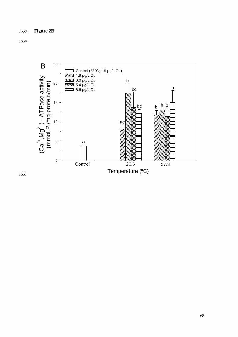
68
Figure 2B 1659
1660
Temperature (ºC)
0
5
10
15
20
25
Control (25°C; 1.9 µg/L Cu)
1.9 µg/L Cu
3.8 µg/L Cu
5.4 µg/L Cu
8.6 µg/L Cu
Control 26.6 27.3
a
b
b
bc
bc b
b
b
ac
(Ca
2+,M
g2
+)
- A
TP
ase a
ctivity
(mm
ol P
i/m
g p
rote
in/m
in)
B
1661

69
Figure 2C 1662
1663
Temperature (ºC)
0
5
10
15
20
25
Control (25°C; 1.9 µg/L Cu)
1.9 µg/L Cu
3.8 µg/L Cu
5.4 µg/L Cu
8.6 µg/L Cu
(Ca
2+,M
g2
+)-
AT
Pa
se
activity
(mm
ol P
i/m
g p
rote
in/m
in)
Control 26.6 27.3
a a
a
a
a
a
a
a
a
C
1664

70
Figure 3A 1665
1666
Time (days)
4 8 12
Yie
ld (
Fv/F
m)
0.0
0.1
0.4
0.5
0.6
0.7
0.8
0.9
1.0
Control
26.6°C
27.3°C
aa a a
a aa a
a* *
A
1667
1668

71
Figure 3B 1669
1670
4 8 12
Yie
ld (
Fv/F
m)
0.00
0.10
0.50
0.55
0.60
0.65
0.70
0.75
0.80
Control (25°C; 1.9 µg/L Cu)
26.6°C/3.8 µg/L Cu
26.6°C/5.4 µg/L Cu
26.6°C/ 8.6 µg/L Cu
27.3°C/ 3.8 µg/L Cu
27.3°C/5.4 µg/L Cu
27.3°C/8.6 µg/L Cu
Time (days)
a
a a a
b
abab
a
b
abababab
aba
b
bcbcbc
ac
ab
* * ** *
B
1671

72
Figure 4A 1672
1673
Temperature (ºC)
0
2
4
6
8
10
12
Control
3.0 µg/L Cu
3.8 µg/L Cu
5.4 µg/L Cu
8.6 µg/L Cu
Control 26.6 27.3
Ch
loro
ph
yl co
nce
ntr
atio
n
(ng/m
g w
w)
aa
a
a
a
a
a
a
a
A
1674 1675

73
Figure 4B 1676
1677
Temperature (ºC)
0
2
4
6
8
10
12
Control
3.0 µg/L Cu
3.8 µg/L Cu
5.4 µg/L Cu
8.6 µg/L Cu
Ch
loro
ph
yl co
nce
ntr
atio
n
(ng/m
g w
w)
Control 26.6 27.3
a
aa
aa
a
a
a
a
B
1678 1679
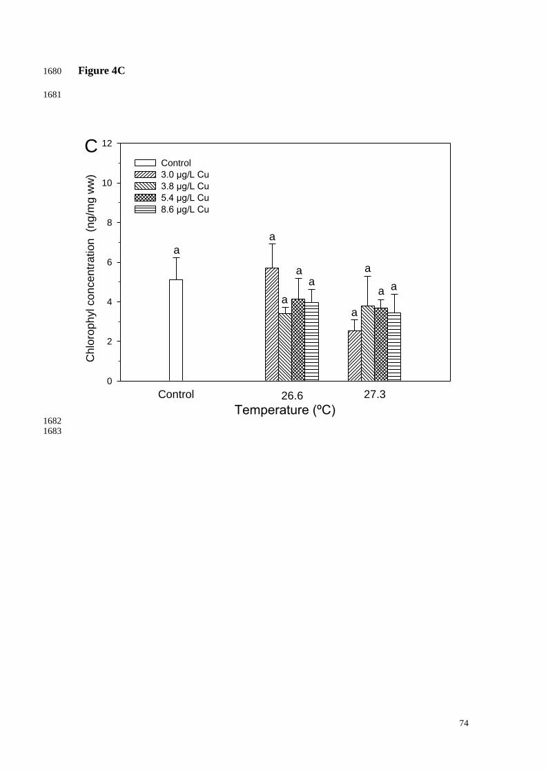
74
Figure 4C 1680
1681
Temperature (ºC)
0
2
4
6
8
10
12
Control
3.0 µg/L Cu
3.8 µg/L Cu
5.4 µg/L Cu
8.6 µg/L Cu
Ch
loro
ph
yl co
nce
ntr
atio
n
(ng
/mg w
w)
Control 26.6 27.3
a
a
a
aa
a
a
a a
C
1682 1683
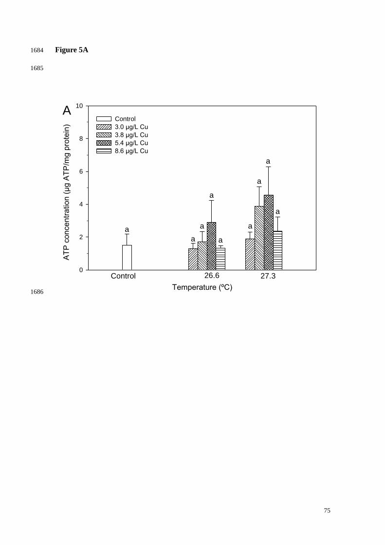
75
Figure 5A 1684
1685
Temperature (ºC)
AT
P c
on
cen
tration (
µg A
TP
/mg p
rote
in)
0
2
4
6
8
10
Control
3.0 µg/L Cu
3.8 µg/L Cu
5.4 µg/L Cu
8.6 µg/L Cu
Control 26.6 27.3
a
a
a
a
a
a
a
a
a
A
1686

76
Figure 5B 1687
1688
Temperature (ºC)
AT
P c
on
ce
ntr
atio
n (
µg
AT
P/m
g p
rote
in)
0
2
4
6
8
10
Control
3.0 µg/L Cu
3.8 µg/L Cu
5.4 µg/L Cu
8.6 µg/L Cu
Control 26.6 27.3
aa
a
a
a
a
a
a
a
B
1689 1690

77
Figure 5C 1691
1692
Temperature (ºC)
AT
P c
oncentr
ation (
µg A
TP
/mg p
rote
in)
0
2
4
6
8
10
Control
3.0 µg/L Cu
3.8 µg/L Cu
5.4 µg/L Cu
8.6 µg/L Cu
Control 26.6 27.3
aa
a
a
a a
a
aa
C
1693

78
Figure 6A 1694
1695
Temperature (ºC)
0.00
0.01
0.02
0.03
0.04
0.05
0.06
Control (25°C; 1.9 µg/L Cu)
1.9 µg/L Cu
3.8 µg/L Cu
5.4 µg/L Cu
8.6 µg/L Cu
AC
AP
(1
/re
lative
are
a)
Control 26.6 27.3
aa
a
a
a
aa
a
a
A
1696 1697

79
Figure 6B 1698
1699
Temperature (ºC)
0.00
0.01
0.02
0.03
0.04
0.05
0.06
Control (25°C; 1.9 µg/L Cu)
1.9 µg/L Cu
3.8 µg/L Cu
5.4 µg/L Cu
8.6 µg/L Cu
AC
AP
(1/r
ela
tive a
rea)
Control 26.6 27.3
a
aa
aa a
a a
a
1700
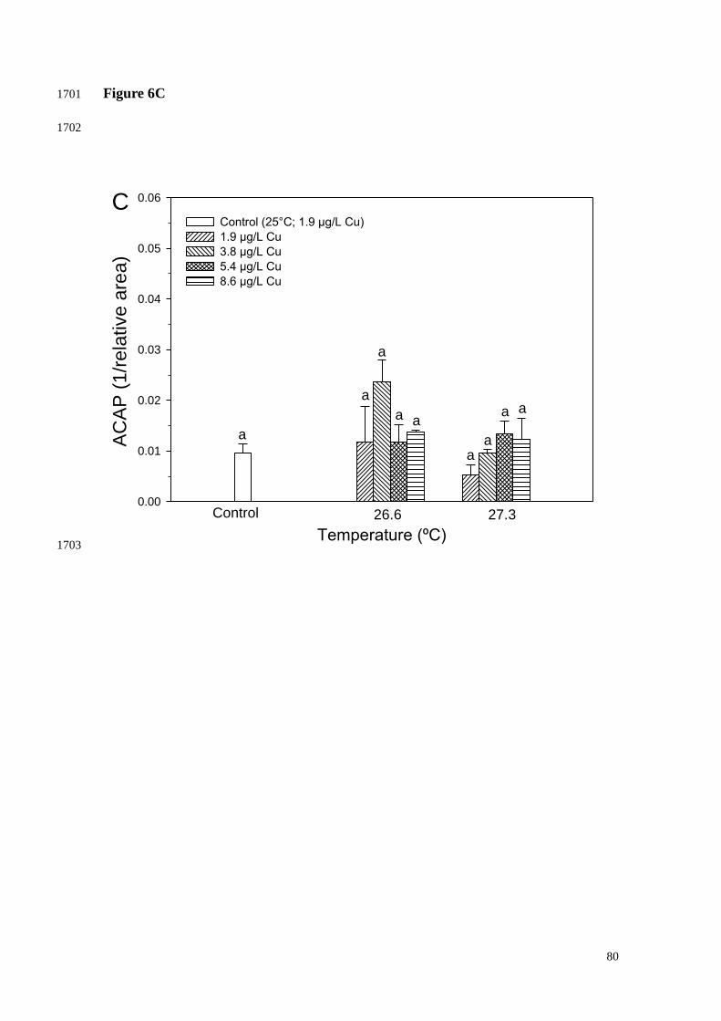
80
Figure 6C 1701
1702
Temperature (ºC)
0.00
0.01
0.02
0.03
0.04
0.05
0.06
Control (25°C; 1.9 µg/L Cu)
1.9 µg/L Cu
3.8 µg/L Cu
5.4 µg/L Cu
8.6 µg/L Cu
AC
AP
(1
/re
lative
are
a)
Control 26.6 27.3
a
a
a
a
a
a
aa
a
C
1703

81
Figure 7A 1704
1705
Temperature (ºC)
0
200
400
600
800
1000
1200
1400
Control (25°C; 1.9 µg/L Cu)
1.9 µg/L Cu
3.8 µg/L Cu
5.4 µg/L Cu
8.6 µg/L Cu
LP
O (
nm
ol M
DA
/mg
pro
tein
)
Control 26.6 27.3
a
b
b
bb
b
a
a
b
A
1706
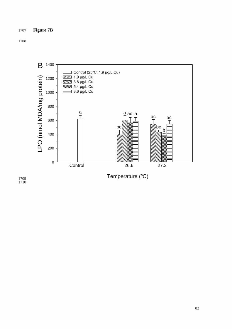
82
Figure 7B 1707
1708
Temperature (ºC)
0
200
400
600
800
1000
1200
1400
Control (25°C; 1.9 µg/L Cu)
1.9 µg/L Cu
3.8 µg/L Cu
5.4 µg/L Cu
8.6 µg/L Cu
LP
O (
nm
ol M
DA
/mg p
rote
in)
Control 26.6 27.3
a
bc bcb
a aacac ac
B
1709 1710

83
Figure 7C 1711
1712
Temperature (ºC)
0
400
800
1200
1600
2000
Control (25°C; 1.9 µg/L Cu)
1.9 µg/L Cu
3.8 µg/L Cu
5.4 µg/L Cu
8.6 µg/L Cu
LP
O (
nm
ol M
DA
/mg p
rote
in)
Control 26.6 27.3
a
b b
c c
ac
acac
ac
C
1713 1714

84
Figure 8 1715
1716

85
Discussão geral: 1717
1718
Considerando-se os resultados do presente estudo com o coral endêmico 1719
Mussismilia harttii, pode-se concluir que a exposição ao aumento da temperatura de 1720
forma isolada e/ou combinada com diferentes concentrações de cobre: (1) aumenta a 1721
atividade de enzimas envolvidas no processo de calcificação, sugerindo um aumento do 1722
metabolismo causado pela exposição aos estressores ou a ativação de mecanismos para 1723
recuperação do processo fotossintético; (2) reduz os danos oxidativos em lipídeos, 1724
possivelmente devido à redução do processo fotossintético ou ao remodelamento de 1725
lipídeos nas membranas biológicas; (3) diminui temporalmente a transferência de 1726
fotossintatos, importantes fontes de energia para o coral, reduzindo assim a energia 1727
disponível para as enzimas de calcificação e, consequentemente, para o crescimento, 1728
bem como para a ativação de mecanismos de proteção contra danos oxidativos, 1729
aumentando assim a susceptibilidade do coral ao branqueamento. Por fim, conclui-se 1730
que vários parâmetros bioquímicos e fisiológicos avaliados no presente estudo se 1731
mostraram sensíveis à exposição aos estressores (aumento de temperatura e exposição 1732
ao cobre dissolvido), indicando que essas ferramentas são potencias biomarcadores para 1733
o monitoramento da saúde de corais, uma vez que sofreram alterações, mesmo antes de 1734
ter sido observada qualquer manifestação de branqueamento. 1735