Judgement in artificial eruption of embedded teeth from an ...
7
12 REVIEW ARTICLE This is an open-access article distributed under the terms of the Creative Commons Attribution Non-Commercial License (http://creativecommons.org/ licenses/by-nc/4.0/), which permits unrestricted non-commercial use, distribution, and reproduction in any medium, provided the original work is properly cited. CC Judgement in artificial eruption of embedded teeth from an oral surgery perspective: review article Basel Mahardawi 1 , Kumar K C 1 , Kanin Arunakul 1 , Teeranut Chaiyasamut 1 , Natthamet Wongsirichat 1,2 1 Department of Oral and Maxillofacial Surgery, Faculty of Dentistry, Mahidol University, 2 Consultant of Walailak University International College of Dentistry, Bangkok, Thailand Abstract (J Korean Assoc Oral Maxillofac Surg 2020;46:12-18) Impacted teeth are a frequent phenomenon encountered by every clinician. The artificial eruption of embedded teeth is the process of directing an im- pacted tooth into normal occlusion. This procedure is currently attracting attention, with the aim of finding the best technique to use according to each case. This article presents key information regarding impacted incisors, canines, and premolars. In addition, we describe the most common techniques to use for artificial eruption, the open and closed techniques. We review the literature concerning these techniques and outline how clinicians can man- age every type of impacted tooth. Key words: Artificial eruption, Closed technique, Impacted, Open technique [paper submitted 2019. 3. 15 / accepted 2019. 5. 10] Copyright © 2020 The Korean Association of Oral and Maxillofacial Surgeons. All rights reserved. https://doi.org/10.5125/jkaoms.2020.46.1.12 pISSN 2234-7550 · eISSN 2234-5930 I. Introduction The normal eruption, position, and morphology of teeth are essential factors of facial esthetics and phonetics, and are the aim of every person to have and every clinician to achieve with their patients. Deviations from normal eruption patterns are frequently observed, and impacted teeth are challenging for clinicians to plan and apply appropriate treatment. It is essential to have a thorough knowledge of eruption distur- bances in order to promptly intervene. When a significant change from the normal shedding pro- cess occurs, diagnosis and treatment should be carried out as soon as possible. Alteration of tooth eruption time could be a symptom of a general condition or, in some cases, an indica- tion of altered physiology and craniofacial development 1 . The process of artificially erupting an impacted tooth and guiding it into occlusion requires a meticulous mul- tidisciplinary approach, requiring the contributions of an oral surgeon, orthodontist, pedodontist, and in some cases a periodontist. The role of the oral surgeon is to uncover the impacted teeth to aid in the orthodontic treatment, which then guides and aligns those teeth in the dental arch, providing better functional and esthetic results. II. Impacted Teeth and Treatment Modalities 1. Impacted canines Following the mandibular third molars, the maxillary permanent canine is the second most impacted tooth, with a prevalence between 0.3%-2.4% 2,3 and showing a higher pre- dilection in females than in males 3,4 . Dachi and Howell 4 indi- cated that impactions of maxillary canines take place unilat- erally in 92% of the cases, and only 8% are bilateral. Canines have a higher tendency to be impacted palatally than labi- ally 5 . Impaction of an anterior tooth can lead to functional or esthetic impairment and possibly further complications like midline diastema, crowding, root resorption on the adjacent permanent teeth 6 , and cysts. Therefore, early and accurate diagnosis of an impacted canine is required and the treatment is often directly initiated by elimination of any obstruction Natthamet Wongsirichat Department of Oral and Maxillofacial Surgery, Faculty of Dentistry, Mahidol University, 6 Yothi Street, Rachathewee District, Bangkok 10400, Thailand TEL: +66-22-007845 FAX: +66-22-007844 E-mail: [email protected] ORCID: https://orcid.org/0000-0003-3005-2680
Transcript of Judgement in artificial eruption of embedded teeth from an ...

12
REVIEW ARTICLE
This is an open-access article distributed under the terms of the Creative Commons Attribution Non-Commercial License (http://creativecommons.org/licenses/by-nc/4.0/), which permits unrestricted non-commercial use, distribution, and reproduction in any medium, provided the original work is properly cited.
CC
Judgement in artificial eruption of embedded teeth from an oral surgery perspective: review article
Basel Mahardawi1, Kumar K C1, Kanin Arunakul1, Teeranut Chaiyasamut1, Natthamet Wongsirichat1,2
1Department of Oral and Maxillofacial Surgery, Faculty of Dentistry, Mahidol University, 2Consultant of Walailak University International College of Dentistry, Bangkok, Thailand
Abstract (J Korean Assoc Oral Maxillofac Surg 2020;46:12-18)
Impacted teeth are a frequent phenomenon encountered by every clinician. The artificial eruption of embedded teeth is the process of directing an im-pacted tooth into normal occlusion. This procedure is currently attracting attention, with the aim of finding the best technique to use according to each case. This article presents key information regarding impacted incisors, canines, and premolars. In addition, we describe the most common techniques to use for artificial eruption, the open and closed techniques. We review the literature concerning these techniques and outline how clinicians can man-age every type of impacted tooth.
Key words: Artificial eruption, Closed technique, Impacted, Open technique[paper submitted 2019. 3. 15 / accepted 2019. 5. 10]
Copyright © 2020 The Korean Association of Oral and Maxillofacial Surgeons. All rights reserved.
https://doi.org/10.5125/jkaoms.2020.46.1.12pISSN 2234-7550 · eISSN 2234-5930
I. Introduction
The normal eruption, position, and morphology of teeth are essential factors of facial esthetics and phonetics, and are the aim of every person to have and every clinician to achieve with their patients. Deviations from normal eruption patterns are frequently observed, and impacted teeth are challenging for clinicians to plan and apply appropriate treatment. It is essential to have a thorough knowledge of eruption distur-bances in order to promptly intervene.
When a significant change from the normal shedding pro-cess occurs, diagnosis and treatment should be carried out as soon as possible. Alteration of tooth eruption time could be a symptom of a general condition or, in some cases, an indica-tion of altered physiology and craniofacial development1.
The process of artificially erupting an impacted tooth
and guiding it into occlusion requires a meticulous mul-tidisciplinary approach, requiring the contributions of an oral surgeon, orthodontist, pedodontist, and in some cases a periodontist. The role of the oral surgeon is to uncover the impacted teeth to aid in the orthodontic treatment, which then guides and aligns those teeth in the dental arch, providing better functional and esthetic results.
II. Impacted Teeth and Treatment Modalities
1. Impacted canines
Following the mandibular third molars, the maxillary permanent canine is the second most impacted tooth, with a prevalence between 0.3%-2.4%2,3 and showing a higher pre-dilection in females than in males3,4. Dachi and Howell4 indi-cated that impactions of maxillary canines take place unilat-erally in 92% of the cases, and only 8% are bilateral. Canines have a higher tendency to be impacted palatally than labi-ally5. Impaction of an anterior tooth can lead to functional or esthetic impairment and possibly further complications like midline diastema, crowding, root resorption on the adjacent permanent teeth6, and cysts. Therefore, early and accurate diagnosis of an impacted canine is required and the treatment is often directly initiated by elimination of any obstruction
Natthamet WongsirichatDepartment of Oral and Maxillofacial Surgery, Faculty of Dentistry, Mahidol University, 6 Yothi Street, Rachathewee District, Bangkok 10400, ThailandTEL: +66-22-007845 FAX: +66-22-007844E-mail: [email protected]: https://orcid.org/0000-0003-3005-2680

Judgement in artificial eruption of embedded teeth from an oral surgery perspective
13
to the normal path of eruption to provide enough space and proper direction for the underlying canine7.
Various procedures in the management of impacted canines are presented. These include the early interceptive approach8, surgical exposure of the canine crown followed by orth-odontic guidance to the dental arch9, and autogenous tooth transplantation10. The canine is a stable arch former and has esthetic value.
If an abnormality cannot be corrected by interceptive treat-ment alone, both surgical and orthodontic approaches are often considered to guide the impacted canine into normal occlusion. This can be achieved by two main surgical tech-niques: the open technique and the closed technique.
1) Open techniqueThis technique involves gingivectomy and apically posi-
tioned flap (also known as apically displaced flap) procedures to expose the impacted tooth. Selection of this procedure is based on a thorough assessment of the labiolingual position-ing of the impacted tooth, the vertical position in relation to the mucogingival junction, the mesio-distal position in rela-tion to the lateral incisor, and the amount and type of gingiva surrounding the site of the tooth.
Gingivectomy is performed to expose the clinical crown by removing the gingiva, which covers the coronal portion of the unerupted tooth, while maintaining adequate keratinized gingiva. In some cases removal of the overlying bone is nec-essary. This is followed by the orthodontic phase to track the tooth into normal occlusion.
The apically displaced flap procedure is conducted by el-evating a partial thickness flap along the tip of the unerupted tooth. A vertical releasing incision is made on either side of the bulged crown of the impacted tooth, ensuring adequate vascularity of the elevated flap. When necessary, osteoplasty may be performed to expose the clinical crown and smooth the marginal bone covering the tooth. The flap is then api-
cally positioned and sutured.
2) Closed techniqueA full-thickness mucoperiosteal flap is elevated and reflect-
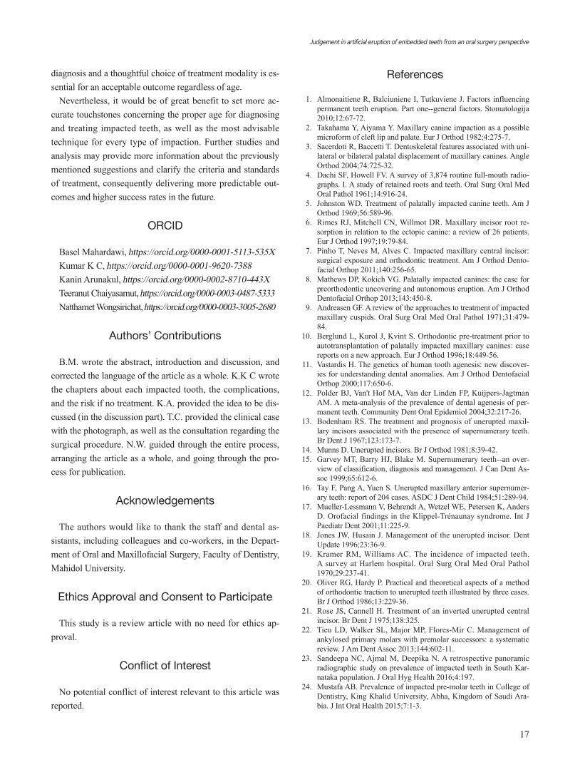
ed to uncover the impacted tooth. This is usually followed by bone removal to expose the clinical part of the crown. Orth-odontic traction tools, such as brackets/attachment/buttons along with a ligature wire, are then bonded to the crown. This wire passes through or under the flap in the desired direction. (Fig. 1. A) Next, the flap is placed back into the original posi-tion and sutured.(Fig. 1. B) Orthodontic traction is initiated in the next stage to drag the tooth into normal occlusion.
2. Impacted central and lateral incisors
Delayed eruption of maxillary incisors also affects indi-vidual facial esthetics and phonetics, and is believed to occur due to permanent tooth agenesis. The incidence of impacted maxillary lateral incisors is 2.2%11, making it the second or third most common occurrence of impaction in the dentition. On the other hand, there is a prevalence of 0.00%-0.01% for a missing central incisor12.
Several reports mention the main cause of delayed maxil-lary incisor eruption as the presence of a supernumerary tooth, with a higher frequency in the maxilla13,14. Certain syndromes are closely associated with supernumerary teeth when compared with the general population. Some studies have proposed that eruption delay can occur as a result of influential factors, for instance, the shape and position of a supernumerary tooth, which acts as a guide for the erup-tion of the incisors15. The tuberculate, or invaginated, type of supernumerary tooth was found to lead to more instances of delayed eruption of the maxillary incisors13. Tay et al.16 stated that the vertical position of supernumerary teeth is more likely to cause the delayed eruption of permanent inci-sors. Tooth malformation or dilacerations usually occur after
Text
A B
Fig. 1. Closed eruption technique. A. Surgical exposure of the canine and placement of orthodontic button. B. Flap closure into the original position.Basel Mahardawi et al: Judgement in artificial eruption of embedded teeth from an oral surgery perspective: review article. J Korean Assoc Oral Maxillofac Surg 2020

J Korean Assoc Oral Maxillofac Surg 2020;46:12-18
14
primary tooth trauma, resulting in an injury to the develop-ing permanent tooth bud which can lead to eruption failure. Moreover, developmental cysts or other pathologies can hin-der the pathway of the developing permanent incisor13. The most common developmental lesion found in the anterior maxilla is the dentigerous cyst17. Treatment planning for an impacted incisor engulfed by a cyst ought to follow a conser-vative approach, as tooth preservation in young patients must be the primary concern. Therefore, marsupialization is the treatment of choice as it is atraumatic and may prevent psy-chological or mental stress. When there is a prolonged and retained primary incisor, it can affect the underlying develop-ing permanent incisor, altering the pathway of eruption, even sometimes preventing it from eruption, which might be due to the formation of dense mucoperiosteum or submucosa that will form a physical eruption barrier18.
A variety of treatment procedures for unerupted maxillary incisors are proposed in the literature; however, successful outcome of treatment is best achieved with early diagnosis and prompt treatment planning11. The most conservative treatment procedure would be the extraction of any obstruc-tion (i.e., supernumerary or deciduous tooth), followed by the creation of space, therefore creating an opportunity for spon-taneous eruption. This is successful in 54%-74% of cases without any further treatment19.
Impacted central and lateral incisors can be managed in three divided phases. Pre-surgical orthodontic measures first create sufficient space for the unerupted tooth, and any ob-structions, mainly supernumerary teeth, are removed. This is followed by surgical uncovering of the impacted tooth by either the open technique (gingivectomy or apically displaced flap) or closed technique. Orthodontic traction and alignment in the post-surgical orthodontic phase complete the treat-ment20. Whenever an unerupted incisor is associated with a considerable pathology, has any chance of damaging the adjacent teeth and causing resorption, or is severely ectopic, extraction of the unerupted tooth is advocated21.
3. Impacted premolars
Although the prevalence of premolar impaction is low, a re-view published by Tieu et al.22 in 2013 proposed that impac-tion of premolars may be caused due to prolonged ankyloses and delayed exfoliation of primary molars. In addition, some studies showed a higher predilection in the mandible than the maxilla23,24. Nevertheless, the number of cases is limited. Although the etiology of ankylosis is not well understood25,26,
possibilities include genetic predisposition27,28, excessive masticatory pressure29, and disturbance in local metabolism30. Tieu et al.22 suggested that as a first step, ankylosed pri-mary molars should undergo close observation for up to six months. A mild to moderate progressive infraocclusion will often occur along with ankylosed primary molars. Extraction of the permanent tooth may become the only option if it has an altered eruption pathway, if the infraocclusion of the anky-losed primary molar becomes severe in relation with the ad-jacent teeth preventing the successor from erupting, or if both conditions exist. Nonetheless, as mentioned before, conserva-tive observation of ankylosed primary molars is suggested22.
III. Complications of Treatment
When making the decision to perform any surgical proce-dure for young children, the psychological consequences of the treatment must be considered. A 2005 study31 to deter-mine patient cognizance during the recovery period follow-ing the closed eruption technique indicated that the surgical procedure used to uncover the impacted tooth will likely have an adverse impact on various aspects of patient health-related quality of life, regardless of the technique performed. Recovery should be expected within three days postopera-tively, and patients should be able to return to school within four to six days. It was found that surgery longer than thirty minutes consequently led to intensified post-operative pain. Furthermore, it was shown that the surgical uncovering of bucally impacted teeth requires a longer recovery period than palatally impacted ones31.
Choosing an appropriate flap design preceding the surgi-cal procedure is vital. The benefits of the apically positioned flap design are in providing a sufficient band of attached gingiva that will surround the crown of the erupted tooth and in employing coronal tension when suturing. This initiates a spontaneous eruption of the incisor in the occlusal direction. This flap permits manipulation of the size of post-operative keratinized gingiva, which plays a role in saving the mu-cogingival complex surrounding the surgically uncovered tooth32, ensuring a healthy long-term periodontal prognosis of the exposed and erupted tooth.
However, this flap design may be associated with poor esthetics, causing more crown length as well as gingival scar-ring12,33. The clinician may choose another procedure, such as the closed eruption technique, as a result of the anterior posi-tion of canines or incisors, in an attempt to eliminate these issues.

Judgement in artificial eruption of embedded teeth from an oral surgery perspective
15
Damage to the adjacent teeth is another possible obstacle which can occur during surgical procedures17, and this can lead to future problems for the patient.
Moreover, periodontal complications may manifest af-ter surgical treatment. These can include a decrease in the amount of keratinized gingiva, recession, and poor oral hy-giene during the orthodontic phase. Periodontal problems are more common with buccally positioned unerupted teeth than with palatally displaced ones34. The aim of any procedure that involves mucogingival tissue intervention should ensure an adequate thickness of keratinized gingiva, thus eliminating any chance of gingival recession.
IV. Risks if No Treatment
Leaving an impacted tooth untreated can lead to several problems including esthetic, functional, and pathological complications. Cyst formation, ankylosis, external tooth resorption, and periodontal problems can all result from untreated impacted teeth. In addition, a prolonged period of having an edentulous space might facilitate migration of dis-tal teeth in a mesial direction, causing obliteration of space35.
There is also an association between having an impacted central incisor and the occurrence of ipsilateral canine impac-tion which the clinician should be aware of to prevent any such effect36.
Individuals facing the problem of impacted teeth are usually in a growing period of mixed dentition; therefore, children may face social and psychological difficulties with unpleasant tooth alignment, especially in the areas visible when smiling. Considering these points, this problem might create a negative outcome if not treated. Thus, individuals at this period must be treated properly, and any of the possible drawbacks of having an impacted tooth should be eliminated.
V. Discussion
Artificial eruption for impacted teeth is a procedure that relies on many factors, as well as experience, in order to choose the proper treatment option. Reviewing the literature, it is noticeable that there is still no complete consensus re-garding the best technique or an absolute procedure for each type or degree of impaction. Taking the impacted maxillary canine for instance, a review published by Counihan et al.37 concluded that when the canine doesn’t overlap the lateral incisor, the crown is between the CEJ (cemento-enamel junc-tion) and half of the root length, the angulation of the canine
is between 0°-15°, and the apex of its root is in the normal position mesio-distally, then the treatment of choice would be only the extraction of the deciduous canine and letting the permanent tooth erupt by itself. This was also stated by Jacobs38, who recommended that in Class I occlusion cases, with a permanent impacted maxillary canine, or when it has erupted buccally or palatally but no crowding is present, the preventive treatment of choice should be the extraction of the deciduous canine between the ages of 10-13 years. In addi-tion, Ericson and Kurol39 treated patients 10-13 years old, and showed that the extraction of the primary canine in 46 cases of palatally impacted maxillary canine led to normalizing its position in 36 of those cases within a 12-month follow-up period, explaining that 91% of the canines which overlapped the roots of the adjacent lateral incisor root by less than half of the root (on the x-rays) moved back to the normal position, and 64% of the cases where the canine overlapped the lateral incisor by more than half of the root width initially normal-ized. Consequently, even when the impacted canine overlaps the lateral incisor, it is still possible to get it to the normal po-sition with interceptive treatment only. On the other hand, the previously mentioned procedure was not in agreement with other opinions. Kinaia et al.40 indicated in their review that when the position of the impacted canine is labial (no over-lapping), the crown is coronal to the muco-gingival junction (between the CEJ and half of the root length), and it is in the normal position mesio-distally, then the treatment of choice is gingivectomy followed by placing the bracket to drag the tooth orthodontically. They were not content with the inter-ceptive treatment only, even in the simplest cases of impac-tion, and presented gingivectomy, apically positioned flap, and the closed technique as treatment options. They stated in this review that when the impacted canine is positioned mesially to the lateral incisor, the proper treatment would be the closed technique. This, in turn, was not consistent with Kokich41, who suggested that in the previously mentioned case, closed eruption or excisional uncovering (gingivec-tomy) would not be advised. Additionally, Yordanova et al.42 treated 3 cases of palatal impaction with the open technique, which is not recommended by Kinaia et al.40 in such cases. Furthermore, Londhe et al.43 compared the open technique (gingivectomy) and closed technique for the treatment of la-bially impacted canines. Patients were selected randomly for each technique. They reported success in all of their cases, and their results were in favor of the closed technique in the case of labial impactions, which is not in consensus with the previously mentioned literature.

J Korean Assoc Oral Maxillofac Surg 2020;46:12-18
16
In addition, the proper age for diagnosing tooth impaction is still uncertain. Ericson and Kurol44 explained that resorp-tion and pathology have a higher incidence in female patients over 14 years old, and in cases with an angulation of more than 25 degrees of the canine in relation to the midline. Shi et al.45 stated that in the case of having impacted maxillary incisors by the age of 12 years, the maxillary incisor roots are fully developed already, and are dilacerated. Therefore, extraction is the only option for a successful outcome. They found that the best results were achieved with the following factors:
1) No more than two-thirds of the entire root length of the impacted tooth is formed.
2) The patient’s age is between 7 and 8 years. However, the mean age of the patients in their study was 6.4-10.4 years old, and all cases were successful.
Moreover, deciding when to carry out the treatment, and the age that will result in the most successful results, is not clearly defined. Kinaia et al.40 presented 4 cases of impacted canines. The patients’ ages were 12 years old in 3 cases and 15 years old in 1 case. No reports of failure or complications were shown. Londhe et al.43 treated 31 patients aged between 14-30 years old with maxillary impacted canines. Patients had undergone surgery using a closed or open technique after ran-dom selection. All cases were successful. Yordanova et al.42 presented 3 cases with palatal canine impactions. Patients’ ages were 13, 15, and 20 years old. They were all treated us-ing the open technique as mentioned earlier, with successful outcomes. Shetty el al.46 reported a case of an 11-year-old pa-tient who had an impacted left central incisor, lateral incisor, and canine. An apically positioned flap was done to expose the impacted canine, while the closed technique was chosen for the impacted central and lateral incisors. The treatment was successful and no complications were reported.
On the other hand, Chaushu and Chaushu47 showed in their
case reports that failure occurred in 2 cases where the patients were 14 and 19 years old. In two other cases, the patients were 22 and 26 years old, yet they were both successful. Chaushu and Chaushu47 indicated that the proper treatment procedure is an important factor that has a significant impact on the outcome.
As mentioned previously, Shi et al.45 conducted their study on the treatment of impacted maxillary incisors. Fifty patients aged between 6.4 and 10.4 years old were included, and all were treated successfully.
Concerning impacted premolars, Burch et al.48 performed artificial eruption on 4 patients aged 11, 12, 9, and 9 years old who had unerupted premolars. Interceptive treatment only was done, and no surgical procedure was performed to expose the impacted premolar and place an orthodontic ap-pliance. All of the cases were successful, and no failure was outlined. A summary of all previously mentioned studies is shown in Table 140,42,43,45-48.
VI. Conclusion
The process of artificial eruption for an impacted tooth is an art that requires the participation of a team, not just an orthodontist. Therefore, the authors would like to emphasize that what matters most is moving the impacted tooth into normal occlusion and having well-aligned teeth in the end, regardless of the technique used. The authors did not suggest criteria or recommend any technique as the debate on which to use in specific cases is ongoing. After reviewing the lit-erature, it is clear that there is no absolute indication for each technique for exposing an impacted tooth, and it is mainly dependent upon experience and preference. In addition, there is no exact evidence on the proper age for the diagnosis and treatment of impacted teeth, but obviously the younger the patient is the better the prognosis. Needless to say, a precise
Table 1. Techniques to expose impacted teeth and treatment outcome in the literature
StudyAge of each patient or
age range (yr)Tooth Type of impaction Technique Success/failure
Burch et al.48 (1994) 11/12/9/9 Premolar N/A Interceptive SuccessChaushu and Chaushu47 (2010) 14-19 Canine Labial N/A Failure
22-26 Canine Palatal N/A SuccessYordanova et al.42 (2011) 13/15/20 Canine Palatal Open SuccessLondhe et al.43 (2014) 14-30 Canine Labial Closed+open SuccessShetty el al.46 (2015) 11 Canine N/A Open+closed SuccessShi et al.45 (2015) 6.4-10.4 Incisor N/A Closed SuccessKinaia et al.40 (2016) 12/12/15/12 Canine Buccal+palatal+
mid alveolarOpen+closed Success
(N/A: not available)Basel Mahardawi et al: Judgement in artificial eruption of embedded teeth from an oral surgery perspective: review article. J Korean Assoc Oral Maxillofac Surg 2020
Judgement in artificial eruption of embedded teeth from an oral surgery perspective
17
diagnosis and a thoughtful choice of treatment modality is es-sential for an acceptable outcome regardless of age.
Nevertheless, it would be of great benefit to set more ac-curate touchstones concerning the proper age for diagnosing and treating impacted teeth, as well as the most advisable technique for every type of impaction. Further studies and analysis may provide more information about the previously mentioned suggestions and clarify the criteria and standards of treatment, consequently delivering more predictable out-comes and higher success rates in the future.
ORCID
Basel Mahardawi, https://orcid.org/0000-0001-5113-535XKumar K C, https://orcid.org/0000-0001-9620-7388Kanin Arunakul, https://orcid.org/0000-0002-8710-443XTeeranut Chaiyasamut, https://orcid.org/0000-0003-0487-5333Natthamet Wongsirichat, https://orcid.org/0000-0003-3005-2680
Authors’ Contributions
B.M. wrote the abstract, introduction and discussion, and corrected the language of the article as a whole. K.K C wrote the chapters about each impacted tooth, the complications, and the risk if no treatment. K.A. provided the idea to be dis-cussed (in the discussion part). T.C. provided the clinical case with the photograph, as well as the consultation regarding the surgical procedure. N.W. guided through the entire process, arranging the article as a whole, and going through the pro-cess for publication.
Acknowledgements
The authors would like to thank the staff and dental as-sistants, including colleagues and co-workers, in the Depart-ment of Oral and Maxillofacial Surgery, Faculty of Dentistry, Mahidol University.
Ethics Approval and Consent to Participate
This study is a review article with no need for ethics ap-proval.
Conflict of Interest
No potential conflict of interest relevant to this article was reported.
References
1. Almonaitiene R, Balciuniene I, Tutkuviene J. Factors influencing permanent teeth eruption. Part one--general factors. Stomatologija 2010;12:67-72.
2. Takahama Y, Aiyama Y. Maxillary canine impaction as a possible microform of cleft lip and palate. Eur J Orthod 1982;4:275-7.
3. Sacerdoti R, Baccetti T. Dentoskeletal features associated with uni-lateral or bilateral palatal displacement of maxillary canines. Angle Orthod 2004;74:725-32.
4. Dachi SF, Howell FV. A survey of 3,874 routine full-mouth radio-graphs. I. A study of retained roots and teeth. Oral Surg Oral Med Oral Pathol 1961;14:916-24.
5. Johnston WD. Treatment of palatally impacted canine teeth. Am J Orthod 1969;56:589-96.
6. Rimes RJ, Mitchell CN, Willmot DR. Maxillary incisor root re-sorption in relation to the ectopic canine: a review of 26 patients. Eur J Orthod 1997;19:79-84.
7. Pinho T, Neves M, Alves C. Impacted maxillary central incisor: surgical exposure and orthodontic treatment. Am J Orthod Dento-facial Orthop 2011;140:256-65.
8. Mathews DP, Kokich VG. Palatally impacted canines: the case for preorthodontic uncovering and autonomous eruption. Am J Orthod Dentofacial Orthop 2013;143:450-8.
9. Andreasen GF. A review of the approaches to treatment of impacted maxillary cuspids. Oral Surg Oral Med Oral Pathol 1971;31:479-84.
10. Berglund L, Kurol J, Kvint S. Orthodontic pre-treatment prior to autotransplantation of palatally impacted maxillary canines: case reports on a new approach. Eur J Orthod 1996;18:449-56.
11. Vastardis H. The genetics of human tooth agenesis: new discover-ies for understanding dental anomalies. Am J Orthod Dentofacial Orthop 2000;117:650-6.
12. Polder BJ, Van't Hof MA, Van der Linden FP, Kuijpers-Jagtman AM. A meta-analysis of the prevalence of dental agenesis of per-manent teeth. Community Dent Oral Epidemiol 2004;32:217-26.
13. Bodenham RS. The treatment and prognosis of unerupted maxil-lary incisors associated with the presence of supernumerary teeth. Br Dent J 1967;123:173-7.
14. Munns D. Unerupted incisors. Br J Orthod 1981;8:39-42.15. Garvey MT, Barry HJ, Blake M. Supernumerary teeth--an over-
view of classification, diagnosis and management. J Can Dent As-soc 1999;65:612-6.
16. Tay F, Pang A, Yuen S. Unerupted maxillary anterior supernumer-ary teeth: report of 204 cases. ASDC J Dent Child 1984;51:289-94.
17. Mueller-Lessmann V, Behrendt A, Wetzel WE, Petersen K, Anders D. Orofacial findings in the Klippel-Trénaunay syndrome. Int J Paediatr Dent 2001;11:225-9.
18. Jones JW, Husain J. Management of the unerupted incisor. Dent Update 1996;23:36-9.
19. Kramer RM, Williams AC. The incidence of impacted teeth. A survey at Harlem hospital. Oral Surg Oral Med Oral Pathol 1970;29:237-41.
20. Oliver RG, Hardy P. Practical and theoretical aspects of a method of orthodontic traction to unerupted teeth illustrated by three cases. Br J Orthod 1986;13:229-36.
21. Rose JS, Cannell H. Treatment of an inverted unerupted central incisor. Br Dent J 1975;138:325.
22. Tieu LD, Walker SL, Major MP, Flores-Mir C. Management of ankylosed primary molars with premolar successors: a systematic review. J Am Dent Assoc 2013;144:602-11.
23. Sandeepa NC, Ajmal M, Deepika N. A retrospective panoramic radiographic study on prevalence of impacted teeth in South Kar-nataka population. J Oral Hyg Health 2016;4:197.
24. Mustafa AB. Prevalence of impacted pre-molar teeth in College of Dentistry, King Khalid University, Abha, Kingdom of Saudi Ara-bia. J Int Oral Health 2015;7:1-3.

J Korean Assoc Oral Maxillofac Surg 2020;46:12-18
18
25. Messer LB, Cline JT. Ankylosed primary molars: results and treat-ment recommendations from an eight-year longitudinal study. Pe-diatr Dent 1980;2:37-47.
26. Kurol J, Thilander B. Infraocclusion of primary molars with apla-sia of the permanent successor. A longitudinal study. Angle Orthod 1984;54:283-94.
27. Kurol J. Infraocclusion of primary molars: an epidemiologic and familial study. Community Dent Oral Epidemiol 1981;9:94-102.
28. Via WF Jr. Submerged deciduous molars: familial tendencies. J Am Dent Assoc 1964;69:127-9.
29. Adamson KT. The problem of impacted teeth in orthodontics. Aust Dent J 1952;56:74-84.
30. Bjerklin K, Kurol J. Prevalence of ectopic eruption of the maxillary first permanent molar. Swed Dent J 1981;5:29-34.
31. Chaushu S, Becker A, Zeltser R, Branski S, Vasker N, Chaushu G. Patients perception of recovery after exposure of impacted teeth: a comparison of closed- versus open-eruption techniques. J Oral Maxillofac Surg 2005;63:323-9.
32. Ong M, Chew MT. Use of the apically repositioned flap in the management of labially impacted maxillary central incisors. Singa-pore Dent J 2004;26:55-9.
33. Kajiyama K, Kai H. Esthetic management of an unerupted maxil-lary central incisor with a closed eruption technique. Am J Orthod Dentofacial Orthop 2000;118:224-8.
34. Kavadia-Tsatala S, Tsalikis L, Kaklamanos EG, Sidiropoulou S, Antoniades K. Orthodontic and periodontal considerations in man-aging teeth exhibiting significant delay in eruption. World J Orthod 2004;5:224-9.
35. Grover PS, Lorton L. The incidence of unerupted permanent teeth and related clinical cases. Oral Surg Oral Med Oral Pathol 1985;59:420-5.
36. Chaushu S, Zilberman Y, Becker A. Maxillary incisor impaction and its relationship to canine displacement. Am J Orthod Dentofa-cial Orthop 2003;124:144-50; discussion 150.
37. Counihan K, Al-Awadhi EA, Butler J. Guidelines for the assess-ment of the impacted maxillary canine. Dent Update 2013;40:770-2, 775-7.
38. Jacobs SG. Reducing the incidence of palatally impacted maxil-lary canines by extraction of deciduous canines: a useful preven-tive/interceptive orthodontic procedure. Case reports. Aust Dent J
1992;37:6-11.39. Ericson S, Kurol J. Early treatment of palatally erupting maxil-
lary canines by extraction of the primary canines. Eur J Orthod 1988;10:283-95.
40. Kinaia BM, Agarwal K, Bushong B, Kapoor N, Hope K, Ambrosio F, et al. Surgical management of impacted canines: a literature re-view and case presentations. J Dent Oral Biol 2016;1:1012.
41. Kokich VG. Surgical and orthodontic management of impacted max-illary canines. Am J Orthod Dentofacial Orthop 2004;126:278-83.
42. Yordanova M, Yordanova S, Vladimirov B. Surgical uncovering and stimulation of physiological eruption of palatally impacted maxillary canines: case reports. J IMAB 2011;17:114-9.
43. Londhe SM, Kumar P, Datana S, Kotwal A, Saxena V. Guided tooth eruption: comparison of open and closed eruption techniques in labially impacted maxillary canines. J Dent Res Rev 2014;1:148-51.
44. Ericson S, Kurol J. Incisor resorption caused by maxillary cuspids. A radiographic study. Angle Orthod 1987;57:332-46.
45. Shi XR, Hu Z, Wang XZ, Sun XY, Zhang CY, Si Y, et al. Evalua-tion of the effect of the closed-eruption technique on impacted im-mature maxillary incisors. Chin J Dent Res 2015;18:111-5.
46. Shetty BK, Somaiah S, Muddaiah S, Parveen S, Sirajuddin. Guided eruption of multiple impacted teeth using a modified miniplate. J Clin Orthod 2015;49:273-80.
47. Chaushu S, Chaushu G. Skeletal implant anchorage in the treat-ment of impacted teeth: a review of the state of the art. Semin Or-thod 2010;16:234-41.
48. Burch J, Ngan P, Hackman A. Diagnosis and treatment planning for unerupted premolars. Pediatr Dent 1994;16:89-95.
How to cite this article: Mahardawi B, K C K, Arunakul K, Chai-
yasamut T, Wongsirichat N. Judgement in artificial eruption of
embedded teeth from an oral surgery perspective: review article.
J Korean Assoc Oral Maxillofac Surg 2020;46:12-18. https://doi.
org/10.5125/jkaoms.2020.46.1.12