Journal of Liver - OMICS International · PDF fileresidues and its correlation with the...
8
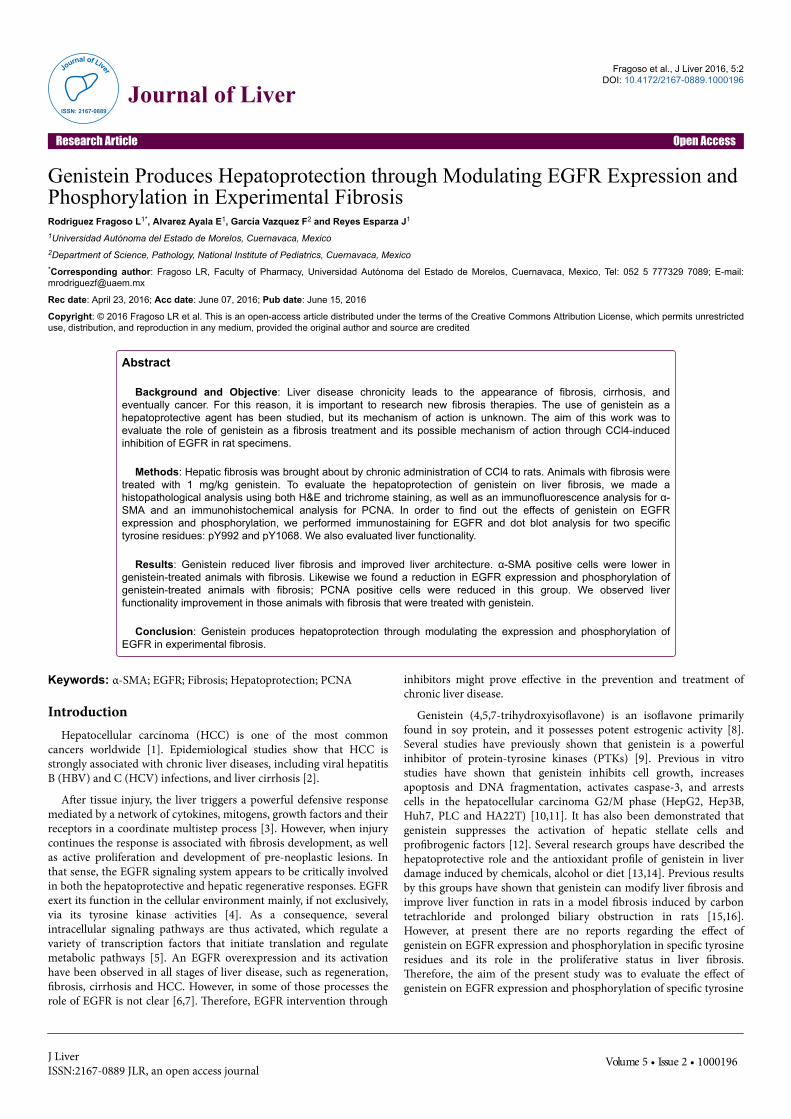
Genistein Produces Hepatoprotection through Modulating EGFR Expression and Phosphorylation in Experimental Fibrosis Rodriguez Fragoso L 1* , Alvarez Ayala E 1 , García Vazquez F 2 and Reyes Esparza J 1 1 Universidad Autónoma del Estado de Morelos, Cuernavaca, Mexico 2 Department of Science, Pathology, National Institute of Pediatrics, Cuernavaca, Mexico * Corresponding author: Fragoso LR, Faculty of Pharmacy, Universidad Autónoma del Estado de Morelos, Cuernavaca, Mexico, Tel: 052 5 777329 7089; E-mail: [email protected] Rec date: April 23, 2016; Acc date: June 07, 2016; Pub date: June 15, 2016 Copyright: © 2016 Fragoso LR et al. This is an open-access article distributed under the terms of the Creative Commons Attribution License, which permits unrestricted use, distribution, and reproduction in any medium, provided the original author and source are credited Abstract Background and Objective: Liver disease chronicity leads to the appearance of fibrosis, cirrhosis, and eventually cancer. For this reason, it is important to research new fibrosis therapies. The use of genistein as a hepatoprotective agent has been studied, but its mechanism of action is unknown. The aim of this work was to evaluate the role of genistein as a fibrosis treatment and its possible mechanism of action through CCl4-induced inhibition of EGFR in rat specimens. Methods: Hepatic fibrosis was brought about by chronic administration of CCl4 to rats. Animals with fibrosis were treated with 1 mg/kg genistein. To evaluate the hepatoprotection of genistein on liver fibrosis, we made a histopathological analysis using both H&E and trichrome staining, as well as an immunofluorescence analysis for α- SMA and an immunohistochemical analysis for PCNA. In order to find out the effects of genistein on EGFR expression and phosphorylation, we performed immunostaining for EGFR and dot blot analysis for two specific tyrosine residues: pY992 and pY1068. We also evaluated liver functionality. Results: Genistein reduced liver fibrosis and improved liver architecture. α-SMA positive cells were lower in genistein-treated animals with fibrosis. Likewise we found a reduction in EGFR expression and phosphorylation of genistein-treated animals with fibrosis; PCNA positive cells were reduced in this group. We observed liver functionality improvement in those animals with fibrosis that were treated with genistein. Conclusion: Genistein produces hepatoprotection through modulating the expression and phosphorylation of EGFR in experimental fibrosis. Keywords: α-SMA; EGFR; Fibrosis; Hepatoprotection; PCNA Introduction Hepatocellular carcinoma (HCC) is one of the most common cancers worldwide [1]. Epidemiological studies show that HCC is strongly associated with chronic liver diseases, including viral hepatitis B (HBV) and C (HCV) infections, and liver cirrhosis [2]. Aſter tissue injury, the liver triggers a powerful defensive response mediated by a network of cytokines, mitogens, growth factors and their receptors in a coordinate multistep process [3]. However, when injury continues the response is associated with fibrosis development, as well as active proliferation and development of pre-neoplastic lesions. In that sense, the EGFR signaling system appears to be critically involved in both the hepatoprotective and hepatic regenerative responses. EGFR exert its function in the cellular environment mainly, if not exclusively, via its tyrosine kinase activities [4]. As a consequence, several intracellular signaling pathways are thus activated, which regulate a variety of transcription factors that initiate translation and regulate metabolic pathways [5]. An EGFR overexpression and its activation have been observed in all stages of liver disease, such as regeneration, fibrosis, cirrhosis and HCC. However, in some of those processes the role of EGFR is not clear [6,7]. erefore, EGFR intervention through inhibitors might prove effective in the prevention and treatment of chronic liver disease. Genistein (4,5,7-trihydroxyisoflavone) is an isoflavone primarily found in soy protein, and it possesses potent estrogenic activity [8]. Several studies have previously shown that genistein is a powerful inhibitor of protein-tyrosine kinases (PTKs) [9]. Previous in vitro studies have shown that genistein inhibits cell growth, increases apoptosis and DNA fragmentation, activates caspase-3, and arrests cells in the hepatocellular carcinoma G2/M phase (HepG2, Hep3B, Huh7, PLC and HA22T) [10,11]. It has also been demonstrated that genistein suppresses the activation of hepatic stellate cells and profibrogenic factors [12]. Several research groups have described the hepatoprotective role and the antioxidant profile of genistein in liver damage induced by chemicals, alcohol or diet [13,14]. Previous results by this groups have shown that genistein can modify liver fibrosis and improve liver function in rats in a model fibrosis induced by carbon tetrachloride and prolonged biliary obstruction in rats [15,16]. However, at present there are no reports regarding the effect of genistein on EGFR expression and phosphorylation in specific tyrosine residues and its role in the proliferative status in liver fibrosis. erefore, the aim of the present study was to evaluate the effect of genistein on EGFR expression and phosphorylation of specific tyrosine Fragoso et al., J Liver 2016, 5:2 DOI: 10.4172/2167-0889.1000196 Research Article Open Access J Liver ISSN:2167-0889 JLR, an open access journal Volume 5 • Issue 2 • 1000196 J o u r n a l o f L i v e r ISSN: 2167-0889 Journal of Liver
Transcript of Journal of Liver - OMICS International · PDF fileresidues and its correlation with the...
Genistein Produces Hepatoprotection through Modulating EGFR Expression andPhosphorylation in Experimental FibrosisRodriguez Fragoso L1*, Alvarez Ayala E1, García Vazquez F2 and Reyes Esparza J1
1Universidad Autónoma del Estado de Morelos, Cuernavaca, Mexico2Department of Science, Pathology, National Institute of Pediatrics, Cuernavaca, Mexico*Corresponding author: Fragoso LR, Faculty of Pharmacy, Universidad Autónoma del Estado de Morelos, Cuernavaca, Mexico, Tel: 052 5 777329 7089; E-mail:[email protected]
Rec date: April 23, 2016; Acc date: June 07, 2016; Pub date: June 15, 2016
Copyright: © 2016 Fragoso LR et al. This is an open-access article distributed under the terms of the Creative Commons Attribution License, which permits unrestricteduse, distribution, and reproduction in any medium, provided the original author and source are credited
Abstract
Background and Objective: Liver disease chronicity leads to the appearance of fibrosis, cirrhosis, andeventually cancer. For this reason, it is important to research new fibrosis therapies. The use of genistein as ahepatoprotective agent has been studied, but its mechanism of action is unknown. The aim of this work was toevaluate the role of genistein as a fibrosis treatment and its possible mechanism of action through CCl4-inducedinhibition of EGFR in rat specimens.
Methods: Hepatic fibrosis was brought about by chronic administration of CCl4 to rats. Animals with fibrosis weretreated with 1 mg/kg genistein. To evaluate the hepatoprotection of genistein on liver fibrosis, we made ahistopathological analysis using both H&E and trichrome staining, as well as an immunofluorescence analysis for α-SMA and an immunohistochemical analysis for PCNA. In order to find out the effects of genistein on EGFRexpression and phosphorylation, we performed immunostaining for EGFR and dot blot analysis for two specifictyrosine residues: pY992 and pY1068. We also evaluated liver functionality.
Results: Genistein reduced liver fibrosis and improved liver architecture. α-SMA positive cells were lower ingenistein-treated animals with fibrosis. Likewise we found a reduction in EGFR expression and phosphorylation ofgenistein-treated animals with fibrosis; PCNA positive cells were reduced in this group. We observed liverfunctionality improvement in those animals with fibrosis that were treated with genistein.
Conclusion: Genistein produces hepatoprotection through modulating the expression and phosphorylation ofEGFR in experimental fibrosis.
Keywords: α-SMA; EGFR; Fibrosis; Hepatoprotection; PCNA
IntroductionHepatocellular carcinoma (HCC) is one of the most common
cancers worldwide [1]. Epidemiological studies show that HCC isstrongly associated with chronic liver diseases, including viral hepatitisB (HBV) and C (HCV) infections, and liver cirrhosis [2].
After tissue injury, the liver triggers a powerful defensive responsemediated by a network of cytokines, mitogens, growth factors and theirreceptors in a coordinate multistep process [3]. However, when injurycontinues the response is associated with fibrosis development, as wellas active proliferation and development of pre-neoplastic lesions. Inthat sense, the EGFR signaling system appears to be critically involvedin both the hepatoprotective and hepatic regenerative responses. EGFRexert its function in the cellular environment mainly, if not exclusively,via its tyrosine kinase activities [4]. As a consequence, severalintracellular signaling pathways are thus activated, which regulate avariety of transcription factors that initiate translation and regulatemetabolic pathways [5]. An EGFR overexpression and its activationhave been observed in all stages of liver disease, such as regeneration,fibrosis, cirrhosis and HCC. However, in some of those processes therole of EGFR is not clear [6,7]. Therefore, EGFR intervention through
inhibitors might prove effective in the prevention and treatment ofchronic liver disease.
Genistein (4,5,7-trihydroxyisoflavone) is an isoflavone primarilyfound in soy protein, and it possesses potent estrogenic activity [8].Several studies have previously shown that genistein is a powerfulinhibitor of protein-tyrosine kinases (PTKs) [9]. Previous in vitrostudies have shown that genistein inhibits cell growth, increasesapoptosis and DNA fragmentation, activates caspase-3, and arrestscells in the hepatocellular carcinoma G2/M phase (HepG2, Hep3B,Huh7, PLC and HA22T) [10,11]. It has also been demonstrated thatgenistein suppresses the activation of hepatic stellate cells andprofibrogenic factors [12]. Several research groups have described thehepatoprotective role and the antioxidant profile of genistein in liverdamage induced by chemicals, alcohol or diet [13,14]. Previous resultsby this groups have shown that genistein can modify liver fibrosis andimprove liver function in rats in a model fibrosis induced by carbontetrachloride and prolonged biliary obstruction in rats [15,16].However, at present there are no reports regarding the effect ofgenistein on EGFR expression and phosphorylation in specific tyrosineresidues and its role in the proliferative status in liver fibrosis.Therefore, the aim of the present study was to evaluate the effect ofgenistein on EGFR expression and phosphorylation of specific tyrosine
Fragoso et al., J Liver 2016, 5:2DOI: 10.4172/2167-0889.1000196
Research Article Open Access
J LiverISSN:2167-0889 JLR, an open access journal
Volume 5 • Issue 2 • 1000196
Journal of Liver
ISSN: 2167-0889
Journal of Liver
residues and its correlation with the hepatoprotective and antifibroticeffects on experimental liver fibrosis.
Material and Methods
AnimalsMale Wistar rats were obtained from Harlan Laboratories Inc.,
Mexico. The animals were housed in a temperature and humiditycontrolled environment and were allowed food (Standard PurinaChow Diet, Mexico) and water ad libitum. The rats’ diet consisted ofapproximately 200 g of PMI® Certified Rabbit Diet per day. Highquality alfalfa hay cubes and tested tap water were offered ad libitum.All procedures were approved by the Institutional Animal Care andUse Committee of the Veterinary Medical School at the NationalAutonomous University of Mexico. The experiments were conductedin accordance with the principles set forth in the Care and Use ofLaboratory Animals Guide [17].
Induction of liver fibrosisThe rat model was established using the method described by
Rojkind et al., [18]. In order to induce liver toxicity, 1.5 mL/Kg i.p. ofCCl4 diluted in mineral oil (1:7, 1:6, 1:5 and 1:4 dilutions) wasemployed. Liver injury and fibrosis were produced by intraperitonealadministration of 0.15 mL solution of CCl4, 3 times a week for 8weeks. Fibrosis grading and staging, as well the grading and staging ofnecrosis were performed based on METAVIR an Ishak Score criteria,as specified in paragraphs below.
Pharmacological treatmentTwenty-four Wistar rats (200–250 g) were randomly divided into
four groups (six rats/group) as follows: Group 1, which served asnormal control, received 1 mL of mineral oil/Kg body weight; Group 2,received a daily oral dose of 1 mg/Kg genistein (Enzo Life Science,Inc.) for four weeks, using an intragastric tubing; Group 3, Fibrosisreceived CCl4: mineral oil i.p. three times per week; Group 4, animalsreceived Fibrosis + genistein, fibrosis was induced by administration ofCCl4 as indicated previously and genistein was given at the same timeas CCl4 during 4 weeks). Genistein (Enzo Life Science, Inc.) wasdissolved in water and administered at a dose of 1 mg/Kg per ratthrough an intragastric tube. Administration of genistein began fourweeks after induction of liver fibrosis and was continued for a furtherfour weeks. After the treatments, animals were deprived of food, butnot of water, for 12 h, and were sacrificed under light ether anesthesia.The rats’ blood samples were centrifuged to separate serum, which waskept at −70°C until analysis. Liver tissues were collected from eachanimal and kept at -4ºC for further studies.
Histopathological analysisSlide evaluation was independently performed by two pathologists
(INP, S.S.) with one investigator (FF, UAEM) blinded with regard tothe length of storage. Overall interobserver difference was 5%. In caseof differing results, consensus was reached by joint evaluation. Tissuesections were reviewed independently for the description of liverinjuries for each treatment group. Fibrosis grading and staging wasperformed based on METAVIR criteria, where: F0 corresponded to nofibrosis; F1 corresponded to fibrous portal expansion; F2 correspondedto few bridges or septa; F3 corresponded to numerous bridges or septa;and F4 corresponded to numerous bridges with regeneration nodules
(cirrhosis). The grading and staging of necrosis was performed inaccordance with Ishak Score criteria, where: 0 corresponded to zeronecrotic cells; 1 corresponded to focal confluent necrosis; 2corresponded to Zone 3 necrosis in some areas; 3 corresponded toZone 3 necrosis in most areas; 4 corresponded to Zone 3 necrosis +occasional portal central bridging; 5 corresponded to Zone 3 necrosis+ multiple portal central bridging; and 6 corresponded to paracinar ormultiacinar necrosis. In the case of inflammation the score was asfollows: 0 corresponded to absence of portal inflammation; 1corresponded to mild, some, or all-portal inflammation areas; 2corresponded to moderate, some, or all-portal inflammation; 3corresponded to moderate/marked all portal inflammation; and 4corresponded to marked all portal inflammation [19].
Immunohistochemistry for activated HSCThe identification of activated HSC in liver tissue was made by
immunohistochemistry using an α-SMA antibody. Analysis wasdeveloped in 2 μm-tick sections; tissue sections were deparaffinizedand rehydrated, antigen retrieval was performed with target retrievalacetate solution (Dako Corporation, CA, and USA), endogenousperoxidase was inactivated with 0.9% H2O2, and washes wereperformed with distilled water. Then slides were allowed to stand for 5min in phosphate-buffered saline (PBS). Tissue sections wereincubated for 45 min with monoclonal anti α-SMA (Santa CruzBiotechnology, CA, USA), diluted 1:100 in 1% bovine serum albuminin phosphate-buffered saline for 60 min at 37ºC for 2h at RT. Boundantibodies were detected using anti-rat biotinylated (JacksonLaboratories Inc. Immuno Research, PA, USA) conjugated with 1:200-diluted streptavidin-peroxidase complex for 20 min, and the reactionwas visualized using 3,3'-diaminobenzidine (Dako CytomationCarpinteria, CA, USA) as substrate under a microscope. The tissueswere counterstained with Lillie-Mayer’s hematoxylin (Biocare Medical,CA, USA). Samples preincubated without immune mouse serum wereused as negative controls.
Immunofluorescence for activated HSCUnstained 2 μm-tick section were deparaffinized and rehydrated,
antigen retrieval was performed with Target Retrieval citrate solution(Dako Corporation CA, USA), and endogenous peroxidase wasinactivated with 0.9% H2O2. The slides were firstly wash with distilledwater then with phosphate-buffered saline (PBS) 5 min. The tissueswere incubated for 45 min with monoclonal anti α-SMA 1:100dilutions (Santa Cruz Biotechnology CA.). Following incubation withthe primary antibody, sections were incubated with FITC conjugatedrabbit anti-mouse 1:200 dilution (Jackson Laboratories Inc. ImmunoResearch USA). Images were taken (40X) using an Olympus IX81inverted fluorescence microscope (Olympus America Inc,Pennsylvania, US) using a ImagePro (Medi cybernetics, Inc.) program.
Dot blot assay for pY992 and pY106-EGFRFor the analysis of dot blot we follow the next procedure: 50 μg of
proteins of liver lysates were spotted onto PVDF membrane and thenallowed to dry. All samples of the groups were deposited in order. Non-specific sites were blocked with 5% BSA in TBS-T during1 h at RT. Themembrane was incubate with primary antibody anti p Y992-EGFR orp- Y1068-EGFR 1:500 dilution (Cell Signaling, Technology Inc.)overnight at 4°C. The membrane was wash three times with TBS-Tduring 5 min. Secondary antibody anti-rabbit HRP (Cell signaling,Technology Inc.) 1:2000 dilution 2 h RT was used. Then the membrane
Citation: Fragoso RL, Ayala AE, Vazquez GF, Esparza JR (2016) Genistein Produces Hepatoprotection through Modulating EGFR Expressionand Phosphorylation in Experimental Fibrosis. J Liver 5: 196. doi:10.4172/2167-0889.1000196
Page 2 of 8
J LiverISSN:2167-0889 JLR, an open access journal
Volume 5 • Issue 2 • 1000196

was wash three times with TBS-T during 5 min. Finally the membranewas exposing to reagent SuperSignal West Femto Maximum SensitivitySubstrate (ThermoFisher Scientific Inc.) and visualized in a Bio-RadChemi Doc Imaging System by Quantity One program.
Immunohistochemical detection of EGFR andphosphorylation of tyrosineThe immunohistochemical stain of tissue samples was performed by
using EGFR, pY992-EGFR and pY1068-EGFR antibodies. Analysiswas performed in 2 μm-tick sections that were mounted on silanized/charged slides and dried for 1 h at RT, followed by 1h at 60°C. Afterdeparaffinization and rehydration, antigen retrieval was performedwith Target Retrieval acetate solution (Dako Corporation CA, USA),endogenous peroxidase was inactivated with 0.9% H2O2, and washeswere performed with distilled water. Then slides were allowed to standfor 5 min in phosphate-buffered saline (PBS). The tissues wereincubated for 45 min in a primary mouse anti-EGFR monoclonal1:100 dilution (Santa Cruz Biotechnology, CA, USA). The secondaryantibody used was 1:200-diluted rabbit biotinylated anti-mouse(Jackson Laboratories Inc. Immuno Research, PA, USA) for 30 min. Inthe case of EGFR-specific tyrosine residues phosphorylation, we usedpolyclonal anti-pY992-EGFR or pY1068-EGFR 1:50 dilution (CellSignaling Technology Inc.) for 60 min. Tissue staining was visualizedwith a DAB substrate chromogen solution. Slides were counterstainedwith hematoxylin, dehydrated, and mounted. Skin tissue served aspositive control to validate each staining as positive or negative.
Detection of proliferating cell nuclear antigen (PCNA)Sections were cut at 4 pm, mounted on slides coated with 3-
aminopropyltriethoxysilane (Sigma-Aldrich Corporation; MO, USA),air-dried at room temperature, and heated at 60°C on a hot plate for afew seconds until the paraffin melted. After deparaffinization andrinses in 100% and 95% ethanol, the slides were incubated in 2%hydrogen peroxide diluted in methanol for 7 min, rehydrated in 95%ethanol, and rinsed again in PBS. When incubation was performed, thesections in 2 N HCl were incubated at room temperature for 30 minand subsequently washed three times in PBS at this point. This wasfollowed by pre-incubation with 5% blocking serum diluted in PBS for30 min and incubation with primary antibody diluted in PBScontaining 1% bovine serum albumin at 4°C overnight. Samples wereincubated for 45 min with monoclonal anti-PCNA 1:200 dilution(Santa Cruz Biotechnology, CA, USA) for 30 min at RT. The sectionswere then incubated with a biotinylated rabbit anti-mouse, diluted1:200 in PBS with 1% bovine serum albumin at 42°C for 20 minutes.This was followed by incubation with avidin DH-biotinylatedhorseradish peroxidase H complex (Vectastain Elite ABC Kit; VectorLaboratories, CA, USA) for 20 min at RT. All steps were followed byappropriate washes in PBS. Finally, the sections were developed in a0.05% diaminobenzidine tetrahydrochloride and 0.01% hydrogenperoxide substrate solution in 0.05M Tris-HC1 (pH 7.6) for 3 min, andthen washed, lightly counterstained with hematoxylin, dehydrated inethanol, and mounted. Control sections of representative tissues wereprepared replacing the primary antibody with dilutions of normalmouse serum or omission of the primary antibody. As positive controltissue, we used a section of regenerating rat liver.
Liver function an biochemical parameters analysisThe enzymatic activity of alanine aminotransferase (ALT), aspartate
aminotransferase (AST) and alkaline phosphatase (AP) was measured
using commercial kits and following the supplier’s instructions (BioVision Inc., CA, USA); absorbance was then obtained with a Victor 3Perkin Elmer (MTX Lab Systems, Inc., VA, USA) plate reader.Triglycerides, cholesterol and glucose levels were quantified by a kitbased on enzymatic-colorimetric methods (Trinder Method) (ELITech, France) in serum from blood samples collected bycentrifugation. Absorbance was then obtained with a Victor 3 PerkinElmer (MTX Lab Systems, Inc., VA, and USA) plate reader.
Statistical analysisThe data were presented as the mean + standard deviation (SD). The
statistical analysis was performed using SPSS 17 Real Stat software(Armonk, NY, USA). The statistical differences between groups weredetermined by ANOVA, followed by Tukey’s test. A p < 0.05 valueswere considered as statistically significant.
Results
Histopathological analysis
Figure 1: Histopathological analysis of liver sections from ratstreated with genistein. Liver tissue sections from: A) Control rat; B)Animal treated with genistein; C) Animal with fibrosis, alterationsin parenchymal, distortion of hepatic architecture with necrosiscells (n) inflammatory infiltrate (i), microvesicular steatosis (s) andbiliar ducts proliferation (*) are observed; D) Animal with fibrosistreated with genistein, is observed a improvement in liver structurewith less number of necrosis cells, inflammatory infiltrate and bileducts. Liver sections were stained with H&E. A representativemicrophotograph of each group was choose, magnification 20X.
Liver samples were stained with H&E and Masson's trichrome toevaluate histopathological changes and fibrosis. The liver sections ofthe control and genistein groups showed a normal parenchymalmorphology (Figures 1A and 1B). Hepatic tissue sections of rats withfibrosis showed many alterations and injuries in the parenchyma,including anisocytosis and anisonucleosis of hepatocytes, hydropicdegeneration and steatosis, ballooning degeneration, dissociation ofhepatic cords, and unicellular necrosis. There was a presence ofinflammatory infiltrates consisting of lymphocytes, plasma cells,macrophages, and neutrophils in a lesser degree (Figure 1C). Thepresence of eosinophils and mast cells was noted in some portal tracts
Citation: Fragoso RL, Ayala AE, Vazquez GF, Esparza JR (2016) Genistein Produces Hepatoprotection through Modulating EGFR Expressionand Phosphorylation in Experimental Fibrosis. J Liver 5: 196. doi:10.4172/2167-0889.1000196
Page 3 of 8
J LiverISSN:2167-0889 JLR, an open access journal
Volume 5 • Issue 2 • 1000196
and perivascular edema in some of the evaluated areas. In the sameway, another set of higher-grade lesions and liver parenchymaconditions were observed, such as the proliferation of bile ducts, whereup to 20 ducts in portal space were appreciated, as well as theproliferation of fibrous tissue with bridging between portal tracts andthe presence of regenerative nodules (with hepatocytes in differentdegrees of degeneration, binucleation and 3-5 mitoses per field). Suchalterations were severe, with multifocal or widespread distributions inthe fibrosis group (Figure 1C). On the other hand, animals withfibrosis and treated with genistein showed a decrease of inflammation,necrosis, steatosis and fibrosis as compared with the fibrosis group, andthe liver tissue morphology and structure improvements were evident(Figure 1D). Genistein also diminished the number of bile ducts in theportal space in animals with fibrosis. Necrosis was determined usingIshak scores (Table 1). The scores for animals with fibrosis were 3-4 innecrosis, and scores for portal inflammation areas were 1-2. However,animals with fibrosis treated with genistein diminished their necrosisscore (2-3), and there was a presence of moderate inflammation insome portal areas scored 0-1.
Necrosis Score
GroupsScore (# of animals)
F0 F1 F2 F3 F4
Control 6 0 0 0 0
Genistein 6 0 0 0 0
Fibrosis 0 0 0 2 4
Fibrosis+Genistein 0 0 4 2 0
Portal Inflammation score
Control 6 0 0 0 0
Genistein 6 0 0 0 0
Fibrosis 0 3 3 0 0
Fibrosis+Genistein 0 6 0 0 0
Grading and stages of fibrosis
Control 6 0 0 0 0
Genistein 6 0 0 0 0
Fibrosis 0 0 0 1 5
Fibrosis+Genistein 0 0 0 6 0
Table 1: Histopathological analysis of liver.
Values represent the number of animals showed necrosiscorresponding to each score based on criteria of Ishak in experimentalgroup. Values represent the number of animals showed fibrosiscorresponding to each stage and grading based on criteria of Metavirin experimental group.
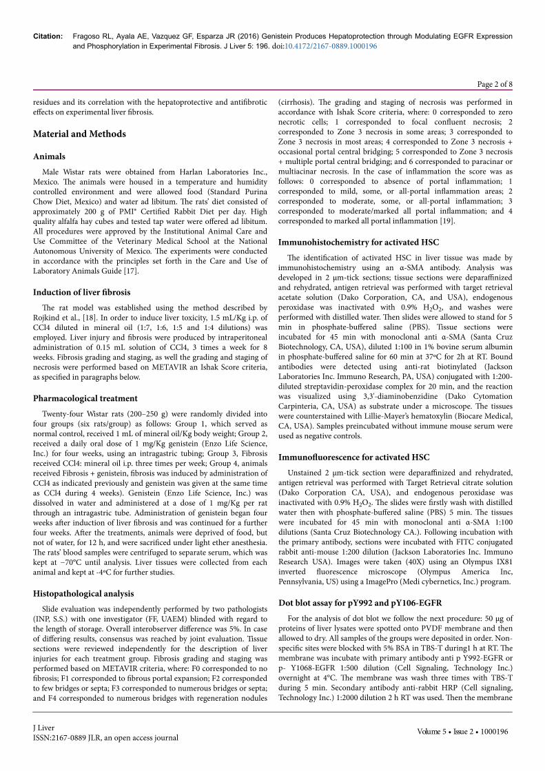
The Masson's trichrome staining of the liver was performed toassess collagen distribution (Figure 2). Animals treated with genisteinshowed the absence of fibrosis in parenchymal liver, and collagen wasobserved only surrounding vessels. However, animals with liverfibrosis exhibited extensive collagen depositions, which weresurrounded by regenerative nodules and collagen bridges between
portal spaces. Fibrosis was evaluated with Metavir criteria (Table 1).Animals with fibrosis had grading scores of F3 (1 animal) and F4 (4animals). However, animals with fibrosis treated with genistein showeda significant reduction in the amount of collagen in the liverparenchyma, and the collagen bridges were thinner; a minor numberof regenerative nodules was also observed. Treatment with genisteindecreased the degree of fibrosis; according to Metavir criteria, thegrading score was F3 for this group.
Figure 2: Effect of genistein on liver fibrosis. Liver tissue sectionsfrom: A) Control animal, is observed normal lobular architectureand a normal distribution of collagen (blue); B) Animal treated withgenistein, normal distribution and deposition of collagen; C)Animal with fibrosis, is observed extensive collagen deposition inparenchyma and formation of bridges of collagen between portalspace, is evident the presence of regenerative nodules; D) Animalwith fibrosis and treated with genistein, is observed a lessdeposition of collagen, there is not the presence of regenerativenodules. Liver tissue was stained with Masson trichrome, collagencan be recognized by blue staining. A representativemicrophotograph of each group was choose, magnification 20X.
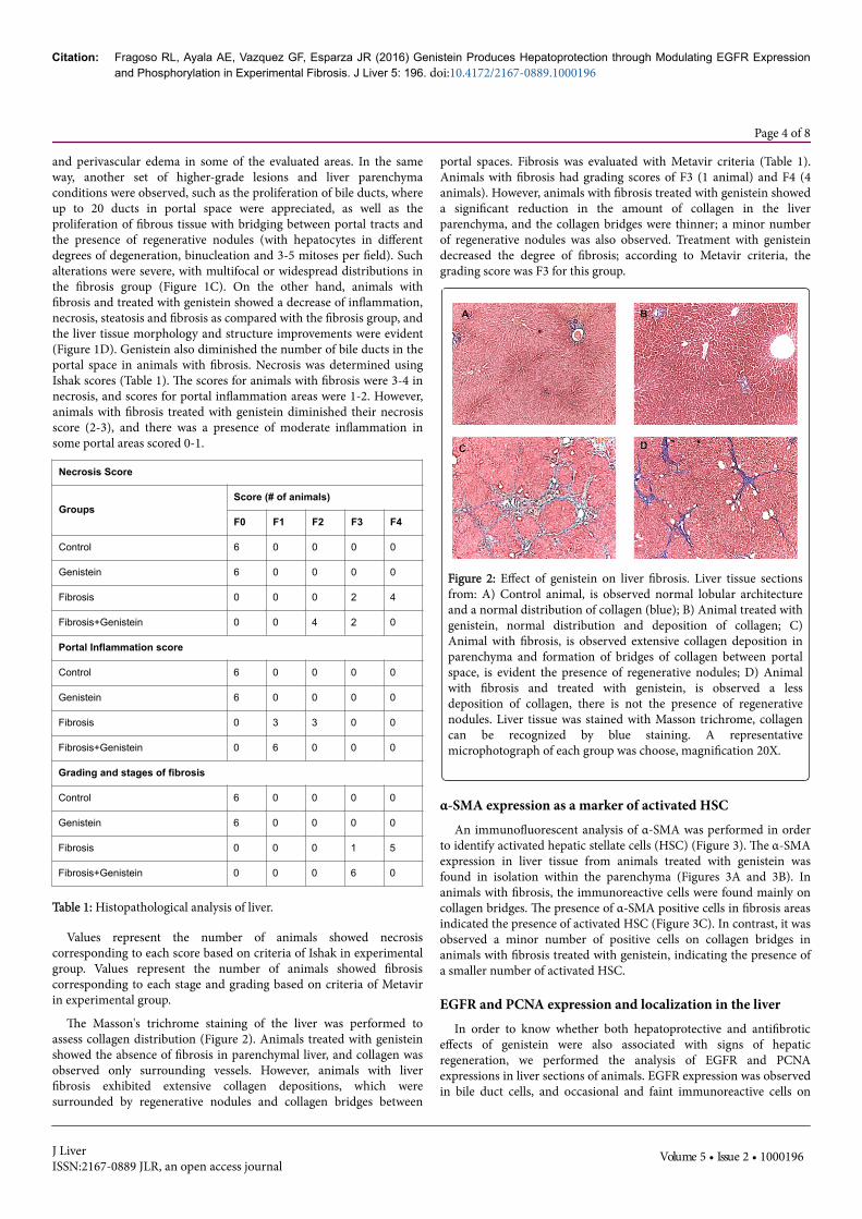
α-SMA expression as a marker of activated HSCAn immunofluorescent analysis of α-SMA was performed in order
to identify activated hepatic stellate cells (HSC) (Figure 3). The α-SMAexpression in liver tissue from animals treated with genistein wasfound in isolation within the parenchyma (Figures 3A and 3B). Inanimals with fibrosis, the immunoreactive cells were found mainly oncollagen bridges. The presence of α-SMA positive cells in fibrosis areasindicated the presence of activated HSC (Figure 3C). In contrast, it wasobserved a minor number of positive cells on collagen bridges inanimals with fibrosis treated with genistein, indicating the presence ofa smaller number of activated HSC.
EGFR and PCNA expression and localization in the liverIn order to know whether both hepatoprotective and antifibrotic
effects of genistein were also associated with signs of hepaticregeneration, we performed the analysis of EGFR and PCNAexpressions in liver sections of animals. EGFR expression was observedin bile duct cells, and occasional and faint immunoreactive cells on
Citation: Fragoso RL, Ayala AE, Vazquez GF, Esparza JR (2016) Genistein Produces Hepatoprotection through Modulating EGFR Expressionand Phosphorylation in Experimental Fibrosis. J Liver 5: 196. doi:10.4172/2167-0889.1000196
Page 4 of 8
J LiverISSN:2167-0889 JLR, an open access journal
Volume 5 • Issue 2 • 1000196
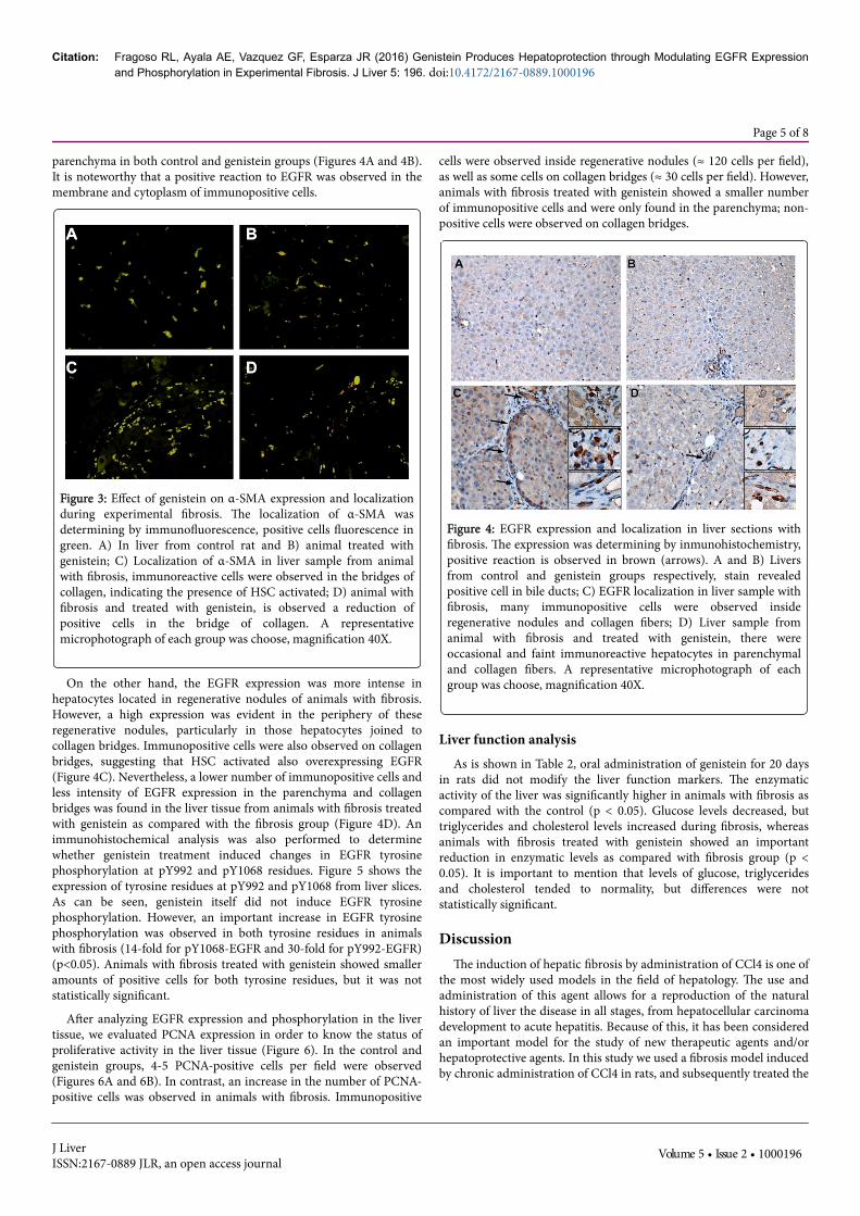
parenchyma in both control and genistein groups (Figures 4A and 4B).It is noteworthy that a positive reaction to EGFR was observed in themembrane and cytoplasm of immunopositive cells.
Figure 3: Effect of genistein on α-SMA expression and localizationduring experimental fibrosis. The localization of α-SMA wasdetermining by immunofluorescence, positive cells fluorescence ingreen. A) In liver from control rat and B) animal treated withgenistein; C) Localization of α-SMA in liver sample from animalwith fibrosis, immunoreactive cells were observed in the bridges ofcollagen, indicating the presence of HSC activated; D) animal withfibrosis and treated with genistein, is observed a reduction ofpositive cells in the bridge of collagen. A representativemicrophotograph of each group was choose, magnification 40X.
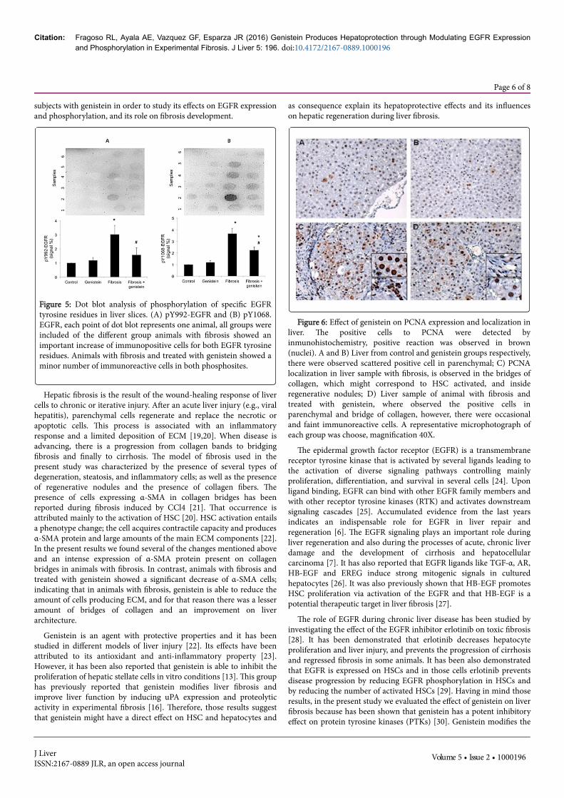
On the other hand, the EGFR expression was more intense inhepatocytes located in regenerative nodules of animals with fibrosis.However, a high expression was evident in the periphery of theseregenerative nodules, particularly in those hepatocytes joined tocollagen bridges. Immunopositive cells were also observed on collagenbridges, suggesting that HSC activated also overexpressing EGFR(Figure 4C). Nevertheless, a lower number of immunopositive cells andless intensity of EGFR expression in the parenchyma and collagenbridges was found in the liver tissue from animals with fibrosis treatedwith genistein as compared with the fibrosis group (Figure 4D). Animmunohistochemical analysis was also performed to determinewhether genistein treatment induced changes in EGFR tyrosinephosphorylation at pY992 and pY1068 residues. Figure 5 shows theexpression of tyrosine residues at pY992 and pY1068 from liver slices.As can be seen, genistein itself did not induce EGFR tyrosinephosphorylation. However, an important increase in EGFR tyrosinephosphorylation was observed in both tyrosine residues in animalswith fibrosis (14-fold for pY1068-EGFR and 30-fold for pY992-EGFR)(p<0.05). Animals with fibrosis treated with genistein showed smalleramounts of positive cells for both tyrosine residues, but it was notstatistically significant.
After analyzing EGFR expression and phosphorylation in the livertissue, we evaluated PCNA expression in order to know the status ofproliferative activity in the liver tissue (Figure 6). In the control andgenistein groups, 4-5 PCNA-positive cells per field were observed(Figures 6A and 6B). In contrast, an increase in the number of PCNA-positive cells was observed in animals with fibrosis. Immunopositive
cells were observed inside regenerative nodules (≈ 120 cells per field),as well as some cells on collagen bridges (≈ 30 cells per field). However,animals with fibrosis treated with genistein showed a smaller numberof immunopositive cells and were only found in the parenchyma; non-positive cells were observed on collagen bridges.
Figure 4: EGFR expression and localization in liver sections withfibrosis. The expression was determining by inmunohistochemistry,positive reaction is observed in brown (arrows). A and B) Liversfrom control and genistein groups respectively, stain revealedpositive cell in bile ducts; C) EGFR localization in liver sample withfibrosis, many immunopositive cells were observed insideregenerative nodules and collagen fibers; D) Liver sample fromanimal with fibrosis and treated with genistein, there wereoccasional and faint immunoreactive hepatocytes in parenchymaland collagen fibers. A representative microphotograph of eachgroup was choose, magnification 40X.
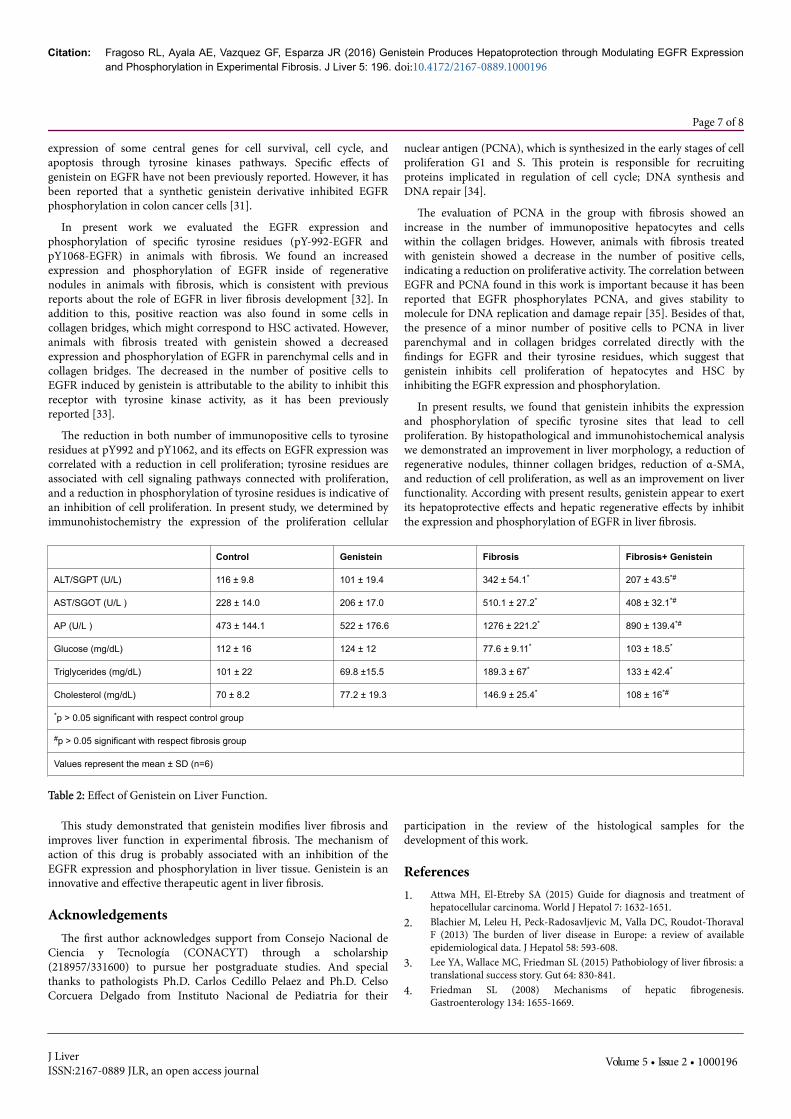
Liver function analysisAs is shown in Table 2, oral administration of genistein for 20 days
in rats did not modify the liver function markers. The enzymaticactivity of the liver was significantly higher in animals with fibrosis ascompared with the control (p < 0.05). Glucose levels decreased, buttriglycerides and cholesterol levels increased during fibrosis, whereasanimals with fibrosis treated with genistein showed an importantreduction in enzymatic levels as compared with fibrosis group (p <0.05). It is important to mention that levels of glucose, triglyceridesand cholesterol tended to normality, but differences were notstatistically significant.
DiscussionThe induction of hepatic fibrosis by administration of CCl4 is one of
the most widely used models in the field of hepatology. The use andadministration of this agent allows for a reproduction of the naturalhistory of liver the disease in all stages, from hepatocellular carcinomadevelopment to acute hepatitis. Because of this, it has been consideredan important model for the study of new therapeutic agents and/orhepatoprotective agents. In this study we used a fibrosis model inducedby chronic administration of CCl4 in rats, and subsequently treated the
Citation: Fragoso RL, Ayala AE, Vazquez GF, Esparza JR (2016) Genistein Produces Hepatoprotection through Modulating EGFR Expressionand Phosphorylation in Experimental Fibrosis. J Liver 5: 196. doi:10.4172/2167-0889.1000196
Page 5 of 8
J LiverISSN:2167-0889 JLR, an open access journal
Volume 5 • Issue 2 • 1000196
subjects with genistein in order to study its effects on EGFR expressionand phosphorylation, and its role on fibrosis development.
Figure 5: Dot blot analysis of phosphorylation of specific EGFRtyrosine residues in liver slices. (A) pY992-EGFR and (B) pY1068.EGFR, each point of dot blot represents one animal, all groups wereincluded of the different group animals with fibrosis showed animportant increase of immunopositive cells for both EGFR tyrosineresidues. Animals with fibrosis and treated with genistein showed aminor number of immunoreactive cells in both phosphosites.
Hepatic fibrosis is the result of the wound-healing response of livercells to chronic or iterative injury. After an acute liver injury (e.g., viralhepatitis), parenchymal cells regenerate and replace the necrotic orapoptotic cells. This process is associated with an inflammatoryresponse and a limited deposition of ECM [19,20]. When disease isadvancing, there is a progression from collagen bands to bridgingfibrosis and finally to cirrhosis. The model of fibrosis used in thepresent study was characterized by the presence of several types ofdegeneration, steatosis, and inflammatory cells; as well as the presenceof regenerative nodules and the presence of collagen fibers. Thepresence of cells expressing α-SMA in collagen bridges has beenreported during fibrosis induced by CCl4 [21]. That occurrence isattributed mainly to the activation of HSC [20]. HSC activation entailsa phenotype change; the cell acquires contractile capacity and producesα-SMA protein and large amounts of the main ECM components [22].In the present results we found several of the changes mentioned aboveand an intense expression of α-SMA protein present on collagenbridges in animals with fibrosis. In contrast, animals with fibrosis andtreated with genistein showed a significant decrease of α-SMA cells;indicating that in animals with fibrosis, genistein is able to reduce theamount of cells producing ECM, and for that reason there was a lesseramount of bridges of collagen and an improvement on liverarchitecture.
Genistein is an agent with protective properties and it has beenstudied in different models of liver injury [22]. Its effects have beenattributed to its antioxidant and anti-inflammatory property [23].However, it has been also reported that genistein is able to inhibit theproliferation of hepatic stellate cells in vitro conditions [13]. This grouphas previously reported that genistein modifies liver fibrosis andimprove liver function by inducing uPA expression and proteolyticactivity in experimental fibrosis [16]. Therefore, those results suggestthat genistein might have a direct effect on HSC and hepatocytes and
as consequence explain its hepatoprotective effects and its influenceson hepatic regeneration during liver fibrosis.
Figure 6: Effect of genistein on PCNA expression and localization inliver. The positive cells to PCNA were detected byinmunohistochemistry, positive reaction was observed in brown(nuclei). A and B) Liver from control and genistein groups respectively,there were observed scattered positive cell in parenchymal; C) PCNAlocalization in liver sample with fibrosis, is observed in the bridges ofcollagen, which might correspond to HSC activated, and insideregenerative nodules; D) Liver sample of animal with fibrosis andtreated with genistein, where observed the positive cells inparenchymal and bridge of collagen, however, there were occasionaland faint immunoreactive cells. A representative microphotograph ofeach group was choose, magnification 40X.
The epidermal growth factor receptor (EGFR) is a transmembranereceptor tyrosine kinase that is activated by several ligands leading tothe activation of diverse signaling pathways controlling mainlyproliferation, differentiation, and survival in several cells [24]. Uponligand binding, EGFR can bind with other EGFR family members andwith other receptor tyrosine kinases (RTK) and activates downstreamsignaling cascades [25]. Accumulated evidence from the last yearsindicates an indispensable role for EGFR in liver repair andregeneration [6]. The EGFR signaling plays an important role duringliver regeneration and also during the processes of acute, chronic liverdamage and the development of cirrhosis and hepatocellularcarcinoma [7]. It has also reported that EGFR ligands like TGF-α, AR,HB-EGF and EREG induce strong mitogenic signals in culturedhepatocytes [26]. It was also previously shown that HB-EGF promotesHSC proliferation via activation of the EGFR and that HB-EGF is apotential therapeutic target in liver fibrosis [27].
The role of EGFR during chronic liver disease has been studied byinvestigating the effect of the EGFR inhibitor erlotinib on toxic fibrosis[28]. It has been demonstrated that erlotinib decreases hepatocyteproliferation and liver injury, and prevents the progression of cirrhosisand regressed fibrosis in some animals. It has been also demonstratedthat EGFR is expressed on HSCs and in those cells erlotinib preventsdisease progression by reducing EGFR phosphorylation in HSCs andby reducing the number of activated HSCs [29]. Having in mind thoseresults, in the present study we evaluated the effect of genistein on liverfibrosis because has been shown that genistein has a potent inhibitoryeffect on protein tyrosine kinases (PTKs) [30]. Genistein modifies the
Citation: Fragoso RL, Ayala AE, Vazquez GF, Esparza JR (2016) Genistein Produces Hepatoprotection through Modulating EGFR Expressionand Phosphorylation in Experimental Fibrosis. J Liver 5: 196. doi:10.4172/2167-0889.1000196
Page 6 of 8
J LiverISSN:2167-0889 JLR, an open access journal
Volume 5 • Issue 2 • 1000196
expression of some central genes for cell survival, cell cycle, andapoptosis through tyrosine kinases pathways. Specific effects ofgenistein on EGFR have not been previously reported. However, it hasbeen reported that a synthetic genistein derivative inhibited EGFRphosphorylation in colon cancer cells [31].
In present work we evaluated the EGFR expression andphosphorylation of specific tyrosine residues (pY-992-EGFR andpY1068-EGFR) in animals with fibrosis. We found an increasedexpression and phosphorylation of EGFR inside of regenerativenodules in animals with fibrosis, which is consistent with previousreports about the role of EGFR in liver fibrosis development [32]. Inaddition to this, positive reaction was also found in some cells incollagen bridges, which might correspond to HSC activated. However,animals with fibrosis treated with genistein showed a decreasedexpression and phosphorylation of EGFR in parenchymal cells and incollagen bridges. The decreased in the number of positive cells toEGFR induced by genistein is attributable to the ability to inhibit thisreceptor with tyrosine kinase activity, as it has been previouslyreported [33].
The reduction in both number of immunopositive cells to tyrosineresidues at pY992 and pY1062, and its effects on EGFR expression wascorrelated with a reduction in cell proliferation; tyrosine residues areassociated with cell signaling pathways connected with proliferation,and a reduction in phosphorylation of tyrosine residues is indicative ofan inhibition of cell proliferation. In present study, we determined byimmunohistochemistry the expression of the proliferation cellular
nuclear antigen (PCNA), which is synthesized in the early stages of cellproliferation G1 and S. This protein is responsible for recruitingproteins implicated in regulation of cell cycle; DNA synthesis andDNA repair [34].
The evaluation of PCNA in the group with fibrosis showed anincrease in the number of immunopositive hepatocytes and cellswithin the collagen bridges. However, animals with fibrosis treatedwith genistein showed a decrease in the number of positive cells,indicating a reduction on proliferative activity. The correlation betweenEGFR and PCNA found in this work is important because it has beenreported that EGFR phosphorylates PCNA, and gives stability tomolecule for DNA replication and damage repair [35]. Besides of that,the presence of a minor number of positive cells to PCNA in liverparenchymal and in collagen bridges correlated directly with thefindings for EGFR and their tyrosine residues, which suggest thatgenistein inhibits cell proliferation of hepatocytes and HSC byinhibiting the EGFR expression and phosphorylation.
In present results, we found that genistein inhibits the expressionand phosphorylation of specific tyrosine sites that lead to cellproliferation. By histopathological and immunohistochemical analysiswe demonstrated an improvement in liver morphology, a reduction ofregenerative nodules, thinner collagen bridges, reduction of α-SMA,and reduction of cell proliferation, as well as an improvement on liverfunctionality. According with present results, genistein appear to exertits hepatoprotective effects and hepatic regenerative effects by inhibitthe expression and phosphorylation of EGFR in liver fibrosis.
Control Genistein Fibrosis Fibrosis+ Genistein
ALT/SGPT (U/L) 116 ± 9.8 101 ± 19.4 342 ± 54.1* 207 ± 43.5*#
AST/SGOT (U/L ) 228 ± 14.0 206 ± 17.0 510.1 ± 27.2* 408 ± 32.1*#
AP (U/L ) 473 ± 144.1 522 ± 176.6 1276 ± 221.2* 890 ± 139.4*#
Glucose (mg/dL) 112 ± 16 124 ± 12 77.6 ± 9.11* 103 ± 18.5*
Triglycerides (mg/dL) 101 ± 22 69.8 ±15.5 189.3 ± 67* 133 ± 42.4*
Cholesterol (mg/dL) 70 ± 8.2 77.2 ± 19.3 146.9 ± 25.4* 108 ± 16*#
*p > 0.05 significant with respect control group
#p > 0.05 significant with respect fibrosis group
Values represent the mean ± SD (n=6)
Table 2: Effect of Genistein on Liver Function.
This study demonstrated that genistein modifies liver fibrosis andimproves liver function in experimental fibrosis. The mechanism ofaction of this drug is probably associated with an inhibition of theEGFR expression and phosphorylation in liver tissue. Genistein is aninnovative and effective therapeutic agent in liver fibrosis.
AcknowledgementsThe first author acknowledges support from Consejo Nacional de
Ciencia y Tecnología (CONACYT) through a scholarship(218957/331600) to pursue her postgraduate studies. And specialthanks to pathologists Ph.D. Carlos Cedillo Pelaez and Ph.D. CelsoCorcuera Delgado from Instituto Nacional de Pediatria for their
participation in the review of the histological samples for thedevelopment of this work.
References1. Attwa MH, El-Etreby SA (2015) Guide for diagnosis and treatment of
hepatocellular carcinoma. World J Hepatol 7: 1632-1651.2. Blachier M, Leleu H, Peck-Radosavljevic M, Valla DC, Roudot-Thoraval
F (2013) The burden of liver disease in Europe: a review of availableepidemiological data. J Hepatol 58: 593-608.
3. Lee YA, Wallace MC, Friedman SL (2015) Pathobiology of liver fibrosis: atranslational success story. Gut 64: 830-841.
4. Friedman SL (2008) Mechanisms of hepatic fibrogenesis.Gastroenterology 134: 1655-1669.
Citation: Fragoso RL, Ayala AE, Vazquez GF, Esparza JR (2016) Genistein Produces Hepatoprotection through Modulating EGFR Expressionand Phosphorylation in Experimental Fibrosis. J Liver 5: 196. doi:10.4172/2167-0889.1000196
Page 7 of 8
J LiverISSN:2167-0889 JLR, an open access journal
Volume 5 • Issue 2 • 1000196
5. Seshacharyulu P, Ponnusamy MP, Haridas D, Jain M, Ganti AK, et al.(2012) Targeting the EGFR signaling pathway in cancer therapy. ExpertOpin Ther Targets 16: 15-31.
6. Komposch K, Sibilia M (2015) EGFR Signaling in Liver Diseases. Int JMol Sci 17.
7. Tanabe KK, Lemoine A, Finkelstein DM, Kawasaki H, Fujii T, et al. (2008)Epidermal growth factor gene functional polymorphism and the risk ofhepatocellular carcinoma in patients with cirrhosis. JAMA 299: 53-60.
8. Ganai AA, Farooqi H (2015) Bioactivity of genistein: A review of in vitroand in vivo studies. Biomed Pharmacother 76: 30-38.
9. Kim HJ, Ahn HS, Choi BH, Hahn SJ (2011) Inhibition of Kv4.3 bygenistein via a tyrosine phosphorylation-independent mechanism. Am JPhysiol Cell Physiol 300: C567-575.
10. Varinska L, Gal P, Mojzisova G, Mirossay L, Mojzis J (2015) Soy andbreast cancer: focus on angiogenesis. Int J Mol Sci 16: 11728-11749.
11. Dastjerdi MN, Kavoosi F, Valiani A, Esfandiari E, Sanaei M, et al. (2015)Inhibitory Effect of Genistein on PLC/PRF5 Hepatocellular CarcinomaCell Line. Int J Prev Med 6: 54.
12. Li Y, Luo Y, Zhang X, Lin X, He M, et al. (2013) Combined taurine,epigallocatechin gallate and genistein therapy reduces HSC-T6 cellproliferation and modulates the expression of fibrogenic factors. Int J MolSci 14: 20543-20554.
13. Saleh DO, Abdel Jaleel GA, El-Awdan SA, Oraby F, Badawi M (2014)Thioacetamide-induced liver injury: protective role of genistein. Can JPhysiol Pharmacol 92: 965-973.
14. Huang Q, Huang R, Zhang S, Lin J, Wei L, et al. (2013) Protective effect ofgenistein isolated from Hydrocotyle sibthorpioides on hepatic injury andfibrosis induced by chronic alcohol in rats. Toxicol Lett 217: 102-110.
15. Salas AL, Ocampo G, Fariña GG, Reyes-Esparza J, RodrÃguez-Fragoso L(2007) Genistein decreases liver fibrosis and cholestasis induced byprolonged biliary obstruction in the rat. Ann Hepatol 6: 41-47.
16. Salas AL, Montezuma TD, Fariña GG, Reyes-Esparza J, Rodríguez-Fragoso L (2008) Genistein modifies liver fibrosis and improves liverfunction by inducing uPA expression and proteolytic activity in CCl4-treated rats. Pharmacology 81: 41-49.
17. Committee on Care and Use of Laboratory Animals of the Institute forLaboratory Animal Resources, Commission on Life Sciences,NationalResearch Council (1996) Guide for the Care and Use of LaboratoryAnimals. Publication 86-23, Maryland US.
18. Rojkind M (1973) Inhibition of liver fibrosis by 1-azetidine-2-carboxylicacid in rats treated with carbon tetrachloride. J Clin Invest 52: 2451-2456.
19. Goodman ZD (2007) Grading and staging systems for inflammation andfibrosis in chronic liver diseases. J Hepatol 47: 598-607.
20. Hernandez-Gea V, Friedman SL (2011) Pathogenesis of liver fibrosis.Annu Rev Pathol 6: 425-456.
21. Fujii T, Fuchs BC, Yamada S, Lauwers GY, Kulu Y, et al. (2010) Mousemodel of carbon tetrachloride induced liver fibrosis: histopathologicalchanges and expression of CD133 and epidermal growth factor. BMCGastroenterol 10: 79.
22. Yalniz M, Bahcecioglu IH, Kuzu N, Poyrazoglu OK, Bulmus O, et al.(2007) Preventive role of genistein in an experimental non-alcoholicsteatohepatitis model. J Gastroenterol Hepatol 22: 2009-2014.
23. Kuzu N, Metin K, Dagli AF, Akdemir F, Orhan C, et al. (2007) Protectiverole of genistein in acute liver damage induced by carbon tetrachloride.Mediators Inflamm 2007: 36381.
24. Shi HY, Xu JW, Ren XX (2008) Effect of genistein on hepatic stellate cellproliferation and lipid peroxidation in vitro. Nan Fang Yi Ke Da Xue XueBao 28: 2066-2068.
25. Schneider MR, Wolf E (2009) The epidermal growth factor receptorligands at a glance. J Cell Physiol 218: 460-466.
26. Almendro V, Garcia-Recio S, Gascon P (2010) Tyrosine kinase receptortransactivation associated to G protein-coupled receptors. Curr DrugTargets 11: 1169-1180.
27. Collin de L'hortet A, Gilgenkrantz H, Guidotti JE (2012) EGFR: A MasterPiece in G1/S Phase Transition of Liver Regeneration. Int J Hepatol 2012:476910.
28. Zhang D, Zhang J, Jiang X, Li X, Wang Y, et al. (2014) Heparin-bindingepidermal growth factor-like growth factor: a hepatic stellate cellproliferation inducer via ErbB receptors. J Gastroenterol Hepatol 29:623-632.
29. Fuchs BC, Hoshida Y, Fujii T, Wei L, Yamada S, et al. (2014) Epidermalgrowth factor receptor inhibition attenuates liver fibrosis anddevelopment of hepatocellular carcinoma. Hepatology 59: 1577-1590.
30. Berasain C, Perugorria MJ, Latasa MU, Castillo J, Goñi S, et al. (2009) Theepidermal growth factor receptor: a link between inflammation and livercancer. Exp Biol Med (Maywood) 234: 713-725.
31. Pavese JM, Farmer RL, Bergan RC (2010) Inhibition of cancer cellinvasion and metastasis by genistein. Cancer Metastasis Rev 29: 465-482.
32. Gruca A, Krawczyk Z, Szeja W, Grynkiewicz G, Rusin A (2014) Syntheticgenistein glycosides inhibiting EGFR phosphorylation enhance the effectof radiation in HCT 116 colon cancer cells. Molecules 19: 18558-18573.
33. Perugorria MJ, Latasa MU, Nicou A, Cartagena-Lirola H, Castillo J, et al.(2008) The epidermal growth factor receptor ligand amphiregulinparticipates in the development of mouse liver fibrosis. Hepatology 48:1251-1261.
34. Moldovan GL, Pfander B, Jentsch S (2007) PCNA, the maestro of thereplication fork. Cell 129: 665-679.
35. Wang SC, Nakajima Y, Yu YL, Xia W, Chen CT, et al. (2006) Tyrosinephosphorylation controls PCNA function through protein stability. NatCell Biol 8: 1359-1368.
Citation: Fragoso RL, Ayala AE, Vazquez GF, Esparza JR (2016) Genistein Produces Hepatoprotection through Modulating EGFR Expressionand Phosphorylation in Experimental Fibrosis. J Liver 5: 196. doi:10.4172/2167-0889.1000196
Page 8 of 8
J LiverISSN:2167-0889 JLR, an open access journal
Volume 5 • Issue 2 • 1000196