Journal of Istanbul Veterınary Scıences An uncommon case ... · 73 of fractures by veterinarians...
6
72 Case Report Volume: 2, Issue: 3 December 2018 Pages: 72-77 An uncommon case: feline tail post-traumac osteomyelis Journal of Istanbul Veterınary Scıences Muhamed Kaca 1 , Žana Stanić 2 , Mehran Shafie 3 , Muamer Obhođaš 4 , Muhamed Očuz 5 1. Department of Pathophysiology, Veterinary Faculty, Sarajevo University, Bosnia and Herzegovina. 2. OBGY Clinic, Clinical Hospital Center Split, Croaa. 3. Internaonal Organizaon for Migraons, Zagreb, Croaa. 4. Surgery Clinic, Veterinary Faculty, Sarajevo University, Bosnia and Herzegovina. 5. Private Veterinary Medicine Pracce „Uvorići d.o.o.“ Visoko, Bosnia and Herzegovina. ABSTRACT Post-traumac osteomyelis (OM) is an uncommon event in cats, usually affecng distal phalanges of extremies. Tail injuries seldom cause bone infecon, but oſten result in neural damage with subsequent tail paralysis, and occasionally in urinary/fecal inconnence. We present a case of old stray cat which developed post-traumac tail OM, and endured it for years. It was an immuno-compromised, neglected, animal strongly infested with larvae of Aelurostrongylus abstrusus and oocistae of Isospora felis. Ulmately, it was treated by tail amputaon, with without any health consequences. Relevance and novel informaon: This report describes the management and outcome of a rare and a life-threatening case of feline post- traumac tail OM which was previously not reported in literature. Keywords: Cat, osteomyelis, tail, injury DOI: 10.30704/hp-www-jivs-net.438470 To cite this arcle: Kaca, M., Stanić, Ž., Shafie, M., Obhođaš, M., Očuz, M., (2018). An uncommon case: feline tail post- traumac osteomyelis. Journal of Istanbul Veterinary Sciences, 2(3), 72-77. Abbreviated Title: J Ist Vet Sci Introducon When it comes to the indoor/house cats, tail trauma is usually caused by accidents with doors (their tail caught in the door), people stepping on them inadvertently or by having their tails hardly pulled. In outdoor cats, tail trauma occurs frequently by scratches and bites from other animals, or, unfortunately, by malicious reasons (Šehić, 2000). Any soſt ssue damage can lead to adjacent bone infecon. Paralyzed tail is oſten soiled with urine and feces promong the infecon if the open wound is present. If the wound is located more towards the end of the tail, it is not exposed that much to urine and feces contaminaon so it usually heals spontaneously, parcularly if the injury is at the very end of the tail, on the last vertebrae. (Simonds, 2014). Posraumac OM is the most common type of OM in small animals. It is usually caused by bacteria, but fungal and viral eology have also been described (Bubenik, 2005). Its pathogenesis involves interacon of an infected wound, avascular bone, and suitable environment (Braden, 1991). Open reducon and internal fixaon *Corresponding Author: Muhamed Kaca E-mail: [email protected] Journal homepage: www.jivs.net hp://dergipark.gov.tr/hp-www-jivs-net Arcle History Received: 05.07.2018 Accepted: 05.10.2018 Available online: 09.10.2018 This work is licensed under the Creave Commons Aribuon 4.0 Internaonal License.
Transcript of Journal of Istanbul Veterınary Scıences An uncommon case ... · 73 of fractures by veterinarians...

72
Case Report
Volume: 2, Issue: 3 December 2018 Pages: 72-77
An uncommon case: feline tail post-traumatic osteomyelitis
Journal of Istanbul Veterınary Scıences
Muhamed Katica1, Žana Stanić2, Mehran Shafie3, Muamer Obhođaš4, Muhamed Očuz5
1. Department of Pathophysiology, Veterinary Faculty, Sarajevo University, Bosnia and Herzegovina. 2. OBGY Clinic, Clinical Hospital Center Split, Croatia. 3. International Organization for Migrations, Zagreb, Croatia. 4. Surgery Clinic, Veterinary Faculty, Sarajevo University, Bosnia and Herzegovina. 5. Private Veterinary Medicine Practice „Uvorići d.o.o.“ Visoko, Bosnia and Herzegovina. ABSTRACT
Post-traumatic osteomyelitis (OM) is an uncommon event in cats, usually affecting distal phalanges of extremities. Tail injuries seldom cause bone infection, but often result in neural damage with subsequent tail paralysis, and occasionally in urinary/fecal incontinence. We present a case of old stray cat which developed post-traumatic tail OM, and endured it for years. It was an immuno-compromised, neglected, animal strongly infested with larvae of Aelurostrongylus abstrusus and oocistae of Isospora felis. Ultimately, it was treated by tail amputation, with without any health consequences. Relevance and novel information: This report describes the management and outcome of a rare and a life-threatening case of feline post-traumatic tail OM which was previously not reported in literature. Keywords: Cat, osteomyelitis, tail, injury
DOI: 10.30704/http-www-jivs-net.438470
To cite this article: Katica, M., Stanić, Ž., Shafie, M., Obhođaš, M., Očuz, M., (2018). An uncommon case: feline tail post-traumatic osteomyelitis. Journal of Istanbul Veterinary Sciences, 2(3), 72-77. Abbreviated Title: J Ist Vet Sci
Introduction
When it comes to the indoor/house cats, tail trauma is usually caused by accidents with doors (their tail caught in the door), people stepping on them inadvertently or by having their tails hardly pulled. In outdoor cats, tail trauma occurs frequently by scratches and bites from other animals, or, unfortunately, by malicious reasons (Šehić, 2000). Any soft tissue damage can lead to adjacent bone infection. Paralyzed tail is often soiled with urine and feces promoting the infection if the open wound is
present. If the wound is located more towards the end of the tail, it is not exposed that much to urine and feces contamination so it usually heals spontaneously, particularly if the injury is at the very end of the tail, on the last vertebrae. (Simonds, 2014). Posttraumatic OM is the most common type of OM in small animals. It is usually caused by bacteria, but fungal and viral etiology have also been described (Bubenik, 2005). Its pathogenesis involves interaction of an infected wound, avascular bone, and suitable environment (Braden, 1991). Open reduction and internal fixation
*Corresponding Author: Muhamed Katica E-mail: [email protected]
Journal homepage: www.jivs.net http://dergipark.gov.tr/http-www-jivs-net
Article History
Received: 05.07.2018 Accepted: 05.10.2018 Available online: 09.10.2018
This work is licensed under the Creative Commons Attribution 4.0 International License.

73
of fractures by veterinarians cause most cases of osteomyelitis (Griffiths and Bellenger, 1979). Contaminated wounds to the limbs of dogs and cats have the potential to cause osteomyelitis. Bite wounds account for a significant portion of these traumatic wounds, especially in cats (Bubenik, 2005). Often the wound appears as a few small skin punctures caused by the teeth. However, the canine teeth, by crushing and tearing action, damage a considerable amount of muscle, tendon and subcutaneous tissue. Bite wounds are contaminated by microorganisms from the attacking animal’s oral flora, the victim’s skin bacteria and often soil organisms. This combination of bacterial contamination, devitalized parosteal tissue and periosteum, dead tissue and inflammatory exudate very likely leads to the development of infection (Johnson et.al., 2006). Skin puncture wounds can heal but the underlying infection can continue to develop to an abscess, either under the periosteum or as an extension to the periosteum from the adjacent subcutaneous tissue. Neglected bite wounds and abscesses, periodontal infection and chronic paronychia are common initiating cause of OM (Sia and Berbari, 2006). Normal, healthy bone in an immune-competent animal is highly resistant to infection, but injury and infection of adjacent soft tissue may induce a posttraumatic OM subsequent to a bite or scratch (Viskjer and Rapp, 2016). However, if the immune-competence of an injured animal is further compromised, as it is with many outdoor cats, the infection spreads easily affecting bone structures underneath inflamated soft tissue. Massive infestation with lung and bowel parasites like Aelurostrongylus abstrusus and Isospora felis, which were abundantly isolated from our patient, exhausts the immunity of the host , therefore facilitating development of OM (Traversa and Di Cesare, 2016). Most cases of osteomyelitis in dogs and cats are chronic at the time of diagnosis (Bubenik, 2005). The main intention of this case report is to present the successful, routine surgical treatment of OM on the cat's tail, what is yet to be described in the literature.
Case
According to oral cavity inspection, an 10-11 years old, 5 kg of body mass, male, domestic short-haired, cat was brought due to sanguinolent-purulent discarge of the tail (Figure 1 and Figure 2). It has been stated that the cat has been in this condition for at least three to four years. Firstly the
cat was admited to a private veterinary outpatient office, where osteomyelitis was diagnosed in the tail. The patient had no fever and the general condition was in good state. It was frightened and agressive, but with good appetite, and no substantial difficulties on physical examination. The open wound with substantial soft tissue defect was dirty, covered with pus and producing unpleasant smell. There was no obvious tail fracture. The perianal and periurethral area were unaffected with trauma. The sensory as much as urinary/fecal continence were preserved.
Figure 1. Street, short-haired cat with obvious tail injuries.
Figure 2. Aspect of long-lasting, untreated sanguinolent-purulent discarge of the tail.
Leukogram showed signs of leukocytosis, neutropenia, monocytosis, basophilia and eosinophilia (Table 1). The animal was
immunocompromized due to long-term chronical infectious disease.Coproculture revealed high degree
Katica et al. 2018 / Journal of Istanbul Veterinary Sciences. Volume 2, Issue 3, pp: 72-77
Table 1. Some hematological parameters of the cat Parameters Measured values Reference values*
RBC (1×10¹²/L) 9. 98 5 - 10
WBC (1×10⁹/L) 29.7 5.5 - 19.5 Platelets (1×10⁹/L) - 300- 800 Hematocrit (%) 39 30 - 45 Hemoglobin (g/L) 137 80 - 150 Neutrophil (%) 34 35 - 37 Lymphocyte (%) 39 20 - 50 Monocyte (%) 7 1 - 4 Eozinophil (%) 18 2 - 12 Basophil (%) 2 0 - 0.5
RBC = Red blood cell; WBC = White blood cell; * Blood & Studdert (1988); O’Brien et. al. (1998).
74
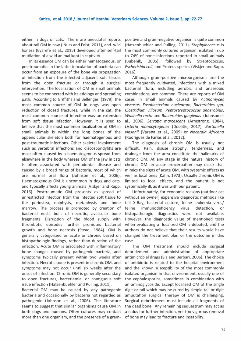
of infestation with larvae of Aelurostrongylus abstrusus and oocistae of Isospora felis. The wound was initially cleaned following the principles of management of dirty surgical wounds. The cat was premedicated with medetomidine (Domitor; Orion Pharma Animal Health) 0.08 mg/kg SC, meloxicam (Metacam; Boehringer Ingelheim Vetmedica) 0.2 mg/kg SC and methadone (Metadon Recip; Meda) 0.3 mg/kg. Ketamine (Ketaminol; Intervet) 5 mg/kg was administered intramuscularly to maintain a dissociative anesthesia. Oxygen was delivered by flow-by with the mask during the procedure. With the cat positioned in right lateral recumbency and the tail directed to the surgeon, the surgical site was lavaged with saline respecting the principles of sepsis and antisepsis. Before incising the skin the intervertebral spaces in front and after the chosen tail vertebra for transection were identified by digital palpation. Two v-shaped skin incisions are made at each side of the tail over the middle region of the vertebra with the tip of the v to the direction of the head of the animal and both dividing branches caudally one to the dorsum of the tail and the other to the ventral part (Figure 3).
Figure 3. The initial steps of the amputation of the tail, the aspect of the two cuts in the „V“ shape.
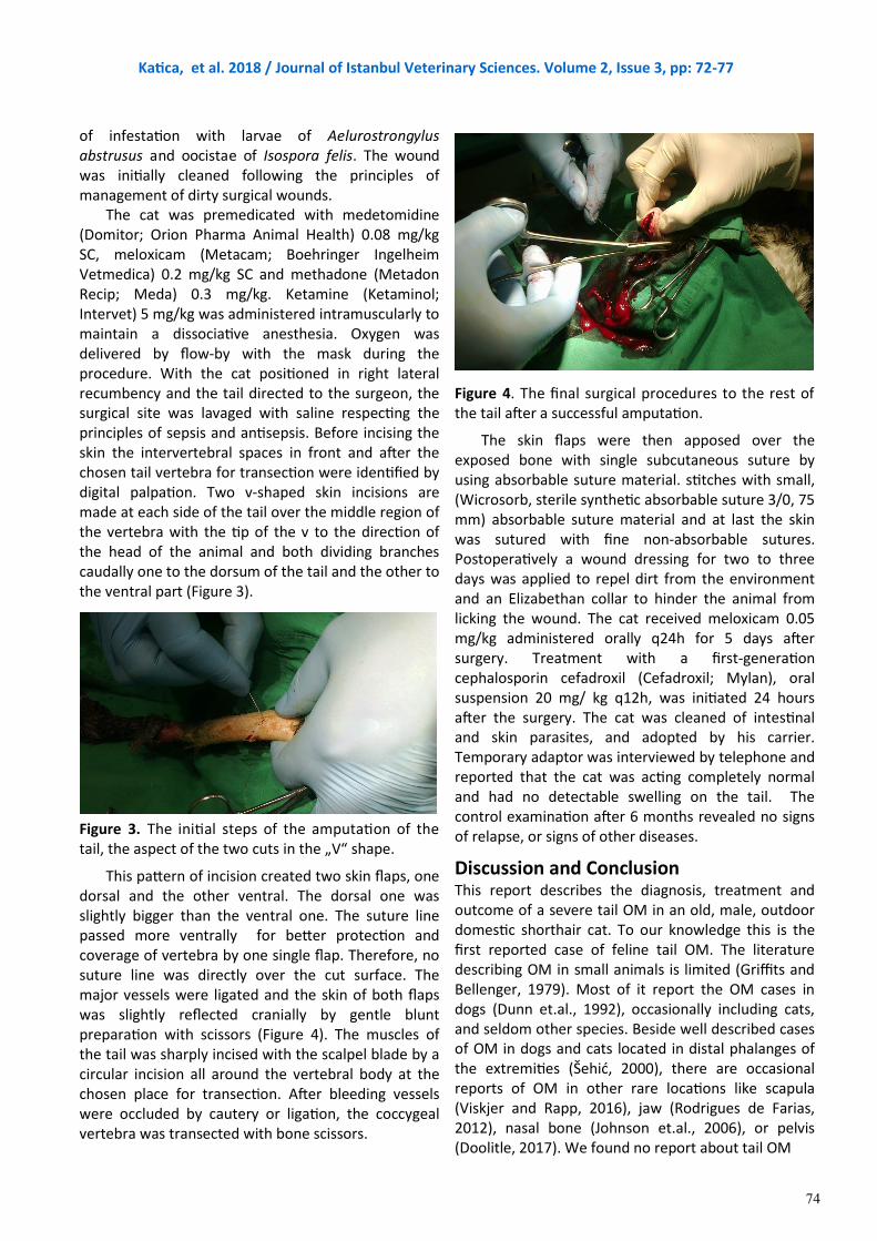
This pattern of incision created two skin flaps, one dorsal and the other ventral. The dorsal one was slightly bigger than the ventral one. The suture line passed more ventrally for better protection and coverage of vertebra by one single flap. Therefore, no suture line was directly over the cut surface. The major vessels were ligated and the skin of both flaps was slightly reflected cranially by gentle blunt preparation with scissors (Figure 4). The muscles of the tail was sharply incised with the scalpel blade by a circular incision all around the vertebral body at the chosen place for transection. After bleeding vessels were occluded by cautery or ligation, the coccygeal vertebra was transected with bone scissors.
Figure 4. The final surgical procedures to the rest of the tail after a successful amputation.
The skin flaps were then apposed over the exposed bone with single subcutaneous suture by using absorbable suture material. stitches with small, (Wicrosorb, sterile synthetic absorbable suture 3/0, 75 mm) absorbable suture material and at last the skin was sutured with fine non-absorbable sutures. Postoperatively a wound dressing for two to three days was applied to repel dirt from the environment and an Elizabethan collar to hinder the animal from licking the wound. The cat received meloxicam 0.05 mg/kg administered orally q24h for 5 days after surgery. Treatment with a first-generation cephalosporin cefadroxil (Cefadroxil; Mylan), oral suspension 20 mg/ kg q12h, was initiated 24 hours after the surgery. The cat was cleaned of intestinal and skin parasites, and adopted by his carrier. Temporary adaptor was interviewed by telephone and reported that the cat was acting completely normal and had no detectable swelling on the tail. The control examination after 6 months revealed no signs of relapse, or signs of other diseases.
Discussion and Conclusion This report describes the diagnosis, treatment and outcome of a severe tail OM in an old, male, outdoor domestic shorthair cat. To our knowledge this is the first reported case of feline tail OM. The literature describing OM in small animals is limited (Griffits and Bellenger, 1979). Most of it report the OM cases in dogs (Dunn et.al., 1992), occasionally including cats, and seldom other species. Beside well described cases of OM in dogs and cats located in distal phalanges of the extremities (Šehić, 2000), there are occasional reports of OM in other rare locations like scapula (Viskjer and Rapp, 2016), jaw (Rodrigues de Farias, 2012), nasal bone (Johnson et.al., 2006), or pelvis (Doolitle, 2017). We found no report about tail OM
Katica, et al. 2018 / Journal of Istanbul Veterinary Sciences. Volume 2, Issue 3, pp: 72-77
75
either in dogs or cats. There are anecdotal reports about tail OM in cow ( Nuss and Feist, 2011), and wild lioness (Eyarefe et al., 2015) developed after self-tail mutilation of a wild animal kept in captivity. In its essence OM can be either hematogenous, or posttraumatic. In the latter inoculation of bacteria can occur from an exposure of the bone via propagation of infection from the infected adjacent soft tissue, from the open fracture or through a surgical intervention. The localization of OM in small animals seems to be connected with its etiology and spreading path. According to Griffiths and Bellenger, (1979), the most common source of OM in dogs was open reduction of closed fractures, while in the cat, the most common source of infection was an extension from soft tissue infection. However, it is used to believe that the most common localization of OM in small animals is within the long bones of the appendicular skeleton both for haematogenous and post-traumatic infections. Other skeletal involvement such as vertebral infections and discospondylitis are most often caused by a haematogenous spread from elsewhere in the body whereas OM of the jaw in cats is often associated with periodontal disease and caused by a broad range of bacteria, most of which are normal oral flora (Johnson et al., 2006). Haematogenous OM is uncommon in dogs and cats, and typically affects young animals (Viskjer and Rapp, 2016). Posttraumatic OM presents as spread of unrestricted infection from the infected soft tissue to the periostea, epiphysis, metaphysis and bone marrow. The process is promoted by creation of bacterial nests built of necrotic, avascular bone fragments. Disruption of the blood supply with thrombotic episodes further promotes bacterial growth and bone necrosis (Stead, 1984). OM is generally categorized as acute or chronic based on histopathologic findings, rather than duration of the infection. Acute OM is associated with inflammatory bone changes caused by pathogenic bacteria, and symptoms typically present within two weeks after infection. Necrotic bone is present in chronic OM, and symptoms may not occur until six weeks after the onset of infection. Chronic OM is generally secondary to open fractures, bacteriemia, or contiguous soft issue infection (Hatzenbuehler and Pulling, 2011). Bacterial OM may be caused by any pathogenic bacteria and occasionally by bacteria not regarded as pathogenic (Johnson et al., 2006). The literature seems to suggest that similar organisms cause OM in both dogs and humans. Often cultures may contain more than one organism, and the presence of a gram-
positive and gram-negative organism is quite common (Hatzenbuehler and Pulling, 2011). Staphylococcus is the most commonly cultured organism, isolated in up to 74% of bone infections reported in small animals (Bubenik, 2005), followed by Streptococcus, Escherichia coli, and Proteus species (Viskjer and Rapp, 2016). Although gram-positive microorganisms are the most frequently cultivated, infections with a mixed bacterial flora, including aerobic and anaerobic combinations, are common. There are reports of OM cases in small animals caused by Actinomyces viscosus, Fusobacterium nucleatum, Bacteroides spp, Clostridium villosum, Peptostreptococcus anaerobius, Wolinella recta and Bacteroides gingivalis (Johnson et al., 2006), Serratia marcescens (Armstrong, 1984), Listeria monocytogenes (Doolitle, 2017), Bartonella vinsonii (Varana et al., 2009) or Nocardia Africana (Rodrigues de Farias et al., 2012). The diagnosis of chronic OM is usually not difficult. Pain, disuse atrophy, tenderness, and drainage from the area constitute the hallmarks of chronic OM. At any stage in the natural history of chronic OM an acute exacerbation may occur that mimics the signs of acute OM, with systemic effects as well as local ones (Kahn, 1973). Usually chronic OM is limited to local effects, and the patient is not systemically ill, as it was with our patient. Unfortunately, for economic reasons (outdoor cat without an owner) expensive diagnostic methods like tail X-Ray, bacterial culture, feline leukemia virus/feline immunodeficiency virus detection, or histopathologic diagnostics were not available. However, the diagnostic value of mentioned tests when evaluating a localized OM is debated, and the authors do not believe that their results would have changed the treatment plan or the outcome in this case. The OM treatment should include surgical debridement and administration of appropriate antimicrobial drugs (Sia and Berbari, 2006). The choice of antibiotic is related to the hospital environment and the known susceptibility of the most commonly isolated organism in that environment; usually one of the cephalosporins, sometimes in combination with an aminoglycoside. Except localized OM of the single digit or tail which may be cured by simple tail or digit amputation surgical therapy of OM is challenging. Surgical debridement must include all fragments of the dead bone. Any remaining sequestrum may act as a nidus for further infection, yet too vigorous removal of bone may lead to fracture and instability.
Katica, et al. 2018 / Journal of Istanbul Veterinary Sciences. Volume 2, Issue 3, pp: 72-77
76
This represents one of the major problems in dealing with chronic OM. The greatest chance for the healing of the infection is through massive removal of dead bone; the greatest chance for nonunion or refracture is also related to massive removal of bone. Fortunately, to our patient, surgical treatment was quite simple. Parasitary infestation reduces the body’s healing response and contributes to chronically open wounds and subsequent soft tissue infection what is supported by frequent presence of eosinophilia in cases with OM developed from neglected fight wounds (Dehghani and Hajighahramani, 2005). These conditions may act synergistically to significantly increase the risk of OM in these patients (Hatzenbuehler and Pulling, 2011). The true prevalence of A. abstrusus infection is unknown. One study in California reported a prevalence of 1.9%, and a second study in Alabama reported a prevalence of 18.5% among stray cats euthanized at a humane shelter (Ellis, 2010). A study in Croatia reported prevalence's, based on histopathologic diagnosis, ranging from 3.9% to 22%, depending on geographic region (Grabarević, 1999). The most frequent signs of Aelurostrongylosis are mild to intense coughing, wheezing, sneezing, nasal discharge, dyspnea and tachypnea. Generalized signs such as lethargy and weight loss have also been described. Severe signs such as open mouth abdominal breathing, tachycardia
and even death may occur in young, debilitated and/or immunosuppressed cats. Aelurostrongylosis has furthermore been implicated in cases of anesthetic-associated deaths, likely due to the decreased surface area for gas exchange and a combination of impaired lung perfusion and ventilation, hypoxia and systemic hypotension, culminating in cardiovascular collapse (Traversa and Di Cesare, 2016). In severely infected, and especially if additionally fractured tails in small animals, it is often recommended to amputate the tail to ultimately cure the locus of permanent infection, and to prevent further injury to the nerves that supply the urethra and anus. After traumatic tail injury, as much as after tail amputation the cat will need to rest in a crate or cage. Cats use their tails for balance and navigation, but with a little adjustment they can learn to manage without their tails, and to function without major handicappe (Simonds, 2014). The causal agent of the injury in this case was not defined. The presence of numerous population of stray dogs in this area (Katica et al., 2017), who present primary predators for the outdoor cats could be the answer, as well as territorial conflict with other stray cats. Malicious events with people or motor vehicle accidents could also play a role in this injury. The patient was successfully treated with simple tail amputation with full recovery.
References
Armstrong, P. J. (1984). Systemic Serratia marcescens infections in a dog and a cat. Journal of the American Veterinary Medical Association,184(9), 1154-1158.
Blood, D. C. & Studdert, V. P. (1988). Bailliere’s Comprehensive Veterinary Dictionary. London, UK: Bailliere Tindall.
Braden, T. D. (1991). Posttraumatic osteomyelitis. Vetinary Clinics of North America: Small Anim Practice, 21(4), 781-811.
Bubenik, L. J. (2005). Infections of the skeletal system Vetinary Clinics of North America: Small Anim Practice, 35, 1039–1049.
De Farias, M. R., Werner, J., Ribeiro, M. G., Rodigheri, S. M., Cavalcante, C. Z., Chi, K. D., Condas, L. A. Z., Gonoi, T., Matsuzama, T. & Yazama, K. (2012). Uncommon mandibular osteomyelitis in cat caused by Nocardia Africana. BMC Veterinary Research, 8, 239.
Dehghani, S. N. & Hajighahramani, S. (2005). Eosinophilia due to osteomyelitis in a dog. Journal of Veterinary Science, 6(3), 255–257.
Doolitle, J. (2017). Osteomyelitis and septicemia secondary
to Listeria Monocytogenes in a young dog. Journal of Dairy, Veterinary & Animal Research, 6(1), 00170.
Dunn, J. K., Dennis, R. & Houlton, J. E. F. (1992). Successful treatment of two cases of metaphyseal osteomyelitis in the dog. Journal of Smal Animal Practice, 33(2), 85-89.
Ellis, A. E, Brown, C. A. & Yabsley, M. J. (2010). Aelurostrongylus abstrusus larvae in the colon of two cats. Journal of Veterinary Diagnostic Investigation, 22(4), 652-655.
Eyarefe, O. D., Oguntoye, Olusa T. A. & Morenikeji, O. A. (2015). Lifesaving tail amputation in an African lioness (Panthera leo L) in captivity. Sokoto Journal of Veterinary Sciences, 13(2), 61-64.
Grabarevic, Z., Curic, S., Tustonja, A., Artukovic, B., Šimec, Z., Ramadan, K. & Zivicnjak T.(1999). Incidence and regional distribution of the lungworm Aelurostrongylus abstrusus in cats in Croatia. Veterinarski Arhiv, 69, 279–287.
Griffiths, G. L. & Bellenger, C. R. (1979). A retrospective study of osteomyelitis in dogs and cats. Australian Veterinary Journal, 55(12), 587-591.
Hatzenbuehler, J. & Pulling, T. J. (2011). Diagnosis and management of osteomyelitis. American Family
Katica, et al. 2018 / Journal of Istanbul Veterinary Sciences. Volume 2, Issue 3, pp: 72-77
77
Johnson, K. A., Lomas, G. R. & Wood, A. K. W. (2006). Osteomyelitis in dogs and cats caused by anaerobic bacteria. Australian Veterinary Journal, 61, 57-61.
Kahn, D. S. & Pritzker, K. P. H. (1973). The pathophysiology of bone infection. Clinical Orthopaedics and Related Research, 96, 12.
Katica, M., Obradovic, Z., Gradascevic, N., Hadzimusic, N., Mujkanovic, R., Mestric, E., Bašić, B., Toplaović, B., Smajkić, R., Dupovac, D. & Celebicic, M. (2017). Assessment of the effect of stray dogs as a risk factor for the health of population in certain areas of Bosnia and Herzegovina. European Journal of Biomedical and Pharmaceutical Sciences, 4(9), 107-111.
Nuss, K. & Feist, M. (2011). Tail amputation for treatment of osteomyelitis of the first and second coccygeal vertebrae in a cow. Tierarztliche Praxis, 39(3), 176-178.
O’Brien, M., Murphy, M. G. & Lowe, J. A. (1998). Hematology and clinical chemistry parameters in the cat (Felis domesticus). Journal of Nutrition, 128, 2678S–2679S.
Šehić, M. (2000). Osteoarthropatije u domaćih životinja. Zagreb, Hrvatska: Skaner Studio.
Sia, I. G. & Berbari, E. F. (2006). Infection and
musculoskeletal conditions: Osteomyelitis. Best Practice & Research: Clinical Rheumatology, 20(6), 1065-1081.
Simons, M. C., Ben-Amotz, R. & Popovitch, C. (2014). Post-operative complications and owner satisfaction following partial caudectomies: 22 cases (2008 to 2013). Journal of Small Animal Practice, 55(10), 509-514.
Stead, A. C. (1984). Osteomyelitis in the dog and cat. Journal of Small Animal Practice, 25, 1-13.
Traversa, D. & Di Cesare, A. (2016). Diagnosis and management of lungworm infections in cats: Cornerstones, dilemmas and new avenues. Journal of Feline Medicine & Surgery, 18, 7-20.
Varanat, M., Travis, A., Lee, W., Maggi, R. G., Bissett, S. A., Linder, K. E. & Breitschwerdt E. B. (2009). Recurrent osteomyelitis in a cat due to infection with Bartonella Vinsonii subsp. Berkhoffii genotype II. Journal of Veterinary Internal Medicine, 23(6), 1273-1277.
Viskjer, S. & Rapp, M. (2016). Scapular osteomyelitis in an immature domestic shorthair cat. Journal of feline medicine and surgery open reports, 2(2), 2055116916668199.
Katica, et al. 2018 / Journal of Istanbul Veterinary Sciences. Volume 2, Issue 3, pp: 72-77