Journal of Hematology & Oncology - University of...
16
BioMed Central Page 1 of 16 (page number not for citation purposes) Journal of Hematology & Oncology Open Access Research Phenotype and functional evaluation of ex vivo generated antigen-specific immune effector cells with potential for therapeutic applications Shuhong Han 1 , Yuju Huang 2 , Yin Liang 2 , Yuchin Ho 2 , Yichen Wang 2 and Lung-Ji Chang* 1 Address: 1 Department of Molecular Genetics and Microbiology, College of Medicine, University of Florida, Gainesville, FL 32610-0266 and 2 Vectorite Biomedica, Inc., Taipei, Taiwan, Republic of China Email: Shuhong Han - [email protected]; Yuju Huang - [email protected]; Yin Liang - [email protected]; Yuchin Ho - [email protected]; Yichen Wang - [email protected]; Lung-Ji Chang* - [email protected] * Corresponding author Abstract Ex vivo activation and expansion of lymphocytes for adoptive cell therapy has demonstrated great success. To improve safety and therapeutic efficacy, increased antigen specificity and reduced non- specific response of the ex vivo generated immune cells are necessary. Here, using a complete protein-spanning pool of pentadecapeptides of the latent membrane protein 2A (LMP2A) of Epstein-Barr virus (EBV), a weak viral antigen which is associated with EBV lymphoproliferative diseases, we investigated the phenotype and function of immune effector cells generated based on IFN-γ or CD137 activation marker selection and dendritic cell (DC) activation. These ex vivo prepared immune cells exhibited a donor- and antigen-dependent T cell response; the IFN-γ- selected immune cells displayed a donor-related CD4- or CD8-dominant T cell phenotype; however, the CD137-enriched cells showed an increased ratio of CD4 T cells. Importantly, the pentadecapeptide antigens accessed both class II and class I MHC antigen processing machineries and effectively activated EBV-specific CD4 and CD8 T cells. Phenotype and kinetic analyses revealed that the IFN-γ and the CD137 selections enriched more central memory T (Tcm) cells than did the DC-activation approach, and after expansion, the IFN-γ-selected effector cells showed the highest level of antigen-specificity and effector activities. While all three approaches generated immune cells with comparable antigen-specific activities, the IFN-γ selection followed by ex vivo expansion produced high quality and quantity of antigen-specific effector cells. Our studies presented the optimal approach for generating therapeutic immune cells with potential for emergency and routine clinical applications. Background The key to the success of immune cell therapy is to increase specific and decrease non-specific immune response. Ex vivo expanded antigen-specific T cells target- ing cytomegalovirus (CMV), Epstein-Barr virus (EBV) and adenovirus have been successfully applied to treating hematopoietic stem cell or solid organ transplant patients who have developed post-transplant viral diseases. [1,2] The labor-intensive and time-consuming process of isolat- ing and expanding antigen-specific immune cells, how- Published: 6 August 2009 Journal of Hematology & Oncology 2009, 2:34 doi:10.1186/1756-8722-2-34 Received: 30 June 2009 Accepted: 6 August 2009 This article is available from: http://www.jhoonline.org/content/2/1/34 © 2009 Han et al; licensee BioMed Central Ltd. This is an Open Access article distributed under the terms of the Creative Commons Attribution License (http://creativecommons.org/licenses/by/2.0 ), which permits unrestricted use, distribution, and reproduction in any medium, provided the original work is properly cited.
Transcript of Journal of Hematology & Oncology - University of...
BioMed Central
Journal of Hematology & Oncology
ss
Open AcceResearchPhenotype and functional evaluation of ex vivo generated antigen-specific immune effector cells with potential for therapeutic applicationsShuhong Han1, Yuju Huang2, Yin Liang2, Yuchin Ho2, Yichen Wang2 and Lung-Ji Chang*1Address: 1Department of Molecular Genetics and Microbiology, College of Medicine, University of Florida, Gainesville, FL 32610-0266 and 2Vectorite Biomedica, Inc., Taipei, Taiwan, Republic of China
Email: Shuhong Han - [email protected]; Yuju Huang - [email protected]; Yin Liang - [email protected]; Yuchin Ho - [email protected]; Yichen Wang - [email protected]; Lung-Ji Chang* - [email protected]
* Corresponding author
AbstractEx vivo activation and expansion of lymphocytes for adoptive cell therapy has demonstrated greatsuccess. To improve safety and therapeutic efficacy, increased antigen specificity and reduced non-specific response of the ex vivo generated immune cells are necessary. Here, using a completeprotein-spanning pool of pentadecapeptides of the latent membrane protein 2A (LMP2A) ofEpstein-Barr virus (EBV), a weak viral antigen which is associated with EBV lymphoproliferativediseases, we investigated the phenotype and function of immune effector cells generated based onIFN-γ or CD137 activation marker selection and dendritic cell (DC) activation. These ex vivoprepared immune cells exhibited a donor- and antigen-dependent T cell response; the IFN-γ-selected immune cells displayed a donor-related CD4- or CD8-dominant T cell phenotype;however, the CD137-enriched cells showed an increased ratio of CD4 T cells. Importantly, thepentadecapeptide antigens accessed both class II and class I MHC antigen processing machineriesand effectively activated EBV-specific CD4 and CD8 T cells. Phenotype and kinetic analysesrevealed that the IFN-γ and the CD137 selections enriched more central memory T (Tcm) cellsthan did the DC-activation approach, and after expansion, the IFN-γ-selected effector cells showedthe highest level of antigen-specificity and effector activities. While all three approaches generatedimmune cells with comparable antigen-specific activities, the IFN-γ selection followed by ex vivoexpansion produced high quality and quantity of antigen-specific effector cells. Our studiespresented the optimal approach for generating therapeutic immune cells with potential foremergency and routine clinical applications.
BackgroundThe key to the success of immune cell therapy is toincrease specific and decrease non-specific immuneresponse. Ex vivo expanded antigen-specific T cells target-ing cytomegalovirus (CMV), Epstein-Barr virus (EBV) and
adenovirus have been successfully applied to treatinghematopoietic stem cell or solid organ transplant patientswho have developed post-transplant viral diseases. [1,2]The labor-intensive and time-consuming process of isolat-ing and expanding antigen-specific immune cells, how-
Published: 6 August 2009
Journal of Hematology & Oncology 2009, 2:34 doi:10.1186/1756-8722-2-34
Received: 30 June 2009Accepted: 6 August 2009
This article is available from: http://www.jhoonline.org/content/2/1/34
© 2009 Han et al; licensee BioMed Central Ltd. This is an Open Access article distributed under the terms of the Creative Commons Attribution License (http://creativecommons.org/licenses/by/2.0), which permits unrestricted use, distribution, and reproduction in any medium, provided the original work is properly cited.
Page 1 of 16(page number not for citation purposes)

Journal of Hematology & Oncology 2009, 2:34 http://www.jhoonline.org/content/2/1/34
ever, has hindered common practice of this medicaladvent.
Donor leukocyte infusion has attained a response rate of>50% in treating post-transplant infections or cancerrelapse of hematopoietic stem cell transplant (HSCT)patients. [3-5] Non-specific leukocyte infusion, however,may cause severe graft-versus-host disease (GvHD) withhigh morbidity and mortality. Adoptive transfer of anti-gen-specific T cell clones combined with clinical regimensto increase immune cell homeostasis has illustrated highresponse rate (50%) in melanoma patients. [6-8] Whilethe amplified T cell clones display increased antigen-spe-cific effector functions, the ex vivo expansion process takeslonger than 4–6 weeks, which often results in terminaldifferentiation of the T cells with reduced in vivo prolifer-ation potential. [9]
A few approaches have been developed to directly isolateantigen-specific immune cells, such as the use of MHC-peptide multimers and selection of IFN-γ-secreting cellswith affinity-magnetic beads. [10-14] Recently, CD137(4-1BB), a member of the tumor necrosis factor receptorfamily, has been reported to be a suitable surface markerfor antigen-specific T cell isolation. [15] Although boththe IFN-γ and the CD137 selection methods generate onlya small number of immune effector cells, these cells maybe further expanded in culture to obtain more functionalcells. [12]
Detailed phenotype and functional characterizations ofthese ex vivo prepared immune effector cells are necessaryto facilitate their clinical applications. Here, we refinedand compared three of the state-of-the-art immune effec-tor cell preparation approaches, the IFN-γ and the CD137selection methods for emergency preparation of therapeu-tic cells, and a DC-immune cell coculture method for theexpansion of antigen-specific immune cells. We targetedEBV using LMP2A pentadecapeptides as antigens becauseEBV-associated lymphoproliferative disorders representone of the most severe problems in HIV/AIDS patientsand transplantation patients. The IFN-γ-selected cellsshowed an increased ratio of CD4 or CD8 effector cellpopulation depending on the donor, whereas the CD137selection method enriched a higher ratio of CD4 T cellsregardless of the donor's T cell dominance response. Bothof the rapid protocols yielded more central memory andeffector memory T cells than did the DC-activationmethod. Our detailed side-by-side comparison concludesthat IFN-γ selection followed by ex vivo expansion repre-sents the preferred method for the generation of antigen-specific immune effector cells with potential for clinicalapplications.
MethodsPeripheral blood mononuclear cells (PBMC) and B lymphoblastoid cell lines (BLCL)Healthy donors' buffy coats were obtained from CivitanBlood Center (Gainesville, FL, USA). PBMC were preparedby gradient density centrifugation in Ficoll-Hypaque (GEHealthcare Bio-Sciences AB, NJ, USA) as previouslydescribed. [16] Viability was determined by trypan bluestaining. Autologous B lymphoblastoid cell line (BLCL)was generated by transforming peripheral blood B lym-phocytes with EBV as described previously. [17] The BLCLwere continuously propagated in RPMI 1640 mediumsupplemented with 2 mM L-glutamine, 100 μg/ml strep-tomycin, 100 IU/ml penicillin and 10% heat inactivatedfetal bovine serum (FBS) at 37°C with 5% CO2.
PeptidesThe mixtures of 11 amino acid overlapping pentade-capeptides (122 peptides) spanning the entire 497 aminoacids (NCBI Accession number P13285) of LMP2A of theEBV (human herpesvirus 4, strain B95-8) and the Wilms'tumor antigen (WT1, 449 amino acids, 110 peptides)were purchased from JPT Peptide Technologies GmbH(Berlin, Germany).
Preparation of 2 day and 5 day dendritic cells (DC)PBMC were plated into 6-well plate at 1 × 10⁷ cells/welland adhered for 2 hours in AIM-V (Gibco-BRL, CA, USA).The non-adherent cells were removed gently and frozen assource of lymphocytes for co-culture use. Adherent mono-cytes were cultured in AIM-V supplemented with 50 ng/mlof GM-CSF and 25 ng/ml IL-4 (eBiosource International,Inc. Camarillo, CA, USA). For the generation of 2 day DC,the adherent cells were cultured with GM-CSF and IL-4 for24 h and incubated for another 24 h with TNFα (50 ng/ml), IL-1β (10 ng/ml), IL-6 (10 ng/ml, all from R&D sys-tems, MN, USA) and PGE2 (1 uM, Sigma-Aldrich, MO,USA) to induce maturation. For the generation of 5 dayDC, cells were cultured with GM-CSF and IL-4 for 5 days.On day 3, half of the medium was replaced with freshmedium containing GM-CSF and IL-4. On day 5, theimmature DC were induced into maturation with TNFα(50 ng/ml), LPS (1 μg/ml, Sigma-Aldrich) and IFN-γ (50ng/ml, R&D systems).
Isolation of antigen-specific IFN-γ secreting or CD137 positive cellsPBMC were resuspended in AIM-V plus 5% human ABserum at 1 × 10⁷ cells/ml and mixed with pentadecapep-tides for EBV-LMP2A (10 ug/ml), mouse anti-humanCD28 antibody (Ab, 1 ug/ml, eBioscience, San Diego,USA) and human β2-microglobulin (1 ug/ml, Sigma) toenhance antigen presentation and costimulation. Thecells were incubated in a 37°C humidified incubator for3–13 hours. IFN-γ secreting cells were enriched with the
Page 2 of 16(page number not for citation purposes)

Journal of Hematology & Oncology 2009, 2:34 http://www.jhoonline.org/content/2/1/34
IFN-γ Catch Reagent and CD137 positive cells with PE-conjugated monoclonal anti-CD137 Ab (clone 4B4-1),followed by affinity isolation using anti-PE microbeadsaccording to the manufacturer's instruction (Miltenyi Bio-tech Inc. Auburn, CA, USA).
Generation of DC-activated antigen-specific immune cellsDC-activated antigen-specific immune cells were gener-ated as previously described. [16] In brief, mature 2 dayDC were loaded with LMP2A peptides (2.5 ug/ml) for 2 hand irradiated (20 Gy, or 2,000 rads). The antigen-pulsedDC were cocultured with autologous non-adherent PBMCat a ratio of 1:20 in AIM-V with 5% human AB serum. Onday 3, half of the medium was replaced with freshmedium supplemented with IL-2 (12.5 U/ml), IL-7 (5 ng/ml) and IL-15 (20 ng/ml, all from Gentaur, Aachen, Ger-many). Half of the medium was replaced with freshmedium with cytokines every other day.
Multi-color flow cytometryMature DC were analyzed using a four-color panel ofmonoclonal Ab including PE-anti-CD14, FITC-anti-HLA-DR, PE-anti-CD86, APC-anti-CD1a, PE-anti-CD 83, APC-anti-CD40 and FITC-anti-HLA-I (BD Biosciences, SanJose, CA, USA), APC-anti-DC-SIGN and PE-cy7-anti-CD11c (eBioscience), and incubated for 30 min at 4°C.Isotype-matched antibodies were used for controls. TheAb-labeled cells were washed twice with PBS containing1% FBS and analyzed with FACSCaliber or FACSAriausing FACSDiva software (BD Biosciences) and Flowjosoftware (Tree Star, Inc., Ashland, OR, USA). For memoryT cell analysis, the cells were stained with PE-anti-IFN-γ orPE-anti-CD137, in combination with APC-anti-CD4(clone RPA-T4), Pacific blue-anti-CD8 (clone RPA-T8),FITC-anti-CD45RA (clone HI100), PE-cy7-anti-CCR7(clone 3D12), Percp-cy5.5-anti-CD28 (clone 293, allfrom BD Biosciences) and Alexa-fluo 750-anti-CD27(clone O323, eBioscience) at 4°C for 30 min and washedtwice with PBS containing 1% FBS. The percentage of dif-ferent T cell subsets was analyzed using FACSAria withFACSDiva and Flowjo softwares.
Immune effector assays: CD107a degranulation and intracellular cytokine stainingThese assay were performed as described. [18] Briefly, 2 ×10⁵ LMP2A-specific T cells were stimulated for 5 h in a 96-well plate with irradiated (20 Gy) antigen-loaded autolo-gous DC. Monensin A (Sigma-Aldrich) and FITC-conju-gated Abs for CD107a or isotype matched Abs (BDPharmingen, San Diego, CA, USA) were added 1 hourafter stimulation and incubated for 5 hours. Cells werethen stained with Abs against CD4 and CD8 and fixed,permeabilized with Cytofix/Cytoperm solution andstained with Ab against IFN-γ (all from BD Pharmingen)at 4°C for 20 min. Unrelated peptide group was included
as a negative control for spontaneous CD107a expressionand/or cytokine production.
Detection of peptide-specific CD8+ T cells by MHC multimer analysisPeptide-major histocompatibility complex (MHC)-pen-tamer conjugate specific for EBV-LMP2A/TYGPVFMCL(HLA-A*2402 restricted) was purchased from Proim-mune (Springfield, VA, USA). T cells were incubated withPE-labeled peptide MHC-pentamer at room temperaturefor 10 min, washed and stained with APC-anti-CD3 andFITC-anti-CD8 Ab (BD Pharmingen) on ice for 30 min,and analyzed using FACSAria. At least 1 × 10⁵ events werecollected for each sample.
T cell proliferation assay with carboxy-fluorescein diacetate succinimidyl ester (CFSE) stainingThe CFSE-based proliferation assay was performed as pre-viously described [19]. Briefly, LMP2A-specific T cellswere washed and labeled with 1 uM CFSE (MolecularProbes, Inc., Eugene, OR, USA). The labeled cells werewashed and plated into 96-well U-bottom wells at 1 × 10⁵
cells per well. Autologous DC were loaded with peptides(2.5 ug/ml) for 2 hours and irradiated (20 Gy). The irradi-ated DC were added to the CFSE-labeled T cells at a ratioof 1:20 and cultured in AIM-V with 5% human AB serum.After 4 days, cells were harvested and analyzed with flowcytometry.
Antigen-specific cytotoxicity assay of immune effector cellsThe immune cell cytotoxicity assay was based on Jedemaet al. with minor modifications. [20] The target cells werewashed with PBS, and labeled with 1 uM CFSE (MolecularProbes) at 5 × 10⁶ cells per ml at 37°C for 15 minutes. Thereaction was stopped with the addition of 10 volumes ofcomplete RPMI containing 10% FCS, followed with a 30min incubation at 37°C. After two washes, the CFSE-labeled target cells were resuspended in AIM-V containing5% human AB serum. The cell concentration was adjustedto 1 × 10⁵ cells/ml before plating into 96-well microtiterplates at 100 ul/per well. The effector cells were thenmixed with target cells at a ratio of 1:1. The plates wereincubated in a humidified atmosphere of 5% CO2 and37°C. The target cells included irradiated (20 Gy) autolo-gous DC loaded with LMP2A peptides or control WT1peptides, and irradiated (100 Gy) autologous BLCL. Theeffector cells included LMP2A peptide-stimulated IFN-γ-selected cells, LMP2A pulsed DC-expanded effector cells,LMP2A peptide-stimulated IFN-γ-negative PBMC, andcontrol PBMC. After 6 hour of incubation, the cells weremixed with 10,000 Flow-Count Fluorospheres (BDPharmingen) and followed by flow cytometry analysis. Tostain for dead cells, 7-AAD (10 ug/ml) was added andincubated for 30 min on ice. For each sample, 5,000
Page 3 of 16(page number not for citation purposes)

Journal of Hematology & Oncology 2009, 2:34 http://www.jhoonline.org/content/2/1/34
microbeads were acquired, facilitating the calculation ofabsolute numbers of target cells. The percentage of sur-vival was determined as the following:
(R refers to different CTL ratio groups; R = 0 refers to theCTL = 0 control)
Statistical analysisData were analyzed using GraphPad Prism 4 analysis soft-ware (GraphPad Software Inc. San Diego, CA) and Stu-dent's t-test. A 2-sided P value of less than 0.05 wasconsidered statistically significant.
ResultsBoth IFN-γ and CD137 are effective markers for the isolation of antigen-specific immune effector cellsThe small population of immune effector cells specific fora particular antigen may be isolated based on expressionof antigen-specific activation markers. IFN-γ and CD137have been identified as antigen-specific activation mark-ers. Upon antigen stimulation, the IFN-γ secreting cellscan be captured with anti-IFN-γ Ab conjugated with ananti-surface marker Ab. Alternatively, CD137 positivecells can be directly isolated using anti-CD137 Ab. Spe-cific T cell immune response to the peptide library span-ning the entire LMP2A sequence has been previouslydocumented. [21] We first established the expressionkinetics of IFN-γ and CD137 in PBMC after stimulationwith a pool of EBV LMP2A pentadecapeptides, as reportsby others, the expression peaked at 6–12 hr for IFN-γ and24 hr for CD137 (data not shown). [15,22] The IFN-γ- andCD137-expressing cells were isolated using Ab-conju-gated magnetic bead affinity columns (Miltenyi Biotech).After examining a large number of donors, we found thatthe antigen-specific response of an individual could beeither CD8 or CD4 T cell dominant, which can be donor-and/or antigen-dependent. Representative results areillustrated in Fig. 1A (a CD4-dominant donor to the left,and a CD8-dominant donor to the right). The results indi-cated that the CD4/CD8 ratio of the IFN-γ-selected cellscorrelated with the donor's T cell dominance phenotype.However, the CD137-enriched cells consistently showedan increased CD4 to CD8 ratio regardless of the donor'sphenotype (Fig. 1A, bottom); the latter could be due tothe increased proportion of CD137 positive CD4 cells inthe total population of T cells before and after stimulation(Fig. 1B). However, quantitative analysis of the expressionlevel of CD137 demonstrated that CD8 T cells expressedhigher density of surface CD137 than did CD4 T cells inboth CD4 and CD8 dominant individuals (see geometricmeans in Fig. 1C).
The antigen-activated IFN-γ and CD137 positive immune cells display different surface phenotypesThe phenotype of the immune cells of the two affinity iso-lation methods has not been characterized in the past dueto the limited number of the total harvested cells. We sub-jected the IFN-γ- and CD137 affinity-purified cells to amulti-color flow cytometry analysis for central memory(Tcm, CCR7+, CD45RA-), effector memory (Tem, CCR7-,CD45RA-), terminal effector (Teff, CCR7-, CD45RA+),differentiation (CD27 and CD28) and migration (CCR7)markers of T cells in addition to CD4, CD8 and IFN-γ (orCD137). We found that there was a trend of increasedearly-differentiated Tcm and Tem cells in the dominantpopulations (Fig. 2A and 2B, CD4- and CD8-dominantdonors, respectively).
Ex vivo expansion of antigen-activated IFN-γ and CD137 positive immune cellsTo see if the enriched immune effector cells could be fur-ther expanded, we cultured them on irradiated autologousfeeder PBMC; the cells expanded approximately 600-foldin two weeks (not shown). Before expansion, the CD137enriched cells contained a higher ratio of CD4 T cells (Fig.1B, 37.6 vs. 13.1); however, the donor's dominant pheno-type was restored after expansion in culture (Fig. 3A). Fur-thermore, we consistently observed an increasedpopulation of CD3-CD56+ cells in the CD137+ cell expan-sion culture compared with the IFN-γ cell expansion cul-ture (Fig. 3A top, 32.7% and 8.5% versus 2.3% and 2.6%,for a CD4-dominant and a CD8-dominant donors,respectively). Compared with the freshly isolated effectorcells that contained more memory effector cells (Fig. 2),the cultured cells differentiated toward Tem and (Teff)cells after expansion (Fig. 3B and 3C). Intracellular stain-ing for effector cytokines showed that the expanded IFN-γand CD137 cells displayed high antigen-specific activities,up to 59% and 34% for IFN-γ and CD137 selected cells,respectively, when restimulated with DC-pulsed with thespecific antigens (LMP2A petadecapeptides) or autolo-gous EBV positive BLCL (Fig. 4A and 4B, control: WT-1peptide-pulsed DC). To demonstrate antigen-specificcytolytic activity, we incubated these cells with differenttarget cells as illustrated in Fig. 4C. The effector cells killedspecific target cells with high specificity including autolo-gous BLCL and LMP2A peptide-pulsed DC, but not con-trol WT-1 peptide-pulsed DC (Fig. 4C, effector to targetratio 1:1).
Ex vivo expansion of antigen-specific immune effector cells with dendritic cells (DC)Antigen-specific immune effector cells can also be gener-ated through DC activation. The latter protocol includes aDC preparation step, followed by antigen exposure andlymphocyte coculture. For the preparation of DC, wecompared the 2 day and the 5 day protocols. [23] We ana-
%# ( )
SurvivalAbsolute of viable CFSE target cells R xAbsolu
= + =tte of viable CFSE target cells R# ( )+ =
×0
100
Page 4 of 16(page number not for citation purposes)

Journal of Hematology & Oncology 2009, 2:34 http://www.jhoonline.org/content/2/1/34
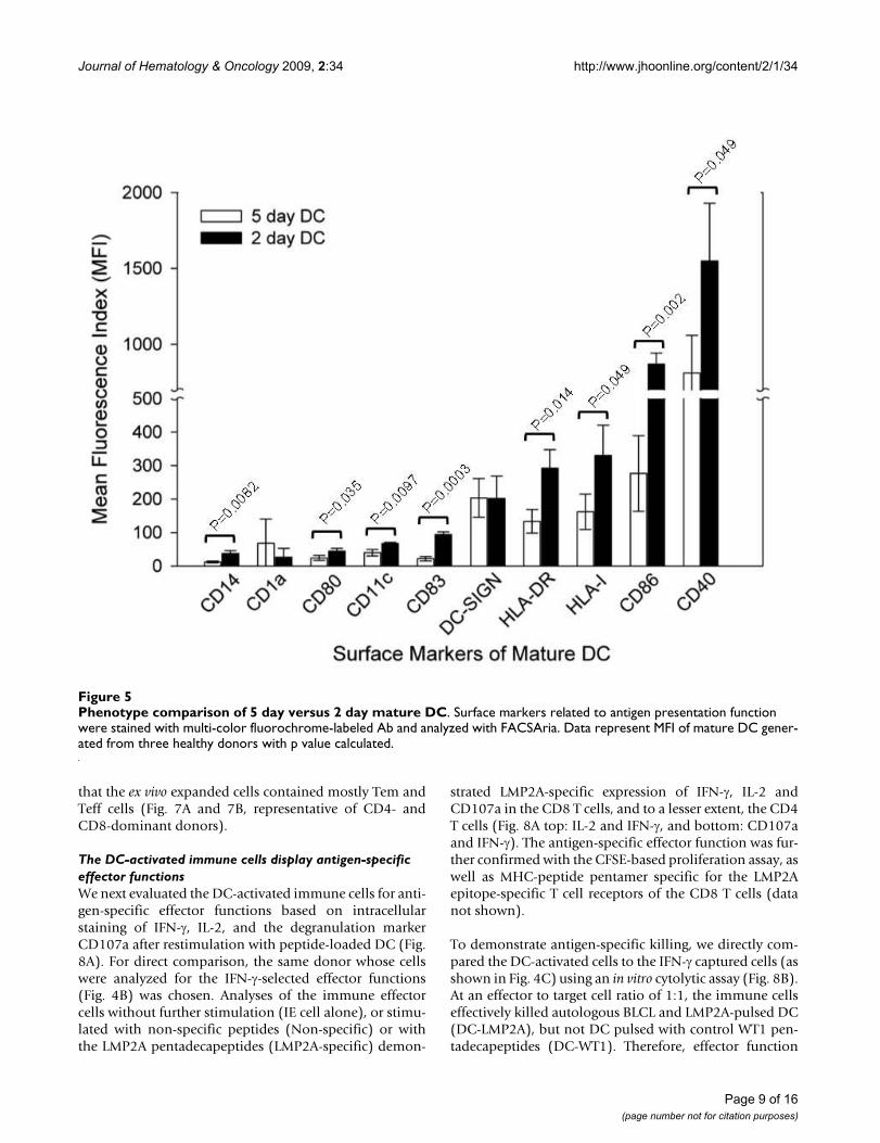
lyzed the surface markers for plastic-adherent monocytesand mature DC with flow cytometry (data shown in Addi-tional file 1), and analyzed phenotypes of the 2 day andthe 5 day mature DC (Fig. 5). The 2 day DC expressedhigher levels of class I/II MHC (HLA-I and HLA-DR), cos-timulatory molecules (CD86 and CD40) and maturationmarker CD83. We also found that the 2 day DC inducedprimary and secondary immune response against viral orcancer antigens at efficiencies equal to or better than the 5day DC (manuscript in preparation). Therefore, the 2 dayDC protocol was adopted for later experiments.
The 2 day DC were pulsed with the pooled LMP2A penta-decapeptides, irradiated and cocultured with autologous
lymphocytes at a ratio of 1:20. The cell number usuallydecreases around day 5 after coculture, followed by anincrease of a few fold around day 17, suggesting a loss ofnon-specific cells followed by expansion of antigen-spe-cific cells (see representative growth curves in Fig. 6A).Flow cytometry analysis of the DC-activated cells in cul-ture from four different donors at day 0, 12 and 19 indi-cated that most donors generated CD3+CD56- T cells, butsome generated a large proportion of CD3-CD56+ NKcells and CD3+CD56+ cells (e.g. donor 3, Fig. 6B). Therelative ratios of CD4+ T cells, CD8+ T cells, NK cells andCD3+CD56+ cells in the coculture appeared to be donor-dependent (Fig. 6B and 6C). Furthermore, phenotypeanalysis with multi-color flow cytometry demonstrated
IFN-γ- or CD137-based enrichment of antigen-specific immune effector cellsFigure 1IFN-γ- or CD137-based enrichment of antigen-specific immune effector cells. (A) CD4 and CD8 T cell distribution in IFN-γ- or CD137-positive cell population after antigen stimulation. The ratios of CD4 to CD8 T cells of five donors were presented. (B) and (C) CD137 expression in CD4 and CD8 T cells before and after Ab affinity column purification. PBMC were mixed with EBV LMP2 pentadecapeptides and 3–24 hr later, IFN-γ-secreting cells or CD137-positive cells were isolated by using MACS magnetic bead affinity columns as described in Materials and Methods. The cells were stained with PE-conjugated anti-IFN-γ or anti-CD137 Ab and CD3, CD4 and CD8 specific Ab and analyzed with flow cytometry.
Page 5 of 16(page number not for citation purposes)

Journal of Hematology & Oncology 2009, 2:34 http://www.jhoonline.org/content/2/1/34
Page 6 of 16(page number not for citation purposes)
Phenotype analysis of IFN-γ- or CD137-selected antigen-specific immune cellsFigure 2Phenotype analysis of IFN-γ- or CD137-selected antigen-specific immune cells. PBMC were stimulated with EBV LMP2A pentadecapeptides and IFN-γ or CD137 positive cells were isolated for analysis. (A) & (B) Memory and effector pheno-type analysis based on seven-color flow cytometry with surface staining for CD27, CD28, CD45RA, CCR7, CD4, CD8, CD137 and intracellular staining for IFN-γ immediately after cell isolation without further culture. The percentage of different popula-tions (IFN-γ or CD137 plus CD4 or CD8 gated) of T cells is illustrated for a CD4-dominant donor (A) and a CD8-dominant donor (B); Tcm, central memory T cells (CD27+/-, CD28+, CCR7+, CD45RA-); Tem, effector memory T cells (CD27+/-, CD28+/-, CCR7-, CD45RA-); Teff, terminal effector T cells (CD27-, CD28-, CCR7+/-, CD45RA+).

Journal of Hematology & Oncology 2009, 2:34 http://www.jhoonline.org/content/2/1/34
Page 7 of 16(page number not for citation purposes)
Phenotype analyses of IFN-γ- or CD137-selected immune effector cells after ex vivo expansionFigure 3Phenotype analyses of IFN-γ- or CD137-selected immune effector cells after ex vivo expansion. (A) Phenotype analysis after ex vivo expansion. After the rapid selection, the antigen-specific immune cells were cultured for fifteen days with irradiated autologous PBMC as feeder cells. The distribution of CD3, CD4, CD8 and CD56 cell populations was determined and representative FACS graphs are shown. (B) and (C) Memory and effector phenotype analysis based on seven-color flow cytometry with surface staining for CD27, CD28, CD45RA, CCR7, CD4, CD8, CD137 and intracellular staining for IFN-γ. Representative results of a CD4-dominant donor (B) and a CD8-dominant donor (C) are shown.

Journal of Hematology & Oncology 2009, 2:34 http://www.jhoonline.org/content/2/1/34
Page 8 of 16(page number not for citation purposes)
Effector function analyses of IFN-γ- or CD137-selected immune effector cells after ex vivo expansionFigure 4Effector function analyses of IFN-γ- or CD137-selected immune effector cells after ex vivo expansion. (A) and (B) Flow cytometry analysis of IFN-γ and CD137 expression after restimulation of the culture expanded IFN-γ or CD137 effector cells. The cells were restimulated with different cells as indicated and subjected to antibody staining and flow cytometry analy-sis; control, WT-1 peptide-pulsed DC; specific, LMP2A peptide-pulsed DC; BLCL, autologous EBV-transformed B cells. (C) Analysis of antigen-specific cytolytic activity. The ex vivo expanded IFN-γ- or CD137-enriched cells were incubated with autol-ogous EBV transformed B cells (BLCL), mature DCs loaded with either LMP2A pentadecapeptides (DC-LMP2A) or WT1 pen-tadecapeptides (DC-WT1, as control) at 1:1 ratio in a cytotoxicity assay based on CFSE labeling as described in Materials and Methods.
Journal of Hematology & Oncology 2009, 2:34 http://www.jhoonline.org/content/2/1/34
that the ex vivo expanded cells contained mostly Tem andTeff cells (Fig. 7A and 7B, representative of CD4- andCD8-dominant donors).
The DC-activated immune cells display antigen-specific effector functionsWe next evaluated the DC-activated immune cells for anti-gen-specific effector functions based on intracellularstaining of IFN-γ, IL-2, and the degranulation markerCD107a after restimulation with peptide-loaded DC (Fig.8A). For direct comparison, the same donor whose cellswere analyzed for the IFN-γ-selected effector functions(Fig. 4B) was chosen. Analyses of the immune effectorcells without further stimulation (IE cell alone), or stimu-lated with non-specific peptides (Non-specific) or withthe LMP2A pentadecapeptides (LMP2A-specific) demon-
strated LMP2A-specific expression of IFN-γ, IL-2 andCD107a in the CD8 T cells, and to a lesser extent, the CD4T cells (Fig. 8A top: IL-2 and IFN-γ, and bottom: CD107aand IFN-γ). The antigen-specific effector function was fur-ther confirmed with the CFSE-based proliferation assay, aswell as MHC-peptide pentamer specific for the LMP2Aepitope-specific T cell receptors of the CD8 T cells (datanot shown).
To demonstrate antigen-specific killing, we directly com-pared the DC-activated cells to the IFN-γ captured cells (asshown in Fig. 4C) using an in vitro cytolytic assay (Fig. 8B).At an effector to target cell ratio of 1:1, the immune cellseffectively killed autologous BLCL and LMP2A-pulsed DC(DC-LMP2A), but not DC pulsed with control WT1 pen-tadecapeptides (DC-WT1). Therefore, effector function
Phenotype comparison of 5 day versus 2 day mature DCFigure 5Phenotype comparison of 5 day versus 2 day mature DC. Surface markers related to antigen presentation function were stained with multi-color fluorochrome-labeled Ab and analyzed with FACSAria. Data represent MFI of mature DC gener-ated from three healthy donors with p value calculated.
Page 9 of 16(page number not for citation purposes)

Journal of Hematology & Oncology 2009, 2:34 http://www.jhoonline.org/content/2/1/34
Page 10 of 16(page number not for citation purposes)
Ex vivo expansion and phenotype analyses of DC-activated immune effector cellsFigure 6Ex vivo expansion and phenotype analyses of DC-activated immune effector cells. EBV LMP2A-specific effector T cells were generated by stimulation of non-adherent PBMC with DC pulsed with LMP2A pentadecapeptides. (A) Growth kinetics of DC-activated immune effector cells. The viable cells were counted with trypan blue staining at different time points after coculture and the growth curves of 5 samples are shown. (B) and (C) CD3, CD56, CD4 and CD8 phenotype analysis. The DC-activated cells from day 0, 12 and 19 were stained with antibodies against CD4, CD8, CD3 and CD56 and analyzed with flow cytometry. The percentages of different lymphocyte subsets were analyzed and shown in bar graphs. Donor 1*, PBMC collected at a different time point from donor 1.

Journal of Hematology & Oncology 2009, 2:34 http://www.jhoonline.org/content/2/1/34
Page 11 of 16(page number not for citation purposes)
Memory and effector T cell analyses of DC-activated immune effector cellsFigure 7Memory and effector T cell analyses of DC-activated immune effector cells. (A) & (B) Memory and effector pheno-type analysis of a CD4 dominant donor (A) and a CD8 dominant donor (B). The DC-activated cells from day 0, 12, and 17 after coculture were analyzed for CD3, CD4, CD8, CD27, CD28, CD45RA, and CCR7 using a seven-color panel of fluorochrome-labeled Ab with FACSAria. The cells were gated for CD3 and CD4 or CD8 as total cells for the percentage analysis. One rep-resentative flow graph of 5 performed experiments is presented.

Journal of Hematology & Oncology 2009, 2:34 http://www.jhoonline.org/content/2/1/34
Figure 8 (see legend on next page)
Page 12 of 16(page number not for citation purposes)

Journal of Hematology & Oncology 2009, 2:34 http://www.jhoonline.org/content/2/1/34
analyses including IFN-γ release and cytotoxicity assayssuggest that the DC-activated cells had lower activitiesthan the expanded IFN-γ effector cells.
DiscussionAdoptive immune cell therapy has shown great promisein treating viral diseases and melanoma. [2,6] Continuedefforts are focused on the generation of sufficient amountof antigen-specific immune cells and optimal condition-ing of immune homeostasis in patients in order to achievea sustained in vivo immune surveillance. [24-26] Here, wecompared three ex vivo immune cell preparation protocolsand phenotypically and functionally characterized thesecells. The rapid protocols based on IFN-γ and CD137selection generate a small number of antigen-specificeffector cells with high percentage of central memory Tcells in a very short period of time. The DC-activation pro-tocol generates more immune cells, albeit, with more dif-ferentiated phenotype and reduced proportion of antigen-specific effector cells.
Based on analyses of a large number of donors, we foundthat individual response to a given antigen could be eitherCD4- or CD8-dominant, which is antigen- and donor-dependent. Immune effector cells isolated based on IFN-γexpression displayed a CD4 or CD8 bias consistent withthe donor's immune dominance. However, antigen-spe-cific CD137 positive cells showed a higher ratio of CD4effector cells regardless of the subject's immune pheno-type; this is in contrast to previous reports that emphasizethe induction of CD8 effector cells after CD137 enrich-ment. [15,22,27] We did, however, show that CD8 T cellsdisplayed higher density of CD137 than did CD4 T cells.It is well documented that CD137 costimulation pro-motes both CD4 and CD8 T cell expansion and long termmemory. [28-30] Our finding that more CD4 T cells thanCD8 T cells are detected in the CD137-positive cell popu-lation suggests a rapid induction of CD137 in the memoryT helper repertoire immediately after antigen stimulation.Although the enriched CD137 immune cells contained ahigher CD4 T cell ratio, further expansion in culturerestored the donor's original dominant phenotype, with a
higher CD3-CD56+ NK cell population than those foundin the expanded IFN-γ enriched immune cells (Fig. 3A).This result suggests that IFN-γ is a more restricted adoptiveimmune response marker and represent less of an innateimmune marker as does CD137.
The ex vivo DC-activation protocol generated differentratios of CD4, CD8 and NK cells in culture, which again,appeared to be donor-dependent. Whether the immunedominance has any effect on in vivo efficacy of the cul-tured immune effector cells awaits further investigation.As CD4 T cells are important for the maintenance of long-term anti-viral CD8 T cell memory [31], therapeuticimmune cells should include polyclonal CD4 and CD8 Tcells. Both IFN-γ and CD137 selection approaches gener-ated increased number of memory type of cells represent-ative of polyclonal CD4 and CD8 T cells that may haveincreased proliferation potential after infusion. Althoughthe antigen-specific memory T cells from PBMC may below; for examples, the average yield of LMP2A-specificIFN-γ positive immune effector cells from healthy EBV-seropositive donors is only 0.22 ± 0.13% (n = 6, after tworounds of affinity column purification), they can beexpanded to more than two orders of magnitude in cul-ture in two weeks and maintain their high antigen specif-icity.
It is evident that the LMP2A pentadecapeptides efficientlyactivate both CD4 and CD8 T cells in a short exposureperiod (3–13 hr). This was surprising since CD8 T cells areactivated through class I MHC loaded with short 9–11amino acid peptide epitopes, different from CD4 T cells,which are activated through class II MHC loaded with 12–15 amino acid peptide epitopes. The pentadecapeptideantigens apparently activated CD8 T cells with high effi-ciency through cross-presentation. This has been con-firmed with various pentadecapeptide antigens(unpublished). The processing of class II MHC peptidesinto class I epitopes for cross-presentation to CD8 T cellsappears to be highly efficient with both the IFN-γ and theCD137 protocols, as with the DC-activation method. The
Functional analyses of DC-activated and ex vivo expanded immune effector cellsFigure 8 (see previous page)Functional analyses of DC-activated and ex vivo expanded immune effector cells. EBV LMP2A-specific effector cells were generated by stimulation of non-adherent PBMC with DC pulsed with LMP2A pentadecapeptides. (A) Analysis of antigen-specific effector cytokines and CD107a expression. The DC-activated immune cells from day 19 coculture were stimulated with autologous DC pulsed with LMP2A peptides, WT1 peptides (non-specific control) or no stimulation (IE cell alone) for 6 hours. The cells were stained with Ab against CD4, CD8, CD107a and IFN-γ. Flow cytometry analyses of IL-2, IFN-γ and CD107a-positive cells in CD4-gated or CD8-gated populations were illustrated as representative of five experiments. (B) Comparison of cytolytic function of IFN-γ-selected (same donor as in Fig. 4C) versus DC-activated LMP2-specific immune effector cells. The LMP2A-specific effector cells were mixed with target cells including autologous BLCL, DC-LMP2A, or con-trol DC-WT1 at 1:1 ratio and analyzed for cytolytic activity based on the CFSE-labeling method as described in Materials and Methods.
Page 13 of 16(page number not for citation purposes)

Journal of Hematology & Oncology 2009, 2:34 http://www.jhoonline.org/content/2/1/34
detailed molecular mechanism of the efficient cross-pres-entation requires further investigation.
To assess differentiation and maturation status of the exvivo generated T cells, we applied multi-color flow cytom-etry to detect differentiation and homeostatic markerCD45RA, trafficking marker CCR7, and costimulatorymarker CD27 and CD28. [32,33] It is not surprising thatboth the IFN-γ- and the CD137-enriched antigen-specificeffector cells displayed more memory markers than didthe DC coculture-expanded cells. While preserving Tcmcells is critical to in vivo therapeutic efficacy,[7,34,35] clin-ical studies have proven that ex vivo expanded effector cellscan persist many years after infusion. [36] Clinical bene-fits of these different protocols will require detailed evalu-ation in a large cohort of patients.
The differentiation status of the ex vivo generated immunecells may contribute to their in vivo therapeutic efficacy.Homeostasis of antigen-specific memory cells can varydepending on antigen source, the immune milieu andindividual donor. It is known that Tcm cells are mainlylocated in lymphoid tissues and Tem cells are distributedin diverse non-lymphoid sites including lung, liver andintestine. [37] In addition, bone marrow has been shownto embrace increased number of anti-cancer or anti-virusmemory T cells. [38-40] After ex vivo expansion, however,wherever the T cells come from, they tend to bestowexhausted proliferation and replicative senescence associ-ated with down-regulation of anti-apoptotic protein Bcl-2and Bcl-xL, and decreased telomere length. [33,34,41]Modification of antigen presentation protocol and culturecondition may help overcome the immune cell exhaus-tion problem. [9,42]
For patients with acute infections or illness, direct isola-tion of antigen-specific immune cells from partly HLA-matched healthy donors represents an attractive emer-gency approach to obtain therapeutic cells. [43,44] Thisapproach offers several advantages including a shortenedhandling time and increased proliferation potential invivo. Although the number of immune cells is limited withthe direct isolation approach, clinical evidence supportsthat only a small number of such immune cells, in therange of 103-104/kg body weight, is sufficient to attaintherapeutic efficacy in transplant patients. [13,45,46]Exvivo expansion of immune cells, nevertheless, may be nec-essary for patients with a compromised immunity.[47,48]
ConclusionThe two rapid immune cell isolation methods generatefunctional effector cells in less than 24–48 hr suitable foremergency immune cell preparation. On the other hand,the DC-activation method expands antigen-specific
immune effector cells while effectively reduce the numberof non-specific cells. Depending on clinical needs, forexamples, the urgency for treatment, patient's bodyweight (e.g. less cells are needed for pediatric patients), orpatient's immune cell proliferative potential in vivo, themethod of immune cell preparation may differ. Our dataindicate that IFN-γ selection followed by ex vivo expansionrepresents the best approach for the generation of highamount of antigen-specific immune effector cells. Furtherefforts to overcome immune tolerance and expand anti-gen-specific immune cells with prolonged in vivo persist-ence are critical to the success of immune cell therapy.
List of abbreviationsIFN-γ: interferon-gamma; IL: interleukine; DC: dendriticcell; CTL: cytotoxic T lymphocyte; MHC: major histocom-patibility complex; Ab: antibody; Ag: antigen; TCR: T cellreceptor; TNF: tumor necrosis factor; BLCL: B lymphoblas-toid cell line; EBV: Epstein-Barr virus; LMP2A: late mem-brane protein 2A; CMV: cytomegalovirus; ICCS:intracellular cytokine staining; CFSE: carboxy-fluoresceindiacetate succinimidyl ester.
Competing interestsYH, YL, YH and YW are employees of Vectorite BiomedicaInc. LJC is consultant to a biotech company.
Authors' contributionsAll authors are accountable for the integrity of the researchresults; Chang is responsible for the conception of theresearch and Han, Huang, Liang, Ho and Wang areresponsible for the execution and for data collection;Chang is responsible for initial drafting and revisions ofthe manuscript.
Additional material
AcknowledgementsWe thank the technical assistance of Liheng Guo, Lily Lien, Fuhung Yang, Yinchieh Fu and Meifang Lin. The study was funded by Vectorite Biomedica Inc. and Yongling Foundation.
Additional file 1Phenotype analysis of monocytes and 2 day and 5 day mature DC. Sur-face markers related to antigen presentation function were analyzed using fluorochrome-labeled Ab. The light-colored lines in the FACS graphs rep-resent control Ab and the numbers represent geometric means with per-centages shown in parentheses. Representatives of two monocyte experiments and three DC experiments are illustrated.Click here for file[http://www.biomedcentral.com/content/supplementary/1756-8722-2-34-S1.tiff]
Page 14 of 16(page number not for citation purposes)
Journal of Hematology & Oncology 2009, 2:34 http://www.jhoonline.org/content/2/1/34
References1. Riddell SR, Greenberg PD: Principles for adoptive T cell therapy
of human viral diseases. Annu Rev Immunol 1995, 13:545-586.2. Fujita Y, Rooney CM, Heslop HE: Adoptive cellular immuno-
therapy for viral diseases. Bone Marrow Transplant 2008,41:193-198.
3. Papadopoulos EB, Ladanyi M, Emanuel D, Mackinnon S, Boulad F,Carabasi MH, Castro-Malaspina H, Childs BH, Gillio AP, Small TN,Young JW, Kernan NA, O'Reilly RJ: Infusions of donor leukocytesto treat Epstein-Barr virus-associated lymphoproliferativedisorders after allogeneic bone marrow transplantation. NEngl J Med 1994, 330:1185-1191.
4. O'Reilly RJ, Small TN, Papadopoulos E, Lucas K, Lacerda J, Koulova L:Biology and adoptive cell therapy of Epstein-Barr virus-asso-ciated lymphoproliferative disorders in recipients of marrowallografts. Immunol Rev 1997, 157:195-216.
5. Collins RH Jr, Shpilberg O, Drobyski WR, Porter DL, Giralt S, Cham-plin R, Goodman SA, Wolff SN, Hu W, Verfaillie C, List A, Dalton W,Ognoskie N, Chetrit A, Antin JH, Nemunaitis J: Donor leukocyteinfusions in 140 patients with relapsed malignancy after allo-geneic bone marrow transplantation. J Clin Oncol 1997,15:433-444.
6. Dudley ME, Wunderlich JR, Robbins PF, Yang JC, Hwu P, Schwartzen-truber DJ, Topalian SL, Sherry R, Restifo NP, Hubicki AM, RobinsonMR, Raffeld M, Duray P, Seipp CA, Rogers-Freezer L, Morton KE,Mavroukakis SA, White DE, Rosenberg SA: Cancer regression andautoimmunity in patients after clonal repopulation with anti-tumor lymphocytes. Science 2002, 298:850-854.
7. Gattinoni L, Powell DJ Jr, Rosenberg SA, Restifo NP: Adoptiveimmunotherapy for cancer: building on success. Nat RevImmunol 2006, 6:383-393.
8. Rosenberg SA, Restifo NP, Yang JC, Morgan RA, Dudley ME: Adop-tive cell transfer: a clinical path to effective cancer immuno-therapy. Nat Rev Cancer 2008, 8:299-308.
9. Klebanoff CA, Gattinoni L, Restifo NP: CD8+ T-cell memory intumor immunology and immunotherapy. Immunol Rev 2006,211:214-224.
10. Yee C, Savage PA, Lee PP, Davis MM, Greenberg PD: Isolation ofhigh avidity melanoma-reactive CTL from heterogeneouspopulations using peptide-MHC tetramers. J Immunol 1999,162:2227-2234.
11. Szmania S, Galloway A, Bruorton M, Musk P, Aubert G, Arthur A, PyleH, Hensel N, Ta N, Lamb L Jr, Dodi T, Madrigal A, Barrett J, Henslee-Downey J, van Rhee F: Isolation and expansion of cytomegalo-virus-specific cytotoxic T lymphocytes to clinical scale froma single blood draw using dendritic cells and HLA-tetramers.Blood 2001, 98:505-512.
12. Rauser G, Einsele H, Sinzger C, Wernet D, Kuntz G, Assenmacher M,Campbell JD, Topp MS: Rapid generation of combined CMV-specific CD4+ and CD8+ T-cell lines for adoptive transferinto recipients of allogeneic stem cell transplants. Blood 2004,103:3565-3572.
13. Feuchtinger T, Matthes-Martin S, Richard C, Lion T, Fuhrer M, Ham-precht K, Handgretinger R, Peters C, Schuster FR, Beck R, SchummM, Lotn R, Jahn G, Lang P: Safe adoptive transfer of virus-specificT-cell immunity for the treatment of systemic adenovirusinfection after allogeneic stem cell transplantation. Br J Hae-matol 2006, 134:64-76.
14. Mackinnon S, Thomson K, Verfuerth S, Peggs K, Lowdell M: Adop-tive cellular therapy for cytomegalovirus infection followingallogeneic stem cell transplantation using virus-specific Tcells. Blood Cells Mol Dis 2008, 40:63-67.
15. Wolfl M, Kuball J, Ho WY, Nguyen H, Manley TJ, Bleakley M, Green-berg PD: Activation-induced expression of CD137 permitsdetection, isolation, and expansion of the full repertoire ofCD8+ T cells responding to antigen without requiring knowl-edge of epitope specificities. Blood 2007, 110:201-210.
16. Han S, Wang B, Cotter MJ, Yang LJ, Zucali J, Moreb JS, Chang L-J:Overcoming immune tolerance against multiple myelomawith lentiviral calnexin-engineered dendritic cells. Mol Ther2008, 16:269-279.
17. Fukushima Y, Ohashi H, Wakui K, Nishida T, Oh-ishi T: A rapidmethod for starting a culture for the establishment ofEpstein-Barr virus-transformed human lymphoblastoid celllines. Jpn J Hum Genet 1992, 37:149-150.
18. Betts MR, Brenchley JM, Price DA, De Rosa SC, Douek DC, RoedererM, Koup RA: Sensitive and viable identification of antigen-spe-cific CD8+ T cells by a flow cytometric assay for degranula-tion. J Immunol Methods 2003, 281:65-78.
19. Lyons AB: Analysing cell division in vivo and in vitro using flowcytometric measurement of CFSE dye dilution. J ImmunolMethods 2000, 243:147-154.
20. Jedema I, Werff NM van der, Barge RM, Willemze R, Falkenburg JH:New CFSE-based assay to determine susceptibility to lysis bycytotoxic T cells of leukemic precursor cells within a heter-ogeneous target cell population. Blood 2004, 103:2677-2682.
21. Straathof KC, Bollard CM, Popat U, Huls MH, Lopez T, Morriss MC,Gresik MV, Gee AP, Russell HV, Brenner MK, Rooney CM, HeslopHE: Treatment of nasopharyngeal carcinoma with Epstein-Barr virus – specific T lymphocytes. Blood 2005, 105:1898-1904.
22. Wehler TC, Karg M, Distler E, Konur A, Nonn M, Meyer RG, HuberC, Hartwig UF, Herr W: Rapid identification and sorting of via-ble virus-reactive CD4(+) and CD8(+) T cells based on anti-gen-triggered CD137 expression. J Immunol Methods 2008,339:23-37.
23. Dauer M, Obermaier B, Herten J, Haerle C, Pohl K, Rothenfusser S,Schnurr M, Endres S, Eigler A: Mature dendritic cells derivedfrom human monocytes within 48 hours: a novel strategy fordendritic cell differentiation from blood precursors. J Immunol2003, 170:4069-4076.
24. Dummer W, Niethammer AG, Baccala R, Lawson BR, Wagner N,Reisfeld RA, Theofilopoulos AN: T cell homeostatic proliferationelicits effective antitumor autoimmunity. J Clin Invest 2002,110:185-192.
25. Klebanoff CA, Khong HT, Antony PA, Palmer DC, Restifo NP: Sinks,suppressors and antigen presenters: how lymphodepletionenhances T cell-mediated tumor immunotherapy. TrendsImmunol 2005, 26:111-117.
26. Wrzesinski C, Paulos CM, Gattinoni L, Palmer DC, Kaiser A, Yu Z,Rosenberg SA, Restifo NP: Hematopoietic stem cells promotethe expansion and function of adoptively transferred antitu-mor CD8 T cells. J Clin Invest 2007, 117:492-501.
27. Watanabe K, Suzuki S, Kamei M, Toji S, Kawase T, Takahashi T,Kuzushima K, Akatsuka Y: CD137-guided isolation and expan-sion of antigen-specific CD8 cells for potential use in adop-tive immunotherapy. Int J Hematol 2008, 88:311-320.
28. Alderson MR, Smith CA, Tough TW, Davis-Smith T, Armitage RJ, FalkB, Roux E, Baker E, Sutherland GR, Din WS: Molecular and biolog-ical characterization of human 4-1BB and its ligand. Eur JImmunol 1994, 24:2219-2227.
29. Wen T, Bukczynski J, Watts TH: 4-1BB ligand-mediated costim-ulation of human T cells induces CD4 and CD8 T cell expan-sion, cytokine production, and the development of cytolyticeffector function. J Immunol 2002, 168:4897-4906.
30. Zhu Y, Zhu G, Luo L, Flies AS, Chen L: CD137 stimulation deliv-ers an antigen-independent growth signal for T lymphocyteswith memory phenotype. Blood 2007, 109:4882-4889.
31. Sun JC, Williams MA, Bevan MJ: CD4+ T cells are required for themaintenance, not programming, of memory CD8+ T cellsafter acute infection. Nat Immunol 2004, 5:927-933.
32. Sallusto F, Geginat J, Lanzavecchia A: Central memory and effec-tor memory T cell subsets: function, generation, and main-tenance. Annu Rev Immunol 2004, 22:745-763.
33. Nascimbeni M, Shin EC, Chiriboga L, Kleiner DE, Rehermann B:Peripheral CD4(+)CD8(+) T cells are differentiated effectormemory cells with antiviral functions. Blood 2004, 104:478-486.
34. Gattinoni L, Klebanoff CA, Palmer DC, Wrzesinski C, Kerstann K, YuZ, Finkelstein SE, Theoret MR, Rosenberg SA, Restifo NP: Acquisi-tion of full effector function in vitro paradoxically impairs thein vivo antitumor efficacy of adoptively transferred CD8+ Tcells. J Clin Invest 2005, 115:1616-1626.
35. Klebanoff CA, Gattinoni L, Torabi-Parizi P, Kerstann K, Cardones AR,Finkelstein SE, Palmer DC, Antony PA, Hwang ST, Rosenberg SA,Waldmann TA, Restifo NP: Central memory self/tumor-reac-tive CD8+ T cells confer superior antitumor immunity com-pared with effector memory T cells. Proc Natl Acad Sci USA 2005,102:9571-9576.
36. Leen AM, Rooney CM, Foster AE: Improving T cell therapy forcancer. Annu Rev Immunol 2007, 25:243-265.
Page 15 of 16(page number not for citation purposes)
http://www.ncbi.nlm.nih.gov/entrez/query.fcgi?cmd=Retrieve&db=PubMed&dopt=Abstract&list_uids=7612234
http://www.ncbi.nlm.nih.gov/entrez/query.fcgi?cmd=Retrieve&db=PubMed&dopt=Abstract&list_uids=7612234
http://www.ncbi.nlm.nih.gov/entrez/query.fcgi?cmd=Retrieve&db=PubMed&dopt=Abstract&list_uids=8093146
http://www.ncbi.nlm.nih.gov/entrez/query.fcgi?cmd=Retrieve&db=PubMed&dopt=Abstract&list_uids=8093146
http://www.ncbi.nlm.nih.gov/entrez/query.fcgi?cmd=Retrieve&db=PubMed&dopt=Abstract&list_uids=8093146
http://www.ncbi.nlm.nih.gov/entrez/query.fcgi?cmd=Retrieve&db=PubMed&dopt=Abstract&list_uids=9255631
http://www.ncbi.nlm.nih.gov/entrez/query.fcgi?cmd=Retrieve&db=PubMed&dopt=Abstract&list_uids=9255631
http://www.ncbi.nlm.nih.gov/entrez/query.fcgi?cmd=Retrieve&db=PubMed&dopt=Abstract&list_uids=9255631
http://www.ncbi.nlm.nih.gov/entrez/query.fcgi?cmd=Retrieve&db=PubMed&dopt=Abstract&list_uids=9053463
http://www.ncbi.nlm.nih.gov/entrez/query.fcgi?cmd=Retrieve&db=PubMed&dopt=Abstract&list_uids=9053463
http://www.ncbi.nlm.nih.gov/entrez/query.fcgi?cmd=Retrieve&db=PubMed&dopt=Abstract&list_uids=9053463
http://www.ncbi.nlm.nih.gov/entrez/query.fcgi?cmd=Retrieve&db=PubMed&dopt=Abstract&list_uids=9973498
http://www.ncbi.nlm.nih.gov/entrez/query.fcgi?cmd=Retrieve&db=PubMed&dopt=Abstract&list_uids=9973498
http://www.ncbi.nlm.nih.gov/entrez/query.fcgi?cmd=Retrieve&db=PubMed&dopt=Abstract&list_uids=9973498
http://www.ncbi.nlm.nih.gov/entrez/query.fcgi?cmd=Retrieve&db=PubMed&dopt=Abstract&list_uids=1327290
http://www.ncbi.nlm.nih.gov/entrez/query.fcgi?cmd=Retrieve&db=PubMed&dopt=Abstract&list_uids=1327290
http://www.ncbi.nlm.nih.gov/entrez/query.fcgi?cmd=Retrieve&db=PubMed&dopt=Abstract&list_uids=1327290
http://www.ncbi.nlm.nih.gov/entrez/query.fcgi?cmd=Retrieve&db=PubMed&dopt=Abstract&list_uids=8088337

Journal of Hematology & Oncology 2009, 2:34 http://www.jhoonline.org/content/2/1/34
Publish with BioMed Central and every scientist can read your work free of charge
"BioMed Central will be the most significant development for disseminating the results of biomedical research in our lifetime."
Sir Paul Nurse, Cancer Research UK
Your research papers will be:
available free of charge to the entire biomedical community
peer reviewed and published immediately upon acceptance
cited in PubMed and archived on PubMed Central
yours — you keep the copyright
Submit your manuscript here:http://www.biomedcentral.com/info/publishing_adv.asp
BioMedcentral
37. Masopust D, Vezys V, Marzo AL, Lefrancois L: Preferential locali-zation of effector memory cells in nonlymphoid tissue. Sci-ence 2001, 291:2413-2417.
38. Letsch A, Keilholz U, Assfalg G, Mailander V, Thiel E, ScheibenbogenC: Bone marrow contains melanoma-reactive CD8+ effectorT cells and, compared with peripheral blood, enriched num-bers of melanoma-reactive CD8+ memory T cells. Cancer Res2003, 63:5582-5586.
39. Slifka MK, Whitmire JK, Ahmed R: Bone marrow contains virus-specific cytotoxic T lymphocytes. Blood 1997, 90:2103-2108.
40. Wood AH, Zhang X, Farber DL, Strome SE: CD8+ memory T lym-phocytes from bone marrow – immune function and thera-peutic potential. Crit Rev Immunol 2007, 27:527-537.
41. Soares MV, Borthwick NJ, Maini MK, Janossy G, Salmon M, Akbar AN:IL-7-dependent extrathymic expansion of CD45RA+ T cellsenables preservation of a naive repertoire. J Immunol 1998,161:5909-5917.
42. van Stipdonk MJ, Sluijter M, Han WG, Offringa R: Development ofCTL memory despite arrested clonal expansion. Eur J Immunol2008, 38:1839-1846.
43. Haque T, Wilkie GM, Taylor C, Amlot PL, Murad P, Iley A, Domba-goda D, Britton KM, Swerdlow AJ, Crawford DH: Treatment ofEpstein-Barr-virus-positive post-transplantation lymphopro-liferative disease with partly HLA-matched allogeneic cyto-toxic T cells. Lancet 2002, 360:436-442.
44. Lucas KG, Salzman D, Garcia A, Sun Q: Adoptive immunotherapywith allogeneic Epstein-Barr virus (EBV)-specific cytotoxicT-lymphocytes for recurrent, EBV-positive Hodgkin disease.Cancer 2004, 100:1892-1901.
45. Cobbold M, Khan N, Pourgheysari B, Tauro S, McDonald D, OsmanH, Assenmacher M, Billingham L, Steward C, Crawley C, Olavarria E,Goldman J, Chakraverty R, Mahendra P, Craddock C, Moss PA:Adoptive transfer of cytomegalovirus-specific CTL to stemcell transplant patients after selection by HLA-peptidetetramers. J Exp Med 2005, 202:379-386.
46. Amrolia PJ, Muccioli-Casadei G, Huls H, Adams S, Durett A, Gee A,Yvon E, Weiss H, Cobbold M, Gaspar HB, Rooney C, Kuehnle L, Ghe-tie V, Schindler J, Krance R, Heslop HE, Veys P, Vitetta E, Brenner MK:Adoptive immunotherapy with allodepleted donor T-cellsimproves immune reconstitution after haploidentical stemcell transplantation. Blood 2006, 108:1797-1808.
47. Overwijk WW: Breaking tolerance in cancer immunotherapy:time to ACT. Curr Opin Immunol 2005, 17:187-194.
48. Rabinovich GA, Gabrilovich D, Sotomayor EM: Immunosuppres-sive strategies that are mediated by tumor cells. Annu RevImmunol 2007, 25:267-296.
Page 16 of 16(page number not for citation purposes)
http://www.ncbi.nlm.nih.gov/entrez/query.fcgi?cmd=Retrieve&db=PubMed&dopt=Abstract&list_uids=9292550
http://www.ncbi.nlm.nih.gov/entrez/query.fcgi?cmd=Retrieve&db=PubMed&dopt=Abstract&list_uids=9292550
http://www.ncbi.nlm.nih.gov/entrez/query.fcgi?cmd=Retrieve&db=PubMed&dopt=Abstract&list_uids=9834071
http://www.ncbi.nlm.nih.gov/entrez/query.fcgi?cmd=Retrieve&db=PubMed&dopt=Abstract&list_uids=9834071