Journal of Hazardous Materials - Marquette University regenerated %
12
Journal of Hazardous Materials 252–253 (2013) 355–366 Contents lists available at SciVerse ScienceDirect Journal of Hazardous Materials jou rn al h om epage: www.elsevier.com/loc ate/jhazmat Chitosan-cellulose composite materials: Preparation, Characterization and application for removal of microcystin Chieu D. Tran a,∗ , Simon Duri a , Ambra Delneri b , Mladen Franko b a Department of Chemistry, Marquette University, P.O. Box 1881, Milwaukee, WI 53201, USA b Laboratory for Environmental Research, University of Nova Gorica, Vipavska 13, 5001 Nova Gorica, Slovenia h i g h l i g h t s • A novel and recyclable synthetic method using an ionic liquid, a Green Solvent. • Ecocomposite materials were syn- thesized from cellulose (CEL) and chitosan (CS). • Adding CEL into CS substantially increases tensile strength of the com- posite. • The composite is much better adsor- bent for cyanotoxins than other materials. • The composite can be reused because adsorbed microcystin can be des- orbed. g r a p h i c a l a b s t r a c t a r t i c l e i n f o Article history: Received 10 December 2012 Received in revised form 12 February 2013 Accepted 25 February 2013 Available online xxx Keywords: Cellulose Chitosan Ionic liquid Green chemisty Cyanotoxins Microcystins a b s t r a c t We developed a simple and one-step method to prepare biocompatible composites from cellulose (CEL) and chitosan (CS). [BMIm + Cl − ], an ionic liquid (IL), was used as a green solvent to dissolve and prepare the [CEL + CS] composites. Since majority (>88%) of IL used was recovered for reuse by distilling the aqueous washings of [CEL + CS], the method is recyclable. XRD, FTIR, NIR, 13 C CP-MAS-NMR and SEM were used to monitor the dissolution and to characterize the composites. The composite was found to have combined advantages of their components: superior mechanical strength (from CEL) and excellent adsorption capability for microcystin-LR, a deadly toxin produced by cyanobacteria (from CS). Specifically, the mechanical strength of the composites increased with CEL loading; e.g., up to 5× increase in tensile strength was achieved by adding 80% of CEL into CS. Kinetic results of adsorption confirm that unique properties of CS remain intact in the composite, i.e., it is not only a very good adsorbent for microcystin but also is better than all other available adsorbents. For example, it can adsorb 4× times more microcystin than the best reported adsorbent. Importantly, the microcystin adsorbed can be quantitatively desorbed to enable the composite to be reused with similar adsorption efficiency. © 2013 Elsevier B.V. All rights reserved. 1. Introduction Microcystin LR, produced by cyanobacteria, is one of the most deadly toxins present in drinking water [1–4]. At least 40 species ∗ Corresponding author. Tel.: +1 424 288 5428; fax: +1 288 7066. E-mail address: [email protected] (C.D. Tran). of cyanobacteria release toxic cyanotoxins into water during algal blooms. Microcystins (MCs) are the most prevalent class of cyan- otoxins. The MCs are very soluble in water and consist of over 80 reported variants; however, four MCs (LR, RR, LA, and YR (See Scheme 1 for structures) are of special concern to the Environmen- tal Protection Agency (EPA) and are on EPA Contaminant Candidate List III. The World Health Organization (WHO) has set a provisional drinking water guideline of 1 g/L for MC-LR. The toxicity of MCs is 0304-3894/$ – see front matter © 2013 Elsevier B.V. All rights reserved. http://dx.doi.org/10.1016/j.jhazmat.2013.02.046
Transcript of Journal of Hazardous Materials - Marquette University regenerated %
Ca
Ca
b
h
•
•
•
•
•
a
ARRAA
KCCIGCM
1
d
0h
Journal of Hazardous Materials 252– 253 (2013) 355– 366
Contents lists available at SciVerse ScienceDirect
Journal of Hazardous Materials
jou rn al h om epage: www.elsev ier .com/ loc ate / jhazmat
hitosan-cellulose composite materials: Preparation, Characterization andpplication for removal of microcystin
hieu D. Trana,∗, Simon Duria, Ambra Delnerib, Mladen Frankob
Department of Chemistry, Marquette University, P.O. Box 1881, Milwaukee, WI 53201, USALaboratory for Environmental Research, University of Nova Gorica, Vipavska 13, 5001 Nova Gorica, Slovenia
i g h l i g h t s
A novel and recyclable syntheticmethod using an ionic liquid, a GreenSolvent.Ecocomposite materials were syn-thesized from cellulose (CEL) andchitosan (CS).Adding CEL into CS substantiallyincreases tensile strength of the com-posite.The composite is much better adsor-bent for cyanotoxins than othermaterials.The composite can be reused becauseadsorbed microcystin can be des-orbed.
g r a p h i c a l a b s t r a c t
r t i c l e i n f o
rticle history:eceived 10 December 2012eceived in revised form 12 February 2013ccepted 25 February 2013vailable online xxx
eywords:ellulose
a b s t r a c t
We developed a simple and one-step method to prepare biocompatible composites from cellulose (CEL)and chitosan (CS). [BMIm+Cl−], an ionic liquid (IL), was used as a green solvent to dissolve and preparethe [CEL + CS] composites. Since majority (>88%) of IL used was recovered for reuse by distilling theaqueous washings of [CEL + CS], the method is recyclable. XRD, FTIR, NIR, 13C CP-MAS-NMR and SEMwere used to monitor the dissolution and to characterize the composites. The composite was found tohave combined advantages of their components: superior mechanical strength (from CEL) and excellentadsorption capability for microcystin-LR, a deadly toxin produced by cyanobacteria (from CS). Specifically,
hitosanonic liquidreen chemistyyanotoxinsicrocystins
the mechanical strength of the composites increased with CEL loading; e.g., up to 5× increase in tensilestrength was achieved by adding 80% of CEL into CS. Kinetic results of adsorption confirm that uniqueproperties of CS remain intact in the composite, i.e., it is not only a very good adsorbent for microcystin butalso is better than all other available adsorbents. For example, it can adsorb 4× times more microcystinthan the best reported adsorbent. Importantly, the microcystin adsorbed can be quantitatively desorbedto enable the composite to be reused with similar adsorption efficiency.
. Introduction
Microcystin LR, produced by cyanobacteria, is one of the mosteadly toxins present in drinking water [1–4]. At least 40 species
∗ Corresponding author. Tel.: +1 424 288 5428; fax: +1 288 7066.E-mail address: [email protected] (C.D. Tran).
304-3894/$ – see front matter © 2013 Elsevier B.V. All rights reserved.ttp://dx.doi.org/10.1016/j.jhazmat.2013.02.046
© 2013 Elsevier B.V. All rights reserved.
of cyanobacteria release toxic cyanotoxins into water during algalblooms. Microcystins (MCs) are the most prevalent class of cyan-otoxins. The MCs are very soluble in water and consist of over80 reported variants; however, four MCs (LR, RR, LA, and YR (See
Scheme 1 for structures) are of special concern to the Environmen-tal Protection Agency (EPA) and are on EPA Contaminant CandidateList III. The World Health Organization (WHO) has set a provisionaldrinking water guideline of 1 �g/L for MC-LR. The toxicity of MCs is
356 C.D. Tran et al. / Journal of Hazardous Materials 252– 253 (2013) 355– 366
cture
dMt
rlradtoo
NembuassauiPnlta[tismdaCu
Scheme 1. Stru
ue to their strong binding and inhibition of protein phosphatases.Cs are also known to promote the growth of tumors; have a geno-
oxic effect as well as strong mutagenicity [1–4].The presence of toxic cyanobacterial blooms in drinking water
eservoirs may represent serious health risks for the human popu-ation. Considerable efforts have, therefore, been made to developemoval methods. Limited success has been made by use of suchdsorbents as activated carbon, oxidation by ozone and chlorineioxide, TiO2 photocatalysis and ultrasonic irradiation [1–4]. Sincehese techniques cannot effectively remove the toxins in water, it isf particular importance that an effective removal technique basedn the use of a biocompatible material be developed.
Chitosan (CS) is a linear amino polysaccharide, obtained by-deacetylation of chitin, and chitin, which is found in thexoskeletons of crustaceans (e.g., crabs and shrimp), is the secondost abundant naturally occurring polysaccharide [5–8]. CS has
een a subject of intense studies for many years [9–24]. The pop-larity stems from the fact that it is biodegradable, biocompatible,nd possesses unique structure and properties which have beenuccessfully exploited in many applications including hemosta-is, wound healing, bactericide and fungicide, drug delivery anddsorbent for organic and inorganic pollutants [9–29]. Of partic-lar interest are reports that CS, when modified with fly ash or
mmobilized on clay, can adsorb and remove algae such as Chlorellayrenoidosa or Microcystis Aeruginosa in water [30–35]. Unfortu-ately, in spite of its potentials, there are drawbacks which severely
imit applications of CS. For example similar to cellulose (CEL),he most abundant substance on earth, in CS, a network of intra-nd inter-hydrogen bonds enables it to adopt an ordered structure6–8]. While such structure is responsible for CS to have aforemen-ioned properties and CEL to have superior mechanical strength,t also makes them insoluble in most solvents [6–8]. As a con-equence, high temperature and strong exotic solvents such asethylmorpholine-N-oxide, dimethylthexylsilyl chloride or LiCl in
imethylacetamide (DMAc) are needed to dissolve CEL whereas ancid such as acetic acid is required to protonate amino groups ofS to facilitate its dissolution in water [6–29]. These methods arendesirable because they are based on the use of corrosive and
of microcystins.
volatile solvents, require high temperature and suffer from sidereactions and impurities which may lead to changes in structureand properties of the polysaccharides. More importantly, it is notpossible to use a single solvent or system of solvents to dissolve bothCEL and CS. Furthermore, CS is known to swell in water which leadsto structural weakening in wet environments [6–21]. To increasethe structural strength of CS products, attempts have been madeto covalently bind or graft CS onto man-made polymers or claysto strengthen its structure [9–29]. Such modification is not desir-able because it may inadvertently alter CS properties, making itnot biocompatible and toxic and lessening or removing its uniqueproperties. A new method which can effectively dissolve both CSand CEL not at high temperature and not by corrosive and volatilesolvents but rather by recyclable “green” solvent is particularlyneeded. This is because such method would facilitate preparation of[CEL + CS] composite material which is not only biocompatible butalso has combined properties of its components, namely mechani-cal strength (from CEL) and hemostasis, wound healing, bactericide,drug delivery and pollutant removal (from CS). Such demand makesrecent report on ability of a simple ionic liquid, butyl methylimmi-dazolium chloride to dissolve up to 10% (w/w) of CEL, particularlyimportant [36].
Butyl methylimmidazolium chloride ([BMIm+Cl−]) belongs toa group of compounds known as ionic liquids (ILs). ILs are organicsalts that are liquid at room temperature [37–40]. They have uniquechemical and physical properties, including being air and moisturestable, a high solubization power, and virtually no vapor pres-sure [37–40]. Because of these properties, they can serve as a“GREEN” recyclable alternative to the volatile organic compoundsthat are traditionally used as industrial solvents [37–40]. Due totheir advantages, ILs have been used for applications which are notpossible with other chemicals. For example, as described above, ILsuch as [BMIm+Cl−] can dissolve up to 10% (w/w) of CEL. This dis-covery is of particular significance as it makes it possible for the first
time that CEL can be dissolved, regenerated and chemically modi-fied by use of a simple and green solvent which has high solubilitypower and low toxicity. Recently, it was found that [BMIm+Cl−] canalso dissolve CS as well [40].
C.D. Tran et al. / Journal of Hazardous Materials 252– 253 (2013) 355– 366 357
eme 2
itatssmvrabrh
2
2
aSawfvbso
2
3Nmpa
Sch
The information presented is indeed provocative and clearlyndicate that it is possible to develop a novel and green methodo prepare novel polysaccharide composite materials for effectivedsorption and removal of MCs. Such considerations prompted uso initiate this study which aims to hasten the breakthrough byystematically and revolutionarily exploit advantages of IL, a greenolvent, to develop a novel, simple, pollution-free and recyclableethod to dissolve both CEL and CS, without using any harmful,
olatile organic solvents and/or strong acid and base, for the prepa-ation of polysaccharide ecocomposite materials containing CELnd CS, and to demonstrate that these materials are not only fullyiocompatible but also superior to currently available materials foremoval of MCs. The results of our initial investigation are reportederein.
. Materials and methods
.1. Chemicals
Microcrystalline CEL (Avicel, DP ≈ 300 [41]), amorphous CELnd chitosan (MW ≈ 310–375 kDa, 75% degree of deacetylation,igma–Aldrich), microcystin-LR (Enzo Life Sciences) were useds received. 1-methylimidazole and 1-chlorobutane (Alfa Aesar)ere further purified by distillation. [BMIm+ Cl−] was synthesized
rom 1-chlorobutane and 1-methylimidazole using method pre-iously reported [42,43]. This IL is known to be hygroscopic andecause any water presence is known to decrease its ability to dis-olve polysaccharides [44], the IL was dried under vacuum at 70 ◦Cvernight before use.
.2. Instruments
UV-visible spectra were measured on a Perkin Elmer Lambda5 UV/VIS spectrometer. NIR spectra were taken on a home-built
IR spectrometer based on an acousto-optic tunable filter. Infor-ation on this NIR spectrometer was described in detail in ourrevious papers [45,46]. Normally, each spectrum was an aver-ge of 30 spectra taken at 1-nm intervals from 1450 to 2450 nm.
.
FTIR spectra were measured on a PerkinElmer 100 spectrometerat 2 cm−1 resolution with either KBr or by a ZnSe single reflectionATR accessory (Pike Miracle ATR). Each spectrum was an averageof 64 spectra. X-ray diffraction (XRD) measurements were takenon a Rigaku MiniFlex II diffractometer utilizing the Ni filtered CuK� radiation (1.54059 A). The voltage and current of the X-ray tubewere 30 kV and 15 mA respectively. The samples were measuredwithin the 2� angle range from 2.0◦ to 40.0◦. The scan rate was5◦/min. Data processing procedures were performed with the Jade8 program package [47]. Scanning electron microscopic images ofsurface and cross section of the polysaccharide composite materi-als were taken under vacuum with an accelerated voltage of 3 kVusing Hitachi S4800 scanning electron microscope (SEM). Tensilestrength measurements were performed on an Instron 5500R Ten-sile Tester.
2.3. Preparation of CEL, CS and [CEL + CS] composite films
Scheme 2 summarizes procedure used to dissolve and toregenerate films of CEL and/or CS with [BMIm+Cl−] as solvent.Specifically, CEL and/or CS were dissolved in [BMIm+Cl−] underargon and magnetic stirring at 100 – 110 ◦C. All polysaccharideswere added in portions of approximately 1 wt% of the IL. Succeedingportions were only added after the previous addition had com-pletely dissolved until the desired concentration has been reached.For composite films, the components were dissolved one after theother, with CEL being dissolved first. Using this procedure, solutionsof CEL (containing up to 10%, w/w (of IL)), CS (up to 4%, w/w) andcomposite solutions containing CEL and CS in various proportionswere prepared in about 6–8 h.
Upon complete dissolution, the homogeneous solutions of thepolysaccharides in [BMIm+ Cl−] were cast on glass slides or Mylarsheets using a RDS stainless steel coating rod with appropriate size
(RDS Specialties, Webster, NY) to produce thin films with differentcompositions and concentrations of CEL and CS. They were thenkept at room temperature for 24 h to allow the solutions to undergogelation (henceforth referred as Gel Films).
358 C.D. Tran et al. / Journal of Hazardous Materials 252– 253 (2013) 355– 366
0
500
1000
1500
2000
2500
3000
3500
0 10 20 30 40
Inte
nsity
Microc rys talline cell ulos e
10% cell ulose ge l fil m
BMIm+ Cl-
Cell ulose dry fil m
A
0
500
1000
1500
2000
2500
0 10 20 30 40
Inte
nsity
le (2 the
chitosan po wde r3% ch itosan ge l fil mBMIm+ Cl-Chitosan dry fil m
B
0
500
1000
1500
2000
2500
3000
3500
0 10 20 30 40
Inte
nsity
Chitosan po wde r
Microc rys talline cellulos eCell ulose + Chitosan dry fil m10%cell ulose + 3%Chitosa n ge l fil m
C
S pow
[fiWr[tWo6fi
a
3
3
ii[iSfFt
ndstCt[tadswapfo[d(o
e
Ang le (2 the ta) deg ree s Ang
Fig. 1. XRD spectra of microcrystalline CEL, C
In addition to the polysaccharides, the Gel Films also containedBMIm+Cl−]. The IL was removed from the films by washing thelms in water for 3 days to yield “[BMIm+Cl−]-free” films (i.e.,et Films). During this period, the washing water was constantly
eplaced with fresh water to maximize the removal of the IL. TheBMIm+Cl−] used was recovered from the washed aqueous solu-ion by distillation. Finally, Dried Films were obtained when the
et Films are allowed to dry. Drying of the Wet Films was carriedut at room temperature in a chamber with humidity controlled at0%. Drying time was found to be dependent on thickness of thelms but generally was in range of 2–3 days.
Detailed information on measurements of swelling, adsorptionnd desorption can be found in the Supplementary Data.
. Results and discussion
.1. Synthesis and characterization of [CEL + CS] composites
Images of CEL, CS and [CEL + CS] composite materials during var-ous stages of preparation are presented in Scheme 2. Specifically,mages of films of one-component (CEL or CS) and two componentCEL + CS] casted right after the polysaccharides were dissolvedn [BMIm+Cl−], i.e., Gel Film, are shown in top center-left of thecheme. After soaking in water for 3 days, [BMIm+Cl−] was removedrom the gel film to yield corresponding Wet Films. Finally, Driedilms were obtained when the wet film was allowed to dry at roomemperature.
XRD, FTIR, NIR and 13C Cross polarization-magic angle spin-ing NMR (CP-MAS-NMR) were used to follow and confirm theissolution process and to characterize the films. Specifically, dis-olution of CEL and CS in [BMIm+Cl−] was confirmed by XRDechnique. Shown in Fig. 1A are XRD spectra of microcrystallineEL (pink curve), the CEL one-component Gel Film (orange curve),he corresponding Dried Film (green curve) as well as that ofBMIm+Cl−] (blue curve) for reference. As illustrated, microscrys-alline CEL exhibits diffraction peaks at 2� = 14.7◦ and 16.3◦, 22.5◦
nd 34.6◦ for (101), (002) and (040) plane, respectively. These bandsisappeared completely when the powder polysaccharide was dis-olved in [BMIm+Cl−]. The disappearance of these bands togetherith the similarity between the spectrum of liquid [BMIm+Cl−]
nd that of the Gel Film clearly indicate that [BMIm+Cl−] com-letely dissolved the polysaccharide. Similar results were alsoound for CS (Fig. 1B), namely, the fact that diffraction bandsf the CS powder disappeared when they were dissolved inBMIm+Cl−], clearly indicate that the [BMIm+Cl−] also completelyissolved this polysaccharide. Similar results were also obtained
Fig. 1C) for two-component composite films containing [CEL + CS]f 10:3.Recently, there have been some reports on toxicity of ILs. How-ver, the IL used in this work, [BMIm+Cl−], is relatively nontoxic
ta) de gree s Ang le (2 the ta) deg ree s
der, regenerated CEL, CS and [CEL + CS] films.
compared to other ILs (its EC-50 and LD50 values are 897.47 ppmand 550 mg/kg, respectively [48]). Nevertheless, it is desirable tocompletely remove the IL from regenerated polysaccharide filmsto ensure the films are biocompatible. Since [BMIm+ Cl−] is totallymiscible with water (the logP, its octanol–water partition coeffi-cient, is −2.4 [49]), it was removed from the Gel Films by washingthe films with water. Washing water (2L for a composite filmof about 10cmX10 cm) was repeatedly replaced with fresh waterevery 24 h until it was confirmed that IL was not detected in thewashed water (by monitoring UV absorption of the IL at 290 nm).It was found that after washing for 72 h, no IL was detected inthe washing water by UV measurements. Since the limit of detec-tion of the spectrophotometer used in this work was estimated tobe about 3 × 10⁻⁵ AU, and the molar absorptivity of [BMIm+Cl−]at 290 nm is 2.6 M−1 cm−1, it is estimated that if any [BMIm+Cl−]remains, its concentration would be smaller than 2 �g/mL of thewashed water and 2 �g/g of the composite film. Since this con-centration is two orders of magnitude lower than the LD50 valueof the [BMIm+Cl−], if any IL remains in the composite films, itwould not pose any harmful effect. UV–vis, FTIR and NIR tech-niques were used to: (1) confirm that when the composite filmswere washed with water, [BMIm+Cl−] was removed from the filmsto a level not detectable by these techniques; and (2) determinechemical composition of composite materials. Shown in Fig. 2A isNIR spectrum of [BMIm+Cl−]. As illustrated, overtone and com-bination bands of aliphatic C H groups of the [BMIm+Cl−] canbe clearly observed at 1388 nm and 1720 nm [50]. Since thesebands are specific for [BMIm+Cl−], they can be used as indicatorsto determine if the IL is present. Also shown in Fig. 2A are NIRspectra of Gel Films of CEL, CS and [CEL + CS]. Spectra of these GelFilms are very similar to that of [BMIm+Cl−] because the IL wasthe main component of these films. As shown in Fig. 2B, Afterwashing with water to remove the IL, and drying, Dried Films ofone-component CEL and CS samples were found to exhibit NIRspectra drastically different from those of their Gel Films shownin Fig. 2A (spectra of Dried Films are shown together with the spec-trum of [BMIm+Cl−] for reference). The fact that NIR spectra of theDried Films (Fig. 2B) exhibit none of the indicator bands specific for[BMIm+Cl−] clearly indicates that washing with water effectivelyremoved the IL from the films to a level which is not detectableby NIR technique. Further confirmation of removal of the ionic liq-uid from the films can also be seen in Fig. 2C which shows FT-IRspectra of the same samples shown in 2B, namely, [BMIm+Cl−]and Dried Films of CEL and CS. As illustrated, [BMIm+Cl−] exhibitsseveral distinct bands at around 756 cm−1, 1173 cm−1, 1468 cm−1
and 1572 cm−1. These bands can be attributed to aromatic C-H
bending, C N stretching, aromatic C-H in-plane bending andaromatic C C stretching, respectively [51,52]. Again, none of thesebands were present in the spectra of both CEL and CS Dried Filmsindicating that by washing with water, [BMIm+Cl−] was removed
C.D. Tran et al. / Journal of Hazardous Mate
Fig. 2. NIR spectra (A and B) and FTIR spectra (C) of: (A): [BMIm+Cl−] solution (blue),CEL Gel Film (red), CS Gel Film (green) and [CEL + CS] Gel Film; (B) [BMIm+Cl−] solu-tion (blue), CEL Dried Film (red) and CS Dried Film (green); and (C) [BMIm+Cl−]solution (blue), CEL Dried Film (red) and CS Dried Film (green). (For interpretationov
fm
seriohpcpra
ni[(n
f the references to color in this figure legend, the reader is referred to the webersion of the article.)
rom the composite films to a level which is not detectable by FTIRethod.The IL used was recovered by distilling the washed aqueous
olution (the IL remained because it is not volatile). The recov-red [BMIm+Cl−] was dried under vacuum at 70oC overnight beforeeuse. If needed, recovered [BMIm+Cl−] was decolored by heat-ng at 70 ◦C with activated carcoal. It was found that at least 88%f [BMIm+Cl−] was recovered for reuse. As such, the method developedere is recyclable because [BMIm+Cl−] is the only solvent used in thereparation and it is recovered for reuse. FTIR and NIR results indi-ate that recovered [BMIm+Cl−] is structurally the same as freshlyrepared[BMIm+Cl−], and that composite materials prepared usingecovered [BMIm+Cl−] were found spectroscopically to be the sames those prepared using freshly made [BMIm+Cl−].
It is important to point out that the method developed here isot only different but also superior to the method recently reported
n which magnetic CEL-CS gel microspheres were prepared usingBMIm+Cl−] as a solvent. This is because that method is not greenvarious chemicals including vacuum pump oil, Span 80, ethanol),ot recyclable (used [BMIm+Cl−] is not recovered for reuse) and
rials 252– 253 (2013) 355– 366 359
can only prepare microspheres [53,54]. Conversely, the methoddeveloped here is green and recyclable (the only chemical usedis [BMIm+Cl−] and at least 88% of it was recovered for reuse) andcan produce [CEL + CS] composite materials in various forms (e.g.thin films, microparticles) which make them particularly suited forvarious applications.
It is noteworthy to add that the cost of [BMIm+Cl−] is comparableto those of solvents traditionally used to dissolve CEL and CS (see,for example, prices quoted in Sigma-Aldrich catalog: USD105/50 gof [BMIm+Cl−] compared to USD46/500 mL of acetic acid (for tra-ditional dissolution of CS); USD225/100 g of metylmorpholine andUSD230/100 g of dimethylthexysilyl chloride (for traditional dis-solution of CEL)). However, the method reported here is much moreeconomical compared to other methods because [BMIm+Cl−] used inthe dissolution is not lost but rather recovered for reuse.
Analysis of the film materials by SEM reveals some interestingfeatures about the texture and morphology of the materials. Shownin Fig. 3 are surface (top three images) and cross section images(bottom three images) of regenerated one component CS film (left)and CEL film (right) as well as 50:50 CEL:CS composite film (mid-dle images). As expected, both surface and cross section imagesclearly indicate that one-component CEL and CS are homogeneous.Chemically, the only difference between CS and CEL is the few –NH2groups in the former. However, their structures, as recorded by theSEM, particularly the cross section images, are substantially differ-ent. Specifically, while CS seems to exhibit smooth structure, CELarranges itself into fibrous structure with fibers having diameterof about ∼0.5–1.0 �m. As expected, the 50:50 CEL:CS compositematerial is not only homogeneous but also its structure is similarto both CEL and CS.
Chemically, the regeneration of both CEL and CS was con-firmed by FTIR spectroscopy. As illustrated in Fig. 4A, the FTIRspectrum of microcrystalline CEL (blue spectrum) exhibits threepronounced bands at around 3400 cm−1, 2850–2900 cm−1 and890–1150 cm−1. These bands can be tentatively assigned to stretch-ing vibrations of O H, C H and O group, respectively [55–57].The fact that the Dried Film (red spectrum in 4A) also exhibitsthese three bands and is very similar to that of the microcrys-talline CEL clearly indicates that CEL was completely regeneratedby this synthetic method. Similarly, the FT-IR spectrum of a CSDried Film (green spectrum in 4B) is similar to the FTIR spectrumof the CS powder (blue spectrum) from which it was made. Thesespectra display characteristic CS bands around 3400 cm−1 (O-Hstretching vibrations), 3250–3350 cm−1 (symmetric and asymmet-ric N-H stretching), 2850–2900 cm−1 (C H stretching), 1657 cm−1
(C O, amide 1), 1595 cm−1 (N H deformation), 1380 cm−1 (CH3symmetrical deformation), 1319 cm−1 (C N stretching, amide III)and 890–1150 cm−1 (ether bonding) [55–57]. These results indicatethat both CEL and CS were successfully regenerated by the syntheticmethod developed here without any chemical transformation.
Results from NIR measurements further confirm regenerationof polysaccharide composite materials. Shown in Fig. 4C are NIRspectra of microcrystalline CEL and regenerated CEL Dried Film (4C)and CS powder together with regenerated CS Dried Film (4D). Asillustrated in 4C, both CEL microcrystalline and regenerated CELfilm exhibit bands around 1492 nm, 1938 nm and around 2104 nm.These can be attributed to the overtone and combination transitionof the OH groups. Since CS also possesses the same O-H groupsin addition to N H2 group, NIR spectra of CS powder and film(4D) also have additional bands at around 1548 nm and 2028 nmwhich expectedly can be attributed to the NH modes [57–60].The similarity between the NIR spectra of starting polysaccharides
and regenerated polysaccharide further confirms that CEL and CSwere successfully regenerated by this preparation method.13C CP-MAS-NMR technique was then employed to further con-firm structure of regenerated polysaccharide composite materials.

360 C.D. Tran et al. / Journal of Hazardous Materials 252– 253 (2013) 355– 366
F imagec
SeBpste
Frit
ig. 3. SEM images of surface (top three images) and cross section (bottom threeomposite film (middle).
hown in Fig. 5 are 13C CP-MAS-NMR spectra obtained for regen-rated Dried Films of CEL (purple spectrum D), CS (red spectrum) and 5:3 CEL:CS composite (light blue spectrum A) together CSowder (green spectrum C) amorphous cellulose powder (black
pectrum E) and microcrystalline cellulose powder (dark blue spec-rum F). Chemical shifts of all six samples are listed in Table 2. Asxpected, the spectra of CS powder, amorphous CEL powder and-0.01
0.04
0.09
0.14
0.19
0.24
0.29
0.34
6501650265036504650565066507650
Abso
rban
ce
wavenumb er (cm-1)
Regenerate d CEL film
Microcrystall ine ce llulo se1307 1503 1770 2130
wavelength, nm
C A
-0.02
0.08
0.18
0.28
0.38
0.48
0.58
0.68
6501650265036504650565066507650
Abso
rban
ce
wavenumb er (cm-1)
Chitosan PowderRgenerate d CS film1307 1503 1770 2130
wavelength, nm
D B
ig. 4. FTIR spectra (A and B) and NIR spectra (C and D) of microcrystalline CEL (blue),egenerated CEL film (red), CS powder (blue) and regenerated CS film (green). (Fornterpretation of the references to color in this figure legend, the reader is referredo the web version of the article.)
s) of regenerated CS film (left), regenerated CEL film (right) and 50:50 [CEL + CS]
microcrystalline CEL powder agree with reported spectra [61–63].The observed similarity between spectra and chemical shifts ofcorresponding carbons of regenerated dried films of CS and CELto those of powder CS and powder of amorphous CEL is a clearindication that CEL and CS were successfully regenerated. Of par-ticular interest is the fact that the spectrum of regenerated CELfilm is similar to that of the amorphous CEL powder, and is dis-tinctly different from that of microcrystalline powder. This can beclearly seen at band corresponding to C4 which is only a broad sin-gle band at 83.9 ppm, 84.4 ppm, respectively for regenerated CELand amorphous CEL, but a doublet at 88.7 ppm and 83.7 ppm forcrystalline CEL powder. These results seem to indicate that whenmicrocrystalline CEL was dissolved by and regenerated from theIL, the regenerated CEL has relatively lower crystallinity than themicrocrystalline CEL because IL disrupted inter- and intramolecularhydrogen bond network during the dissolution process. As a conse-quence, the regenerated CEL adapts an amorphous structure. Theseresults not only confirm conclusion based on XRD results presentedin previous section but also are in agreement with other previousstudies [61–63]. CEL has relatively different chemical shifts com-pared to those of CS, particularly C2 and C6; i.e., in CEL, a singleC6 band is at 62.0 ppm, C2 is together with C3 and C5 in a largeband at 74.1 ppm whereas in CS, C2 and C6 are together in a bandat around 61.0 ppm, and the large band at 75.4 ppm is due only toC3 and C5. Since these bands can be resolved into individual bands
corresponding to C2, C3, C5 and C6 for each CEL and CS componentof composite materials, it is possible to determine concentrationof each polysaccharide component in composite materials usingthe CP-MAS-NMR technique. Of particular interest is the presenceTable 1Tensile strength and swelling parameters for [CEL + CS] composites.
% CS in [CEL + CS] composite Tensilestrength (MPa)
Equilibrium watercontent (EWC(%))
100 12.0 73.667 17.1 69.950 23 62.240 41.429 52.3 58.520 72.20 82.6 46.4

C.D. Tran et al. / Journal of Hazardous Mate
F ), mic[
oCTnbvtwc
3
pusfimstttbstg
bfstewqora
relative concentration of CS in the material with 100% CEL hav-ing the lowest value of 80% and 100% CS with the highest value of270%. Values for other composites are between these two values.More quantitative assessment can be obtained from equilibrium
0 100 200 300 13860
50
100
150
200
250
300
Sw
ellin
g r
ati
o (
%)
100% CS
67% CS
50% CS
29% CS
0% CS
B
y = -0.2735x + 75 .997R² = 0.9489
0
20
40
60
80
100
0
20
40
60
80
100
0 20 40 60 80 100
Tens
ile S
tren
gth
(Mpa
)
EWC
(%)
% CEL
Equilibr ium wat er content, EWC(%)Tensile s trength(MPa)
A
ig. 5. 13C CP MAS NMR spectra of chitosan powder (C), amorphous CEL powder (ECEL + Cs] composite films (A).
f the band corresponding to carbon of CH3 group in CS andEL:CS composite material (but not in CEL) at around 22.7 ppm.his band can be attributed to remaining acyl groups of chitin,amely, chitosan obtained from commercial sources is not 100%ut, as specified by the manufacturer in our case, is only >75% con-erted from chitin. In fact, by accumulating spectra for longer time,his CH3 band can be clearly observed, and it can be used, togetherith FTIR method to determine exact degree of deacetylation of
hitosan.
.2. Properties of [CEL + CS] composites
As described above, mechanical strength of CS is so poor thatractically it cannot be used by itself for applications based on itsnique properties. Measurements were made to determine tensiletrength of pure CS film and (CS + CEL) composite films with dif-erent CEL concentrations in order to determine if by adding CELnto CS, the [CEL + CS] composite material would have adequate
echanical strength for practical applications. Results obtained,hown in Fig. 6A, clearly indicate that adding CEL into CS substan-ially increase its tensile strength. For example, up to 5× increase inensile strength can be achieved by adding 80% of CEL into CS, andhat the tensile strength of the composite material can be adjustedy adding judicious amount of CEL. More importantly, the tensiletrengths of [CS + CEL] composite materials are comparable withhose of existing CS materials including those prepared by eitherrafting or copolymerization with other chemicals [64–69].
As described above, CS has relatively poor mechanical strengthecause it undergoes swelling in water. Adding CEL into CS wasound to improve mechanical strength of the composite. However,ince the [CEL + CS] composite still swell in water, it is importanto investigate the swelling of [CEL + CS] composites with differ-nt compositions in order to determine the optimal CEL/CS ratiohich, in spite of swelling, can still provide a composite with ade-
uate mechanical strength for its utilization. Accordingly, kineticsf swelling of [CEL + CS] composites with different compositionsanging from 0% to 100% CS were measured and results obtainedre shown in Fig. 6B which plot swelling ratio (S%) as a function of
rials 252– 253 (2013) 355– 366 361
rocrystalline CEL powder (F), regenerated CEL film (D), regenerated CS film (B) and
time. As illustrated, for all composites, the water swelling occurredrapidly, reaching equilibrium in water uptake at about 120 min. Asexpected, the swelling ratio was found to be proportional to the
Time, min
Fig. 6. (A) Plot of tensile strength and equilibrium water content (EWC%) as a func-tion of CEL concentration and (B) swelling kinetics of different [CEL + CS] compositefilms.

362 C.D. Tran et al. / Journal of Hazardous Materials 252– 253 (2013) 355– 366
Table 213C CP-MAS-NMR Chemical shifts of starting materials and regenerated CEL, CS and [CEL + CS] composite.
Carbon chemical shifts (ppm)
C1 C2 C3 C4 C5 C6 CH3
Regenerated cellulose film 104.6 74.1 74.1 83.9 74.1 62.0Amorphous cellulose powder 105.7 74.8 74.8 84.4 74.8 63.0Microcrystalline cellulose powder 104.9 72.1 74.6 83.7, 88.7 74.6 64.7, 62.3
75.74.75.
w[Cttottisolitrust
sscCtmdid
3
paahtop
l
wrla
h
Regenerated chitosan film 104.7 61.0
Chitosan powder 104.9 60.1
Regenerated cellulose + chitosan film 104.5 56.4
ater content EWC % values. EWC % and tensile strength values forCEL + CS] composites with different compositions from 0 to 100%S are listed in Table 1 and plotted together, as a function of CEL inhe composites (Fig. 6A). It is evident from the figure that the rela-ionship between the tensile strength with CEL concentration ispposite to that of the swelling, namely, increasing CEL concentra-ion leads to the increase in the tensile strength and the decrease inhe swelling. However, the relative increase in the tensile strengths much larger than the decrease in the swelling. For example, ten-ile strength of 100% CEL is 82.6 MPa which is 588% higher than thatf 0% CEL (12.0 MPa) whereas its EWC % value of 46.4% is only 58%ower than the value for 73.6% for 0%CEL. This finding is particularlymportant because it clearly indicates that mechanical strength ofhe composite can be substantially strengthened by adding onlyelatively small amount of CEL. The composite still can retain itsnique properties because the concentration of CEL added is somall to have any pronounced effect on the unique properties ofhe composites which are due solely to CS.
Taken together, results presented clearly indicate that we haveuccessfully developed a novel, green and recyclable method to dis-olve CEL and CS and to prepare biocompartible all polysaccharideomposite materials containing CEL and CS. As anticipated, addingEL to CS material substantially increases mechanical strength ofhe CS-based composite. It is expected that the [CEL + CS] composite
ay retain its unique properties including bactericide, fungicide,rug delivery as well as good adsorbent for pollutants and tox-
ns (from CS). Initial evaluation of their ability to adsorb MC-LR isescribed in following section.
.3. Adsorption of microcystin-LR by [CEL + CS] composites
Experiments were designed to determine: (1) if [CEL + CS] com-osite materials can adsorb MC; (2) if they can, mechanism ofdsorption processes in terms of rate constants and adsorbedmounts at equilibrium; and (3) composite material which givesighest adsorption. These were accomplished by fitting kinetic datao both pseudo-first order and pseudo-second order models inrder to determine appropriate reaction order for the adsorptionrocesses based on R2 and MSC values [70–72].
For pseudo-first order:
n(qe − qt) = ln qe − k1t (1)
For pseudo-second order
t
qt= 1
(k2q2r )
+ t
qe(2)
here k1 and k2 are pseudo-first order and pseudo-second orderate constant of sorption (g/mg.min), qe is the amount of ana-yte adsorbed at equilibrium (mg/g), qt is the amount of analyte
dsorbed at any time t (mg/g).If the initial adsorption rate h is
= k2q2e (3)
4 83.6 75.4 61.0 22.77 84.0 74.7 60.1 23.45 83.9 75.5 61.5 23.0
Then Eq. (2) can be rearranged as
t
qt= 1
h+ 1
qet (4)
A linear plot can be obtained by plotting t/qt against t. qe and h,can be obtained from the slope and intercept; k2 can be calculatedfrom h and qe according to Eq. (3).
As described in experimental section, kinetics of adsorption ofMC by composite materials were determined by measuring change(i.e., decrease) in absorbance at 238 nm as a function of time in asolution containing a composite film. From measured absorbances,the amount of adsorbed MC at time t (i.e., qt) can then be calculated.Shown in Fig. 7A is plot of qt as a function of time for six different[CEL + CS] materials with different composition ranging from 0% to67% CS. It is evident from this plot that increasing CS concentrationin the composite material led to increase in amount of microcystinadsorbed at equilibrium. Detailed information on adsorption kinet-ics can be obtained by fitting the data to both pseudo-first order(Eq. (1)) and pseudo-second order (Eq. (4)) model. Listed in Table 3are results obtained for [CEL + CS] composite materials with dif-ferent composition ranging from 20% to 67% of CS. It was foundthat 0% chitosan (i.e., 100% CEL) material does not adsorb MC, andadsorption by composite material with < 20% CS was too small to bemeasured. Typical linearized plots for pseudo-1st order and psedo-2nd order for two samples are shown in Fig. 8. It is evident from theplots as well as from the fact that R2 and model selection criteria(MSC) values in all cases are relatively higher for pseudo-2nd orderthan for pseudo-1st order indicating that adsorption process fol-lows pseudo-2nd order model. Results obtained clearly show thatCS composite materials can adsorb MC very well. Material with-out CS (i.e., 0% CS or 100% CEL) does not adsorb MC at all (yellowcurve in Fig. 7A), and that up to 96 mg of MC can be adsorbed perg of the composites. The adsorptivity was found to be dependenton the concentration of CS in the composite, namely, increasingconcentration of CS leads to an increase in amount of adsorbedMC. For example, increase of CS concentration from 20% to 67%led to 128% increase in the amount of adsorbed MC. This is asexpected because adsorption is due mainly to CS; CEL does nothave any adsorption ability toward MC. The role of CEL in thecomposite is, as explained above, to strengthen mechanical prop-erties of the material. Again there is a concern that CS had twoopposite effects on the properties of the composites: namely, it isresponsible for the adsorption of MC but it also undergoes swellingin water which leads to weakening structure of the composites.However, this should not be a problem because the increase inadsorption efficiency with the CS concentration is much larger thanthat of the swelling (i.e., EWC %). Specifically, increase of CS con-centration from 20% to 67% led to 128% increase in the amountof adsorbed MC but only 29.2% increase in swelling (from 54.1 to69.9%).
As described in the introduction, currently there are severalabsorbents available for removal of MC. Table 4 summarizessome of more popular adsorbents together with amount (mg)of MC which can be removed per gram of the adsorbent. It is
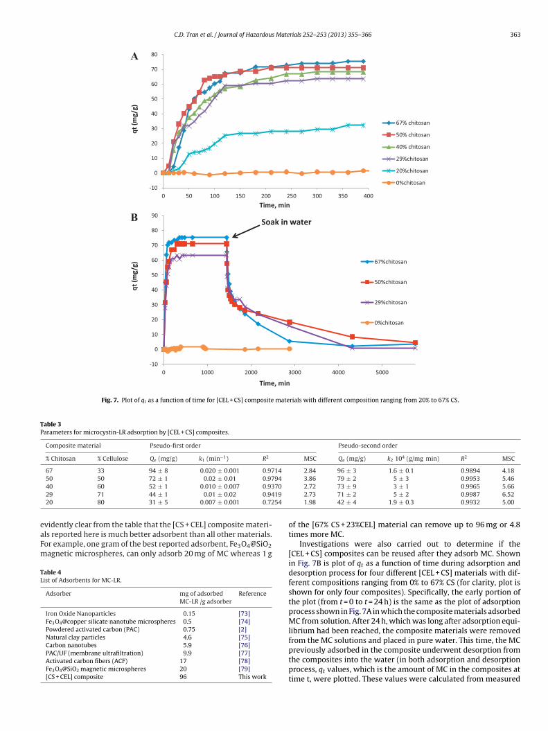
C.D. Tran et al. / Journal of Hazardous Materials 252– 253 (2013) 355– 366 363
-10
0
10
20
30
40
50
60
70
80
0 50 100 150 200 250 300 350 400
qt (m
g/g)
Time, min
67% c hit osan
50% c hit osan
40% c hit osan
29%chit osan
20%chit osan
0%chit osan
-10
0
10
20
30
40
50
60
70
80
90
0 1000 2000 3000 4000 5000
qt (m
g/g)
Time, min
67%chit osan
50%chit osan
29%chit osan
0%chit osan
Soak in wa ter
A
B
Fig. 7. Plot of qt as a function of time for [CEL + CS] composite materials with different composition ranging from 20% to 67% CS.
Table 3Parameters for microcystin-LR adsorption by [CEL + CS] composites.
Composite material Pseudo-first order Pseudo-second order
% Chitosan % Cellulose Qe (mg/g) k1 (min−1) R2 MSC Qe (mg/g) k2 104 (g/mg min) R2 MSC
67 33 94 ± 8 0.020 ± 0.001 0.9714 2.84 96 ± 3 1.6 ± 0.1 0.9894 4.1850 50 72 ± 1 0.02 ± 0.01 0.9794 3.86 79 ± 2 5 ± 3 0.9953 5.46
70
19
54
eaFm
TL
40 60 52 ± 1 0.010 ± 0.007 0.9329 71 44 ± 1 0.01 ± 0.02 0.9420 80 31 ± 5 0.007 ± 0.001 0.72
vidently clear from the table that the [CS + CEL] composite materi-ls reported here is much better adsorbent than all other materials.or example, one gram of the best reported adsorbent, Fe3O4@SiO2agnetic microspheres, can only adsorb 20 mg of MC whereas 1 g
able 4ist of Adsorbents for MC-LR.
Adsorber mg of adsorbedMC-LR /g adsorber
Reference
Iron Oxide Nanoparticles 0.15 [73]Fe3O4@copper silicate nanotube microspheres 0.5 [74]Powdered activated carbon (PAC) 0.75 [2]Natural clay particles 4.6 [75]Carbon nanotubes 5.9 [76]PAC/UF (membrane ultrafiltration) 9.9 [77]Activated carbon fibers (ACF) 17 [78]Fe3O4@SiO2 magnetic microspheres 20 [79][CS + CEL] composite 96 This work
2.72 73 ± 9 3 ± 1 0.9965 5.662.73 71 ± 2 5 ± 2 0.9987 6.521.98 42 ± 4 1.9 ± 0.3 0.9932 5.00
of the [67% CS + 23%CEL] material can remove up to 96 mg or 4.8times more MC.
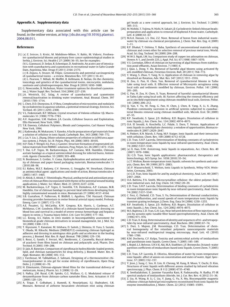
Investigations were also carried out to determine if the[CEL + CS] composites can be reused after they adsorb MC. Shownin Fig. 7B is plot of qt as a function of time during adsorption anddesorption process for four different [CEL + CS] materials with dif-ferent compositions ranging from 0% to 67% CS (for clarity, plot isshown for only four composites). Specifically, the early portion ofthe plot (from t = 0 to t = 24 h) is the same as the plot of adsorptionprocess shown in Fig. 7A in which the composite materials adsorbedMC from solution. After 24 h, which was long after adsorption equi-librium had been reached, the composite materials were removedfrom the MC solutions and placed in pure water. This time, the MC
previously adsorbed in the composite underwent desorption fromthe composites into the water (in both adsorption and desorptionprocess, qt values, which is the amount of MC in the composites attime t, were plotted. These values were calculated from measured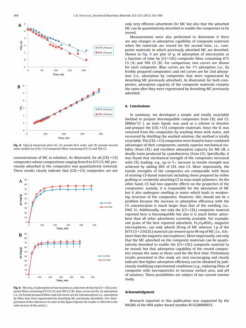
364 C.D. Tran et al. / Journal of Hazardous Mate
-1
0
1
2
3
4
5
0 50 100 150 200
ln(q
e-q t
)
Time, min
67% c hit osan
50% c hit osan
A
0
1
2
3
4
5
6
0 100 200 300 400
t/q t
Time, min
67% c hit osan
50% c hit osan
B
Fo
ccvT
Fp(bpw
ig. 8. Typical linearized plots for (A) pseudo-first order and (B) pseudo-secondrder models for [CEL + CS] composite films containing 67% CS and 50% CS.
oncentrations of MC in solution). As illustrated, for all [CEL + CS]
omposites whose compositions ranging from 0 to 67% CS, MC pre-iously adsorbed in the composites was quantitatively removed.hese results clearly indicate that [CEL + CS] composites are not0
10
20
30
40
50
60
70
80
90
0 100 200 300 400 500
qt (m
g/g)
Time, min
1st Ads
2nd Ads
0
10
20
30
40
50
60
70
80
90
0 100 200 300 400 500 600
qt (m
g/g)
Time, min
1st Ads
2nd Ads
A
B
ig. 9. Plot of qt of adsorption of microcystin as a function of time by [CS + CEL] com-osite films containing 67% CS (A) and 50% CS (B): blue curves are for 1st adsorptioni.e., by freshly prepared films) and red curves are for 2nd adsorption (i.e., adsorptiony films that were regenerated by desorbing MC previously adsorbed). (For inter-retation of the references to color in this figure legend, the reader is referred to theeb version of the article.)
rials 252– 253 (2013) 355– 366
only very efficient adsorbents for MC but also that the adsorbedMC can be quantitatively desorbed to enable the composites to bereused.
Measurements were also performed to determine if thereare any changes in adsorption capability of composite materialswhen the materials are reused for the second time, i.e., com-posite materials in which previously adsorbed MC are desorbed.Shown in Fig. 9 are plot of qt of adsorption of microcystin asa function of time by [CS + CEL] composite films containing 67%CS (A) and 50% CS (B). For comparison, two curves are shownfor each composite: Blue curves are for 1st adsorption (i.e., byfreshly prepared composites) and red curves are for 2nd adsorp-tion (i.e., adsorption by composites that were regenerated bydesorbing MC previously adsorbed). As illustrated, for both com-posites, adsorption capacity of the composite materials remainsthe same after they were regenerated by desorbing MC previouslyadsorbed.
4. Conclusions
In summary, we developed a simple and totally recyclablemethod to prepare biocompatible composites from CEL and CS.[BMIm+Cl−], an ionic liquid, was used as a solvent to dissolveand prepare the [CEL + CS] composite materials. Since the IL wasremoved from the composites by washing them with water, andrecovered by distilling the washed solution, the method is totallyrecyclable. The [CEL + CS] composites were found to have combinedadvantages of their components, namely superior mechanical sta-bility (from CEL) and excellent adsorption capacity for MC-LR, adeadly toxin produced by cyanobacteria (from CS). Specifically, itwas found that mechanical strength of the composites increasedwith CEL loading; e.g., up to 5× increase in tensile strength wasachieved by adding 80% of CEL into CS. More importantly, thetensile strengths of the composites are comparable with thoseof existing CS-based materials including those prepared by eithergrafting or covalently attaching CS to man-made polymers. On theother hand, CS had two opposite effects on the properties of thecomposites: namely, it is responsible for the adsorption of MCbut it also undergoes swelling in water which leads to weaken-ing structure of the composites. However, this should not be aproblem because the increase in adsorption efficiency with theCS concentration is much larger than that of the swelling (i.e.,EWC %). Additionally, not only the [CS + CEL] composite materialreported here is biocompatible but also it is much better adsor-bent than all other adsorbents currently available. For example,one gram of the best reported adsorbent, Fe3O4@SiO2 magneticmicrospheres, can only adsorb 20 mg of MC whereas 1 g of the[67% CS + 23%CEL] material can remove up to 96 mg of MC (i.e., 4.8×more than the magnetic microspheres). More importantly, not onlythat the MC adsorbed on the composite materials can be quanti-tatively desorbed to enable the [CS + CEL] composite material tobe reused, but that adsorption capability of the reused compos-ites remain the same as those used for the first time. Preliminaryresults presented in this study are very encouraging and clearlyindicate that higher adsorption efficiency can be obtained by judi-ciously modifying experimental conditions (e.g., replacing films ofcomposite with microparticles to increase surface area, and pHof solution). These possibilities are subject of our current intensestudy.
Acknowledgment
Research reported in this publication was supported by theNIGMS of the NIH under Award number R15GM099033.
s Mate
A
f2
R
[
[
[
[
[
[
[
[
[
[
[
[
[
[
[
[
[
[
[
[
[
[
[
[
[
[
[
[
[
[
[
[
[
[
[
[
[
[
[
[
[
[
C.D. Tran et al. / Journal of Hazardou
ppendix A. Supplementary data
Supplementary data associated with this article can beound, in the online version, at http://dx.doi.org/10.1016/j.plantsci.004.08.011.
eferences
[1] (a) Z. Svircev, S. Krstic, M. Malidinov-Mikov, V. Baltic, M. Vidovic, Freshwa-ter cyanobacterial blooms and primary liver cancer epidemiological studies inSerbia, J. Environ. Sci. Health C 27 (2009) 36–55, See for examples;(b) L. Giannuzzi, D. Sedan, R. Echenique, D. Andrinolo, An acute case of intoxica-tion with cyanobacteria and cyanotoxins in recreational water in Salto GrandeDam, Argentina, Mar. Drugs 9 (2011) 2164–2175;(c) B. Zegura, A. Straser, M. Filipic, Genotoxicity and potential carcinogenicityof cyanobacterial toxins – a review, Mutation Res. 727 (2011) 16–41;(d) L. Pearson, T. Mihali, M. Moffitt, R. Kellmann, B. Neilan, On the chemistry,toxicology and genetics of the cyanobacterial toxins, microcystin, nodularin,saxitoxin and cylindrospermopsin, Mar. Drugs 8 (2010) 1650–1680.
[2] G. Newcombe, B. Nicholson, Water treatment options for dissolved cyanotox-ins, J. Water Suppl. Res. Technol. 534 (2004) 227–239.
[3] J.A. Westrick, D.C. Szlag, A review of cyanobacteria and cyanotoxinsremoval/inactivation in drinking water treatment, Anal. Bioanal. Chem. 397(2010) 1705–1714.
[4] L. Chen, D.D. Dionysiou, K. O’Shea, Complexation of microcystins and nodularinby cyclodextrins in aqueous solution, a potential removal strategy, Environ. Sci.Technol. 45 (2011) 2293–2300.
[5] V.L. Finkenstadt, R.P. Millane, Crystal structure of Valonia cellulose 1�, Macro-molecules 31 (1998) 7776–7783.
[6] A.V. Augustine, S.M. Hudson, J.A. Cuculo, Cellulose Sources and Exploitation,Ellis Horwood, New York, 1990.
[7] T.R. Dawsey, Cellulosic Polymers, Blends and Composites, Carl Hanser Verlag,New York, 1994.
[8] J. Kadowaka, M. Mukarami, Y. Kaneko, A facile preparation of gel materials froma solution of cellulose in ionic liquid, Carbohydr. Res. 343 (2008) 769–772.
[9] J. Cai, Y. Liu, L. Zhang, Dilute solution properties of cellulose in LiOH/urea aque-ous system, J. Polym. Sci. B: Polym. Phys. 44 (2006) 3093–30105.
10] H.P. Fink, P. Weigel, H.J. Purz, J. Ganster, Structure formation of regenerated cel-lulose materials from NMMO- solutions, Prog. Polym. Sci. 26 (2001) 1473–1524.
11] T. Dai, G.P. Tegos, M. Barkatovskaya, A.P. Castano, M.R. Hamblin, Chitosanacetate bandage as a topical antimicrobial dressing for infected burns, Antimi-crob. Agents Chemother. 53 (2009) 393–400.
12] N. Bordenave, S. Grelier, V. Coma, Hydrophobization and antimicrobial activ-ity of chitosan and paper-based packaging materials, Biomacromolecules 11(2010) 88–96.
13] E.I. Rabea, M.E.T. Badawy, C.V. Stevens, G. Smagghe, W. Steurbaut, Chitosanas antimicrobial agent: applications and mode of action, Biomacromolecules 4(2003) 1457–1465.
14] D. Altiok, E. Altiok, F. Tihminlioglu, Physical, antibacterial and antioxidant prop-erties of chitosan films incorporated with thyme oil for potential wound healingapplications, J. Mater. Sci: Mater. Med. 21 (2010) 2227–2236.
15] M. Burkatovskaya, G.P. Tegos, E. Swietlik, T.N. Demidova, A.P. Castano, M.R.Hamblin, Use of chitosan bandage to prevent fatal infections developing fromhighly contaminated wounds in mice, Biomaterials 27 (2006) 4157–4164.
16] S.B. Gustafson, P. Fulkerson, R. Bildfell, L. Aguilera, T.M. Hazzard, Chitosandressing provides hemostasis in swine femoral arterial injury model, Prehosp.Emerg. Care 11 (2007) 172–178.
17] A.E. Pusateri, S.J. McCarthy, K.W. Gregory, R.A. Harris, L. Cardenas, A.T.McManus, C.W. Goodwin, Effect of a chitosan-based haemostatic dressing onblood loss and survival in a model of severe venous hemorrhage and hepaticainjury in swine, J. Trauma Injury Infect. Crit. Care 54 (2003) 177–182.
18] L.C. Keong, A.S. Halim, In vitro models in biocompatibility assessment forbiomedical-grade chitosan derivatives in wound management, Int. J. Mol. Sci.10 (2009) 1300–1313.
19] T. Kiyozumi, Y. Kanatani, M. Ishihara, D. Saitoh, J. Shimizu, H. Yura, S. Suzuki,Y. Okada, M. Kikuchi, Medium (DMEM/F12)-containing chitosan hydrogel asadhesive and dressing in autologous skin grafts and accelerator in the healingprocess, J. Biomed. Mat. Res. B: Appl. Biomat. 79B (2006) 129–136.
20] S. Rossi, G. Sandri, F. Ferrari, M.C. Benferonic, C. Caramella, Buccal deliveryof acyclovir from films based on chitosan and polyacrylic acid, Pharm. Dev.Technol. 8 (2003) 199–208.
21] D. Jain, R. Banerjee, Comparison of ciprofloxacin hydrochloride-loaded protein,lipid, and chitosan nanoparticles for drug delivery, J. Biomed. Mater. Res. B:Appl. Biomater. 86 (2008) 105–112.
22] J. Varshosaz, M. Tabbakhian, Z. Salmani, Designing of a thermosensitive chi-tosan/poloxamer in situ gel for ocular delivery of ciprofloxacin, Open DrugDeliv. J. 2 (2008) 61–70.
23] H. Elmotasem, Chitosan-alginate blend films for the transdermal delivery ofmeloxicam, Asian J. Pharm. Sci. 3 (2008) 12–29.
24] S. Naficy, J.M. Razal, G.M. Spinks, G.G. Wallace, G. G. Modulated release ofdexamethasone from chitosan-carbon nanotube films, Sens. Actuators A 155(2009) 120–124.
25] A. Tirgar, F. Golbabaei, J. Hamedi, K. Nourijelyani, S.J. Shahtaheri, S.R.Moosavi, Removal of airborne hexavalent chromium mist using chitosan
[
[
rials 252– 253 (2013) 355– 366 365
gel beads as a new control approach, Int. J. Environ. Sci. Technol. 3 (2006)305–313.
26] M. Nishiki, T. Tojima, N. Nishi, N. Sakairi, �-Cyclodextrin-linked chitosan beads:preparation and application to removal of bisphenol A from water, Carbohydr.Lett. 4 (2000) 61–67.
27] M.A.A. Hassan, L.S. Hui, Z.Z. Noor, Removal of boron from industrial waste-water by chitosan via chemical precipitation, J. Chem. Nat. Res. Eng. 4 (2009)1–11.
28] R.P. Dhakal, T. Oshima, Y. Baba, Synthesis of unconventional materials usingchitosan and crown ether for selective removal of precious metal ions, World.Acad. Sci. Eng. Technol. 56 (2009) 204–208.
29] W.W.S. Ngah, I.M. Isa, Comparison study of copper ion adsorption on chitosan,Dowex A-1, and Zerolit 225, J, Appl. Pol. Sci. 67 (1998) 1067–1070.
30] V.S. Govindan, Effect of chitosan on harvesting of algal biomass from stabiliza-tion pond effluents, Asian Environ. 7 (1985) 4.
31] J. Qiao, L. Dong, Y. Hu, Removal of harmful algal blooms using activated flyash-modified chitosan, Fresenius Environ. Bull. 20 (2011) 764–772.
32] Y. Wang, S. Zhuo, Y. Yang, N. Li, Application of chitosan in removing algae bydissolved air flotation, Adv. Mat. Res. 347 (2012) 1911–1916.
33] H. Zou, G. Pan, H. Chen, Yan, Removal of cyanobacterial blooms in TaihuLake using local soils. II. Effective removal of Microsystis aeruginosa usinglocal soils and sediments modified by chitosan, Environ. Pollut. 141 (2006)201–205.
34] G. Pan, H. Zou, H. Chen, X. Yuan, Removal of harmful cyanobacterial bloomsin Taihu Lake using local soils. III. Factors affecting the removal efficiency andan in situ field experiment using chitosan-modified local soils, Environ. Pollut.141 (2006) 206–212.
35] Q. Yan, Y. Yu, W. Feng, G. Pan, H. Chen, J. Chen, B. Yang, X. Li, X. Zhang,Plankton community sucession in artificial systems subjected to cyanobac-terial blooms removal using chitosan modified soils, Microb. Ecol. 58 (2009)47–55.
36] R.P. Swatloski, S. Spear, J.D. Holbrey, R.D. Rogers, Dissolution of cellulose inionic liquids, J. Am. Chem. Soc. 124 (2002) 4974–4975.
37] O.A. El Seould, A. Koschella, L.C. Fidale, S. Dorn, T. Heinze, Applications ofionic liquids in carbohydrate chemistry: a window of opportunities, Biomacro-molecules 8 (2007) 2629–2647.
38] A. Pinkert, K.N. Marsh, S. Pang, M.P. Staiger, Ionic liquids and their interactionwith cellulose, Chem. Rev. 109 (2009) 6712–6728.
39] C.D. Tran, S.H.P. Lacerda, Determination of binding constants of cyclodextrinsin room temperature ionic liquids by near-infrared spectrometry, Anal. Chem.74 (2002) 5337–5341.
40] (a) X. Han, D.W. Armstrong, Ionic liquids in separations, Acc. Chem. Res. 40(2007) 1079–1086;(b) C.D. Tran, Ionic liquids applications: pharmaceutical, therapeutics andbiotechnology, ACS Symp. Ser. 1038 (2010) 35–54;(c) T. Welton, Room-temperature ionic liquids. solvents for synthesis and catal-ysis, Chem. Rev. 99 (1999) 2071–2083;(d) P. Wasserscheid, T. Welton, Ionic Liquids in Synthesis, Wiley-VCH, Wein-heim, Germany, 2003;(e) C.D. Tran, Ionic liquids for and by analytical chemistry, Anal. Lett. 40 (2007)2447–2464.
41] O.A. Battista, P.A. Smith, Microcrystalline cellulose: the oldest polymer findsnew industrial uses, Ind. Eng. Chem. 54 (1962) 20–29.
42] C.D. Tran, S.H.P. Lacerda, Determination of binding constants of cyclodextrinsin room temperature ionic liquids by near-infrared spectrometry, Anal. Chem.74 (2002) 5337–5341.
43] C. Frez, G. Diebold, C.D. Tran, S. Yu, Determination of thermal conductivities,thermal diffusivities, and sound speeds of room temperature ionic liquids bytransient grating technique, J.Chem. Eng. Data 54 (2006) 1250–1255.
44] R.P. Swatloski, S. Spear, J.D. Holbrey, R.D. Rogers, Dissolution of cellulose inionic liquids, J. Am. Chem. Soc. 124 (2002) 4974–4975.
45] M.S. Baptista, C.D. Tran, G.H. Gao, Near Infrared detection of flow injection anal-ysis by acousto-optic tunable filter based spectrophotometry, Anal. Chem. 68(1996) 971–976.
46] C.D. Tran, X. Kong, Determination of identity and sequences of tri- and tetrapep-tides by near-infrared spectrometry, Anal. Biochem. 286 (2000) 67–74.
47] S. Duri, S. Majoni, J.M. Hossenlopp, C.D. Tran, Determination of chem-ical homogeneity of fire retardant polymeric nanocomposite materialsby near-infrared multispectral imaging microscopy, Anal. Lett. 43 (2010)1780–1789.
48] K.M. Docherty, C.F. Kulpa, Toxicity and antimicrobial activity of imidazoliumand pyridinium ionic liquids, Green Chem. 7 (2005) 185–189.
49] L. Ropel, L.S. Belveze, S.N.V.K. Aki, M.A. Stadtherr, J.F. Brennecke, Octanol–waterpartition coefficients of imidazolium-based ionic liquids, Green Chem. 7 (2005)83.
50] C.D. Tran, S.P. Lacerda, D. Oliveira, Absorption of water by room-temperatureionic liquids: affect of anions on concentration and states of water, Appl. Spec-trosc. 57 (2003) 152–157.
51] Y. Jeon, J. Sung, C. Seo, H. Lim, H. Cheong, M. Kang, B. Moon, Y. Ouchi, D. Kim,Structures of ionic liquids with different anions studied by Infrared vibrationspectroscopy, J. Phys. Chem. B 112 (2008) 4735–4740.
52] K. Seethalakshmi, E. Jasmine Vasantha Rani, R. Padmavathy, N. Radha, FT-IRspectral analysis of imidazolium chloride, Int. J. Cur. Res. Rev. 4 (2012) 31–36.
53] Z. Liu, H. Wang, B. Li, C. Liu, Y. Jiang, G. Yu, X. Mu, Biocompatible magneticcellulose-chitosan hybrid gel microspheres reconstituted from ionic liquids forenzyme immobilization, J. Mater. Chem. 22 (2012) 15085–15091.

3 s Mate
[
[
[
[
[
[
[
[
[
[
[
[
[
[
[
[
[
[
[
[
[
[
[
[
[
66 C.D. Tran et al. / Journal of Hazardou
54] Z. Liu, H. Wang, C. Liu, Y. Jiang, G. Yu, X. Mu, X. Wang, Magnetic cellulose-chitosan hydrogels prepared from ionic liquids as reusable adsorbents forremoval of heavy metal ions, Chem. Commun. 48 (2012) 7530–7532.
55] A.L. Da Roz, F.L. Leite, L.V. Pereiro, P.A.P. Nascente, V. Zucolotto, O.N. Oliveira,A.J.F. Carvalho, Adsorption of chitosan on spin-coated cellulose films, Carbo-hydr. Polym. 80 (2010) 65–70.
56] S. Dreve, I. Kacso, I. Bratu, E. Indrea, Chitosan-based delivery systems fordiclofenac delivery: peparation and characterization, J. Phys.: Conf.Ser. 182(2009) 1–4.
57] D.A. Burns, E.W. Ciurczak, Handbook of Near-Infrared Analysis, Marcell Dekker,New York, 1992.
58] J.W. Ellis, Infra-red absorption by the N H bond II in aryl, alkyl and aryl-alkylamines, J.Am. Chem. Soc. 50 (1928) 685–695.
59] M.S. Baptista, C.D. Tran, G.H. Gao, Near infrared detection of flow injection anal-ysis by acousto-optic tunable filter based spectrophotometry, Anal. Chem. 68(1996) 971–976.
60] C.D. Tran, X. Kong, Determination of identity and sequences of tri- and tetrapep-tides by near-infrared spectrometry, Anal. Biochem. 286 (2000) 67–74.
61] L. Zhang, D. Ruan, S. Gao, Dissolution and regeneration of cellulose inNaOH/Thiourea aqueous solution, J. Polym. Sci: Part B: Polym. Phys. 40 (2002)1521–1529.
62] T. Mori, E. Chikayama, Y. Tsuboi, N. Ishida, N. Shisa, Y. Noritake, S. Moriya amd,J. Kikuchi, Exploring the conformationa; space of amorphous cellulose usingNMR chemical shifts, Carbohydr. Polym. 90 (2012) 1197–1203.
63] G. Sebe, F. Ham-Pichavant, E. Ibarboure, A.L.C. Koffi, P. Tingaut, Supramolecu-lar structure characterization of cellulose II nanowhiskers producued by acidhydrolysis of cellulose I substrates, Biomacromolecules 13 (2012) 570–578.
64] H. Kiuchi, W. Kai, Y. Inoue, Preparation and characterization of poly(ethyleneglycol) crosslinked chitosan films, J. Appl. Polym. Sci. 107 (2008)3823–3830.
65] S. Khamhan, Y. Baimark, S. Chaichanadee, P. Phinyocheep, S.W. Kittipoom,Water vapor permeability and mechanical properties of biodegradable chi-
tosan/methoxy poly(ethylene glycol)-b-poly(�-caprolactone) nanocompositefilms, Int. J. Polym. Anal. Charact. 13 (2008) 224–231.66] R. Alam, A.M. Khan, A.R. Khan, S. Ghoshal, M.I.H. Mondal, Study on thephysico-mechanical properties of photo-cured chitosan films with oligomerand acrylate monomer, J. Polym. Environ. 16 (2008) 213–219.
[
rials 252– 253 (2013) 355– 366
67] S. Liang, L. Liu, Q. Huang, L.K. Yam, Preparation of single or double- network chi-tosan/poly(vinyl alcohol) gel films through selectively cross-linking method,Carbohydr. Polym. 77 (2009) 718–724.
68] M.A. Khan, R. Alam, F. Noor, R.A. Khan, A.Md. Rahman, Modification and char-acterization of chitosan films using 3-trimethoxysilylpropyl methacrylate, J.Macrom. Sci. Part A: Pure Appl. Chem. 46 (2009) 751–758.
69] C. Radhakumary, D.N. Prabha, C.P.N. Reghunadhan, S. Mathew, Chitosan- comb-graft-polyethylene glycol monomethacrylate-Synthesis, characterization, andevaluation as a biomaterial for hemodialysis applications, J. Appl. Polym. Sci.114 (2009) 2873–2886.
70] Y.S. Ho, G. McKay, The kinetics of sorption of divalent metal ions onto sphagnummoss peat, Water Res. 34 (2000) 735–742.
71] K.V. Kumar, S. Sivanesan, Pseudo second order kinetics and pseudo isothermsfor malachite green onto activated carbon: comparison of linear and non- linearregression methods, J. Hazard. Mater. B 136 (2006) 721–726.
72] Q. Li, L. Sun, Y. Zhang, Y. Qian, J. Zhai, Characteristics of equilibrium, kineticsstudies for adsorption of Hg(II) and Cr(VI) by polyaniline/humic acid composite,Desalination 266 (2011) 188–194.
73] Y. Gao, N. Gao, J. Gu, Y. Shen, S. Wang, Adsorption of microcystin-LR from waterwith iron oxide nanoparticles, Water Environ. Res. 84 (2012) 562–568.
74] H. Chen, X. Lu, C. Deng, X. Yan, Facile synthesis of uniform microsphere com-posed of magnetite core and copper silicate nanotube shell for removal ofMicrocystins in water, J. Phys. Chem. C 113 (2009) 21068–21073.
75] R.J. Morris, D.E. Williams, H.A. Luu, C.F.B. Holmes, R.J. Andersen, S.E. Calvert,The adsorption of microcystin-LR by natural clay particles, Toxicon 38 (2000)303–308.
76] H. Yan, A. Gong, H. He, J. Zhou, Y. Wei, Adsorption of microcystins by carbonnanotubes, Chemosphere 62 (2006) 142–148.
77] J. Lee, H. Walker, Effects of process variables and natural organic matteron removal of microcystin-LR by PAC-UF, Environ. Sci. Technol. 40 (2006)7336–7342.
78] D. Pyo, D. Moon, Adsorption of microcystin LR by activated carbon fibers, Bull.
Korean Chem. Soc. 26 (2005) 2089–2092.79] Y.H. Deng, D.W. Qi, C. Deng, X.M. Zhang, D.Y. Zhao, Superparamagnetic high-magnetization microspheres with an Fe3O4@SiO2 core and perpendicularlyaligned mesoporous SiO2 shell for removal of microcystins, J. Am. Chem. Soc.130 (2008) 28–29.