Journal of Controlled Release - -ORCAorca.cf.ac.uk/80771/1/Disarmed anthrax toxin delivers...
13
Disarmed anthrax toxin delivers antisense oligonucleotides and siRNA with high efficiency and low toxicity Paul D.R. Dyer a,1 , Thomas R. Shepherd a,1 , Alexander S. Gollings a,1 , Susan A. Shorter a,1 , Monique A.M. Gorringe-Pattrick a , Chun-Kit Tang a , Beatrice N. Cattoz c , Les Baillie b , Peter C. Griffiths c , Simon C.W. Richardson a,* a Intercellular Delivery Solutions Laboratory, Faculty of Engineering and Science, University of Greenwich, Central Avenue, Chatham Maritime, Kent, ME4 4TB, UK b School of Pharmacy and Pharmaceutical Sciences, Cardiff University, King Edward VII Avenue, Cardiff CF10 3AX, UK c Department of Pharmaceutical, Chemical and Environmental Science, Faculty of Engineering and Science, University of Greenwich, Central Avenue, Chatham Maritime, Kent ME4 4TB, UK abstract article info Article history: Received 28 July 2015 Received in revised form 26 October 2015 Accepted 28 October 2015 Available online xxxx Keywords: Antisense RNAi Anthrax toxin PEG-dilemma Non-viral Inefficient cytosolic delivery and vector toxicity contribute to the limited use of antisense oligonucleotides (ASOs) and siRNA as therapeutics. As anthrax toxin (Atx) accesses the cytosol, the purpose of this study was to evaluate the potential of disarmed Atx to deliver either ASOs or siRNA. We hypothesized that this delivery strat- egy would facilitate improved transfection efficiency while eliminating the toxicity seen for many vectors due to membrane destabilization. Atx complex formation with ASOs or siRNA was achieved via the in-frame fusion of either Saccharomyces cerevisiae GAL4 or Homo sapien sapien PKR (respectively) to a truncation of Atx lethal factor (LFn), which were used with Atx protective antigen (PA). Western immunoblotting confirmed the production of: LFN-GAL4, LFn-PKR and PA which were detected at ~45.9 kDa, ~37 kDa, and ~83 kDa respectively and small angle neutron scattering confirmed the ability of PA to form an annular structure with a radius of gyration of 7.0 ± 1.0 nm when placed in serum. In order to form a complex with LFn-GAL4, ASOs were engineered to contain a double-stranded region, and a cell free in vitro translation assay demonstrated that no loss of antisense activity above 30 pmol ASO was evident. The in vitro toxicity of both PA:LFn-GAL4:ASO and PA:LFn-PKR:siRNA complexes was low (IC 50 N 100 μg/mL in HeLa and Vero cells) and subcellular fractionation in conjunction with microscopy confirmed the detection of LFn-GAL4 or LFn-PKR in the cytosol. Syntaxin5 (Synt5) was used as a model target gene to determine pharmacological activity. The PA:LFn-GAL4:ASO complexes had transfection efficiency approximately equivalent to Nucleofection ® over a variety of ASO concentrations (24 h post- transfection) and during a 72 h time course. In HeLa cells, at 200 pmol ASO (with PA:LFN-GAL4), 5.4 ± 2.0% Synt5 expression was evident relative to an untreated control after 24 h. Using 200 pmol ASOs, Nucleofection ® reduced Synt5 expression to 8.1 ± 2.1% after 24 h. PA:LFn-GAL4:ASO transfection of non- or terminally-differen- tiated THP-1 cells and Vero cells resulted in 35.2 ± 19.1%, 36.4 ± 1.8% and 22.9 ± 6.9% (respectively) Synt5 expression after treatment with 200 pmol of ASO and demonstrated versatility. Nucleofection ® with Stealth RNAi™ siRNA reduced HeLa Synt5 levels to 4.6 ± 6.1% whereas treatment with the PA:LFn-PKR:siRNA resulted in 8.5 ± 3.4% Synt5 expression after 24 h (HeLa cells). These studies report for the first time an ASO and RNAi delivery system based upon protein toxin architecture that is devoid of polycations. This system may utilize regulated membrane back-fusion for the cytosolic delivery of ASOs and siRNA, which would account for the lack of toxicity observed. High delivery efficiency suggests further in vivo evaluation is warranted. © 2015 The Authors. Published by Elsevier B.V. This is an open access article under the CC BY license (http://creativecommons.org/licenses/by/4.0/). Journal of Controlled Release 220 (2015) 316–328 Abbreviations: ASOs, antisense oligonucleotides; Atx, anthrax toxin; BME, beta-mercaptoethanol; MTT, 3-(4,5-dimethylthiazol-2-yl)-2,5-diphenyltetrazolium bromide; E7, early 7; EEA1, early endosomal antigen 1; LFn, Atx lethal factor domain I; GFP, Green fluorescent protein; PKR, Homo sapien sapien protein kinase-R; HPV, human papilloma virus; ILVs, intraluminal vesicles; LBPA, lysobisphosphatidic acid; LAMP2, lysosome associated membrane protein 2; LF, lethal factor; MVBs, multivesicular bodies; PEI, poly(ethyleneimine); PNS, post-nuclear su- pernatant; PA, protective antigen; GAL4, Saccharomyces cerevisiae galactose metabolism DNA binding protein; SANS, small-angle neutron scattering; (si)RNA, small interfering; Synt5, Syntaxin5; t-GFP, turbo-GFP. ⁎ Corresponding author. E-mail address: [email protected] (S.C.W. Richardson). 1 These authors contributed equally to this manuscript. http://dx.doi.org/10.1016/j.jconrel.2015.10.054 0168-3659/© 2015 The Authors. Published by Elsevier B.V. This is an open access article under the CC BY license (http://creativecommons.org/licenses/by/4.0/). Contents lists available at ScienceDirect Journal of Controlled Release journal homepage: www.elsevier.com/locate/jconrel
-
Upload
nguyenthien -
Category
Documents
-
view
217 -
download
0
Transcript of Journal of Controlled Release - -ORCAorca.cf.ac.uk/80771/1/Disarmed anthrax toxin delivers...
Journal of Controlled Release 220 (2015) 316–328
Contents lists available at ScienceDirect
Journal of Controlled Release
j ourna l homepage: www.e lsev ie r .com/ locate / jconre l
Disarmed anthrax toxin delivers antisense oligonucleotides and siRNAwith high efficiency and low toxicity
Paul D.R. Dyer a,1, Thomas R. Shepherd a,1, Alexander S. Gollings a,1, Susan A. Shorter a,1,Monique A.M. Gorringe-Pattrick a, Chun-Kit Tang a, Beatrice N. Cattoz c, Les Baillie b,Peter C. Griffiths c, Simon C.W. Richardson a,*
a Intercellular Delivery Solutions Laboratory, Faculty of Engineering and Science, University of Greenwich, Central Avenue, ChathamMaritime, Kent, ME4 4TB, UKb School of Pharmacy and Pharmaceutical Sciences, Cardiff University, King Edward VII Avenue, Cardiff CF10 3AX, UKc Department of Pharmaceutical, Chemical and Environmental Science, Faculty of Engineering and Science, University of Greenwich, Central Avenue, Chatham Maritime, Kent ME4 4TB, UK
Abbreviations: ASOs, antisense oligonucleotides; Atx,EEA1, early endosomal antigen1; LFn, Atx lethal factor domvesicles; LBPA, lysobisphosphatidic acid; LAMP2, lysosomepernatant; PA, protective antigen; GAL4, Saccharomyces cSyntaxin5; t-GFP, turbo-GFP.⁎ Corresponding author.
E-mail address: [email protected] (S1 These authors contributed equally to this manuscript
http://dx.doi.org/10.1016/j.jconrel.2015.10.0540168-3659/© 2015 The Authors. Published by Elsevier B.V
a b s t r a c t
a r t i c l e i n f oArticle history:Received 28 July 2015Received in revised form 26 October 2015Accepted 28 October 2015Available online xxxx
Keywords:AntisenseRNAiAnthrax toxinPEG-dilemmaNon-viral
Inefficient cytosolic delivery and vector toxicity contribute to the limited use of antisense oligonucleotides(ASOs) and siRNA as therapeutics. As anthrax toxin (Atx) accesses the cytosol, the purpose of this study was toevaluate the potential of disarmed Atx to deliver either ASOs or siRNA. We hypothesized that this delivery strat-egy would facilitate improved transfection efficiency while eliminating the toxicity seen for many vectors due tomembrane destabilization. Atx complex formation with ASOs or siRNA was achieved via the in-frame fusion ofeither Saccharomyces cerevisiaeGAL4 orHomo sapien sapien PKR (respectively) to a truncation of Atx lethal factor(LFn), which were used with Atx protective antigen (PA). Western immunoblotting confirmed the productionof: LFN-GAL4, LFn-PKR and PA which were detected at ~45.9 kDa, ~37 kDa, and ~83 kDa respectively andsmall angle neutron scattering confirmed the ability of PA to form an annular structure with a radius of gyrationof 7.0 ± 1.0 nm when placed in serum. In order to form a complex with LFn-GAL4, ASOs were engineered tocontain a double-stranded region, and a cell free in vitro translation assay demonstrated that no loss of antisenseactivity above 30 pmol ASO was evident. The in vitro toxicity of both PA:LFn-GAL4:ASO and PA:LFn-PKR:siRNAcomplexes was low (IC50 N 100 μg/mL in HeLa and Vero cells) and subcellular fractionation in conjunctionwith microscopy confirmed the detection of LFn-GAL4 or LFn-PKR in the cytosol. Syntaxin5 (Synt5) was usedas amodel target gene to determine pharmacological activity. The PA:LFn-GAL4:ASO complexes had transfectionefficiency approximately equivalent to Nucleofection® over a variety of ASO concentrations (24 h post-transfection) and during a 72 h time course. In HeLa cells, at 200 pmol ASO (with PA:LFN-GAL4), 5.4 ± 2.0%Synt5 expression was evident relative to an untreated control after 24 h. Using 200 pmol ASOs, Nucleofection®
reduced Synt5 expression to 8.1± 2.1% after 24 h. PA:LFn-GAL4:ASO transfection of non- or terminally-differen-tiated THP-1 cells and Vero cells resulted in 35.2 ± 19.1%, 36.4 ± 1.8% and 22.9 ± 6.9% (respectively) Synt5expression after treatment with 200 pmol of ASO and demonstrated versatility. Nucleofection® with StealthRNAi™ siRNA reduced HeLa Synt5 levels to 4.6 ± 6.1% whereas treatment with the PA:LFn-PKR:siRNA resultedin 8.5 ± 3.4% Synt5 expression after 24 h (HeLa cells). These studies report for the first time an ASO and RNAidelivery system based upon protein toxin architecture that is devoid of polycations. This system may utilizeregulated membrane back-fusion for the cytosolic delivery of ASOs and siRNA, which would account for thelack of toxicity observed. High delivery efficiency suggests further in vivo evaluation is warranted.
© 2015 The Authors. Published by Elsevier B.V. This is an open access article under the CC BY license(http://creativecommons.org/licenses/by/4.0/).
anthrax toxin; BME, beta-mercaptoethanol; MTT, 3-(4,5-dimethylthiazol-2-yl)-2,5-diphenyltetrazolium bromide; E7, early 7;ain I; GFP, Greenfluorescent protein; PKR,Homo sapien sapien protein kinase-R; HPV, humanpapilloma virus; ILVs, intraluminalassociatedmembrane protein 2; LF, lethal factor; MVBs, multivesicular bodies; PEI, poly(ethyleneimine); PNS, post-nuclear su-erevisiae galactose metabolism DNA binding protein; SANS, small-angle neutron scattering; (si)RNA, small interfering; Synt5,
.C.W. Richardson)..
. This is an open access article under the CC BY license (http://creativecommons.org/licenses/by/4.0/).
317P.D.R. Dyer et al. / Journal of Controlled Release 220 (2015) 316–328
1. Introduction
Inefficient cytosolic delivery, suboptimal pharmacokinetics andpharmacodynamics and vector toxicity contribute to the limited appli-cation of antisense oligonucleotides (ASOs) and small interfering(si)RNAs as routinely used clinical tools [1,2]. A variety of non-viraldelivery systems have been explored in relation to the intracellular de-livery of siRNA and ASOs. However, to date, despite three antisensedrugs being licensed by the FDA [3,4,5], no ASO intracellular deliverysystems have entered into routine clinical use [1,2].
Anthrax toxin (Atx) has evolved to mediate the cytosolic delivery ofmacromolecules [6] and the attenuation of its intrinsic toxicity usingrecombinant technology is facile i.e. using recombinant PCR to removeLethal Factor (LF) domains II–IV to give rise to the truncated proteinLFn [7]. The translocation of LFn into the cytosol requires Bacillusanthracis protective antigen (PA) and has been described [6–10]. Thisoperation requires the association of PA with one of its three knownreceptors (β1-integrin [11], tumor endothelial marker 8 (TEM8) [12]or capillary morphogenesis gene-2 (CMG-2) [13], which are almostubiquitously expressed in mammals. PA has been reported to formhomoheptamers [6] or homooctamers prior to, or during cell receptorassociation [14], which are then trafficked onto the limiting membraneof intraluminal vesicles (ILVs)withinmultivesicular bodies (MVBs) [8,9,10]. A pH-driven PA conformational change results in PA membraneinsertion (pre-pore to pore transition) [15] and the translocation ofLFn over the ILV limiting membrane, into the lumen of the ILV. A subse-quent back-fusion event between the ILV and the limitingmembrane ofthe MBV releases LF(n) into the cytosol [8,9,10]. Given that all of theevents culminating in back-fusion and LFn cytosolic release are highlyregulated, it may be possible to exploit these trafficking events for notonly the transport of peptides into the cytosol [16,17,18] but also themovement of nucleic acids,without the aid of any polycationic condens-ing agents [19,20,21].
Consequently, the aim of this study was to evaluate the potentialof Atx components to deliver ASOs and siRNA to the cytosol withoutgenerating the toxicity resulting from non-specific membrane
Fig. 1.Exploitingmembraneback-fusion for thedelivery of ASOs and siRNA. This cartoon is adapstudy it is used to illustrate the possible route taken to the cytosol by the Atx derived delivery
destabilization common to many forms of non-viral delivery technolo-gy. A cartoon representing the proposed delivery strategy, and themembrane back-fusion event acting as an “airlock” into the cytosol, isshown (Fig. 1) and is based upon the trafficking of LF [8,9,10]. To thisend, ASOs or siRNA was joined to LFn via the in-frame fusion of a DNAor RNA binding protein requiring Saccharomyces cerevisiae galactosemetabolismDNAbindingprotein (GAL4) (for ASOs) [21] orHomo sapiensapien protein kinase-R (PKR) [22] (for RNA) to be fused in frame to LFnand used in conjunction with PA.
For the first time, we report the ability of disarmed Atx to deliverASOs and siRNA to a variety of primate cell lines (HeLa, THP-1 andVero) without the aid of any polycationic or lipidic helpers. The PA,LFn-GAL4, LFn-PKR proteins and their complexes with ASO and StealthRNAi™ siRNA were characterized in relation to structure and generaltoxicity inHela andVero cells in vitro. Fluorescencemicroscopy and sub-cellular fractionation were used to verify cytosolic entry of LFn-GAL4and LFn-PKR or their Texas Red®-labeled analogues and the traffickingof PA to lysobisphosphatidic acid (LBPA) positive endocytic vesiclesover time. Finally the ability of the disarmed Atx to deliver ASOs andsiRNA to a variety of primate cell lines (HeLa, THP-1 and Vero) wasstudied by measuring the knockdown of Syntaxin5 (Synt5) expressionas a model to quantitate pharmacological activity in relation toNucleofection® (and lipofection) as a benchmark available commercial-ly to anyone wishing to gauge the efficiency of their system in relationto the data reported herein.
2. Materials and methods
2.1. Oligonucleotides
ASOs are described (Table 1) and were supplied by Invitrogen(Paisley, UK). The antisense sequence against Synt5 has previouslybeen published [23]. Synt5 specific Stealth RNAi™ siRNA modifiedRNA (4392420; Life Technologies, Paisley, UK) was purchasedcommercially.
ted fromdata describing the cytosolic translocation of LF(n) [6–10]. For thepurposes of thissystems described herein i.e. PA:LFn-GAL4:ASO or PA:LFn-PKR:siRNA.
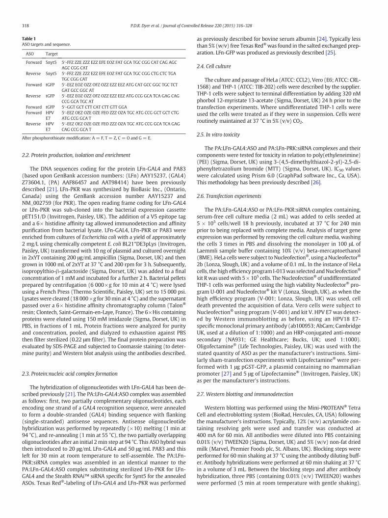
Table 1ASO targets and sequence.
ASO Target
Forward Snyt5 5′-FFZ ZZE ZZZ EZZ EFE EOZ FAT GCA TGC CGG CAT CAG AGCAGC CGG CAT
Reverse Snyt5 5′-FFZ ZZE ZZZ EZZ EFE EOZ FAT GCA TGC CGG CTG CTC TGATGC CGG CAT
Forward tGFP 5′-EEZ EOZ OZZ OFZ OZZ EZZ EEZ ATG CAT GCC GGC TGC TCTGAT GCC GGC AT
Reverse tGFP 5′-EEZ EOZ OZZ OFZ OZZ EZZ EEZ ATG CCG GCA TCA GAG CAGCCG GCA TGC AT
Forward tGFP 5′-GGT GCT CTT CAT CTT GTT GGAForward HPV
E75′-EEZ OEZ OZE OZE FEO ZZZ OZA TGC ATG CCG GCT GCT CTGATG CCG GCA T
Reverse HPVE7
5′-EEZ OEZ OZE OZE FEO ZZZ OZA TGC ATG CCG GCA TCA GAGCAG CCG GCA T
After phosphorothioate modification: A = F, T = Z, C = O and G = E.
318 P.D.R. Dyer et al. / Journal of Controlled Release 220 (2015) 316–328
2.2. Protein production, isolation and enrichment
The DNA sequences coding for the protein LFn-GAL4 and PA83(based upon GenBank accession numbers: (LFn) AAY15237, (GAL4)Z73604.1, (PA) AAF86457 and AAT98414) have been previouslydescribed [21]. LFn-PKR was synthesized by BioBasic Inc., (Ontario,Canada) using the GenBank accession number AAY15237 andNM_002759 (for PKR). The open reading frame coding for LFn-GAL4or LFn-PKR was sub-cloned into the bacterial expression cassettepET151/D (Invitrogen, Paisley, UK). The addition of a V5 epitope tagand a 6× histidine affinity tag allowed immunodetection and affinitypurification from bacterial lysate. LFn-GAL4, LFn-PKR or PA83 wereenriched from cultures of Escherichia coli with a yield of approximately2 mg/L using chemically competent E. coli BL21*DE3pLys (Invitrogen,Paisley, UK) transformed with 10 ng of plasmid and cultured overnightin 2xYT containing 200 μg/mL ampicillin (Sigma, Dorset, UK) and thengrown in 1000 mL of 2xYT at 37 °C and 200 rpm for 3 h. Subsequently,isopropylthio-β-galactoside (Sigma, Dorset, UK) was added to a finalconcentration of 1 mM and incubated for a further 2 h. Bacterial pelletsprepared by centrifugation (6 000×g for 10 min at 4 °C) were lysedusing a French Press (Thermo Scientific, Paisley, UK) set to 15 000 psi.Lysateswere cleared (18 000×g for 30min at 4 °C) and the supernatantpassed over a 6× histidine affinity chromatography column (Talon®
resin; Clontech, Saint-Germain-en-Laye, France). The 6×His containingproteins were eluted using 150 mM imidazole (Sigma, Dorset, UK) inPBS, in fractions of 1 mL. Protein fractions were analyzed for purityand concentration, pooled, and dialyzed to exhaustion against PBSthen filter sterilized (0.22 μm filter). The final protein preparation wasevaluated by SDS-PAGE and subjected to Coomassie staining (to deter-mine purity) andWestern blot analysis using the antibodies described.
2.3. Protein:nucleic acid complex formation
The hybridization of oligonucleotides with LFn-GAL4 has been de-scribed previously [21]. The PA:LFn-GAL4:ASO complex was assembledas follows: first, two partially complementary oligonucleotides, eachencoding one strand of a GAL4 recognition sequence, were annealedto form a double-stranded (GAL4) binding sequence with flanking(single-stranded) antisense sequences. Antisense oligonucleotidehybridization was performed by repeatedly (×10) melting (1 min at94 °C), and re-annealing (1 min at 55 °C), the two partially overlappingoligonucleotides after an initial 2min step at 94 °C. This ASO hybrid wasthen introduced to 20 μg/mL LFn-GAL4 and 50 μg/mL PA83 and thisleft for 30 min at room temperature to self-assemble. The PA:LFn-PKR:siRNA complex was assembled in an identical manner to thePA:LFn-GAL4:ASO complex substituting sterilized LFn-PKR for LFn-GAL4 and the Stealth RNAi™ siRNA specific for Synt5 for the annealedASOs. Texas Red®-labeling of LFn-GAL4 and LFn-PKR was performed
as previously described for bovine serum albumin [24]. Typically lessthan 5% (w/v) free Texas Red®was found in the salted exchanged prep-aration. LFn-GFP was produced as previously described [25].
2.4. Cell culture
The culture and passage of HeLa (ATCC: CCL2), Vero (E6; ATCC: CRL-1568) and THP-1 (ATCC: TIB-202) cells were described by the supplier.THP-1 cells were subject to terminal differentiation by adding 320 nMphorbol 12-myristate 13-acetate (Sigma, Dorset, UK) 24 h prior to thetransfection experiments. Where undifferentiated THP-1 cells wereused the cells were treated as if they were in suspension. Cells wereroutinely maintained at 37 °C in 5% (v/v) CO2.
2.5. In vitro toxicity
The PA:LFn-GAL4:ASO and PA:LFn-PRK:siRNA complexes and theircomponents were tested for toxicity in relation to poly(ethyleneimine)(PEI) (Sigma, Dorset, UK) using 3-(4,5-dimethylthiazol-2-yl)-2,5-di-phenyltetrazolium bromide (MTT) (Sigma, Dorset, UK). IC50 valueswere calculated using Prism 6.0 (GraphPad software Inc., Ca, USA).This methodology has been previously described [26].
2.6. Transfection experiments
The PA:LFn-GAL4:ASO or PA:LFn-PKR:siRNA complex containing,serum-free cell culture media (2 mL) was added to cells seeded at5 × 105 cells/well 18 h previously, incubated at 37 °C for 240 minprior to being replaced with complete media. Analysis of target geneexpression was performed by removing the cell culture media, washingthe cells 3 times in PBS and dissolving the monolayer in 100 μL ofLaemmli sample buffer containing 10% (v/v) beta-mercaptoethanol(BME). HeLa cells were subject to Nucleofection®, using a Nucleofector®
2b (Lonza, Slough, UK) and a volume of 0.1 mL. In the instance of HeLacells, the high efficiency program I-013was selected andNucleofection®
kit Rwas usedwith 5 × 10⁵ cells. TheNucleofection® of undifferentiatedTHP-1 cells was performed using the high viability Nucleofector® pro-gram U-001 and Nucleofector® kit V (Lonza, Slough, UK), as when thehigh efficiency program (V-001; Lonza, Slough, UK) was used, celldeath prevented the acquisition of data. Vero cells were subject toNucleofection® using program (V-001) and kit V. HPV E7 was detect-ed by Western immunoblotting as before, using an HPV18 E7-specificmonoclonal primary antibody (ab100953; AbCam; CambridgeUK, used at a dilution of 1:1000) and an HRP-conjugated anti-mousesecondary (NA931; GE Healthcare; Bucks, UK; used 1:1000).Oligofectamine® (Life Technologies, Paisley, UK) was used with thestated quantity of ASO as per the manufacturer's instructions. Simi-larly sham-transfection experiments with Lipofectamine® were per-formed with 1 μg pGST-GFP, a plasmid containing no mammalianpromoter [27] and 5 μg of Lipofectamine® (Invitrogen, Paisley, UK)as per the manufacturer's instructions.
2.7. Western blotting and immunodetection
Western blotting was performed using the Mini-PROTEAN® TetraCell and electroblotting system (BioRad, Hercules, CA, USA) followingthe manufacturer's instructions. Typically, 12% (w/v) acrylamide con-taining resolving gels were used and transfer was conducted at400 mA for 60 min. All antibodies were diluted into PBS containing0.01% (v/v) TWEEN20 (Sigma, Dorset, UK) and 5% (w/v) non-fat driedmilk (Marvel, Premier Foods plc, St. Albans, UK). Blocking steps wereperformed for 60min shaking at 37 °C using the antibody diluting buff-er. Antibody hybridizations were performed at 60 min shaking at 37 °Cin a volume of 3 mL. Between the blocking steps and after antibodyhybridization, three PBS (containing 0.01% (v/v) TWEEN20) washeswere performed (5 min at room temperature with gentle shaking).
319P.D.R. Dyer et al. / Journal of Controlled Release 220 (2015) 316–328
The final wash prior to autographic detection was conducted with PBSonly. Detection was performed using pico-stable ECL reagent (ThermoScientific, Paisley, UK) and X-ray film (Kodak, Rochester, NY, USA)following the manufacturer's instructions. Exposed X-ray film was de-veloped, scanned and analyzed using NIH ImageJ, before normalizingthe expression level of the target gene to that of a housekeeper (earlyendosomal antigen 1 (EEA1)), detected using a monoclonal anti-EEA1primary antibody (610456; BD Bioscience; Oxford, UK) at a dilution of1:1000. As before an anti-mouse HRP-conjugated secondary was usedto detect the primary anti-EEA1 antibody. Care was taken to not exceedthe dynamic range of the detection system. Synt5 expression was de-tected using a rabbit polyclonal Synt5-specific primary (13259648;Thermo Scientific; Paisley, UK) and an anti-rabbit-specific, HRP-conjugated secondary antibody (NA934v; GE Healthcare; (Bucks, UK))diluted 1:1000. The immunodetection of PA was performed using arabbit anti-PA83 primary antibody (ab13808; AbCam; Cambridge, UK,diluted 1:1000), and an anti-rabbit-HRP-conjugated secondary anti-body as before. LFn-GAL4 (or LFN-PKR) was immunodetected using amonoclonal anti-V5 primary antibody (ab13808; AbCam; Cambridge,UK, diluted 1:1000) with an anti-mouse-HPR-conjugated secondary asbefore. For Western blotting and immunodetection typically 100 ngfor recombinant protein was used as a reference.
Fig. 2. Characterization of LFn-GAL4 & LFn-PKR, PA83 and ASOs used herein. Here PA:LFn-GAL4(PA83, LFn-GAL4 and LFn-PKR) and (ii) SANS (PA) (b).Western blotting and immunodetectionSANS was performed by assessing the ability of recombinant, deuterated PA63 (i.e. “activated” Pcircles). An IVT kitwas usedmeasuring the inhibition of tGFP expression relative to the additionfor tGFP, one strand of the hybrid described (closed square), the ASO hybrid described (open circ(c). For all of the antisense treatments described, equal numbers of antisense sequence were u
2.8. SANS and data analysis
Deuterated PA83 protein was grown in 2xYT containing 50% (v/v)deuterium oxide (Sigma, Dorset, UK) and was subject to isolation andpurification as before. Proteolytic cleavage was performed using 1 μLtrypsin (0.025% (w/v) trypsin and 0.01% (w/v) EDTA) (Invitrogen,Paisley, UK) in PBS per mg of deuterated PA83. The PA digest was leftfor 20 min at room temperature to produce deuterated PA63. The reac-tionwas stoppedby adding an excess of Complete™ EDTA-free proteaseinhibitor cocktail (Roche, BurgessHill, UK). The liberated PA20 fragmentwas removed by affinity chromatography as before. LFn-GFPwas addedto excess aswell as fetal bovine serum (FBS) (Invitrogen, Paisley, UK), toa final concentration of 50% (v/v) FBS, giving a final concentration of1 mg/mL deuterated PA63. This was in contrast to the deuteratedPA63 thatwas diluted to 1mg/mL in PBS. Bothpreparationswere placedinto separate “banjo” cuvettes (Hellma, Essex, UK). The SANS measure-ments were performed on the fixed-geometry, time-of-flight LOQdiffractometer (ISIS Spallation Neutron Source, Oxfordshire, UK). Allmeasurements were carried out at 25 °C. Experimental measuringtimes were approximately 80 min. All scattering data were normalizedfor the sample transmission and incident wavelength distribution, aswell as corrected for instrumental and sample backgrounds using a
and PA:LFn-PKR components were characterized by both: (i) Western blotting (a, inset)revealed the enrichedproteins to be of the predictedmolecularweight. Characterization byA83) to form oligomers in either 50% (v/v) fetal bovine serum (open circles) or PBS (blackof a negative control (PBS). The inhibitionmediated by; a 21merASO (closed circle) specificle), and theASO hybrid described (Fig. 1a)mixedwith LFn-GAL4 (open square) are shownsed.
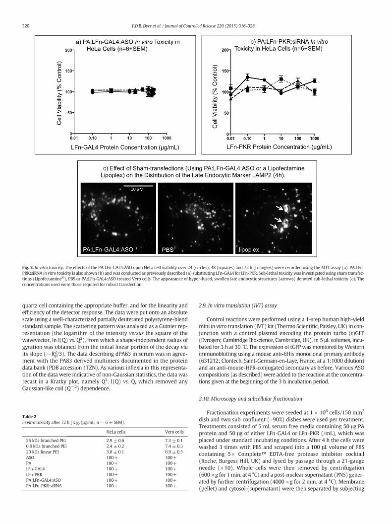
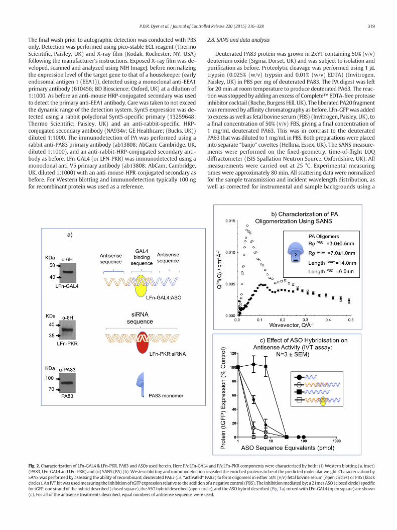
Fig. 3. In vitro toxicity. The effects of the PA:LFn-GAL4:ASO upon HeLa cell viability over 24 (circles), 48 (squares) and 72 h (triangles) were recorded using the MTT assay (a). PA:LFn-PRK:siRNA in vitro toxicity is also shown (b) and was conducted as previously described (a) substituting LFn-GAL4 for LFn-PKR. Sub-lethal toxicity was investigated using sham transfec-tions (Lipofectamine®), PBS or PA:LFn-GAL4:ASO treated Vero cells. The appearance of hyper-fused, swollen late endocytic structures (arrows) denoted sub-lethal toxicity (c). Theconcentrations used were those required for robust transfection.
320 P.D.R. Dyer et al. / Journal of Controlled Release 220 (2015) 316–328
quartz cell containing the appropriate buffer, and for the linearity andefficiency of the detector response. The data were put onto an absolutescale using a well-characterized partially deuterated polystyrene-blendstandard sample. The scattering pattern was analyzed as a Guinier rep-resentation (the logarithm of the intensity versus the square of thewavevector, ln I(Q) vs. Q2), from which a shape-independent radius ofgyration was obtained from the initial linear portion of the decay viaits slope (−Rg
2/3). The data describing dPA63 in serum was in agree-ment with the PA83 derived multimers documented in the proteindata bank (PDB accession 1TZN). As various inflexia in this representa-tion of the data were indicative of non-Gaussian statistics, the data wasrecast in a Kratky plot, namely Q2. I(Q) vs. Q, which removed anyGaussian-like coil (Q−2) dependence.
Table 2In vitro toxicity after 72 h (IC50 (μg/mL; n = 6 ± SEM).
HeLa cells Vero cells
25 kDa branched PEI 2.9 ± 0.6 7.3 ± 0.10.8 kDa branched PEI 2.4 ± 0.2 7.4 ± 0.320 kDa linear PEI 3.0 ± 0.1 6.9 ± 0.5ASO 100+ 100+PA 100+ 100+LFn-GAL4 100+ 100+LFn-PKR 100+ 100+PA:LFn-GAL4:ASO 100+ 100+PA:LFn-PKR:siRNA 100+ 100+
2.9. In vitro translation (IVT) assay
Control reactions were performed using a 1-step human high-yieldmini in vitro translation (IVT) kit (Thermo Scientific, Paisley, UK) in con-junction with a control plasmid encoding the protein turbo (t)GFP(Evrogen; Cambridge Bioscience, Cambridge, UK), in 5 μL volumes, incu-bated for 3 h at 30 °C. The expression of tGFP was monitored byWesternimmunoblotting using a mouse anti-6His monoclonal primary antibody(631212; Clontech, Saint-Germain-en-Laye, France, at a 1:1000 dilution)and an anti-mouse-HPR-conjugated secondary as before. Various ASOcompositions (as described)were added to the reaction at the concentra-tions given at the beginning of the 3 h incubation period.
2.10. Microscopy and subcellular fractionation
Fractionation experiments were seeded at 1 × 10⁶ cells/150 mm2
dish and two sub-confluent (~90%) dishes were used per treatment.Treatments consisted of 5 mL serum free media containing 50 μg PAprotein and 50 μg of either LFn-GAL4 or LFn-PKR (/mL), which wasplaced under standard incubating conditions. After 4 h the cells werewashed 3 times with PBS and scraped into a 100 μL volume of PBScontaining 5× Complete™ EDTA-free protease inhibitor cocktail(Roche, Burgess Hill, UK) and lysed by passage through a 21-gaugeneedle (×10). Whole cells were then removed by centrifugation(600 ×g for 1min. at 4 °C) and a post-nuclear supernatant (PNS) gener-ated by further centrifugation (4000 ×g for 2 min. at 4 °C). Membrane(pellet) and cytosol (supernatant) were then separated by subjecting
321P.D.R. Dyer et al. / Journal of Controlled Release 220 (2015) 316–328
PNS to sedimentation at 200,000 ×g for 60 min at 4 °C. Both the mem-brane and cytosol fractions were then adjusted to 100 μL total volumeusing Lamellae sample buffer containing 20% (v/v) BME. The fractionswere analyzed using the following primary antibodies: α-LDH (L7016;Sigma; Dorset, UK), as a cytosolic marker, α-V5 (ab9116; AbCam;Cambridge, UK) to detect either LFn-PKR or LFn-GAL4 and an α-transferrin receptor (TfR) primary (612124; BD-Transduction labs;Oxford, UK), was used to detect the presence of the transferrin receptor,a marker for cell membrane. Microscopy was performed after seeding1 × 105 Vero cells onto a sterile coverslip and incubating the cellsovernight in complete media. The following day PA was added to thecells to a final concentration of 50 μg/mL aswell as either Texas Red®-la-beled LFn-PKR or LFn-GAL4 at a concentration of 50 μg/mL in serum freemedia. After 4 h the cells were fixed using formalin [24]. Antibodies spe-cific for EEA1 (E41120; BD-Transduction labs; Oxford, UK)were used asa counter immunostain and were used at a dilution of 1:500. EEA1-specific primary antibodies were detected using a 1:500 dilution ofAlexa488-conjugated goat anti-mouse specific secondary antibodies(A11001; Invitrogen, Paisley, UK). Sub-lethal toxicity experiments wereperformed by seeding Vero cells as before and the followingday conducting transfection experiments using the preparations asdescribed. After 4 h the cells were fixed using cold methanol [24]and immunostained using an anti- lysosome associated membraneprotein 2 (LAMP2) specific antibody at a dilution of 1:10 (H4B4; DHSB,University of Iowa, IA, USA). This primary antibody was detected using a1:500 dilution of an anti-mouse Texas Red®-labeled secondary (T-862;
Fig. 4. PA mediated LFn-GAL4 or LFN-PKR cytosolic translocation by subcellular fractionation. Vdocumented and show the detection of LFn-GAL4 (containing an N-terminal V5-epitope) in botwithin the cytosolic fractions derived from this methodology was recorded over time (b). Omidemonstrating specificity. The cytosolic trafficking of PA:LFn-PKR:ASOwas alsomeasured by coLFn-GAL4. LFn-PKR also contained an N-terminal V5-epitope.
Invitrogen, Paisley, UK). PA localization to LBPA positive endocyticvesicles was undertaken by pulsing Vero cells with PA (50 μg/mL)with 200 μM Leupeptin 4 or 8 h prior to fixing the cells with 2%formalin (w/v) and then processed as previously described [24].Immunodetection was performed using both (rabbit) α-PA (ab13808;AbCam; Cambridge) and (mouse) α-LBPA antibodies (clone 6C4,α-LBPA antibody; Merck Millipore, Watford, UK). Texas Red-conjugatedanti-rabbit and Alex488-conjugated anti-mouse secondary antibodieswere used to visualize both LBPA and PA.
Cells were imaged using an Eclipse 90i overhead fluorescent micro-scope (Nikon, Japan), using a Nikon Digital Camera (DS-Qi1Nc). Theobjective used in imaging was an oil immersion CFI Plan ApochromatVC 60XN2 (NA 1.4 WD 0.13 mm) (Nikon, Japan).
3. Results
3.1. Characterization of LFn-GAL4 & LFn-PKR, PA83 and ASOs
A cartoon depicting the LFN-GAL4:ASO complex, the LFn-PKR:siRNAcomplex and the PA monomer are shown (Fig. 2a). Inset are Westernblots where LFn-GAL4 (~45.9 kDa), LFn-PKR (~37 kDa) and PA(~83 kDa) were detected at their predicted molecular weights.
Further characterization of the PA component of this systemwas conducted by SANS, and showed PA63 (i.e. activated PA83)oligomerizing in serum (Fig. 2b) [6]. Analysis of the SANS data plottedas a Guinier representation allowed the derivation of shape-independent
ero subcellular fractionations derived from cells exposed to PA:LFn-GAL4:ASO have beenh themembrane and cytosolic fractions, after 4 h (a). The temporal residence of FLn-GAL4tting PA prevented the cytosolic translocation of LFn-GAL4 as did keeping the cells at 0 °Cnducting subcellular fractionation (b) as previously described (a), substituting LFn-PKR for
322 P.D.R. Dyer et al. / Journal of Controlled Release 220 (2015) 316–328
radius of gyration measurements which were: RgPBS = 3.0 ± 0.5 nm
and Rgserum = 7.0 ± 1.0 nm (data not shown). The absence of highly
extended structure (Q−1 dependence), was also demonstrated by thedecaying (rather than increasing) background and the almost oscillato-ry nature of the data was indicative of highly non-Gaussian structures.Peaks present in the Kratky plot resulting from the scattering fromdPA63 in PBS and in serum (Fig. 2b) reported peaks shifting to smallerQ (serum), indicating a slightly larger annular structure [28], in agree-ment with previously published descriptions of PA heptamer [6].By noting that the peak positions Qmax = 2π/L correlate to distancesL = 14.0 nm in serum and 6.0 nm in PBS, it is likely that in PBS, themajority of the oligomers contain less than 7 members. For reasons ofclarity and narration, PA heptamers or octamers have been depictedhere (Figs. 1 & 2) as a blue “mushroom”. A more concise descriptionof this annular structure has been recently published [6].
Further characterization was conducted by examining the effect ofASO configuration and the presence of LFn-GAL4 upon antisense activityusing a cell-free in vitro translation assay (1-step human high-yield miniin vitro translation (IVT) kit; Thermo Scientific, Paisley, UK) (Fig. 2c).Here, the inhibition of tGFP expression was measured relative to the
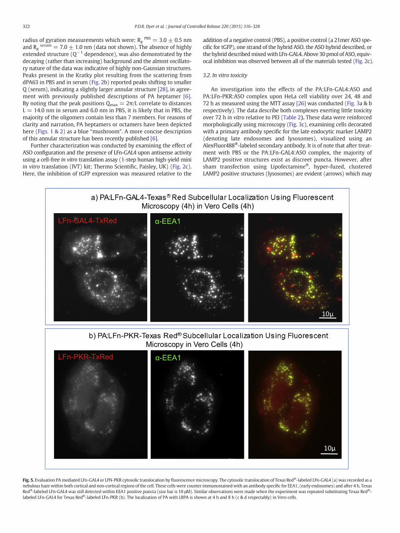
Fig. 5. Evaluation PAmediated LFn-GAL4 or LFN-PKR cytosolic translocation by fluorescencemicnebulous hazewithin both cortical and non-cortical regions of the cell. These cells were counterRed®-labeled LFn-GAL4 was still detected within EEA1 positive puncta (size bar is 10 μM). Simlabeled LFn-GAL4 for Texas Red®-labeled LFn-PKR (b). The localization of PA with LBPA is show
addition of a negative control (PBS), a positive control (a 21mer ASO spe-cific for tGFP), one strand of the hybrid ASO, the ASO hybrid described, orthe hybrid describedmixedwith LFn-GAL4. Above 30 pmol of ASO, equiv-ocal inhibition was observed between all of the materials tested (Fig. 2c).
3.2. In vitro toxicity
An investigation into the effects of the PA:LFn-GAL4:ASO andPA:LFn-PKR:ASO complex upon HeLa cell viability over 24, 48 and72 h as measured using the MTT assay [26] was conducted (Fig. 3a & brespectively). The data describe both complexes exerting little toxicityover 72 h in vitro relative to PEI (Table 2). These data were reinforcedmorphologically using microscopy (Fig. 3c), examining cells decoratedwith a primary antibody specific for the late endocytic marker LAMP2(denoting late endosomes and lysosomes), visualized using anAlexFluor488®-labeled secondary antibody. It is of note that after treat-ment with PBS or the PA:LFn-GAL4:ASO complex, the majority ofLAMP2 positive structures exist as discreet puncta. However, aftersham transfection using Lipofectamine®, hyper-fuzed, clusteredLAMP2 positive structures (lysosomes) are evident (arrows) which may
roscopy. The cytosolic translocation of Texas Red®-labeled LFn-GAL4 (a) was recorded as aimmunostainedwith an antibody specific for EEA1, (early endosomes) and after 4 h, Texasilar observations were made when the experiment was repeated substituting Texas Red®-n at 4 h and 8 h (c & d respectably) in Vero cells.
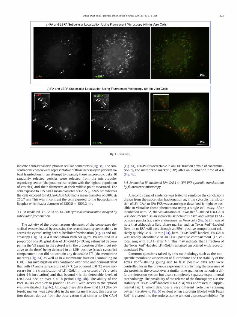
Fig. 5 (continued).
323P.D.R. Dyer et al. / Journal of Controlled Release 220 (2015) 316–328
indicate a sub-lethal disruption in cellular homeostasis (Fig. 3c). The con-centrations chosenwere representative of those necessary to perform ro-bust transfection. In an attempt to quantify these microscopic data, 10randomly selected vesicles were selected from the microtubule-organizing center (the juxtanuclear region with the highest populationof vesicles) and their diameters at their widest point measured. Thecells exposed to PBS had a mean diameter of 623.5 ± 224.5 nmwhereasthe cells exposed to PA:LFn-GAL4:ASO had a mean diameter of 688.0 ±250.7 nm. This was in contrast the cells exposed to the liposectaminelipoplex which had a diameter of 2300.5 ± 1505.2 nm.
3.3. PA mediated LFn-GAL4 or LFn-PKR cytosolic translocation assayed bysubcellular fractionation
The activity of the proteinaceous elements of the complexes de-scribed was evaluated by assessing the recombinant system's ability toaccess the cytosol using both subcellular fractionation (Fig. 4) and mi-croscopy (Fig. 5). A 4 h incubation with 50 μg/mL PA resulted in aproportion of a 50 μg/mL dose of LFn-GAL4 (~100 ng, estimated by com-paring the V5 signal in the cytosol with the proportion of the input rel-ative to the dose) being detected in an LDH-positive (crude cytosolic)compartment that did not contain any detectable TfR (the membranemarker) (Fig. 4a) as well as in a membrane fraction (containing noLDH). This investigation was continued over time and demonstratedthat both PA and a temperature of 37 °C (as opposed to 0 °C) were nec-essary for the translocation of LFn-GAL4 in the cytosol of Vero cells(after 4 h incubation) and that beyond 4 h, the detectable levels ofLFn-GAL4 decline over a 40 h period (Fig. 4b). The ability of thePA:LFn-PKR complex to provide LFn-PKR with access to the cytosolwas investigated (Fig. 4c). Although these data show that LDH (the cy-tosolic marker) was detectable in the membrane fraction, this observa-tion doesn't detract from the observation that similar to LFn-GAL4
(Fig. 4a), LFn-PKR is detectable in an LDH fraction devoid of contamina-tion by the membrane marker (TfR) after an incubation time of 4 h(Fig. 4c).
3.4. Evaluation PA mediated LFn-GAL4 or LFN-PKR cytosolic translocationby fluorescence microscopy
A second string of evidence was tested to reinforce the conclusionsdrawn from the subcellular fractionation as, if the cytosolic transloca-tion of LFn-GA:4 or LFn-PKRwas occurring as described, itmight be pos-sible to visualize these phenomena using a single cell assay. Afterincubation with PA, the visualization of Texas Red®-labeled LFn-GAL4was documented as an intracellular nebulous haze and within EEA1-positive puncta (i.e. early endosomes) in Vero cells (Fig. 5a). It was ofnote that although a fluid phase marker such as Texas Red®-labeledDextran or BSA will pass through an EEA1 positive compartment rela-tively quickly i.e. 5–10 min [24], here, Texas Red®-labeled LFn-GAL4was readily identifiable in an EEA1 positive compartment (i.e. co-localizing with EEA1) after 4 h. This may indicate that a fraction ofthe Texas Red®-labeled LFn-GAL4 remained associated with receptorassociated PA.
Common questions raised by this methodology such as the non-specific membrane association of fluorophore and the stability of theTexas Red®-labeling giving rise to false positive data sets werecontrolled for in the previous experiment, confirming the presence ofthe protein in the cytosol over a similar time span using not only a dif-ferent detection system but also a completely separate experimentalmethodology. The possibility of the release of the fluorophore (i.e. thestability of Texas Red®-labeled LFn-GAL4) was addressed in Supple-mental Fig. 1, which describes a very different (reticular) stainingpattern (relative to Fig. 5) evident when a protein labeled with TexasRed® is chased into the endolysosome without a protease inhibitor. To
324 P.D.R. Dyer et al. / Journal of Controlled Release 220 (2015) 316–328
repeat the previous observation using Texas Red®-labeled LFn-PKR andPA, a second experimentwas undertaken. This resulted in a second formof experimental evidence documenting Texas Red®-LFn-PKR in the cy-tosol of Vero cells using fluorescence microscopy (Fig. 5b) against anEEA1 counter stain. With morphology similar to Texas Red®-labeledLFn-GAL4 (Fig. 5a), Texas Red®-labeled LFn-PKRwas also detected ubiq-uitously within the cells (Fig. 5b).
Supplemental Fig. 2 further controls for the possibility that the redsignal detected in Fig. 5 was autofluorescence, documenting an increaseand then decreasing in red signal from Texas Red-labeled LFn-GAL4over time in live cells. Fig. 5c & d further control of red cytosolic autoflu-orescence in fixed cells as well as demonstrate that after 4 h a limitedamount of co-localization can been seen between a pulse of PA andLBPA (a marker for MVBs and a lipid critical for back-fusion [9]). Afteran additional 4 h a substantially higher degree of co-localizationbetween PA and LBPA can be observed (Fig. 5d).
3.5. PA:LFn-GAL4:ASO antisense activity against a panel of primate cells
An initial experiment was preformed to evaluate the baseline levelof ASO activity in HeLa cells. Here 200 pmol of ASO (or separatelysiRNA) was added to serum free cell culture media, whichwas replacedby complete media after 4 h. After 24 h a statistically significant varia-tion in target gene expression was evident relative to a PBS treatedcontrol (Supplemental Fig. 3 and supplemental Table 1), where base-lines of 71.3% and 69.0% Synt5 expression were recorded for cells treat-ed with only ASO or siRNA respectively.
Having established background levels of ASO transfection (Supple-mental Fig. 3 and Supplemental Table 1), the effect of increasingamounts of ASO in concert with static concentrations of PA:LFn-GAL4
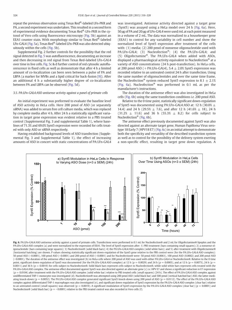
Fig. 6. PA:LFn-GAL4:ASO antisense activity against a panel of primate cells. Transfections werePA:LFn-GAL4:ASO complex (a) and were normalized to the expression of EEA1. The level of Syigonucleotide (bars containing large squares); 3) Nucleofection® (solid black bars); 4) the PA(horizontal hatching) are shown. P values showing statistically significant down-regulation50 pmol ASO (b0.0001), 100 pmol ASO (b0.0001) and 200 pmol of ASO (b0.0001) and for N(b0.0001). The duration of the antisense effect was investigated (b) in HeLa cells where 200 ppoint, significant down-regulation of Synt5 was documented (for the PA:LFn-GAL4:ASO com0.0011) and 36 h (p = 0.0019) for cells subject to Nucleofection®. Solid black bars represenPA:LFn-GAL4:ASO complex. The antisense effect documented against Synt5 was also directed a(p = 0.0104) after treatment with the PA:LFn-GAL4:ASO complex (solid white bar) relative toundifferentiated THP-1 monocytes was investigated (d). Nucleofection®was attempted usingating knock-down (p = 0.0052). The PA:LFn-GAL4:ASO complex generated significant Synt5complex against differentiated THP-1 macrophages was also investigated (e), and significant dto an untreated control (small squares) was observed (p = 0.0019). A significant modulatioNucleofection® (solid black bar) (p = b0.0001) relative to the PBS treated control was also re
was investigated. Antisense activity directed against a target gene(Synt5) was assayed using a HeLa model over 24 h (Fig. 6a). Here,50 μg of PA and 20 μg of LFn-GAL4were used/mL at each pointmeasuredin a volume of 2 mL. The data was normalized to a housekeeper gene(EEA1) to control for any variability in cell number and show thenormalized level of Synt5 expression after treatment of the cellswith: (1) media; (2) 200 pmol of nonsense oligonucleotide used withPA:LFn-GAL4; (3) Nucleofection®; (4) the PA:LFn-GAL4; and(5) Oligofectamine®. The PA:LFn-GAL4 when added with ASOs,displayed a pharmacological activity equivalent to Nucleofection® at avariety of ASO concentrations (24 h post-transfection). In HeLa cells,at 200 pmol ASO (+PA:LFn-GAL4), 5.4 ± 2.0% Synt5 expression wasrecorded relative to an untreated control 24 h after transfection. Usingthe same number of oligonucleotides and over the same time frame,the Nucleofection® system reduced Synt5 expression to 8.1 ± 2.1%(Fig. 6a). Nucleofection® was performed in 0.1 mL as per themanufacturer's instructions.
The duration of the antisense effect was also investigated in HeLacells (Fig. 6b) using the same transfection conditions i.e. 200 pmol ASO.
Relative to the 0 time point, statistically significant down-regulationof Synt5 was documented using PA:LFn-GAL4:ASO at: 12 h (30.0% ±14.4) and 24 h (29.5% ± 7.6), and after 12 h (41.6% ± 18), 24 h(31.2% ± 11.6) and 36 h (55.3% ± 8.2) for cells subject toNucleofection® (Fig. 6b).
The antisense effect previously documented against Synt5 was alsodirected against an alternate target gene, Human Papilloma Virus sero-type 18 Early 7 (HPV18 E7) (Fig. 6c) in an initial attempt to demonstrateboth the specificity and versatility of the described transfection systemas well as to control for the possibility of the delivery system exertinga non-specific effect, resulting in target gene down regulation. A
performed in 0.1 mL for Nucleofection® and 2 mL for Oligofectamine® lipoplex and thent5 expression after: 1) PBS treatment (bars containing small squares); 2) a nonsense ol-:LFn-GAL4:ASO complex (solid white bars); and 5) after treatment with Oligofectamine®of the Synt5 gene relative to the PBS control were (for the PA:LFn-GAL4:ASO complex):ucleofection® were: 50 pmol ASO (0.0061), 100 pmol ASO (0.0002) and 200 pmol ASOmol of ASO was used with either PA:LFn-GAL4 or Nucleofection®. Relative to the 0 timeplex) at 12 h (p = 0.0029) and 24 h (p = 0.0003), and at 12 h (p = 0.0073), 24 h (p =t cells subject to Nucleofection®, while solid white bars represent cells treated with thegainst an alternate gene (c) i.e.HPV E7 and shows a significant reduction in E7 expressionPBS treated cells (small squares) (24 h). The effect of PA:LFn:GAL4:ASO complex against200 pmol ASO (solid black bar) and 500 pmol (vertical hatched bar) ASO, the latter medi-knock-down using 200 pmol of ASO (p = 0.0112). The effect of the PA:LFn-GAL4:ASO
own-regulation of Synt5 expression by the PA:LFn-GAL4:ASO complex (clear bar) relativen of Synt5 expression by the PA:LFn-GAL4:ASO complex (clear bar) (p = 0.0001) andcorded in Vero cells (f).
Fig. 6 (continued).
325P.D.R. Dyer et al. / Journal of Controlled Release 220 (2015) 316–328
statistically significant reduction in E7 expression level was recorded(65.3% ± 4.4) after treatment with the PA:LFn-GAL4:ASO relative toan untreated control after 24 h.
The ability of PA:LFn-GAL4:ASO to down-regulate a target gene in avariety of primate cell lines was also undertaken in order to demon-strate activity in non-HeLa cells. To this end, PA:LFn-GAL4:ASO wasdirected against Synt5 using undifferentiated THP-1 monocytes(Fig. 6d). Separately, Nucleofection® was also attempted using200 pmol ASO in the same undifferentiated cells. No Synt5 knock-down was evident after this first attempt at Nucleofection®. When theamount of ASO used was increased to 500 pmol, Synt5 knock-downwas evident (25.3% ± 16.8 of Synt5 remained) after 24 h. This was incontrast to the PA:LFn-GAL4:ASO complex, which left a significantlysmaller amount of detectable Synt5 (35.2% ± 19.1) after treatmentwith 200 pmol of ASO after 24 h.
The effect of the Synt5 targeted PA:LFn-GAL4:ASO complex againstdifferentiated THP-1 macrophages was also investigated (Fig. 6e).Here, (35.2%±19.1) Synt5 expressionwas documented after treatmentwith the PA:LFn-GAL4:ASO complexmade with 200 pmol ASO (relativeto an untreated control). All attempts at the Nucleofection® of differen-tiated THP-1 cells resulted in a very high degree of cell death, as would
be expected when using this transfection recalcitrant cell line [29].Consequently, in this instance, no transfection was recorded.
The effect of the PA:LFn-GAL4:ASO complex directed against Synt5in Vero cells was explored (Fig. 6f). Synt5 expression levels in PA:LFn-GAL4:ASO complex treated cells, relative to an untreated control wererecorded (22.9% ± 6.9) as well as the effect of Nucleofection® usingSynt5 specific ASOs (16.2% ± 1.7). PA:LFn-GAL4:ASO complex activityin Vero cells demonstrated functionality in a non-human primate spe-cies, using a cell line well characterized in our and many otherlaboratories.
3.6. PA:LFn-PKR:siRNA mediated Synt5 down-regulation
The ability of the PA:LFn-PKR:siRNA complex to deliver pharmaco-logically active siRNA was examined (Fig. 7). After 24 h; 8.5 ± 3.4%Synt5 expression was recorded after treatment with the PA:LFn-PKR:siRNA complex (containing 200 pmol of Stealth RNAi™ siRNA).This was measured against the Nucleofection® of siRNA using200 pmol of Stealth RNAi™ siRNA. Treatment with the PA:LFn-PKR:siRNA complex resulted in an almost equivalent level of Synt5expression of Synt5 relative to Nucleofection® (i.e. 4.6 ± 6.1%) using
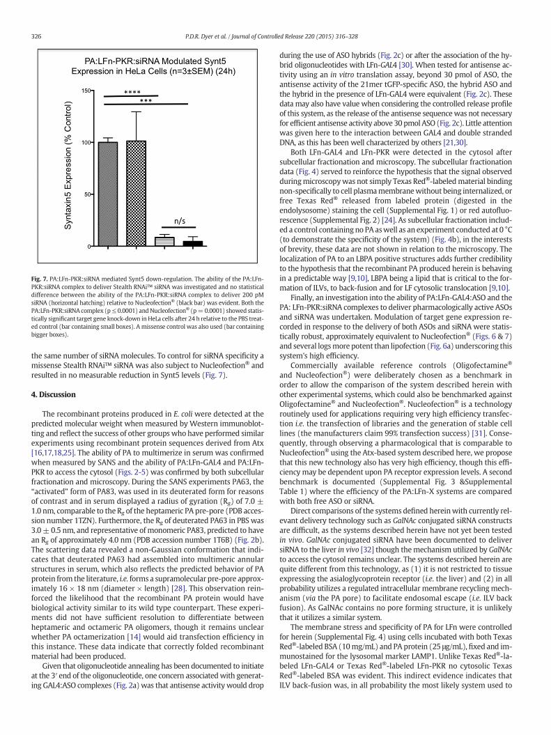
Fig. 7. PA:LFn-PKR:siRNA mediated Synt5 down-regulation. The ability of the PA:LFn-PKR:siRNA complex to deliver Stealth RNAi™ siRNA was investigated and no statisticaldifference between the ability of the PA:LFn-PKR:siRNA complex to deliver 200 pMsiRNA (horizontal hatching) relative to Nucleofection® (black bar) was evident. Both thePA:LFn-PKR:siRNA complex (p≤ 0.0001) andNucleofection® (p=0.0001) showed statis-tically significant target gene knock-down inHeLa cells after 24 h relative to the PBS treat-ed control (bar containing small boxes). A missense control was also used (bar containingbigger boxes).
326 P.D.R. Dyer et al. / Journal of Controlled Release 220 (2015) 316–328
the same number of siRNA molecules. To control for siRNA specificity amissense Stealth RNAi™ siRNA was also subject to Nucleofection® andresulted in no measurable reduction in Synt5 levels (Fig. 7).
4. Discussion
The recombinant proteins produced in E. coli were detected at thepredicted molecular weight when measured by Western immunoblot-ting and reflect the success of other groupswho have performed similarexperiments using recombinant protein sequences derived from Atx[16,17,18,25]. The ability of PA to multimerize in serum was confirmedwhen measured by SANS and the ability of PA:LFn-GAL4 and PA:LFn-PKR to access the cytosol (Figs. 2-5) was confirmed by both subcellularfractionation and microscopy. During the SANS experiments PA63, the“activated” form of PA83, was used in its deuterated form for reasonsof contrast and in serum displayed a radius of gyration (Rg) of 7.0 ±1.0 nm, comparable to the Rg of the heptameric PA pre-pore (PDB acces-sion number 1TZN). Furthermore, the Rg of deuterated PA63 in PBS was3.0± 0.5 nm, and representative of monomeric PA83, predicted to havean Rg of approximately 4.0 nm (PDB accession number 1T6B) (Fig. 2b).The scattering data revealed a non-Gaussian conformation that indi-cates that deuterated PA63 had assembled into multimeric annularstructures in serum, which also reflects the predicted behavior of PAprotein from the literature, i.e. forms a supramolecular pre-pore approx-imately 16 × 18 nm (diameter × length) [28]. This observation rein-forced the likelihood that the recombinant PA protein would havebiological activity similar to its wild type counterpart. These experi-ments did not have sufficient resolution to differentiate betweenheptameric and octameric PA oligomers, though it remains unclearwhether PA octamerization [14] would aid transfection efficiency inthis instance. These data indicate that correctly folded recombinantmaterial had been produced.
Given that oligonucleotide annealing has been documented to initiateat the 3′ end of the oligonucleotide, one concern associatedwith generat-ing GAL4:ASO complexes (Fig. 2a)was that antisense activity would drop
during the use of ASO hybrids (Fig. 2c) or after the association of the hy-brid oligonucleotides with LFn-GAL4 [30]. When tested for antisense ac-tivity using an in vitro translation assay, beyond 30 pmol of ASO, theantisense activity of the 21mer tGFP-specific ASO, the hybrid ASO andthe hybrid in the presence of LFn-GAL4 were equivalent (Fig. 2c). Thesedata may also have value when considering the controlled release profileof this system, as the release of the antisense sequencewas not necessaryfor efficient antisense activity above 30 pmol ASO (Fig. 2c). Little attentionwas given here to the interaction between GAL4 and double strandedDNA, as this has been well characterized by others [21,30].
Both LFn-GAL4 and LFn-PKR were detected in the cytosol aftersubcellular fractionation and microscopy. The subcellular fractionationdata (Fig. 4) served to reinforce the hypothesis that the signal observedduringmicroscopywas not simply Texas Red®-labeledmaterial bindingnon-specifically to cell plasmamembranewithout being internalized, orfree Texas Red® released from labeled protein (digested in theendolysosome) staining the cell (Supplemental Fig. 1) or red autofluo-rescence (Supplemental Fig. 2) [24]. As subcellular fractionation includ-ed a control containing no PA aswell as an experiment conducted at 0 °C(to demonstrate the specificity of the system) (Fig. 4b), in the interestsof brevity, these data are not shown in relation to the microscopy. Thelocalization of PA to an LBPA positive structures adds further credibilityto the hypothesis that the recombinant PA produced herein is behavingin a predictable way [9,10], LBPA being a lipid that is critical to the for-mation of ILVs, to back-fusion and for LF cytosolic translocation [9,10].
Finally, an investigation into the ability of PA:LFn-GAL4:ASO and thePA: LFn-PKR:siRNA complexes to deliver pharmacologically active ASOsand siRNA was undertaken. Modulation of target gene expression re-corded in response to the delivery of both ASOs and siRNA were statis-tically robust, approximately equivalent to Nucleofection® (Figs. 6 & 7)and several logsmore potent than lipofection (Fig. 6a) underscoring thissystem's high efficiency.
Commercially available reference controls (Oligofectamine®
and Nucleofection®) were deliberately chosen as a benchmark inorder to allow the comparison of the system described herein withother experimental systems, which could also be benchmarked againstOligofectamine® and Nucleofection®. Nucleofection® is a technologyroutinely used for applications requiring very high efficiency transfec-tion i.e. the transfection of libraries and the generation of stable celllines (the manufacturers claim 99% transfection success) [31]. Conse-quently, through observing a pharmacological that is comparable toNucleofection® using the Atx-based system described here, we proposethat this new technology also has very high efficiency, though this effi-ciency may be dependent upon PA receptor expression levels. A secondbenchmark is documented (Supplemental Fig. 3 &SupplementalTable 1) where the efficiency of the PA:LFn-X systems are comparedwith both free ASO or siRNA.
Direct comparisons of the systems defined hereinwith currently rel-evant delivery technology such as GalNAc conjugated siRNA constructsare difficult, as the systems described herein have not yet been testedin vivo. GalNAc conjugated siRNA have been documented to deliversiRNA to the liver in vivo [32] though themechanism utilized by GalNActo access the cytosol remains unclear. The systems described herein arequite different from this technology, as (1) it is not restricted to tissueexpressing the asialoglycoprotein receptor (i.e. the liver) and (2) in allprobability utilizes a regulated intracellular membrane recyclingmech-anism (via the PA pore) to facilitate endosomal escape (i.e. ILV backfusion). As GalNAc contains no pore forming structure, it is unlikelythat it utilizes a similar system.
The membrane stress and specificity of PA for LFn were controlledfor herein (Supplemental Fig. 4) using cells incubated with both TexasRed®-labeled BSA (10mg/mL) and PA protein (25 μg/mL), fixed and im-munostained for the lysosomal marker LAMP1. Unlike Texas Red®-la-beled LFn-GAL4 or Texas Red®-labeled LFn-PKR no cytosolic TexasRed®-labeled BSA was evident. This indirect evidence indicates thatILV back-fusion was, in all probability the most likely system used to
327P.D.R. Dyer et al. / Journal of Controlled Release 220 (2015) 316–328
access the cytosol (rather than membrane destabilization). Previouslywe have demonstrated the release of fluorescent-labeled Gelonin intothe cytosol using similar methodology (i.e. fluorescence microscopy)to monitor polymer-mediated membrane perturbation and cytosoliccargo transfer [33]. Efforts to investigate the role of ILV back-fusion(more specifically, a dependence on LBPA andALIX [9] for pharmacolog-ical activity of both PA:LFn-GAL4:ASO and PA:LFn-PKR:siRNA) arecurrently underway.
The pharmacological activity of the Atx-based transfection systemdescribed was shown to be reproducible during studies investigatingASO time-dependency and propensity to transfect a selection of primatecell lines (Fig. 6), and (conceptually) via the successful delivery of siRNAby PA:LFn-PKR (Fig. 7). Given that the HeLa cells used to document theduration of antisense activity were in an exponential growth phase, itremains possible that the duration of the observed antisense effect(Fig. 6b) may well be extended in vivo, though other variables, suchas mRNA and protein abundance and half-life, would also need tobe considered. Differences in mRNA and protein half-life, abundanceand protein-specific biology may also account for the variation in pro-tein knockdown observed when Synt5 is contrasted with HPV18 E7(Fig. 6a & c).
Relative to the delivery of plasmids, the effective delivery of siRNAand ASOs requires different parameters to be met [34]. Consequently,the use of smaller (relative to a plasmid) pieces of nucleic acid (i.e.ASOs) and the absence of polycations [19,20,21] differentiates thiswork from that of others. In order to condense and conjugate plasmidDNA to disarmed toxins, previous studies have used a polycationicelement sometimes described as an “affinity handle” [21]. The lack ofa condensing agent to mediate nuclease protection here is counteredthrough the use of chemical modification within the nucleic acid back-bone i.e. through the use of phosphorothioate ASOs. Similarly, as ASOsand siRNAs were being used, a condensing agent to facilitate theendocytic capture of the PA:LFn-GAL4:ASO or PA:LFn-PKR:siRNA wasnot necessary. This was important as previous studies [19,20,21] con-taining polycationic condensing agents i.e. poly(Lysine) in conjunctionwith toxin components may have lent ambiguity to the conclusionthat protein architecture alone was responsible for the translocation ofthe nucleic acid cargo over biological barriers. This is due to the possibil-ity that the toxin-polycation delivery efficiency reported, was beingaugmented (even indirectly by stressingmembrane) by the polycation.Here, this possibility has been controlled for (Supplemental Fig. 4).Condensing agents aside, LFn-GAL4:ASO and LFn-PKR:siRNA size wasstill contentious as the literature states that the complexes generatedherein should be too large to go though the internal lumen of the PApore, its minimum diameter being approximately 0.6 nm at the phiclamp [6]. In the wild, LF and EF may undergo a molten globular transi-tion, losing higher order structure in order to “ratchet” thorough theinternal lumen of the PA pore [6,35]. Given the size of a supramolecularassembly composed of a pair of ASOs and two LFn-GAL4 molecules, (orthe LFn-PKR:siRNA equivalent) the data presented herein begins toquestion the conventional wisdom describing the transition of materialover the PA pore, hinting at the possibility of the pore dilating to allowthe transit of larger material. Equivalently, it is also possible that the at-tachment of the ASO or Stealth RNAi™ siRNA to LFn served only to bringnucleic acid into proximal contact with the PA pore interior, facilitatingtranslocation into the lumen of the ILV [36]. Given the diameter of anucleic acid duplex (~2.0 nm) the DNA would be required to “melt” topass through the internal lumen of a static PA pore which, like themol-ten globular transition of LFn-GFP at pH 5.5 may be thermodynamicallyimprobable [25]. Previous studies also failed to address the question ofhow an interpolyelectrolyte complex (typically 100-200 nm) could“ratchet” through the internal (negatively charged) lumen of the PA(or similar) pore [6,21].
The scope of polycations to interact withmembranes [37]mediatingtoxicity (such as PEI; Table 2), or transfection [38] has been well docu-mented. It was shown here that there was, under the parameters
conducive to transfection, very little in vitro toxicity when eitherPA:LFn-GAL4:ASO or PA:LFn-PKR:siRNA were used (Fig. 3), sheddingnew light upon the “PEG-dilemma” [2,39]. The ramifications of thisobservation may also stretch beyond toxicity to body distribution andthe ability to transfect tissue currently beyond the reach of establishednon-viral delivery technology [1,2]. The body distribution of the PA pro-tein has been well documented [40,41] and the studies reported hereconfirmed that PA:LFn-GAL4 and PA:LFn-PKRhave the potential to de-liver ASO and siRNA in a variety of different cell types (includingmono-cytes and macrophages (Fig. 6d & e) as efficiently as Nucleofection®.Consequently, experiments will be designed to establish proof ofconcept in specific disease model in vivo in the future.
In relation to loading capacity, assuming PA forms a heptamer [6],three LFn molecules associate with it directly, each forming a dimerwith a second LFn-GAL4 molecule binding the ASO hybrid [21,30].Consequently one discreet PA(7):LFn-GAL4(6):ASO(6) complex has thecapacity, when considered as a static unit, to contain 6 antisense se-quences. Although the “drug” loading capacity of the PA:LFn-GAL4:ASOcomplex is undoubtedly less than that of an IPEC, the lack of toxicity, thepredictable, regulated nature of membrane translation and lack of a netpositive charge (or prevalent hydrophobicity),may serve to counterbal-ance the efficiency of the PA:LFn-GAL4:ASO complex in relation to theefficiency of the Nucleofection® and lipofection systems reported(Fig. 6). The PA pore may also be able to sustain the delivery of LFn-fusion molecules over time. Efforts are currently underway to quantifyhow many cytosolic ASO molecules can be detected as a function ofdelivery in relation to an observed pharmacological effect. This informa-tion may be critical when considering the variance of PA receptordensity and type in relation to determining the delivery efficiency nec-essary to treat a specific pathology. Once target mRNA abundance andprotein half-life are considered in the light of delivery efficiency, in-formed decisions about dosemay be possible, further reducing unwant-ed off-target effects due to an excess of “drug”. Ultimately, this approachmay also allow the precise control over the level of modulation exertedover target gene expression, in a given cell, tissue type or person.
In contrast to LFn, PA has been documented as not being particularlyimmunogenic in Humans [42]. If repeat dosing is required, immunoge-nicity may be something that needs to be addressed, but is, for the timebeing, beyond the remit of this study. If immunogenicity does become aproblem, there are potentially several ways of dealing with it withoutcompromising the efficacy of the existing anthrax vaccines (that alsocontain PA and LF) [42]. One possible solution may be to PEGylateassemblies of PA:LFn-X:ASO/siRNA.
5. Conclusion
The novel antisense and siRNA delivery technology describeddisplays high transfection efficiency and little toxicity in vitro. As thiscytosolic delivery platform is a departure from conventional chargedor hydrophobic, non-viral drug delivery technology, it is also possiblethat it may not be subject to many of the limitations that have thus farprevented non-viral drug delivery technology from translating intoroutinely used clinical tools.
Supplementary data to this article can be found online at http://dx.doi.org/10.1016/j.jconrel.2015.10.054.
Acknowledgments
S.C.W.R. and P.C.G. would like to thank the Science and TechnologyFundingCouncil (STFC) for their support, funding theneutron scatteringwork through awards: RB1220030 and RB1320038, to P.C.G. andS.C.W.R., respectively. P.D.R.D., T.R.S., S.A.S., M.P., C.-K.T. and A.S.G.would like to thank The University of Greenwich for funding. S.C.W.R.would like to thank GUEL for supporting this project. Prof. StephenLeppla (National Institute of Allergy and Infectious Diseases, USA) is
328 P.D.R. Dyer et al. / Journal of Controlled Release 220 (2015) 316–328
also thanked for the kind gift of a LF encoding plasmid. S.C.W.R wouldlike to thank Prof. Ruth Duncan for critically reading this manuscript.
References
[1] H. Yin, et al., Non-viral vectors for gene based therapy, Nat. Rev. Genet. 15 (2014)541–555, http://dx.doi.org/10.1038/nrg3763.
[2] O.M. Merkel, T. Kissel, Quo vadis polyplex? J. Control. Release 190 (2014) 415–423,http://dx.doi.org/10.1016/j.jconrel.2014.06.009.
[3] R. Geary, et al., Fomivirsen: clinical pharmacology and potential drug interactions,Clin. Pharmacokinet. 41 (4) (2002) 255–260.
[4] I. Marafini, et al., Antisense approach to inflammatory bowel disease: prospects andchallenges, Drugs 75 (7) (2015) 723–730, http://dx.doi.org/10.1007/s40265-015-0391-0.
[5] D. Rader, J. Kastelein, Lomitapide and mipomersen: two first-in-class drugs forreducing low-density lipoprotein cholesterol in patients with homozygous familialhypercholesterolemia, Circulation 129 (9) (2014) 1022–1032, http://dx.doi.org/10.1161/circulationaha.113.001292.
[6] J. Jiang, et al., Atomic structure of anthrax protective antigen pore elucidates toxintranslocation, Nature 521 (2015) 545–549, http://dx.doi.org/10.1038/nature14247.
[7] N. Aurora, S.H. Leppla, Residues 1–254 of anthrax toxin lethal factor are sufficient tocause cellular uptake of fused polypeptides, J. Biol. Chem. 268 (1993) 3334–3341.
[8] T. Falguieres, et al., Molecular assemblies and membrane domains in multivesicularendosome dynamics, Exp. Cell Res. 315 (2009) 1567–1573, http://dx.doi.org/10.1016/j.yexcr.2008.12.006.
[9] Abrami L., et al., Membrane insertion of anthrax protective antigen and cytoplasmicdelivery of lethal factor occur at different stages of the endocytic pathway. J. CellBiol., 66(5): 645–651, (2004). doi/http://dx.doi.org/10.1083/jcb.200312072.
[10] J. Gruenberg, F.G. van der Goot, Mechanisms of pathogen entry through theendosomal compartments, Nat. Rev. Mol. Cell Biol. 7 (7) (2006) 495–504, http://dx.doi.org/10.1038/nrm1959.
[11] M. Martchenko, S.-Y. Jeonga, S.N. Cohen, Heterodimeric integrin complexes contain-ing β1-integrin promote internalization and lethality of anthrax toxin, Proc. Natl.Acad. Sci. U. S. A. 107 (2010) 15583–15588, http://dx.doi.org/10.1073/pnas.1010145107.
[12] S. Liu, S.H. Leppla, Cell surface tumor endothelium marker 8 cytoplasmic tail-independent anthrax toxin binding, proteolytic processing, oligomer formation,and internalization, J. Biol. Chem. 287 (2003) 5227–5234.
[13] S. Liu, et al., Capillary morphogenesis protein-2 is the major receptor mediatinglethality of anthrax toxin in vivo, Proc. Natl. Acad. Sci. U. S. A. 106 (2009)12424–12429, http://dx.doi.org/10.1073/pnas.0905409106.
[14] A.F. Kintzer, et al., The protective antigen component of anthrax toxin forms func-tional octameric complexes, J. Mol. Biol. 392 (2009) 614–629, http://dx.doi.org/10.1016/j.jmb.2009.07.037.
[15] M. Qadan, et al., Membrane insertion by anthrax protective antigen in cultured cells,Mol. Cell. Biol. 25 (13) (2005) 5492–5498.
[16] W.P. Verdurmen, M. Luginbühl, A. Honegger, A. Plückthun, Efficient cell-specificuptake of binding proteins into the cytoplasm through engineered modular trans-port systems, J. Control. Release 200 (2015) 13–22, http://dx.doi.org/10.1016/j.jconrel.2014.12.019.
[17] X. Liao, et al., Delivery of antibody mimics into mammalian cells via anthrax toxinprotective antigen, ChemBioChem 15 (2014) 2458–2466, http://dx.doi.org/10.1002/cbic.201402290.
[18] D.E. Peters, et al., Comparative toxicity and efficacy of engineered anthrax lethaltoxin variants with broad anti-tumor activities, Toxicol. Appl. Pharmacol. 279(2014) 220–229, http://dx.doi.org/10.1016/j.taap.2014.06.010.
[19] L.B. Barrett, et al., CTb targeted non-viral cDNA delivery enhances transgene expres-sion in neurons, J. Gene Med. 6 (4) (2004) 429–438.
[20] J. Fominaya, W.W. Target, D.N.A. Cell-specific, Transfer mediated by a chimericmultidomain protein, J. Biol. Chem. 271 (18) (1996) 10560–10568.
[21] R. Gaur, P. Gupta, A. Goyal, W. Wels, Y. Singh, Delivery of nucleic acid into mamma-lian cells by anthrax toxin, Biochem. Biophys. Res. Commun. 297 (2002) 1121–1127.
[22] H.J. Stunden, E. Latz, PKR stirs up inflammasomes, Cell Res. 23 (2) (2013) 168–170,http://dx.doi.org/10.1038/cr.2012.125.
[23] K. Suga, et al., RNA interference-mediated silencing of the syntaxin 5 gene inducesGolgi fragmentation but capable of transporting vesicles, FEBS Lett. 579 (20)(2005) 4226–4234.
[24] S.C. Richardson, et al., The use of fluorescence microscopy to define polymer locali-zation to the late endocytic compartments in cells that are targets for drug delivery,J. Control. Release 127 (1) (2008) 1–11, http://dx.doi.org/10.1016/j.jconrel.2007.12.015.
[25] I. Zornetta, et al., Imaging the cell entry of the anthrax oedema and lethal toxinswith fluorescent protein chimeras, Cell. Microbiol. 12 (2010) 1435–1445, http://dx.doi.org/10.1111/j.1462-5822.2010.01480.x.
[26] S.C. Richardson, et al., Potential of low molecular mass chitosan as a DNA deliverysystem: biocompatibility, body distribution and ability to complex and protectDNA, Int. J. Pharm. 178 (2) (1999) 231–243.
[27] M.W. Pettit, et al., Construction and physiochemical characterization of a multi-composite, potential oral vaccine delivery system (VDS), Int. J. Pharm. 468 (1–2)(2014) 264–271, http://dx.doi.org/10.1016/j.ijpharm.2014.03.046.
[28] V. Receveur-Brechot, D. Durand, How random are intrinsically disordered proteins?A small angle scattering perspective, Curr. Protein Pept. Sci. 13 (2012) 55–75, http://dx.doi.org/10.2174/138920312799277901.
[29] M.B. Maeß, et al., Optimization of the transfection of human THP-1 macrophages byapplication of Nunc UpCell technology, Anal. Biochem. 479 (2015) 40–42, http://dx.doi.org/10.1016/j.ab.2014.12.023.
[30] T. Pan, J.E. Coleman, Structure and function of the Zn(II) binding site with in theDNA-binding domain of the GAL4 transcription factor, PNAS 86 (1989) 3145–3149.
[31] http://www.lonza.com/products-services/bio-research/transfection/nucleofector-technology.aspx
[32] Nair, et al., Multivalent N-acetylgalactosamine-conjugated siRNA localizes in hepa-tocytes and elicits robust RNAi-mediated gene silencing, JACS 136 (2014)16958–16961.
[33] Richardson, S. C. W., Pattrick, N. G., Lavignac, N., Ferruti, P., & Duncan, R. (2010).Intracellular fate of bioresponsive poly(amidoamine)s in vitro and in vivo. J. Control.Release, 142(1), 78–88. doi.org/http://dx.doi.org/10.1016/j.jconrel.2009.09.025.
[34] C. Scholz, E. Wagner, Therapeutic plasmid DNA versus siRNA delivery: common anddifferent tasks for synthetic carriers, J. Control. Release 161 (2012) 554–565, http://dx.doi.org/10.1016/j.jconrel.2011.11.014.
[35] B.A. Krantz, et al., Acid-induced unfolding of the amino-terminal domains of thelethal and edema factors of anthrax toxin, J. Mol. Biol. 344 (3) (2004) 739–756.
[36] D.G.Wright, et al., Effective delivery of antisense peptide nucleic acid oligomers intocells by anthrax protective antigen, Biochem. Biophys. Res. Commun. 376 (2008)200–205, http://dx.doi.org/10.1016/j.bbrc.2008.08.124.
[37] Y. Marikovsky, D. Danon, A. Katchalsky, Agglutination by polylysine of young andold red blood cells, Biochim. Biophys. Acta 124 (1966) 154–159.
[38] O. Boussif, et al., A versatile vector for gene and oligonucleotide transfer into cells inculture and in vivo: polyethyleneimine, Proc. Natl. Acad. Sci. U. S. A. 92 (1995) 7297-7230.
[39] T. Liu, B. Thierry, A solution to the PEG dilemma: efficient bioconjugation of largegold nanoparticles for biodiagnostic applications using mixed layers, Langmuir 28(44) (2012) 15634–15642, http://dx.doi.org/10.1021/la301390u.
[40] M. Moayeri, et al., Anthrax protective antigen cleavage and clearance from the bloodof mice and rats, Infect. Immun. 75 (2007) 5175–5184.
[41] E. Dadachova, et al., In vitro evaluation, biodistribution and scintigraphic imaging inmice of radiolabeled anthrax toxins, Nucl. Med. Biol. 37 (2009) 755–761, http://dx.doi.org/10.1016/j.nucmedbio.2008.07.001.
[42] L.W. Baillie, et al., An anthrax subunit vaccine candidate based on protective regionsof Bacillus anthracis protective antigen and lethal factor, Vaccine 28 (2010)6740–6748.
[43] Dyer, P. D. R., Kotha, A. K., Pettit, M. W., & Richardson, S. C. W. Imaging selectMammalian organelles using fluorescent microscopy: application to drug delivery.Methods Mol. Biol. (Clifton, N.J.), 991, 195–209. (2013). doi.org/http://dx.doi.org/10.1007/978-1-62703-336-7_19.


![James Bacque - Other Losses [the Mass Deaths of Disarmed German Soldiers & Civilians] (1991)](https://static.fdocuments.in/doc/165x107/577d1f571a28ab4e1e906735/james-bacque-other-losses-the-mass-deaths-of-disarmed-german-soldiers-.jpg)