JOURNAL OF CHEMISTRY Vol No. 9, 10, 4321-4328, 1981 ... · THE JOURNAL OF BIOLOGICAL CHEMISTRY...
8
THE JOURNAL OF BIOLOGICAL CHEMISTRY Prlnted m U.S.A. Vol 256, No. 9, Issue of May 10, pp. 4321-4328, 1981 Adrenal Mitochondrial Cytochrome P-450,,, CHOLESTEROL AND ADRENODOXIN INTERACTIONS AT EQUILIBRIUM AND DURING TURNOVER* (Received for publication, July 28, 1980) Israel HanukogluS, Vitaly Spitsbergg, John A. Bumpusl, Karl M. Dusfll, and Colin R. Jefcoate** From the Department of Pharmacology, University of Wisconsin MedicalSchool, Madison, Wisconsin 53706 and the IEdward A. Doisy Department of Biochemistry, St. Louis University Medical School, St. Louis, Missouri 63104 Purified cytochrome P-450,,, from bovine adrenal cortex mitochondria after treatment with BrCN yielded a core peptide which retains heme.The amino acid composition of this peptide was similar to that of the analogous peptide isolated from cytochrome P-450,- of Pseudomonas putida. Adrenodoxin and cholesterol association with P- 450,, was analyzed in nonionic Tween 20 micelles where cholesterol appears to be fully in equilibrium with the cytochrome. Adrenodoxin binding to choles- terol-free P-450.,, was observed by a type I spectral shift in the cytochrome (Kd = 4 X lo” M). Binding to the cholesterol-P-450,, complex was over 10 times stronger (Kd = 3 X M). Binding of adrenodoxin to both free enzyme and cholesterol complex was unaf- fected by Tween 20, indicating a clear separation of the adrenodoxin binding site from the hydrophobic mem- brane binding domain. Adrenodoxin binding is driven by a large increase in entropy (AS = 30 e.u., AH = 0 kcal), while cholesterol activation of this binding is a consequence of a further increase in AS (from 30 to 53 e.u.) which more than offsets an increase in AH (5.7 kcal). The spin states of the complexes of cytochrome P-450.,, with both cholesterol and adrenodoxin and of the ternary complex were insensitive to changes of temperature (5-35”C),but the high spin content of the cholesterol complex in 4-(2-hydroxyethyl)-l-pipera- zineethanesulfonic acid buffer was raised by increased ionic strength (200 m~ KC1,83%)and by the binding of adrenodoxin (92%). The Kd for adrenodoxin-P-450,,, complex and the apparent K,,, for adrenodoxin in cho- lesterol side chain cleavage reaction both increased exponentially with ionic strength. In contrast, the anal- ogous constants for cholesterol were insensitive to ionic strength. The initial velocity patterns with varied adrenodoxin and cholesterol intersected below the horizontal axis, the apparent K,,, for each reactant increasing with in- creasing concentrations of the second reactant. The true K,,, for each reactant was manyfold greater than the respective Kd for complex formation with P-450,,,. * The costs of publication of this article were defrayed in part by the payment of page charges. This article must therefore be hereby marked “aduertisement” in accordance with 18 U.S.C. Section 1734 solely to indicate this fact. $ Present address, University of Chicago, 950 E. 59th St., Box 407, Chicago, Illinois 60637. 5 Present address, Edward A. Doisy Department of Biochemistry, St. Louis University Medical School, St. Louis, Missouri 63104. 11 Recipient of National Science Foundation Grant PCM-75 23480 and National Institutes of Health Grant GM-21726. ** Recipient of National Institutes of Health GrantAM-18585 and Research Career Development Award CA-00250. To whom corre- spondence should be addressed. The results, overall, are consistent with a random non- rapid equilibrium mechanism with positive cooperativ- ity (synergism) for the binding of adrenodoxin and cholesterol to P-450,,, during turnover. The rate-limiting step in steroidogenesis, cholesterol side chain cleavage, is a complex process which consumes 3 NADPH, 3 02, and 3 H+, one each for three consecutive monooxygenase reactions (1, 2). In the adrenal cortex, this process is catalyzedby a mitochondrial cytochrome P-450 which is specific for cholesterol sidechain cleavage (P-450,,,)’ and distinct from the other mitochondrial cytochrome(s) P- 450 that catalyze llp- and 18-hydroxylation of A4-3-ketoste- roids (3-6). The mitochondrial cytochromes P-450 are in many respects similar to the Pseudomonas putida P-450,,, which functions in camphor hydroxylation (6). We have recently shown im- munological cross-reactivity between cytochromes P-450,,,. and P-450,,, (7). BrCN cleavage of bacterial aswell as micro- somal cytochromes P-450 yields a heme-binding peptide con- taining 40-50 amino acids which probably retains certain structural featuresof the heme and substrate binding sites of the cytochrome (8-10). In this paper, we describe the isolation of a similar peptide from cytochrome P-450,,,, the first tobe isolated from a mitochondrial cytochrome P-450. Monooxygenation by mitochondrial P-450 hemeproteins requires both a flavoprotein (adrenodoxin reductase) and a ferredoxin-type iron-sulfur oxidation-reduction protein (adre- nodoxin) which function in the transport of electrons from NADPH to P-450 (6, 11). Also, in this respect, the mitochon- drial monooxygenases resemble the camphor hydroxylation system of P. putida (6, 11, 12). However, in contrast to the bacterial enzymes which are soluble, the mitochondrial en- zymes are membrane-bound adrenodoxin reductase and ADX appear tobe peripheral membrane proteins and P-450,,, is a n integral membrane protein (11, 13, 14). Recent evidence sug- gests that ADX transports electrons from adrenodoxin reduc- tase to P-450 by shuttling between these two enzymes (14- 17). According to Kid0 and Kimura, ADX binding to cyto- chrome P-45OS,, requires both cholesterol and a detergent or a phospholipid (18, 19), while in apparent contrast, Lambeth et al. (20) report that ADX can bind to cholesterol-free cytochrome P-450,, in phospholipid vesicles. In this paper, we examine the interactions of ADX and cholesterol with cytochrome P-450,,, in the presence and The abbreviations used are: P-450,,,, cytochrome P-450 specific for cholesterol side chain cleavage; P-450,,,, Pseudomonas putida cytochrome P-450 specific for camphor hydroxylation; ADX, adre- nodoxin; Hepes, 4-(2-hydroxyethyl)-l-piperazineethanesulfonic acid. 4321
Transcript of JOURNAL OF CHEMISTRY Vol No. 9, 10, 4321-4328, 1981 ... · THE JOURNAL OF BIOLOGICAL CHEMISTRY...

THE JOURNAL OF BIOLOGICAL CHEMISTRY
Prlnted m U.S.A. Vol 256, No. 9, Issue of May 10, pp. 4321-4328, 1981
Adrenal Mitochondrial Cytochrome P-450,,, CHOLESTEROL AND ADRENODOXIN INTERACTIONS AT EQUILIBRIUM AND DURING TURNOVER*
(Received for publication, July 28, 1980)
Israel HanukogluS, Vitaly Spitsbergg, John A. Bumpusl, Karl M. Dusfll, and Colin R. Jefcoate** From the Department of Pharmacology, University of Wisconsin Medical School, Madison, Wisconsin 53706 and the IEdward A. Doisy Department of Biochemistry, St. Louis University Medical School, St. Louis, Missouri 63104
Purified cytochrome P-450,,, from bovine adrenal cortex mitochondria after treatment with BrCN yielded a core peptide which retains heme. The amino acid composition of this peptide was similar to that of the analogous peptide isolated from cytochrome P-450,- of Pseudomonas putida.
Adrenodoxin and cholesterol association with P- 450,, was analyzed in nonionic Tween 20 micelles where cholesterol appears to be fully in equilibrium with the cytochrome. Adrenodoxin binding to choles- terol-free P-450.,, was observed by a type I spectral shift in the cytochrome (Kd = 4 X lo” M). Binding to the cholesterol-P-450,, complex was over 10 times stronger (Kd = 3 X M). Binding of adrenodoxin to both free enzyme and cholesterol complex was unaf- fected by Tween 20, indicating a clear separation of the adrenodoxin binding site from the hydrophobic mem- brane binding domain. Adrenodoxin binding is driven by a large increase in entropy (AS = 30 e.u., A H = 0 kcal), while cholesterol activation of this binding is a consequence of a further increase in A S (from 30 to 53 e.u.) which more than offsets an increase in A H (5.7 kcal). The spin states of the complexes of cytochrome P-450.,, with both cholesterol and adrenodoxin and of the ternary complex were insensitive to changes of temperature (5-35”C), but the high spin content of the cholesterol complex in 4-(2-hydroxyethyl)-l-pipera- zineethanesulfonic acid buffer was raised by increased ionic strength (200 m~ KC1,83%) and by the binding of adrenodoxin (92%). The Kd for adrenodoxin-P-450,,, complex and the apparent K,,, for adrenodoxin in cho- lesterol side chain cleavage reaction both increased exponentially with ionic strength. In contrast, the anal- ogous constants for cholesterol were insensitive to ionic strength.
The initial velocity patterns with varied adrenodoxin and cholesterol intersected below the horizontal axis, the apparent K,,, for each reactant increasing with in- creasing concentrations of the second reactant. The true K,,, for each reactant was manyfold greater than the respective Kd for complex formation with P-450,,,.
* The costs of publication of this article were defrayed in part by the payment of page charges. This article must therefore be hereby marked “aduertisement” in accordance with 18 U.S.C. Section 1734 solely to indicate this fact.
$ Present address, University of Chicago, 950 E. 59th St., Box 407, Chicago, Illinois 60637.
5 Present address, Edward A. Doisy Department of Biochemistry, St. Louis University Medical School, St. Louis, Missouri 63104.
11 Recipient of National Science Foundation Grant PCM-75 23480 and National Institutes of Health Grant GM-21726.
* * Recipient of National Institutes of Health Grant AM-18585 and Research Career Development Award CA-00250. To whom corre- spondence should be addressed.
The results, overall, are consistent with a random non- rapid equilibrium mechanism with positive cooperativ- ity (synergism) for the binding of adrenodoxin and cholesterol to P-450,,, during turnover.
The rate-limiting step in steroidogenesis, cholesterol side chain cleavage, is a complex process which consumes 3 NADPH, 3 02, and 3 H+, one each for three consecutive monooxygenase reactions (1, 2). In the adrenal cortex, this process is catalyzed by a mitochondrial cytochrome P-450 which is specific for cholesterol side chain cleavage (P-450,,,)’ and distinct from the other mitochondrial cytochrome(s) P- 450 that catalyze l lp- and 18-hydroxylation of A4-3-ketoste- roids (3-6).
The mitochondrial cytochromes P-450 are in many respects similar to the Pseudomonas putida P-450,,, which functions in camphor hydroxylation (6). We have recently shown im- munological cross-reactivity between cytochromes P-450,,,. and P-450,,, (7). BrCN cleavage of bacterial as well as micro- somal cytochromes P-450 yields a heme-binding peptide con- taining 40-50 amino acids which probably retains certain structural features of the heme and substrate binding sites of the cytochrome (8-10). In this paper, we describe the isolation of a similar peptide from cytochrome P-450,,,, the first to be isolated from a mitochondrial cytochrome P-450.
Monooxygenation by mitochondrial P-450 hemeproteins requires both a flavoprotein (adrenodoxin reductase) and a ferredoxin-type iron-sulfur oxidation-reduction protein (adre- nodoxin) which function in the transport of electrons from NADPH to P-450 (6, 11). Also, in this respect, the mitochon- drial monooxygenases resemble the camphor hydroxylation system of P. putida (6 , 11, 12). However, in contrast to the bacterial enzymes which are soluble, the mitochondrial en- zymes are membrane-bound adrenodoxin reductase and ADX appear to be peripheral membrane proteins and P-450,,, is an integral membrane protein (11, 13, 14). Recent evidence sug- gests that ADX transports electrons from adrenodoxin reduc- tase to P-450 by shuttling between these two enzymes (14- 17). According to Kid0 and Kimura, ADX binding to cyto- chrome P-45OS,, requires both cholesterol and a detergent or a phospholipid (18, 19), while in apparent contrast, Lambeth et al. (20) report that ADX can bind to cholesterol-free cytochrome P-450,, in phospholipid vesicles.
In this paper, we examine the interactions of ADX and cholesterol with cytochrome P-450,,, in the presence and
The abbreviations used are: P-450,,,, cytochrome P-450 specific for cholesterol side chain cleavage; P-450,,,, Pseudomonas putida cytochrome P-450 specific for camphor hydroxylation; ADX, adre- nodoxin; Hepes, 4-(2-hydroxyethyl)-l-piperazineethanesulfonic acid.
4321

4322 Cytochrome P-450,,,: Cholesterol and Adrenodoxin Interactions
absence of the detergent Tween 20.' This detergent provides optimal activity for cholesterol side chain cleavage (17, 21) and its nonionic nature permits the analysis of ion effects on the enzymes independent of interactions of ions and proteins with charged head groups of phospholipid vesicles. Our results indicate that Tween 20 micelles do not significantly affect the affinity of adrenodoxin to P-450,,, and provide an environment where cholesterol appears to be fully in equilibrium with the cytochrome. The binding of ADX to P-450,,, is shown to be much more sensitive to changes in metal ion concentrations than is cholesterol binding, and this difference is observed to be maintained in the catalytic constants. Steady state kinetics is used to examine the sequence of cholesterol and ADX binding to cytochrome P-450,,, during side chain cleavage, and the results are compared with the monooxygenase scheme for camphor hydroxylation by P-450,.,.
METHODS" A n l l l n e Sepharore--An~l1ne-Sepharare w a s prepared by a mod l f l ca t l on o f t he me thod o f
CUatreCdPas ( 2 2 ) . Washed Sepharose 48 (100 m l ) *ai d l l u t e d 1:l w I t h water. BrCN ( 2 . 5 ml ,
1 glnl a c e t o n l t r l l e ) was added with r a p l d i t l r r r n g and t h e s l u r r y was m i n t a l n e d a t pH 11
: 0.5 by drapwlre l d d l t l o n of 5 N NdOH and a t Z O ' C b y d d d l t l o n O f ice. A f t e r 20 mln. Sepharole was rapidly washed with 1.5 I l t e r l 0.2 M NIHC03 ( p H 9.5) and then added t o an
a n l l l n e - b u f f e r mixture (IO m l f r e s h l y r e d l l t l l l e d a n l l l n e 1s *,red v l t h 125 m l water.
fo l lowed by 1W m1 0.2 M NaHC03). A f t e r I t l r r l n g O v e r n l g h t a t 4'C. t he
an l l l ne -Sepharo re #ai washed I ~ L L e s s l ~ e l y w i t h 500 m1 O f 0 .1 N sodium ace ta te (pH 4.0).
was suspended I n 50 nM K phosphate W f f e r , pH 7.9. and 0.02% I o d l m azlde. 2 N uvea. and 0.1 N NaHC03, each c a n t a l n l n g 0.5 N NaCI. F r n a l l y , t he an l l l ne -Sephara re
dialyzed against 0.1 M NaHC03, pH 1.8. 0.4 M K C I . Sephdrole-4B (30 m l packed voIme1
Adrenodorln-Sepharare--A m l u t l o n Of A O X (5 nl. 1.7 vml with Az801A414 1.21 YIP
w I I activated w I t h BrCN a s dercr lbed by March e t . (231. BTCN-activated SepharoPe was
washed and e q u l l l b r d t e d with 30 m1 caupllng b u f f e r ( 0 . 1 M NaHC03, pH 7.8, 0.4 M KC1).
The POX IO lU t lOn was added and coupllng war performed a t 4'C f o r 40 h . A f t e r Coupling.
recovered ~n t he wash, l n d l c d t l n g coupling O f 84% O f t h e A O X p r e p d r d t l o n . A f t e r
t h e s l u r r y was f l l t e r e d under Y ~ C U U ~ . Only 16% O f A O X , as determined b y A414, was
f l l t r d t l O n , t h e gel * d l stirred w l t h 30 rnl O f Coupling b u f f e r c o n t a l n l n g 0.5 M q l y c l n e a t 4'C f o r 3 h t o mark unredcted groups.
AOX-SWha?OSe *dl k e p t I n IO nH I r i s . pH 7.5, 0.5 M K C l , 0.02% sodium dz!de.
bacterial growth. Under these COndltlOns, AOX-Sepharore i s s t a b l e f o r seyer.1 m n t h P . l m l d l d t e l y a f t e r USlng a Column. t h l l b u f f e r was run through the column t o prevent
Adrenodoxln and Adrenodoxin Reductase-40X was prepared a c c o ~ d ~ n g t o t h e method of
Ome-JohnSan and Belnert except tha t Sephadex 6-100 Chromatography wa5 substituted f o r t h e
g e l e l e C t m p h O r e l ~ l Step (24). A f l na l ch romatog raphy on Sephddex G.50 ~n 10 mn Trlr
* d l prepared according t o t h e p r o c e d u r e O f H1Watalhl etal. ( 2 5 ) t o A272/A450 8.4.
bu f fe r . pH 7.5, Con td ln lng 0.5 M KC1 improved the A 2801A415 r a t l o from 2.8 t o 1.2 . AR
Cytochrome P-450~cr--Cytochrone P-45OI,, --was prepared b y a m o d l f l c d t i o n o f t h e
procedure Of T d k m o r I etal. (26). Beef ddrenocor t l cd l sc rap ing6 ( I kg f rom loo a d r e n a l r )
were washed w i t h several llters O f 0.25 M I Y C ~ O S F . hornoqenlzed (Uar lng b lender ) , and
centrifuged It 9W X g f o r IO m l n . The r e s u l t a n t s u p e r n a t a n t was centrifuged a t
16.0W X 9 fo r 10 mln. The rn7 tochondr la l pe l l e t was suspended t o a f m a l C o n c e n t r a t i o n o f
30-40 "9 p r o t e l n f m l I n 100 nH I: phosphate, pH 7.3, 200 UM EDTA. l e f t O v e r n l g h t a t 4'C and
then sonicated (power output, 140 I a t t l : Heat Sy r ten r . U l t r a i o n l c r ) i n 5O+l p o r t l a m I "
d 50-nl beaker f o r 5 mln. Ourlng IOnlCat lOn, the beaker was kep t on I L ~ , b u t t h e
temperdtUre O f t he SuIpenllOn rose t o 35'C a t t he end Of the IOnlCat lOn. The Ion lCa ted
5uSPennon was centrifuged a t 60,WO X q f o r 60 mln. The superna tan t was k e p t f o r A R
PUTlf lCatlOn. The p e l l e t was suspended I n 1W n*l K phosphate. pH 7.3, 200 UM EDTA t o a f l n d l protern concentration O f 30-40 mglnl and s t o r e d I " 30-60-ml p + r t l O n l a t -7O.C.
pH 7.3. 1W VM EDTA, 100 U M d l t h l o t h r e l t o l ( b u f f e r AI a t 15 mg p r o t e l n l l n l w I t h 0.5 rng
The m l t K h 0 n d r l a l p e l l e t (1400 mg) was thawed and I O l U b l l l z e d ~n 50 mM K phosphate,
c h o l a t e l m g p r o t e i n f o r 1 112 h. The suspension was centrifuged (105,OOOg X 60 mln] ,
diluted 1 1 with water and a p p l l e d t o an an l l l ne -Sephara re column (15 cm X 2 .2 cm)
e q u r l 7 b r a t e d w I t h b u f f e r A ( a Column of th7S size can accomodd te t he Cho la te ex t rac t o f
a t l e a s t up t o 6 g of m l tochondr la l protein). The column *ai Nashed with 50 m1 o f
Duf fe r A and then 50 ml a f b u f f e r A - 0.3% c h o l a t e - 50 nM KCI. The red band I n t he t op
q u a r t e r o f t h e column "as n o t I h l f t e d w h l l e t h e e l u a t e C o n t a m e d h e n a p r a t e w v l t h hmar
420 nn. Cytochrome P-450 was t h e n e l u t e d f r o m t h e column w t t h ZW m1 O f bu f fe r A - 0.38 c h o l a t e - 1 M K C I . The red band s I o * l y moved t h r o u g h t h e Column preceded by a ye l l ow
band. Yhen t h e r p e t t r a O f e l u t l n g f r a c t l o n l were examined. a f r a c t l o n ( 3 0 m i ) with I l l g h t
* These studies were presented in part at the American Society of Biological Chemists Minisymposium on Cytochrome P-450 in New Orleans, Louisiana, June 5-8, 1980, by I. Hanukoglu, C. T. Privalle, and C. R. Jefcoate.
Portions of this section are presented in miniprint as prepared by the authors. Miniprint is easily read with the aid of a standard magnifying glass. Full size photocopies are available from the Journal of Biological Chemistry, 9650 Rockville Pike, Bethesda, Md. 20014. Request Document No. 80M-1559, cite author(s), and include a check or money order for $1.60 per set of photocopies. Full size photocopies are also included in the microfilm edition of the Journal that is available from Waverly Press.
turbidity and l o w IPeCI f lC dc t l v l t y p receded a fraction I " WhlCh A
A2B01A3g0 3.0. The Second fTdCtlDn * d l pooled ( 6 1 m i 490 "mol) and f r d c t l o n d t e d 3901A415 = 1.5 and
with * m D n l U m S u l f a t e (25-45%1 (pH YdS m d l n t a l n e d a t 7.2 b y d r o p * l r e a d d l t i a n o f 2 n NnqOHi. The 25-45 f r dC t lOn was gent ly resuspended ~n b u f f e r A . 1 0 6 glycerOl (15 n~l)
and d l d l y l e d aga,nSt 1 l l t e r Of t he Same b u f f e r overnight (210 n m l ) . T h j r ~ ~ l ~ t l ~ ~
remlned c o v l e t e l y c l e a r and was w l w d t o an an l l l ne -Sephara re (IO X 1.5 C m )
e q u l l i b l a t e d W I t h t h e d l a l y s 1 6 b u f f e r . P-45OsCc bound a s 1 tight red band *hlch was
e l u t e d * I t h 0.2 M KC), 0.2% Cholate. The f r a c t l m s w t h A280/A393 = 1.1 less
*ere m b l n e d (22 m l , 100 nmgl), dialyzed dga lns t 50 v~ lu rnes or mgre of buf fer A . IW g l y c e r o l and s t o r e d a t -20.C. When P-45OsCc w a s needeq one d l l q w t of 9&50 m o l thawed and app l l ed onto an ADX-Sepharore 10.9 X IO ern) co lumn equl l lbrated * , th buf fer A .
9lYcera l . The L O I u m was washed w i t h 50 m l o f t h e same b u f f e r and p . 4 5 0 ~ ~ ~
* I t h b u f f e r A - 1% BlYCelOl - PO0 mM KC1 COl leC t lng 1.2-nl f r a c t i o n s . F ? a c t l o n s " 7 t h
A2801A393 - 1.2 *ere combined and d i a l y z e d 3 t o 4 h against 100 V ~ l U m e P Of buffer A .
The d b m r b m C e r a t l o O f 1.2 corresponds t o 13 n m l P-4501CClmg p r o t e m a s deternlned by
1% 9lYcerOl. P-45OsCc *dI s t a b l e I n t h l l b u f f e r a t 4'C f o v a t l e a s t a few weeks.
t he l ou ry assay (27 ) and the reduced-C0 difference spectrum 91 ( 2 8 ) .
9lYLWOl. * I S diluted * i t h an equal volume Of H20 and p laced ~n a 3.ml cuve t te . *Ox , A R
and WOPH *ere added I n t h a t O r d e r t o b o t h t h e r e f e r e n c e and sample r w e t t e (final
COnCentrdt lDnl Of enZY7WS Were 5.5. 1.2 and 0.04 uM, r e r p e c t l v e l y . NADPH was 0.13 nH) and
t he U'09rerS Of the CelCt lOn Y d l W n i t o r e d b y scanning t h e s p e c t r a a t 350.660 nm a t
10 n m f i . Under these C m d l t l o m . t h e P-4505cc spec t ra became f u l l y low spln v ~ t h l n 5 to
n l t h b u f f e r A ~ 1W 9 lYcera l and 0.4 M K C I . The e l u t e d enzyne contalned no s l g n l f l c m t
7 m ) , t h e C o l ~ m n was washed w I t h 15 ml b u f f e r A . 108 g l y c e r o l , and p.45osCc
amount O f P-420 ( l e s s t h a n 5 % ) . dnd I n t h e s o l v e n t e x t v d c t s o f t h e e n z y w s o i u t l o n , no
c h a l e r t e r a l or pregnenolone c o u l d be detected.
P repdrd t lOn Of Cho les te ro l -F ree P-4501CC--The p u r l f l e d P-450,,, I " bu f fe r A . 1 0 8
10 "7" d t t eWWdtYW. Th lS IO lU t lDn Of P-450 was a p p l i e d on AOX-Sepharore (0.9 x
Hmepept lde--Core pept lder C O n t d l n l n q heme (hemepeptlderl xe le prepared b y BrCN
Cleavage I48 h l o f P u r i f i e d P-45OsCc and rubreguent column cwomatog rmhy on srphader G-75 equlllbrated w I t h 2 W dCetlC a c i d I n the dark I S dercr>ned f o r p h o t o a f f l n l t y
l abe led P-45OCam by Swanion and 0"s (291. H m e con ten t of the hemepeptlde w a s mearlrred
using t h e pyridine hemochrome procedure (30 ) . The hemepeptlde war submitted t o SDS-PAGE ~n h l g h l y c T o s s - l l n k e d g e l s 11581 131).
A m i Q O Ac id Ana ly re r - -P r io r to m l n o a c l d a ~ a l y s ~ s , t h e P-45OsCc preparation vas
PdSSed O w e l d C O h m o f SeDhdOer G-150 (1.5 X 7 5 cm) e q Y I 1 I b r a t e d w I t h 2 0 8 acetic d c l d
t o le7TDVe high mg1eCUldT weight t race Contamlndnt l which were somet?me$ notpd I " n e a v l l y overloaded SOSWAGE ge ls .
a r i d analyzer On I l l qUOts o f t h e h m e p r o t e l n and t h e hernepeptlde. hyd ro l yzed ~n 5 . 7 N H C I
Amino acld l n d l y I e r were C d r l l e d ou t ~n duplicate with a Beckman, Model 12OC m l n o
f o r 24 and 48 h without p"Or removal O f t h e heme.
Spectra-All optical spectra were carried out on a DW-2 spectro- photometer (Aminco) operating either in the dual wavelength or split beam modes. ADX binding curves were determined from the AA (390-420 nm) responses upon addition of concentrated solutions of ADX in 10 mM Tris buffer (500 p ~ ) to P-450,,. Cholesterol binding to P-450,, was determined by measuring the changes at 420 and 390 nm in direct spectra of solutions made up in the appropriate concen- tration of cholesterol. For ADX binding, it was necessary to calculate the concentrations of bound and free proteins. AAmaX and hence AE for complex formation was determined by measuring the response induced by saturating concentrations of ADX. The small direct con- tribution of the ADX absorption was measured independently and subtracted from the type I response. The concentration of P-450,, was quantitated by the reduced CO difference spectrum (28). ADX and adrenodoxin reductase concentrations were determined using E
= 10 and 11 mM" cm" at 414 and 450 nm, respectively (32, 33). Analysis of Kinetic Data-The steady state kinetic nomenclature
used in this paper is that of Cleland (34). The initial velocity pattern presented in Fig. 11 was analyzed by SEQUEN and PINGPONG programs in Fortran (35). These programs fit the data to the inter- secting and parallel patterns described by sequential and ping-pong mechanisms, respectively, using a nonlinear least squares method. The lines in Fig. 10 are based on the SEQUEN fit. The lines in Fig. 8 were fit to the data using the Fortran program HYPER (35). The concentration of ADXf,, in Figs. 8 and 10 was calculated as previously described (17). The Kd values for the adrenodoxin reductase-ADX complex (2.5-40 X lo-@ M ) were taken from Fig. 2 of Lambetb et al. (16), and Kd values for the ADX P-450,,, complex (1.5-58 X M ) were estimated by binding experiments (Fig. 8, inset).
Other Procedures-Lipid content was measured by analysis of organic phosphate after Folch extraction (36, 37) and cholesterol content by gas-liquid chromatography using [3H]cholesterol to quan- titate recoveries after extraction by methanol and hot ethyl acetate. Unless otherwise specified, all protein determinations were done using the biuret procedure (38). Cholesterol side chain cleavage was assayed in 0.3% Tween 20 at 37°C. The preparation of Tween 20-cholesterol solution and the assays of [3H]cholesterol conversion to [3H]pregnen- olone were carried out as previously described (39).
RESULTS
In the present studies, cytochrome P-450,, has been puri- fied to high specific activity (13 nmol of P-450/mg of protein,
Cytochrome P-450,,,: Cholesterol and Adrenodoxin Interactions 4323
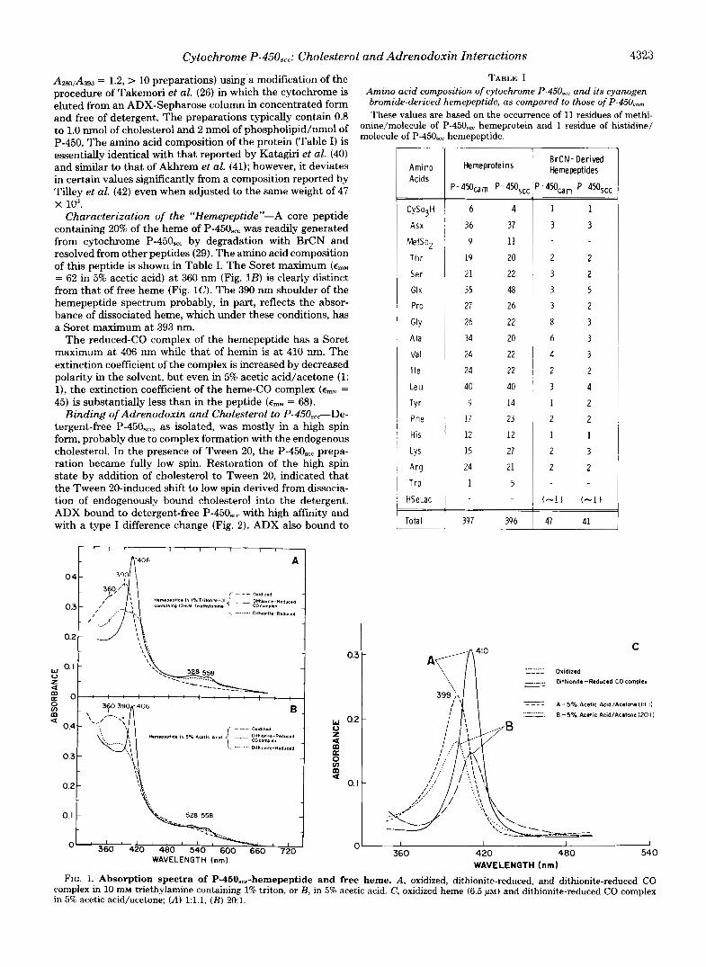
A280,A393 = 1.2, > 10 preparations) using.a modification of the TABLE I procedure of Takemori et al. (26) in which the cytochrome is Amino acid composition of cytochrome P-45OS,, and its cyanogen eluted from an ADX-Sepharose column in concentrated form bromide-deriued hemepeptide, as compared to those of P-45Oc,,
and free of detergent. The preparations typically contain 0.8 These values are based on the occurrence of 11 residues of methi- to 1.0 nmol of cholesterol and 2 nmol of p ~ o s p ~ o ~ ~ p ~ ~ ~ n m o ~ of onine/molecule of P-45Osc, hemeprotein and 1 residue of histidine/
P-450. The amino acid composition of the protein (Table I) is essentially identical with that reported by Katagiri et al. (40)
molecule of P-450,,, hemepeptide.
and similar to that of Akhrem et al. (41); however, it deviates in certain values significantly from a composition reported by Tilley et al. (42) even when adjusted to the same weight of 47
Characterization of the "Hemepeptide"-A core peptide containing 20% of the heme of P-450,,, was readily generated from cytochrome P-450,,, by degradation with BrCN and resolved from other peptides (29). The amino acid composition of this peptide is shown in Table I. The Soret maximum (ernM = 62 in 5% acetic acid) at 360 nm (Fig. 1B) is clearly distinct from that of free heme (Fig. 1 0 . The 390 nm shoulder of the hemepeptide spectrum probably, in part, reflects the absor- bance of dissociated heme, which under these conditions, has a Soret maximum at 393 nm.
The reduced-CO complex of the hemepeptide has a Soret maximum at 406 nm while that of hemin is at 410 nm. The extinction coefficient of the complex is increased by decreased polarity in the solvent, but even in 5% acetic acid/acetone (1: l), the extinction coefficient of the heme-CO complex (ernM = 45) is substantially less than in the peptide (ernM = 68).
Binding of Adrenodoxin and Cholesterol to P-45OS,,-De- tergent-free P-450,,,, as isolated, was mostly in a high spin form, probably due to complex formation with the endogenous cholesterol. In the presence of Tween 20, the P-450,,, prepa- ration became fully low spin. Restoration of the high spin state by addition of cholesterol to Tween 20, indicated that the Tween 20-induced shift to low spin derived from dissocia- tion of endogenously bound cholesterol into the detergent. ADX bound to detergent-free P-450,,, with high affinity and with a type I difference change (Fig. 2). ADX also bound to
X lo3.
0.3 -
w 0.2
m a
-
0 t
L s m a
0.1 -
Amino Acids
CySo3H
Asx
Metsop
T h r
Ser
Glx
Pro
GlY
Ala
Va I
I le
Leu
TY r Phe
His
LYS
Hemeproteins
P - 4 5 0 a m P - 450,,,
6 4
36 37
9 11
19 20
21 22
55 48
27 26
26 22
34 20
24 22
24 22
40 40
9 14
17 23
12 12
15 27
24 21
1 5
39 7 39 6
T
! I
BrCN- Derived Hernepeptides
2 2
3 2
3 5
3 2
8 3
6 3
4 3
2 2
3 4
1 2
2 2
1 1
2 3
2 2
(-1J (-1)
47 41
C
.......... _ _ _ _ Oxidlzed
._._. Dilhionllr-Reduced CO complex - - ---- A - 5 % Acellc Acid/Acelone(l.l I ) ............ E -5% Acetic Acid/Acalone(20 I) ............ E -5% Acetic Acid/Acalone(20 I)
" 360 420 400 54 0
WAVELENGTH (nm) FIG. 1. Absorption spectra of P-450.,,-hemepeptide and free heme. A, oxidized, dithionite-reduced, and dithionite-reduced CO
complex in 10 mM triethylamine containing 1% triton, or B, in 5% acetic acid. C, oxidized heme (6.5 ~ L M ) and dithionite-reduced CO complex in 5% acetic acid/acetone; (A) l:l.l, ( B ) 20:l.

4324 Cytochrome P-450,,,: Cholesterol and Adrenodoxin Interactions
P-450,,, which was fully depleted of endogenously bound cholesterol (see "Methods"), as observed by a type I difference spectrum (A,,, 385, Ami, 425 nm) which was shifted by the presence of 0.3% Tween 20 (A,,, 383, Ami,, 417 nm) (Fig. 2).
The affinity of cholesterol-free P-450 for ADX was not significantly affected by 0.3% Tween 20 (& = 0.4 p~ in both cases). The affinity for ADX was greatly enhanced by the presence of cholesterol (e.g. 6 times by 90 p~ cholesterol, Fig. 3). The Kd for ADX binding at saturation of cholesterol, calculated from the plot in Fig. 3 (inset) was enhanced 13-fold
I * * I
60 I I No Additions
I I
/ I
OBONM .17pM 0 CAdrenodor inF,eeT' , I "
10 20
FIG. 2. Adrenodoxin binding to cytochrome P-450.,, in the presence and absence of Tween 20. ADX was added to P-450,, (0.16 p ~ ) at 35°C in 25 mM K phosphate (0- - -0) and with 0.3% Tween 20 and 200 p~ cholesterol (t".). Difference spectra (inset) show addition of ADX (0.5 p~ increments) to cholesterol-depleted P- 450,,, (0.82 p ~ ) with (A) and without 0.3% Tween 20 (B) . The base- line prior to addition of ADX is indicated ( b ) .
/ Tween Cholesterol
, 1 I 013,~M I 04pM 0 2 4 6 8 IO 12 14
0 3,uM [Adrenodoxin,,,]",~M"
FIG. 3. Effect of cholesterol on adrenodoxin binding to cy- tochrome P-450.,,. Type I spectral responses were measured at 35°C for addition of ADX to cytochrome P-450,,, (0.15 p ~ ) in 0.3% Tween 20,5 mM K phosphate, 50 mM KCl, and 0,15,36, or 90 p~ cholesterol. [ADXr,,.] was calculated as described under "Methods." The values at the x axis intercept represent the Kd for the ADX-P-450 complex at each cholesterol concentration. Inset, replot of Kd uersus choles- terol concentration (C) based on the relationship (modified from Ref. 53)
[Kc + c ] / K d = C/KCA + KAC/KCA
where &A is the constant for dissociation of ADX from the P-450- cholesterol complex and KAC is the constant for dissociation of cho- lesterol from the P-450-ADX complex. The slope of the graph is 1/ KCA .
over binding in absence of cholesterol. Similarly, 0.5 p~ ADX enhanced the affinity of P-450,,, for cholesterol by 6 to 7 times (data not shown). Affinity of ADX to P-450,,, in Tween 20 with 200 p~ cholesterol was 2 times stronger than to P-450,,, formed in a complex with endogenous cholesterol in absence of Tween 20 (the percentage of P-450,, formed in a complex with cholesterol was similar under the two conditions), sug- gesting a small stabilizing effect of Tween 20 on the ternary complex of ADX-cholesterol-P-450scc (Fig. 2).
The spin states of the various P-450,,, complexes have been calculated from changes in the Soret absorption bands relative to the spectrum of a fully low spin cholesterol-free cytochrome (Table 11). The proportions of high spin state induced at saturation with only ADX or only cholesterol were 20% and 6776, respectively; whereas at saturation of both, it was 92%, suggesting an additive effect of the two ligands on this param- eter (Table 11).
Effect of Temperature on Complex Formation-The bind- ing of ADX to cytochrome P-450,,, in the absence of choles- terol was insensitive to temperature changes from 22-35°C. However, below 22"C, complex formation increased with de- crease of temperature. The differences shown in Fig. 4 be- tween complex formation at 6°C and 22°C were measured by changing the temperature on a single sample and were there- fore directly observable. Spectral changes produced by wann- ing and cooling the sample in the range 6-35°C were fully reversible.
Elevated temperature decreased the affinity of P-450,,, for cholesterol (& (36°C) = 100 pM, K d (5OC) = 35 pM) (Fig. 5). Expression of this data as a Van't Hoff plot provided A H =
TABLE 11 Spin states of cytochrome P-450,, complexes
Complex Temperature [KC11 High spin state"
"C r n M %
ADX-P-450 5-30 0-50 20 Chol-P-450' 5 50 73
30 50 67 30 0 60 30 200 83
Chol-ADX-P-45ObS e 5 50 90 30 50 92
a High spin state is calculated from absolute absorption spectra in 0.3% Tween, 5 mM Hepes (pH 7.2) from the change ( A A ) in A (390- 420 nm) relative to the fully low spin spectrum of P-450,,, without added cholesterol or ADX. The increase in percentage high spin state is AA/nmol P-450/0.11 X 100, which is based on AE = 110 cm" mM-' for a complete spin state change. ' Calculated from the extrapolation of AA to saturation with cho-
lesterol. 2.0 p~ ADX.
[Adrenodoxintree ]$M"
FIG. 4. Temperature dependence of adrenodoxin binding to cytochrome P-450.,,. Cytochrome (0.55 PM) was in 13 mM Hepes (pH 7.2), 0.3% Tween 20, 0.5 mM dithiothreitol, and 50 mM KC1. Measurements at 6"C, 22"C, and 30°C were all carried out on the same solution.

Cytochrome P-450,,,: Cholesterol and Adrenodoxin Interactions 4325
I I I
'?!I/ 43
4 2 A H = - 7 4 kc01
41
4 0
AS = - 6 e u
3ZO 330 340 350 360
FIG. 5. Temperature dependence of cholesterol binding to cytochrome P-450,,,. Cytochrome (0.37 p ~ ) was in the buffer de- scribed in Fig. 4. The inset shows Van't Hoff plots of these data and for a parallel experiment with 0.5 /.LM ADX.
-7.4 kcal mol" and A S = -5.9 e.u. In the presence of ADX (0.5 p ~ ) , Kd was less temperature-sensitive with A H = -1.7 kcal mol" and A S = 17 e.u. (Fig. 5). The increase in temper- ature induced only a slight decrease in the proportion of high spin state in the cholesterol-P-450,,, complex formed by sat- urating levels of cholesterol (73% at 5°C to 67% at 30°C). The spin state of the ternary cholesterol-ADX-P-450,,, complex was also relatively insensitive to temperature (Table 11).
Effect of Ions on Complex Formation-Increasing concen- trations of NaCl increased the Kd for ADX both in the absence and presence of 45 p~ cholesterol (Fig. 6). Effects of NaCl on the optical change at saturation of ADX, both with and without cholesterol, were negligible (Fig. 6). In contrast, al- though NaCl progressively increased the proportion of high spin cytochrome at saturation with cholesterol (60-85%), it did not significantly change the Kd for cholesterol (Fig. 7). NaCl also increased the proportion of high spin cytochrome P-450 when added to the cholesterol complex in the absence of Tween 20. CaClz (1-5 mM) had no effect on either the affinity of the cytochrome for cholesterol or the spectrum of the complex.
Effect of Ionic Strength on Adrenodoxin and Cholesterol Dependence of Side Chain Cleavage Activity-When choles- terol side chain cleavage was reconstituted from purified adrenodoxin reductase, ADX, and cytochrome P-450,,, in the presence of 0.3% Tween 20, activity in 5 mM Hepes buffer was greatly enhanced by the addition of univalent ions. At 5 p~ ADX, we observed a similar bell-shaped dependence on the concentration of added NaCl to that previously reported by Takikawa et al. (21) with peak activity between 80 and 150 mM KC1. However, the fall-off in activity at high salt was caused by an increase in the K,,, for ADX which paralleled the increase in the Kd for ADX (Fig. 8, inset). Indeed, at 200 mM NaCl and 200 p~ cholesterol, the V,,, in terms of saturation with ADX was at the highest value of 30 nmol of pregneno- lone/nmol P-450/min. The same dependence of activity on ionic strength was obtained when Hepes/NaCl was replaced by increasing concentrations of K phosphate buffer. In this buffer, salt had only a slight effect on the K,,, for cholesterol up to 42 mM K phosphate (ionic strength equivalent to 100 mM NaC1) (Fig. 9). However, at 82 mM K phosphate (equiva- lent to 200 mM NaCl), there was a decrease in the apparent K,,, for cholesterol. Thus, a 2-fold increase in ionic strength (100-200) produces a 15-fold increase in K, for ADX and, at most, a 2-fold decrease in K,,, for cholesterol.
Side Chain Cleavage Activity: Initial Velocity Patterns- In order to determine the kinetic mechanism of cholesterol and ADX additions during each cycle of cholesterol side chain cleavage, initial velocity pattern studies were carried out. Plots of v-l versus [ADXf,,,]" exhibited a decreasing slope
P 1 . 1 ,
50 2x P450
I I ./
Io No NaCl 10
0 2 4 6 8 1 0
[Adranodoxinm,]-',fiM"
FIG. 6. Effect of NaCl on adrenodoxin binding to cytochrome P-450.,,. A, in absence of cholesterol; B, in presence of 45 p~ cholesterol. Type I spectral changes were measured with cytochrome P-450,,, ( A = 0.24 p ~ ; = 0.12 pM) present in 0.3% Tween 20, 5 mM Hepes (pH 7.2), and 0.5 mM dithiothreitol at 30°C.
zero
-10 0 IO 20 30 40
[Cholesterol]-!rnM-' FIG. 7. Effect of NaCl on cholesterol-binding to cytochrome
P-450,,,. Type I spectral changes were measured with cytochrome P- 450,,, (0.28 p ~ ) present in 13 mM Hepes (pH 7.2), 0.3% Tween 20 at 30°C.
'0.3pM 51u'M0 I 2 3 4 5 6 5 [Adrenodoxin f,ee]",~M"
FIG. 8. Effect of NaCl on adrenodoxin dependence of choles- terol side chain cleavage activity. The assays were carried out in 10 mM Hepes buffer (pH 7.2) with 0.3% Tween 20,200 p~ cholesterol, 0.3 p~ adrenodoxin reductase, and 0.2 p~ P-450,,,. The concentrations of NaCl are indicated next to each reciprocal plot. Inset shows the K, values determined by these reciprocal plots and the Kd values deter- mined in the same buffer with 200 p~ cholesterol and 0.125 p~ P- 45Oxc.

4326 Cytochrome P-45OS,,: Cholesterol and Adrenodonin Interactions
t c 30
I I 4 2 5 4
~
Y 0 E , 4 2 m M -x
-15 C 0 0 c -
-05 5 E - -
I
51pM 130pM [Cholesterol]‘~mM” - 93,,M’ 0 IO 20 30 40
FIG. 9. Effect of ionic strength on cholesterol dependence of cholesterol side chain cleavage activity. The assays were carried out in 27,42, or 82 mM K phosphate buffer (pH 7.2) with 10 mM KCI, 0.3% Tween 20, 200 p~ cholesterol, 0.25 p~ adrenodoxin reductase, 4.5 p~ ADX, and 0.22 p~ P-450,,.
FIG. 10. Initial velocity pattern analysis of cholesterol side chain cleavage reaction with varying adrenodoxin at constant cholesterol concentrations. The inset shows some of the same data plotted with varying cholesterol at constant ADXfre concentrations. The assays were carried out in 10 mM Hepes buffer (pH 7.2) with 100 m~ NaC1, 0.3% Tween 20, 0.3 p~ adrenodoxin reductase, and 0.2 p~ P-450,cc.
with increasing cholesterol, intersecting at a single point (Fig. 10). The same data replotted as u-l uersus [cholesterol]” at various [ADXr,,,] also provides a set of intersecting lines (Fig. 10, inset). A notable feature of these plots is that the apparent K,,, increases in both cases as the concentration of the fmed component is increased. The true K,,, values corresponding to saturation of the second component were computed to be approximately 390 ~ L M for cholesterol and 1.2 ~ L M for ADX. The Vmax at saturation of both cholesterol and ADX was 68 nmol of pregnenolone/min/nmol of P-450.
DISCUSSION
Cytochrome P-450,, and the proteins mediating transfer of electrons from NADPH to P-450,,,, adrenodoxin reductase and ADX are located on the matrix side of the inner mem- branes of adrenal cortical mitochondria (6, 14, 43). P-450,,, is
an integral membrane protein, whereas adrenodoxin reductase and ADX appear to be peripheral membrane proteins (14). Cholesterol, the substrate, is very insoluble in water (CMC z 30 nm, Ref. 44) and more readily gains access to the cytochrome within detergent micelles or phospholipid bilayers ( 14).
The extensive studies on P. putida ferredoxin-cytochrome P-45OC,,-catalyzed camphor monooxygenation provide the basis for our current understanding of the catalytic cycle for this class of reactions (12). Many similarities are apparent between cytochromes P-450,,, and P-450,,,, including immu- nological cross-reactivity (7) and inter-relationship of sub- strate and ferredoxin binding to the cytochromes. Cyto- chromes P-450,,, and P-450,., have nearly identical sues while the amino acid composition of P-450,,, differs most noticeably from that of P-450,,, in the large increases in aromatic amino acids and lysine as compared to a substantial decrease in alanine (Table I).
Hemepeptide-P-450,,, also shares with P-450,,, and P- 4 5 0 ~ ~ a structural feature which permits release with BrCN of a very hydrophobic, heme-binding core peptide which in each case comprises about 10% of the total proteins (7,8, 29). The peculiar spacing of methionine residues in the surround- ing sequences possibly makes this domain fortuitously acces- sible via BrCN cleavage. The purified hemepeptide of P-450,,, was found to contain 20% of the heme of the native hemepro- tein. However, recoveries of up to 80% have been observed in hemepeptides released from P-450,, photocovalently labeled at a site close to the heme binding site by an azido derivative of the inhibitor, aminoglutethimide. For other P-450 cyto- chromes, a substrate-based photoaffinity probe also greatly increases this recovery, thus further relating the peptide to the functional center of the cytochrome (8, 29).
Adrenodoxin and Cholesterol Binding to P-450,,,-There has been a conflict in previous reports (18-20) concerning the binding of ADX to substrate depleted P-45OS,,. This study clearly shows that ADX binds to P-450,,, metabolically de- pleted of endogenous cholesterol in the presence or absence of detergent Tween 20 (Fig. 2). The kinetics of cholesterol association with P-450,,, in Tween 20 (Fig. 5) indicate that in Tween 20 micelles cholesterol is fully in equilibrium with the cytochrome. The positive cooperativity we observe between the interactions of cholesterol and ADX with P-450,,, in Tween 20 is quantitatively similar to that reported by Lam- beth et al. for P-450,,, in lecithin-cholesterol vesicles (20). In the present studies, we also show that Tween slightly activates rather than inhibits the binding of ADX to the P-450,,- cholesterol complex and that it has no effect on binding to the substrate-free cytochrome. This insensitivity to Tween 20 implies that the hydrophobic domain which anchors the cy- tochrome to membranes is clearly separated from the ADX binding site. The small perturbation of the Soret band of the substrate-free cytochrome by Tween 20 does, however, sug- gest a communication between the membrane environment and the active center (Fig. 2, inset). Seybert et al. have established that cholesterol gains access to the substrate bind- ing site through the membrane (14).
ADX binding to P-450.,, is driven by a large increase in entropy. Consideration of the full cycle (Fig. 11) indicates that cholesterol activation of this binding is a consequence of a further increase in A S (from 30 to 53 e.u.) which more than offsets an increase in AH (5.7 kcal). The high negative charge on the surface of ADX and the observed competition between ions and ADX for the cytochrome suggest that the entropy increase may derive from a release of water and ions during the interaction of complementary charged domains on the two proteins.

Cytochrome P-450,,,: Cholesterol and Adrenodoxin Interactions 4327 AH=-74kcal
AS=-Geu
+CHOL P450, 'P450 CHOL
A H ' I ~ ~ = 3 e e u 1 l+ADX AS=62e.u AH=84 kea' j \A
AH= 0 ASZ3Oeu IB AH=5 kcal j,
P450 ADX- P450. :EEL hS.53 eu j
+CHOL AH=- I7 kcal
ASZ17eu
FIG. 11. Entropy and enthalpy changes for binding of cho- lesterol (CHOL) and adrenodoxin to cytochrome P-450.,,. The values were calculated from data represented in Figs. 4 and 5. A at 22"C, B at 35°C.
The spin state change from low to high spin is associated with an increase in entropy (14-30 e.u.) and a decrease in enthalpy (2.5-10 kcal) (46, 53).4 Subtraction of the contribu- tion from the spin state change (60%) indicates that choles- terol binding is associated with large opposing changes in enthalpy and entropy. The transference of cholesterol from detergent to binding site therefore seems to be associated with restricted movement, possibly of both cholesterol and protein, which is partially relieved by prior binding of ADX.
The spin state of cytochrome P-450 is an indicator of the configuration of ligands around the heme (12). Increases in the proportion of the high spin state are also frequently associated with facilitated reduction of the cytochrome (45). Both ADX and cholesterol shift the spin state equilibrium of P-450,,, to high spin form (17-20, 51). This provides a differ- ence from P-450,,, where camphor stabilizes the high spin form but putidaredoxin causes a shift to the low spin form (12, 52).
The high spin state induced by a fxed concentration of cholesterol declines with rising temperature. In Tween 20, this change derives from an increased dissociation of cholesterol rather than from an intrinsic change in the spin state of the substrate complex, as has been reported for P-450,,, (46). The similar temperature-dependent change to low spin observed for the P-450,,, complex with endogenous cholesterol in ab- sence of Tween 20 (51) presumably also derives from move- ment of cholesterol out of the substrate site. However, choles- terol remains bound to the protein under these conditions, suggesting that cholesterol moves to a secondary site on the protein associated with a low spin complex. ADX and ions increase the proportion of high spin state in the cholesterol- P-450,, complex by comparable amounts (Table IT) suggest- ing that an ionic effect on the ADX site may alone be sufficient to communicate a change in heme configuration from the surface of the cytochrome. However, changes in spin state in absence of cholesterol and activation of cholesterol binding are induced by ADX but not by elevation of ionic strength.
Ionic Effects on Adrenodoxin and Cholesterol Dependence 0 f P - 4 5 0 , ~ ~ Activity-Previous studies by Takikawa et al. (21) have indicated an unusual "bell-shaped'' dependence of cho- lesterol side chain cleavage activity on univalent cation con- centration. However, we have recently shown that although, on the basis of ionic strength, activation by Mg2+ follows a pattern similar to Univalent metal ions, Ca2+ causes very little activation and inhibits side chain cleavage activity optimally activated by 100 mM NaCl (54). Activation at low ionic strength can in part be ascribed to the activation of ADX
These authors use the sign convention opposite to that used here. Our data derive from the conventional relationship AG = -RT In K,,
A H - TAS.
reduction (16). Activation of ADX reduction also results in decreased levels of oxidized ADX which we have previously shown to inhibit monooxygenase activity (17, 47). Here, we show that the inactivation at high concentrations of univalent ions is only apparent and is due entirely to an over 50-fold increase in the K,,, for ADX which parallels the increase in the Kd for ADX (Fig. 8). Indeed, V,,, at 200 pM cholesterol continues to increase up to the highest salt concentration examined. In contrast, the apparent K, and the K d for choles- terol are both relatively insensitive to changes in salt concen- tration (Figs. 7 and 9). The shift in the apparent K, for cholesterol at the highest salt concentration examined is in the same direction as that caused by decreased [ADX] (Fig. 10). Thus, this shift is probably due to an increase in the K , for ADX (Fig. 8) (which would render the [ADX] used in this experiment less than saturating), rather than a true decrease in the K, for cholesterol.
Complex Formation during Enzyme Turnover-The cata- lytic cycle for camphor monooxygenation at P-450,,, has been presented as an ordered sequence of substrate binding fol- lowed by putidaredoxin binding and first electron transfer (12, 48). However, our results, as well as those of Lambeth et al. (20), clearly indicate that ADX can bind to P-450,,, that is free of substrate cholesterol. Since on the basis of these results there appears to be no obligatory order for the binding of ADX and cholesterol to P-450,,,, the sequence of additions of these two reactants would have to be considered as random rather than ordered (34, 49). Superficially, the intersecting initial velocity pattern and the linearity of the plots (Fig. 10) are consistent with a futed order of binding of cholesterol and ADX (Ref. 49, p. 564). However, apparent order can be reconciled with random binding if, at these relative concentra- tions of reactants under these experimental conditions, the addition of one of the reactants occurs first with much higher frequency. These reaction conditions are far removed from those found in the adrenal cortex mitochondria where high local concentrations of both ADX and P-450,,, on the surface of the inner membrane and a low steady state level of choles- terol may result in frequent binding of ADX to cholesterol- free P-450,,.
Given the substantial synergism in the binding of choles- terol and ADX to P-450,,, the observed initial velocity pat- terns in which cholesterol increases the K, for ADX and vice uersa (point of intersection below the abscissa, Fig. 10) are atypical for a random rapid equilibrium mechanism (Ref. 49, p. 274). Indeed, there is strong evidence that, unlike camphor hydroxylation (48), rapid equilibration of these reactants does not occur during cholesterol side chain cleavage. The true K , values for cholesterol and ADX are manyfold larger than the respective K d values for either binary or ternary complex formation. This is normally observed when the rate-limiting step is faster than the dissociation of the substrates by similar factors. Simulation of kinetic patterns corresponding to such nonrapid equilibrium conditions shows that synergism in the binding of reactants can be consistent with an intersection point below the abscissa (50).
The apparent slow dissociation of reduced ADX from P- 450,,, raises the important question of whether oxidized ADX dissociates from the cytochrome during the catalytic cycle. Possibly, dissociation may be accelerated by either reduction of P-450,,, or oxidation of cholesterol. In view of the evidence that adrenodoxin reductase, ADX, and P-450,,, do not readily form a ternary complex (14-17), the dissociation of oxidized ADX from P-450,,, must be considered a potential rate-lim- iting factor in the cholesterol side chain cleavage mechanism.
Acknowledgments-We are grateful to Dr. W. W. Cleland for reviewing the initial velocity pattern analysis and for providing copies

4328 Cytochrome P-450,,,: Cholesterol and Adrenodoxin Interactions
of his computer programs. We also gratefully acknowledge the expert secretarial assistance of Margaret O’Donnell.
REFERENCES
1. Shikita, M., and Hall, P. (1974) Proc. Natl. Acad. Sci. U. S. A .
2. Burstein, S., and Gut, M. (1976) Steroids 28, 115-131 3. Jefcoate, C. R., Hume, R., and Boyd, G. S. (1970) FEBS Lett. 9,
41-44 4. Suhara, K., Gomi, T., Sato, H., Itagaki, E., Takemori, S., and
Katagiri, M. (1978) Arch. Biochem. Biophys. 190, 290-299 5. Watanuki, M., Granger, G. A,, and Hall, P. F. (1978) J . Biol.
Chem. 253, 2927-2931 6. Mitani, F. (1979) Mol. Cell. Biochem. 24, 21-43 7. Dus, K. M., Litchfield, W. J., Hippenmeyer, P. J., Bumpus, J . A.,
Obidoa, O., Spitsberg, V., and Jefcoate, C. R. (1980) Eur. J. Biochem. 111,307-314
8. Dus, K., Carey, D., Goewert, R., and Swanson, R. A. (1977) in Microsomes and Drug Oxidations (Ullrich, V., ed) pp. 95-102, Pergamon Press, Oxford
9. Dus, K. (1976) in Enzymes of Biological Membranes (Martonosi, A,, ed) Vol. 4, Part A, Chap. 7, pp. 199-238, Plenum Publishing Co., New York
71, 1441-1445
10. Dus, K. (1975) Adu. Exp. Med. Biol. 58, 287-309 11. Simpson, E. R. (1979) Mol. Cell. Endocrinol. 13,213-227 12. Gunsalus, I. C., and Sligar, S. G. (1978) Adu. Enzymol. Relat.
13. Hall, P. F., Watanuki, M., and Hamkalo, B. A. (1979) J. Biol.
14. Seybert, D. W., Lancaster, J. R., Jr., Lambeth, J . D., and Kamin,
15. Seybert, D. W., Lambeth, J. D., and Kamin, H. (1978) J. Biol.
16. Lambeth, J . D., Seybert, D. W., and Kamin, H. (1979) J. Biol.
17. Hanukoalu. I.. and Jefcoate, C. R. (1980) J. Biol. Chem. 255,
Areas Mol. Biol. 47, 1-44
Chem. 254, 547-552
H. (1980) J. Biol. Chem. 254, 12088-12098
Chem. 253,8355-8358
Chem. 254,7255-7264
3057-3061 18. Kido. T.. Arakawa. M.. and Kimura. T. (1979) J . Biol. Chem. 254.
8377-8385 19. Kido, T., and Kimura, T. (1979) J. Biol. Chem. 254,11806-11815 20. Lambeth, J . D., Seybert, D. W., and Kamin, H. (1980) J. Biol.
21. Takikawa, O., Gomi, T., Suhara, K., Itagaki, E., Takemori, S., and
22. Cuatrecasas, P., and Anfinsen, C. B. (1971) Methods Enzymol.
23. March, S. C., Parikh, I. , and Cuatrecasas, P. (1974) Anal. Bio-
24. Orme-Johnson, W. H., and Beinert, H. (1969) J. Biol. Chem. 244,
25. Hiwatashi, A., Ichikawa, Y., Maruya, N., Yamano, T., and Aki, K.
26. Takemori, S., Suhara, K., Hashimoto, S., Hashimoto, M., Sato,
Chem. 255, 138-143
Katagiri, M. (1978) Arch. Biochem. Biophys. 190,300-306
22,345-385
chem. 60, 149-152
6143-6148
(1976) Biochemistry 15, 3082-3090
H., Gomi, T., and Katagiri, M. (1975) Biochem. Biophys. Res. Commun. 63,588-593
27. Lowry, 0. H., Rosebrough, N. J., Farr, A. L., and Randall, R. J. (1951) J. Biol. Chem. 193, 265-275
28. Omura, T., and Sato, R. (1964) J . Biol. Chem. 239,2370-2378 29. Swanson, R. A,, and Dus, K. M. (1979) J . B i d . Chem. 254,7238-
30. Falk, J. E. (1964) Porphyrins and Metalloporphyrins, pp. 181-
31. Laemmli, U. K. (1970) Nature 227,680-685 32. Suhara, K., Takemori, S., and Katagiri, M. (1972) Biochim. Bio-
33. Chu, J.-W., and Kimura, T. (1973) J . Biol. Chem. 248, 2089-2094 34. Cleland, W. W. (1963) Biochim. Biophys. Acta 67, 104-137 35. Cleland, W. W. (1979) Methods Enzymol. 63, 103-138 36. Folch, J., Lees, M., and Sloane-Stanley, G. H. (1957) J. Biol.
37. Bartlett, G. R. (1958) J. Biol. Chem. 234,466-468 38. Gornall, A. G., Bardawill, C. J., and David, M. M. (1949) J. Biol.
39. Hanukoglu, I., and Jefcoate, C. R. (1980) J . Chromatogr. 190,
40. Katagiri, M., Takemori, S., Itagaki, E., Suhara, K., Gomi, T., and Sato, H. (1976) Adu. Exp. Med. Biol. 74,281-289
41. Akhrem, A. A., Lapko, V. N., Lapko, A. G., Shkumatov, V. M., and Chashchin, V. L. (1979) Acta Biol. Med. Germ. 38, 257- 273
42. Tilley, B. E., Watanuki, M., and Hall, P. F. (1977) Biochim. Biophys. Acta 488,330-339
43. Churchill, P. F., and Kimura, T. (1979) J. Biol. Chem. 254,10443- 10448
44. Haberland, M. E., and Reynolds, J. A. (1973) Proc. Natl. Acad. Sci. U. S. A. 70,2313-2316
45. Rein, H., Ristau, O., Misselwitz, R., Buder, E., and Ruckpaul, K. (1979) Acta Bid. Med. Germ. 38, 187-200
46. Sligar, S. G. (1976) Biochemistry 15,5399-5406 47. Hanukoglu, I., Privalle, C. T., and Jefcoate, C. R. (1980) Fed Proc.
39, 1825 48. Pederson, T. C., Austin, R. H., and Gunsalus, I. C. (1977) in
Microsomes and Drug Oxidations (Ullrich, V., Roots, I., Hil- debrandt, A,, Estabrook, R. W., and Conney, A. H., eds) pp. 275-283, Pergamon Press, Oxford
49. Segel, I. H. (1975) Enzyme Kinetics: Behavior and Analysis of Rapid Equilibrium and Steady-State Enzyme Systems, pp. 957, John Wiley & Sons, New York
50. Bar-Tana, J., and Cleland, W. W. (1974) J. Biol. Chem. 249,
51. Katagirl, M., Takikawa, O., Sato, H., and Suhara, K. (1977)
52. Lipscomb, J. D. (1980) Biochemistry 19,3590-3559 53. Ristau, O., Rein, H., Greschner, S., Janig, G.-R., and Ruckpaul,
54. Hanukoglu, I., Privalle, C. T., and Jefcoate C. R. (1981) J . Biol.
7246
188, Elsevier, Amsterdam
phys. Acta 263,272-278
Chem. 233,497-509
Chem. 177, 751-766
256-262
1271-1276
Biochem. Biophys. Res. Commun. 77,804-809
K. (1979) Acta Biol. Med. Germ. 38, 177-184
Chem. 256,4329-4335