· Journal of American Science, 2011;7(10) 255 [email protected]
Journal of American Science, 2011;7(9) http://www.americanscience.org
http://www.americanscience.org [email protected] 584
Factors Affecting the resolution of SPECT Imaging
H. E. Mostafa*1, H. A. Ayoub2 and Sh.Magraby1
1Kasr El-Ini Center for Oncology, Cairo University, 2Faculty of Science, Suez Canal University [email protected] h. [email protected]
Abstract: SPECT technique provides 3D view of the organ/structure (depth information), which facilitates the quantification and the improvement of the contrast and resolution of output images. Because of the importance of spatial resolution in SPECT studies, it is essential to periodically verify that there has been no deterioration in this parameter. Spatial resolution is commonly quantified from the full-width-at-half-maximum (FWHM) of the line spread response function. Many acquisition factors degrade SPECT images both qualitatively and quantitatively. The present work was done by using the performance Jasezczak and Resolution phantoms filled with a 99mTc solution to simulate the tumor (lesion) and calculate its spatial resolution (FWHM). The image acquired by using different acquisition matrices, mode of rotation, radius of rotation, number of projection, time per projection and type of collimator. The reconstruction of image was done by filter back projection method where Butterworth filter used with cutoff frequency 0.40 at order 5. The performance phantoms and imaging process were done by founding of the attenuation and scattering media (water). The earned results indicated 60 views which gave the best image quality rather than 30 or 45 views. Smaller pixel size 128 x 128 can display more image details and good resolution than the 64 x 64. Time per view (TPV) 30 or 40 s is lead to improved image quality (resolution). Image quality is being worse by inhancement the radius of rotation for gamma camera. Ultra high-resolution collimator is recommended to provide good spatial resolution rather than the general all purpose collimator. [H. E. Mostafa, H. A. Ayoub and Sh.Magraby, Factors Affecting the resolution of SPECT Imaging. Journal of American Science 2011; 7(9):584-591].(ISSN: 1545-1003). http://www.americanscience.org. Keywords: radionuclide imaging/ SPECT/ contrast.
1. Introduction
SPECT (Single Photon Emission Computed Tomography) is a technique for tracing radioactive materials in the body and provide three dimensional maps of in vivo radiopharmaceutical distributions (Bartlett et al., 1995). The fundamental goal of all tomographic imaging systems is to portray the distribution of the radioactivity in the patient more accurately (Collier et al., 1984). SPECT has highlighted the need to improve gamma camera performance.
Physical properties are important for the assessment of the potential of emission computed tomography implemented by collimated detector system which; includes sensitivity, statistical and angular sampling requirements, attenuation compensation, resolution, uniformity, and multi-section design constraints. Also, poor energy resolution translates directly into degraded spatial resolution through reduced ability to reject scattered photons on the basis of pulse height analysis (Thrall & Zienssman 1987).
Various factors limit the quality of the SPECT images. Among these is radius of rotation, number of projections, time per projection, number of projection, matrix size, type of collimators, (Amersham Healthcare, 1994). In order to overcome patient study limitation, the cylindrical Perspex SPECT phantoms has been used.
2. Material and Methods
Phillips axis dual head gamma camera was used in this study. Two types of phantoms (jasezczak& resolution phantoms) were used. The jasezczak phantom consists of a commercially supplied cylindrical Perspex container. It has three distinct sections; the cold-rod section, a uniformity section and a hot rod section. It has a cavity and three screw-filling plugs that can be filled and then sealed. The total fluid filling volume is about 4.0 liters. While the resolution phantom is an upright cylindrical phantom with diameter of 22 cm and a height of 31-cm. The phantom is constructed of Plexiglass with a wall thickness of 1 cm and 3 stainless steel capillary tubes with diameter of 1mm embedded inside the cavity of the cylinder, so that the spacing between each two of them is fixed to be 10 cm.
99mTc having gamma ray energy of 140 KeV, and half-life 6.02 hrs was used. The radioactive material used in the tests must be prepared and measured in a clean and well aerated area (Laminar flow). Eight patients (males and females) were examined for brain imaging using Tc- HMPAO as the radiopharmaceutical. Phantom imaging:
The SPECT jasezczak phantom, filled with water containing approximately 925 MBq (25mCi)

Journal of American Science, 2011;7(9) http://www.americanscience.org
http://www.americanscience.org [email protected] 585
of 99mTc while the resolution phantom filled 1 mCi of each capillary tube, were positioned on the gamma camera SPECT imaging table. The cylinder axis of the phantoms was positioned parallel to the axis of rotation of the gamma camera detector. Within the rotational field of view, we acquired 24 SPECT acquisitions to evaluate the different parameters. Image acquisition:
The image acquired by using different acquisition matrices such as mode of rotation, radius of rotation, number of projection, time per projection and collimator type. 1- Acquisition matrix: The matrices of 64 x64 and 128 x 128 pixels were applied on the SPECT images in two acquisitions to detect the effect of matrix size on the resolution of the images. 2- Number of views (NOV):
The effect of the number of acquired views on the SPECT images was evaluated. The SPECT phantom had been imaged five times using fixed parameters in both acquisition and reconstruction. The only difference was the number of views per scan. In this parameter 30,36,45,60 and 90 views were used per acquired image. 3- Motion type (mode of rotation):
To study the effect of the motion type on the SPECT images, with SPECT image with step&shot, step&shot/con and continues applied to the phantom, and another SPECT image in a fixed state were obtained. This protocol was applied to the phantom three times within the acquisition. 4- Radius of Rotation: Four images were compared to study the effect of radius on SPECT obtained for SPECT phantom. The radii of rotation for gamma camera were 15, 20, 25 and 30 cm. 5- Type of collimator:
The effect of the collimator type on SPECT images was studied. The SPECT phantoms were used with the fixed parameters for both acquisition
and reconstruction. The data was recorded with low energy high-
resolution collimator (LEHR) = (LEGAP) and low energy ultra high-resolution collimators (LEUHR). Image reconstruction:
The image reconstruction method used was the filtered back projection, using the upper back projection parameters, and filter information parameters. The reconstruction matrix: 128 x 128 Number of slices: 25 Filter type: Butterworth Cut of frequency Hz: 0.40& order 5. The reconstructed image was saved as a transverse image, which was used for analysis. Image analysis software:
The SPECT transverse image number 18 was chosen (which is located within the hot rod section for Jasezczak and resolution phantoms) for each acquisition study, Because of the several SPECT acquisition parameters measured, only the percentage variation of some data was used for statistical analysis 3. Results and Discussion The following factors affect SPECT images both qualitatively and quantitatively: 1- Image matrix (pixels):
The results obtained (Fig. 1─4) showed a great difference between images taken by an image matrix 64x64 and that taken by another value of 128x128. This difference showed due to image resolution. A smaller pixels dimension of 128x128 could display more image detail than the 64x64. There was great loose of resolution for the 64 x 64 images, although maximum profiles (opposite to resolution) were larger than in case of 128x128 pixels. This could be explained due to the fact that the chosen pixel (matrix) should be less than 1/3 of the expected FWHM .This is matched with the matrix of (128x128).the same results were found by Farag et al., 2003. These results are also consistent with the study of the effect of different matrices using clinical bone image as a test pattern which showed that smaller pixels caused improvement in the resolution of images (Mark et al., 1998)

Journal of American Science, 2011;7(9) http://www.americanscience.org
http://www.americanscience.org [email protected] 586
Matrix 64x64 Matrix 128x128
Fig. (1): Spatial resolution of the cold lesion of SPECT J-phantom as a function of matrix.
Matrix 64x64 Matrix 128x128
Fig. (2): Spatial resolution of the hot lesion of SPECT J-phantom as a function of matrix.
Fig. (3): Mean FWHM versus matrix (pixels) for R-phantom. Fig. (4): Mean FWHM versus matrix (pixels) for R-phantom 2- Number of views (NOV): Increasing the number of views leads to the best definition of the SPECT images. This parameter showed improvements of counts and resolution in the case of 90 and 60 views rather than in 45, 36 and 30 views. The blur (noise artifact lines) appeared when 36 and 30 views are used because the angular step increased. From this factor No. of view, it was known that 60 NOV give the best image quality. In practice, approximately 60 views were adequate for
most clinical studies. This is in agreement with the results obtained by Bernier et al., 1994. The whole time of acquisition should be suitable to patient stability during imaging to get clinical image quality. Study case No. 2
• A male patient 60 years old with history of diabetes. He started to get weakness and parathesia in right side for which cerebral blood flow examination was done.
control control
36
37
38
39
40
41
42
43
Matrix 64*64 Matrix 128*128
matrix size
Mea
n FW
HM
mm
controlcontrol
0
2
4
6
8
10
12
Matrix64*64 Matrix128*128
Matrix size
Mea
n FW
HM
mm

Journal of American Science, 2011;7(9) http://www.americanscience.org
http://www.americanscience.org [email protected] 587
• Cerebral blood flow with 60 views, which revealed an area of hypo- perfusion in left frontotempral region (arrowed) with normal perfusion in the rest of brain.
• Whereas using 30 views revealed heterogeneous uptake through all the different regions of the brain.
Number of view or angular step60 views
30 views
Plate No. 2 represents case No. 2.
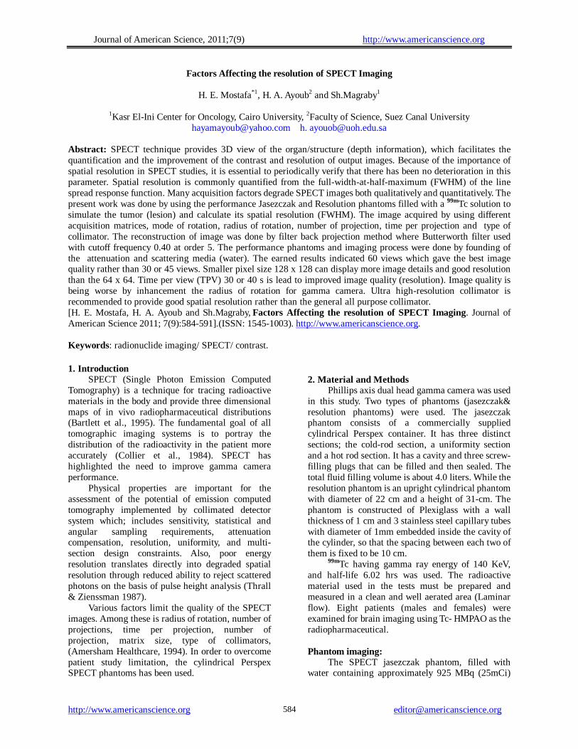
3- Motion type (mode of rotation): In present work, low resolution in addition to blurred lines were observed due to slow motion of gamma camera (continuous motion mode). By using the other two motion
types(step&shot/cont., and step& shot)these blurred lines disappeared and resolution can be increased, due to stop of gamma camera at each projection (view).
Step & shot / cont. continuous Step & shot
Fig. (5): spatial resolution of the cold lesion of J- phantom as a function of motion type.
control control control
7
7.2
7.4
7.6
7.8
8
8.2
8.4
8.6
Step & shot Step & shot cont. Continues
Motion type
Mea
n F
WH
M m
m
control control control
37.6
37.8
38
38.2
38.4
38.6
38.8
39
39.2
Step & shot Step & shot cont. Continues
Motion type
Mea
n F
WH
M m
m
Fig. (6): Mean FWHM versus motion type for R-phantom. Fig. (7): Mean FWHM versus motion type for J-phantom.
Study case No. 3 • female patient 50 years old with hypertension
followed by a history of stroke, patient was clinically had weakness and parathesia in his right side.
• Cerebral blood flow investigated by the use 99mTc- HMPAO according to step&shot / cont. motion type , showed an area of hypo- perfusion in left fronto-tempral region (arrowed).
Journal of American Science, 2011;7(9) http://www.americanscience.org
http://www.americanscience.org [email protected] 588
Motion typeStep & shot / cont
Continues
Plate No. 3 represents case No.3.
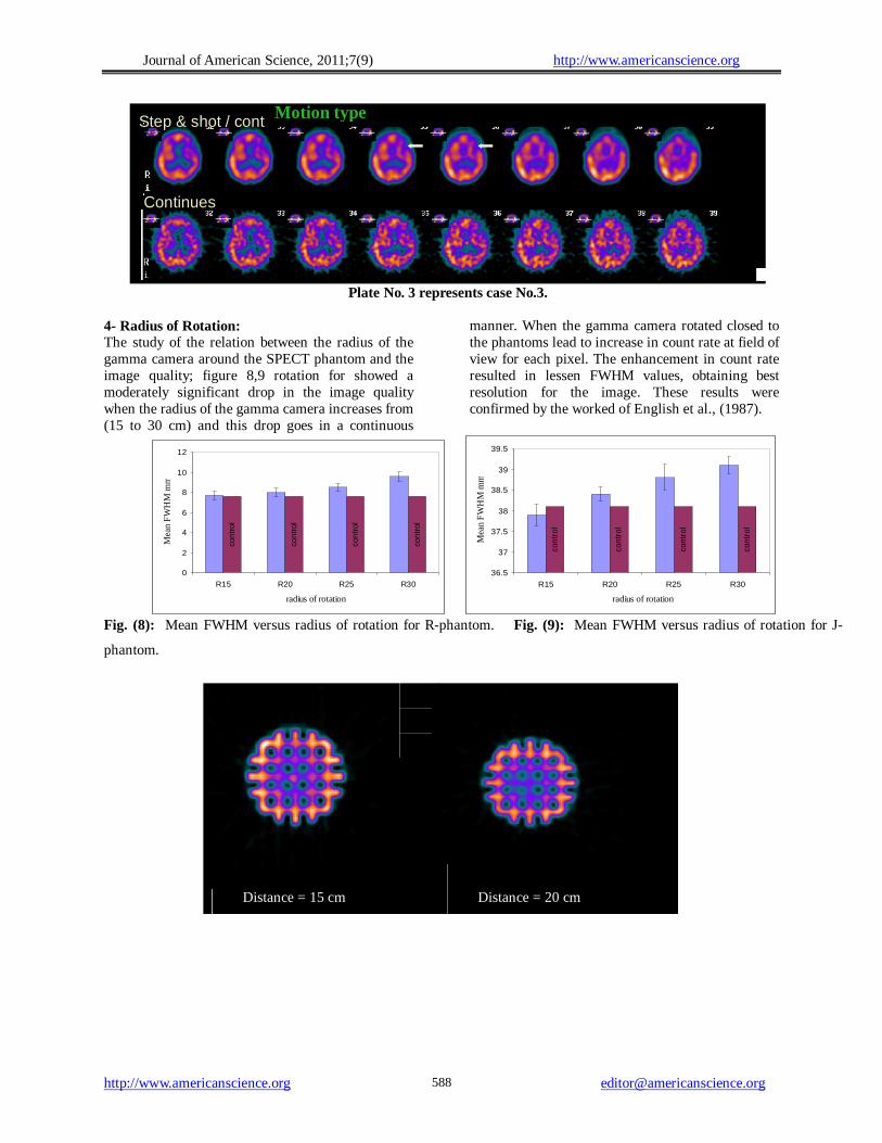
4- Radius of Rotation: The study of the relation between the radius of the gamma camera around the SPECT phantom and the image quality; figure 8,9 rotation for showed a moderately significant drop in the image quality when the radius of the gamma camera increases from (15 to 30 cm) and this drop goes in a continuous
manner. When the gamma camera rotated closed to the phantoms lead to increase in count rate at field of view for each pixel. The enhancement in count rate resulted in lessen FWHM values, obtaining best resolution for the image. These results were confirmed by the worked of English et al., (1987).
con
tro
l
con
tro
l
con
tro
l
con
tro
l
0
2
4
6
8
10
12
R15 R20 R25 R30
radius of rotation
Mea
n F
WH
M m
m
con
trol
con
trol
con
trol
con
trol
36.5
37
37.5
38
38.5
39
39.5
R15 R20 R25 R30
radius of rotation
Mea
n F
WH
M m
m
Fig. (8): Mean FWHM versus radius of rotation for R-phantom. Fig. (9): Mean FWHM versus radius of rotation for J-
phantom.
Distance = 15 cm Distance = 20 cm

Journal of American Science, 2011;7(9) http://www.americanscience.org
http://www.americanscience.org [email protected] 589
Distance = 25 cm Distance = 30 cm
Fig. (10): Spatial resolution of the cold lesion of SPECT phantom as a function of Radius of Rotation.
Study case No. 4 • A male patient 57 years old with hypertension
followed by a history of stroke, patient was clinically had weakness and parathesia in his left side.
• Cerebral blood flow investigated by the use 99mTc- HMPAO according to radius of rotation
was 15cm, showed an area of hypo- perfusion in right fronto-tempral region (arrowed).
• When using radius 25cm of rotation was used, cerebral blood flow didn't show the same area of hypo perfusion with heterogeneous uptake through all the brain.
Radius of rotation15 cm
25 cm
Plate No. 4 represents case No. 4.
5- Type of collimator: By changing the type of collimators LEAGP
(low energy higher resolution) or LEUHR (low energy ultra high resolution) the obtained results revealed that by changing the type of collimator, lead to an increase in the resolution of image .This increment was due to smaller distance of holes for
LEUHR collimator. The Ultra high-resolution collimator (LEUHR) should be recommended to provide good spatial resolution. This collimator could help in imaging smaller structures such as the brain. This is in agreement with the results obtained by El Gohary et al., 2008 in practices. This could help in imaging smaller structures of the brain.

Journal of American Science, 2011;7(9) http://www.americanscience.org
http://www.americanscience.org [email protected] 590
Fig. (12): Mean FWHM versus type of collimator for R-phantom. Fig. (13): Mean FWHM versus type of collimator for J-
phantom
Study case No. 5 • A female patient 62 years with history of
cerebra-vascular stroke to get right side hemiplegia (weakness and parathesia in right side).
• When LEGAP and LEUHR collimators were used, this showed similar cerebra-vascular syndrome with marked hypo perfusion in left tempera-parietal region while the rest of brain showed normal perfusion.
Collimator typeLEGAP
LEUHR
Plate No. 5 represents Case No. 5.
LEAGP LEUHR
Fig. (11): spatial resolution of the cold lesion of J-phantom as a function of Collimator type.
control control
7
7.2
7.4
7.6
7.8
8
8.2
8.4
LEUHR LEAGP
collimator type
Mean
FW
HM
mm
control control
37.6
37.7
37.8
37.9
38
38.138.2
38.3
38.4
38.5
38.6
LEUHR LEAGP
collimator type
Mean
FW
HM
mm
Journal of American Science, 2011;7(9) http://www.americanscience.org
http://www.americanscience.org [email protected] 591
CONCLUSION • It has been concluded that most acquisition
parameters affect the SPECT image quality. Pixel size 128 x 128 can display more image details and good resolution than the 64 x 64.
• When the number of view (NOV) increased (60&90 views), a better resolution was obtained in this parameter.
• At distance smaller than 25 cm, the resolution was gradually improved, while at longer distance it will diminish.
• The step&shot/cont. for mode of rotation is giving best resolution of image which was obtained.
• Ultra high-resolution collimator (LEUHR), rather than high-resolution collimator (LEAGP), is better to provide good spatial resolution.
• Patient motion occurring during data acquisition in SPECT could cause serious reconstruction artifacts.
• Also, whole time of acquisition should be suitable for patient stability, in evaluating the acquisition parameters.
Corresponding author H. A. Ayoub Faculty of Science, Suez Canal University [email protected] h. [email protected] References 1. Amersham healthcare (1994): "Functional brain
imaging with ceretec and SPECT " Amersham-England Artifactual in-homogeneities in myocardial PET and SPECT scan in normal subjects" J. Nucl. Med., 36: 188-195.
2. Bartlett M. L., Bacharach S. L., Voipio-Pulkki L. M., and Dilsizain V. (1995): Principles and Practice of nuclear medicine.
3. Bernier D. R, Christian P. E., Langan J. K. (1994) "Nuclear medicine technology and techniques” Mosby 1-110.
4. Bernier D. R.; (1994): Christian P. E., Langan J. K.; "Nuclear medicine technology and techniques” Mosby 1-110.
5. Collier B. D., Palmer D. W., Konbel J; (1984): Gamma camera energy windows for 99mTc bone scan scintigraphy ; effect of asymmetry on contrast resolution " Radiology; 151: 495-497.
6. English RJ., Brown SE. SPECT: (1987). Single-photon emission computed tomography: a primer. New York, NY: Society Nuclear Medicine;
7. El Gohary M., Mostafa H., Abdou S., El Faid A., Fouda M., Saad I., (2008): collimator selection for high quality brain SPECT imaging. Egypt .J. Biophys.Biomed. Engng. 9, 25-40
8. Farag H, Khalil W, Ali R, Al-Lehyani S and Shousha H. ,(2003): Effect of Acquisition Orbits & matrix on the Accuracy of SPECT Imaging: Quantitative Evaluation in cardiac Phantom. Workshop on materials science and radiation physics, 20-22 Dec. Assuit
9. Mark F. Smith, David R. Gilland, R. Edward Coleman and Ronald J. Jaszczak. (1998): Quantitative Imaging of Iodine-131 Distributions in Brain Tumors with Pinhole SPECT. J Nucl .Med.39:856-864
10. Thrall H. J., Zienssman A. H.;(1987) " Nuclear medicine ; the requisites" Mosby ; 1- 30 .
8/25/2011