
Journal of Alloys and Compounds - eng.usf.edu
7
Temperature effects on magnetic properties of Fe 3 O 4 nanoparticles synthesized by the sol-gel explosion-assisted method Ping Hu a, b, * , Tian Chang a, b , Wen-Jing Chen a, b , Jie Deng a, b , Shi-Lei Li a, b , Ye-Gai Zuo a , Lu Kang a , Fan Yang a, b , Megan Hostetter c , Alex A. Volinsky c a School of Metallurgy Engineering, Xi'an University of Architecture and Technology, Xi'an, 710055, China b State Local Joint Engineering Research Center for Functional Materials Processing, Xi'an, 710055, China c Department of Mechanical Engineering, University of South Florida, Tampa, FL, 33620, USA article info Article history: Received 4 August 2018 Received in revised form 17 September 2018 Accepted 18 September 2018 Available online 19 September 2018 Keywords: Sol-gel explosion-assisted Monodispersed Verwey transition Magnetic behavior abstract Fe 3 O 4 nanoparticles were successfully synthesized by the sol-gel explosion-assisted method. The phase composition of products with different ratios of dry gel and explosive agent (3:1, 6:1,12:1) was studied. Highly pure, well-crystallized, spherical and monodispersed Fe 3 O 4 3e20 nm nanoparticles were obtained at the 12:1 ratio. X-ray photoelectron spectroscopy characterization of the as-synthesized nanoparticles demonstrated consistency with stoichiometric Fe 3 O 4 surface composition. Zero-field cooled and field cooled measurements at 200 Oe validate that the anisotropy energy is greater than thermal energy up to 300 K for the ~10 nm samples. The Verwey transition (metal-insulator) of magnetite nanoparticles takes place at 128 K (T v ). The effects of different temperatures of 5 K, 128 K, and 300 K on magnetic behavior were studied in detail. The results show that hysteresis behavior weakens as the temperature increases. Saturation magnetization (M s ) of 86.2 emu/g is the highest at T v . Initial susceptibility (c a ) increases as a function of temperature, whereas coercivity (H c ) decreases. © 2018 Elsevier B.V. All rights reserved. 1. Introduction Nano-sized magnetic materials have been immensely researched because of their different and improved functionality compared to their bulk counterparts. Fe 3 O 4 is among the most widely studied metal oxide nanoparticle systems and has gained more scientific and technological attention due to its wide range of applications in magnetic resonance imaging [1], drug delivery [2], environmental remediation [3,4] and heterogeneous catalysis [5]. It also attracts academic interest because of the special Verwey transition, which is a metal to insulator transition with a sharp change in conductivity, heat capacity, coercivity and magnetization [6]. Fe 3 O 4 belongs to the inverse spinel family with the [Fe 3þ ] Tetra [Fe 2þ Fe 3þ ] Octra O 4 chemical formula. The material is conductive and ferromagnetic with a high Curie temperature of T c ¼ 850 K, with nearly full spin polarization at room temperature [7]. Recently, several methods have been used to prepare Fe 3 O 4 nanostructures, including hydrothermal [8], co-precipitation [9], sol-gel [10], thermal decomposition [11] and micro-emulsion [12] methods. However, all the above synthesis methods often require long production cycle, complicated operating steps, and poor reproducibility, which rendered them unsuitable for large-scale production. In this study, based on the nano-powder synthesis technology of diamond and graphite [13], we introduced a weak explosive agent (picric acid) into the synthesis of Fe 3 O 4 nanoparticles, and devel- oped a sol-gel explosion-assisted method for rapid synthesis of small particles with good dispersibility of Fe 3 O 4 nanoparticles. This method has high yield in the sol-gel process; and the action of the explosion field prevents magnetic nanoparticles tendency to easily agglomerate. The phase composition of products with different ratios of dry gel and explosive agent (3:1, 6:1, 12:1) was analyzed. The micro- structure along with the synthesis mechanism was also studied. The metal-insulator transition (the Verwey transition) temperature (T v ) of Fe 3 O 4 was confirmed in this work. The effects of different temperatures (T < T v , T v , and T > T v ) on magnetic behavior have been thoroughly studied. * Corresponding author. School of Metallurgy Engineering, Xi'an University of Architecture and Technology, Xi'an, 710055, China. E-mail address: [email protected] (P. Hu). Contents lists available at ScienceDirect Journal of Alloys and Compounds journal homepage: http://www.elsevier.com/locate/jalcom https://doi.org/10.1016/j.jallcom.2018.09.238 0925-8388/© 2018 Elsevier B.V. All rights reserved. Journal of Alloys and Compounds 773 (2019) 605e611
Transcript of Journal of Alloys and Compounds - eng.usf.edu
lable at ScienceDirect
Journal of Alloys and Compounds 773 (2019) 605e611
Contents lists avai
Journal of Alloys and Compounds
journal homepage: http: / /www.elsevier .com/locate/ ja lcom
Temperature effects on magnetic properties of Fe3O4 nanoparticlessynthesized by the sol-gel explosion-assisted method
Ping Hu a, b, *, Tian Chang a, b, Wen-Jing Chen a, b, Jie Deng a, b, Shi-Lei Li a, b, Ye-Gai Zuo a,Lu Kang a, Fan Yang a, b, Megan Hostetter c, Alex A. Volinsky c
a School of Metallurgy Engineering, Xi'an University of Architecture and Technology, Xi'an, 710055, Chinab State Local Joint Engineering Research Center for Functional Materials Processing, Xi'an, 710055, Chinac Department of Mechanical Engineering, University of South Florida, Tampa, FL, 33620, USA
a r t i c l e i n f o
Article history:Received 4 August 2018Received in revised form17 September 2018Accepted 18 September 2018Available online 19 September 2018
Keywords:Sol-gel explosion-assistedMonodispersedVerwey transitionMagnetic behavior
* Corresponding author. School of Metallurgy EngArchitecture and Technology, Xi'an, 710055, China.
E-mail address: [email protected] (P. Hu).
https://doi.org/10.1016/j.jallcom.2018.09.2380925-8388/© 2018 Elsevier B.V. All rights reserved.
a b s t r a c t
Fe3O4 nanoparticles were successfully synthesized by the sol-gel explosion-assisted method. The phasecomposition of products with different ratios of dry gel and explosive agent (3:1, 6:1, 12:1) was studied.Highly pure, well-crystallized, spherical and monodispersed Fe3O4 3e20 nm nanoparticles were obtainedat the 12:1 ratio. X-ray photoelectron spectroscopy characterization of the as-synthesized nanoparticlesdemonstrated consistency with stoichiometric Fe3O4 surface composition. Zero-field cooled and fieldcooled measurements at 200 Oe validate that the anisotropy energy is greater than thermal energy up to300 K for the ~10 nm samples. The Verwey transition (metal-insulator) of magnetite nanoparticles takesplace at 128 K (Tv). The effects of different temperatures of 5 K, 128 K, and 300 K on magnetic behaviorwere studied in detail. The results show that hysteresis behavior weakens as the temperature increases.Saturation magnetization (Ms) of 86.2 emu/g is the highest at Tv. Initial susceptibility (ca) increases as afunction of temperature, whereas coercivity (Hc) decreases.
© 2018 Elsevier B.V. All rights reserved.
1. Introduction
Nano-sized magnetic materials have been immenselyresearched because of their different and improved functionalitycompared to their bulk counterparts. Fe3O4 is among the mostwidely studied metal oxide nanoparticle systems and has gainedmore scientific and technological attention due to its wide range ofapplications in magnetic resonance imaging [1], drug delivery [2],environmental remediation [3,4] and heterogeneous catalysis [5]. Italso attracts academic interest because of the special Verweytransition, which is a metal to insulator transition with a sharpchange in conductivity, heat capacity, coercivity and magnetization[6]. Fe3O4 belongs to the inverse spinel family with the[Fe3þ]Tetra[Fe2þFe3þ]OctraO4 chemical formula. The material isconductive and ferromagnetic with a high Curie temperature ofTc¼ 850 K, with nearly full spin polarization at room temperature[7].
Recently, several methods have been used to prepare Fe3O4
ineering, Xi'an University of
nanostructures, including hydrothermal [8], co-precipitation [9],sol-gel [10], thermal decomposition [11] and micro-emulsion [12]methods. However, all the above synthesis methods often requirelong production cycle, complicated operating steps, and poorreproducibility, which rendered them unsuitable for large-scaleproduction.
In this study, based on the nano-powder synthesis technology ofdiamond and graphite [13], we introduced a weak explosive agent(picric acid) into the synthesis of Fe3O4 nanoparticles, and devel-oped a sol-gel explosion-assisted method for rapid synthesis ofsmall particles with good dispersibility of Fe3O4 nanoparticles. Thismethod has high yield in the sol-gel process; and the action of theexplosion field prevents magnetic nanoparticles tendency to easilyagglomerate.
The phase composition of products with different ratios of drygel and explosive agent (3:1, 6:1, 12:1) was analyzed. The micro-structure along with the synthesis mechanism was also studied.The metal-insulator transition (the Verwey transition) temperature(Tv) of Fe3O4 was confirmed in this work. The effects of differenttemperatures (T< Tv, Tv, and T> Tv) onmagnetic behavior have beenthoroughly studied.
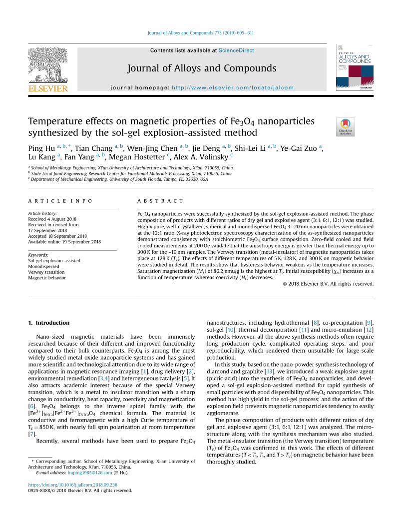
Fig. 1. XRD patterns of products with different picric acid ratios and holding time: (a)3:1 and 60, 40, and 20min, (b) 6:1, (c) 12:1.
P. Hu et al. / Journal of Alloys and Compounds 773 (2019) 605e611606
2. Experimental
2.1. Synthesis of Fe3O4 nanoparticles
Ferric nitrate and citric acid with a molar ratio of 1:0.8 weredissolved in 113ml of deionized water, and the material was fullydissolved by electromagnetic stirring at 35 �C for 30min. Ammoniawas added dropwise to adjust the solution pH to 7.3. Then the re-action was carried out at 68 �C for 4 h. Afterwards, the solution wasevaporated to a viscous state at 95 �C. When a sol-like substancewas formed, it was quickly poured onto a tray. The dry gel particlesand picric acid were homogeneously mixed in the autoclave filledwith Ar. The autoclave was moved to a heating furnace and kept at450 �C for a while, and finally loose black powder products wereobtained after water cooling to room temperature. The productswerewashed with ethanol in an ultrasonic cleaner three times, andseparated with a magnet. The products prepared in this work werenamed Yi-Xi, where Yi (i¼ 1, 2, 3) represent the mass ratio of dry geland picric acid, corresponding to 3:1, 6:1, and 12:1, respectively. Xi(i¼ 1, 2, 3) means holding time, which is 60, 40, and 20min,respectively.
2.2. Materials characterization
The phase composition of the Fe3O4 nanoparticles was analyzedusing X-ray diffractometer (XRD, D8, Buber, Germany, Cu Ka radi-ation, l¼ 0.15414 nm). The element states were investigated by theX-ray photoelectron spectroscopy (XPS, PHI Quantera SXM) withthe Al-Ka excitation source. The morphology and size of particleswere observed by transmission electron microscope (TEM, JEM-2100 Plus, Japan) operated at 200 kV. The intrinsic magneticproperties of the samples were measured with a vibrating samplemagnetometer (VSM, MPMS-SQUID VSM-094, USA).
3. Results and discussion
3.1. Structure characterization
XRD patterns of products with different picric acid ratios andholding times are shown in Fig. 1. At Y1¼3:1, FeO and FeCO3appeared in addition to the Fe3O4
* phase. Spinel iron oxide (Fe3O4and Fe2O3) cannot be distinguished by XRD results alone, so it isassumed here as Fe3O4
*. Moreover, the relative intensity of thediffraction peaks of FeO decreased with shorter holding time,which means that longer holding time and higher picric acid ratiolead to over-reduction of the product.
Therefore, the heat treatment at different holding times wasalso performed at Y¼ 6:1 and 12:1. It can be seen from Fig. 1(b) thatcompared with the ratio of 3:1, the number of FeO diffraction peaksat the 6:1 ratio is significantly reduced and the relative intensity isweakened. When the ratio was reduced to 12:1, Fe3O4
* phase wassuccessfully synthesized. Fig. 1 (c) shows that the diffraction peaksof the Y3-X1 samples are consistent with the Fe3O4
* standard peak.Strong and sharp diffraction peaks indicate that the Fe3O4
* is wellcrystallized.
X-ray photoelectron spectroscopy (XPS) is a versatile surfaceanalysis technique that can be used for compositional and chemicalstates analysis. It has been shown in previous studies that the peakpositions of Fe 2p1/2 and Fe 2p3/2 depend on the ionic states of Fe[14e17]. The positions of the satellite Fe 2p1/2 and Fe 2p3/2 peaks arealso very sensitive to the oxidation states and these peaks havebeen used to qualitatively determine the ionic states of iron. It hasbeen previously reported that Fe 2p3/2 in Fe3O4 does not have asatellite peak [18]. The absence of the satellite peak has been alsoconfirmed in this study in Fig. 2(a). The peak positions of Fe 2p1/2
and Fe 2p3/2 are 710.2 and 724 eV, respectively. They are locatedbetween the values for the 2FeO$SiO2 and Fe2O3 standard samples[18].
The 2p peaks are separated into two peaks. To obtain the total
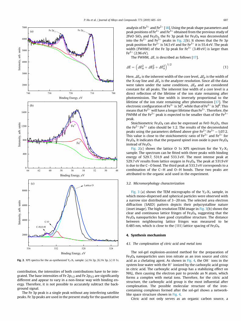
Fig. 2. XPS spectra for the as-synthesized Y3-X1 sample: (a) Fe 2p, (b) Fe 3p, (c) O 1s.
P. Hu et al. / Journal of Alloys and Compounds 773 (2019) 605e611 607
contribution, the intensities of both contributions have to be inte-grated. The base intensities of Fe 2p1/2 and Fe 2p3/2 are significantlydifferent and appear to vary in a non-linear way with binding en-ergy. Therefore, it is not possible to accurately subtract the back-ground signal.
The Fe 3p peak is a single peak without any interfering satellitepeaks. Fe 3p peaks are used in the present study for the quantitative
analysis of Fe3þ and Fe2þ [18]. Using the peak shape parameters andpeak positions of Fe2þ and Fe3þ obtained from the previous study of2FeO$SiO2 and Fe2O3, the Fe 3p peak for Fe3O4 was deconvolutedinto the Fe2þ and Fe3þ peaks in Fig. 2(b). It shows that the Fe 3ppeak position for Fe2þ is 54.5 eV and for Fe3þ it is 55.4 eV. The peakwidth (FWHM) of the Fe 3p peak for Fe3þ (3.49 eV) is larger thanFe2þ (2.96 eV).
The FWHM, DE, is described as follows [17].
DE ¼�DE2n þ DE2P þ DE2a
�1=2(1)
Here,DEn is the inherent width of the core level, DEp is the width ofthe X-ray line and DEa is the analyzer resolution. Since all the datawere taken under the same conditions, DEp and are consideredconstant for all peaks. The inherent line width of a core level is adirect reflection of the lifetime of the ion state remaining afterphotoemission. The line width is inversely proportional to thelifetime of the ion state remaining after photoemission [17]. Theelectronic configuration of Fe3þ is 3d5, while that of Fe2þ is 3d6. Thismeans that Fe2þwill have a longer lifetime than Fe3þ. Therefore, theFWHM of the Fe2þ peak is expected to be smaller than of the Fe3þ
peak.Stoichiometric Fe3O4 can also be expressed as FeO$Fe2O3, thus
the Fe2þ:Fe3þ ratio should be 1:2. The results of the deconvolutedpeaks using the parameters defined above give Fe2þ:Fe3þ ¼1.07:2.This value is close to the stoichiometric ratio of Fe2þ and Fe3þ forFe3O4. It indicates that the prepared spinel iron oxide is pure Fe3O4
instead of Fe2O3.Fig. 2(c) shows the lattice O 1s XPS spectrum for the Y3-X1
sample. The spectrum can be fitted with three peaks with bindingenergy of 529.7, 531.9 and 533.3 eV. The most intense peak at529.7 eV results from lattice oxygen in Fe3O4. The peak at 531.9 eVis due to the CeO bond. The third peak at 533.3 eV corresponds to acombination of the CeH and OeH bonds. These two peaks areattributed to the organic acid used in the experiment.
3.2. Micromorphology characterization
Fig. 3 (a) shows the TEM micrographs of the Y3-X1 sample, inwhich mono-dispersed and spherical particles were observed witha narrow size distribution of 3e20 nm. The selected area electrondiffraction (SAED) pattern depicts their polycrystalline nature(inset image). The high resolution TEM image in Fig. 3(b) shows theclear and continuous lattice fringes of Fe3O4, suggesting that theFe3O4 nanoparticles have good crystalline structure. The distancebetween neighbouring lattice fringes was measured to be0.485 nm, which is close to the (111) lattice spacing of Fe3O4.
4. Synthesis mechanism
4.1. The complexation of citric acid and metal ions
The sol-gel explosion-assisted method for the preparation ofFe3O4 nanoparticles uses iron nitrate as an iron source and citricacid as a chelating agent. As shown in Fig. 4, the OH� ions in thesystem lose water with the Hþ ionized by the carboxylic acid groupin citric acid. The carboxylic acid group has a stabilizing effect onNH4
þ, then causing the electron pair to provide an N atom, whichforms a complex with metal ions. Therefore, for the citric acidstructure, the carboxylic acid group is the most influential aftercomplexation. The possible molecular structure of the iron-containing complexes formed after the sol-gel shows a network-like space structure shown in Fig. 4.
Citric acid not only serves as an organic carbon source, a
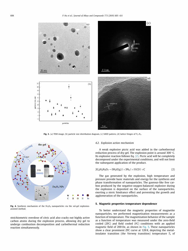
Fig. 3. (a) TEM image, (b) particle size distribution diagram, (c) SAED pattern, (d) lattice fringes of Y3-X1.
Fig. 4. Synthesis mechanism of the Fe3O4 nanoparticles via the sol-gel explosion-assisted method.
P. Hu et al. / Journal of Alloys and Compounds 773 (2019) 605e611608
stoichiometric overdose of citric acid also cracks out highly activecarbon atoms during the explosion process, allowing dry gel toundergo combustion decomposition and carbothermal reductionreaction simultaneously.
4.2. Explosion action mechanism
A weak explosive picric acid was added in the carbothermalreduction process of dry gel. The explosion point is around 300 �C.Its explosive reaction follows Eq. (2). Picric acid will be completelydecomposed under the experimental conditions, and will not limitthe subsequent application of the product.
2C6H3N3O7/3H2O(g)[þ3N2[þ11CO[þC (2)
The gas generated by the explosion, high temperature andpressure provide basic materials and energy for the synthesis andphase transformation of nanoparticles. The gaseous-like free car-bon produced by the negative-oxygen-balanced explosive duringthe explosion is deposited on the surface of the nanoparticles,exerting a steric hindrance effect and preventing the growth andagglomeration of the nanoparticles.
5. Magnetic properties temperature dependence
To better understand the magnetic properties of magnetitenanoparticles, we performed magnetization measurements as afunction of temperature. The magnetization behavior of the sampleas a function of temperature was measured under the zero-fieldcooled (ZFC) and field cooled (FC) conditions with an appliedmagnetic field of 200 Oe, as shown in Fig. 5. These nanoparticlesshow a clear prominent ZFC curve at 128 K, depicting the metal-insulator transition (the Verwey transition) temperature Tv of
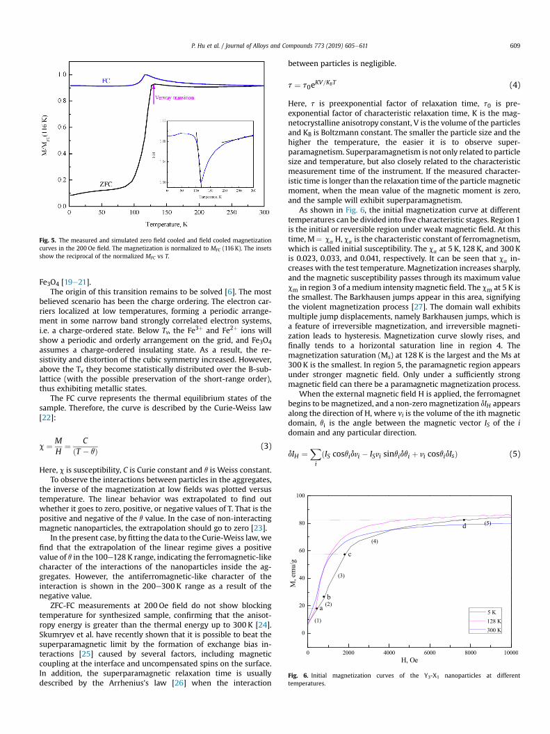
Fig. 5. The measured and simulated zero field cooled and field cooled magnetizationcurves in the 200 Oe field. The magnetization is normalized to MFC (116 K). The insetsshow the reciprocal of the normalized MFC vs T.
Fig. 6. Initial magnetization curves of the Y3-X1 nanoparticles at differenttemperatures.
P. Hu et al. / Journal of Alloys and Compounds 773 (2019) 605e611 609
Fe3O4 [19e21].The origin of this transition remains to be solved [6]. The most
believed scenario has been the charge ordering. The electron car-riers localized at low temperatures, forming a periodic arrange-ment in some narrow band strongly correlated electron systems,i.e. a charge-ordered state. Below Tv, the Fe3þ and Fe2þ ions willshow a periodic and orderly arrangement on the grid, and Fe3O4assumes a charge-ordered insulating state. As a result, the re-sistivity and distortion of the cubic symmetry increased. However,above the Tv they become statistically distributed over the B-sub-lattice (with the possible preservation of the short-range order),thus exhibiting metallic states.
The FC curve represents the thermal equilibrium states of thesample. Therefore, the curve is described by the Curie-Weiss law[22]:
c ¼ MH
¼ CðT � qÞ (3)
Here, c is susceptibility, C is Curie constant and q is Weiss constant.To observe the interactions between particles in the aggregates,
the inverse of the magnetization at low fields was plotted versustemperature. The linear behavior was extrapolated to find outwhether it goes to zero, positive, or negative values of T. That is thepositive and negative of the q value. In the case of non-interactingmagnetic nanoparticles, the extrapolation should go to zero [23].
In the present case, by fitting the data to the Curie-Weiss law, wefind that the extrapolation of the linear regime gives a positivevalue of q in the 100e128 K range, indicating the ferromagnetic-likecharacter of the interactions of the nanoparticles inside the ag-gregates. However, the antiferromagnetic-like character of theinteraction is shown in the 200e300 K range as a result of thenegative value.
ZFC-FC measurements at 200 Oe field do not show blockingtemperature for synthesized sample, confirming that the anisot-ropy energy is greater than the thermal energy up to 300 K [24].Skumryev et al. have recently shown that it is possible to beat thesuperparamagnetic limit by the formation of exchange bias in-teractions [25] caused by several factors, including magneticcoupling at the interface and uncompensated spins on the surface.In addition, the superparamagnetic relaxation time is usuallydescribed by the Arrhenius's law [26] when the interaction
between particles is negligible.
t ¼ t0eKV=KBT (4)
Here, t is preexponential factor of relaxation time, t0 is pre-exponential factor of characteristic relaxation time, K is the mag-netocrystalline anisotropy constant, V is the volume of the particlesand KB is Boltzmann constant. The smaller the particle size and thehigher the temperature, the easier it is to observe super-paramagnetism. Superparamagnetism is not only related to particlesize and temperature, but also closely related to the characteristicmeasurement time of the instrument. If the measured character-istic time is longer than the relaxation time of the particle magneticmoment, when the mean value of the magnetic moment is zero,and the sample will exhibit superparamagnetism.
As shown in Fig. 6, the initial magnetization curve at differenttemperatures can be divided into five characteristic stages. Region 1is the initial or reversible region under weak magnetic field. At thistime, M¼ ca H, ca is the characteristic constant of ferromagnetism,which is called initial susceptibility. The ca at 5 K, 128 K, and 300 Kis 0.023, 0.033, and 0.041, respectively. It can be seen that ca in-creases with the test temperature. Magnetization increases sharply,and the magnetic susceptibility passes through its maximum valuecm in region 3 of amedium intensitymagnetic field. The cm at 5 K isthe smallest. The Barkhausen jumps appear in this area, signifyingthe violent magnetization process [27]. The domain wall exhibitsmultiple jump displacements, namely Barkhausen jumps, which isa feature of irreversible magnetization, and irreversible magneti-zation leads to hysteresis. Magnetization curve slowly rises, andfinally tends to a horizontal saturation line in region 4. Themagnetization saturation (Ms) at 128 K is the largest and the Ms at300 K is the smallest. In region 5, the paramagnetic region appearsunder stronger magnetic field. Only under a sufficiently strongmagnetic field can there be a paramagnetic magnetization process.
When the external magnetic field H is applied, the ferromagnetbegins to bemagnetized, and a non-zeromagnetization dIH appearsalong the direction of H, where vi is the volume of the ith magneticdomain, qi is the angle between the magnetic vector IS of the idomain and any particular direction.
dIH ¼Xi
ðIS cosqidvi � ISvi sinqidqi þ vi cosqidIsÞ (5)
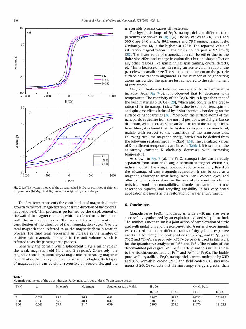
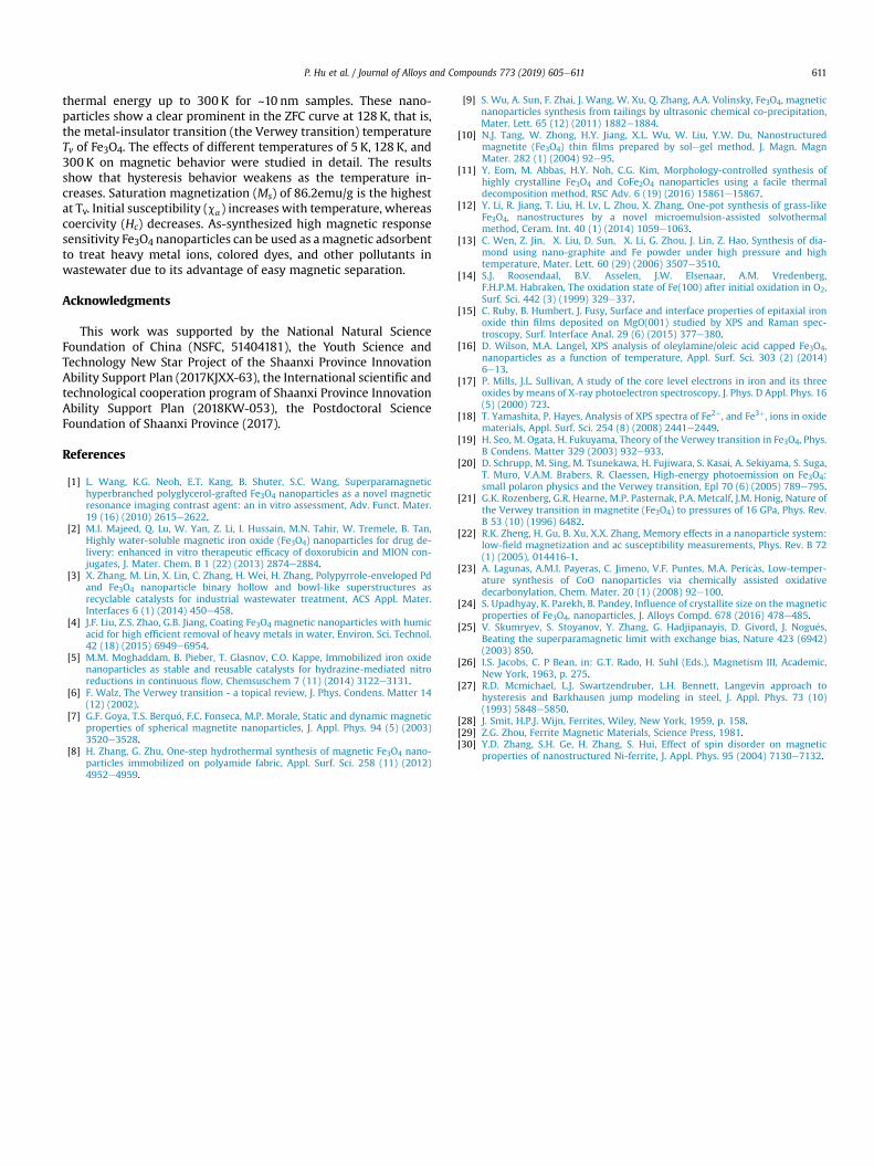
Fig. 7. (a) The hysteresis loops of the as-synthesized Fe3O4 nanoparticles at differenttemperatures, (b) Magnified diagram at the origin of hysteresis loops.
P. Hu et al. / Journal of Alloys and Compounds 773 (2019) 605e611610
The first term represents the contribution of magnetic domaingrowth to the total magnetization near the direction of the externalmagnetic field. This process is performed by the displacement ofthewall of the magnetic domain, which is referred to as the domainwall displacement process. The second term represents thecontribution of the direction of the magnetization vector IS to thetotal magnetization, referred to as the magnetic domain rotationprocess. The third term represents an increase in the number ofpositive spin magnetic moments in the unit volume, which isreferred to as the paramagnetic process.
Generally, the domain wall displacement plays a major role inthe weak magnetic field (1, 2 and 3 regions). Conversely, themagnetic domain rotation plays a major role in the strongmagneticfield. That is, the energy required for rotation is higher. Both typesof magnetization can be either reversible or irreversible, and the
Table 1Magnetic parameters of the as-synthesized Fe3O4 nanoparticles under different temper
T (K) ca Ms emu/g Mr emu/g Squareness ra
5 0.023 84.6 36.6 0.43128 0.033 86.2 40.8 0.47300 0.041 79.7 31.7 0.39
irreversible process causes all hysteresis.The hysteresis loops of Fe3O4 nanoparticles at different tem-
peratures are shown in Fig. 7(a). The Ms values at 5 K, 128 K and300 K are 84.6 emu/g, 86.2 emu/g and 79.7 emu/g, respectively.Obviously, the Ms is the highest at 128 K. The reported value ofsaturation magnetization in their bulk counterpart is 92 emu/g[28]. The lower value of magnetization can be either due to thefinite size effect and change in cation distribution, shape effect orany other reasons like spin pinning, spin canting, crystal defects,etc. This is because of the increasing surface to volume ratio of theparticle with smaller size. The spin moment present on the particlesurface have random alignment as the number of neighbouringatoms surrounded the spin are less compared to the spin momentof core atoms.
Magnetic hysteresis behavior weakens with the temperatureincrease. From Fig. 7(b), it is observed that Hc decreases withtemperature. The coercivity of the Fe3O4 NPs is larger than that ofthe bulk materials (<10 Oe) [29], which also occurs in the prepa-ration of ferrite nanoparticles. This is due to spin barriers, spin tiltand spin glass effects induced by in situ chemical disordering on thesurface of nanoparticles [30]. Moreover, the surface atoms of thenanoparticles deviate from the normal positions, resulting in latticedistortion, which increases the surface barrier of the nanoparticles.In addition, it is found that the hysteresis loops are asymmetrical,mainly with respect to the translation of the transverse axis.Following N�eel, the magnetic energy barrier can be defined fromthe following relationship: Hc¼ 2K/Ms [24]. The calculated valuesof K at different temperature are listed in Table 1. It is seen that theanisotropy constant K obviously decreases with increasingtemperature.
As shown in Fig. 7 (a), the Fe3O4 nanoparticles can be easilyseparated from solutions using a permanent magnet within 5 s,indicating that it has a highmagnetic response sensitivity. Based onthe advantage of easy magnetic separation, it can be used as amagnetic adsorber to treat heavy metal ions, colored dyes, andother pollutants in wastewater. Because of the non-toxic charac-teristics, good biocompatibility, simple preparation, strongadsorption capacity and recycling capability, it has very broadapplication prospects in the restoration of water environment.
6. Conclusions
Monodisperse Fe3O4 nanoparticles with 3e20 nm size weresuccessfully synthesized by an explosion-assisted sol-gel method.The synthesis mechanism is a joint action of complexation of citricacid withmetal ions and the explosive field. A series of experimentswere carried out under different ratios of dry gel and explosiveagent (3:1, 6:1, 12:1). The peak positions of Fe 2p1/2 and Fe 2p3/2 are710.2 and 724 eV, respectively. XPS Fe 3p peak is used in this workfor the quantitative analysis of Fe3þ and Fe2þ. The results of thedeconvoluted peaks give Fe2þ:Fe3þ¼1.07:2, and this value is closeto the stoichiometric ratio of Fe2þ and Fe3þ for Fe3O4. The highlypure, well-crystallized Fe3O4 nanoparticles were confirmed by XRDand XPS. Zero-field cooled (ZFC) and field cooled (FC) measure-ments at 200 Oe validate that the anisotropy energy is greater than
atures.
tio Mr/Ms Hc, Oe K¼Ms$Hc/2
Hc (�) Hc (þ) K (�) K (þ)
584.7 598.5 24732.8 25316.6338.1 351.8 14572.1 15162.6280.3 278.6 11170 11102.2
P. Hu et al. / Journal of Alloys and Compounds 773 (2019) 605e611 611
thermal energy up to 300 K for ~10 nm samples. These nano-particles show a clear prominent in the ZFC curve at 128 K, that is,the metal-insulator transition (the Verwey transition) temperatureTv of Fe3O4. The effects of different temperatures of 5 K, 128 K, and300 K on magnetic behavior were studied in detail. The resultsshow that hysteresis behavior weakens as the temperature in-creases. Saturation magnetization (Ms) of 86.2emu/g is the highestat Tv. Initial susceptibility (ca) increases with temperature, whereascoercivity (Hc) decreases. As-synthesized high magnetic responsesensitivity Fe3O4 nanoparticles can be used as amagnetic adsorbentto treat heavy metal ions, colored dyes, and other pollutants inwastewater due to its advantage of easy magnetic separation.
Acknowledgments
This work was supported by the National Natural ScienceFoundation of China (NSFC, 51404181), the Youth Science andTechnology New Star Project of the Shaanxi Province InnovationAbility Support Plan (2017KJXX-63), the International scientific andtechnological cooperation program of Shaanxi Province InnovationAbility Support Plan (2018KW-053), the Postdoctoral ScienceFoundation of Shaanxi Province (2017).
References
[1] L. Wang, K.G. Neoh, E.T. Kang, B. Shuter, S.C. Wang, Superparamagnetichyperbranched polyglycerol-grafted Fe3O4 nanoparticles as a novel magneticresonance imaging contrast agent: an in vitro assessment, Adv. Funct. Mater.19 (16) (2010) 2615e2622.
[2] M.I. Majeed, Q. Lu, W. Yan, Z. Li, I. Hussain, M.N. Tahir, W. Tremele, B. Tan,Highly water-soluble magnetic iron oxide (Fe3O4) nanoparticles for drug de-livery: enhanced in vitro therapeutic efficacy of doxorubicin and MION con-jugates, J. Mater. Chem. B 1 (22) (2013) 2874e2884.
[3] X. Zhang, M. Lin, X. Lin, C. Zhang, H. Wei, H. Zhang, Polypyrrole-enveloped Pdand Fe3O4 nanoparticle binary hollow and bowl-like superstructures asrecyclable catalysts for industrial wastewater treatment, ACS Appl. Mater.Interfaces 6 (1) (2014) 450e458.
[4] J.F. Liu, Z.S. Zhao, G.B. Jiang, Coating Fe3O4 magnetic nanoparticles with humicacid for high efficient removal of heavy metals in water, Environ. Sci. Technol.42 (18) (2015) 6949e6954.
[5] M.M. Moghaddam, B. Pieber, T. Glasnov, C.O. Kappe, Immobilized iron oxidenanoparticles as stable and reusable catalysts for hydrazine-mediated nitroreductions in continuous flow, Chemsuschem 7 (11) (2014) 3122e3131.
[6] F. Walz, The Verwey transition - a topical review, J. Phys. Condens. Matter 14(12) (2002).
[7] G.F. Goya, T.S. Berqu�o, F.C. Fonseca, M.P. Morale, Static and dynamic magneticproperties of spherical magnetite nanoparticles, J. Appl. Phys. 94 (5) (2003)3520e3528.
[8] H. Zhang, G. Zhu, One-step hydrothermal synthesis of magnetic Fe3O4 nano-particles immobilized on polyamide fabric, Appl. Surf. Sci. 258 (11) (2012)4952e4959.
[9] S. Wu, A. Sun, F. Zhai, J. Wang, W. Xu, Q. Zhang, A.A. Volinsky, Fe3O4, magneticnanoparticles synthesis from tailings by ultrasonic chemical co-precipitation,Mater. Lett. 65 (12) (2011) 1882e1884.
[10] N.J. Tang, W. Zhong, H.Y. Jiang, X.L. Wu, W. Liu, Y.W. Du, Nanostructuredmagnetite (Fe3O4) thin films prepared by solegel method, J. Magn. MagnMater. 282 (1) (2004) 92e95.
[11] Y. Eom, M. Abbas, H.Y. Noh, C.G. Kim, Morphology-controlled synthesis ofhighly crystalline Fe3O4 and CoFe2O4 nanoparticles using a facile thermaldecomposition method, RSC Adv. 6 (19) (2016) 15861e15867.
[12] Y. Li, R. Jiang, T. Liu, H. Lv, L. Zhou, X. Zhang, One-pot synthesis of grass-likeFe3O4, nanostructures by a novel microemulsion-assisted solvothermalmethod, Ceram. Int. 40 (1) (2014) 1059e1063.
[13] C. Wen, Z. Jin, X. Liu, D. Sun, X. Li, G. Zhou, J. Lin, Z. Hao, Synthesis of dia-mond using nano-graphite and Fe powder under high pressure and hightemperature, Mater. Lett. 60 (29) (2006) 3507e3510.
[14] S.J. Roosendaal, B.V. Asselen, J.W. Elsenaar, A.M. Vredenberg,F.H.P.M. Habraken, The oxidation state of Fe(100) after initial oxidation in O2,Surf. Sci. 442 (3) (1999) 329e337.
[15] C. Ruby, B. Humbert, J. Fusy, Surface and interface properties of epitaxial ironoxide thin films deposited on MgO(001) studied by XPS and Raman spec-troscopy, Surf. Interface Anal. 29 (6) (2015) 377e380.
[16] D. Wilson, M.A. Langel, XPS analysis of oleylamine/oleic acid capped Fe3O4,nanoparticles as a function of temperature, Appl. Surf. Sci. 303 (2) (2014)6e13.
[17] P. Mills, J.L. Sullivan, A study of the core level electrons in iron and its threeoxides by means of X-ray photoelectron spectroscopy, J. Phys. D Appl. Phys. 16(5) (2000) 723.
[18] T. Yamashita, P. Hayes, Analysis of XPS spectra of Fe2þ, and Fe3þ, ions in oxidematerials, Appl. Surf. Sci. 254 (8) (2008) 2441e2449.
[19] H. Seo, M. Ogata, H. Fukuyama, Theory of the Verwey transition in Fe3O4, Phys.B Condens. Matter 329 (2003) 932e933.
[20] D. Schrupp, M. Sing, M. Tsunekawa, H. Fujiwara, S. Kasai, A. Sekiyama, S. Suga,T. Muro, V.A.M. Brabers, R. Claessen, High-energy photoemission on Fe3O4:small polaron physics and the Verwey transition, Epl 70 (6) (2005) 789e795.
[21] G.K. Rozenberg, G.R. Hearne, M.P. Pasternak, P.A. Metcalf, J.M. Honig, Nature ofthe Verwey transition in magnetite (Fe3O4) to pressures of 16 GPa, Phys. Rev.B 53 (10) (1996) 6482.
[22] R.K. Zheng, H. Gu, B. Xu, X.X. Zhang, Memory effects in a nanoparticle system:low-field magnetization and ac susceptibility measurements, Phys. Rev. B 72(1) (2005), 014416-1.
[23] A. Lagunas, A.M.I. Payeras, C. Jimeno, V.F. Puntes, M.A. Peric�as, Low-temper-ature synthesis of CoO nanoparticles via chemically assisted oxidativedecarbonylation, Chem. Mater. 20 (1) (2008) 92e100.
[24] S. Upadhyay, K. Parekh, B. Pandey, Influence of crystallite size on the magneticproperties of Fe3O4, nanoparticles, J. Alloys Compd. 678 (2016) 478e485.
[25] V. Skumryev, S. Stoyanov, Y. Zhang, G. Hadjipanayis, D. Givord, J. Nogu�es,Beating the superparamagnetic limit with exchange bias, Nature 423 (6942)(2003) 850.
[26] I.S. Jacobs, C. P Bean, in: G.T. Rado, H. Suhl (Eds.), Magnetism III, Academic,New York, 1963, p. 275.
[27] R.D. Mcmichael, L.J. Swartzendruber, L.H. Bennett, Langevin approach tohysteresis and Barkhausen jump modeling in steel, J. Appl. Phys. 73 (10)(1993) 5848e5850.
[28] J. Smit, H.P.J. Wijn, Ferrites, Wiley, New York, 1959, p. 158.[29] Z.G. Zhou, Ferrite Magnetic Materials, Science Press, 1981.[30] Y.D. Zhang, S.H. Ge, H. Zhang, S. Hui, Effect of spin disorder on magnetic
properties of nanostructured Ni-ferrite, J. Appl. Phys. 95 (2004) 7130e7132.


![Journal of Alloys and Compounds - · PDF file Journal of Alloys and Compounds 509 (2011) ... a College of Math and Physics, ... the below formula [17]:](https://static.fdocuments.in/doc/165x107/5aa05da77f8b9a62178e072c/journal-of-alloys-and-compounds-journal-of-alloys-and-compounds-509-2011-.jpg)


![Journal of Alloys and Compounds - nimte.ac.cn...thermal transport properties between magnetic refrigerants and heat-exchange medium [24]. Therefore, the Fe-based glassy alloys with](https://static.fdocuments.in/doc/165x107/60d2bd31873414242c6a7eb3/journal-of-alloys-and-compounds-nimteaccn-thermal-transport-properties-between.jpg)


