journal 09 Nov NEW 2 - Dermatological Society of Malaysia
34
27 MJD 2009 December Vol 23 Malaysian Journal of Dermatology Keywords cetirizine, fixed drug eruptions Introduction A fixed drug eruption (FDE) is a distinct drug induced reaction pattern that characteristically recurs at the same site on the skin or mucosa. We report a case of bullous FDE following ingestion of cetirizine, a common treatment for allergic disorders but a rare causative agent for cutaneous adverse drug reaction. Case report A 30 year old lady presented with multiple painful blisters in the oral cavity and over the right wrist and left breast for 4 days duration. The lesions developed within 3-4 hours after taken a single tablet of Cetirizine Hydrochloride which was prescribed for her allergic rhinitis. This was the 2nd episode. She had a similar problem about 1 month ago. She took Cetirizine for allergic rhinitis. The similar blistering lesions occurred in the oral cavity and over the right wrist which resolved after 1 week with minimal residual hyperpigmentation. She did not seek medical attention during the first episode. In the past she had taken multiple courses of “clarinase” and “clarityn” without any complaint. On examination, she was afebrile and well. There were multiple tense bullae at the buccal mucosa and right wrist of various sizes ranging from 4mm - 10mm (Figure 1 & 2). A hyperpigmented patch was noted over the left breast. The blisters were surrounded by an erythematous rim. There was no lesions elsewhere. No target lesion were seen and no eye and genital involvement were detected. Skin biopsy, patch test and oral provocation test were not done in this patient because of the clear drug history and the patient did not consent for the tests. ADVERSE DRUG REACTIONS - Case Report Cetirizine induced bullous fixed drug eruptions Tan WC, MRCP, Chan LC, MMed The patient was counselled on medication avoidance and possible cross-reactions of similar medications such as cetirizine, hydroxyzine, levocetirizine and ethylenediamine. She was given a short course of oral prednisolone, in a tapering dose, for 2 weeks. The lesions improved and healed with residual hyperpigmentation. Correspondence Tan Wooi Chiang, MRCP Department of Dermatology, Penang Hospital Jalan Residensi, 10990 Penang E-mail : [email protected] Figure 1 Tense bullae with erythematous rim over the right wrist Figure 2 Circular hyperpigmented lesions over the lip with superficial erosions
Transcript of journal 09 Nov NEW 2 - Dermatological Society of Malaysia
27MJD 2009 December Vol 23
Malaysian Journal of Dermatology
Keywords cetirizine, fixed drug eruptions
IntroductionA fixed drug eruption (FDE) is a distinct druginduced reaction pattern that characteristicallyrecurs at the same site on the skin or mucosa. Wereport a case of bullous FDE following ingestion ofcetirizine, a common treatment for allergicdisorders but a rare causative agent for cutaneousadverse drug reaction.
Case reportA 30 year old lady presented with multiple painfulblisters in the oral cavity and over the right wristand left breast for 4 days duration. The lesionsdeveloped within 3-4 hours after taken a singletablet of Cetirizine Hydrochloride which wasprescribed for her allergic rhinitis. This was the 2ndepisode. She had a similar problem about 1 monthago. She took Cetirizine for allergic rhinitis. Thesimilar blistering lesions occurred in the oral cavityand over the right wrist which resolved after 1 weekwith minimal residual hyperpigmentation. She didnot seek medical attention during the first episode.In the past she had taken multiple courses of“clarinase” and “clarityn” without any complaint.
On examination, she was afebrile and well. Therewere multiple tense bullae at the buccal mucosa andright wrist of various sizes ranging from 4mm -10mm (Figure 1 & 2). A hyperpigmented patch wasnoted over the left breast. The blisters weresurrounded by an erythematous rim. There was nolesions elsewhere. No target lesion were seen andno eye and genital involvement were detected. Skinbiopsy, patch test and oral provocation test were notdone in this patient because of the clear drug historyand the patient did not consent for the tests.
ADVERSE DRUG REACTIONS - Case Report
Cetirizine induced bullous fixed drug eruptions
Tan WC, MRCP, Chan LC, MMed
The patient was counselled on medicationavoidance and possible cross-reactions of similarmedications such as cetirizine, hydroxyzine,levocetirizine and ethylenediamine. She was given ashort course of oral prednisolone, in a taperingdose, for 2 weeks. The lesions improved and healedwith residual hyperpigmentation.
CorrespondenceTan Wooi Chiang, MRCPDepartment of Dermatology, Penang HospitalJalan Residensi, 10990 PenangE-mail : [email protected]
Figure 1 Tense bullae with erythematous rim over the right wrist
Figure 2 Circular hyperpigmented lesions over the lip with superficial erosions

28 MJD 2009 December Vol 23
Malaysian Journal of Dermatology
DiscussionBrocq first introduced the term “fixed drugeruption” in 18941. The term FDE describes thedevelopment of one or more annular or ovalerythematous patches as a result of systemicexposure to a drug. These reactions normallyresolve with hyperpigmentation and may recur atthe same site with re-exposure to the drug2.Repeated exposure to the offending drug may causenew lesions to develop in addition to “lighting up”the older hyperpigmented lesions.
The pathogenesis of FDE is still enigmatic. Themost commonly accepted hypothesis is thepersistence of memory T cell in the affected skin3.CD8+ cells phenotypically resembling effectormemory T cells have been shown to be greatlyenhanced in the lesions of FDE.
The skin lesion of FDE usually starts as anerythematous macule then subsequently evolvesinto a plaque. Vesicles and bullae develop at a laterstage. The lesions can occur on any part of the skinand mucous membranes. The sites of predilectionare the genitalia, limbs, sacral region, palmar andplantar area. The oral mucosa may be involved inassociation with skin lesions or in isolation.
More than 100 drugs have been implicated in FDEbut the causative drugs that are commonlyassociated with FDE include co-trimoxazole,tetracycline, non-steroidal anti inflammatory drugs(NSAIDs), phenytoin, griseofulvin, salicylates,penicillin and phenolphthalein4. It has also beendescribed as a side-effect of some anti-H1-antihistamine drugs, such as cyclizine lactate,diphenhydramine hydrochloride, phenothiazines,hydroxyzine5 and loratadine6. To the best of ourknowledge, there were only a few cases of FDEsecondary to cetirizine being reported7-8.
Cetirizine is a specific histamine H1-receptorantagonist and a second-generation antihistamineand generates the lowest rate of cutaneousreactions. Cetirizine is used worldwide in thetreatment of allergic disorders and is generally welltolerated without much problem. Although topicalantihistamines commonly leads to sensitization andcan cause contact and photo-contact dermatitis9,skin reactions provoked by their systemic use israre10-11.
Cross-reactions among ethylenediamine, cetirizineand hydroxyzine had been reported by Bark-LinLew et al12. Cutaneous reactivity to the H1-antihistamines is caused by the fact that they sharethe same chemical piperazine structure and similarpharmacologic profiles12-13. In vivo, 45% ofhydroxyzine is transformed into cetirizine andlevocetirizine is the active (R)-enantiomer ofcetirizine5.
Oral challenge can be used to confirm the etiologyof FDE14. But there are risks involved in thisapproach, mainly anaphylactic reactions or intenselesional reactivation with a significant increase inthe number of lesions.
Patch testing is not regularly performed.Reappearance of skin lesions with re-challengeidentifies offending agent. Furthermore, reactivityof patch tests in FDE is variable. Some studyshowed patch testing at the site of a previous lesionyields a positive response in up to 43% of cases15,but the reactivity depends on the drug and thevehicle. Reactivity is usually seen before 24 hoursand is observed exclusively on the lesional skin.Patch testing is safer than oral provocation tests. Italso allows the study of several drugs at the sametime.
Physicians should watch carefully for eruptionsfrom antihistamines and not to misinterpret them asunresponsiveness to the medications. Because oralantihistamines are one of the most commonmedications used to treat itchy dermatoses, it isprudent to listen to patients when they indicate thatthese medications seem to worsen their condition.
This report highlights an uncommon causativeagent of FDE.
References
1. Brocq I. Eruption erythemato-pigmentee fixe due al’antipyrine. Ann Dermatol Venereol. 1894; 5: 308-13.Quoted from: Shiohara T, Nickoloff BJ, Sagawa Y,Gomi T et al. Fixed drug eruption. Expression ofepidermal keratinocyte intercellular adhesionmolecule-1 (ICAM-1). Arch Dermatol 1989; 25:1371-6
2. Lee AY. Fixed Drug Eruptions: Incidence, recognition,and avoidance. Am J Clin Dermatol 2000; 1(5):277-85

29MJD 2009 December Vol 23
Malaysian Journal of Dermatology
3. Shiohara T, Nickoloff BJ, Sagawa Y, Gomi T et al. Fixeddrug eruption. Expression of epidermal keratinocyteintercellular adhesion molecule-1 (ICAM-1). ArchDermatol 1989; 25: 1371-6
4. Magee P. Drug-induced skin disorders. In: Walker R,Edwards C ‘editors’. Clinical Pharmacy andTherapeutics. 3rd edition. Phildelphia: ChurchillLivingstone 2003; 843-52
5. Mahajan VK, Sharma NL, Sharma VC. Fixed drugeruption: a novel side-effect of levocetirizine. Int JDermatol 2005; 44: 796-798
6. Pionetti CH, Kien MC, Alonso A. Fixed drug eruptiondue to loratadine. Allergol Immunopathol (Madr) 2003;31: 291-293
7. Inamadar AC, Palit A, Athanikar SB, Sampagavi VV,Deshmukh NS. Multiple fixed drug eruptions due tocetirizine. Br J Dermatol 2002; 147: 1025-6
8. Kranke B, Kern T. Multilocalized fixed drug eruptionto the antihistamine cetirizine. J Allergy Clin Immunol2000; 106: 988
9. Kulthanan K, Tiprungkorn P, Linpiyawan R. Cutaneousreaction to oral antihistamine. Clin Exp Dermatol2003; 28: 229-30
10. Spencer CM, Faulds D, Peters DH. Cetirizine: areappraisal of its pharmacological properties andtherapeutic use in selected allergic disorders. Drugs1993; 46: 1055-80
11. Campoli-Richards DM, Buckley MMT, Fitton A.Cetirizine: a review of its pharmacological propertiesand clinical potential in allergic rhinitis, pollen-inducedasthma, and chronic urticaria. Drugs 1990; 40: 762-81
12. Lew BL, Haw CR, Lee MH. Cutaneous drug eruptionfrom cetirizine and hydroxyzine. J Am Acad Dermatol.2004; 50(6): 953-6
13. Assouere MN, Mazereeuw-Hautier J, Bonafe JL.Cutaneous drug eruption with two antihistaminic drugsof the same chemical family: cetirizine andhydroxyzine. Ann Dermatol Venereol 2002; 129:1295-1298
14. Kauppinen K, Stubb S. Fixed eruptions: causativedrugs and challenge tests. Br J Dermatol 1985; 112:575-578.
15. Barbaud A, Reichert-Penetrat S, Trechot P, et al. Theuse of skin testing in the investigation of cutaneousadverse drug reactions. Br J Dermatol 1998; 139: 49-58.

30 MJD 2009 December Vol 23
Malaysian Journal of Dermatology
Vitamin K injection is a common treatment for theprevention of bleeding. It acts by promoting theformation of liver coagulation factor II, VII, IX andX. Anaphylactic reaction with the use of thisproduct is rare and suspected to be caused by itsnon-ionic solubilizing and emulsifying agent;Cremophor EL formulated in Vitamin K Injection(KISAN® 10mg/ml)1,2. The main component ofCremophor EL is glycerol-polyethylene glycolricinoleate. It is widely used as a formulationvehicle for various poorly-water soluble drugsincluding the lipid soluble vitamin A, D, E and K.Extra precautions should be taken whenadministering pharmaceutical products containing
ADVERSE DRUG REACTIONS - Update
Intravenous Vitamin K Administration
Norkasihan Ibrahim, Fadilah Othman, Norliza Mat Ariffin, Zainon Abudin
Cremophor EL. This agent has been found to causesevere anaphylactoid-like reaction especially whenhigh doses are administered via bolus infusion1,2. Itis recommended to dilute Vitamin K Injection(KISAN® 10mg/ml) in normal saline, dextrose 5%or dextrose saline to a concentration not exceeding1mg/ml. The diluted product should be infused fornot less than 30 minutes1,2.
References
1. KISAN® Injection 10mg/ml Product InformationLeaflet
2. Cremophor EL the drawbacks and advantages ofvehicle selection for drug formulation.
CorrespondenceNorkasihan Ibrahim Department of Pharmacy, Selayang Hospital SelangorE-mail : [email protected]

31MJD 2009 December Vol 23
Malaysian Journal of Dermatology
Dear Editor,We have encountered a 42 year old Malay lady withantiphospholipid syndrome for the past 3 yearspresented with red cutaneous swelling at bothdeltoid regions a week after given intramuscularVitamin K injection. She was referred to us 6 weeksfollowing the Vitamin K injection. Psoriasiformplaques with perilesional warm erythematous skinwere noted over both deltoid regions (Figure 1). Askin biopsy (Figure 2a & 2b) showed subepidermalvesicular change with formation of bullae including
ADVERSE DRUG REACTIONS - Short Communication
Hypersensitivity reaction to intramuscular Vitamin K
Raoul RS1, MBBS, Rohna R, MRCP, Shalini Kumar2, MPath
basal cell vacuolation and necrotic keratinocytes.The upper dermis was infiltrated with mononuclearinflammatory cells. The features are in keepingwith a hypersensitivity reaction.
A diagnosis of hypersensitivity reaction to InjectionVitamin K was made based on the history and skinbiopsy. The skin lesions flattened and becamehyperpigmented after two weeks application ofpotent topical steroid and oral antihistamine.
CorrespondenceRaoul Roger Sibert, MBBS 1Department of DermatologySelayang Hospital, SelangorE-mail : [email protected]
2Department of Pathology, Selayang Hospital
Figure 1 Lesion at right deltoid Figure 2A HPE of skin biopsy at 4X magnification
Figure 2B HPE of skin biopsy at 40X magnification

32 MJD 2009 December Vol 23
Malaysian Journal of Dermatology
DiscussionVitamin K1 (Phytomenadione) is a fat soluble,naturally occurring vitamin used to treat certaincoagulation disorders1. Injectable vitamin K cancause skin reactions whatever the dose and mode ofinjection2. The first few cases of hypersensitivity tovitamin K were reported in patients with liverdisease. The pathophysiological mechanism of theacute form would involves type IV allergy toPhytomenadione3. There are three distinct types ofcutaneous reactions to vitamin K1: localizedeczematous, localized morphea-form4-5, and, veryrarely, diffuse maculopapular eruption2. Theeczematous type appears at the site of injection.
The morphea-form type is a localized morphea-form patch that appears at the site of injection. Thediagnosis of an adverse cutaneous reaction tovitamin K can be made if the possibility isconsidered. Many of these reactions are very slowto clear up and some may persist as a chronicsclerodermoid change4-5. Managing may befrustrating for both the patient and the clinician.
In our patient, the localized psoriasiform dermatitiswith perilesional erythema and oedema was treatedwith topical steroids and resolved with post-inflammatory hyperpigmentation.
References
1. Gimenez-Arnau, A.M., A. Toll, and R.M. Pujol,Immediate cutaneous hypersensitivity response tophytomenadione induced by vitamin K in skindiagnostic procedure. Contact Dermatitis 2005; 52(5):284-5
2. Moreau-Cabarrot, A., F. Giordano-Labadie, and J.Bazex, [Cutaneous hypersensitivity at the site ofinjection of vitamin K1]. Ann Dermatol Venereol,1996; 123(3): 177-9
3. Bruynzeel, I., et al., Cutaneous hypersensitivityreactions to vitamin K: 2 case reports and a review ofthe literature. Contact Dermatitis 1995; 32(2):78-82.
4. Finkelstein, H., M.C. Champion, and J.E. Adam,Cutaneous hypersensitivity to vitamin K1 injection. JAm Acad Dermatol, 1987; 16(3 Pt 1): 540-5.
5. BK Pang, V Munro, S Kossard. Pseudosclerodermasecondary to phytomenadione (vitamin K1) injections:Texier's disease. Australasian Journal of Dermatology,1996 - interscience.wiley.com

33MJD 2009 December Vol 23
Malaysian Journal of Dermatology
Keywords autoimmune bullous disease,pemphigus, thymoma, paraneoplastic
IntroductionPemphigus foliaceous (PF) is an autoimmuneblistering disease resulting from acquiredimmunoglobulin G autoantibodies againstdesmoglein 1 of the skin, which is one of theadhesion molecules of keratinocytes. Clinicallypatients with PF develop crusted and scaly erosionsmainly over the seborrhoeic distribution i.e. theface, scalp and upper trunk. Mild cases of PF maybe localized but in some cases it may progress toerythrodermic exfoliative dermatitis. There ishowever no mucosal involvement in PF in contrastto pemphigus vulgaris and paraneoplasticpemphigus. Light microscopy of lesional biopsyshows subcorneal acantholysis. Directimmunofluorescence study of perilesional skinreveals presence of intraepithelial intercellulardeposit of IgG and C3. We describe 2 cases of PF inthe presence of thymoma, a relatively rareassociation, which could further support the fact ofthymoma associated autoimmune disease.
Case report 1A 35-year-old Malay painter, presented to us inAugust 2008 with a month history of flaccid blistersstarting over the anterior chest, progressivelyinvolving the face, arms, abdomen, legs and thenbecame generalized. There was no oral or genitalinvolvement. The lesions were not aggravated bysun exposure. There was no significant drughistory. There were no other constitutionalsymptoms. On further questioning, the patient wasdiagnosed to have a thymoma after being
AUTOIMMUNE DISORDERS - Case Report
Pemphigus foliaceous and thymoma: a report of 2 cases
Tang MM1, AdMDerm, Lee YY2, AdMDerm, Suganthi T1, MMed (UKM)
investigated for difficulty in swallowing andchanges of voice in 2006. He was advised forsurgical resection of the mediastinal mass bycardiothoracic surgeon. He however declined anyform of surgery and defaulted subsequent followup.
On examination, the patient was afebrile. His bloodpressure was normal but has tachycardia. He waserythrodermic with erosions and crusts over theface, scalp, chest, back, arms and thighs sparing theoral mucosal and genitalia. Examination of othersystems revealed no abnormality.
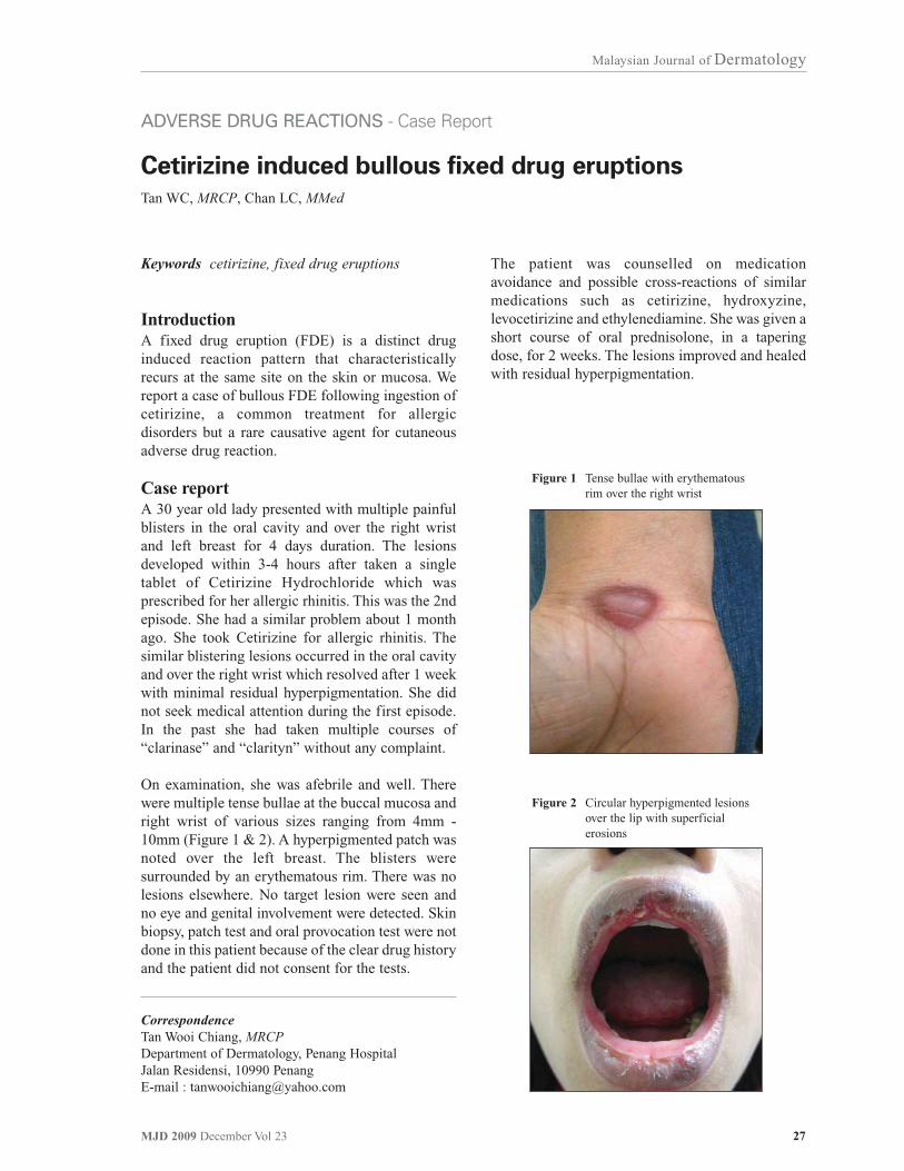
Skin biopsy of lesion demonstrated subcornealbullae with acantholytic cells (Figure 1). The directimmunofluorescence study showed strongintraepidermal IgG deposits (Figure 2).
These confirmed the clinical diagnosisof pemphigus foliaceous. His indirectimmunofluorescence study showed a titre of 1:320.His blood counts were normal with ESR of35mm/hr. Full blood picture showed leucocytosiswith predominant neutrophils, toxic granulationwere seen but immature cell were absent. Hisalanine aminotransferase was 283U/l (normal 0-41U/l) Serology tests for HepBsAg, Anti Hep Cantibody, anti-HIV-1&2, anti-smooth muscleantibody were negative. Ultrasound of abdomenshowed normal liver echogenicity and echotexturewith no other abnormality. His chest radiographrevealed a mass at the right mid zone, obliteratingthe right heart border (Figure 3).
Due to the severity of his skin lesions, the patientwas admitted and initially given intravenoushydrocortisone 100mg 8hrly. Multiple courses ofintravenous antibiotic were given for thesuperimposed bacterial infection of the eroded skin.The skin lesions progressed further despite withabove treatment and aggressive skin nursing care.Steroid sparing immunosuppresants such asazathioprine, mycophenolate mofetil andmethotrexate were not considered due to hepatitis.Due to the poor response after 3 weeks ofintravenous hydrocortisone, we initiated pulsedexamethasone
CorrespondenceTang MM, AdMDerm1Department of Dermatology, Hospital Kuala LumpurE-mail : [email protected]
2Department of DermatologyUniversity Malaya Medical Centre
34 MJD 2009 December Vol 23
Malaysian Journal of Dermatology
Figure 1 The skin biopsy showed superficial subcorneal bulla with collection of neutrophils. Haematoxylin and Eosin stain x400
Figure 3 Presence of a mass at the right mid zone on the chest radiograph
Figure 4 Computed tomography of thorax showed a heterogeneously enhancing anterior mediastinal mass measuring 5.5x5.6cm on the right mid thoracic level
Figure 2 Positive deposition of Ig G of the intraepithelial and intercellular spaces. (Direct immunofluorescence stain IgG x200)

35MJD 2009 December Vol 23
Malaysian Journal of Dermatology
dexamethasone-cyclophosphamide (DCP) regimeafter obtained consent from the patient. This regimewas modified to include oral prednisolone at0.5mg/kg/day, after the first 2 pulses, the skinlesions stopped progressing and healed gradually.Further investigation included a CT thorax whichshowed a heterogeneously enhancing anteriormediastinal mass measuring 5.5 x 5.6cm on theright mid thoracic level. There was calcificationwithin the mass. There was clear demarcation withthe adjacent vessels. The mediastinal lymph nodeswere not enlarged and there were no lung nodules orpleural effusion (Figure 4). Fatty liver was alsonoted in the CT scan. His carcinoembryonic antigen(CEA) was 11.1U/l Units (twice above the uppernormal limit) Other tumour markers includedlactate dehydrogenase and alpha fetoprotein werenot elevated. Computed tomography guided biopsyconfirmed a type A thymoma.
The patient was repeatedly counseled on the needfor surgical excision of the thymoma as thepemphigus foliaceous which could be aparaneoplastic manifestation may probably resolvepost-thymectomy. However the patient adamantlyrefused surgical intervention. He subsequentlyunderwent 11 pulses of DCP and he has been lesionfree for the past 4 months.
Case report 2A 43-year-old Chinese lady presented to us inMarch year 2004 with 2-week history of erosionsstarted over the neck which became generalized.There were however no oral or genital ulcers. Shewas diagnosed to have malignant thymoma in 1997and had thymectomy done in the same year. Shewas also suffering from end stage renal failure dueto chronic glomerulonephritis in 1999 and had acadaveric renal transplant done in China in 2001.After the renal transplant, she was prescribed a fewimmunosuppressants which included oralprednisolone, high dose of oral cyclosporine andoral mycophenolate mofetil. She had recurrence ofthymoma in 2002 and was re-operated followed by30 fractions of radiotherapy. Unfortunately shedeveloped another recurrence of the thymoma at theend of 2003 and the tumour was deemedinoperable. She was referred to Oncology team andwas offered chemotherapy. However she declined.The dosage of cyclosporine was reduced andmycophenolate mofetil was stopped.
Clinically she was anemic. She was not jaundiced.There were no lymph nodes palpable. She had
erosions with crust over the face, chest, back,abdomen and limbs. There was crust over thelower lip but there were no oral or genital ulcers.There was also no hepatosplenomegaly orlymphadenopathy.
Skin biopsy demonstrated subcorneal blister withpresence of intraepithelial intercellular deposits ofimmunoglobulin G and C3 and these confirmed thediagnosis of pemphigus foliaceous. Herhaemoglobin was 6.7g/dl with reticulocyte count of6.8%. Her direct Coomb’s test was positive but theserum bilirubin was not raised. Her full bloodpicture revealed normocromic normocytic anaemiawith features of combined iron and folatedeficiency, and there was no active haemolysisnoted. Her upper endoscopy and colonoscopyshowed normal findings. She had a positivehomogenous antinuclear antibody test but herextractable nuclear antibody and anti ds-DNAantibody were non reactive. Her renal profile andliver function tests were also normal. Computedtomography of the thorax showed a solid mass atanterior lower mediastinum with multiple rightparacardiac lymph nodes.
Prednisolone at the dose of 1mg/kg/day was startedinitially and new erosions ceased to form and pre-existing lesion healed slowly. There were no othersteroid sparing agents added while the cyclosporinewas maintained at 25mg bd. Nevertheless shedeveloped new lesions each time when theprednisolone was tapered below 20mg/day. She wasat the same time co-managed by oncology team andnephrology team. The malignant thymoma hadincreased in size in 2007 and she had right pleuraleffusion, enlarged mediastinal lymph nodes andliver metastasis. She was given 6 cycles of palliativechemotherapy consisting of Doxorubicin, Cisplatinand Cyclophosphamide every 3 weeks. At the endof the chemotherapy, the thymoma shrank, the livermetastasis resolved and the right lower thoraxpleural effusion was slightly improved. The skincondition was well controlled at that point of timewith oral prednisolone 15-20mg a day.
Unfortunately she deteriorated rapidly inSeptember 2008 when she developed ascites, ankleoedema and right pleural effusion. She wasdependant on continuous nasal oxygen therapy andher symptoms were only slightly controlled withsymptomatic treatment and oral dexamethasone.She was then finally succumbed to the disease athome in October 2008.

36 MJD 2009 December Vol 23
Malaysian Journal of Dermatology
DiscussionThymoma is the commonest primary mediastinaltumour in the adult population with thoseassociated with paraneoplastic syndromes, tend tooccur at younger age group1. It has been associatedwith many paraneoplastic syndromes such as theneuromuscular syndrome with Myasthenia Gravisbeing the most common (30-45%); hematologicsyndromes; collagen and autoimmune disorders;immune deficiency syndromes; dermatologicdisorders; endocrine disorders; gastrointestinaldisorders and renal disease2. Pemphigus, alopecia,chronic candidiasis. have rarely been reported tosporadically occur in the presence of thymoma.
Both our patients presented with history ofthymoma a few years prior to the onset of cutaneousmanifestations. They developed superficial erosionswithout any mucous membrane involvement. Therewas no history to suggest myasthenia gravis. Thehistopathologic and direct immunofluorescencefeatures confirmed pemphigus foliaceus. Theclinical manifestations were not suggestive of atypical natural history paraneoplastic pemphigus(PNP). There was no intractable stomatitis which isthe most constant feature of PNP3. Besides, thecutaneous lesions in PNP are usually polymorphicincluding macules, tense blisters, erosions, EM-like, lichenoid and GVHD-like3. In both our cases,there were only erosions with no other form oflesions. In addition, the histopathology of PNP isalso variable including a combination of PV-like,EM-like, LP like histological features i.e.intraepidermal suprabasal acantholytic blister;interface dermatitis with vacuolar degeneration ofbasement membrane zone, necrotic keratinocytes,superficial perivascular lymphocytic infiltrates andlichenoid dermatitis3. Direct immunofluorescenceshows IgG and complement on intercellular spacesof keratinocytes and basement membranezone. These were not present in our patients.Positive staining of rat urothelium on indirectimmunofluorescence is another diagnostic criteriafor PNP but this test is not available in this country.
The coexistence of thymoma and pemphigus wasfirst reported in 1964 by Kough & Barners andsince then case reports have appeared sporadicallyin the literature. The subtypes of pemphigus thatwere indentified to be associated with thymoma inthe literature include pemhigus vulgaris, pemphigusfoliaceous and pemphigus erythematosus. The
types of thymoma reported ranged from benign tomalignant. It is still not understood exactly howthymic neoplasms are associated with pemphigus.The thymus gland is important in theimmunological make-up of an individual. It ispostulated that the altered constituents of thethymus may act as antigens which may mimicepidermal intercellular adhesion molecules inpemphigus, which induce autoantibodies that reactagainst the epidermal intercellular adhesionmolecules. This is supported by the development ofantibodies against intercellular adhesion moleculesin 3 patients with thymomas without clinicallyapparent pemphigus reported by Imamura et al4.Takeshita K et al in 2000 also reported a case ofthymoma associated with pemphigus foliaceouswho had underwent total thymectomy whichresulted in the resolution of cutaneous lesions andreduction of serum anti-Dsg 1 antibody5.
In our first patient, his skin lesions were not wellcontrolled with high dose of systemic steroid. Pulsedexamethasone-cyclophosphamide (DCP) was usednot only because of the severity of the skincondition but also because other agents such asazathioprine and MMF were contraindicated in theface of hepatitis. The skin lesions resolved on theDCP regime with oral prednisolone and oralcyclophosphamide in between pulses. The patientrefuses surgical intervention despite extensivecounseling. We are currently exploring othermodality of treatment such as chemotherapy orradiotherapy for the thymoma. Interestingly,Loehrer PJ et al reported a 50% response rate inpatients with unresectable / advance thymomatreated with the PAC regime (combination ofcisplatin, doxorubicin and cylcophosphamide)6.This paper made us wonder if thecyclophosphamide, which is a known agent in thetreatment of thymoma administered in our patient,halted the progression of his thymoma through itsantimitotic property.
Our second patient had a rather complexcombination of malignant thymoma, chronicglomerulonephritis leading to end stage renalfailure with renal transplant and pemphigusfoliaceus. It is unclear whether the chronicglomerulonephritis could be part of the disordersassociated to epithelial thymic tumour. Besides, hertreatment with immunosuppressive agents such ashigh dose cyclosporine, mycophenolate mofetil and

37MJD 2009 December Vol 23
Malaysian Journal of Dermatology
prednisolone post renal transplant might play a rolein escalating her tumour progression as sheexperienced the first relapse of thymoma a yearafter the renal transplant. Studies have shown thatorgan-transplant recipients have an increasedincidence of cancer as compared with an age-matched healthy population or with patientsundergoing dialysis. London NJ et al found thatafter 20 years of immunosuppressive therapy, 40percent of recipients had cancer7. Sasaki et alreported immune suppression may have been acontributing factor in the induction of thymoma8. Inour patient, the dose of cyclosporine was reducedand the MMF was taken off successfully withoutcompromising the renal graft function. However thethymoma progressed for the next 4 years since thediagnosis of pemphigus folicaceous and shesuccumbed to her malignancy with metastases. Theaggressive nature of her malignant thymoma wasthe main contributing factor to the mortality. Herskin lesions were well controlled few months beforeshe passed away, probably because she received acycle of palliative chemotherapy consisting ofDoxorubicin, Carboplatin and Cyclophosphamideand also dexamethasone in her palliative careperiod.
In conclusion, we reported 2 cases of pemphigusfoliaceous associated with thymic neoplasms whichcould be the manifestations of immune systeminstability. Based on our experience managing these2 patients, it is pertinent for us to seriously considerthe possibility of thymoma if there is abnormal
mediastinal widening or mass in the chestradiograph of patients presenting with pemphigusfoliaceous. Further studies are needed to analyzethe pathogenesis of the natural course of pemphigusfoliaceous co-existing with, or after discovery ofthymoma and also the role of thymectomy inrelation to the natural course of the cutaneousmanifestations.
References
1. Detterbeck FC, Parsons AM. Thymic tumors. AnnThorac Surg 2004;77: 1860-9
2. Venuta F, Anile M, Diso Daniele et al. Thymoma andthymic carcinoma. Eur J Cardiothorac Surg (2009),doi:10.1016/ j.ejcts.2009.05.038
3. Camisa C, Helm TN. Paraneoplastic pemphigus is adistinct neoplasia-induced autoimmune disease. ArchDermatol 1993;129:883-6
4. Imamura S, Takigawa M, Ikai K, Yoshinaga H, YamadaM. Pemphigus foliaceus, myasthenia gravis, thymomaand red cell aplasia. Clin Exp Dermatol 1978; 3: 285-291
5. Takeshita K, Amana M, Shimizu T et al. Thymoma withpemhigus foliaceous. Int Med 2000; 39:742-747
6. Loehrer Sr PJ, Chen M, Kim K et al. Cisplatin,doxorubicin and cyclophosphamide plus thoracicradiation therapy for limited stage unresectablethymoma: and intergroup trial. J Clin Oncol 1997;15:3093-9
7. London NJ, Farmery SM, Will EJ, Davison AM, LodgeJP. Risk of neoplasia in renal transplant patients. Lancet1995;346:403-6
8. Sasaki J, Saito A, Hoshino M, Yanagawa S, MizoguchiM, Takahashi H. A case of pemphigus vulgaris withmediastinal tumor, a case report and literature review.Hifukano Rinsho (Rincho Derma) 31: 479-483, 1989
38 MJD 2009 December Vol 23
Malaysian Journal of Dermatology
IntroductionPemphigus erythematosus (PE) is an autoimmunebullous disease where the antibody is directed atdesmoglein 1, a desmosomal protein important inkeratinocyte adhesion, resulting in intraepidermalbullae. Pemphigus erythematosus also knownas Senear-Usher syndrome, is a variant ofsuperficial pemphigus with features of bothlupus erythematosus and pemphigus. The skinbiopsy exhibits histopathological and directimmunofluoresence features of both lupuseythematosus and pemphigus i.e. granular IgG andC3 at the basement membrane zone andintercellular IgG and C3 on the cell surface ofkeratinocytes with circulating antinuclearantibodies in the blood. We describe an interestingcase of a Myanmar refugee with pemphiguserythematosus presenting with cutaneous featuresresembling lupus erythematosus.
AUTOIMMUNE DISORDERS - Case Report
Mimicry of the great mimicker
Lee YY, AdMDerm, Priya GP, MRCP, Suganthi T, MMed
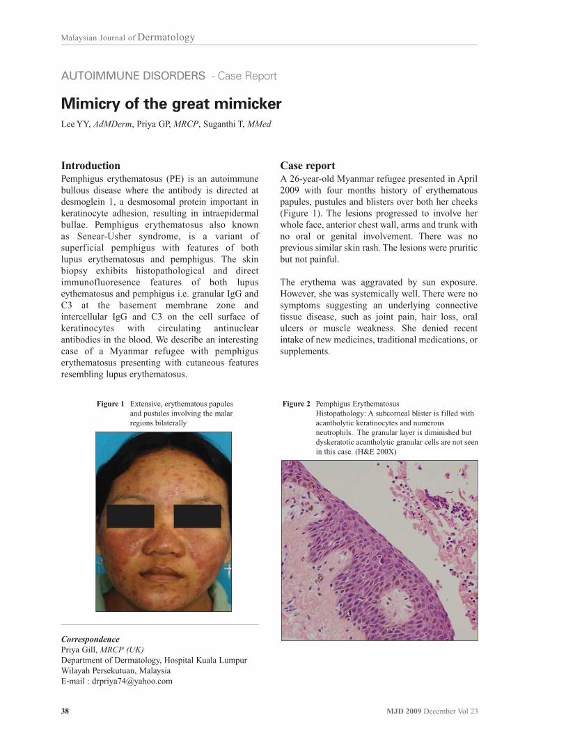
Case reportA 26-year-old Myanmar refugee presented in April2009 with four months history of erythematouspapules, pustules and blisters over both her cheeks(Figure 1). The lesions progressed to involve herwhole face, anterior chest wall, arms and trunk withno oral or genital involvement. There was noprevious similar skin rash. The lesions were pruriticbut not painful.
The erythema was aggravated by sun exposure.However, she was systemically well. There were nosymptoms suggesting an underlying connectivetissue disease, such as joint pain, hair loss, oralulcers or muscle weakness. She denied recentintake of new medicines, traditional medications, orsupplements.
CorrespondencePriya Gill, MRCP (UK)Department of Dermatology, Hospital Kuala LumpurWilayah Persekutuan, MalaysiaE-mail : [email protected]
Figure 1 Extensive, erythematous papules and pustules involving the malar regions bilaterally
Figure 2 Pemphigus ErythematosusHistopathology: A subcorneal blister is filled with acantholytic keratinocytes and numerous neutrophils. The granular layer is diminished but dyskeratotic acantholytic granular cells are not seen in this case. (H&E 200X)

39MJD 2009 December Vol 23
Malaysian Journal of Dermatology
Clinically, there were extensive erosions witherythematous crusts and scales involving the malarregion, anterior trunk and entire back. There werealso a few intact flaccid bullae on the upper limbsand back. However, there was no Raynaud’sphenomenon or cutaneous vasculitic changes.Examination of other systems was normal.The provisional diagnosis was pemphiguserythematosus with a differential of pemphigusfoliaceus and acute cutaneous lupus erythematosus.
Skin biopsies were performed from 2 sites; anerythematous, scaly patch from the left cheek and aflaccid bulla from the right forearm. The firstbiopsy showed follicular plugging, mild oedemaof the upper dermis with perivascularand periappendegeal infiltration by chronicinflammatory cells plus focal basal cellvacuolation. The second biopsy demonstratedsubcorneal bullae containing few neutrophilsand acantholytic cells (Figure 2). Directimmunofluorescence showed moderately strongintraepidermal and dermo-epidermal junction IgGdeposits. These confirmed the clinical diagnosis ofpemphigus erythematosus.
The full blood counts, renal profile, liver functionand serum complements were normal. Anti nuclearantibody and extractable nuclear antibody were nonreactive. There was microscopic hematuria whichcould be attributed to a Klebsiella urinary tractinfection.
The patient was admitted to the dermatology wardand subjected to intensive skin nursing. She wasprescribed intravenous hydrocortisone 100mg 8hourly and oral Cefuroxime Axetil (for urinary tractinfection) and improved dramatically over the next5 days. The patient requested an early discharge andwas discharged on Prednisolone 45mg daily,calcium carbonate 600mg twice daily, _-calcidiol0.5mg daily, titanium dioxide as a sun block andtopical betamethasone valerate 0.025% twice daily.We planned to add on oral Azathioprine 100mgdaily as a steroid sparing agent on follow up.
Unfortunately, she defaulted her follow up and wasnot contactable thereafter.
DiscussionPemphigus erythematosus (PE), also known asSenear-Usher syndrome is an antibody-mediatedautoimmune bullous disease with combined
features of pemphigus foliaceous andlupus erythematosus. The term ‘pemphiguserythematosus’ was initially coined to describepatients with immunological features of both lupuserythematosus and pemphigus foliaceous. PErepresents approximately 8% of all cases ofpemphigus. In pemphigus foliaceous, antibodieswere directed at desmoglein 1, a desmosomalprotein important in keratinocyte adhesion.
Histopathology and direct immunofluorescenceexamination of PE elucidate features of both lupuserythematosus and pemphigus, i.e. granular IgGand C3 at the basement membrane zone andintercellular IgG and C3 on the cell surface ofkeratinocytes in a fishnet appearance 1.
PE affects mainly middle-aged adults and manifestsitself on sun-exposed areas as flaccid bullae withscales and crusts. The main areas of distributioninclude the scalp, face, upper chest and back. Facialmanifestations are localized to the typical butterflydistribution as seen in lupus erythematosus (LE)2,3.Pemphigus and LE have been reported to coexist inthe same patient. However, the incidence of the co-existence is low. Most of these patients are non-Caucasian females in their reproductive age groups,which is similar to LE. In a review conducted byMohsin Malik et al, majority of these patients hadpemphigus vulgaris (PV) and less commonly theother variants of pemphigus4.
Our patient, presented with a history and clinicalexamination suggestive of LE such asphotosensitivity and malar rash. The bullae also ledus to think of bullous lupus erythematosus.However, the connective tissue screening wasunremarkable with a negative anti nuclear factor(ANA) and extractable nuclear antibody (ENA).The confirmatory diagnosis of PE was onlyelucidated from the skin histopathological anddirect immunofluorescence examination wherebythere were features of both pemphigus foliaceousand lupus erythematosus.
The natural course of PE is relatively milder, moresteroid sensitive and carries a better prognosis5.Treatment for PE has relied heavily on the useof systemic steroids, with adjuvant steroidsparing drugs such as dapsone, azathioprine,cyclophosphamide and methotrexate. In ourpatients, oral prednisolone at 0.5mg/kg/day wassuccessful

40 MJD 2009 December Vol 23
Malaysian Journal of Dermatology
successful in controlling the initial eruption.Unfortunately, we are unable to monitor her longterm disease progress. This patient is an excellentexample of how the great mimicker (LE) ismimicked (PE).
References
1. Amagai M. Pemphigus. In: Dermatology (Bolognia JL,Jorizzo J, Rapini RP, eds). London: Mosby, 2003; 449-62
2. Senear FE, Usher B. An unusual type of pemphigus:coming features of lupus erythematosus. ArchDermatol Syphilol 1926; 13: 761
3. Chorzelski J, Jacblonska S, Blaszczyk M.Immunopathological investigations in the Senear-Ushersyndrome (coexistence of pemphigus and lupuserythematosus). Br J Dermatol 1968; 80: 211)
4. Mohsin Malik, A. Razzaque Ahmed. Concurrence ofSystemic Lupus Erythematosus and Pemphigus:Coincidence or Correlation? Dermatology 214:231-239, 2007
5. Ahmed AR, Salm M. Juvenile pemphigus. J Am AcadDermatol 1983; 8: 799-807

41MJD 2009 December Vol 23
Malaysian Journal of Dermatology
CONTACT DERMATITIS & OCCUPATIONAL DERMATOSES - Original Article
Clinical pattern and causative allergens of hand and / or
feet eczema identified from patch test - a retrospective
5 year study
Priya G, MRCP, Asmah J, MMED, Gangaram HB, FRCP, Suraiya HH, FRCP
Abstract
Background Hand and/or feet eczema may be due to contact dermatitis, either irritant or allergic innature. Difficulties often arise in distinguishing purely endogenous eczema from the possibility ofcontact dermatitis clinically. Patch test is carried out to detect the presence of allergic contactdermatitis. This is important for optimum patient care and to obtain a favourable outcome.
Objectives To identify the demography, clinical characteristics and causative allergens of handand/or feet eczema among patients from the patch test clinic.
Methods Patients who attended the patch test clinic in the Department of Dermatology, HospitalKuala Lumpur from 2003 to 2007 were evaluated retrospectively. All of them were having handand/or feet eczema. Data were collected for their demography, sites affected and patch test findings.
Results 379 patients were included in the study. The age of patients ranged from 6 years to 78 yearswith an average of 36.7 years. Their occupations ranged from blue collar (20.3%) and white collar(38.3%) workers, housewives (9.5%), pensioners (7.1%) and students (20.3%). Clinicalpresentations included isolated hand eczema (34.6%), isolated feet eczema (21.9%), hand and feeteczema (19.0%), and hand and/or feet with eczema with involvement of other parts of the body(24.5%).The mean duration of eczema was 3.8 years. The rate of positive patch test was 58.0 %( n= 220/379).Clinically relevant allergens were identified in 123 (32.5%) patients only. Fifty twopercent of the clinically relevant allergens were identified from the European Standard Series patchtest, 9.0% from the Specific Series patch test and 39.0 % from the patients’ own personal productsthat were tested. The most common source was metal items containing nickel (33.3%), followed bytoiletries (14.6%) and detergents (10.6%). Other sources include fragrance, cosmetics, rubber,medicaments and hair dye. In 256 (67.5%) patients, there were no underlying causes detected, andthey were managed as endogenous hand and feet eczema. There is a possibility that the causativeallergen was not suspected/tested and hence not detected.
Conclusion Hand and/or feet eczema can affect any age group and patch testing forms a veryimportant diagnostic tool in the management.
Keywords Clinical pattern, allergens, hand and/or feet eczema, patch test
CorrespondencePriya Gill, MRCP (UK)Department of Dermatology, Hospital Kuala LumpurWilayah Persekutuan, MalaysiaE-mail : [email protected]
IntroductionThe hand and feet are often exposed to variouspotential allergens and irritants. Exclusive handeczema affects 9.1% of patients and another 9.0%
have exclusive feet eczema. Difficulties often arisein distinguishing endogenous eczema from theprobability of contact dermatitis clinically andpatch testing is of much help in these situations1,2.
The aim of this study is to determine thedemography, clinical pattern and causativeallergens identified from patch test in hand and / orfeet eczema.

42 MJD 2009 December Vol 23
Malaysian Journal of Dermatology
Materials and methodsThis is a retrospective study on patients from thePatch Test clinic in the Department of Dermatology,Hospital Kuala Lumpur. All patients with handand/or feet eczema who underwent Patch Test fromJanuary 2003 to December 2007 were included.Patch Test was carried out according to therecommendations made by the InternationalContact Dermatitis Research Group using Trolab®
allergens. All patients were patch tested against theEuropean Standard Series allergens and ifnecessary, with an additional specific series e.g.hairdressing chemicals, shoe allergens, rubberchemicals etc and their own products. The readingswere done on day 3 and day 5.
Data was analyzed using SPSS. The patients wereassessed in terms of demographic data, clinicalpresentations, patch test results, clinically relevantpatch test results and source of allergens.
ResultsA total of 379 patients were included in this review(259 female,120 male). Two hundred and twentypatients had a positive patch test (164 female, 56male). The racial distribution of patients in eachcategory mimicked the racial attendance of theOutpatient specialist clinic, Department ofDermatology, Hospital Kuala Lumpur in 2007 with58.6% Malays, 21.9% Chinese, 17.4% Indian and2.1% of patients belonging to other races. They
were categorized into patients with only handeczema or only feet eczema, hand and feet eczemaand hand and / or feet eczema with generalization.The demography of patients is shown in Table 1.There is a female preponderance and the mean agegroups were in their thirties for all categories. Theage distribution is shown in Figure 1.
Table 2 details the patients’ occupations. They wereclassified into white collar workers (e.g.supervisors, clerks, nurses, professionals, teachers,secretaries, executives, film producers etc), bluecollar workers (soldiers, labourers, machineoperators, welders, attendants, taxi drivers, babysitters, cleaners etc), housewives, pensioners andstudents.The white collar patient subgrouppresented mainly with hand and feet eczema whilethose who held blue collar jobs had mainly feeteczema. Interestingly, the many of the students hadfeet eczema.
The working diagnosis for all the patients is shownin Table 3. Patch Test was undertaken only when acontact element was suspected.
Two hundred and twenty patients had a positivePatch Test (58%). The positive Patch Test wasclinically relevant in only 123 patients (32.5 %)(Table 4). Fifty two percent of the relevant allergenswere identified from the European Standard Series,39% from the patients’ personal products and 9%from the Specific Series.
Table 1 Patient demographics
Type
Number
Male: Female ratio
Age (years)
Range
Mean
Hand Eczema
131
(34.6%)
1: 2.4
(38 male, 93 female)
7 - 72
34.4
Feet Eczema
83
(21.9%)
1:1.4
(35 male, 48 female)
6 - 72
34.4
Hand & Feet Eczema
72
(19.0%)
1:2.0
(24 male, 48 female)
7 - 66
36.0
Hand and/or Feet Eczema
with generalization
93
(24.5%)
1:3.0
(23 male, 70 female)
7 -78
38.0

43MJD 2009 December Vol 23
Malaysian Journal of Dermatology
The top 10 allergens detected from the EuropeanStandard Series were nickel, fragrance mix, balsamof Peru, paraben, chromium, neomycin, colophony,cobalt, thiuram mix and formaldehyde which wereclinically relevant in 41.3%, 31.0%, 42.9%, 35.0%,41.2%, 31.3%, 33.3%, 28.6%, 50.0% and 42.9%respectively. (Table 5). Only the top 20 allergensidentified from the European Standard Series andtheir clinical relevance are shown in Table 5.
Fifty four patients were tested with rubberchemicals; clinically relevant in 4 patients (9%)(Table 6). A total of 26 patients were tested withshoe allergens; clinically relevant in 2 patients [n =2/26, (8%)]. Twenty nine patients were tested withthe textile and leather dyes allergens; relevant inonly 1 patient [n = 1/29, (3%)]. Only 4 patients weretested with hairdressing allergens and two of themwere clinically relevant. [n= 2/4, (50%)].
Figure 1 Age distribution
Table 2 Occupation
Occupational field
White Collar
Blue Collar
Housewife
Pensioner
Student
Not available
145 (38.3%)
77 (20.3%)
36 (9.5%)
27 (7.1%)
77 (20.3%)
17 (4.5%)
57 (43.5%)
27 (20.6%)
9 (6.9%)
13 (9.9%)
16 (12.2%)
9 (6.9%)
18 (21.7%)
21 (25.3%)
7 (8.4%)
5 (6.0%)
29 (35.0%)
3 (3.6%)
32 (44.4%)
9 (12.5%)
8 (11.1%)
5 (7.0%)
17 (23.6%)
1 (1.4%)
38 (40.9%)
20 (21.5%)
12 (12.9%)
4 (4.3%)
15 (16.1%)
4 (4.3%)
N=379 Hand Eczema
N = 131
Feet Eczema
N=83
Hand & Feet Eczema
N = 72
Hand & / or Feet Eczema
with eczema elsewhere
N=93
7
50
90
60
80
61
28
3

44 MJD 2009 December Vol 23
Malaysian Journal of Dermatology
Table 3 Working diagnosis and Patch test results
72 (58.6%)
16 (13.0%)
13 (10.6%)
18 (14.6%)
0 (0%)
2 (1.6%)
2 (1.6%)
0 (0%)
Relevant Allergen identified
N=123
163 (43.0%)
75 (19.8%)
62 (16.3%)
59 (15.6%)
6 (1.6%)
8 (2.1%)
5 (1.3%)
1 (0.3%)
All patients
N=379
Diagnosis
Contact Dermatitis
Hand Eczema
Feet Eczema
Hand & Feet Eczema
Discoid Eczema
Atopic Eczema
Photodermatitis
Juvenile Plantar Dermatitis
Table 4 The top 20 allergens from the European Standard Series
Allergens
Nickel
Fragrance mix
Balsam of Peru
Paraben
Chromium
Neomycin
Colophony
Cobalt
Thiuram Mix
Formaldehyde
Isothiazolin
Flavin
Wool Alcohol
Mercaptobenzothiazole
Mercapto Mix
Sesquiterpene Lactone Mix
Paratertiary Phenol Formaldehyde Resin ( PPFR)
N-Isopropyl-N2-phenyl paraphenylenediamine ( PPD)
Hydroxymethylpentylhexenecarboxyaldehyde
Benzocaine
104 (47.3)
42 (19.1)
21 (9.5)
20 (9.1)
17 (7.7)
16 (7.3)
15 (6.8)
14 (6.4)
14 (6.4)
14 (6.4)
10 (4.5)
8 (3.6)
7 (3.2)
5 (2.3)
4 (1.8)
3 (1.4)
1 (0.5)
1 (0.5)
2 (0.9)
3 (1.4)
43 (41.3)
13 (31.0)
9 (42.9)
7 (35.0)
7 (41.2)
5 (31.3)
5 (33.3)
4 (28.6)
7 (50.0)
6 (42.9)
3 (30.0)
1 (12.5)
4 (57.1)
3 (60.0)
4 (100.0)
3 (100.0)
1 (100.0)
1 (100.0)
0
0
+ve PT (%) Clinically
(%)
Relevant

45MJD 2009 December Vol 23
Malaysian Journal of Dermatology
Table 5 Patch Test result for rubber allergens
Frequency
1
2
1
1
1
2
1
2
1
1
%
25
50
50
25
25
50
25
50
25
25
Allergens
Hexamethylenetetramine
Diphenylthiourea
Dibutylthiourea
1, 3 - Diphenylguanidine
Bis (diethyldithiocarbamato) Zinc
N, N - Diphenyl Paraphenylenediamine
Bis (dibuhyldithiocarbamato)Zinc
Cyclohexyl Thiophtalimide
4, 4’- Dihydroxybiphenly
Zinc dibenzyldithiocarbamate
Rubber Chemicals, n = 54, clinically relevant in 4 patients
Table 6 Patch Test result for Hand eczema
Allergens
Nickel
Fragrance mix
Balsam of Peru
Paraben
Thiuram Mix
Cobalt
Colophony
Formaldehyde
Isothiazolin
Neomycin
Chromium
Wool Alcohol
Benzocaine
Flavin
Sesquiterpene Lactone Mix
Special Series: Rubber Chemicals
Special Series : Hairdressing
32 (39.0)
20 (24.4)
9 (11.0)
8 (9.8)
7 (8.5)
7 (8.5)
6 (7.3)
6 (7.3)
6 (7.3)
4 (4.9)
4 (4.9)
4 (4.9)
3 (3.7)
3 (3.7)
2 (2.4)
4 (4.9)
2 (2.4)
10 (31.3)
4 (20.0)
2 (22.2)
2 (25.0)
3 (42.9)
0
1 (16.7)
2 (33.3)
2 (33.3)
2 (50.0)
0
3 (75.0)
0
0
1 (50.0)
3 (75.0)
2 (100)
+ve PT(%) ( N = 82)
Clinically relevant (%)
Source
Coins, Costume jewellery, Watch Strap,Belt buckle, pins etc
Perfume & After shavePerfume & After shave / Cosmetics
Cosmetics
Rubber Gloves
-
Medicament
Cosmetics
Cosmetics /Detergents
Medicament
-
Cosmetics
-
-
Cosmetics
Rubber Gloves
Hair Dye
46 MJD 2009 December Vol 23
Malaysian Journal of Dermatology
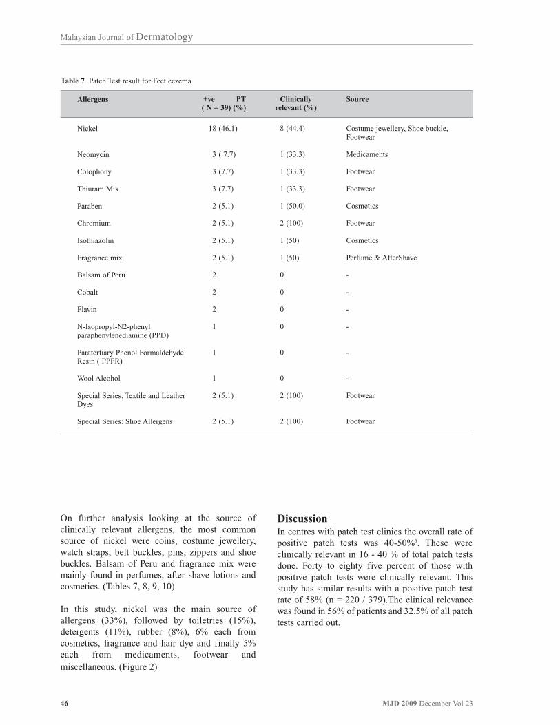
Table 7 Patch Test result for Feet eczema
Allergens
Nickel
Neomycin
Colophony
Thiuram Mix
Paraben
Chromium
Isothiazolin
Fragrance mix
Balsam of Peru
Cobalt
Flavin
N-Isopropyl-N2-phenylparaphenylenediamine (PPD)
Paratertiary Phenol FormaldehydeResin ( PPFR)
Wool Alcohol
Special Series: Textile and LeatherDyes
Special Series: Shoe Allergens
18 (46.1)
3 ( 7.7)
3 (7.7)
3 (7.7)
2 (5.1)
2 (5.1)
2 (5.1)
2 (5.1)
2
2
2
1
1
1
2 (5.1)
2 (5.1)
+ve PT( N = 39) (%)
8 (44.4)
1 (33.3)
1 (33.3)
1 (33.3)
1 (50.0)
2 (100)
1 (50)
1 (50)
0
0
0
0
0
0
2 (100)
2 (100)
Clinically relevant (%)
Source
Costume jewellery, Shoe buckle,Footwear
Medicaments
Footwear
Footwear
Cosmetics
Footwear
Cosmetics
Perfume & AfterShave
-
-
-
-
-
-
Footwear
Footwear
On further analysis looking at the source ofclinically relevant allergens, the most commonsource of nickel were coins, costume jewellery,watch straps, belt buckles, pins, zippers and shoebuckles. Balsam of Peru and fragrance mix weremainly found in perfumes, after shave lotions andcosmetics. (Tables 7, 8, 9, 10)
In this study, nickel was the main source ofallergens (33%), followed by toiletries (15%),detergents (11%), rubber (8%), 6% each fromcosmetics, fragrance and hair dye and finally 5%each from medicaments, footwear andmiscellaneous. (Figure 2)
DiscussionIn centres with patch test clinics the overall rate ofpositive patch tests was 40-50%3. These wereclinically relevant in 16 - 40 % of total patch testsdone. Forty to eighty five percent of those withpositive patch tests were clinically relevant. Thisstudy has similar results with a positive patch testrate of 58% (n = 220 / 379).The clinical relevancewas found in 56% of patients and 32.5% of all patchtests carried out.

47MJD 2009 December Vol 23
Malaysian Journal of Dermatology
Table 8 Patch Test result for Hand & Feet eczema
Allergens
Nickel
Fragrance mix
Balsam of Peru
Paraben
Chromium
Isothiazolin
Formaldehyde
Cobalt
Mercaptobenzothiazole
Mercapto Mix
Flavin
Colophony
Wool Alcohol
Neomycin
Benzocaine
Special series: Rubber Chemicals
Special Series: Shoe Allergens
17 (42.5)
8 (20.0)
6 (15.0)
6 (15.0)
5 (12.5)
2 (5.0)
2 (5.0)
2 (5.0)
2 (5.0)
2 (5.0)
2 (5.0)
2 (5.0)
1 (2.5)
1 (2.5)
1 (2.5)
1 (2.5)
1 (2.5)
6 (35.3)
2 (25.0)
4 (66.7)
4 (66.7)
2 (40.0)
1 (50.0)
2 (100)
2 (100)
2 (100)
2 (100)
0
0
0
0
0
1 (100)
1 (100)
+ve PT(%) ( N = 40)
Clinically relevant (%)
Source
Coins, Costume jewellery, Zipper,Watch Strap, Belt & shoe buckle etc
Fragrance/Cosmetics
Toiletries/Cosmetics/Perfume & After shave
Cosmetics
Cement/Industrial oils
Cosmetics
Cosmetics
Personal Items/Industrial oils
Rubber Gloves
Rubber Gloves
-
-
-
-
-
Rubber Gloves
Footwear
Figure 2 Sources of clinically relevant positive Patch test in Hand & / or Feet Eczema
Medicaments 5%
Hair Dye 6%
Fragrance 6%
Cosmetics 6%
Rubber 8%
Detergent 10%
Toileteries 15%
Nickel 34%
Footware 5%Miscellanous 5%

48 MJD 2009 December Vol 23
Malaysian Journal of Dermatology
Table 9 Patch Test result for Hand & / or Feet Eczema with generalization
Allergens
Nickel
Fragrance mix
Neomycin
Chromium
Formaldehyde
Paraben
Colophony
Balsam of Peru
Thiuram Mix
Mercaptobenzothiazole
Cobalt
Hydroxymethylpentylhexenecarboxyal-dehyde
Mercapto Mix
Sesquiterpene Lactone Mix
Flavin
Wool Alcohol
Special Series: Metal Compounds
37 (51.4)
12 (20.3)
8 (13.6)
7 (11.9)
6 (10.2)
4 (6.8)
4 (6.8)
4 (6.8)
4 (6.8)
3 (5.1)
3 (5.1)
2 (3.4)
2 (3.4)
1 (1.7)
1 (1.7)
1 (1.7)
1 (1.7)
19 (51.4)
1 (8.3)
3 (38.0)
2 (28.6)
0
2 (50.0)
0
0
2 (50.0)
1 (33.3)
0
1 (50.0)
1 (50.0)
1 (100)
0
0
1 (100)
+ve PT(%) ( N = 59)
Clinically relevant (%)
Source
Coins, Costume jewellery, Zipper,Watch Strap, Belt & shoe buckle etc
Perfume & After shave
Medicaments
Dental Material/ Leather Strap
-
Medicaments
-
-
Gloves
Leather Strap
Dental Material
Perfume & After shave
Leather Strap
Flower
-
-
Factory machinery
The predominance of young women corresponds tothe cohort of Meding et al6. In the South India studylooking at lower leg and feet eczema, the averagepatient age was 40.49 years with a female to maleratio of 1.6:14. However there are studies that with amale preponderance. Smith et al reported a male tofemale ratio of 1.25:1 in their study on descriptiveepidemiology of hand eczema7. Goh CL fromSingapore observed a male preponderance as wellin the prevalence of hand eczema in his cohort ofpatients. (Male 56.0%, Female 44.0%)8. Thedifference between studies could be explained bydifferent jobs being carried out by men and womenand their tolerance to develop hand and / or feetirritant contact dermatitis with different types ofexposure to allergens / irritants.
The working diagnosis for our patients was contactdermatitis (43.0%), hand eczema (19.8%), feeteczema (16.3%), hand and feet eczema (15.6%)with about 5% of patients having a diagnosis ofdiscoid eczema/ atopic eczema/photodermatitis/juvenile dermatitis. As these patients wereidentified from patch test, those with one type ofendogenous eczema do not have to undergo thisprocedure unless suspected to have contactdermatitis. Therefore, those with hand and feeteczema are more likely to undergo patch test.

49MJD 2009 December Vol 23
Malaysian Journal of Dermatology
Chougule et al reported that among patients withlower leg and foot eczema in South India, the mostcommon working diagnosis was lichen simplexchronicus (36%), followed by discoid eczema(18.5%), stasis eczema (7.5%), juvenile plantardermatoses (5%), hyperkeratotic eczema (3%) andunclassified endogenous eczema (3%)4. The NorthAmerican Contact Dermatitis Group (NACDG)looked at 6953 patients with hand eczema only(1994-2004) and their common working diagnosiswere allergic contact dermatitis (27.8%), irritantcontact dermatitis (19.7%), psoriasis (3.3%), atopiceczema (2.5%), pompholyx (1.7%) and otherdermatitis/dermatoses (8.9%)5. In this paper,however, the authors were not specifically lookingfor contact dermatitis but rather at the range ofconditions that presented with hand eczema.Different centres will have different workingdiagnosis prevalence as a lot of factors willinfluence their decision to carry out patch test.
Majority of patients were in the white collar group(38.3%), followed by the blue collar group andstudents (20.3% each), housewives (9.5%) andfinally pensioners (7.1%). Our results are differentcompared to studies by Cherry et al and Smith et al.Cherry concluded that contact dermatitis is by farthe most commonly reported occupational skindisease, especially in the blue collar group. Theirstudy showed that women were most likely to havedermatitis attributed to wet work, and men to oilsand related substances. In Smith’s paper, the mostfrequent types of employment associated withoccupational hand eczema were caterers, metalworkers, mechanics, builders and printers (bluecollar workers)7,9. Unfortunately, as this is aretrospective review, we are unable to ascertain forsure the relationship of eczema in our group ofpatients to the occupation. The majority of patientsin our review were from white collar occupations.This can be attributed to our patient population thatunderwent patch testing in general being from thewhite collar group as this department is located inthe heart of Kuala Lumpur, the biggest city inMalaysia. Smith et al also suggested that low officehumidity as an irritant factor and therecommendation of office work as a “clean, safejob” might need to be reviewed7.
The most common presentation for white collarworkers, blue collar workers and pensioners washand eczema. Feet eczema was the most commonsubtype among students and not surprisingly,
housewives presented most commonly with handand/ or feet eczema with eczema elsewhere. Thiscould be due to the possible contact to variouspotential irritants in everyday household chores likewashing clothes and utensils with detergents andwater. Many housewives as well squat on theground while doing their chores; and their feet arecontinuously exposed to detergents and water whileother areas are affected by splashing of the water.
The highest proportion of clinically relevantallergens were identified in patients who were beingmanaged as contact dermatitis (44.2%), comparedto hand eczema (21.3%), feet eczema (21.0%),hand & feet eczema (30.5%) and atopic dermatitis(25.0%).This could be due to a lower index ofsuspicion for a contact element in those diagnosedto have endogenous eczema. However, studies haveshown that endogenous eczema is commonlycomplicated by a contact element and eliminatingthe causative factor will greatly facilitate themanagement10,11.
The most common positive patch test allergens inthis study were nickel (47.3%), fragrance mix(19.1%) and balsam of Peru (7.9%). However, forthose with feet eczema, the most common allergenswere nickel, chromium, shoe allergens and textileand leather dyes. The results in our centre differedslightly compared to Osmania General Hospital,Hyderabad, India where a similar study was doneusing the Indian Standard Series Patch testallergens. Their most common allergens were nickeland chromium (25% each), followed by fragrancemix (21.4%) and wool alcohol (14.3%).Theseresults were probably due to the different seriesbeing used for Patch test and the different patientfactors, commercial products and environmentalfactors the two populations are exposed to2.
The most common sources of allergens werepersonal items containing nickel (33%), toiletries(15%) and detergents (15%). Our findings aresimiliar that of the North American ContactDermatitis Group. They found the most commonsources to be soaps, cleaners, detergents, solvents,oil and lubricants5. These are common everydayitems individuals are exposed to.
There are several limitations to this study. The datais retrospective with some even incompleteinformation; therefore a more definite causalrelationship cannot be determined. The studysample
50 MJD 2009 December Vol 23
Malaysian Journal of Dermatology
sample was drawn only from patients with handand/or feet eczema who were patch tested; as such,they are not representative of the general populationor the general dermatology population. Moreresearch in a clinical setting is needed to addresssome of the issues raised in our review, especiallyprospective research.
ConclusionsHand and/ or feet eczema affects all ages anddifferent types of occupations. This disease cancause significant disability and economic loss toboth individuals and society. Patch testing is a veryimportant diagnostic tool to exclude allergic contactdermatitis in those suspected as having endogenouseczema or other types of dermatoses. The specificseries patch test allergens and the patient’s ownproducts are of added benefit if clinically indicated.Contact avoidance remains the most importantmeasure in the prevention and management ofallergic and irritant contact dermatitis. Therefore,education about avoidance of, or protection from,the most common antigens is critical, especially inhigh risk occupations
References
1. Ng SK, Goh CL, M Shahidullah. Clinical patterns ofFeet Eczema in Singapore. Bulletin for medicalpractitioners 1998; 9 (1)
2. G Vani , Rao GA, V Gowri TSS Lakshmi. Allergiccontact dermatitis of the hands and/or feet - commonsensitizers. Contact Dermatitis 2005; 52 (1):50-52
3. Ormond P, Hazelwood E et al. The importance of adedicated patch test clinic. Br J Derm 2002; 146: 304-307
4. Chougule A, Thappa DM. Patterns of lower leg and footeczema in south India. Indian J Dermatol VenereolLeprol 2008; 74:458-61
5. Warshaw ME, Ahmed AL, Belsito DV et al. Contactdermatitis of the hands: Cross-sectional analyses ofNorth American Contact Dermatitis Group Data,1994-2004; J Am Acad Dermatol 2007; 57; 301-14)
6. Veien NK, Hattel T, Laurberg G. Hand eczema: causes,course, and prognosis I. Contact Dermatitis 2008; 58;330-334
7. Smith HR, Armstrong DKB, WakelinSH etal. Descriptive epidemiology of hand dermatitis at theSt John’s contact dermatitis clinic. Br J Dermatol 2000;142: 284-287
8. Goh CL. An epidemiological comparison betweenoccupational and non-occupational hand eczema. Br JDermatol 1989; 120: 77-82
9. Cherry N, Meyer JD, Adidesh A et al. Surveillance ofoccupational skin disease: EPIDERM and OPRA. Br JDerm 2000; 142:1128-1134
10. Shackelford KE, Belsito DV. The etiology of allergic-appearing foot dermatitis: a 5-year retrospective study.J Am Acad Dermatol 2002; 47 (5): 715-721
11. Fowler JF, Ghosh A, Sung J et al. Impact of chronichand dermatitis on quality of life, work productivity,activity impairment and medical costs. J Am AcadDermatol 2006; 54 (30): 448-457

51MJD 2009 December Vol 23
Malaysian Journal of Dermatology
Keywords Uveitis, lymphadenopathy, leprosy,sarcoidosis
IntroductionSarcoidosis is a chronic systemic disorderof unknown etiology, characterizedhistopathatologically by non-caseating, epithelioidgranulomatous infiltration in various organs.1,2
Cutaneous sarcoidosis is also known as adermatologic masquerader because the lesions canexhibit many different morphologies.3 We report apatient who was initially diagnosed as havingtuberculoid leprosy based on histological findings.He was treated with multi-drug therapy for 18months without clinical improvement. In addition,he had left panuveitis and mediastinallymphadenopathy.
Case reportA 28 year old gentleman with no previous medicalillness first presented to the ophthalmologist withcomplaint of acute onset of left eye redness andpain. He was diagnosed and treated for leftpanuveitis. He was referred to us two months laterfor further management of non-pruriticerythematous skin rashes of 1 year duration. At thesame time, he was referred to the respiratory teamfor further work up of possible tuberculosis.
The skin eruption first started over the upper limbsand subsequently involved the trunk, sparing theface, palm and soles. There was no associated limbnumbness or weakness and no fever, cough orshortness of breath. His appetite was normal andthere was no significant weight loss. Review ofother systems was unremarkable.
Clinically, he was pink and not jaundiced. Hisheight was 108 cm and weight 102 kg. There weremultiple well-defined erythematous papules and
GRANULOMATOUS DISEASE - Case Report
Cutaneous Sarcoidosis Mimicing Tuberculoid Leprosy
Chong YT, MRCP, Tey KE, MRCP, Choon SE, FRCP
plaques distributed symmetrically over the trunkand upper limbs. Sensation was normal and therewas no peripheral nerve thickening. Lymph nodeswere not palpable. Examinations of the cardio-respiratory system and abdomen did not review anyabnormalities.
Blood investigations revealed hypochromicmicrocytic anaemia with a hemoglobin of13.1gm/dl and eosinophilia. His erythrocytesedimentation rate (ESR) was 30mm per hour. Hehas hyperglobulinaemia (51 g/L) and slightly raisedalanine aminotransferase (40 IU/L). The renalprofile, serum calcium and fasting blood glucosewere normal. Serological screen for viral hepatitis,rapid reagent test for syphilis (RPR) and anti-HIVwere negative. Mantoux test was negative (2mm).The slit skin smear was negative for acid fastbacilli.
The skin biopsy was reported as tuberculoid leprosywhich showed numerous epitheliod granulomaaround neurovascular bundles with occasionalLangerhan’s giant cells, many epitheliod cells andlymphocytes. There was no caseation necrosis andthe Fite stain was negative for acid fact bacilli.Tissue culture for mycobacteria and fungus wasalso negative.
Based on the histology report, multi-drug therapy(MDT) for paucibacillary leprosy (rifampicin,clofazimine and dapsone) was commenced. At thesame time, he was treated with prednisolone forpanuveitis by the ophthalmologist. His skin lesionsimproved initially. However, two months intotreatment, he developed raised liver enzymes.Ultrasound of the liver showed fatty changes. TheMDT was withheld for one month and laterrestarted with minocycline and dapsone. After oneyear of treatment, his skin lesions did not showmuch improvement despite the initial response. Arepeat skin biopsy showed similar findings as theinitial one.
Further investigations with CT scan and endoscopicultrasound showed multiple small mediastinallymph nodes. Fine-needle aspiration cytology of thesub-carinal lymph node was reported as agranulomatous lesion. PCR test from a repeat skinbiopsy was negative for Mycobacteria DNA.
CorrespondenceChong Yew Thong Department of DermatologyHospital Sultanah Aminah80100 Johor Bahru, JohorE-mail : [email protected]
52 MJD 2009 December Vol 23
Malaysian Journal of Dermatology
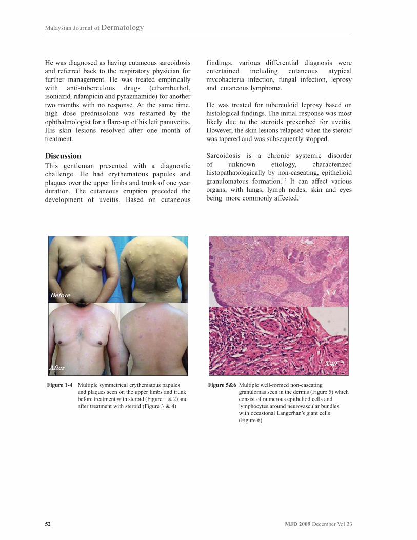
He was diagnosed as having cutaneous sarcoidosisand referred back to the respiratory physician forfurther management. He was treated empiricallywith anti-tuberculous drugs (ethambuthol,isoniazid, rifampicin and pyrazinamide) for anothertwo months with no response. At the same time,high dose prednisolone was restarted by theophthalmologist for a flare-up of his left panuveitis.His skin lesions resolved after one month oftreatment.
DiscussionThis gentleman presented with a diagnosticchallenge. He had erythematous papules andplaques over the upper limbs and trunk of one yearduration. The cutaneous eruption preceded thedevelopment of uveitis. Based on cutaneous
findings, various differential diagnosis wereentertained including cutaneous atypicalmycobacteria infection, fungal infection, leprosyand cutaneous lymphoma.
He was treated for tuberculoid leprosy based onhistological findings. The initial response was mostlikely due to the steroids prescribed for uveitis.However, the skin lesions relapsed when the steroidwas tapered and was subsequently stopped.
Sarcoidosis is a chronic systemic disorderof unknown etiology, characterizedhistopathatologically by non-caseating, epithelioidgranulomatous formation.1,2 It can affect variousorgans, with lungs, lymph nodes, skin and eyesbeing more commonly affected.4
Figure 1-4 Multiple symmetrical erythematous papules and plaques seen on the upper limbs and trunk before treatment with steroid (Figure 1 & 2) and after treatment with steroid (Figure 3 & 4)
Figure 5&6 Multiple well-formed non-caseating granulomas seen in the dermis (Figure 5) which consist of numerous epitheliod cells and lymphocytes around neurovascular bundles with occasional Langerhan’s giant cells(Figure 6)

53MJD 2009 December Vol 23
Malaysian Journal of Dermatology
Sarcoidosis affects all races, both sexes and allages. Incidence reported ranged from 64/100, 000in Sweden to 1.4/100, 000 in Japan.1
Cutaneous involvement occurs in 20 to 35 percentof patients with systemic sarcoidosis but it mayoccur without systemic involvement.3 It is mostcommonly seen at the onset of the disease process.1
Various morphologies have been described,including papules, follicular papules, plaques,nodules, ulcerative lesions and alopecia.1
The eye and adnexa are involved in 25 to 80% ofpatients with sarcoidosis and anterior uveitis is themost common manifestation, occurring up to 65%of patients with ophthalmologic involvement. Inabout 10 to 15%, both the anterior and posterior(panuveitis) segments may be involved.11
In developed countries, sarcoidosis was reported tobe the second most common non-infectious causeof uveitis, after sero-negative spondyloarthropathy,especially in US, Netherland and Japan.10
There is no single test to confirm the diagnosis ofsarcoidosis.3 Sarcoidal granulomas have no uniquehistologic features to differentiate them from othergranulomas. Patients are diagnosed withsarcoidosis on the basis of compatible clinical,radiologic findings, supported by histologicevidence of non-caseating granulomas, and whenother potential causes, such as infections, areexcluded.11
Cutaneous sarcoidosis has quite often beenmisdiagnosed as leprosy because of near similarskin lesions and histologic findings of non-caseating granuloma. Diagnosis of sarcoidosis wassuspected when the patient did not respond to acourse of anti-leprosy drugs.6,7,8,9
Cutaneous sarcoidosis is rare in South-East Asia,including Malaysia. Liam et al 12 reported 14 casesof sarcoidosis from a single centre in eighteen years(1972 to 1990), out of which, twelve patients hadpulmonary involvement and five developederythema nodosum. A case series of 25 patientswith cutaneous sarcoidosis in twenty three years(1980 to 2003) was also reported in Singapore byChong et al.5 10 out of 25 patients had extracutaneous manifestation.
As this condition is rare, the clinical diagnosis ofcutaneous sarcoidosis is often not suspected and ismade on subsequent biopsy excluding other causes,in particular tuberculoid leprosy and othermycobacterial infections which are more prevalent.5
In summary, our patient presented witherythematous skin eruption associated with uveitisand bilateral hilar lymphadenopathy. His skinbiopsy revealed a non-caseating granulomatousreaction. He was treated with anti-leprosy and anti-tuberculous drugs without clinical improvement.Thus, he was diagnosed as having sarcoidosis afterthe infectious causes were ruled out. Cutaneoussarcoidosis is rare and a high index of suspicionwith clinical correlation of various features isimportant to make a correct diagnosis.
References
1. English III JC, Patel PJ, Greer KE. Sarcoidosis. J AmAcad Dermatol 2001; 44:725-743
2. Young RJ III, Gilson RT, Yanase D, Elston DM.Cutaneous sarcoidosis. Int J Dermatol 2001; 40:249-253
3. Katta, R. Cutaenous sarcoidosis: A dermatologicmasquerader. Am Fam Physician 2002; 65:1581-1584
4. Mahajan VK, Sharma NL, Sharma RC, Sharma VC.Cutaneous sarcoidosis: Clinical profile of 23 Indianpatients. Indian J Dermatol Venereol Leprol 2007;73:16-21
5. Chong WS, Tan HH, Tan SH. Cutaneous sarcoidosis inAsians: a report of 25 patients from Singapore. ClinExp Dermatol 2005; 30:120-124
6. Singh K, Raina V, Narulla AK, Singh R. Sarcoidosismasquerading as leprosy, pulmonary tuberculosis andurolothiasis. J Assoc Physicians India 1990; 38(9):657-9 [Abstract]
7. Ramanujam K. Tuberculoid leprosy or sarcoidosis? Adiagnostic dilemma. Lepr India 1982; 54(2):318-23[Abstract]
8. Kaur S, Dhar S, Bambery P, Kanwar AJ, Khajuria A.Cutaneous sarcoidosis masquerading as relapsedborderline tuberculoid leprosy? Int J Lepr MycobactDis 1993; 61(3):455-458 [Abstract]
9. Thaipisuttikul Y, Kateruttanakul, P. Sarcoidosis mimicslepromatous leprosy: a case report. J Med Assoc Thai2007; 90:171-174
10. Rathinam SR, Namperumalsamy P. Global variationand pattern changes in epidemiology of uveitis. IndianJ Ophthalmol 2007; 55:173-183
11. Lannuzi, MC, Rybicki, BA, Teirstein AS. Sarcoidosis.N Engl J Med 2007; 357(21):2153-2165
12. Liam CK, Menon A. Sarcoidosis: A review of casesseen at the University Hospital, Kuala Lumpur. SingMed J 1993; 34:153-156
54 MJD 2009 December Vol 23
Malaysian Journal of Dermatology
Assessment of doctors’ performance is an integralpart of quality improvement in health care.Traditionally, evaluation of competency inperforming procedures involves informal peerreview and formal credentialing. However, thesemethods are often subjective and are withoutexplicit reference to universally accepted standardsof practice1,2. Recently, statistical process controltechniques which have long been used inmanufacturing industry, have gained popularity inquality improvement in health care3. One of thetechniques used in the objective monitoring ofdoctors’ performance is the Cumulative Sum(CUSUM) method. First described by E.S. Page in1954, CUSUM is based on sequential monitoring ofa cumulative performance measure over time2. Agraphical representation of CUSUM in a line chartis designed to detect any early change inperformance associated with an unacceptable rateof adverse outcome2. Early warning of poorperformance ensures patient safety with minimummorbidity, and timely corrective actions can betaken to improve the doctor’s performance.
Skin biopsy is an important diagnostic andtherapeutic procedure in dermatology. Although itis a minor surgical procedure, its outcome dependsgreatly on the surgeon’s skill and competency. Agood outcome of a skin biopsy can be defined bythe absence of post-biopsy wound infection,presence of cosmetically acceptable scar, and thetissue sample collected being representative as wellas adequate for histopathological interpretation. ASkin Biopsy Registry has been established by agroup of dermatologists and it has the followingobjectives:1) To monitor the performance and competency
of doctors in performing skin biopsies2) To determine factors affecting the outcome of
skin biopsies
ANNOUNCEMENT - Administrative Update
Monitoring doctors’ performance in skin biopsy using
CUSUM technique
Chang CC, AdvMDerm (UKM), Azizan NZ, AdvMDerm (UKM), Roshidah B, FRCP
3) To assess the concordance rates between pre-biopsy clinical diagnosis and histopathological diagnosis from skin biopsy
Individual doctors are required to collect data on theskin biopsies he or she performs. Data on eachbiopsy is collected prospectively at three separateoccasions.
Firstly, patient demographic and clinical data aswell as details of biopsy and post-biopsy care aredocumented immediately after biopsy. Next, duringpost-biopsy review when patient returns for sutureremoval, any complications such as wounddehiscence or infection are recorded.
Lastly, during histopathological review, the biopsytissue sample is evaluated by the pathologistwhether it is adequate and representative forhistopathological interpretation.
For the purpose of CUSUM charting, two outcomeparameters of skin biopsy are measured:
1) Wound infection rate within 14 days post-biopsy
2) Rate of biopsy tissue sample being representative of skin lesion and adequate for histopathological interpretation
As there are no universally accepted standards forthese outcome measures in the published literature,the acceptable and unacceptable failure rates can bedetermined by performing a retrospective audit ofavailable data or a consensus decision ofdermatologists4. Following implementation ofCUSUM charting, these rates may be revised andadjusted based on the average performance of thetrainees and dermatologists.
The CUSUM chart is a plot of a cumulative scoreversus the index number of a series of consecutiveprocedures. The CUSUM score is determined bythe following formula when the outcome of eachconsecutive procedure is known2:
Cn = max ( 0, Cn-1 + Xn - k )
CorrespondenceDr Chang Choong Chor, AdvMDermDepartment of Dermatology, Hospital Kuala LumpurJalan Pahang, 50586 Kuala Lumpur, MalaysiaE-mail : [email protected]

55MJD 2009 December Vol 23
Malaysian Journal of Dermatology
Xn is the outcome measure for the nth procedure.
Xn is 0 for success and 1 for failure.
k is the reference value determined by pre-definedstandards of performance in terms of acceptableand unacceptable failure rates.
At the start, CUSUM C0 = 0.
When an individual performs at an acceptable levelof performance, the CUSUM curve runs at ahorizontal trend. However, when the failure ratesreach an unacceptable level, the curve slopesupward and crosses a horizontal line called decisioninterval, h, signaling unacceptable performance.When this occurs, the individual is required todetermine and correct the cause of poorperformance.
Figure 1 shows an example of typical CUSUMcurves of a trainee learning to perform a procedureand a consultant who is an expert on the procedure.The consultant’s CUSUM curve is flat, indicatingthat he has been performing within the specifiedstandards. In contrast, the trainee’s CUSUM curverises initially crossing two horizontal lines (decisionintervals). This indicates poor performancerequiring close supervision and re-training.Nevertheless, this trainee seems to have acquiredthe competence after a certain period, as shown byhis CUSUM curve which later plateaus to ahorizontal trend.
With the financial and technical support receivedfrom the Ministry of Health through the Clinical
Research Centre, the Skin Biopsy Registry iscurrently being developed with an online electronicdatabase. This is then linked to the eCUSUM webapplication (http://app.acrm.org.my/eCUSUM)which will obviate complex manual calculationsand tedious charting on paper graphs. Individualdoctor can evaluate his or her own performance bylogging in to the eCUSUM application to view theCUSUM charts. Supervisors can have access totheir trainees’ CUSUM charts for periodicmonitoring.
A 3-month pilot study is currently being conductedin the Department of Dermatology, Hospital KualaLumpur. CUSUM for skin biopsy will be used inthe public hospitals in the very near future. It is ourfervent hope that CUSUM will encompass otherdiagnostic and therapeutic procedures indermatology and will interest dermatologists inprivate practice as well.
References
1. Williams SM, Parry BR, Schlup MMT. Quality control:an application of the CUSUM. Br Med J 1992; 304:1359-1361
2. Lim TO, Soraya A, Ding LM, Morad Z. Assessingdoctors’ competence: application of CUSUM techniquein monitoring doctors’ performance. Int J QualityHealth Care 2002; 14(3):251-258
3. Lim TO. Statistical process control tools for monitoringclinical performance. Int J Quality Health Care 2002;15(1):1-2
4. Kestin IG. A statistical approach to measuring thecompetence of anaesthetic trainees at practicalprocedures. Br J Anaesth 1995; 75:805-809
Figure 1 CUSUM chart for a procedure performed by a trainee and a consultant

56 MJD 2009 December Vol 23
Malaysian Journal of Dermatology
ANNOUNCEMENTContinuous Professional Development
22nd World Congress of Dermatology
Organizers : International Leagues of Dermatological Societies (ILDS) with the support of the Korean Dermatological Association (KDA)
Theme : Connecting the World Through Innovative Dermatology
Venue : Seoul, Korea
Date : 24 - 29 May 2011
Program : website: www.wcd2011.org
Abstract submission deadlines for Free papers: 31 October 2010

57MJD 2009 December Vol 23
Malaysian Journal of Dermatology
Answer is given on page 60
Quiz
Dermatology Care Plan Quiz for nurses
State the nursing care plan required by each patient.
e.g.Nursing problem Nursing intervention Expected Outcome CommentWeeping lesion Wet wrap with astringent Dry scaly lesion Stop using astringent
solution regularly when lesions are dry
Slide A Patient is admitted for management of erysipelas at his left leg
Slide B Patient has fixed drug eruptions on hisgenital

58 MJD 2009 December Vol 23
Malaysian Journal of Dermatology
Answers to Clinical Diagnositic Skill Test
Slide A ADRtumourappendageal disordersbacterial infectionautoimmune disorderspsedolymphomalupus ertyhematosuslymphomaleprosyrosacea
Slide B ADRtumourappendageal disordersbacterial infectionautoimmune disorderspsedolymphomalupus ertyhematosuslymphomaleprosyrosacea
Slide C psoriasiskeratodermaacanthosiserythrodermascabieslichen planuscontact dermatitisfungal infectionsecondary syphiliskeratoderma blenorrhagica
Slide D pomphylxkeratodermaacanthosiserythrodermascabieslichen planuscontact dermatitisvasculitisbullous diseasescalding
This autoimmune bullous disease is easilymistaken for common chronic inflammatorydisease. The round erosions and crusting on theseerosions should prompt the diagnosis of bullousdisease.
Opaque scaling at finger webs that may extend topalms is a sign of Norwegian scabies. If mistakenfor dermatitis, psoriasis or fungal infection willresult in prolonged morbidity and spread ofscabies to others including healthcare providers.
Differential diagnosis of facial plaques includeleprosy, lupus erythematosus, granuloma fasciale,polymorphic light reaction, benigh lymphocytomacutis, lymphoma. By suspecting this condition willpatient be investigated fully with regular follow-up.
This patient has roasacea as evidence by theacneiform-like papules in the absence ofcomedone and the slightly large and reddish nosewhich can result in rhinophyma. It is important torecognise this condition because of the risk ofassociate keratitis. Rosacea patient may respond toantbiotic, topical metronidazole gel or retin Acream.
0
0
1
1
0
0
0
0
02
0
1
0
1
1
1
1
1
2-3
-2
1
1
0
2
0
-2
-2
0-2
-3
-3
-3
-3
-3
-3
-3
-3
2-3
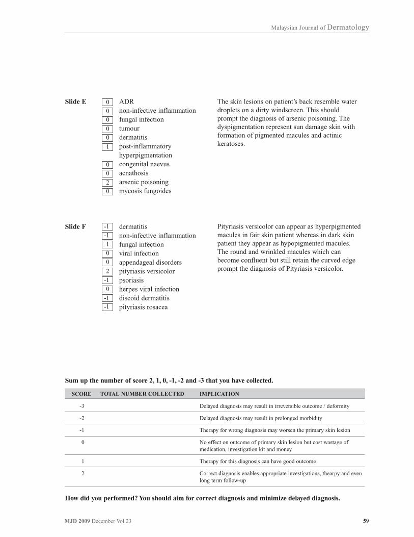
59MJD 2009 December Vol 23
Malaysian Journal of Dermatology
Slide E ADRnon-infective inflammationfungal infectiontumourdermatitispost-inflammatory hyperpigmentationcongenital naevusacnathosisarsenic poisoningmycosis fungoides
Slide F dermatitisnon-infective inflammationfungal infectionviral infectionappendageal disorderspityriasis versicolorpsoriasisherpes viral infectiondiscoid dermatitispityriasis rosacea
Pityriasis versicolor can appear as hyperpigmentedmacules in fair skin patient whereas in dark skinpatient they appear as hypopigmented macules.The round and wrinkled macules which canbecome confluent but still retain the curved edgeprompt the diagnosis of Pityriasis versicolor.
The skin lesions on patient’s back resemble waterdroplets on a dirty windscreen. This shouldprompt the diagnosis of arsenic poisoning. Thedyspigmentation represent sun damage skin withformation of pigmented macules and actinickeratoses.
0
0
0
0
0
1
0
0
20
-1
-1
1
0
0
2
-1
0
-1-1
How did you performed? You should aim for correct diagnosis and minimize delayed diagnosis.
Sum up the number of score 2, 1, 0, -1, -2 and -3 that you have collected.
SCORE
-3
-2
-1
0
1
2
TOTAL NUMBER COLLECTED IMPLICATION
Delayed diagnosis may result in irreversible outcome / deformity
Delayed diagnosis may result in prolonged morbidity
Therapy for wrong diagnosis may worsen the primary skin lesion
No effect on outcome of primary skin lesion but cost wastage of medication, investigation kit and money
Therapy for this diagnosis can have good outcome
Correct diagnosis enables appropriate investigations, thearpy and even long term follow-up

60 MJD 2009 December Vol 23
Malaysian Journal of Dermatology
Answers to Dermatology Care Plan Quiz
Slide A Erysipelas
Nursing problem
Pain in the leg
Swollen leg
Blistering & weeping lesions
Dry scaly skin
Nursing intervention
analgesics
Rest & elevate leg
Wrap lesion with diluted KMNO4 solutions Wet
Use moisturising soap
Expected Outcome
Improve pain score
Reduced calf circumference
Dry wrinkled scaly skin
Moist non-scaly skin
Comment
Stop analgesic when pain has subside
Do not elevate the leg.
Stop wet wraps once swellingand blisters have resolved
Stop moisturizing soap whenskin normalise
Slide B Fixed drug eruptions on the genitals
Nursing problem
Weeping lesion
Adverse drug eruptions (ADR)
Nursing intervention
Wash genitals with astringent solution (normal saline or diluted KMNO4 solutions)
Educate patient to avoid drug by giving allergy card
Expected Outcome
Dry scaly skin
Prevent recurrent ADR
Comment
Stop astringent solution once skin dries up
Inform patient the possibility of cross reactions with sulphurdrugs and black dye if patienthas ADR to bactrim