JOJOURNALURNAL Apoptosis regulation in tetraploid...
12
Apoptosis regulation in tetraploid cancer cells Maria Castedo 1,6 , Arnaud Coquelle 1,6 , Sonia Vivet 1 , Ilio Vitale 1 , Audrey Kauffmann 1 , Philippe Dessen 1 , Marie O Pequignot 1 , Noelia Casares 1 , Alexandre Valent 2 , Shahul Mouhamad 1 , Elise Schmitt 3 , Nazanine Modjtahedi 1 , William Vainchenker 4 , Laurence Zitvogel 5 , Vladimir Lazar 2 , Carmen Garrido 3 and Guido Kroemer 1, * 1 CNRS, UMR8125, Institut Gustave Roussy,Villejuif, France, 2 Unite ´ de Ge ´nomique Fonctionnelle, Institut Gustave Roussy, Villejuif, France, 3 INSERM U-517, Faculty of Medicine and Pharmacy, Dijon, France, 4 INSERM U362, Institut Gustave Roussy, Villejuif, France and 5 INSERM ERM0208, Institut Gustave Roussy, Villejuif, France Tetraploidy can result in cancer-associated aneuploidy. As shown here, freshly generated tetraploid cells arising due to mitotic slippage or failed cytokinesis are prone to undergo Bax-dependent mitochondrial membrane per- meabilization and subsequent apoptosis. Knockout of Bax or overexpression of Bcl-2 facilitated the survival of tetraploid cells at least as efficiently as the p53 or p21 knockout. When tetraploid cells were derived from diploid p53 and Bax-proficient precursors, such cells exhibited an enhanced transcription of p53 target genes. Tetraploid cells exhibited an enhanced rate of spontaneous apoptosis that could be suppressed by inhibition of p53 or by knockdown of proapoptotic p53 target genes such as BBC3/Puma, GADD45A and ferredoxin reductase. Unexpectedly, tetraploid cells were more resistant to DNA damaging agents (cisplatin, oxaliplatin and camp- tothecin) than their diploid counterparts, and this differ- ence disappeared upon inhibition of p53 or knockdown of p53-inducible ribonucleotide reductase. Tetraploid cells were also more resistant against UVC and c-irradiation. These data indicate the existence of p53-dependent altera- tions in apoptosis regulation in tetraploid cells. The EMBO Journal (2006) 25, 2584–2595. doi:10.1038/ sj.emboj.7601127; Published online 4 May 2006 Subject Categories: genome stability & dynamics; differentiation & death Keywords: apoptosis; bax; chemoresistance; mitochondria; p53 Introduction Cancer cells characteristically provide their own growth signals, ignore growth-inhibitory signals, replicate without limit, sustain angiogenesis, invade tissues, proliferate in unnatural locations, and avoid cell death (Hanahan and Weinberg, 2000). Another characteristic of cancer is genomic instability, which is frequently characterized by numeric and structural chromosomal aberrations. Among these hallmarks, the relationship between cell death resistance and genomic instability remains elusive (Zhivotovsky and Kroemer, 2004). Although it is intrinsically difficult to reconstruct the process of aneuploidization by characterizing aneuploid cancer cells (which have survived a Darwinian selection in which most if not all of the intermediates have been lost), there are multiple examples of aneuploid cancer cells generated through asym- metric division of or progressive chromosomal loss from tetraploid precursors (Lin et al, 2001; Castedo et al, 2004; Imkie et al, 2004; Storchova and Pellman, 2004; Watanabe et al, 2004; Fujiwara et al, 2005). This is particularly well documented for cancer development from Barrett’s esopha- gus (Barrett et al, 2003; Maley et al, 2004) and oral leuko- plakia (Sudbo et al, 2001; Sudbo et al, 2004). Tetraploidy can be induced through two distinct processes, namely illicit fusion of two diploid cells (Duelli and Lazebnik, 2003; Ogle et al, 2005) or, more likely, by duplication of the normal chromosomal number in the absence of nuclear and cellular division. This can occur physiologically through endoreplication (DNA replication without mitosis) or endo- mitosis (karyokinesis without cytokinesis) or, pathologically, through mitotic failure (Storchova and Pellman, 2004). For instance, activation of the spindle assembly checkpoint (SAC) usually arrests mitosis during the metaphase until the pro- blems accounting for SAC activation have been solved and mitotic division can ensue correctly. However, a prolonged arrest due to the impossibility to satisfy the SAC leads to checkpoint ‘adaptation’, ‘slippage’ or ‘leakage’ with the con- sequent exit of mitosis and fixation of a tetraploid state (Rieder and Maiato, 2004; Weaver and Cleveland, 2005). Although it has been debated whether a so-called ‘tetra- ploidy checkpoint’ exists (Uetake and Sluder, 2004), it is widely acknowledged that tetraploid cells arrest their cell cycle and that this arrest depends on the tumor suppressor protein p53 (Cross et al, 1995; Yin et al, 1999; Andreassen et al, 2001; Meraldi et al, 2002; Vogel et al, 2004; Sphyris and Harrison, 2005). p53 is a multifunctional transcription factor, which induces multiple target genes. Depending on the cellular context, p53 transactivation can stimulate DNA re- pair (e.g. by induction of p53R2), cell cycle arrest (e.g. by induction of p21) as well as apoptosis (e.g. by induction of proapoptotic members of the Bcl-2 family such as Bax and Puma/BBC3) (Vogelstein et al, 2000; Vousden and Lu, 2002; Yu et al, 2003). Failure of the ‘tetraploidy checkpoint’ due to a missing cell cycle arrest has been accused to be (co)respon- sible for the genomic instability induced by inactivating p53 mutations (Cross et al, 1995; Yin et al, 1999; Andreassen et al, 2001; Vogel et al, 2004; Sphyris and Harrison, 2005). Based on these premises, we decided to re-evaluate the relationship between tetraploidization and p53. We discov- ered that one of the processes that aborts polyploid cells is apoptosis and that suppression of apoptosis by inhibition of Received: 30 September 2005; accepted: 11 April 2006; published online: 4 May 2006 *Corresponding author. CNRS-UMR8125, Institut Gustave Roussy, PR1, 39 rue Camille Desmoulins, 94805 Villejuif, France. Tel.: þ 33 1 42 11 60 46; Fax: þ 33 1 42 11 60 47; E-mail: [email protected] 6 These authors contributed equally to this work The EMBO Journal (2006) 25, 2584–2595 | & 2006 European Molecular Biology Organization | All Rights Reserved 0261-4189/06 www.embojournal.org The EMBO Journal VOL 25 | NO 11 | 2006 & 2006 European Molecular Biology Organization EMBO THE EMBO JOURNAL THE EMBO JOURNAL 2584
-
Upload
nguyenhanh -
Category
Documents
-
view
213 -
download
0
Transcript of JOJOURNALURNAL Apoptosis regulation in tetraploid...

Apoptosis regulation in tetraploid cancer cells
Maria Castedo1,6, Arnaud Coquelle1,6,Sonia Vivet1, Ilio Vitale1, AudreyKauffmann1, Philippe Dessen1,Marie O Pequignot1, Noelia Casares1,Alexandre Valent2, Shahul Mouhamad1,Elise Schmitt3, Nazanine Modjtahedi1,William Vainchenker4, Laurence Zitvogel5,Vladimir Lazar2, Carmen Garrido3
and Guido Kroemer1,*1CNRS, UMR8125, Institut Gustave Roussy,Villejuif, France, 2Unite deGenomique Fonctionnelle, Institut Gustave Roussy, Villejuif, France,3INSERM U-517, Faculty of Medicine and Pharmacy, Dijon, France,4INSERM U362, Institut Gustave Roussy, Villejuif, France and 5INSERMERM0208, Institut Gustave Roussy, Villejuif, France
Tetraploidy can result in cancer-associated aneuploidy.
As shown here, freshly generated tetraploid cells arising
due to mitotic slippage or failed cytokinesis are prone to
undergo Bax-dependent mitochondrial membrane per-
meabilization and subsequent apoptosis. Knockout of
Bax or overexpression of Bcl-2 facilitated the survival of
tetraploid cells at least as efficiently as the p53 or p21
knockout. When tetraploid cells were derived from diploid
p53 and Bax-proficient precursors, such cells exhibited an
enhanced transcription of p53 target genes. Tetraploid
cells exhibited an enhanced rate of spontaneous apoptosis
that could be suppressed by inhibition of p53 or by
knockdown of proapoptotic p53 target genes such as
BBC3/Puma, GADD45A and ferredoxin reductase.
Unexpectedly, tetraploid cells were more resistant to
DNA damaging agents (cisplatin, oxaliplatin and camp-
tothecin) than their diploid counterparts, and this differ-
ence disappeared upon inhibition of p53 or knockdown of
p53-inducible ribonucleotide reductase. Tetraploid cells
were also more resistant against UVC and c-irradiation.These data indicate the existence of p53-dependent altera-
tions in apoptosis regulation in tetraploid cells.
The EMBO Journal (2006) 25, 2584–2595. doi:10.1038/
sj.emboj.7601127; Published online 4 May 2006
Subject Categories: genome stability & dynamics;
differentiation & death
Keywords: apoptosis; bax; chemoresistance; mitochondria; p53
Introduction
Cancer cells characteristically provide their own growth
signals, ignore growth-inhibitory signals, replicate without
limit, sustain angiogenesis, invade tissues, proliferate in
unnatural locations, and avoid cell death (Hanahan and
Weinberg, 2000). Another characteristic of cancer is genomic
instability, which is frequently characterized by numeric and
structural chromosomal aberrations. Among these hallmarks,
the relationship between cell death resistance and genomic
instability remains elusive (Zhivotovsky and Kroemer, 2004).
Although it is intrinsically difficult to reconstruct the process
of aneuploidization by characterizing aneuploid cancer cells
(which have survived a Darwinian selection in which most if
not all of the intermediates have been lost), there are multiple
examples of aneuploid cancer cells generated through asym-
metric division of or progressive chromosomal loss from
tetraploid precursors (Lin et al, 2001; Castedo et al, 2004;
Imkie et al, 2004; Storchova and Pellman, 2004; Watanabe
et al, 2004; Fujiwara et al, 2005). This is particularly well
documented for cancer development from Barrett’s esopha-
gus (Barrett et al, 2003; Maley et al, 2004) and oral leuko-
plakia (Sudbo et al, 2001; Sudbo et al, 2004).
Tetraploidy can be induced through two distinct processes,
namely illicit fusion of two diploid cells (Duelli and Lazebnik,
2003; Ogle et al, 2005) or, more likely, by duplication of the
normal chromosomal number in the absence of nuclear and
cellular division. This can occur physiologically through
endoreplication (DNA replication without mitosis) or endo-
mitosis (karyokinesis without cytokinesis) or, pathologically,
through mitotic failure (Storchova and Pellman, 2004). For
instance, activation of the spindle assembly checkpoint (SAC)
usually arrests mitosis during the metaphase until the pro-
blems accounting for SAC activation have been solved and
mitotic division can ensue correctly. However, a prolonged
arrest due to the impossibility to satisfy the SAC leads to
checkpoint ‘adaptation’, ‘slippage’ or ‘leakage’ with the con-
sequent exit of mitosis and fixation of a tetraploid state
(Rieder and Maiato, 2004; Weaver and Cleveland, 2005).
Although it has been debated whether a so-called ‘tetra-
ploidy checkpoint’ exists (Uetake and Sluder, 2004), it is
widely acknowledged that tetraploid cells arrest their cell
cycle and that this arrest depends on the tumor suppressor
protein p53 (Cross et al, 1995; Yin et al, 1999; Andreassen
et al, 2001; Meraldi et al, 2002; Vogel et al, 2004; Sphyris and
Harrison, 2005). p53 is a multifunctional transcription factor,
which induces multiple target genes. Depending on the
cellular context, p53 transactivation can stimulate DNA re-
pair (e.g. by induction of p53R2), cell cycle arrest (e.g. by
induction of p21) as well as apoptosis (e.g. by induction of
proapoptotic members of the Bcl-2 family such as Bax and
Puma/BBC3) (Vogelstein et al, 2000; Vousden and Lu, 2002;
Yu et al, 2003). Failure of the ‘tetraploidy checkpoint’ due to a
missing cell cycle arrest has been accused to be (co)respon-
sible for the genomic instability induced by inactivating p53
mutations (Cross et al, 1995; Yin et al, 1999; Andreassen
et al, 2001; Vogel et al, 2004; Sphyris and Harrison, 2005).
Based on these premises, we decided to re-evaluate the
relationship between tetraploidization and p53. We discov-
ered that one of the processes that aborts polyploid cells is
apoptosis and that suppression of apoptosis by inhibition ofReceived: 30 September 2005; accepted: 11 April 2006; publishedonline: 4 May 2006
*Corresponding author. CNRS-UMR8125, Institut Gustave Roussy, PR1,39 rue Camille Desmoulins, 94805 Villejuif, France.Tel.: þ 33 1 42 11 60 46; Fax: þ 33 1 42 11 60 47;E-mail: [email protected] authors contributed equally to this work
The EMBO Journal (2006) 25, 2584–2595 | & 2006 European Molecular Biology Organization |All Rights Reserved 0261-4189/06
www.embojournal.org
The EMBO Journal VOL 25 | NO 11 | 2006 &2006 European Molecular Biology Organization
EMBO
THE
EMBOJOURNAL
THE
EMBOJOURNAL
2584

mitochondrial outer membrane permeabilization (MOMP)—
the major apoptotic checkpoint (Green and Kroemer, 2004;
Jiang and Wang, 2004)—is permissive for the survival and
propagation of tetraploid cells. When characterizing the
epigenetic regulation of tetraploid genomes, we found that
tetraploid cells are intrinsically prone to activate p53 and p53
target genes, leading to an increased spontaneous apoptosis
and—paradoxically—to an increased resistance against DNA
damage-induced cell death.
Results
Apoptosis inhibition is permissive for experimental
polyploidization
HCT116 cells exposed to the microtubule poison nocodazole
first activate the spindle checkpoint (and hence arrest in the
metaphase) and then undergo mitotic slippage to become
hyperploid. In response to acute nocodazole treatment (48 h),
both cells with a regular DNA content (2–4N) and cells with
a hyperploid DNA content (44N) tended to lose their mito-
chondrial transmembrane potential (DCm), as detectable
with the DCm-sensitive dye DiOC6(3) (Figure 1A), thus
demonstrating signs of ongoing cell death (Green and
Kroemer, 2004). This cell death was only partially inhibited
by the pan-caspase inhibitor Z-VAD-fmk (but not by t
Z-VDVAD and Z-FA-fmk, which inhibit caspase-2 and cathe-
psin B, respectively).
In this setting, the knockout of p53, Bax or p21 (but less
than that of 14.3.3s) strongly reduced cell death and aug-
mented the percentage of viable, 44N cells elicited by a 48-h
incubation with nocodazole (Figure 1A–C). Z-VAD-fmk failed
to enhance the percentage of polyploid cells (Figure 1B). We
FACS-purified viable (DCmhigh) nocodazole-treated cells with
an B8N DNA content and subjected them to fluorescent
in situ hybridization (FISH) with centromere-specific probes
for chromosomes 9 and 18. These experiments revealed the
presence of four rather than eight FISH-discernible signals per
cell for chromosome 9 and 18 (in 490% of the cases). Thus,
this population was composed by tetraploid cells in G2/M
(before separation of centromeres) rather than by octoploid
cells in G1. The FACS-purified population with an B8N DNA
content was cultured in the absence of nocodazole for 24 h,
and the entry of cells into apoptosis was monitored
(Figure 1D). These results confirmed that de novo formed
tetraploid cells tend to die (as indicated by DCm dissipation)
and that the removal of p53 or Bax from the system greatly
reduces the death of such cells. Of note, in this setting,
nocodazole did not induce a DNA damage response, as
indicated by the absence of DNA damage foci staining for
phosphorylated histone H2AX (Supplementary Figure 1S).
Moreover, the FACS-purified B8N population did not
increase its DNA content upon re-culture, in line with the
FISH data indicating that these cells are in G2/M rather than
in the G1 phase of the cell cycle (Figure 1D). Very similar data
suggesting that p53 and Bax are required for the death of
tetraploid cells were obtained when polyploidization was
induced by cytochalasin D, an inhibitor of cytokinesis
(Supplementary Figure 2S). Thus, p53 and Bax inhibition
are permissive for experimental polyploidization.
Of note, neither p53 nor Bax did influence the expression
level of BubR1 and its nocodazole-induced phosphorylation
(Supplementary Figure 3S), although BubR1 has been sug-
gested to be a major negative regulator of polyploidization
(Shin et al, 2003). Upon nocodazole treatment, Bax-proficient
cells with a 44N DNA content translocated cytochrome c
from mitochondria and activated caspase-3, while Bax-defi-
cient cells retained cytochrome c in mitochondria and failed
to activate caspase-3, as determined by confocal immuno-
fluorescence (Figure 2A). In this system, Z-VAD-fmk only
partially inhibited cytochrome c release, although it fully
blocked caspase-3 activation (Figure 2A), indicating that
MOMP can occur without caspase activation.
The survival of cells with a hyperploid DNA content
(44N) was facilitated by Bax inhibition in several experi-
mental systems. Thus, nocodazole-treated HeLa cells that
overexpress two distinct Bax antagonists (Bcl-2 or the cyto-
megalovirus-derived mitochondrial inhibitor of apoptosis,
vMIA) (Poncet et al, 2004) did not undergo apoptosis when
they accumulated 44N DNA in response to nocodazole
(Figure 2B). Mouse embryonic fibroblasts (MEF) in which
both Bax and its structural homolog Bak were subjected to a
double knockout (DKO) (Wei et al, 2001), also generated
more cells with a 44N DNA content in response to nocoda-
zole than wild-type MEF (Figure 2C). When nocodazole was
replaced by another spindle poison, docetaxel (Figure 2D),
the absence of Bax again facilitated the generation of cells
with 44N DNA.
In short-term experiments (48 h), the p53 and the Bax
knockout were equivalently permissive for DNA accumula-
tion 44N (Figure 1, Supplementary Figure 2S). However,
upon prolonged culture (10 days) of cells transiently exposed
to nocodazole (2 days), Bax-negative HCT116 cells had
generated more polyploid cells than p53-negative cells, and
these Bax-negative polyploid cells were undergoing less
spontaneous death than p53-negative polyploid cells
(Figure 3A). Note that at this time point (10 days), diploid
cells that had been exposed transiently to nocodazole did not
undergo a higher rate of apoptosis than untreated control
cells, as determined by FACS purification of the cells with
a 2N DNA content and re-culture of the cells for 24 h
(Figure 3B). The fact that Bax KO protected 44N cells from
spontaneous apoptosis more efficiently than the p53 KO
(Figure 3A), suggested the existence of a p53-independent
Bax activation pathway. Accordingly, a substantial fraction of
p53-KO cells with a 44N DNA content demonstrated posi-
tivity for activated Bax (detectable with an antibody specific
for the N-terminus, which is exposed upon activation and
mitochondrial membrane insertion of Bax) as late as 10 days
after nocodazole treatment (Figure 3C and D). In contrast, the
percentage (but not the absolute number) of tetraploid cells
exhibiting the activating phosphorylation of p53 on serine 15
was significantly reduced in Bax KO cells as compared to
wild-type controls, 10 days after nocodazole treatment
(Figure 3C and D), presumably as a result of the outgrowth
of viable tetraploid cells, Altogether, these data indicate that
cells with a 44N DNA content are usually aborted by
apoptosis executed through a Bax-dependent mitochondrial
pathway.
Establishment of tetraploid cell lines and
characterization of their transcriptome
Although nocodazole or cytochalasin D-treated wild-type
HCT116 cells massively succumbed to apoptosis upon accu-
mulation of a 44N DNA content (Figures 1–3), we could
Apoptosis and tetraploidyM Castedo et al
&2006 European Molecular Biology Organization The EMBO Journal VOL 25 | NO 11 | 2006 2585

A
WT
WT+Z-V
AD
WT+Z-V
DVAD
WT+Z-F
A
Bax K
O
p53 K
O
p21 K
O
14.3.
3σ K
OW
T
WT+Z-V
AD
WT+Z-V
DVAD
WT+Z-F
A
Bax K
O
p53 K
O
p21 K
O
14.3.
3σ K
O0
20
40
60ControlNocodazole
0
10
20
30 DiOC6(3)low
PIhigh
% p
olyp
loid
y (>
4N)
% o
f ce
lls
CB
*
*
*
* **
*
Nocodazole
25 12 5 6
0 1000
p21 KO 14.3.3σ KO
0 1000
WT FCCP
0 1000
WT Z-VDVAD
0 0 10001000
WT FCCP
DNA content (Hoechst 33342)
28 5 1493100
101
102
103
104
100
101
102
103
1042N 4N
ΔΨm
(D
iOC
6(3)
)
WT WT WT Z-VAD p53 KO Bax KO
15±4% 52±11% 13±3%
2N 4N
Purification and culture for 24 h
8N 8N8N
WTWT BaxKO p53KO
Nocodazole
23±4%
D
0 10000 1000 0 1000 0 1000
DNA content (Hoechst 33342)
100
101
102
103
104
100
101
102
103
104
ΔΨm
(D
iOC
6(3)
)
Figure 1 Acute mortality of polyploid cells modulated by p53 and Bax. Wild-type (WT) HCT116 cells or cells manipulated to lose expression ofp53, p21, 14.3.3s or Bax were cultured in the absence or presence of nocodazole, alone or combined with the protonophore FCCP (whichdissipates the DCm) or the protease inhibitors Z-VAD-fmk, Z-VDVAD-CHO or Z-FA-fmk for 48 h, followed by three-color staining with Hoechst33342 (which measures DNA content), DiOC6(3) (which measures DCm) and propidium iodine (PI, a vital dye that incorporates only into deadcells) and cytofluorometric evaluation. (A) Representative pictograms showing the polyploid cells (DNA content 44N) with a normal DCm
(upper window), as well as polyploid cells with reduced DCm (lower window). Numbers refer to the percentage of polyploid DiOC6(3)low cells
(considering 100% as the sum of all polyploid cells). (B) Percentage of polyploid cells among the total population of cells treated as in (A).(C) Frequency of dying (DiOC6(3)
low) and dead (PIhigh) cells among the polyploid population elicited by nocodazole, as determined by FACSanalysis. Values are X7s.e.m. of five independent experiments. Asterisks refer to significant effects (paired Student’s t-test, Po0.001). (D) Fateof viable polyploid cells elicited by nocodazole. Cells with the indicated genotype were cultured for 30 h in the absence or presence ofnocodazole and then stained with Hoechst 33342 and DiOC6(3), followed by FACS purification of the euploid (2N) or polyploidy (44N)DiOC6(3)
high cells (as indicated by the windows in the upper panels). These purified cells were then cultured for 24 h and reanalyzed afterrestaining with Hoechst 33342 and DiOC6(3), as indicated in the lower panels. Numbers indicate the percentage of cells with a DiOC6(3)
low
phenotype (X7s.e.m., n¼ 3).
Apoptosis and tetraploidyM Castedo et al
The EMBO Journal VOL 25 | NO 11 | 2006 &2006 European Molecular Biology Organization2586

isolate a few clones of tetraploid HCT116 wild-type cells
(Figure 4A). Such cells arose either after treatment with
nocodazole (clones N1 and N2) or cytochalasin D (clones
C1 and C2) at a frequency of less than 1 in 10 000 and were
found to express unmutated, normal Bax levels (not shown).
In contrast, we were unable to generate stable clones with a
higher-order polyploidy (e.g. octoploidy), irrespective of the
HCT116 genotype investigated. We observed that another
diploid colon carcinoma cell line, RKO, spontaneously con-
tained B5% of tetraploid cells. Limiting dilution-based clon-
ing led to the isolation of several stable tetraploid clones (T1–
7) and several diploid clones (D1–D7, which after several
passages again contained B5% of tetraploid cells). One
of these latter clones (D5) was transfected with a histone
H2B-GFP fusion construct (which allows to visualize
chromosomes and to measure chromatin content) and then
FACS-separated to generate H2B-GFP-positive tetraploid (TA,
TB, TC, TD) and diploid (DW, DY, DX, DZ) subclones
A
B
C
D
Neo0
10
20
15
5
NocodazoleControl
Neo Bcl2 vMIA
% p
olyp
loid
y
% o
f ce
lls
0
0
10
20
30
Bcl2 vMIA
% p
olyp
loid
y
0
10
20
30
40
50
WT DKO
DiOC6(3)low
PIhigh
DiOC6(3)low
PIhigh
DiOC6(3)low
PIhigh
40
20
60
80%
of
cells
NocodazoleControl
% p
olyp
loid
y
0
10
20
30
40
50
0
10
30
20
DocetaxelControl
% o
f ce
lls
WT p53KOBaxKO WT p53KOBaxKO
WT DKO
WTControl
WT Bax KO
% o
f ce
lls
40
0
Released Cyt-cCasp-3a
WT Bax KO WT Bax KOControl Nocodazole
10
20
30
Cyt-c Casp-3a Hoechst 10 μm
Nocodazole50
WT Bax KONocodazole+ Z-VAD
Figure 2 Mitochondrial cell death regulators and the fate of polyploid cells. (A) Evidence for MOMP in nocodazole-treated cells. Untreatedcontrol or nocodazole treated HCT116 cells (either wild type or Bax KO) were treated for 48 h with nocodazole alone or in combination withZ-VAD-fmk, followed by confocal immunofluorescence staining with antibodies specific for cytochrome c (Cyt c) and proteolytically activecaspase-3 (Casp-3a). The percentage of cells exhibiting diffuse Cyt c staining or positivity for Casp-3a was determined among the entirepopulation in controls and among nocodazole treated cells that exhibited a larger nucleus than controls, and that were considered as polyploid(X7s.e.m., n¼ 3). (B) Effect of Bcl-2 overexpression and vMIA expression on the fate of polyploid cells. HeLa cells transfected with Bcl-2 orvMIAwere treated for 48 h with nocodazole. Then, the frequency (X7s.e.m., n¼ 4) of polyploid cells among the total population (left panels)or that of dying (DiOC6(3)
low) and dead (PIhigh) cells among the polyploid population elicited by nocodazole (right panels) was determined asin Figure 1A–C. (C) Comparison of wild-type or Bax�/�Bak�/� DKO MEF in their response to nocodazole. Cells were treated and monitored asin (B). (D) Effect of the Bax and p53 knockout on docetaxel-induced polyploidization. HCT116 cells with the indicated genotype were treatedfor 48 h with 100 nM docetaxel and the frequency (X7s.e.m., n¼ 4) of polyploid cells among the total population (left panels) or that of dying(DiOC6(3)
low) and dead (PIhigh) cells among polyploid cells (right panel) was determined.
Apoptosis and tetraploidyM Castedo et al
&2006 European Molecular Biology Organization The EMBO Journal VOL 25 | NO 11 | 2006 2587

(Figure 4B). Again, there was no consistent difference in the
expression level of Bax, Bcl-2, and BubR1 between diploid
and tetraploid cells (Figure 4C).
Karyograms and comparative genomic hybridization
(CGH) performed on representative RKO clones indicate
that tetraploid cells contained an exact duplication of the
diploid set of chromosomes, without any CGH-detectable
duplication or deletion (Supplementary Figure 4S). We then
determined whether tetraploidization would induce epige-
netic changes in the transcriptome, by performing microarray
p53S15Nucleus Bax
A
B
WT BaxKO Noco p53KO Noco WT Noco
10 μm
0
20
40
60 Active bax P53S15P
WT Bax KO
p53KO
WT Bax KO
p53KO
Noco – – – + + +
% o
f ce
lls
DiOC6(3)low
PIhigh
60
40
20
0
% o
f ce
lls
Num
ber
of c
ells
(a.u
.) 4
21
WT BaxKO
% p
olyp
loid
y
p53KO
p21KO
14.3.3σKO
0
3
56
10
20
30
40
50
0WT Bax
KOp53KO
p21KO
14.3.3σKO
C
D
100101102103104
100101102103104
100101102103104
100
100
101
101
102
102
103
103
104
104100101102103104 100101102103104100101102103104 100101102103104
WTWT BaxKONocodazole
DNA content (Hoechst 33342)
ΔΨm
(D
iOC
6(3)
) ΔΨ
m (
DiO
C6(
3))
Plasma membrane permeabilization (PI)
2N 8N 2N 8N2N
10±2% 11±3% 9±2%27±4% 8±2%
0 10000 10000 1000
8N2N8N2N2N
Purification and culture for 24 h
Figure 3 Long-term effects of Bax and p53 on the survival of polyploid cells. (A) Chronic mortality of polyploid cells modulated by p53 andBax. HCT116 cells with the indicated genotype were treated for 48 h with nocodazole, then washed extensively (5� ) and cultured duringfurther 8 days in normal culture medium, followed by determination of the frequency (X7s.e.m., n¼ 5) of live, dying and dead polyploid cellsas in Figure 1A–C. Note that less than 5% of cells with an B2N DNA content were dying (DiOC6(3)
low) or dead (PIhigh) cells and that dying ordead cells were almost exclusively found in the polyploid (44N) population. (B) Fate of viable polyploidy cells induced by nocodazole. HCT116WTor HCT116 BaxKO cells were transiently exposed to nocodazole for 2 days and then cultured for 8 days without nocodazole, stained withDiOC6(3) versus Hoechst 33342 followed by FACS purification of cells with 2N or 8N DNA content, cultured for 24 h and relabelled withDiOC6(3) and PI to determine the frequency of dying and dead cells. (C, D) p53-dependent and -independent Bax activation in a fraction ofpolyploid cells. HCT116 cells (WT, Bax KO or p53 KO) were cultured for 48 h in nocodazole and then for 8 days without nocodazole, followedby immunofluorescence staining of activated Bax and S15-phosphorylated p53. Representative micrographs are shown in (C), and data arequantified in (D). The percentage of cells exhibiting a punctate cytoplasmic staining for activated Bax or a nuclear staining for S15-phosphorylated p53 was assessed among the entire population in untreated controls and among nocodazole-exposed, polyploid cells(X7s.e.m., n¼ 3).
Apoptosis and tetraploidyM Castedo et al
The EMBO Journal VOL 25 | NO 11 | 2006 &2006 European Molecular Biology Organization2588

comparisons of four diploid and four tetraploid HCT116
clones generated by treatment with either nocodazole or
cytochalasin D (Supplementary Figure 5S). Similarly, we
compared the transcriptome of four diploid and four tetra-
ploid H2B-GFP-positive RKO subclones (Supplementary
Figure 6S). Surprisingly, there were little changes in the
transcriptome of tetraploid HCT116 or RKO cells. The com-
bined comparison of the 16 diploid and tetraploid clones led
to a short list of genes that were consistently up- or down-
regulated by more than 20% as a function of ploidy. The gene
that was most downregulated in tetraploid cells was the
winged helix transcription factor FOXD1 (by 40%) and
the two genes that were most upregulated were ZNF395,
the zinc-finger protein 395 (by 40%), and RRM2B, the gene
encoding p53R2, the p53-inducible ribonucleotide reductase-
2 (by 30%). These results were confirmed by independent
quantitative PCR analyses (not shown). Although the differ-
ences were small (20–25%), we found that three previously
identified p53-inducible transcripts were significantly
(Po0.0001, ANOVA test) incremented in tetraploid cells:
BBC3 (which encodes the proapoptotic BH3 only protein
Puma) (Nakano and Vousden, 2001; Yu et al, 2001),
GADD45A (a cell cycle regulator), and FDXR (which encodes
the mitochondrion-localized ferredoxin reductase) (Hwang
et al, 2001) (Figure 4D). Intriguingly, 12 out of the 29 genes
(41%) contained in the short list of ploidy regulated genes
were found to contain a p53 consensus binding site or a
putative p53-responsive tetrarepeat motif in the promoter
region (Figure 4D). These data point to a major dysregulation
of the p53 system in tetraploid cells.
100
0 10000
DiploidC
ount
s
A
DNA content
BRKO95% Diploid 5% Tetraploid
D1 D2 D3 D4 D5 D6 D7 T1 T2 T3 T4 T5 T6 T7
DXDYDW DZ TA TB TC TD
GFP-H2B transfectionFACS separationLimiting dilution
Bax
Bcl2
BubR1
GAPDH
TA TC TDTBDW DY DZDX
Diploid TetraploidC
Tetraploid
TA
D
Fold changeThumbnail view
–2.00000 2.000000
0.68
0.35
0.03
–0.3
–0.62
– 0.7
– 0.36
– 0.02
0.32
0.66
Figure 4 Establishment and characterization of tetraploid cell lines. (A) Comparison of the DNA content of a representative tetraploid HCT116cell line and of its diploid control. (B) Genealogy of diploid and tetraploid RKO clones. The wild-type control cell line containing B5%tetraploid cells was subcloned by limiting dilution into diploid and tetraploid clones; The D5 clone was transfected with a cDNA encoding anH2B-GFP chimera, FACS-separated into subsets of cells enriched in a diploid or tetraploid DNA content, and again subcloned to generate diploidand tetraploid H2B-GFP-expressing clones. Representative fluorescence micrographs of such cells are shown (the width of the squares is25mm). (C) Western blot determination of the expression levels of Bax, Bcl-2, p53 and BubR1, with GAPDH as a loading control. (D) Microarrayanalyses of the transcriptome of diploid versus tetraploid cells. The clones HCT116 C1 and C2 and N1 and N2 are tetraploid, generated uponcytochalasin D or nocodazole treatment, respectively. Clones Co1–4 are diploid. The RKO clones are described in (B). The overlap between thegenes coming from an ANOVA test with a threshold P-value of 10�3 on the HCT116 samples (Supplementary Figure 5S) and the same test on theRKO samples (Supplementary Figure 6S) led to the selection of 29 genes. These genes were subjected to a hierarchical cluster analysis usinga calculation based on cosine correlation and the agglomerative method of the average link. Each row represents the combination of twodye-swap experimental samples and each column represents a single accession number. *marks genes that carry at least one consensus p53-binding consensus sequence (RRRCWWGYYY), within the 2000 nucleotides upstream of the transcription start. # marks genes the promoter ofwhich contains putative p53 binding sites, as determined by another procedure bases on a sliding profile of four matrixes of the sequenceRRRCA/TT/AGYYY, as detailed in the Materials and methods.
Apoptosis and tetraploidyM Castedo et al
&2006 European Molecular Biology Organization The EMBO Journal VOL 25 | NO 11 | 2006 2589

Enhanced spontaneous apoptosis of tetraploid cells
As compared to their diploid precursors, a higher percentage
of tetraploid cells exhibited the activating phosphorylation of
p53 on serine 15 (p53S15P) or serine 46 (p53S46P), detect-
able by immunofluorescence staining with antibodies specific
for p53 phospho-neoepitopes. This was observed in RKO
(Figure 5A–C) and HCT116 cells (not shown). In contrast,
we found no difference in the phosphorylation of histone
H2AX that would be indicative of DNA damage (not shown).
p53 phosphorylation was mainly detected in tetraploid cells
that were terminating cytokinesis (Figure 5B), suggesting that
p53 activation resulted from deficient mitoses. The increased
phosphorylation of p53 on serine 15 was accompanied by
a general increase in p53 expression levels, as determined by
immunoblot (Figure 5D). An increased percentage of tetra-
ploid cells died spontaneously from apoptosis, as indicated
by the DCm loss (Figure 5E), chromatin condensation, and
incorporation of the vital dye PI (not shown). The caspase
inhibitor Z-VAD-fmk did not affect the spontaneous mortality
of tetraploid cells. However, there was an effect of cyclic
pifithrin-a, which palliated the overmortality of tetraploid
clones down to the diploid level both in RKO (Figure 5E)
and in HCT116 cells (not shown). siRNA-mediated knock-
down of Puma and of the Puma target Bax also reduced the
spontaneous apoptosis of tetraploid cells. Similarly, knock-
down of FDXR and GADD45A reduced the spontaneous
apoptosis of tetraploid cells (Figure 5F), indicating that these
p53 target genes contribute to the death of tetraploid cells.
Selective resistance of tetraploid cells against
DNA-damaging agents
Since p53 controls the cell death induced by DNA damage
(Vogelstein et al, 2000; Vousden and Lu, 2002), we evaluated
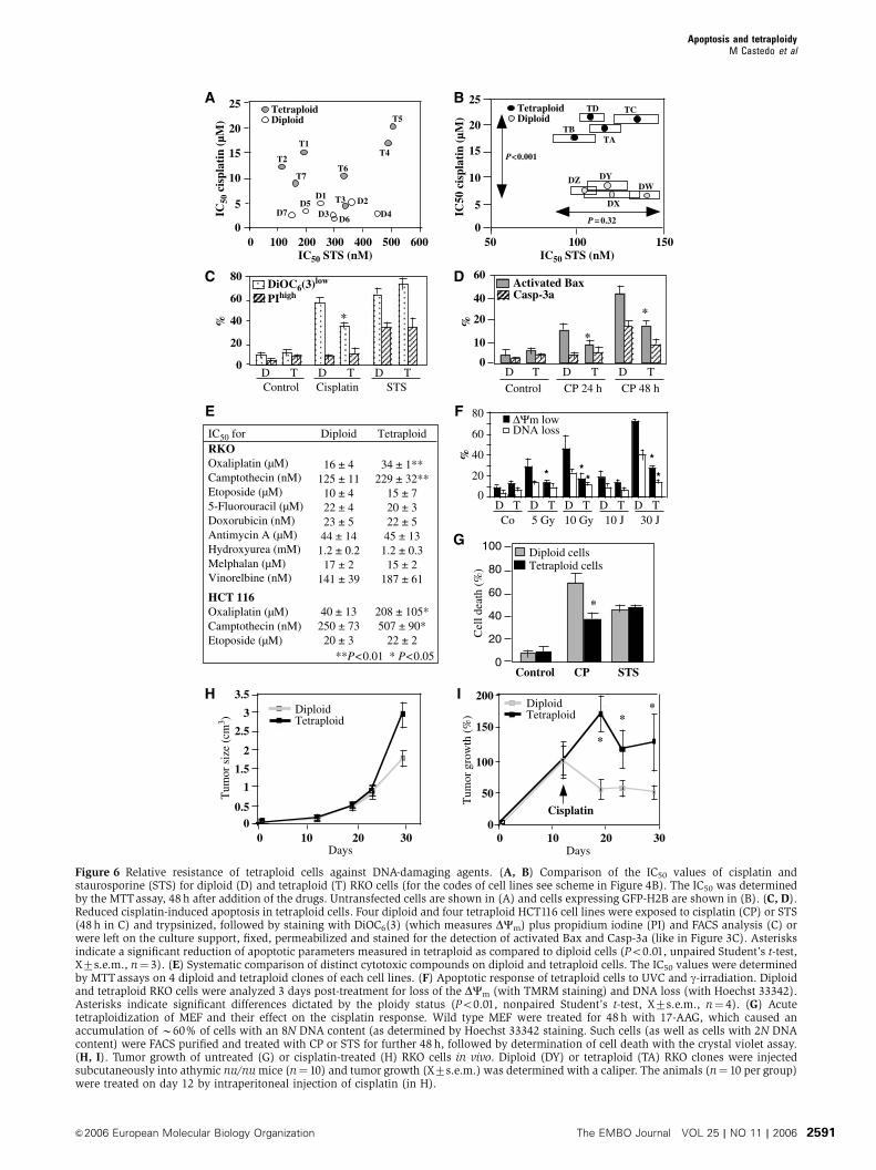
the apoptotic response of diploid and tetraploid cells. While
there was no difference in the apoptotic response to the pan-
tyrosine kinase inhibitor staurosporine (STS), tetraploid RKO
clones without (Figure 6A) or with H2B-GFP (Figure 6B)
as well as tetraploid HCT116 clones (Figure 6C) died less in
response to cisplatin than their diploid counterparts. When
exposed to cisplatin, tetraploid cells manifested a less pro-
nounced DCm dissipation (Figure 6C), Bax activation and
proteolytic caspase-3 maturation than their diploid controls
(Figure 6D). This relative resistance of tetraploid cells was
also observed when cisplatin was replaced by oxaliplatin
(which, as cisplatin, is a platinum compound) or the topo-
isomerase-1 inhibitor camptothecin. In contrast, there was no
ploidy-specific difference in the apoptotic response to a
number of other toxic compounds including staurosporine
(Figure 6A–C), etoposide, doxorubicin and hydroxyurea
(Figure 6E). Tetraploid RKO and HCT116 cells were also
more resistant against apoptosis induced by UVC or g-irradia-tion (Figure 6F). The relative cisplatin resistance was found
again in FACS-purified MEF with an 8N DNA content. Such
hyperploid cells were obtained by short-term (2 days) culture
with 17-allylamino-17-demethoxygeldanamycin, and com-
pared to FACS-purified diploid controls cells that had under-
gone an identical treatment (Figure 6G). Thus, even acute
polyploidization caused cisplatin resistance. When injected
into immunodeficient mice, both diploid and tetraploid RKO
cells generated tumors (Figure 6H). Tetraploid tumors were
relatively more resistant to cisplatin chemotherapy than
diploid cancers (Figure 6I). Hence, the increased cisplatin
resistance of tetraploid cells could be confirmed in vivo.
To explore the molecular mechanisms of drug resistance,
we investigated the cisplatin response in diploid and
tetraploid cells that were cocultured. Diploid RKO clones
0
1
2
3
4
5
Control Z-VAD Pifithrin 10
TM
RM
low
(%
) TetraploidDiploid
Pifithrin 30
TetraploidDiploid
0
10
20
Co Bax FDXR GADD45 Puma
TM
RM
low
(%
)
E F
siRNA: p53R2
** **** **
**
*P<0.05**P<0.005
01234567
% o
f ce
lls
p53S15Pp53S46P
2N 4N
4N2N
20 μM
p53S15P Hoechst
**
**
A
β-tubulin P53S15P
C
p53S15P
p53
GAPDH
DDiploid Tetraploid
Hoechst
B
Figure 5 Constitutive p53 activation in tetraploid cells. (A, B) Staining of cells with an antibody recognizing p53 phophorylated on serine 15(p53S15P). Representative images are shown for diploid and tetraploid RKO populations in (A). In (B), a representative tetraploid cells positivefor p53S15P and counterstained with b-tubulin is shown. (C) Frequency of cells with positive nuclear staining for p53S15P or p53phophorylated on serine 46 (p53S46P) is shown for diploid (n¼ 4) and tetraploid (n¼ 4) RKO cells. Asterisks indicate significant (Po0.01)differences between diploid and tetraploid cells (unpaired Student’s t-test). (D) Immunoblot confirmation of the p53 activation. Representativediploid (DW, DY) and tetraploid (TA, TD) cells were subjected to immunoblot detection of p53, p53S15P and GAPDH. (E) Spontaneous celldeath in RKO cells and its suppression by cyclic pifithrin-a. Cells were cultured for 2 days in the presence of Z-VAD-fmk or pifithrin-a, and thefrequency of dying cells was measured by staining with TMRM. Asterisks indicate significant differences determined by ploidy (Po0.01,Student’s t-test, X7s.e.m., n¼ 4). (F) Effect of siRNAs on the spontaneous death of tetraploid RKO cells. Cells were transfected with siRNAsspecific for emerin (negative control, Co), ferredoxin reductase (FDXR), GADD45A, Puma, Bax or p53R2 and 72 h later the spontaneousmortality of cells was assessed as in (E). Asterisks in (E) and (F) indicate significant inhibitory effects as compared to control cells (untreated in(E) and Emerin siRNA-transfected in (F)).
Apoptosis and tetraploidyM Castedo et al
The EMBO Journal VOL 25 | NO 11 | 2006 &2006 European Molecular Biology Organization2590
C
A
E
G
B
F
H
D
100 300 600
IC50
cis
plat
in (
μM)
15
25
20
10
5
0200 400 5000
TetraploidDiploid
IC50 STS (nM)
T1
T2T6
T7
T4
T5
T3 D2D1
D7D5
D3D6
D4
0
50
100
150
200
TetraploidDiploid
0 10 20 30
Tum
or g
row
th (
%)
Days
Cisplatin
*
**
15
50 100 150
IC50
cis
plat
in (
μM)
25
20
10
5
0
P<0.001
P = 0.32
IC50 STS (nM)
TetraploidDiploid
TB
TD
TA
TC
DWDYDZ
DX
0 10 20 30
0.5
1
1.5
2
2.5
3
3.5
TetraploidDiploid
Tum
or s
ize
(cm
3 )
Days
0
0
20
40
60
80
100
Cel
l dea
th (
%) Tetraploid cells
Control CP STS
*
Diploid cells
I
DNA loss
***
ΔΨm low
5 Gy 10 Gy 10 J 30 J
%
Co
0
20
40
60
80
* *
D T D T D T D T D T
40
20
10
0
60Activated BaxCasp-3a
D D T D T
Control CP 24 h CP 48 h
*
*
%
T
60
40%
D D T D T
*
0
20
80
Control Cisplatin STS
DiOC6(3)low
PIhigh
T
IC50 for DiploidRKOOxaliplatin (μM)Camptothecin (nM)Etoposide (μM)5-Fluorouracil (μM)Doxorubicin (nM)Antimycin A (μM)Hydroxyurea (mM)Melphalan (μM)Vinorelbine (nM)
HCT 116Oxaliplatin (μM)Camptothecin (nM)Etoposide (μM)
**P<0.01 * P<0.05
34 ± 1**229 ± 32**
15 ± 720 ± 322 ± 545 ± 131.2 ± 0.315 ± 2
187 ± 61
16 ± 4125 ± 1110 ± 422 ± 423 ± 544 ± 141.2 ± 0.217 ± 2
141 ± 39
40 ± 13250 ± 7320 ± 3
208 ± 105*507 ± 90*
22 ± 2
Tetraploid
Figure 6 Relative resistance of tetraploid cells against DNA-damaging agents. (A, B) Comparison of the IC50 values of cisplatin andstaurosporine (STS) for diploid (D) and tetraploid (T) RKO cells (for the codes of cell lines see scheme in Figure 4B). The IC50 was determinedby the MTTassay, 48 h after addition of the drugs. Untransfected cells are shown in (A) and cells expressing GFP-H2B are shown in (B). (C, D).Reduced cisplatin-induced apoptosis in tetraploid cells. Four diploid and four tetraploid HCT116 cell lines were exposed to cisplatin (CP) or STS(48h in C) and trypsinized, followed by staining with DiOC6(3) (which measures DCm) plus propidium iodine (PI) and FACS analysis (C) orwere left on the culture support, fixed, permeabilized and stained for the detection of activated Bax and Casp-3a (like in Figure 3C). Asterisksindicate a significant reduction of apoptotic parameters measured in tetraploid as compared to diploid cells (Po0.01, unpaired Student’s t-test,X7s.e.m., n¼ 3). (E) Systematic comparison of distinct cytotoxic compounds on diploid and tetraploid cells. The IC50 values were determinedby MTTassays on 4 diploid and tetraploid clones of each cell lines. (F) Apoptotic response of tetraploid cells to UVC and g-irradiation. Diploidand tetraploid RKO cells were analyzed 3 days post-treatment for loss of the DCm (with TMRM staining) and DNA loss (with Hoechst 33342).Asterisks indicate significant differences dictated by the ploidy status (Po0.01, nonpaired Student’s t-test, X7s.e.m., n¼ 4). (G) Acutetetraploidization of MEF and their effect on the cisplatin response. Wild type MEF were treated for 48 h with 17-AAG, which caused anaccumulation of B60% of cells with an 8N DNA content (as determined by Hoechst 33342 staining. Such cells (as well as cells with 2N DNAcontent) were FACS purified and treated with CP or STS for further 48 h, followed by determination of cell death with the crystal violet assay.(H, I). Tumor growth of untreated (G) or cisplatin-treated (H) RKO cells in vivo. Diploid (DY) or tetraploid (TA) RKO clones were injectedsubcutaneously into athymic nu/numice (n¼ 10) and tumor growth (X7s.e.m.) was determined with a caliper. The animals (n¼ 10 per group)were treated on day 12 by intraperitoneal injection of cisplatin (in H).
Apoptosis and tetraploidyM Castedo et al
&2006 European Molecular Biology Organization The EMBO Journal VOL 25 | NO 11 | 2006 2591

(H2B-GFP positive) and tetraploid RKO clones (H2B-GFP
negative) were admixed at different ratios and then cocul-
tured during several days in the absence or presence of
cytotoxic drugs. Cultures initiated at a 50:50 ratio (diploid:
tetraploid) shifted to a predominance of diploidy after 5 days
(Figure 7A), in accord with the observation that the mean
duplication time of diploid cells (23.2 h) was shorter than that
of tetraploid cells (26.4 h), and that tetraploid cells exhibit a
higher rate of spontaneous apoptosis than diploid controls
(see above). This shift towards a predominance of diploid
cells, as observed in untreated cultures, was not affected by
STS. However, addition of cisplatin favored the relative out-
growth of tetraploid cells, in line with the interpretation that
tetraploid cells are fitter than diploid cells in conditions of
DNA damage (Figure 7A and B). Similar results were ob-
tained for mixed diploid/tetraploid cultures of HCT116 clones
0
10
20
30
%
Day 0Co Co CP Campt
0
5
10
STS
%
Day 5
0
20
40
60
80
100
%
TetraploidDiploid
Cisplatin
100 102 104 100 102 104 100 102 104
GFP
100
101
102
103
104
TM
RM
STSControl
36 29
15 20
19 51
2281
7524
% o
f te
trap
loid
cel
ls
20
40
60
80Pifithrin 10
100Co
20 5025
Pifithrin 30
10 (μM)0Cisplatin Oxaliplatin
**
**
0
20
40
60
Diploid Co siRNA
205 100Cisplatin (μM)
Tetraploid Co siRNADiploid p53R2 siRNATetraploid p53R2 siRNA
p53R2GAPDH
Co p53R2siRNA for
*
*
80
% T
MR
Mlo
w%
TM
RM
low
Co Bax FDXR GADD45 PumasiRNA:0
20
40
60Diploid Tetraploid
+Cisplatin
** * *
A
B C
D
E
Figure 7 Mechanisms of cisplatin resistance in tetraploid cells. (A, B) Assay for the simultaneous detection of diploid and tetraploid cells dyingwith cisplatin. The GFP-H2B-expressing diploid RKO clone DY was mixed with the GFP-negative tetraploid RKO clone T1 at a 1:1 ratio (in A),followed by culture for 5 days in the absence of cytotoxic drugs (control) or in the presence of cisplatin or staurosporine (STS), and then stainedwith TMRM to determine the proportion of dying diploid and tetraploid cells. Note the increase in the relative frequency of diploid cells inuntreated control cultures, irrespective of the initial diploid: tetraploid (D:T) ratio (1:1 in (A) or 9:1, 1:1 or 1:9 in (B)), while cisplatin increasesthe percentage of tetraploid cells in the cultures. Similar results indicating an enhanced resistance of tetraploid cells against cisplatin wereobtained when tetraploid H2B-GFP-expressing clones were co-cultured with diploid GFP-negative clones (not shown). (C) Effect of cyclicpifithrin-a (Pif) on the D:Tratio. Cells (inital ratio 1:1) were cultured in the absence of presence of cisplatin, oxaliplatin and/or cyclic pifithrin-a(doses in mg), and the D:Tratio of the cultures was determined after 5 days. (D) Effect of small-interfering RNA designed to downregulate p53R2on the death of representative diploid (DY, D5) and tetraploid RKO (TA, T1) clones. Cells were transfected with siRNAs specific for p53R2(440)or emerin, and 36 h later the cells were treated with cisplatin for 2 days, and the frequency of dying (TMRMlow) cells was measured. Theimmunoblot demonstrates the efficacy of p53R2 440 on p53R2 protein expression, as tested on a pool of RKO-derived clones. Similar functionalresults were obtained for a second p53R2-specific siRNA (UTR, not shown). (E) Effect of other p53 target genes on the cisplatin response.Specific siRNAs were used to downmodulate emerin (Co), FDXR, GADD45A, BAX or Puma, and the frequence of dying (TMRMlow) cells(X7s.e.m., n¼ 3) was determined after addition of 20 mM cisplatin for 2 days. Note that the values of spontaneous apoptosis (obtained in theabsence of cisplatin) are contained in Figure 5F. Asterisks in (C–D) indicate significant (Po0.01) apoptosis-inhibitory effects.
Apoptosis and tetraploidyM Castedo et al
The EMBO Journal VOL 25 | NO 11 | 2006 &2006 European Molecular Biology Organization2592

in which either of the two clones was labeled with the stable
fluorescence marker CMFDA (not shown) or when cisplatin
was replaced by oxaliplatin (Figure 7C). Addition of the p53
inhibitor cyclic pifithrin-a prevented the cisplatin- or oxali-
platin-induced shift in favor of tetraploid cells (Figure 7C),
suggesting that p53 target genes determine the differential
behavior of diploid versus tetraploid cells. One of the four p53
target genes overexpressed in tetraploid cells, p53R2, has
previously been shown to confer cisplatin resistance (Lin
et al, 2004; Okumura et al, 2005). We therefore knocked
down p53R2 expression by siRNA. Down modulation of
p53R2 sensitized tetraploid cells to cisplatin-induced cell
death and annihilated the difference in the cisplatin
response between diploid and tetraploid cells (Figure 7D).
As expected, downmodulation of Bax, Puma, GADD45A and
FDXR reduced cisplatin-induced apoptosis, both in diploid
and in tetraploid cells (Figure 7E). Altogether, these
data indicate that subtle differences in the p53 transcriptome
and in particular in p53R2 expression may explain the
relative resistance of tetraploid cells against DNA-damaging
agents.
Discussion
Based on the data shown in this paper, it appears plausible
that disabled apoptosis could be permissive for the genera-
tion of tetraploid cells, while tetraploidization has an intrinsic
effect on apoptosis regulation. Acute tetraploidization caused
activation of p53 (see phosphorylation of p53 in Figure 3D),
in accord with the literature (Cross et al, 1995; Yin et al, 1999;
Andreassen et al, 2001; Du and Hannon, 2004; Vogel et al,
2004; Sphyris and Harrison, 2005). Acute tetraploidization
also resulted in the (partially p53-dependent and partially
p53-independent, Figure 3D) activation of Bax, which adopts
its proapoptotic conformation (Figures 2A and 3D) and
triggers MOMP with cytochrome c release and consequent
caspase activation (Figure 2A). MOMP is likely to lead to
caspase activation in an amplification loop in which caspase
activation, in turn, feeds back on MOMP (Adams, 2003;
Danial and Korsmeyer, 2004), explaining the partial inhibi-
tory effects of caspase inhibition (Figures 1A–C and 2A).
Since the knockout of Bax or the overexpression of Bax
antagonists (such as Bcl-2 or vMIA) blocks cell death and
increases the survival of tetraploid cells at least as efficiently
as does the knockout of p53 (and that of p21) (Figures 1–3), it
appears that one mechanisms that suppresses nonphysiolo-
gical tetraploidization is the apoptotis. In this context, apop-
tosis would be as a mechanism of quality control (in accord
with the rule ‘better dead than wrong’) (Thompson, 1995)
which, when perturbed, is permissive for the generation of
tetraploid cells.
Although disabled apoptosis enhances the probability of
stable tetraploidization, we were able to derive tetraploid
clones from diploid parental lines that were apoptosis-com-
petent (Figure 4). These tetraploid clones apparently main-
tained the capacity to undergo apoptosis (and hence
remained fully susceptible to cell death induction by staur-
osporine or antimycin A but also drugs such as etoposide and
vinorelbine) (Figure 6), in accord with the observation that
they had no major cytogenetic defects (Figure 4S), expressed
normal Bax and enhanced p53 levels (Figures 4C and 5D),
and exhibited no downregulation of gene products with
essential functions in apoptosis (Figure 4D). Tetraploidy-
associated changes in the transcriptome were subtle (with
the highest variation by less than a factor of 2), which
contrasts with reports showing that acute tetraploidization
by etoposide treatment has dramatic effects on multiple
transcripts (Chen et al, 2003). However, in our system, we
investigated the long-term effects of tetraploidization, several
weeks after removal of the tetraploidy-inducing agent. We
found a significant, yet minor (20–40%) increase in the
expression of several p53 target genes (Figure 4D). p53
would be activated among a fraction of tetraploid cells,
based on three lines of evidence, namely an activating p53
phosphorylation (on serine 15 and 46) (Figure 5A–D), an
increased overall p53 protein level (Figure 5D), and an
increased spontaneous mortality, which was blocked by p53
inhibition (Figure 5E) or by inhibition of several proapoptotic
p53 target genes (Bax, BBC3/Puma, GADD45A, FDXR)
(Figure 5F). The mechanism of the p53 phosphorylation is
not clear. However, p53 was phosphorylated in cells that were
completing mitosis (Figure 5B), suggesting that p53 activa-
tion would be linked to an intrinsic difficulty in managing the
division of tetraploid genomes.
Unexpectedly, tetraploid cells did exhibit an increase in the
IC50 for DNA-damaging agents such as cisplatin, oxaliplatin
and camptothecin, by a factor of approximately two
(Figure 6E). This was not a cloning artifact because it could
be reproduced in distinct cell lines, as well as in separate sub-
subcloning procedures (Figure 6). Moreover, it was probably
not related to issues of drug uptake and efflux because
tetraploid cells were also more resistant to physical DNA
damage by UVC or g-irradiation (Figure 6F). Inhibition of p53
reversed the relative resistance of tetraploid cells to DNA-
damaging agents such as cisplatin (Figure 7C), an effect that
could be related to the expression of one particular p53 target
gene, p53R2 (Figure 7D). Thus, among the few p53 target
genes that are induced in tetraploid cells, the antiapoptotic
p53R2 functionally dominates over proapoptotic genes such
as BBC3/Puma and FDXR when the cisplatin response is
assessed.
Altogether, the data contained in this paper suggest a dual
implication of p53-triggered mitochondrial apoptosis in tetra-
ploid cells. First, shortly after tetraploidization, p53 contri-
butes to the activation of the Bax-dependent mitochondrial
pathway. At this stage, there are probably also p53-indepen-
dent mechanisms that can lead to the activation of Bax and to
the induction of apoptotic MOMP. Second, when tetraploid
cells have become relatively stable and enter a logarithmic
phase of growth, p53 is activated at a low level. At this stage,
p53 causes the transcriptional activation of several genes,
some of which trigger the apoptotic pathway at the mito-
chondrial level. This applies to FDXR, whose gene product
perturbs the mitochondrial redox equilibrium (Hwang et al,
2001), as well as to BBC3/Puma, which is well known to
activate Bax through direct physical interactions and hence to
stimulate Bax-mediated MOMP (Nakano and Vousden, 2001;
Yu et al, 2001). Thus, at this stage, p53 activation accounts for
the reduced fitness of tetraploid cells that exhibit an elevated
rate of spontaneous apoptosis and proliferate less than di-
ploid parental cells. As a side effect of the enhanced p53
activation and enhanced expression of p53R2, however,
tetraploid cells develop a relative resistance against DNA-
damaging agents.
Apoptosis and tetraploidyM Castedo et al
&2006 European Molecular Biology Organization The EMBO Journal VOL 25 | NO 11 | 2006 2593

In a larger context, our data fit into a scenario in which
apoptosis activated through the mitochondrial pathway ac-
tively contributes to the elimination of polyploid cells, while
tetraploidy causes a shift towards apoptosis resistance. Along
the same line, it can be speculated that asymmetric division
or chromosome loss from tetraploid cells would trigger
apoptosis as a default pathway and that only a few, stress-
resistant cells would survive aneuploidization and hence
contribute to the oncogenic process.
Materials and methods
Cell lines, culture and treatmentDerivatives of the HCT116 cell line (parental, Bax�/�, p53�/�, p21�/�
or 14-3-3s�/�) were a kind gift by B Vogelstein (Chan et al, 1999;Zhang et al, 2000) and were grown in McCoy’s 5A mediumsupplemented with 10%FCS. HeLa cells transfected with Bcl-2 orvMIA (Poncet et al, 2004) and wild type of Bax�/�Bak�/� DKOMEFs (Wei et al, 2001) were maintained in DMEM and RPMI1640,respectively, with 10% FCS. Cell lines were treated with nocodazole(100 nM, Sigma) or docetaxel (100 nM) for 48 h in the presence orin the absence of Z-VAD-fmk (100mM) or other protease-inhibitors(Enzyme Systems). Unless specified differently, the standardconcentrations were the following: camptothecin: 50nM; cisplatin:20 mM; cyclic pifithrin-a: 30mM; staurosporine:1mM. To generatetetraploid and diploid clones, the cell line RKO containing B5%tetraploid cells was subcloned by limiting dilution into diploid andtetraploid clones. One diploid clone was transfected with a cDNAencoding H2B-GFP (Pharmingen), selected in blasticidine (20mg/ml, InVitrogene), FACS-separated into subsets of cells enriched in adiploid or tetraploid DNA content to generate diploıd and tetraploidH2B-GFP-expressing clones. Tetraploid HCT116 clones were gener-ated upon treatment with cytochalasin D (0.6 mg/ml, 48 h) ornocodazole (100 nM, 48 h). A colorimetric assay for quantificationof cell viability based on the cleavage of the tetrazolium salt WST-1(Roche Diagnostics, Germany) was used to measure the IC50 forDNA-damaging agents.
siRNAsThe knockdown of Bax, Puma, FDXR, Gadd45A or p53R2 wasperformed with siRNAs purchased from Proligo (Bax sense 50-GGUGCCGGAACUGAUCAGATT-30, anti-sense: 50UCUGAUCAGUUCCGGCACCTT-30; FDXR: sense 50-CUGGAGGCCCUCCUUUUGTT-30,antisense 50-ACAAAAGGAGGGCCUCCAGTT-30; GADD45A: sense50-CGACCUGCAGUUUGCAAUATT-30, antisense 50-UAUUGCAAACUGCAGGUCGTT-30); PUMA: sense 50UCUCAUCAUGGGACUCCUGTT-30 or 50-UUGAGGUCGUCCGCCAUCCTT-30; p53R2 440: sense50-GCAGAAGAGGUCGACUUAUTT-30, antisense 50-AUAAGUCGACCUCUUCUGCTT-30; p53R2 UTR : sense 50-GAACAUGGUAGGGAUUAUUTT-30, antisense 50-AAUAAUCCCUACCAUGUUCTT-30). As acontrol, an siRNA specific for emerin (Elbashir et al, 2001) as wellas scrambled control siRNAs were used. Diploid and tetraploid RKOwere cultured in 12-well plates and transfected at 30–40%confluence by adding oligofectamine (Invitrogen) complexed withsiRNA (final 150nM). After 72 h, the efficiency of transfection wasdetermined by immunoblot, yielding in each case 480% of down-modulation of the target gene product.
Microarray analyses and identification of putative p53 bindingsitesThe microarray data (see Materials and methods in the legend toSupplementary Figure 5S) were submitted to the EBI database andare available under the access code E-TABM-70 IGR_PLOIDY.Putative p53 binding sites were searched within the 2000nucleotides upstream of the transcription start of each transcript,
using two distinct methods. The first method was based on a slidingprofile of four matrices of 10 nucleotides in length, spaced between0 and 13, with each matrix position having a score of 1 for eachmatch but 10 for the central C and G positions (RRRCA/TT/AGYYY). A threshold of 104/112 has been set for this analysis(Bourdon et al, 1997). The second method was based on theidentification of at least one p53 consensus binding sequence(RRRCWWGYYY), using the tfsccan program of the EMBOSS suite(Rice et al, 2000).
Staining of live cells and immunofluorescenceLive cells were stained with DCm-sensitive dies (40 nM3,30dihexiloxalocarbocyanine iodide, DiOC6(3), or 150 nM tetra-methylrhodamine methylester, TMRM, Molecular Probes), vitaldies (2mg/ml propidium iodine, PI, Sigma; or 10 mM 4,6-diamino-2phenylindole, DAPI, Molecular Probes), or Hoechst 33342 (2 mM,Molecular Probes) for 30min at 371 (Castedo et al, 2002a).Cytofluorometric analyses were performed on a FACS Vantage(Becton Dickinson) equipped with a 70 mm nozzle and CellQuestsoftware. Cells were fixed with paraformaldehyde (4% w:v) andthen stained with rabbit antisera specific for p53 phophorylated onserine 15 or serine 46 (Cell Signaling Technology, MA, USA), Casp-3a (Cell Signaling Technology), all revealed with a goat anti-rabbitIgG conjugated to Alexa 568 (red) from Molecular Probes. Cellswere also stained for the detection of activated Bax (mAb 6A7, BDPharmingen), H2AXP (rabbit polyclonal, Trevigen), cytochrome c(mAb, BD Transduction Laboratories), and revealed with an anti-mouse IgG Alexa conjugated to Alexa 488 (green) from MolecularProbes (Castedo et al, 2002b).
Quantitation of protein expressionProtein samples were prepared from HCT116 or RKO cells in lysisbuffer. Aliquots of protein extracts (50mg/lane) were subjected toimmunoblots using antibodies specific for p53 (DO-1, Santa CruzBiotechnology, CA), p53R2 (goat polyclonal N-16, Santa Cruz),Chk1 (rabbit polyclonal antibody FL-476, Santa Cruz), BubR1(mAb, BD Transduction Laboratories), Bax (N-20, Santa Cruz), Bcl-2 (mAb 100, Santa Cruz), VDAC (anti-porin 31HL, Calbiochem) andGAPDH (Chemicon, CA).
In vivo modelAthymic nu/nu 6-week-old female mice (IGR animal facility) wereinoculated s.c. in 200ml of PBS with 3�106 diploid or tetraploidRKO cells into the lower flank. When tumors reached 125mm3,mice received i.p. either 200ml of PBS1X or 5mg/kg of cisplatinthree times a week during 3 weeks. Tumor growth was measuredwith a caliper. The mean of the tumor volume at each point wasnormalized in each group to the mean volume measured at the firstinjection. Then, the tumor growth of the diploid and tetraploidtreated group was normalized to the growth of the nontreateddiploid or tetraploid group, respectively. All animals were main-tained in specific pathogen-free conditions and all experimentsfollowed the FELASA guidelines.
Supplementary dataSupplementary data are available at The EMBO Journal Online.
Acknowledgements
We thank Stanley Korsmeyer (Harvard University, Boston, MA) forBax/Bak-deficient MEF and Bernd Vogelstein (John Hopkin’sUniversity, Baltimore, MD) for genetically modified HCT116 cells.GK is supported by Ligue Nationale contre le cancer, EuropeanCommunity (Impaled, Active p53, RIGHT), and Institut GustaveRoussy. NC received a fellowship from Fondation pour la RechercheMedicale.
References
Adams JM (2003) Ways of dying: multiple pathways to apoptosis.Genes Dev 17: 2481–2495
Andreassen PR, Lohez OD, Lacroix FB, Margolis RL (2001)Tetraploid state induces p53-dependent arrest of non-transformedmammalian cells in G1. Mol Biol Cell 12: 1315–1328
Barrett MT, Pritchard D, Palanca-Wessels C, Anderson J,Reid BJ, Rabinovitch PS (2003) Molecular phenotype ofspontaneously arising 4N (G2-tetraploid) intermediates ofneoplastic progression in Barrett’s esophagus. Cancer Res 63:4211–4217
Apoptosis and tetraploidyM Castedo et al
The EMBO Journal VOL 25 | NO 11 | 2006 &2006 European Molecular Biology Organization2594

Bourdon JC, Deguin-Chambon V, Lelong JC, Dessen P, May P,Debuire B, May E (1997) Further characterisation of the p53responsive element—identification of new candidate genes fortrans-activation by p53. Oncogene 14: 85–94
Castedo M, Ferri K, Roumier T, Metivier D, Zamzami N, Kroemer G(2002a) Quantitation of mitochondrial alterations associated withapoptosis. J Immunol Methods 265: 39–47
Castedo M, Perfettini J-L, Roumier T, Valent A, Raslova H,Yakushijin K, Horne DA, Feunteun J, Lenoir G, Vainchenker W,Kroemer G (2004) Mitotic catastrophe. A special case of apoptosispreventing aneuploidy. Oncogene 23: 4362–4370
Castedo M, Roumier T, Blanco J, Ferri KF, Barretina J, Andreau K,Perfettini J-L, Armendola A, Nardacci R, LeDuc P, Ingber DE, EsteJA, Modjtahedi N, Piacentini M, Kroemer G (2002b) Sequentialinvolvement of Cdk1, mTOR and p53 in apoptosis induced by thehuman immunodeficiency virus-1 envelope. EMBO J 21: 4070–4080
Chan TA, Hermeking H, Lengauer C, Kinzler KW, Vogelstein B(1999) 14-3-3Sigma is required to prevent mitotic catastropheafter DNA damage. Nature 401: 616–620
Chen JG, Yang CP, Cammer M, Horwitz SB (2003) Gene expressionand mitotic exit induced by microtubule-stabilizing drugs. CancerRes 63: 7891–7899
Cross SM, Sanchez CA, Morgan CA, Schimke MK, Ramel S, IdzerdaRL, Raskind WH, Reid BJ (1995) A p53-dependent mouse spindlecheckpoint. Science 267: 1353–1356
Danial NN, Korsmeyer S (2004) Cell death: critical control points.Cell 116: 205–219
Du J, Hannon GJ (2004) Suppression of p160ROCK bypasses cellcycle arrest after Aurora-A/STK15 depletion. Proc Natl Acad SciUSA 101: 8975–8980
Duelli D, Lazebnik Y (2003) Cell fusion: a hidden enemy? CancerCell 3: 445–448
Elbashir SM, Harborth J, Lendeckel W, Yalcin A, Weber K, Tuschl T(2001) Duplexes of 21-nucleotide RNAs mediate RNA interferencein cultured mammalian cells. Nature 411: 494–498
Fujiwara T, Bandi M, Natti M, Ivanova EV, Bronson RT, Pellman D(2005) Cytokinesis failure generating tetraploids promotes tumor-igenesis in p53 null mice. Nature 437: 1043–1047
Green DR, Kroemer G (2004) The pathophysiology of mitochondrialcell death. Science 305: 626–629
Hanahan D, Weinberg RA (2000) The hallmarks of cancer. Cell 100:57–70
Hwang PM, Bunz F, Yu J, Rago C, Chan TA, Murphy MP, Kelso GF,Smith RA, Kinzler KW, Vogelstein B (2001) Ferredoxin reductaseaffects p53-dependent, 5-fluorouracil-induced apoptosis in color-ectal cancer cells. Nat Med 7: 1111–1117
Imkie M, Davis MK, Persons DL, Cunningham MT (2004) Biphasicacute myeloid leukemia with near-tetraploidy and immunophe-notypic transformation. Arch Pathol Lab Med 128: 448–451
Jiang W, Wang X (2004) Cytochrome C-mediated apoptosis. AnnuRev Biochem 73: 87–106
Lin H, de Cavalho P, Kho D, Tai C-Y, Pierre P, Fink GR, Pellman D(2001) Polyploids require Bik1 for kinetochore–microtubule at-tachment. J Cell Biol 155: 1173–1184
Lin ZP, Belcourt MF, Cory JG, Sartorelli AC (2004) Stable suppres-sion of the R2 subunit of ribonucleotide reductase by R2-targetedshort interference RNA sensitizes p53(�/�) HCT-116 colon can-cer cells to DNA-damaging agents and ribonucleotide reductaseinhibitors. J Biol Chem 279: 27030–27038
Maley CC, Galipeau PC, Li X, Sanchez CA, Paulson TG, Blount PL,Reid BJ (2004) The combination of genetic instability and clonalexpansion predicts progression to esophageal adenocarcinoma.Cancer Res 64: 7629–7633
Meraldi P, Honda R, Nigg EA (2002) Aurora-A overexpressionreveals tetraploidization as a major route to centrosome amplifi-cation in p53(�/�) cells. EMBO J 21: 483–492
Nakano K, Vousden KH (2001) PUMA, a novel proapoptotic gene, isinduced by p53. Mol Cell 7: 683–694
Ogle BM, Cascalho M, Platt JL (2005) Biological implications of cellfusion. Nat Rev Mol Cell Biol 6: 567–575
Okumura H, Natsugoe S, Matsumoto M, Mataki Y, Takatori H,Ishigami S, Takao S, Aikou T (2005) The predictive value ofp53, p53R2, and p21 for the effect of chemoradiation therapyon oesophageal squamous cell carcinoma. Br J Cancer 92:284–289
Poncet D, Larochette N, Pauleau AL, Boya P, Jalil AA, Cartron PF,Vallette F, Schnebelen C, Bartle LM, Skaletskaya A, Boutolleau D,Martinou JC, Goldmacher VS, Kroemer G, Zamzami N (2004) Ananti-apoptotic viral protein that recruits Bax to mitochondria.J Biol Chem 279: 22605–22614
Rice P, Longden I, Bleasby A (2000) EMBOSS: The EuropeanMolecular Biology Open Software Suite. Trends Genet 16:276–277
Rieder CL, Maiato H (2004) Stuck in division or passing through:what happens when cells cannot satisfy the spindle assemblycheckpoint. Dev Cell 7: 637–651
Shin HJ, Baek KH, Jeon AH, Park MT, Lee SJ, Kang CM, Lee HS, YooSH, Chung DH, Sung YS, McKeon F, Lee CW (2003) Dual roles ofhuman BubR1, a mitotic checkpoint kinase, in the monitoring ofchromosomal instability. Cancer Cell 4: 483–497
Sphyris N, Harrison DJ (2005) p53 deficiency exacerbates pleio-tropic mitotic defects, changes in nuclearity and polyploidyin transdifferentiating pancreatic acinar cells. Oncogene 24:2184–2194
Storchova Z, Pellman D (2004) From polyploidy to aneuploidy,genome instability and cancer. Nat Rev Mol Cell Biol 5: 45–54
Sudbo J, Kildal W, Risberg B, Koppang HS, Danielsen HE, Reith A(2001) DNA content as a prognostic marker in patients with oralleukoplakia. N Engl J Med 344: 1270–1278
Sudbo J, Lippman SM, Lee JJ, Mao L, Kildal W, Sudbo A, Sagen S,Bryne M, El-Naggar A, Risberg B, Evensen JF, Reith A (2004) Theinfluence of resection and aneuploidy on mortality in oral leuko-plakia. N Engl J Med 350: 1405–1413
Thompson CB (1995) Apoptosis in the pathogenesis and treatmentof disease. Science 267: 1456–1462
Uetake Y, Sluder G (2004) Cell cycle progression after cleavagefailure: mammalian somatic cells do not possess a ‘tetraploidycheckpoint’. J Cell Biol 165: 609–615
Vogel C, Kienitz A, Hofmann I, Muller R, Bastians H (2004)Crosstalk of the mitotic spindle assembly checkpoint with p53to prevent polyploidy. Oncogene 23: 6845–6853
Vogelstein B, Lane D, Levine AJ (2000) Surfing the p53 network.Nature 408: 307–310
Vousden KH, Lu X (2002) Live or let die: the cell’s response to p53.Nat Rev Cancer 2: 594–604
Watanabe A, Inokuchi K, Yamaguchi H, Mizuki T, Tanosaki S,Shimada T, Dan K (2004) Near-triploidy and near-tetraploidy inhematological malignancies and mutation of the p53 gene. ClinLab Haematol 26: 25–30
Weaver BA, Cleveland DW (2005) Decoding the links betweenmitosis, cancer, and chemotherapy: the mitotic checkpoint, adap-tation, and cell death. Cancer Cell 8: 7–12
Wei MC, Zong W-X, Cheng EH-Y, Lindsten T, Panoutsakopoulou V,Ross AJ, Roth KA, MacGregor GR, Thompson CB, Korsmeyer SJ(2001) Proapoptotic BAX and BAK: a requisite gateway to mito-chondrial dysfunction and death. Science 292: 727–730
Yin XY, Grove L, Datta NS, Long MW, Prochownik EV (1999) C-mycoverexpression and p53 loss cooperate to promote genomicinstability. Oncogene 18: 1177–1184
Yu J, Wang Z, Kinzler KW, Vogelstein B, Zhang L (2003) PUMAmediates the apoptotic response to p53 in colorectal cancer cells.Proc Natl Acad Sci USA 100: 1931–1936
Yu J, Zhang L, Hwang PM, Kinzler JW, Vogelstein B (2001) PUMAinduces the rapid apoptosis of colorectal cancer cells. Mol Cell 7:673–682
Zhang L, Yu J, Park BH, Kinzler KW, Vogelstein B (2000) Role ofBAX in the apoptotic response to anticancer agents. Science 290:989–992
Zhivotovsky B, Kroemer G (2004) Apoptosis and genomicinstability. Nat Rev Mol Cell Biol 117: 4461–4468
Apoptosis and tetraploidyM Castedo et al
&2006 European Molecular Biology Organization The EMBO Journal VOL 25 | NO 11 | 2006 2595