
ARTICULATIONS. CLASSIFICATION OF JOINTS FUNCTIONAL VS STRUCTURAL.
JointsChapter 14
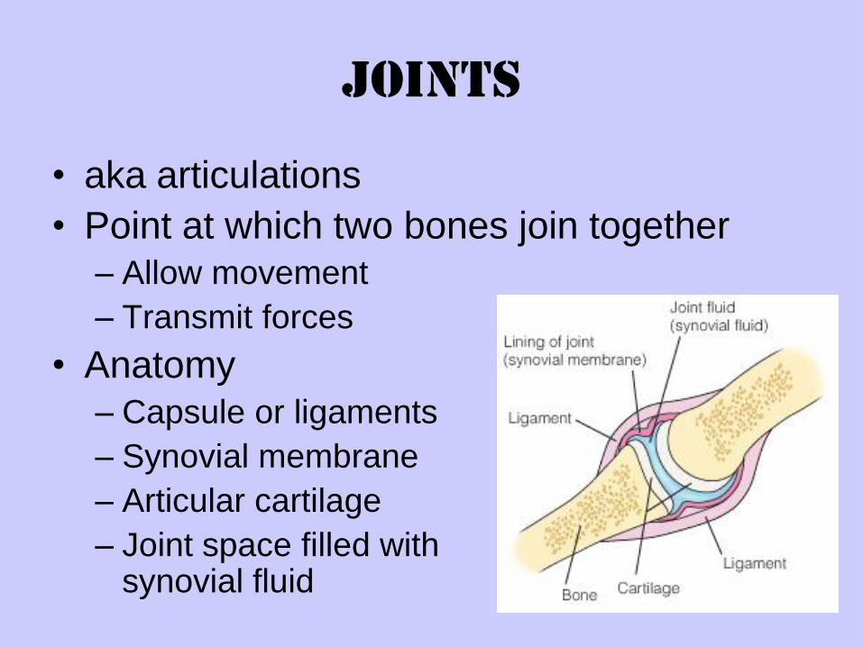
Joints
• aka articulations
• Point at which two bones join together
– Allow movement
– Transmit forces
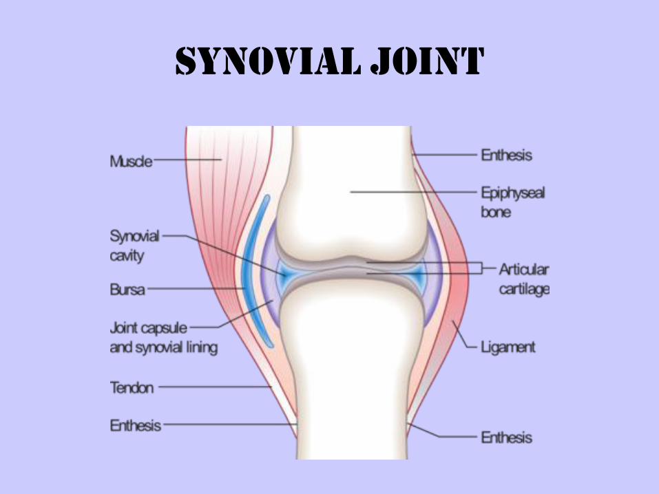
• Anatomy
– Capsule or ligaments
– Synovial membrane
– Articular cartilage
– Joint space filled with synovial fluid
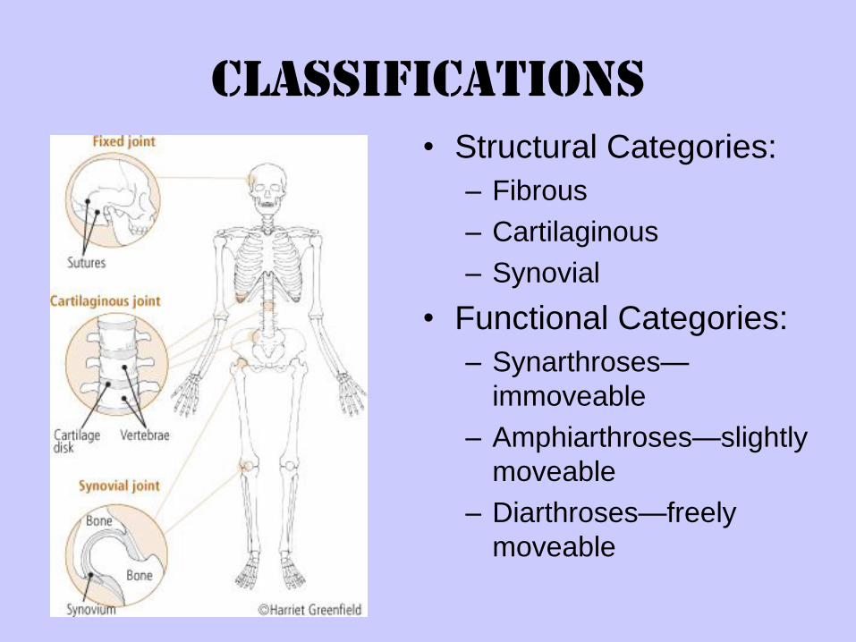
Classifications
• Structural Categories:
– Fibrous
– Cartilaginous
– Synovial
• Functional Categories:
– Synarthroses—
immoveable
– Amphiarthroses—slightly
moveable
– Diarthroses—freely
moveable
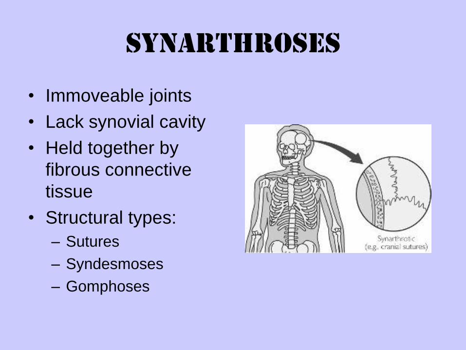
Synarthroses
• Immoveable joints
• Lack synovial cavity
• Held together by
fibrous connective
tissue
• Structural types:
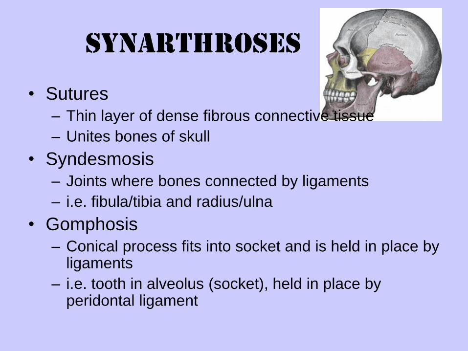
– Sutures
– Syndesmoses
– Gomphoses
Synarthroses
• Sutures– Thin layer of dense fibrous connective tissue
– Unites bones of skull
• Syndesmosis– Joints where bones connected by ligaments
– i.e. fibula/tibia and radius/ulna
• Gomphosis – Conical process fits into socket and is held in place by
ligaments
– i.e. tooth in alveolus (socket), held in place by peridontal ligament
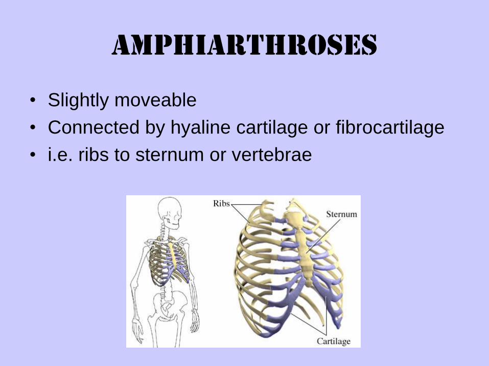
Amphiarthroses
• Slightly moveable
• Connected by hyaline cartilage or fibrocartilage
• i.e. ribs to sternum or vertebrae
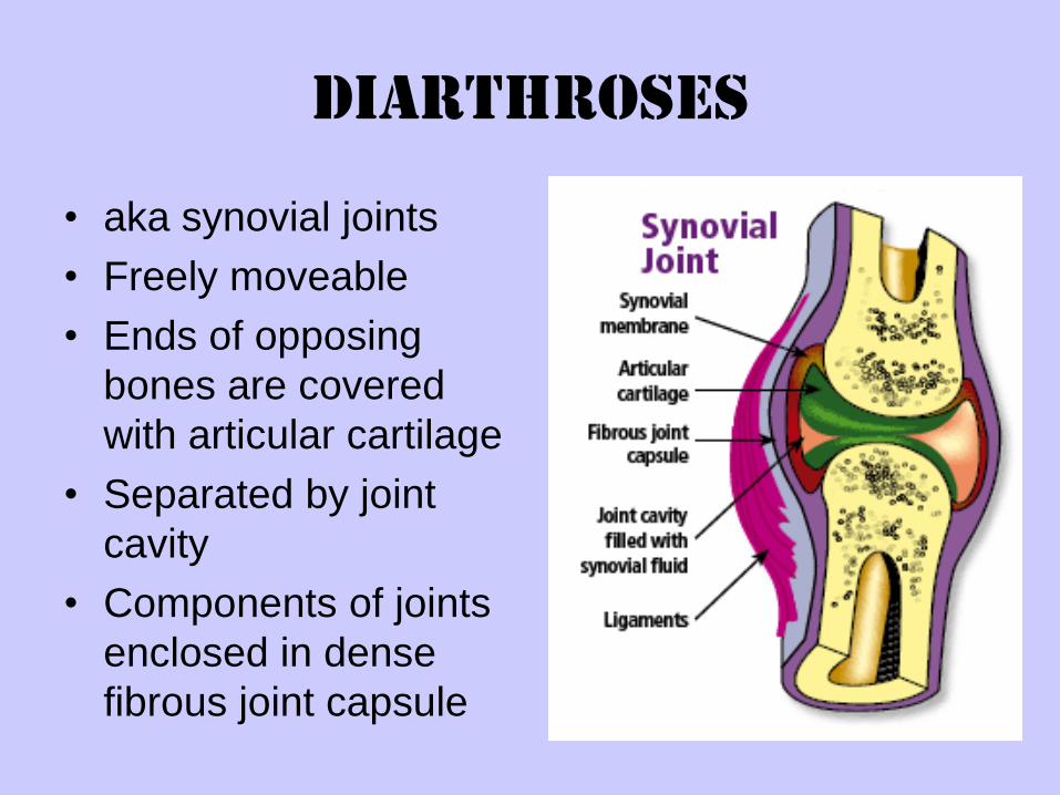
Diarthroses
• aka synovial joints
• Freely moveable
• Ends of opposing
bones are covered
with articular cartilage
• Separated by joint
cavity
• Components of joints
enclosed in dense
fibrous joint capsule
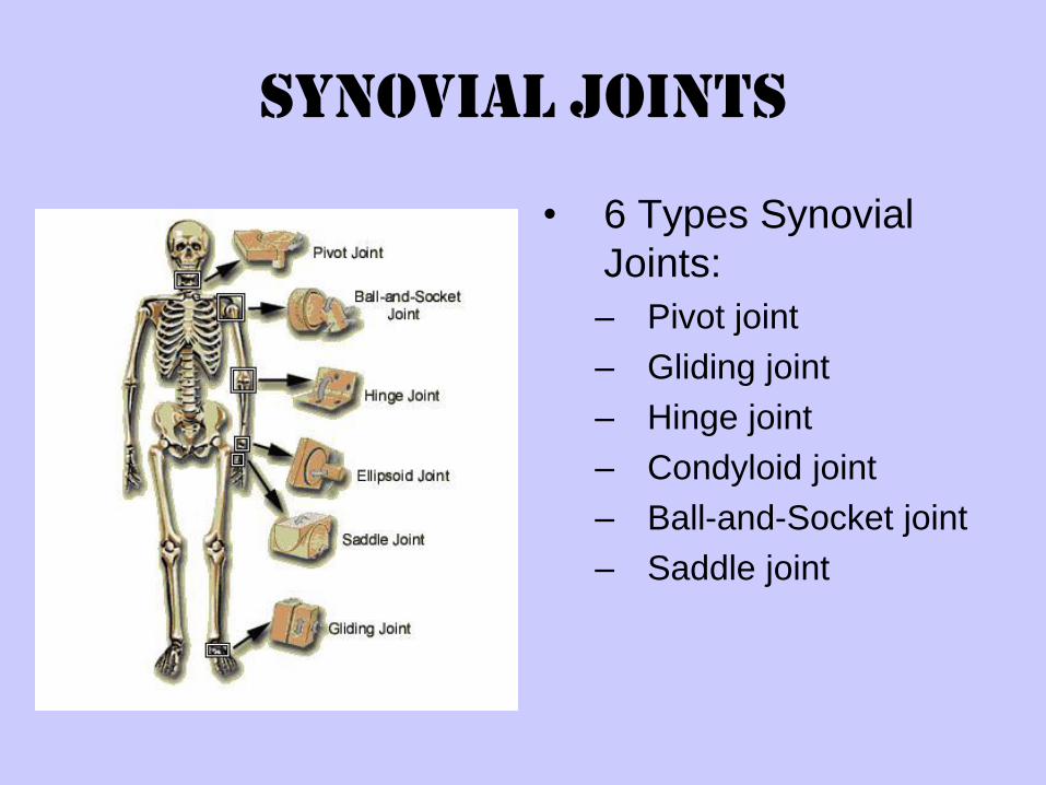
Synovial Joints
• 6 Types Synovial
Joints:
– Pivot joint
– Gliding joint
– Hinge joint
– Condyloid joint
– Ball-and-Socket joint
– Saddle joint
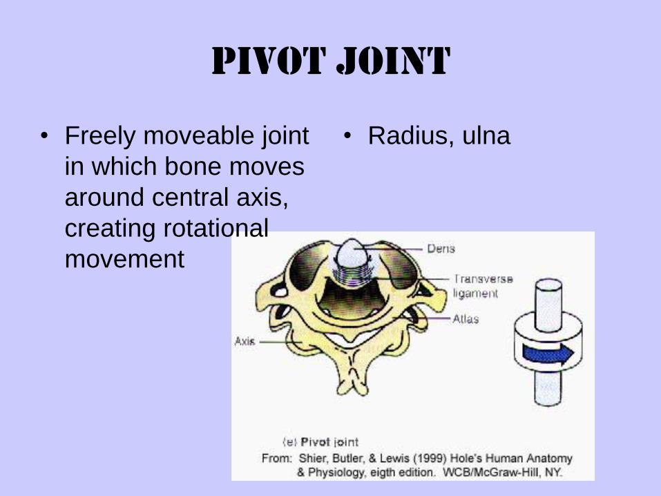
Pivot Joint
• Radius, ulna• Freely moveable joint
in which bone moves
around central axis,
creating rotational
movement
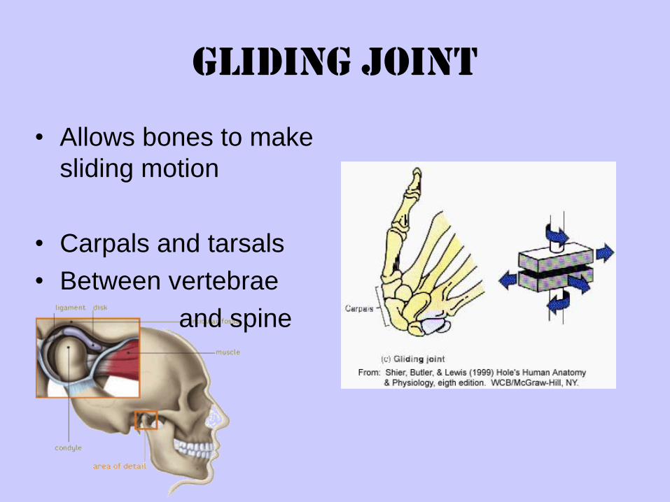
Gliding Joint
• Allows bones to make
sliding motion
• Carpals and tarsals
• Between vertebrae
and spine
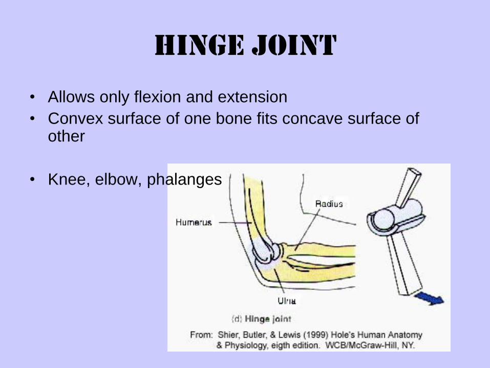
Hinge Joint
• Allows only flexion and extension
• Convex surface of one bone fits concave surface of other
• Knee, elbow, phalanges
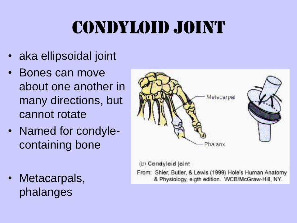
Condyloid Joint
• aka ellipsoidal joint
• Bones can move
about one another in
many directions, but
cannot rotate
• Named for condyle-
containing bone
• Metacarpals,
phalanges
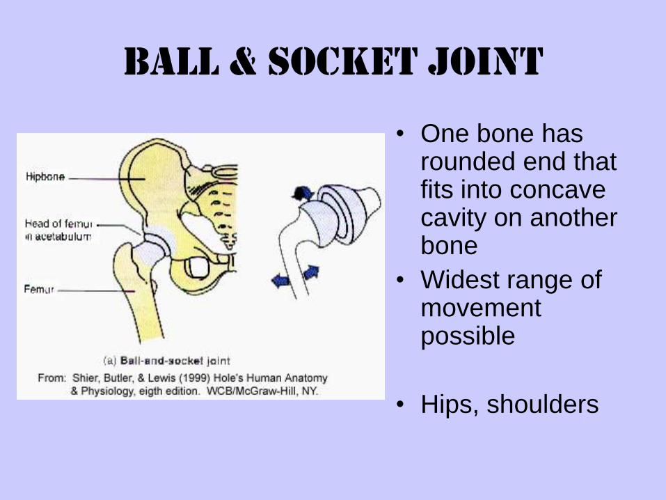
Ball & Socket Joint
• One bone has rounded end that fits into concave cavity on another bone
• Widest range of movement possible
• Hips, shoulders
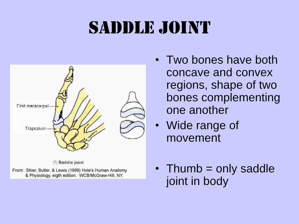
Saddle Joint
• Two bones have both concave and convex regions, shape of two bones complementing one another
• Wide range of movement
• Thumb = only saddle joint in body
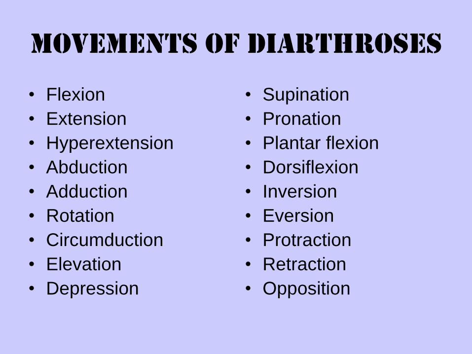
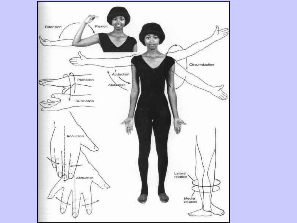
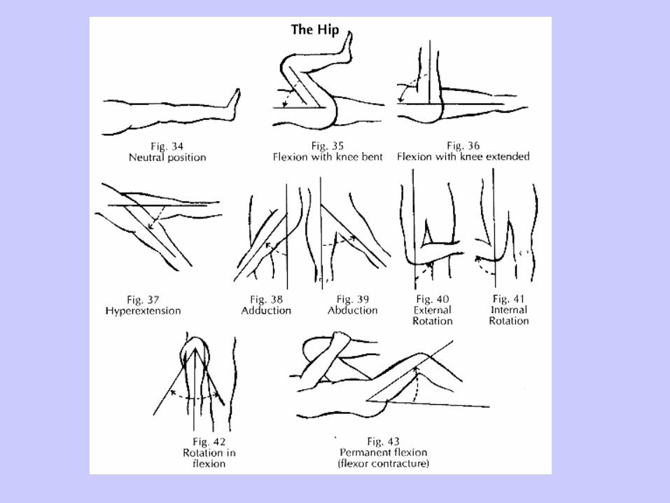
Movements of Diarthroses
• Flexion
• Extension
• Hyperextension
• Abduction
• Adduction
• Rotation
• Circumduction
• Elevation
• Depression
• Supination
• Pronation
• Plantar flexion
• Dorsiflexion
• Inversion
• Eversion
• Protraction
• Retraction
• Opposition
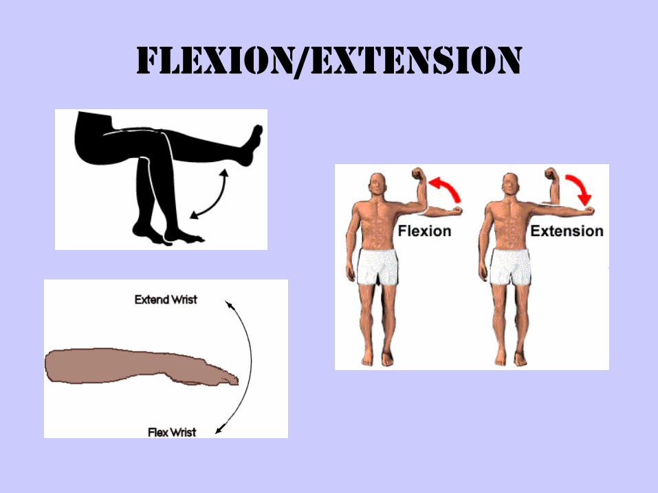
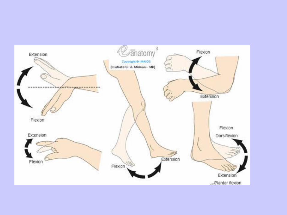
Flexion/Extension
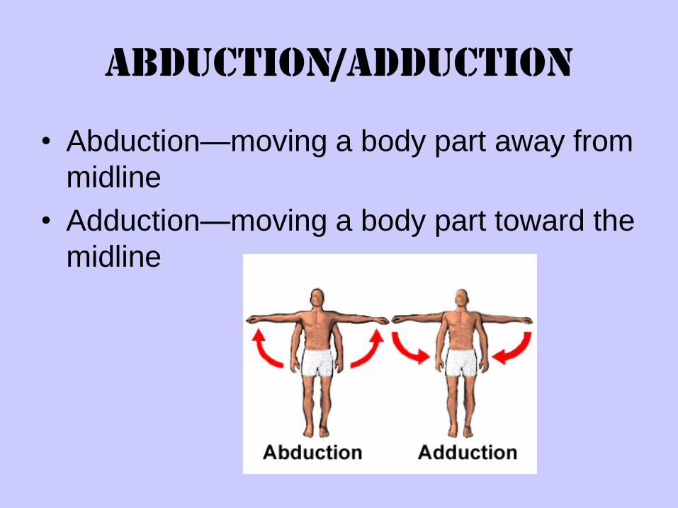
Abduction/Adduction
• Abduction—moving a body part away from
midline
• Adduction—moving a body part toward the
midline
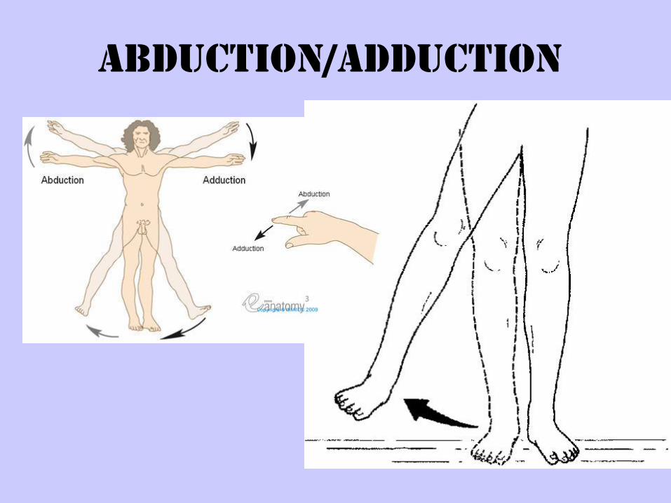
Abduction/Adduction
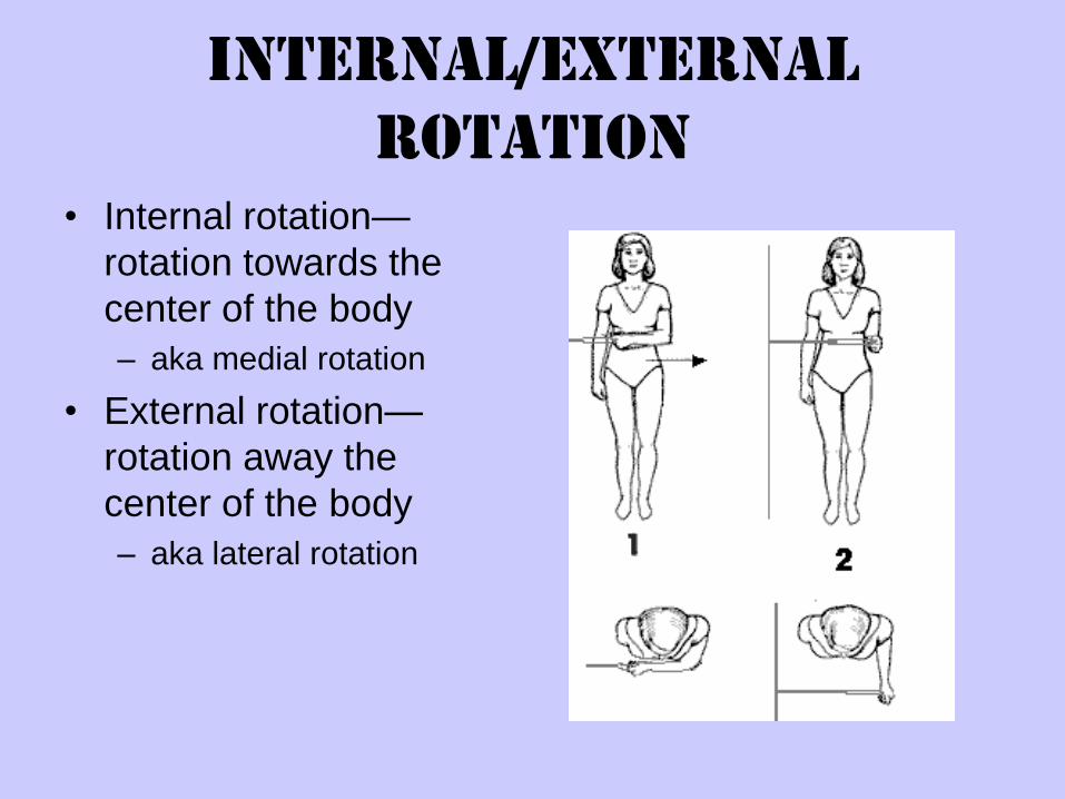
Internal/External
Rotation
• Internal rotation—
rotation towards the
center of the body
– aka medial rotation
• External rotation—
rotation away the
center of the body
– aka lateral rotation
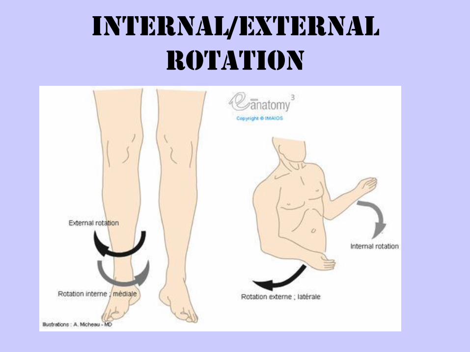
Internal/External
Rotation
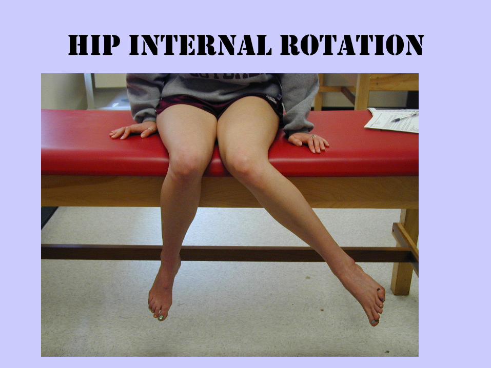
Hip Internal Rotation
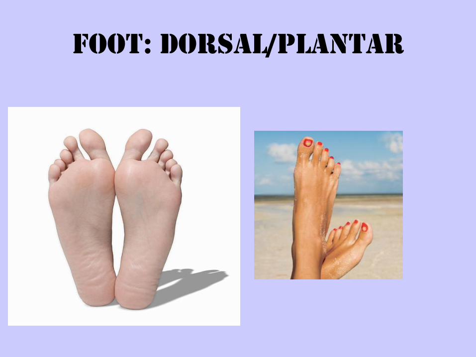
Foot: Dorsal/Plantar
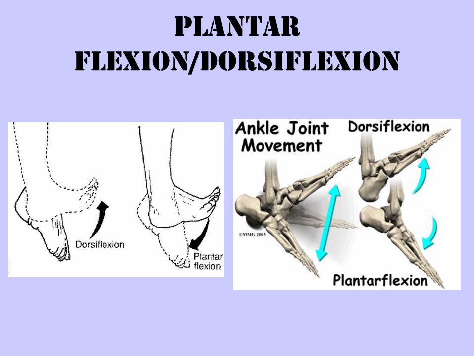
Plantar
Flexion/Dorsiflexion
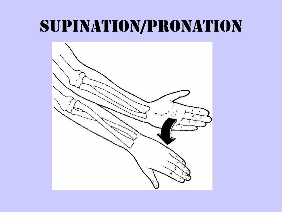
Supination/Pronation
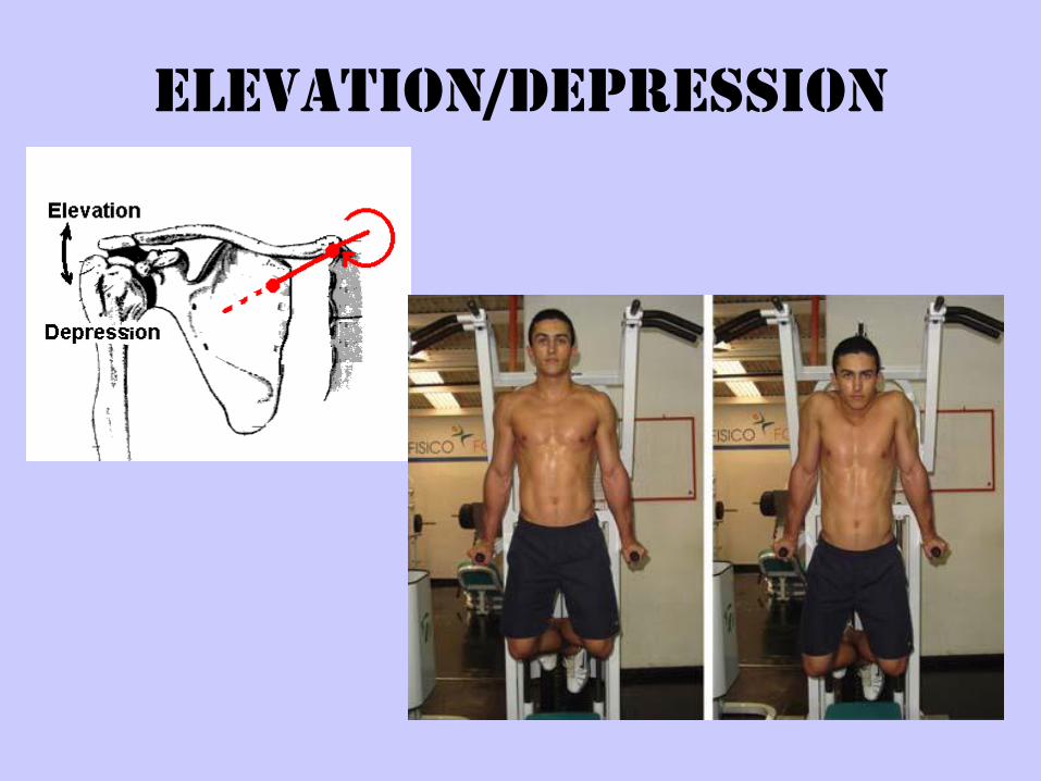
Elevation/Depression
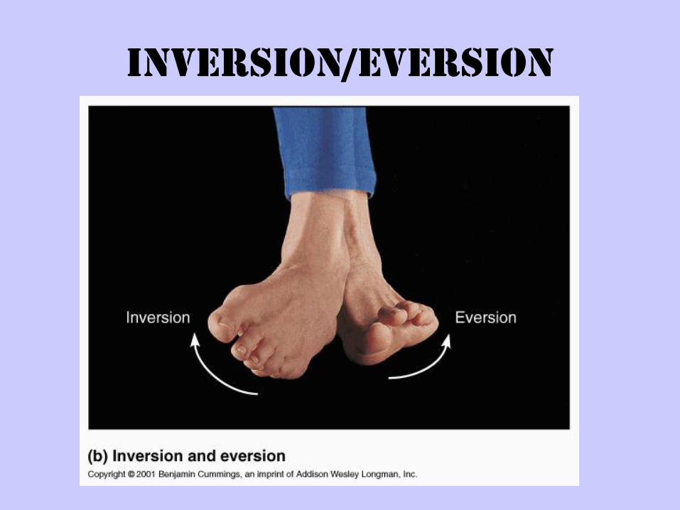
Inversion/Eversion
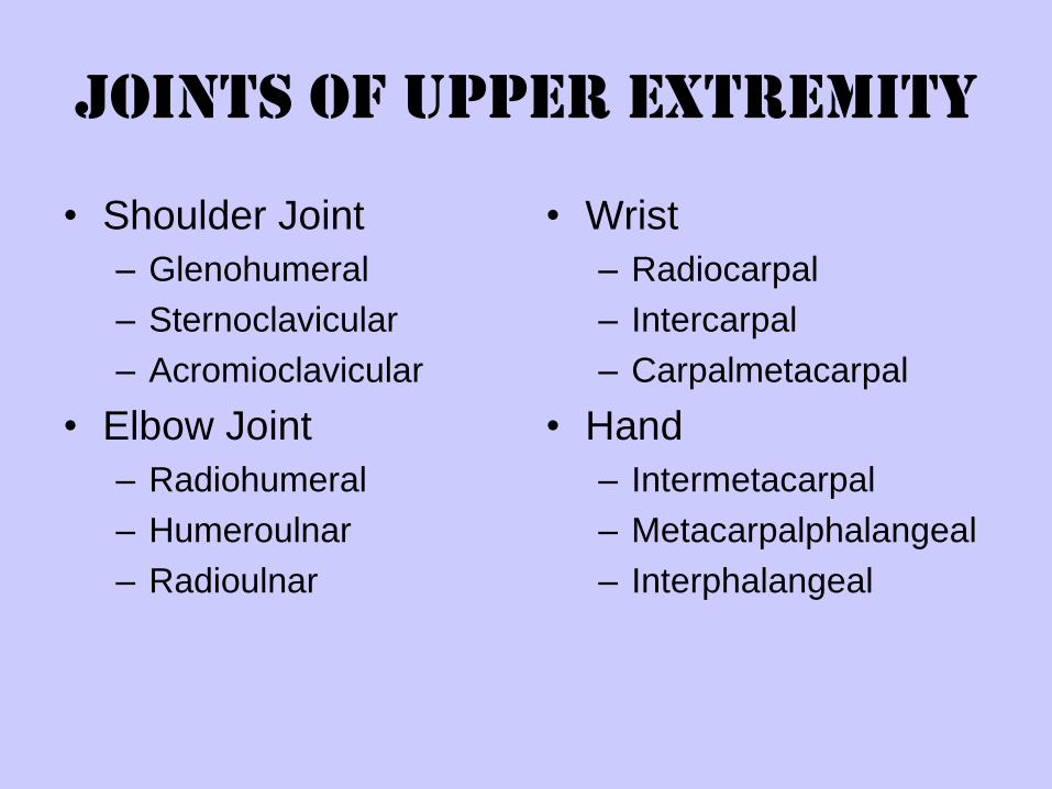
Joints of Upper Extremity
• Shoulder Joint
– Glenohumeral
– Sternoclavicular
– Acromioclavicular
• Elbow Joint
– Radiohumeral
– Humeroulnar
– Radioulnar
• Wrist
– Radiocarpal
– Intercarpal
– Carpalmetacarpal
• Hand
– Intermetacarpal
– Metacarpalphalangeal
– Interphalangeal
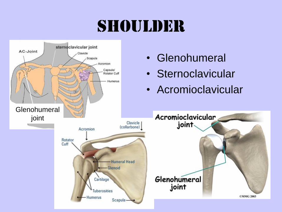
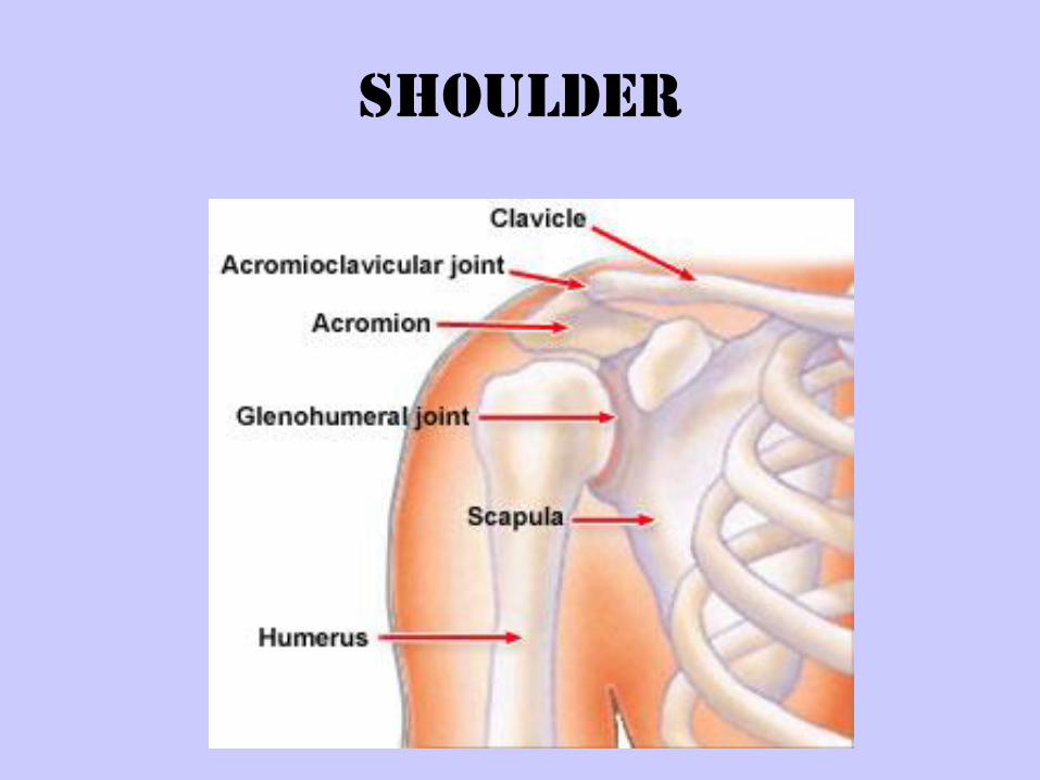
Shoulder
• Glenohumeral
• Sternoclavicular
• Acromioclavicular
Glenohumeral
joint
shoulder
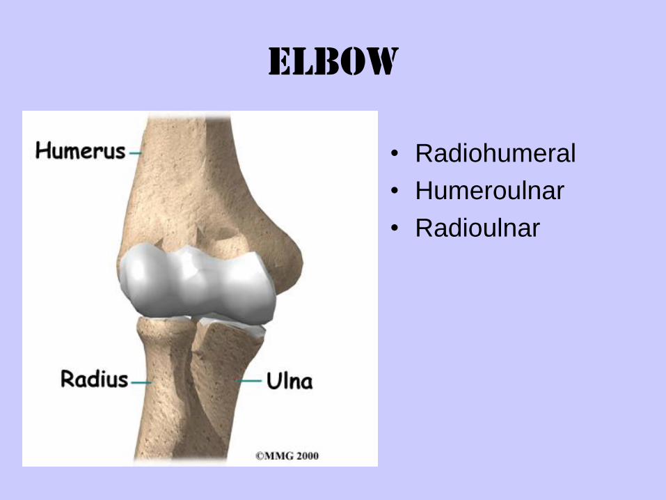
Elbow
• Radiohumeral
• Humeroulnar
• Radioulnar
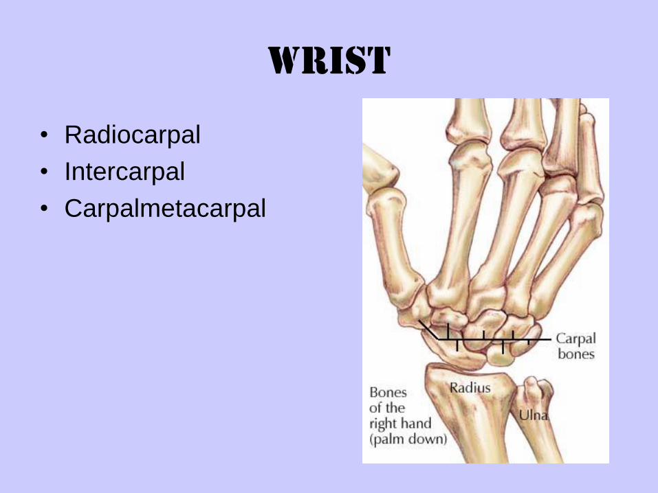
Wrist
• Radiocarpal
• Intercarpal
• Carpalmetacarpal
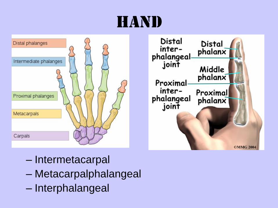
Hand
– Intermetacarpal
– Metacarpalphalangeal
– Interphalangeal
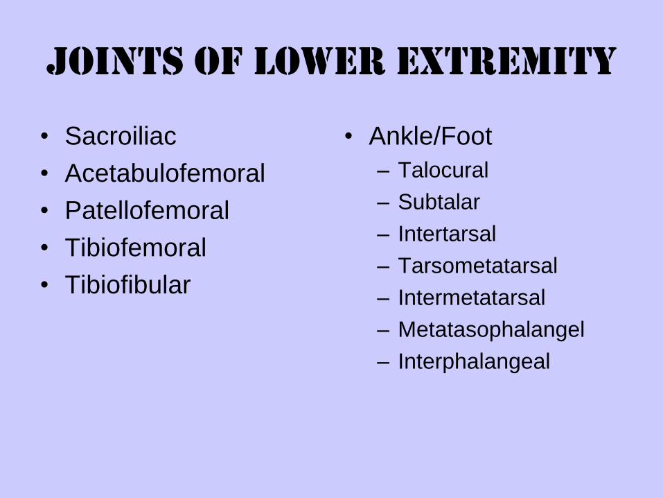
Joints of Lower Extremity
• Sacroiliac
• Acetabulofemoral
• Patellofemoral
• Tibiofemoral
• Tibiofibular
• Ankle/Foot
– Talocural
– Subtalar
– Intertarsal
– Tarsometatarsal
– Intermetatarsal
– Metatasophalangel
– Interphalangeal
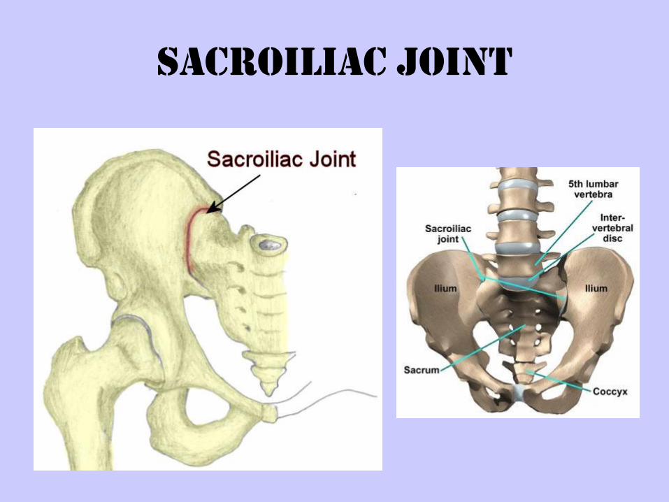
Sacroiliac joint
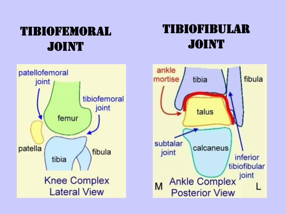
Tibiofemoral
joint
TIBIOFIBULAR
JOINT
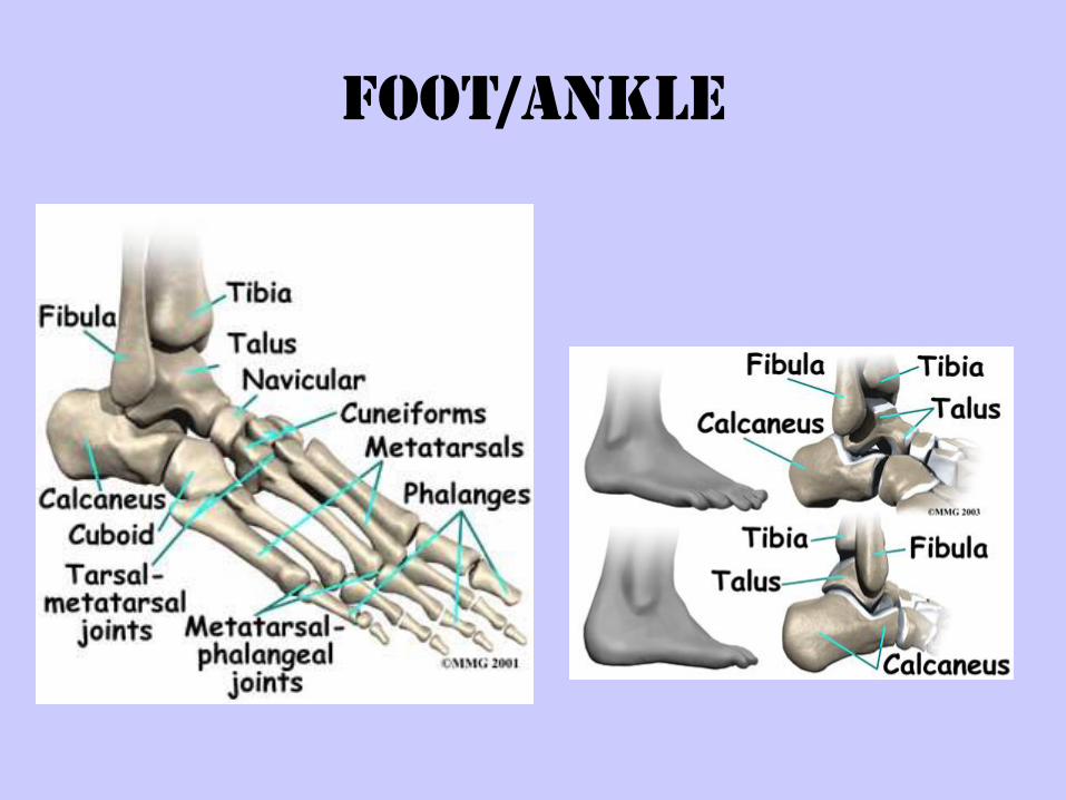
Foot/Ankle
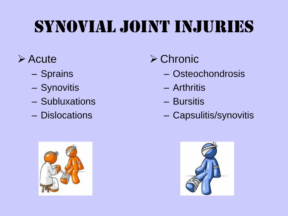
Synovial Joint Injuries
Acute
– Sprains
– Synovitis
– Subluxations
– Dislocations
Chronic
– Osteochondrosis
– Arthritis
– Bursitis
– Capsulitis/synovitis
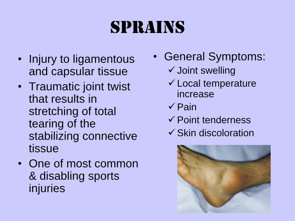
Sprains
• Injury to ligamentous and capsular tissue
• Traumatic joint twist that results in stretching of total tearing of the stabilizing connective tissue
• One of most common & disabling sports injuries
• General Symptoms: Joint swelling
Local temperature increase
Pain
Point tenderness
Skin discoloration
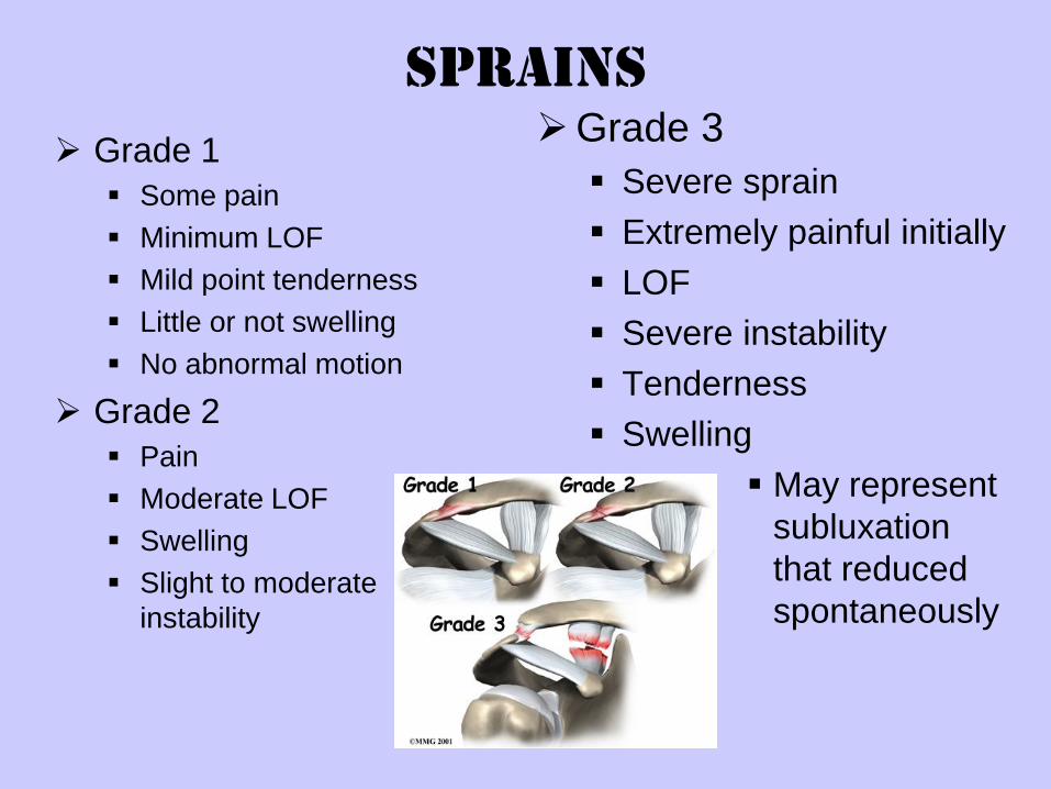
Sprains
Grade 1
Some pain
Minimum LOF
Mild point tenderness
Little or not swelling
No abnormal motion
Grade 2
Pain
Moderate LOF
Swelling
Slight to moderate
instability
Grade 3
Severe sprain
Extremely painful initially
LOF
Severe instability
Tenderness
Swelling
May represent
subluxation
that reduced
spontaneously
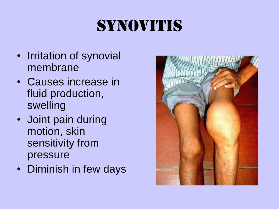
Synovitis
• Irritation of synovial membrane
• Causes increase in fluid production, swelling
• Joint pain during motion, skin sensitivity from pressure
• Diminish in few days
Acute Joint Injuries
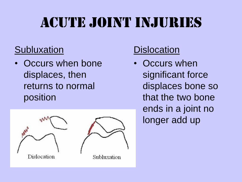
Subluxation
• Occurs when bone
displaces, then
returns to normal
position
Dislocation
• Occurs when
significant force
displaces bone so
that the two bone
ends in a joint no
longer add up
Osteochondrosis
• Degenerative changes in the ossification centers
of the epiphysis of bones
• During periods of rapid growth in children
• Osteochondritis dissecans
• Suggested causes—
– aseptic necrosis: circulation to epiphysis disrupted
– Trauma causes particles of articular cartilage to fx,
resulting in fissures that penetrate to subchondral
bone
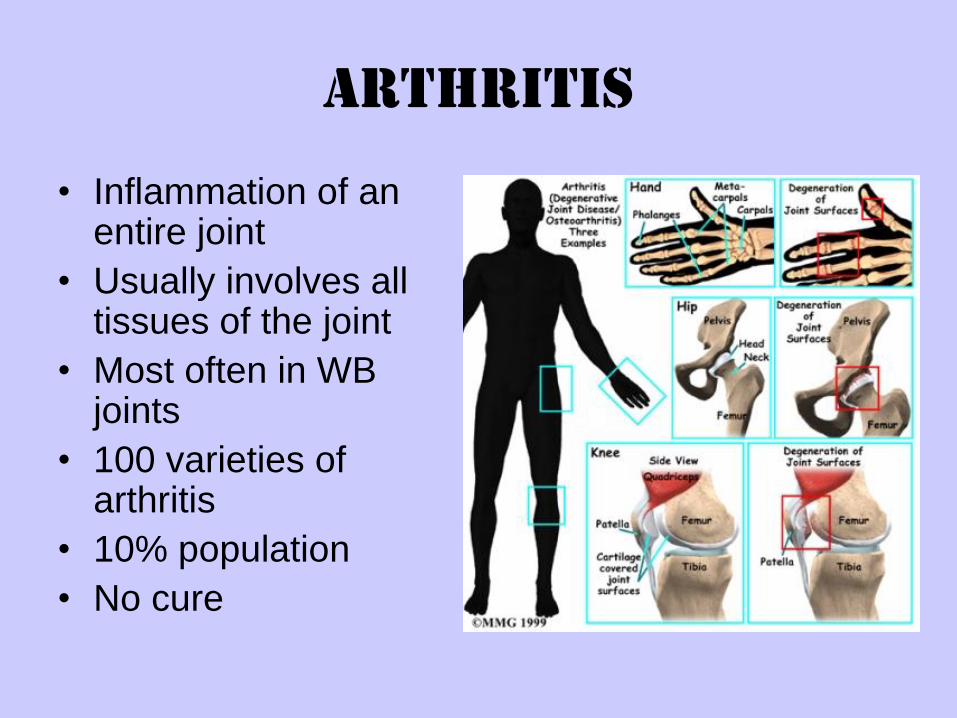
Arthritis
• Inflammation of an entire joint
• Usually involves all tissues of the joint
• Most often in WB joints
• 100 varieties of arthritis
• 10% population
• No cure
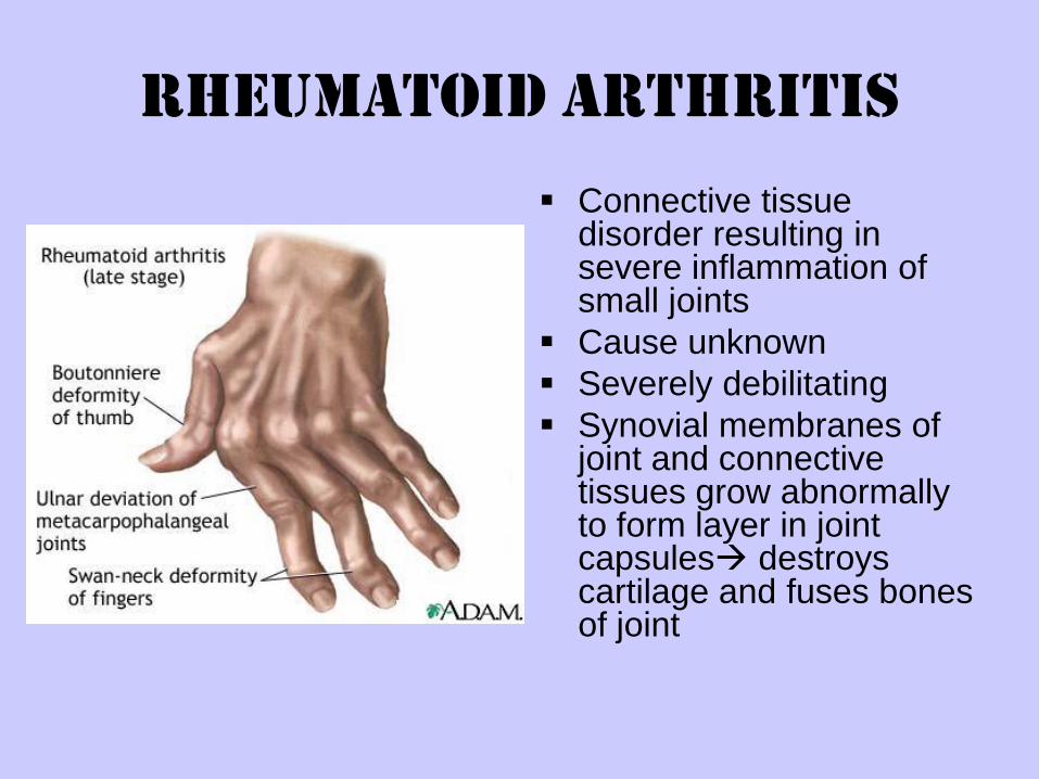
Rheumatoid Arthritis
Connective tissue disorder resulting in severe inflammation of small joints
Cause unknown
Severely debilitating
Synovial membranes of joint and connective tissues grow abnormally to form layer in joint capsules destroys cartilage and fuses bones of joint
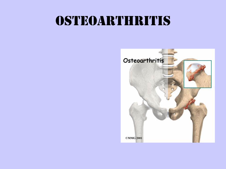
Osteoarthritis
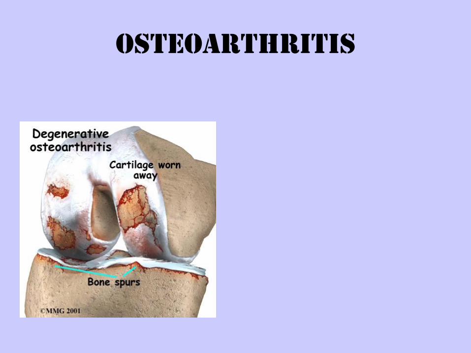
Osteoarthritis
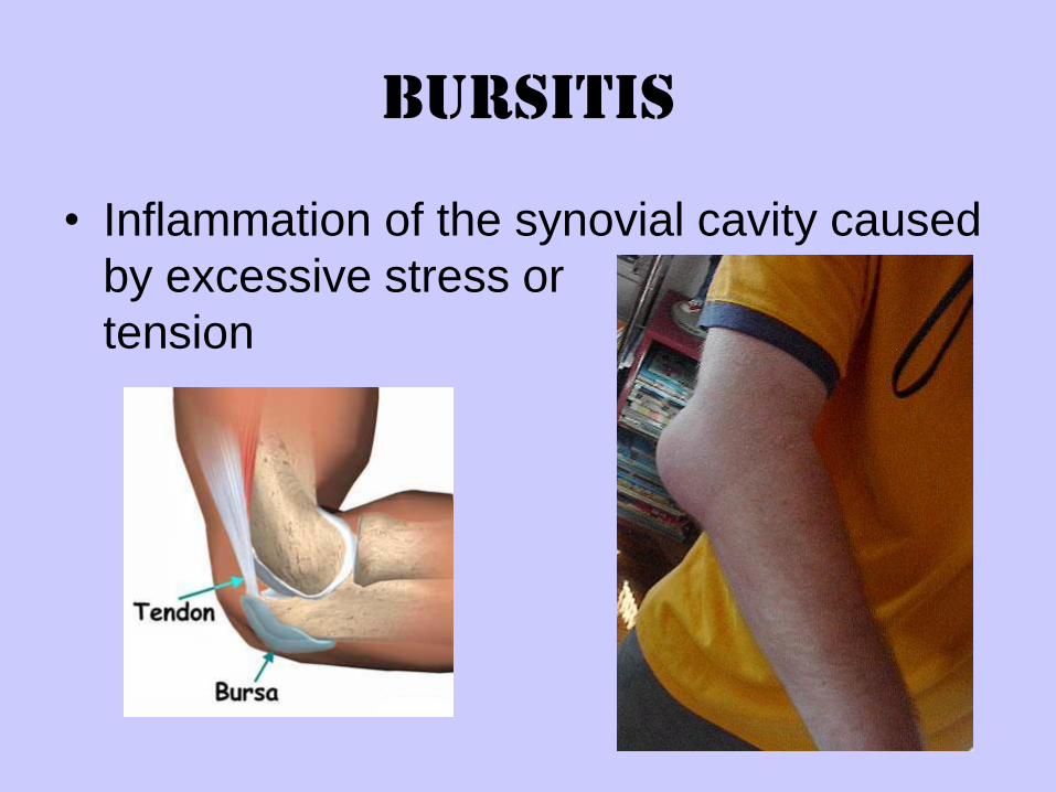
Bursitis
• Inflammation of the synovial cavity caused
by excessive stress or
tension
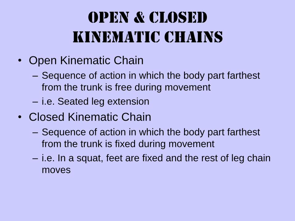
Open & Closed
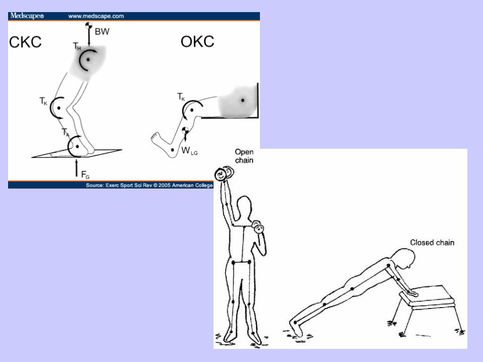
Kinematic Chains
• Open Kinematic Chain
– Sequence of action in which the body part farthest
from the trunk is free during movement
– i.e. Seated leg extension
• Closed Kinematic Chain
– Sequence of action in which the body part farthest
from the trunk is fixed during movement
– i.e. In a squat, feet are fixed and the rest of leg chain
moves