Joints & Joint Movements Human Anatomy Sonya Schuh-Huerta, Ph.D.
108
Joints & Joint Movements Joints & Joint Movements Human Anatomy Human Anatomy Sonya Schuh-Huerta, Ph.D. Sonya Schuh-Huerta, Ph.D.
-
Upload
phoebe-shute -
Category
Documents
-
view
228 -
download
3
Transcript of Joints & Joint Movements Human Anatomy Sonya Schuh-Huerta, Ph.D.
Joints & Joint MovementsJoints & Joint Movements
Human AnatomyHuman AnatomySonya Schuh-Huerta, Ph.D.Sonya Schuh-Huerta, Ph.D.
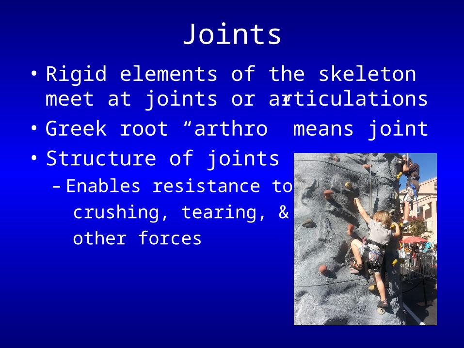
Joints• Rigid elements of the skeleton meet at
joints or articulations
• Greek root “arthro” means joint
• Structure of joints– Enables resistance to
crushing, tearing, &
other forces
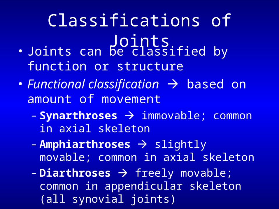
Classifications of Joints
• Joints can be classified by function or structure
• Functional classification based on amount of movement– Synarthroses immovable; common in
axial skeleton– Amphiarthroses slightly movable;
common in axial skeleton– Diarthroses freely movable; common in
appendicular skeleton (all synovial joints)
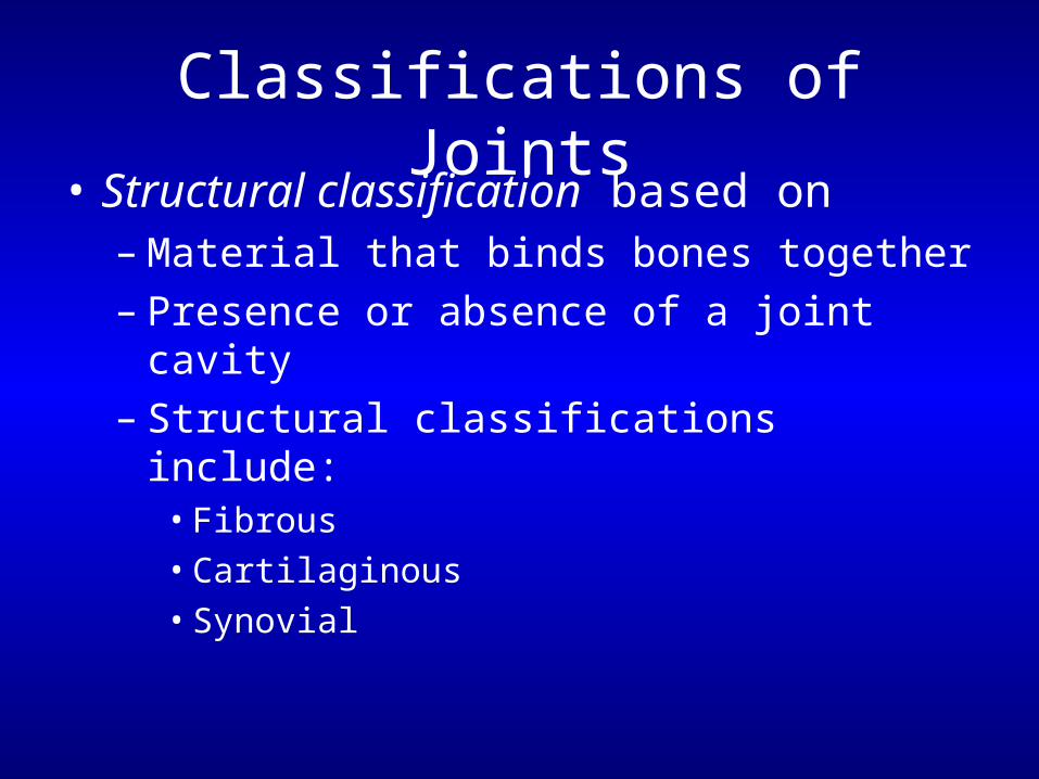
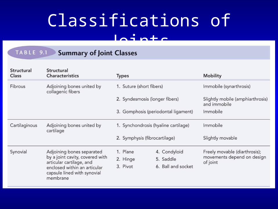
Classifications of Joints
• Structural classification based on– Material that binds bones together– Presence or absence of a joint cavity– Structural classifications include:
• Fibrous• Cartilaginous• Synovial
Classifications of Joints
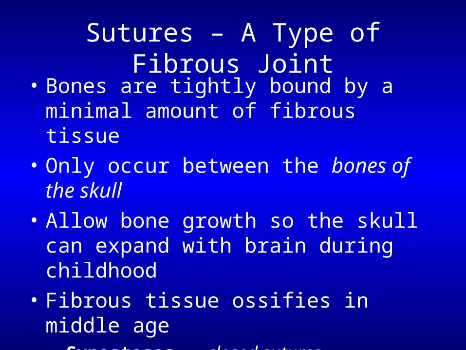
Sutures – A Type of Fibrous Joint
• Bones are tightly bound by a minimal amount of fibrous tissue
• Only occur between the bones of the skull
• Allow bone growth so the skull can expand with brain during childhood
• Fibrous tissue ossifies in middle age– Synostoses = closed sutures
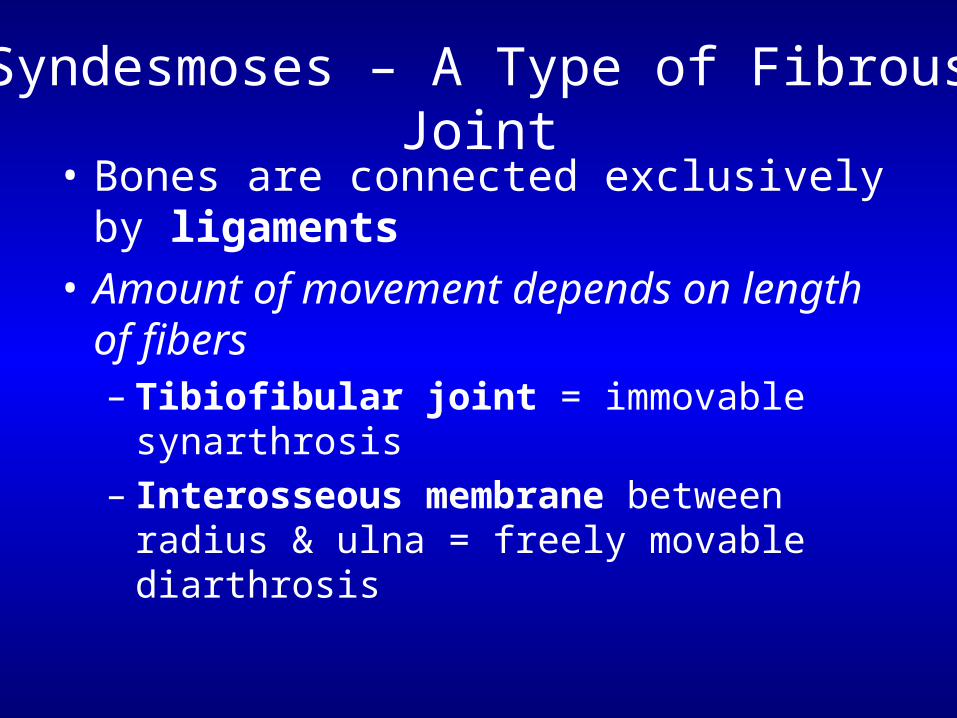
Syndesmoses – A Type of Fibrous Joint
• Bones are connected exclusively by ligaments
• Amount of movement depends on length of fibers– Tibiofibular joint = immovable synarthrosis– Interosseous membrane between radius &
ulna = freely movable diarthrosis

Gomphoses – A Type of Fibrous Joint
• Tooth in a socket
• Connecting ligament the periodontal ligament
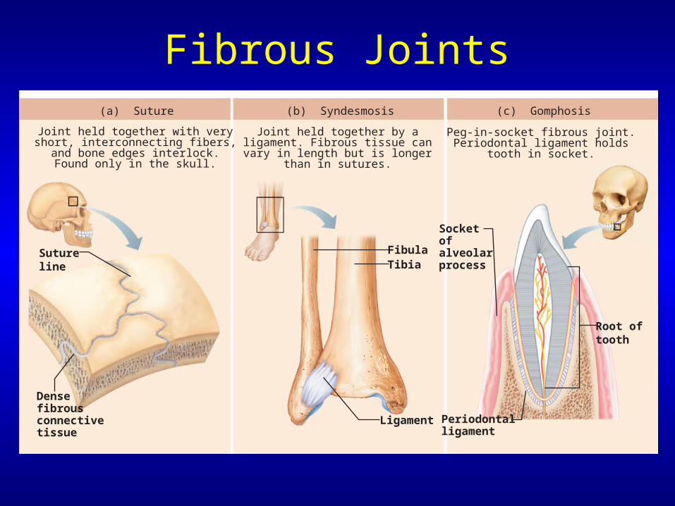
Fibrous Joints
Densefibrousconnectivetissue
Sutureline
Root oftooth
Socket of alveolarprocess
Periodontalligament
FibulaTibia
Ligament
(a) Suture
Joint held together with veryshort, interconnecting fibers,
and bone edges interlock.Found only in the skull.
(b) Syndesmosis
Joint held together by aligament. Fibrous tissue canvary in length but is longer
than in sutures.
(c) Gomphosis
Peg-in-socket fibrous joint.Periodontal ligament holds
tooth in socket.

Cartilaginous Joints
• Bones are united by cartilage
• Lack a joint cavity
• 2 types– Synchondroses– Symphyses
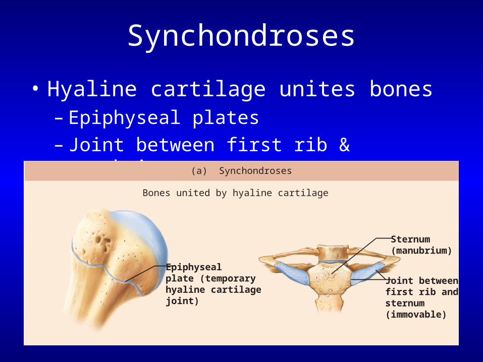
Synchondroses
• Hyaline cartilage unites bones– Epiphyseal plates– Joint between first rib & manubrium
Epiphysealplate (temporaryhyaline cartilagejoint)
Sternum(manubrium)
Joint betweenfirst rib and sternum (immovable)
(a) Synchondroses
Bones united by hyaline cartilage
Symphyses
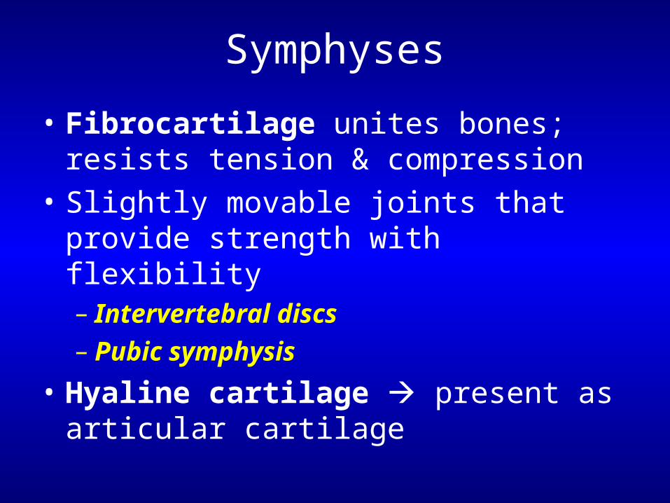
• Fibrocartilage unites bones; resists tension & compression
• Slightly movable joints that provide strength with flexibility– Intervertebral discs– Pubic symphysis
• Hyaline cartilage present as articular cartilage
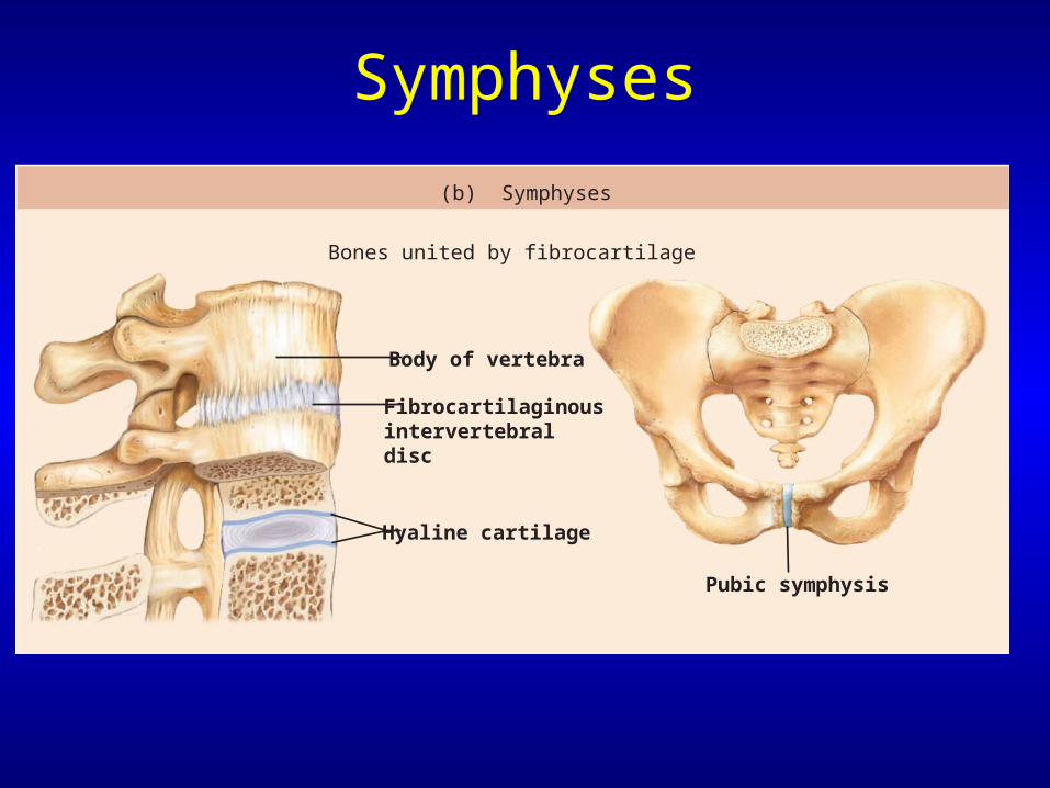
Symphyses
Fibrocartilaginousintervertebraldisc
Pubic symphysis
Body of vertebra
Hyaline cartilage
(b) Symphyses
Bones united by fibrocartilage
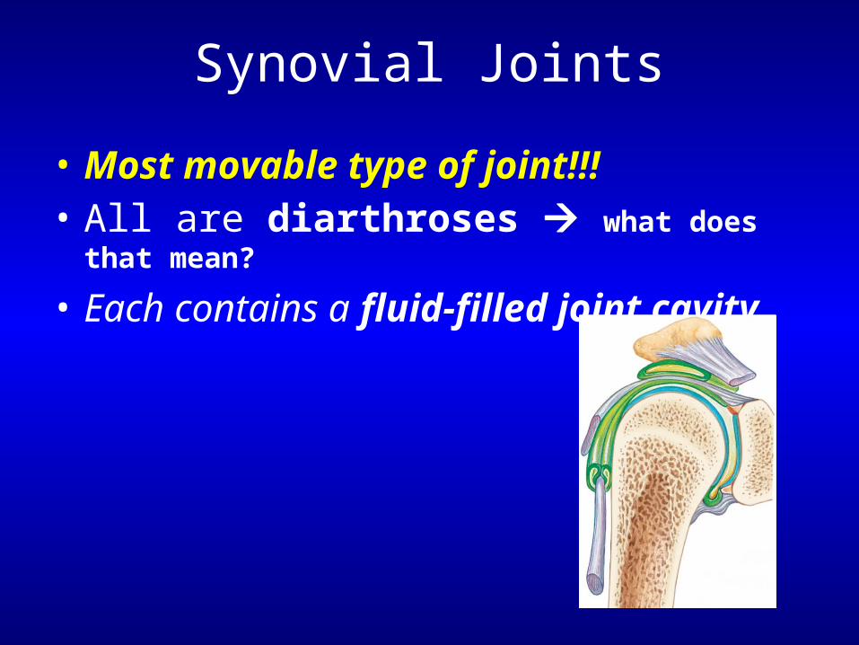
Synovial Joints
• Most movable type of joint!!!• All are diarthroses what does that mean?
• Each contains a fluid-filled joint cavity
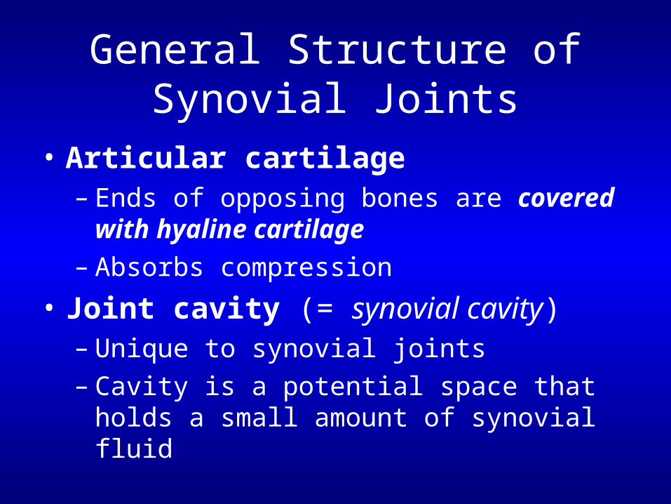
General Structure of Synovial Joints
• Articular cartilage– Ends of opposing bones are covered with
hyaline cartilage– Absorbs compression
• Joint cavity (= synovial cavity)– Unique to synovial joints– Cavity is a potential space that holds a small
amount of synovial fluid
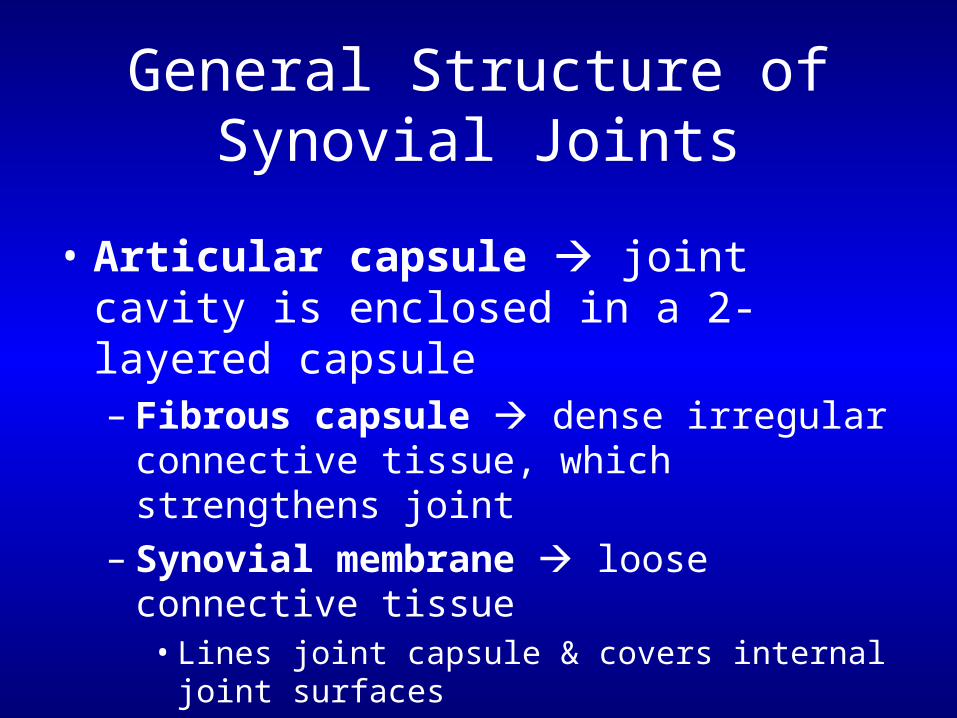
General Structure of Synovial Joints
• Articular capsule joint cavity is enclosed in a 2-layered capsule– Fibrous capsule dense irregular
connective tissue, which strengthens joint– Synovial membrane loose connective
tissue• Lines joint capsule & covers internal joint surfaces• Makes synovial fluid
General Structure of Synovial Joints
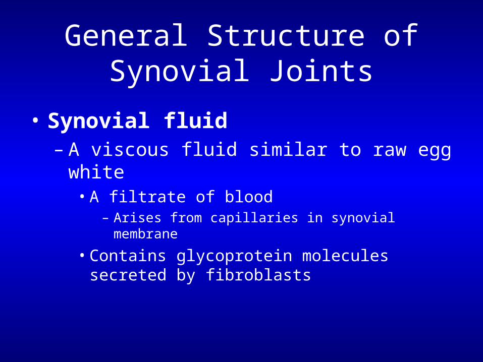
• Synovial fluid– A viscous fluid similar to raw egg white
• A filtrate of blood– Arises from capillaries in synovial membrane
• Contains glycoprotein molecules secreted by fibroblasts
General Structure of Synovial Joints
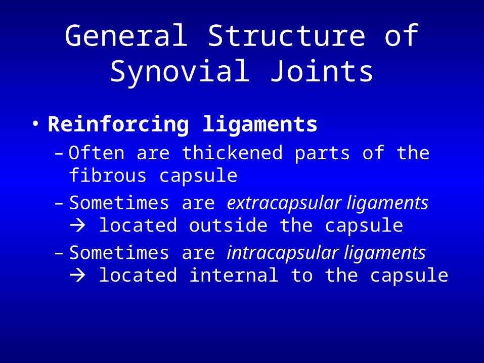
• Reinforcing ligaments– Often are thickened parts of the fibrous
capsule– Sometimes are extracapsular ligaments
located outside the capsule– Sometimes are intracapsular ligaments
located internal to the capsule
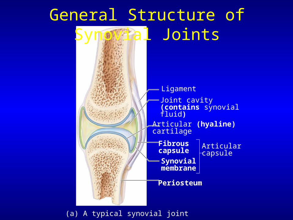
Periosteum
Ligament
Fibrouscapsule
Synovialmembrane
Joint cavity (contains synovial fluid)
Articular (hyaline)cartilage
Articularcapsule
(a) A typical synovial joint
General Structure of Synovial Joints
General Structure of Synovial Joints
• Richly supplied with sensory nerves– Detect pain– Most monitor how much the capsule is being
stretched
General Structure of Synovial Joints
• Have a rich blood supply– Most supply the synovial membrane– Extensive capillary beds produce basis of
synovial fluid– Branches of several major nerves & blood
vessels
Synovial Joints with Articular Discs
• Some synovial joints contain an articular disc– Temporomandibular joint & Knee joint– Occur in joints whose articulating bones have
somewhat different shapes
How Synovial Joints Function
• Synovial joints lubricating devices
• Friction could overheat & destroy joint tissue & bone ends
• Are subjected to compressive forces• Fluid is squeezed out as opposing cartilages touch• Cartilages ride on the slippery film
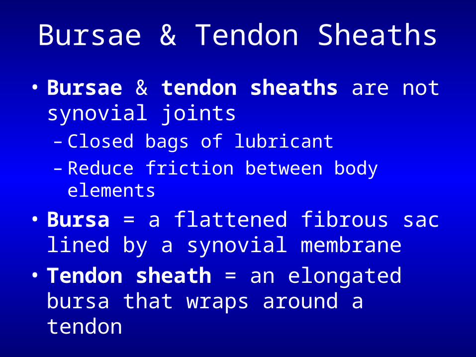
Bursae & Tendon Sheaths
• Bursae & tendon sheaths are not synovial joints– Closed bags of lubricant– Reduce friction between body elements
• Bursa = a flattened fibrous sac lined by a synovial membrane
• Tendon sheath = an elongated bursa that wraps around a tendon
Bursae & Tendon Sheaths
Acromionof scapula
Joint cavitycontainingsynovial fluid
Synovialmembrane
Fibrouscapsule
Humerus
Hyalinecartilage
Coracoacromialligament
Subacromialbursa
Fibrousarticular capsule
Tendonsheath
Tendon oflong headof bicepsbrachii muscle
(a) Frontal section through the right shoulder joint
Coracoacromialligament
Subacromialbursa
Cavity inbursa containingsynovial fluid
(b) Enlargement of (a), showing how a bursa eliminates friction where a ligament (or other structure) would rub against a bone
Humerus resting
Humerus moving
Bursa rollsand lessens friction.
Humerus headrolls medially as arm abducts.
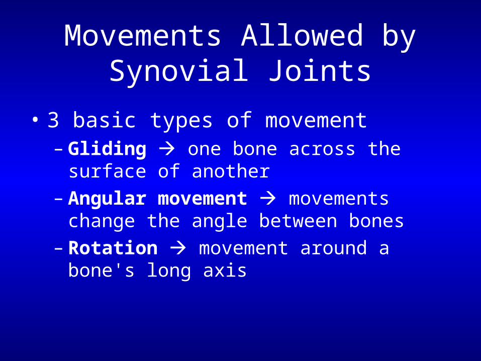
Movements Allowed by Synovial Joints
• 3 basic types of movement– Gliding one bone across the surface of
another– Angular movement movements change
the angle between bones– Rotation movement around a bone's long
axis
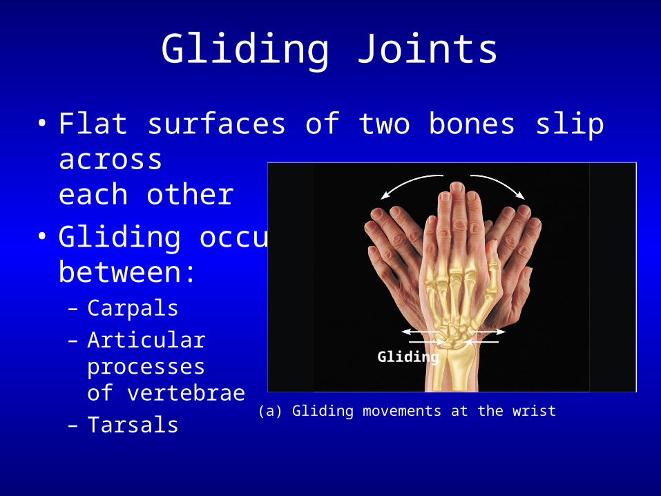
Gliding Joints
• Flat surfaces of two bones slip across each other
• Gliding occurs between: – Carpals– Articular
processes of vertebrae
– Tarsals
Gliding
(a) Gliding movements at the wrist
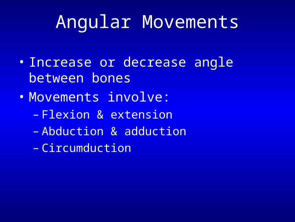
Angular Movements
• Increase or decrease angle between bones
• Movements involve:– Flexion & extension– Abduction & adduction– Circumduction
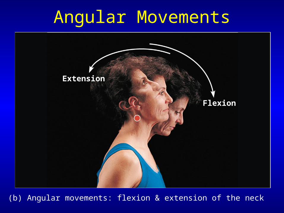
Angular Movements
(b) Angular movements: flexion & extension of the neck
Extension
Flexion

Angular Movements
Flexion
Extension
(c) Angular movements: flexion & extension of the trunk
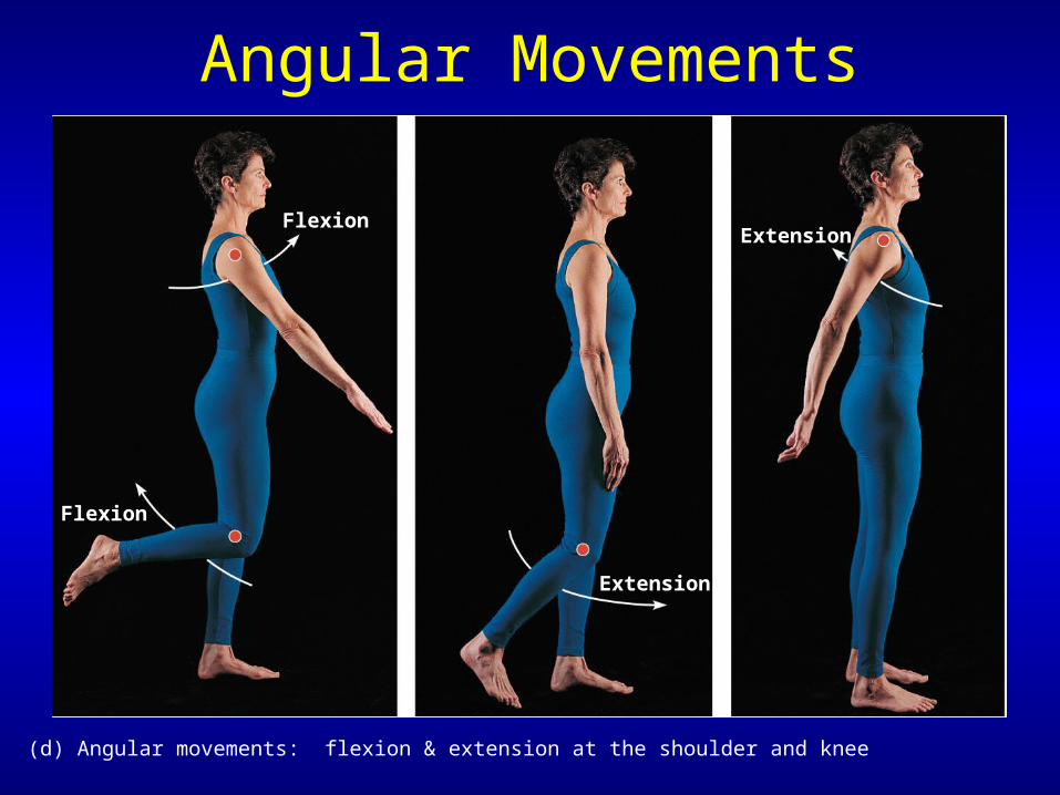
Angular Movements
Extension
Extension
Flexion
Flexion
(d) Angular movements: flexion & extension at the shoulder and knee

Angular Movements
Abduction
Adduction
(e) Angular movements: abduction, adduction, & circumduction of the upper limb at the shoulder
Circumduction
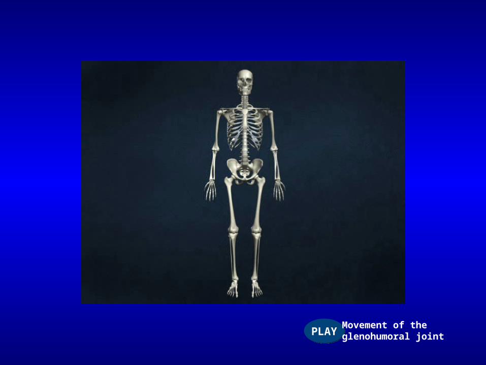
PLAYPLAYMovement of theglenohumoral joint
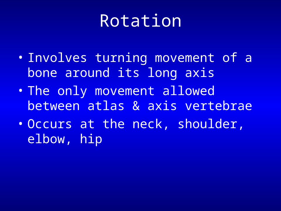
Rotation
• Involves turning movement of a bone around its long axis
• The only movement allowed between atlas & axis vertebrae
• Occurs at the neck, shoulder, elbow, hip
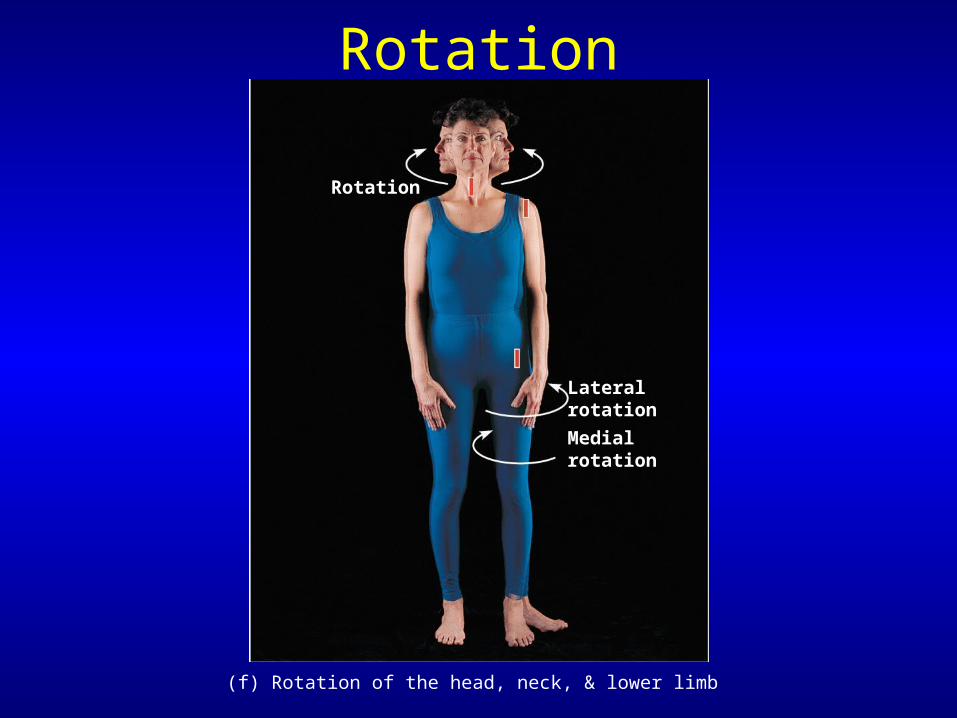
Rotation
Lateralrotation
Medialrotation
(f) Rotation of the head, neck, & lower limb
Rotation
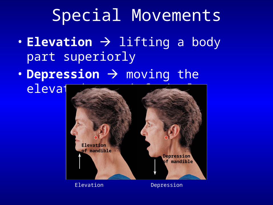
Special Movements
• Elevation lifting a body part superiorly
• Depression moving the elevated part inferiorly
Elevationof mandible
Depressionof mandible
Elevation
Depression

Special Movements
• Protraction nonangular movement anteriorly
• Retraction nonangular movement posteriorly
Protractionof mandible
Retractionof mandible
ProtractionMoving a body part in theanterior direction
RetractionMoving a body part in theposterior direction
Special Movements
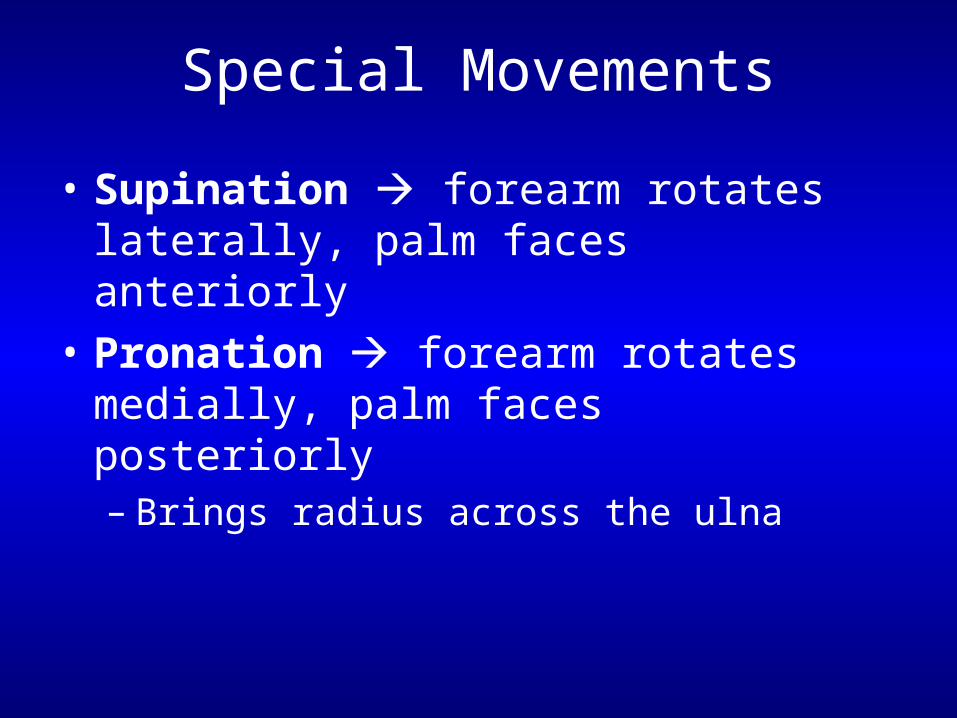
• Supination forearm rotates laterally, palm faces anteriorly
• Pronation forearm rotates medially, palm faces posteriorly– Brings radius across the ulna
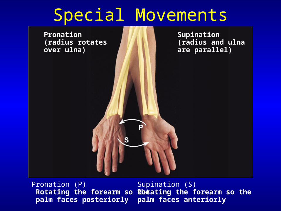
Special MovementsSupination(radius and ulnaare parallel)
Pronation (P) Rotating the forearm so the palm faces posteriorly
Supination (S)Rotating the forearm so thepalm faces anteriorly
Pronation(radius rotatesover ulna)
Special Movements
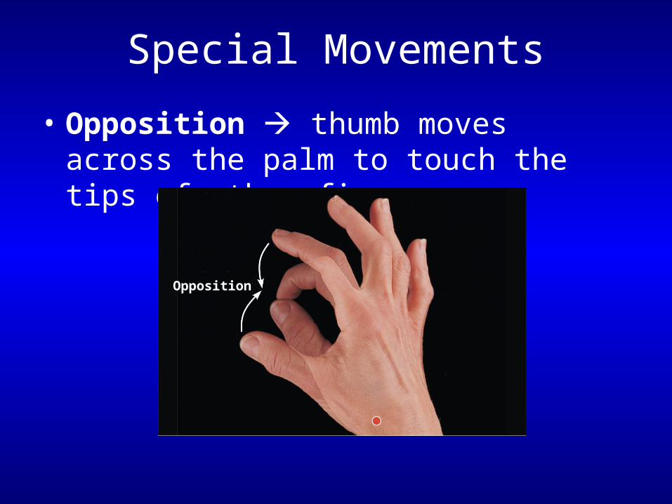
• Opposition thumb moves across the palm to touch the tips of other fingers
Opposition
Special Movements
• Inversion & eversion– Special movements at the foot
• Inversion turns sole medially• Eversion turns sole laterally
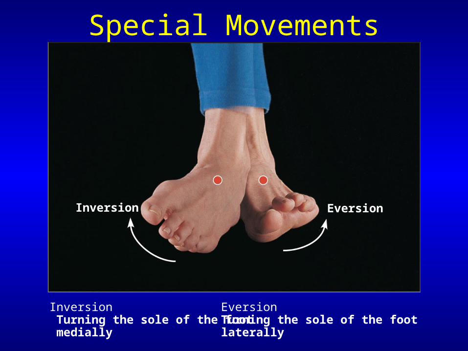
EversionInversion
Inversion Turning the sole of the foot medially
EversionTurning the sole of the footlaterally
Special Movements
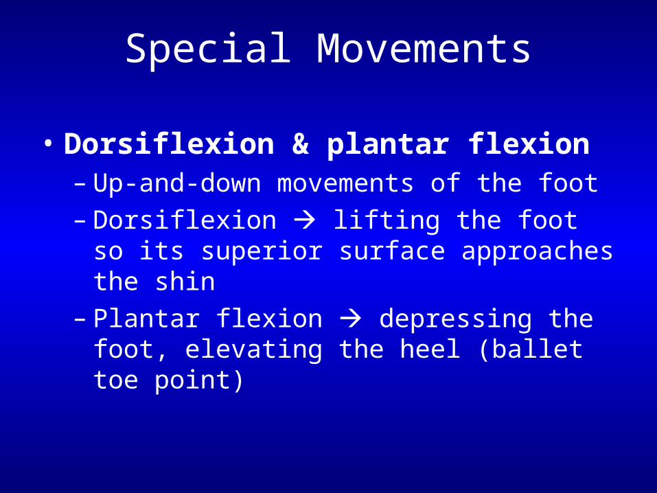
Special Movements
• Dorsiflexion & plantar flexion– Up-and-down movements of the foot– Dorsiflexion lifting the foot so its superior
surface approaches the shin– Plantar flexion depressing the foot,
elevating the heel (ballet toe point)
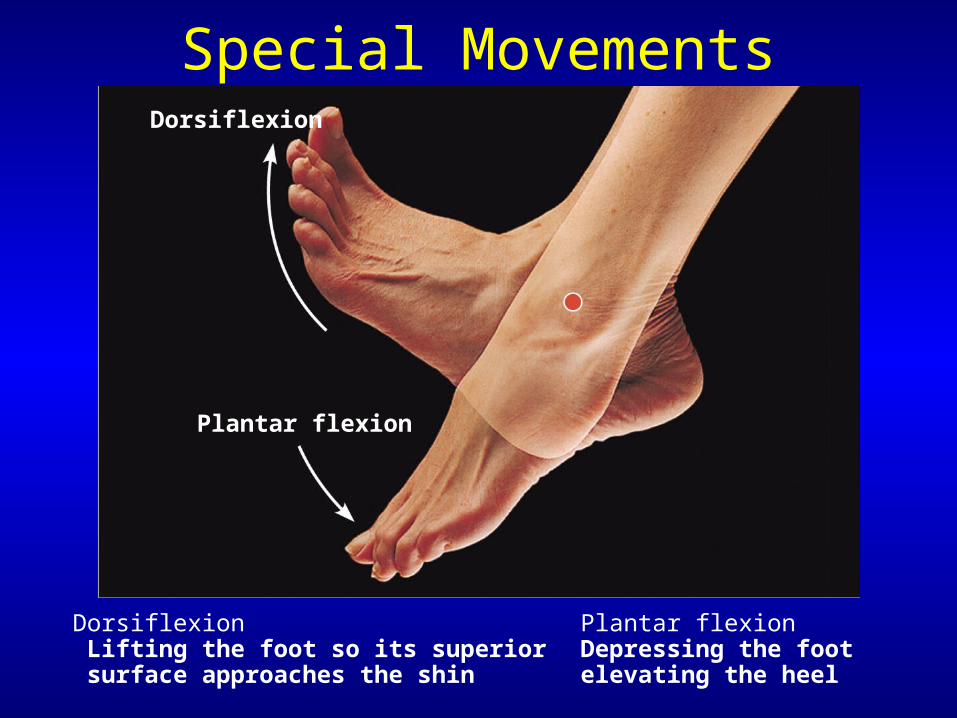
Special MovementsDorsiflexion
Plantar flexion
Dorsiflexion Lifting the foot so its superior surface approaches the shin
Plantar flexionDepressing the footelevating the heel

PLAYPLAYEversion ofAnkle joint (5a)
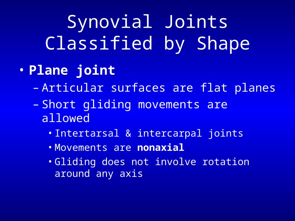
Synovial Joints Classified by Shape
• Plane joint– Articular surfaces are flat planes– Short gliding movements are allowed
• Intertarsal & intercarpal joints• Movements are nonaxial• Gliding does not involve rotation around any axis
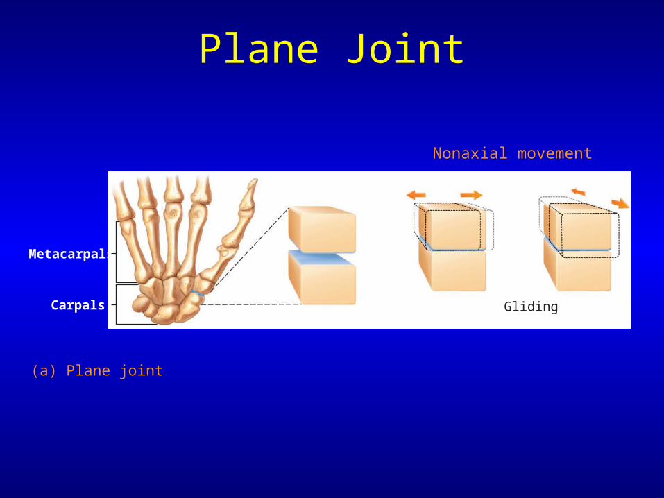
Plane Joint
(a) Plane joint
Gliding
Metacarpals
Carpals
Nonaxial movement
Synovial Joints Classified by Shape
• Hinge joints– Cylindrical end of one bone fits into a trough
on another bone– Angular movement is allowed in one plane– Elbow, ankle, & joints between phalanges– Movement is uniaxial allows movement
around one axis only

Hinge Joint
(b) Hinge joint
Medial/lateralaxis
Flexion & extension
Humerus
Ulna
Uniaxial movement
Synovial Joints Classified by Shape
• Pivot joints– Classified as uniaxial – rotating bone only
turns around its long axis– Examples
• Proximal radioulnar joint• Joint between atlas & axis
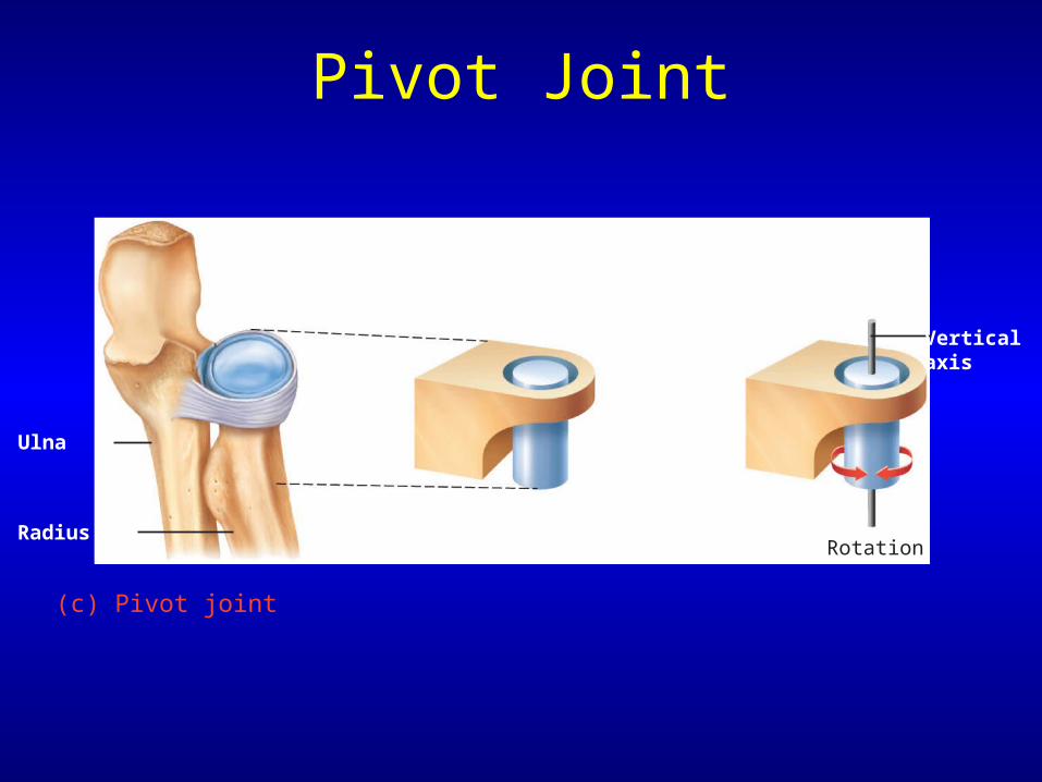
Pivot Joint
(c) Pivot joint
Ulna
Verticalaxis
RotationRadius
Synovial Joints Classified by Shape
• Condyloid joints– Allow moving bone to travel:
• Side to side abduction-adduction• Back & forth flexion-extension
– Classified as biaxial = movement occurs around 2 axes
– Phalanges
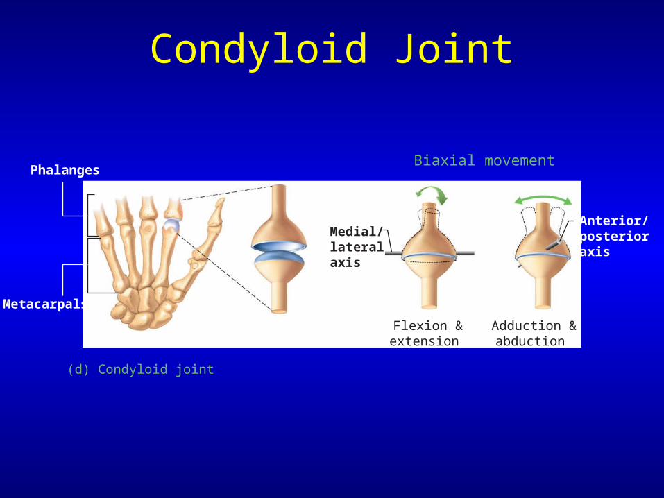
Condyloid Joint
(d) Condyloid joint
Medial/lateralaxis
Adduction &abduction
Flexion &extension
Metacarpals
Phalanges
Anterior/posterioraxis
Biaxial movement
Synovial Joints Classified by Shape
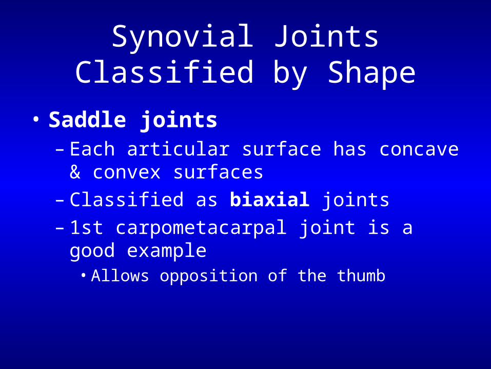
• Saddle joints– Each articular surface has concave & convex
surfaces– Classified as biaxial joints– 1st carpometacarpal joint is a good example
• Allows opposition of the thumb
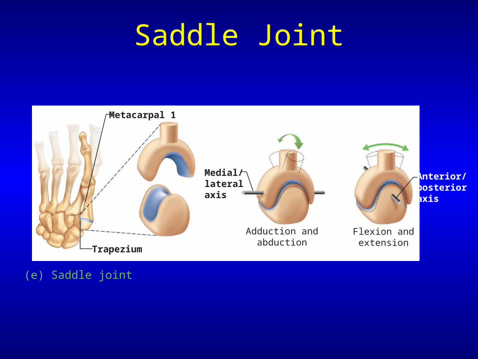
Saddle Joint
(e) Saddle joint
Anterior/posterior axis
Medial/lateralaxis
Adduction andabduction
Metacarpal 1
Trapezium
Flexion andextension
Synovial Joints Classified by Shape
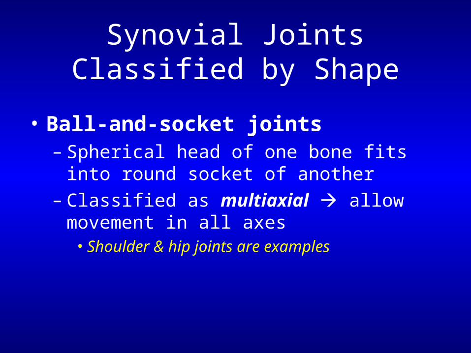
• Ball-and-socket joints– Spherical head of one bone fits into round
socket of another– Classified as multiaxial allow movement in
all axes• Shoulder & hip joints are examples
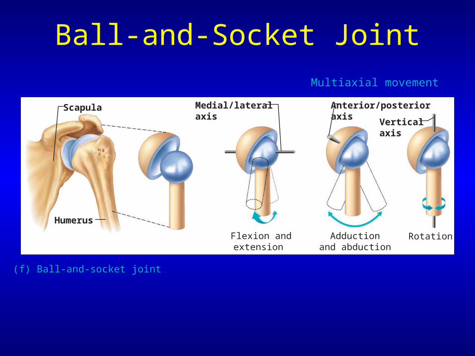
Ball-and-Socket Joint
(f) Ball-and-socket joint
Medial/lateralaxis
Anterior/posterioraxis
Verticalaxis
RotationAdductionand abduction
Flexion andextension
Scapula
Humerus
Multiaxial movement
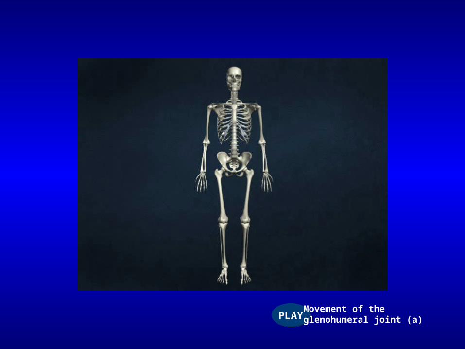
PLAYPLAYMovement of theglenohumeral joint (a)
Factors Influencing Stability of Synovial Joints
• Articular surfaces– Shapes of articulating surfaces determine
movements possible

Factors Influencing Stability of Synovial Joints
• Ligaments– Capsules & ligaments prevent excessive
motions– On the medial or inferior side of a joint
prevent excessive abduction– Lateral or superiorly located resist
adduction
Factors Influencing Stability of Synovial Joints
• Ligaments (cont…)– Anterior ligaments resist extension &
lateral rotation– Posterior ligaments resist flexion & medial
rotation
• The more ligaments usually the stronger & more stable
Factors Influencing Stability of Synovial Joints
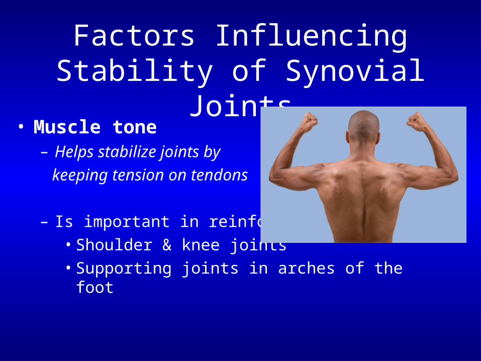
• Muscle tone– Helps stabilize joints by
keeping tension on tendons
– Is important in reinforcing:• Shoulder & knee joints• Supporting joints in arches of the foot

Selected Joints
Selected Synovial Joints
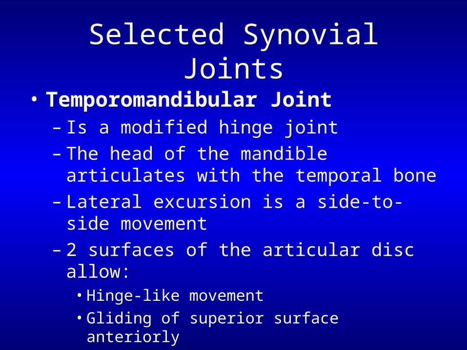
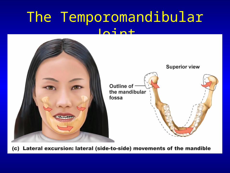
• Temporomandibular Joint– Is a modified hinge joint– The head of the mandible articulates with the
temporal bone– Lateral excursion is a side-to-side movement– 2 surfaces of the articular disc allow:
• Hinge-like movement• Gliding of superior surface anteriorly
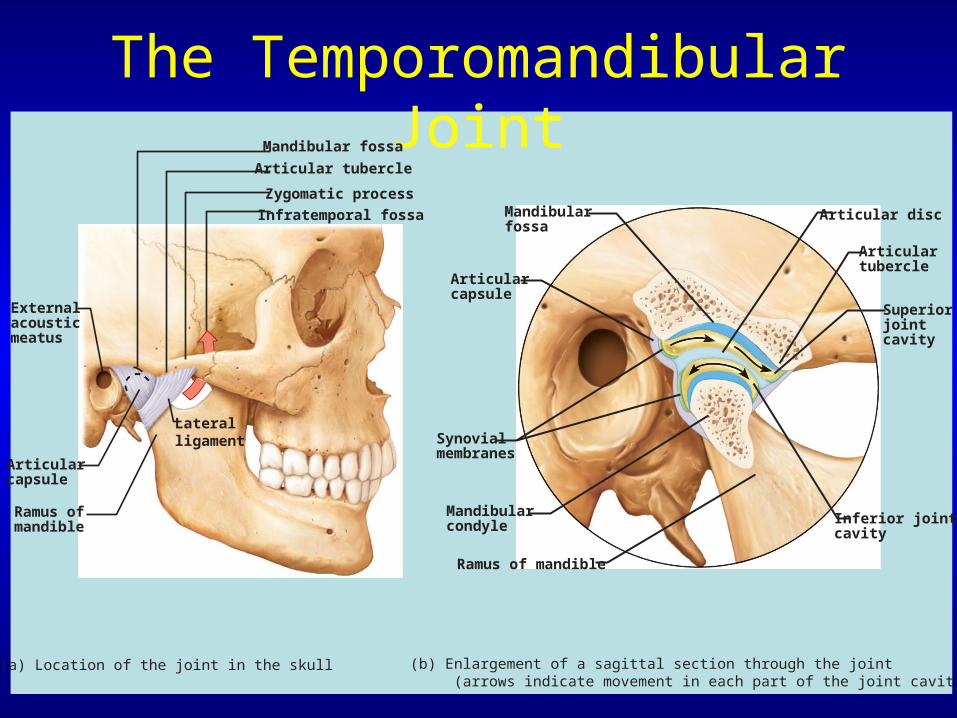
The Temporomandibular Joint
Zygomatic process
Mandibular fossa
Articular tubercle
Infratemporal fossa
Externalacousticmeatus
Articularcapsule
Ramus ofmandible
(a) Location of the joint in the skull
Lateralligament
Articularcapsule
Mandibularfossa
Articular disc
Articulartubercle
Superiorjointcavity
Inferior jointcavity
Mandibularcondyle
Ramus of mandible
Synovialmembranes
(b) Enlargement of a sagittal section through the joint (arrows indicate movement in each part of the joint cavity)
The Temporomandibular Joint
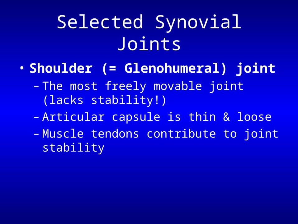
Selected Synovial Joints
• Shoulder (= Glenohumeral) joint– The most freely movable joint (lacks stability!)– Articular capsule is thin & loose– Muscle tendons contribute to joint stability
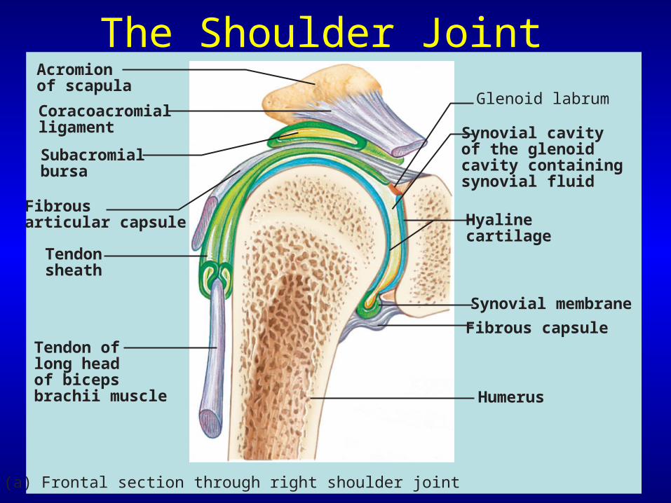
The Shoulder JointAcromionof scapula
Synovial membrane
Fibrous capsule
Hyalinecartilage
Coracoacromialligament
Subacromialbursa
Fibrousarticular capsule
Tendonsheath
Tendon oflong headof bicepsbrachii muscle
(a) Frontal section through right shoulder joint
Synovial cavityof the glenoidcavity containingsynovial fluid
Glenoid labrum
Humerus
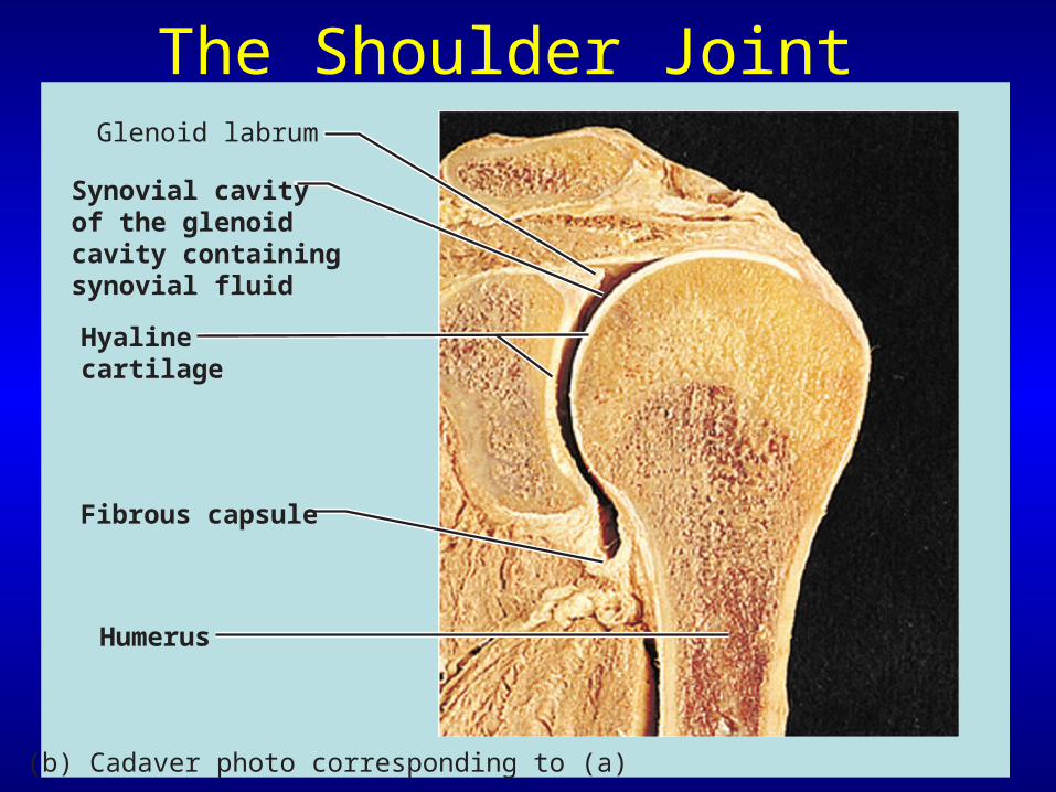
Fibrous capsule
Hyalinecartilage
Synovial cavityof the glenoidcavity containingsynovial fluid
Glenoid labrum
Humerus
(b) Cadaver photo corresponding to (a)
The Shoulder Joint
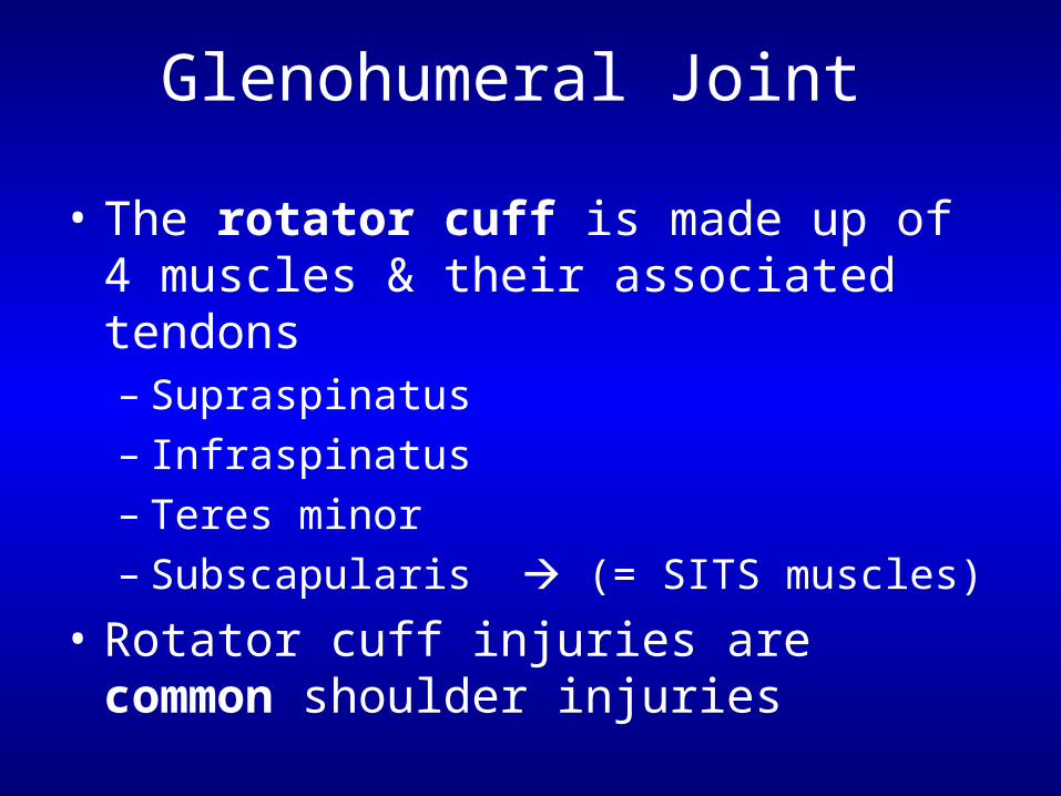
Glenohumeral Joint
• The rotator cuff is made up of 4 muscles & their associated tendons– Supraspinatus– Infraspinatus– Teres minor– Subscapularis (= SITS muscles)
• Rotator cuff injuries are common shoulder injuries
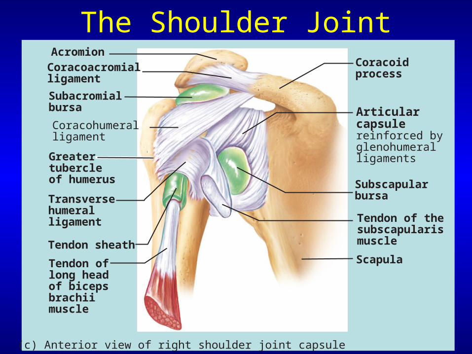
The Shoulder JointAcromionCoracoacromialligament
Subacromialbursa
Coracohumeralligament
Greatertubercleof humerus
Transversehumeralligament
Tendon sheath
Tendon oflong headof bicepsbrachiimuscle
Articularcapsulereinforced byglenohumeralligaments
Subscapularbursa
Tendon of thesubscapularismuscle
Scapula
Coracoidprocess
(c) Anterior view of right shoulder joint capsule
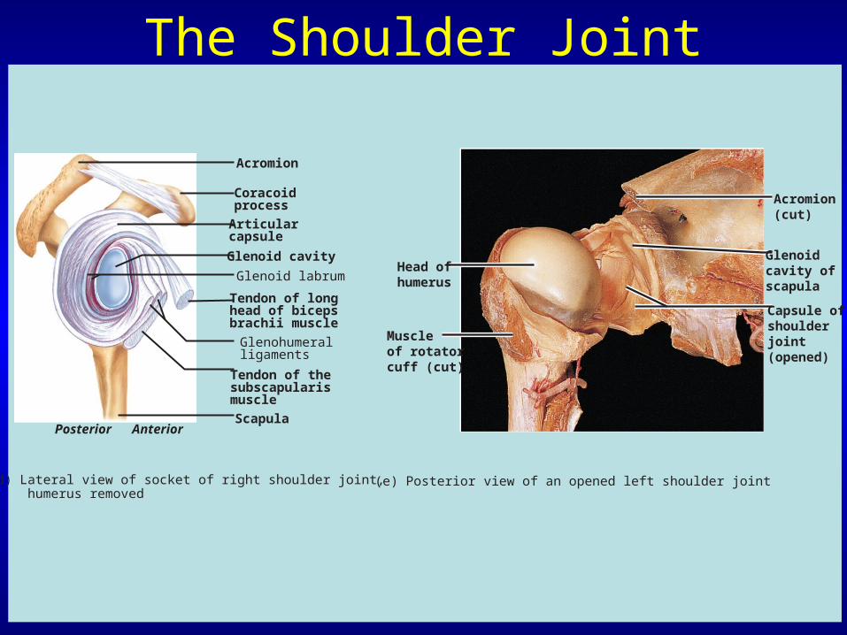
The Shoulder Joint
Acromion
Coracoidprocess
Articularcapsule
Glenoid cavity
Glenoid labrum
Tendon of longhead of bicepsbrachii muscle
Glenohumeralligaments
Tendon of thesubscapularismuscle
ScapulaPosterior Anterior
(d) Lateral view of socket of right shoulder joint, humerus removed
(e) Posterior view of an opened left shoulder joint
Head ofhumerus
Muscleof rotatorcuff (cut)
Acromion(cut)
Glenoidcavity ofscapula
Capsule ofshoulderjoint(opened)

Selected Synovial Joints
• Elbow joint– Allows flexion & extension– The humerus’ articulation with the trochlear
notch of the ulna forms the hinge– Tendons of biceps & triceps brachii provide
stability
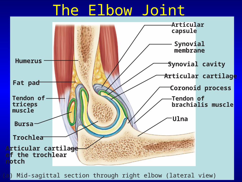
The Elbow JointArticularcapsule
Synovialmembrane
Synovial cavity
Articular cartilage
Coronoid process
Tendon ofbrachialis muscle
Ulna
Humerus
Fat pad
Tendon oftricepsmuscle
Bursa
Trochlea
Articular cartilageof the trochlearnotch
(a) Mid-sagittal section through right elbow (lateral view)
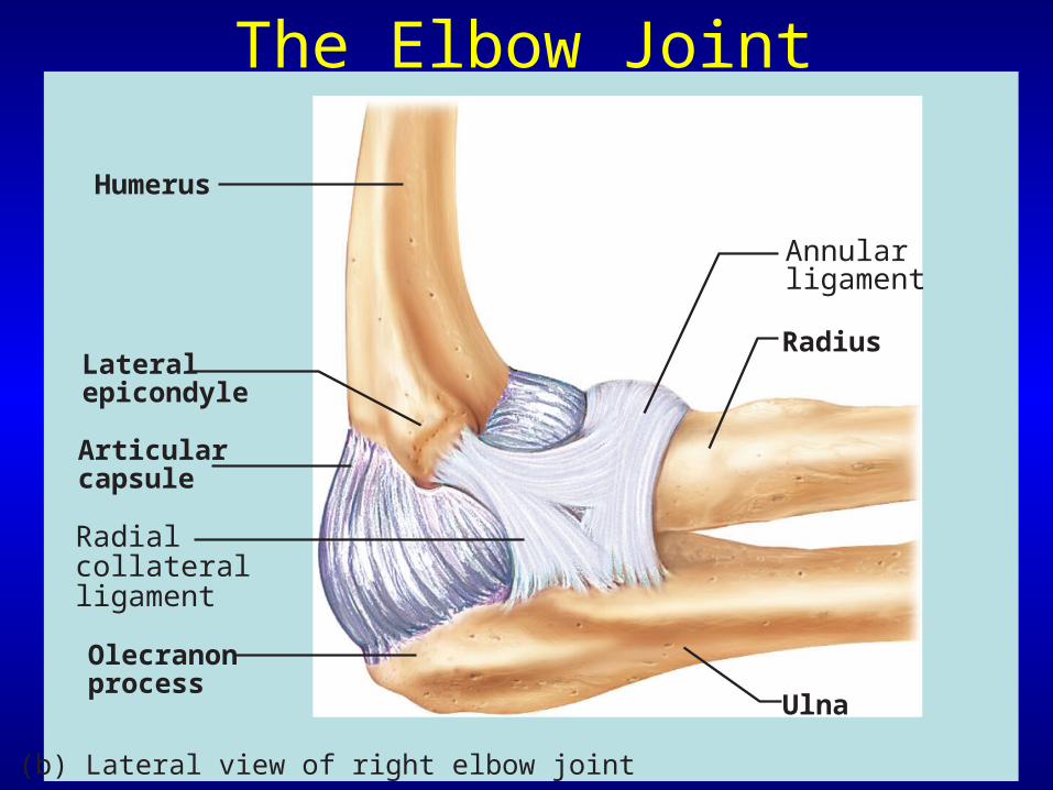
The Elbow Joint
Humerus
Lateralepicondyle
Articularcapsule
Radialcollateralligament
Olecranonprocess
(b) Lateral view of right elbow joint
Annularligament
Radius
Ulna
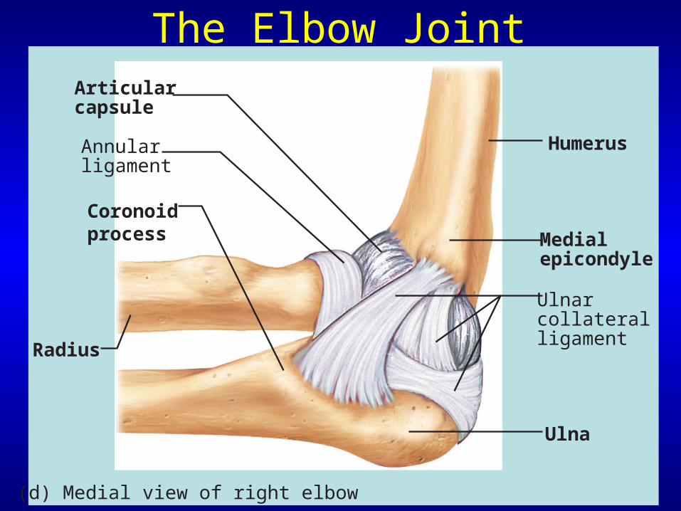
The Elbow JointArticularcapsule
Annularligament
Coronoidprocess
(d) Medial view of right elbow
Radius
Humerus
Medialepicondyle
Ulnarcollateralligament
Ulna
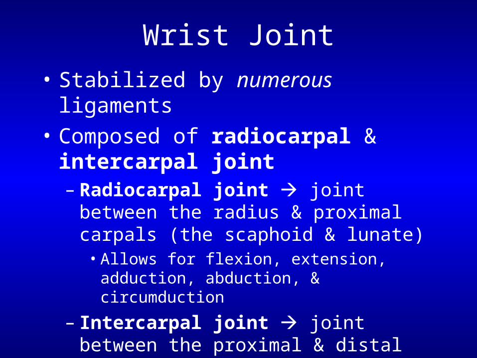
Wrist Joint
• Stabilized by numerous ligaments
• Composed of radiocarpal & intercarpal joint– Radiocarpal joint joint between the
radius & proximal carpals (the scaphoid & lunate)
• Allows for flexion, extension, adduction, abduction, & circumduction
– Intercarpal joint joint between the proximal & distal rows of carpals
• Allows for gliding movement
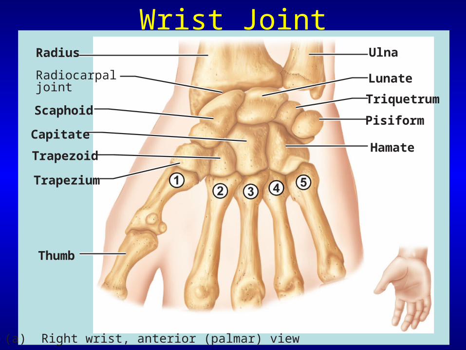
Wrist JointRadius Ulna
Lunate
Triquetrum
Pisiform
HamateCapitate
Scaphoid
Trapezoid
Trapezium
Thumb
Radiocarpaljoint
(a) Right wrist, anterior (palmar) view

Wrist JointDistalradioulnarjoint
Ulnarcollateralligament
ArticulardiscRadial
collateralligament
Radiocarpaljoint
Intercarpaljoint
(b) Wrist joints, coronal section
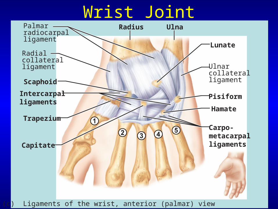
Wrist Joint
Hamate
Carpo-metacarpalligaments
Pisiform
Lunate
Radius Ulna
Ulnarcollateralligament
Radialcollateralligament
Palmarradiocarpalligament
Intercarpalligaments
Trapezium
Capitate
Scaphoid
(c) Ligaments of the wrist, anterior (palmar) view
Selected Synovial Joints
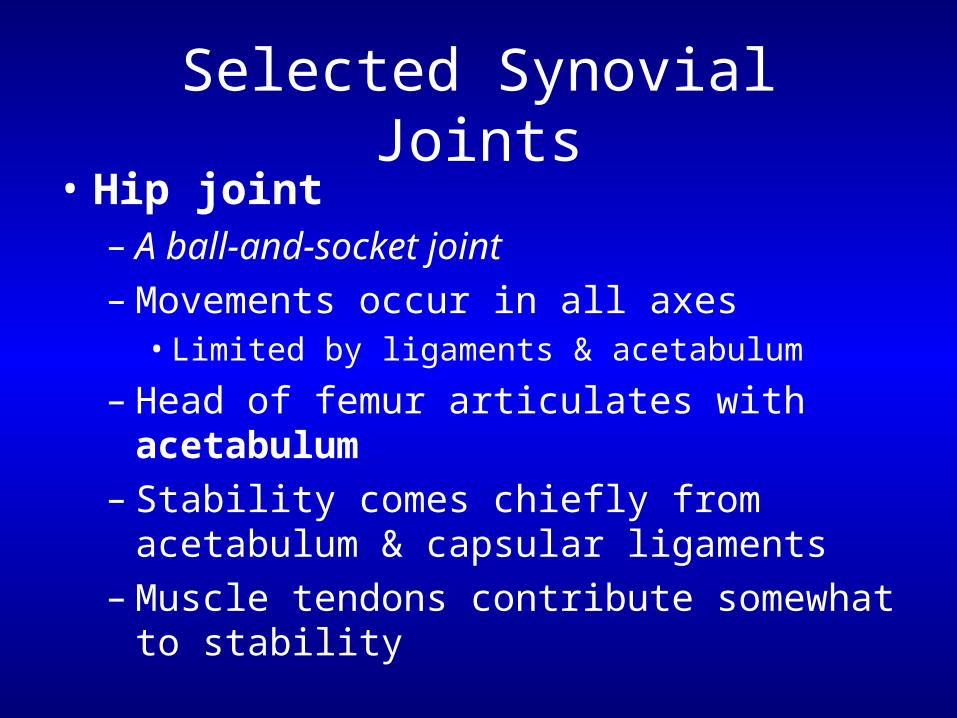
• Hip joint– A ball-and-socket joint– Movements occur in all axes
• Limited by ligaments & acetabulum
– Head of femur articulates with acetabulum– Stability comes chiefly from acetabulum &
capsular ligaments – Muscle tendons contribute somewhat to
stability
PLAYPLAYMovement of thehip joint
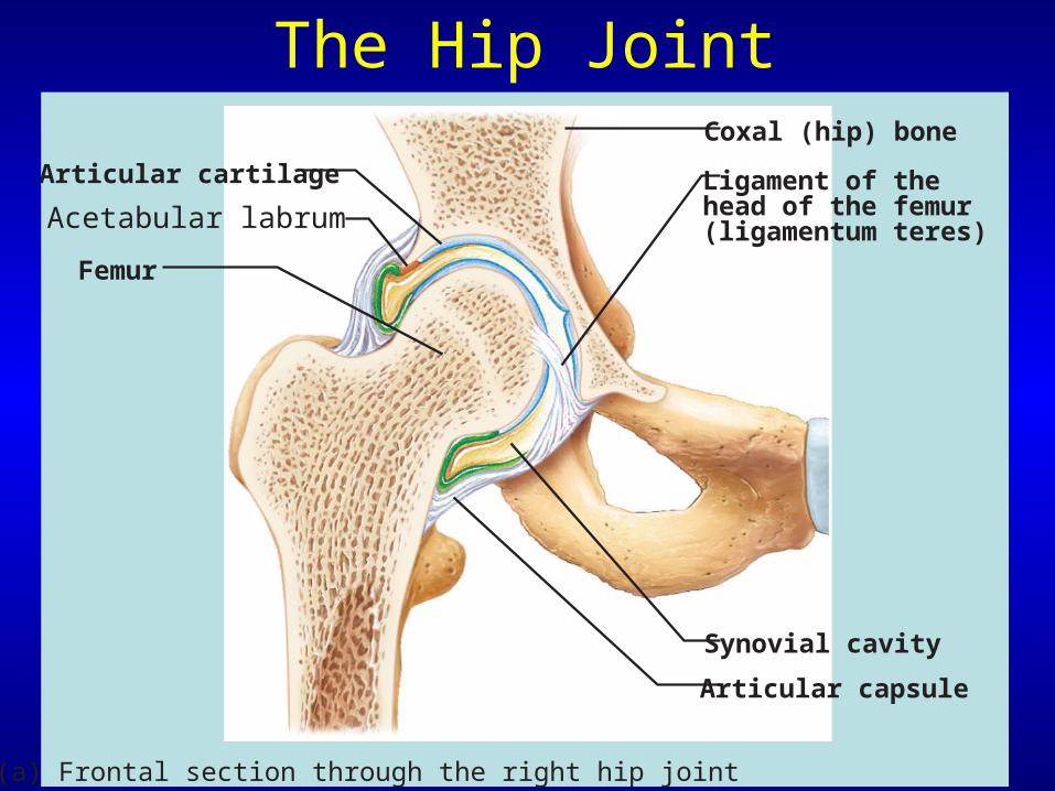
The Hip Joint
Articular cartilage
Coxal (hip) bone
Ligament of thehead of the femur(ligamentum teres)
Synovial cavity
Articular capsule
Acetabular labrum
Femur
(a) Frontal section through the right hip joint
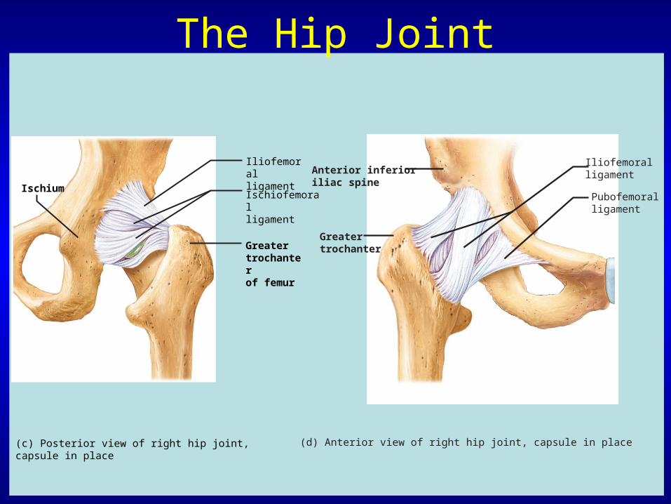
Anterior inferioriliac spine
Iliofemoralligament
Pubofemoralligament
Greatertrochanter
(d) Anterior view of right hip joint, capsule in place
The Hip Joint
Ischium
Iliofemoralligament
Ischiofemoralligament
Greatertrochanterof femur
(c) Posterior view of right hip joint, capsule in place
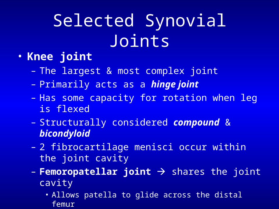
Selected Synovial Joints
• Knee joint– The largest & most complex joint– Primarily acts as a hinge joint– Has some capacity for rotation when leg is flexed– Structurally considered compound & bicondyloid– 2 fibrocartilage menisci occur within the joint cavity– Femoropatellar joint shares the joint cavity
• Allows patella to glide across the distal femur
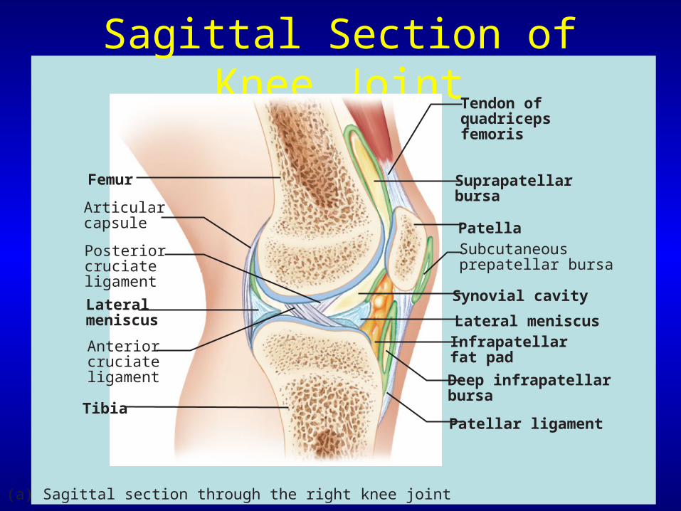
Sagittal Section of Knee Joint
(a) Sagittal section through the right knee joint
Femur
Tendon ofquadricepsfemoris
Suprapatellarbursa
PatellaSubcutaneousprepatellar bursa
Synovial cavity
Lateral meniscus
Posteriorcruciateligament
Infrapatellarfat pad
Deep infrapatellarbursa
Patellar ligament
Articularcapsule
Lateralmeniscus
Anteriorcruciateligament
Tibia
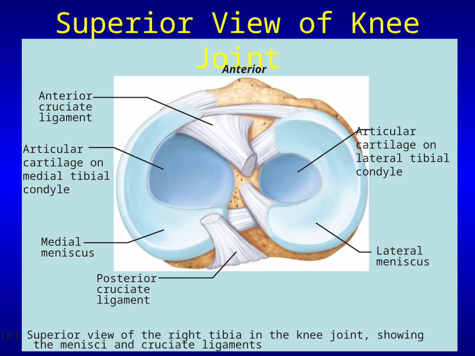
Superior View of Knee Joint
(b) Superior view of the right tibia in the knee joint, showing the menisci and cruciate ligaments
Medialmeniscus
Articularcartilage on medial tibial condyle
Anterior
Anteriorcruciateligament
Articularcartilage onlateral tibialcondyle
Lateralmeniscus
Posteriorcruciateligament
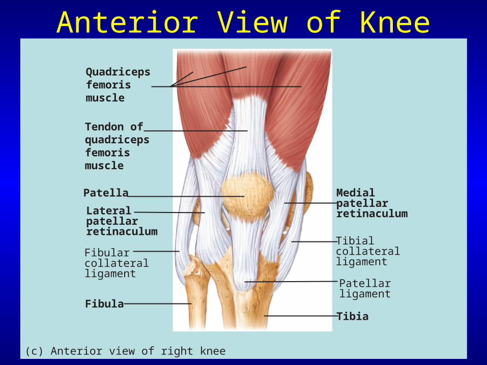
Anterior View of Knee
Quadricepsfemorismuscle
Tendon ofquadricepsfemorismuscle
Patella
Lateralpatellarretinaculum
Medialpatellarretinaculum
Tibialcollateralligament
Tibia
Fibularcollateralligament
Fibula
(c) Anterior view of right knee
Patellarligament
Knee Joint
• Ligaments of the knee joint:– Become taut when knee is extended– These extracapsular & capsular ligaments are:
• Fibular & tibial collateral ligaments• Oblique popliteal ligament• Arcuate popliteal ligament
Knee Joint
• Intracapsular ligaments– Cruciate ligaments
• Cross each other like an “X”
– Each cruciate ligament runs from the proximal tibia to the distal femur
• Anterior cruciate ligament (ACL) • Posterior cruciate ligament (PCL)
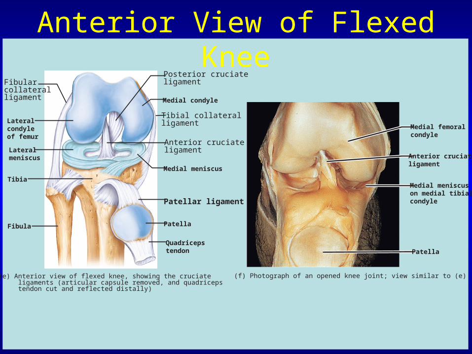
Anterior View of Flexed Knee
Fibularcollateralligament
Posterior cruciateligament
Medial condyle
Tibial collateralligament
Anterior cruciateligament
Medial meniscus
Patellar ligament
Patella
Quadricepstendon
Lateralcondyleof femur
Lateralmeniscus
Fibula
(e) Anterior view of flexed knee, showing the cruciate ligaments (articular capsule removed, and quadriceps tendon cut and reflected distally)
Tibia
Medial femoralcondyle
Anterior cruciateligament
Medial meniscuson medial tibialcondyle
Patella
(f) Photograph of an opened knee joint; view similar to (e)
Knee Joint
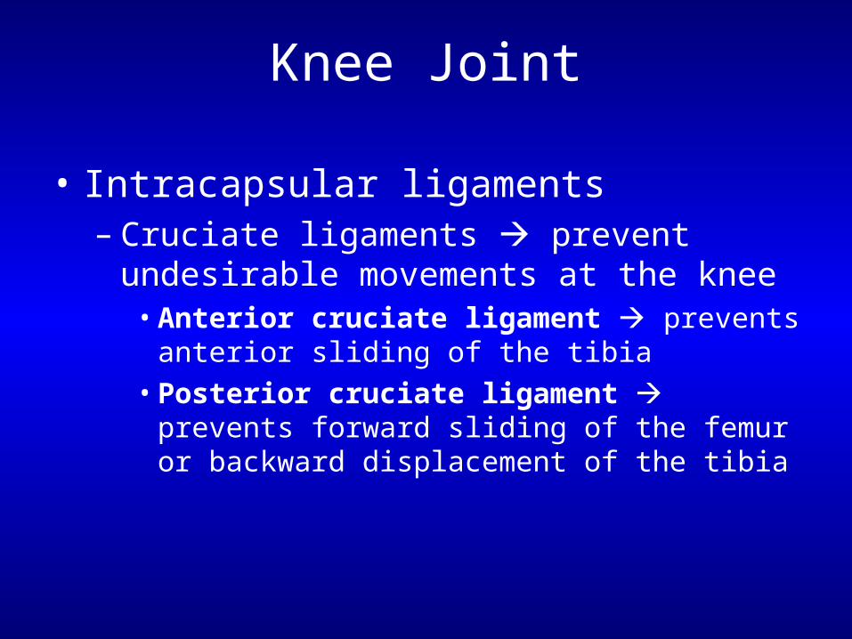
• Intracapsular ligaments– Cruciate ligaments prevent undesirable
movements at the knee• Anterior cruciate ligament prevents anterior
sliding of the tibia• Posterior cruciate ligament prevents forward
sliding of the femur or backward displacement of the tibia
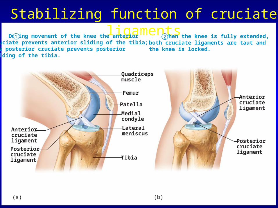
Stabilizing function of cruciate ligaments During movement of the knee the anteriorcruciate prevents anterior sliding of the tibia; the posterior cruciate prevents posterior sliding of the tibia.
Anteriorcruciateligament
(a)
Posteriorcruciateligament
Quadricepsmuscle
Femur
Patella
Lateralmeniscus
Tibia
Medialcondyle
(b)
Anteriorcruciateligament
Posteriorcruciateligament
When the knee is fully extended,both cruciate ligaments are taut and the knee is locked.
1 2
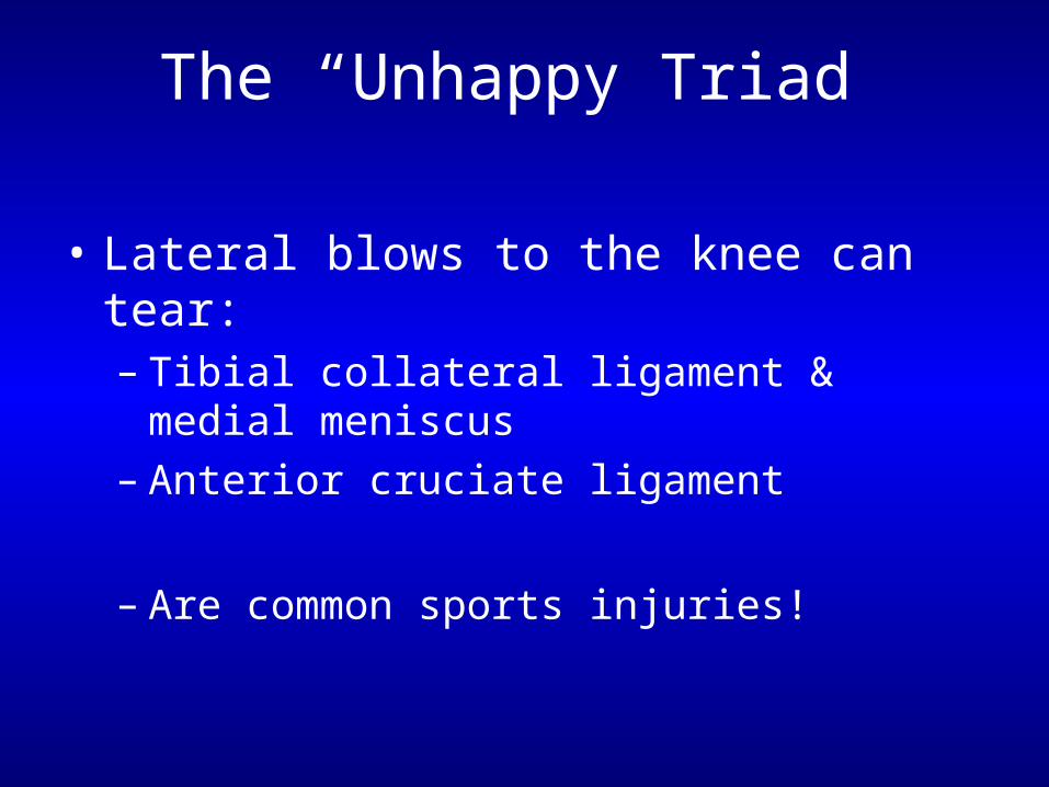
The “Unhappy Triad”
• Lateral blows to the knee can tear:– Tibial collateral ligament & medial meniscus– Anterior cruciate ligament
– Are common sports injuries!
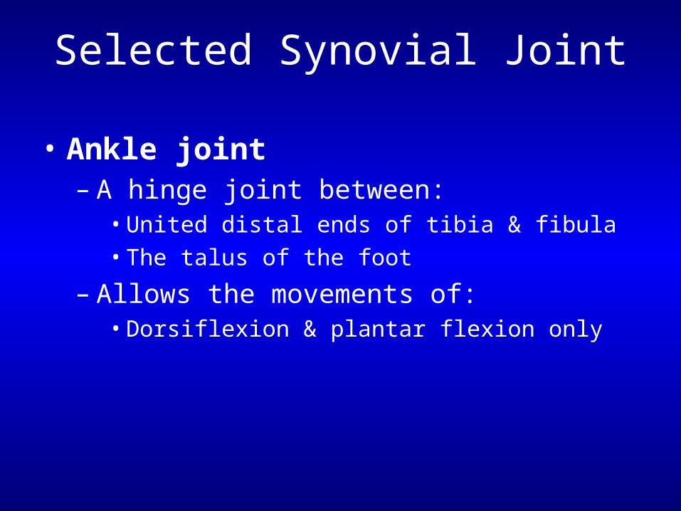
Selected Synovial Joint
• Ankle joint– A hinge joint between:
• United distal ends of tibia & fibula • The talus of the foot
– Allows the movements of:• Dorsiflexion & plantar flexion only
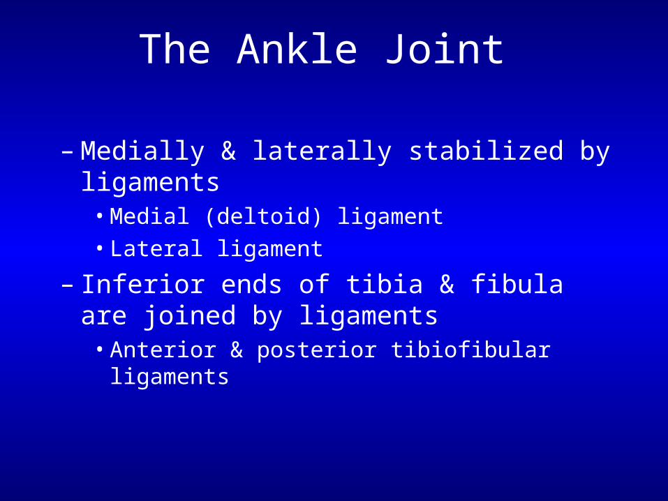
The Ankle Joint
– Medially & laterally stabilized by ligaments• Medial (deltoid) ligament• Lateral ligament
– Inferior ends of tibia & fibula are joined by ligaments
• Anterior & posterior tibiofibular ligaments
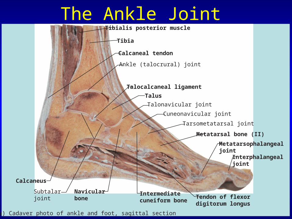
The Ankle Joint Tibia
Calcaneal tendon
Calcaneus
Navicularbone
Subtalarjoint
(a) Cadaver photo of ankle and foot, sagittal section
Intermediatecuneiform bone
Tendon of flexordigitorum longus
Metatarsal bone (II)
Interphalangeal joint
Metatarsophalangealjoint
Cuneonavicular joint
Talonavicular joint
Talocalcaneal ligament
Tibialis posterior muscle
Ankle (talocrural) joint
Tarsometatarsal joint
Talus
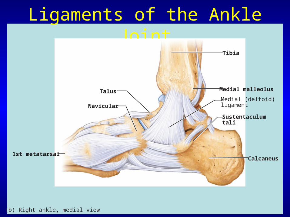
Ligaments of the Ankle Joint
Medial malleolus
Calcaneus
Sustentaculumtali
Medial (deltoid)ligament
Talus
Navicular
Tibia
1st metatarsal
(b) Right ankle, medial view
Ligaments of the Ankle Joint
Anterior tibiofibularligament
Anterior talofibularligament
Calcaneus
Metatarsals
Lateral malleolus
Posterior tibiofibularligament
Posterior talofibularligament Calcaneofibularligament
Tibia
Talus
Cuboid
Fibula
(c) Right ankle, lateral view
Lateralligament
Disorders of Joints
• Structure of joints makes them prone to traumatic stress
• Function of joints makes them subject to friction and wear & tear
• Affected by inflammatory & degenerative processes
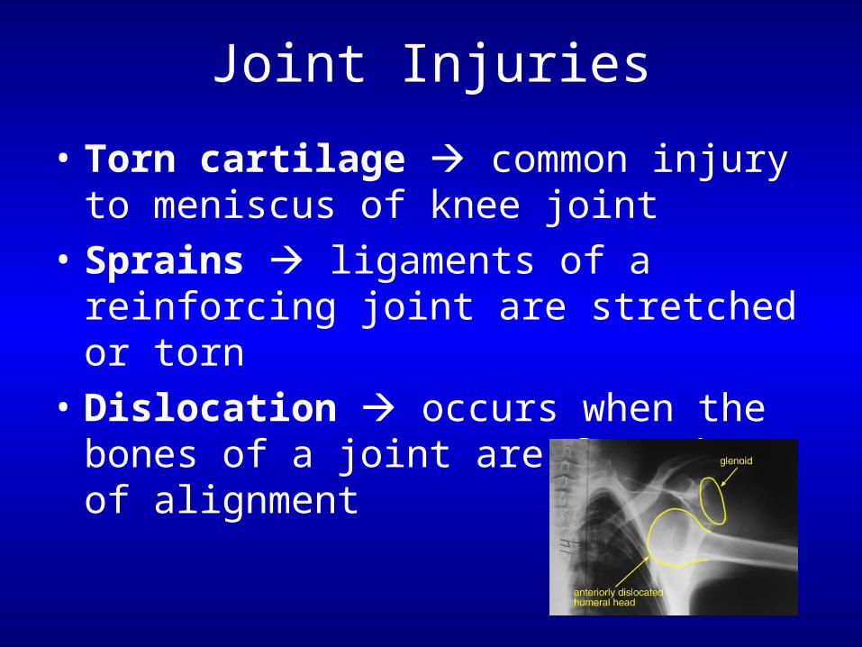
Joint Injuries
• Torn cartilage common injury to meniscus of knee joint
• Sprains ligaments of a reinforcing joint are stretched or torn
• Dislocation occurs when the bones of a joint are forced out of alignment
Inflammatory & Degenerative Conditions
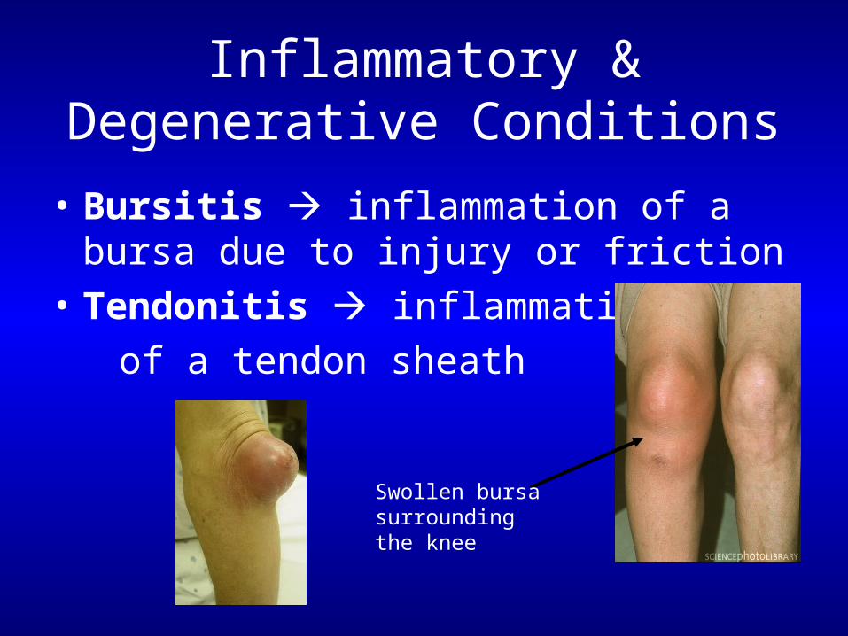
• Bursitis inflammation of a bursa due to injury or friction
• Tendonitis inflammation
of a tendon sheath
Swollen bursa surroundingthe knee
Inflammatory & Degenerative Conditions
• Arthritis describes over 100 kinds of joint-damaging diseases– Osteoarthritis most common type of “wear
& tear” arthritis– Rheumatoid arthritis a chronic
inflammatory disorder – Gouty arthritis (gout) uric acid build-up
causes pain in joints• Lyme disease inflammatory disease often
resulting in joint pain
The Joints Throughout Life
• Synovial joints develop from mesenchyme
• By week 8 of fetal development, joints resemble adult joints– Outer region of mesenchyme becomes fibrous
joint capsule– Inner region becomes the joint cavity
The Joints Throughout Life
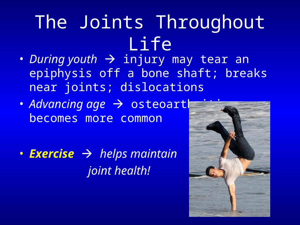
• During youth injury may tear an epiphysis off a bone shaft; breaks near joints; dislocations
• Advancing age osteoarthritis becomes more common
• Exercise helps maintain
joint health!
Keeping Your Joints Healthy
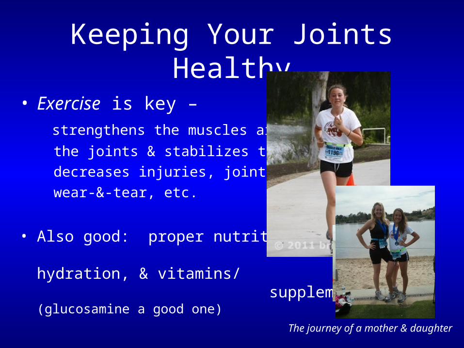
• Exercise is key –
strengthens the muscles around
the joints & stabilizes them;
decreases injuries, joint disorders,
wear-&-tear, etc.
• Also good: proper nutrition, hydration, & vitamins/ supplements (glucosamine a good one)
The journey of a mother & daughter

Keeping Your Joints Healthy
Questions…?
What’s Next?Lab: Finish Bones & JointsWed Lecture: Skeletal muscle Wed Lab: Skeletal muscle tissue & muscles