Joints Hip region Knee region Ankle region. sacroiliac joints hip joint pubic symphysis Hip region.
63
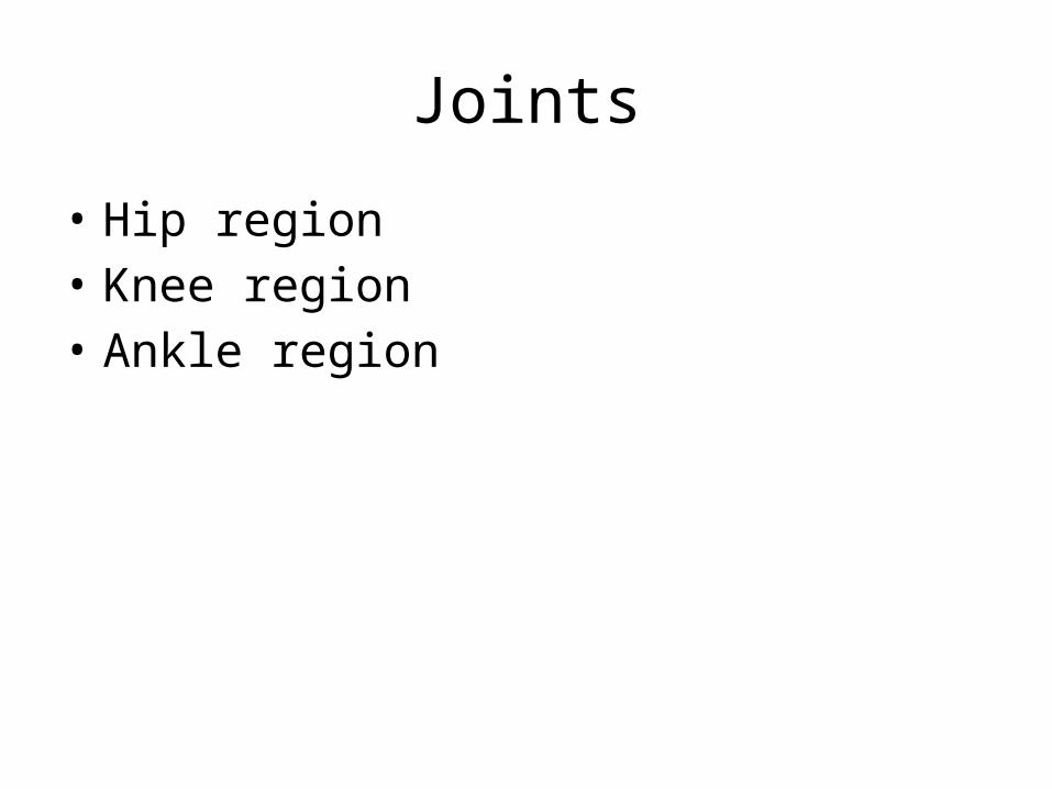
Joints • Hip region • Knee region • Ankle region
-
Upload
gonzalo-dimery -
Category
Documents
-
view
228 -
download
1
Transcript of Joints Hip region Knee region Ankle region. sacroiliac joints hip joint pubic symphysis Hip region.
Joints
• Hip region• Knee region• Ankle region
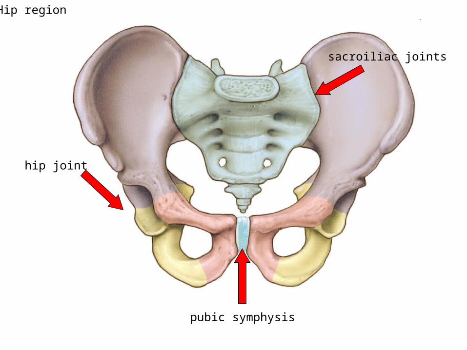
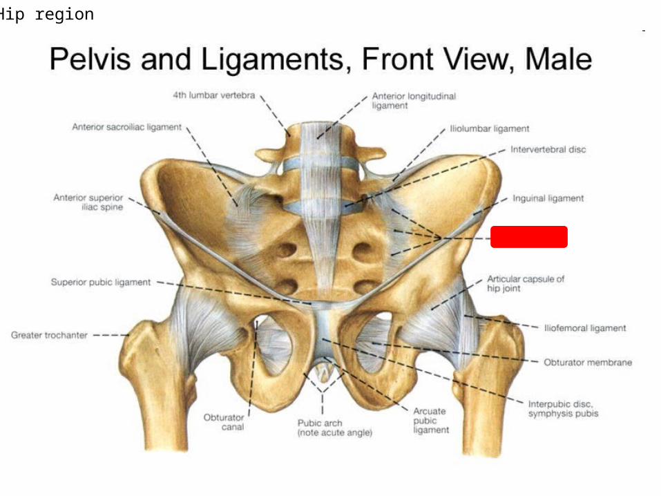
sacroiliac joints
hip joint
pubic symphysis
Hip region
Hip region
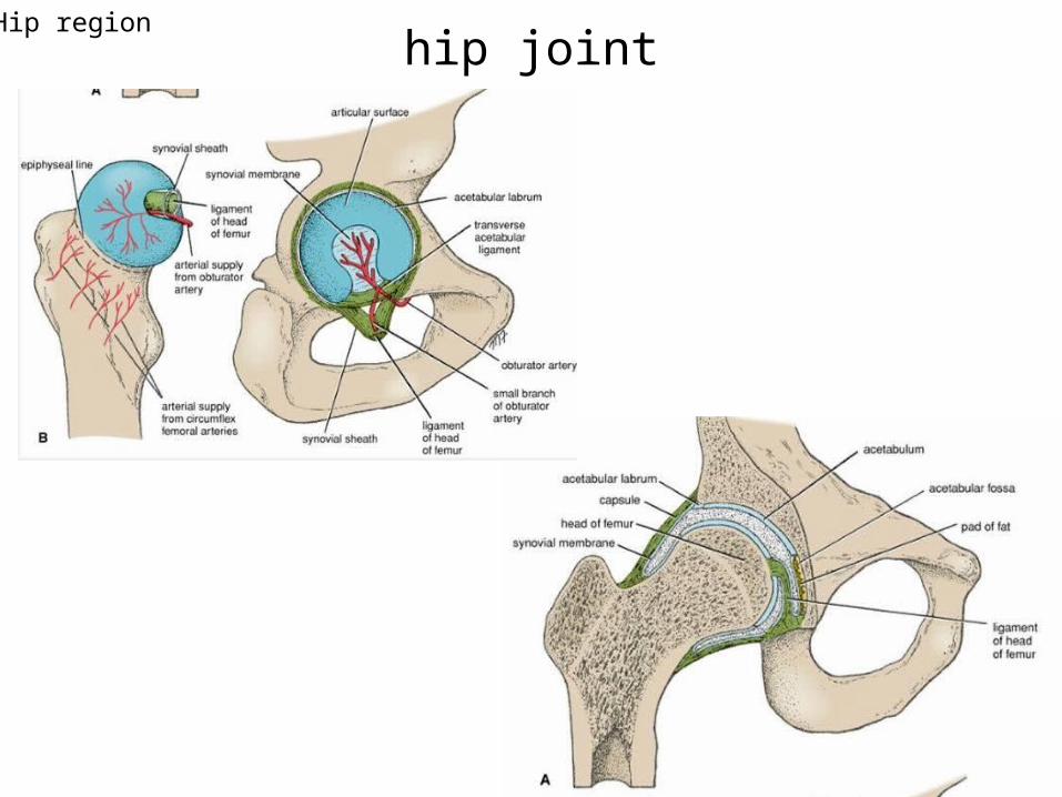
hip jointHip region
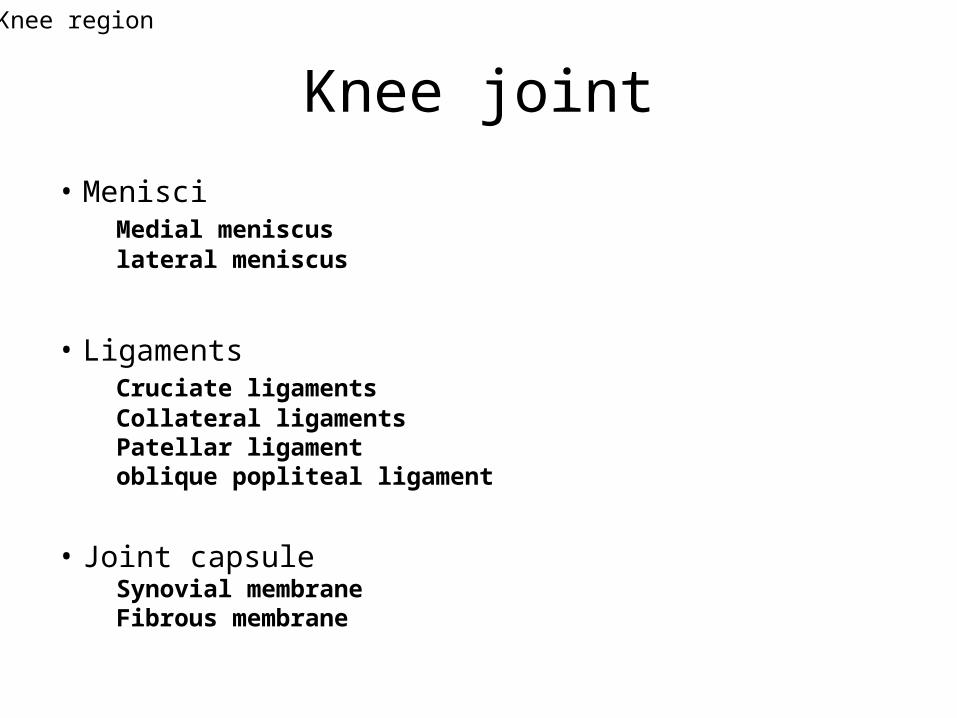
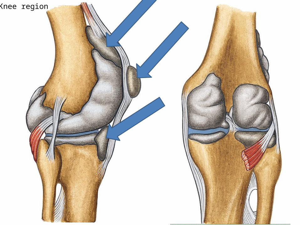
Knee joint
• MenisciMedial meniscuslateral meniscus
• Ligaments Cruciate ligamentsCollateral ligaments Patellar ligamentoblique popliteal ligament
• Joint capsuleSynovial membraneFibrous membrane
Knee region
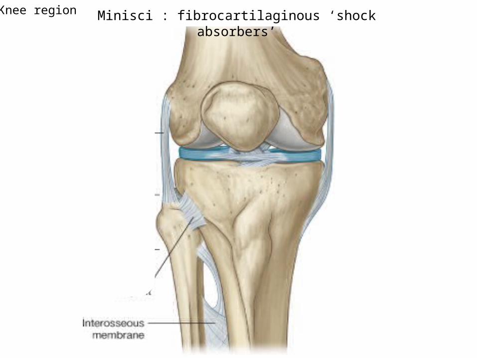
Minisci : fibrocartilaginous ‘shock absorbers’Knee region

semilunar cartilages
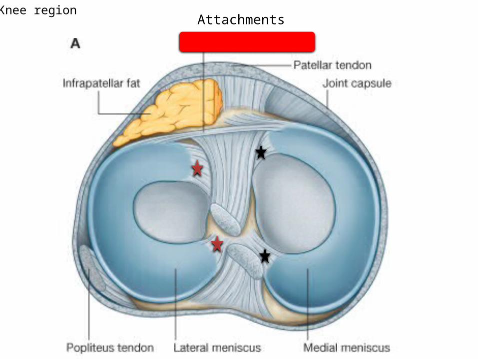
AttachmentsKnee region
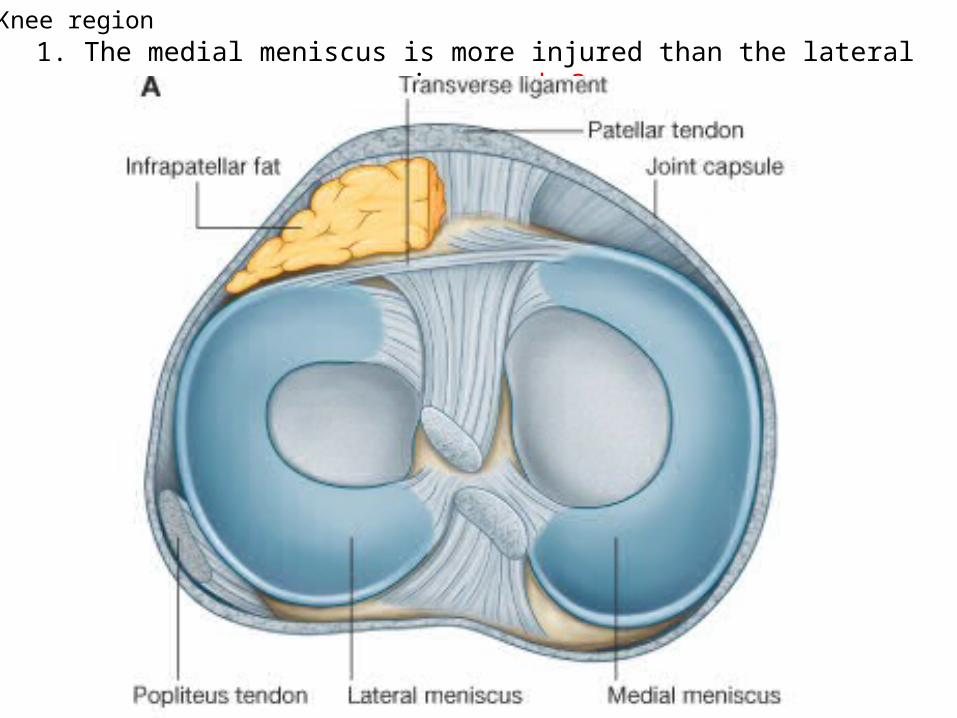
1. The medial meniscus is more injured than the lateral meniscus, why?Knee region
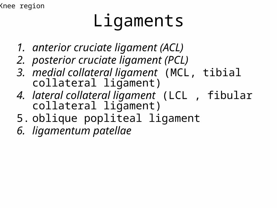
Ligaments1. anterior cruciate ligament (ACL)2. posterior cruciate ligament (PCL)3. medial collateral ligament (MCL, tibial collateral
ligament)4. lateral collateral ligament (LCL , fibular collateral
ligament) 5. oblique popliteal ligament6. ligamentum patellae
Knee region
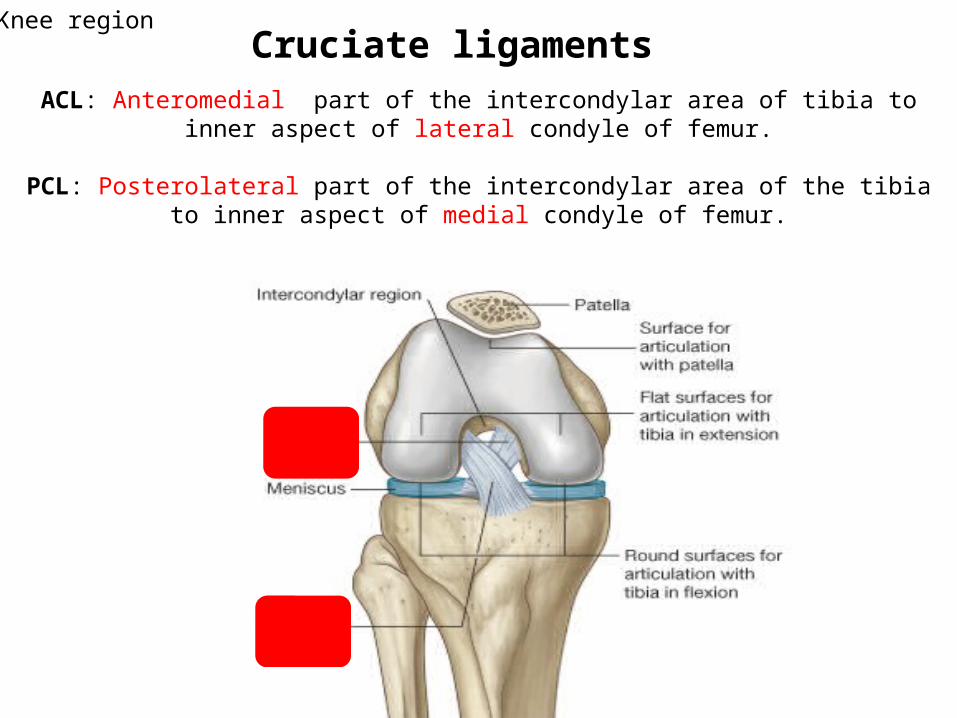
ACL: Anteromedial part of the intercondylar area of tibia to inner aspect of lateral condyle of femur.
PCL: Posterolateral part of the intercondylar area of the tibia to inner aspect of medial condyle of femur.
Cruciate ligamentsKnee region
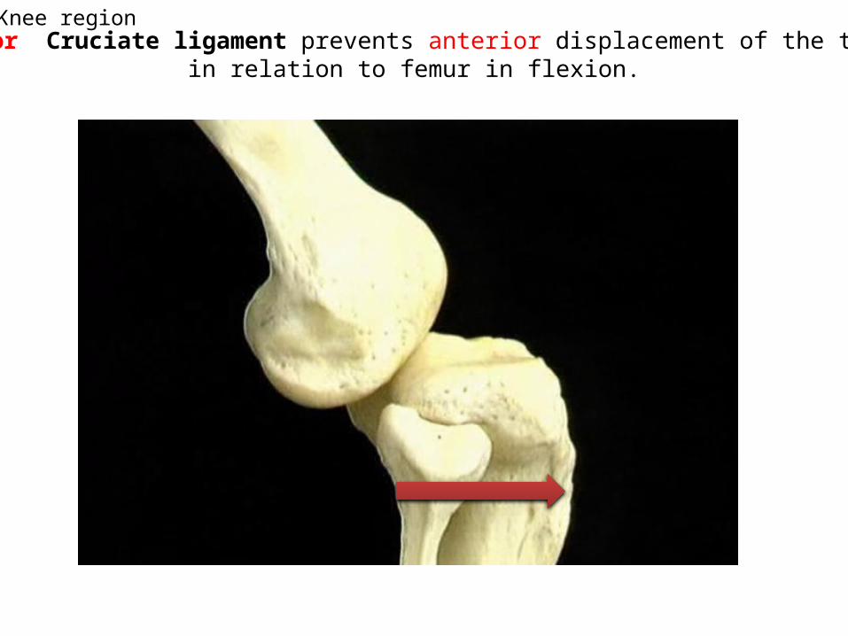
Anterior Cruciate ligament prevents anterior displacement of the tibia in relation to femur in flexion.
Knee region
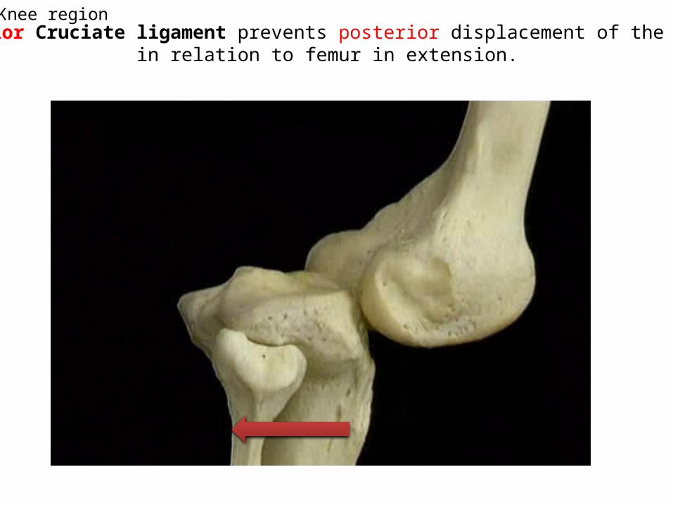
Posterior Cruciate ligament prevents posterior displacement of the tibia in relation to femur in extension.
Knee region
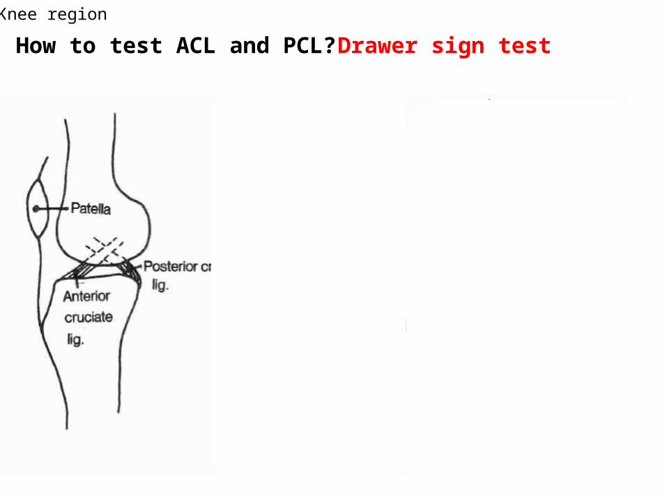
How to test ACL and PCL? Drawer sign testKnee region
15
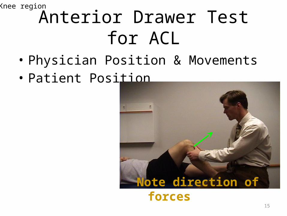
Anterior Drawer Test for ACL
• Physician Position & Movements• Patient Position
Note direction of forces
Knee region
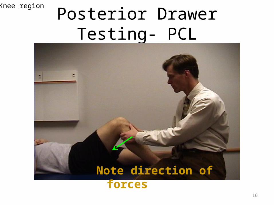
16
Posterior Drawer Testing- PCL
Note direction of forces
Knee region
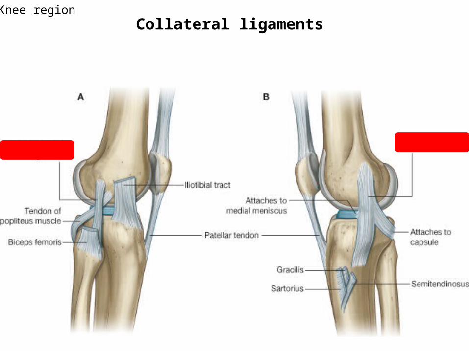
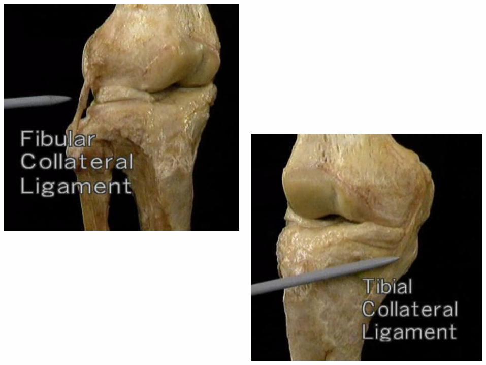
Collateral ligaments Knee region
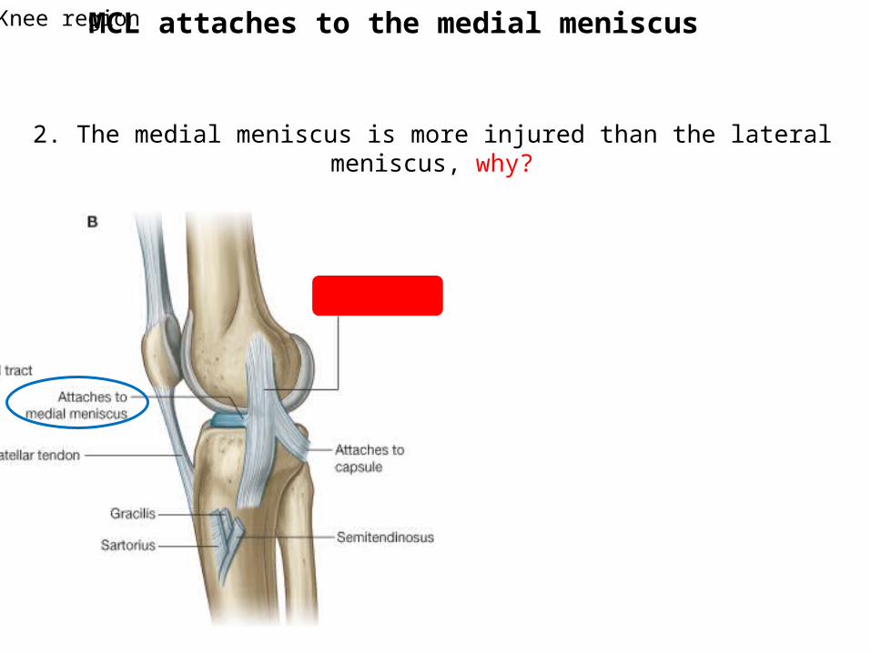
MCL attaches to the medial meniscus
2. The medial meniscus is more injured than the lateral meniscus, why?
Knee region
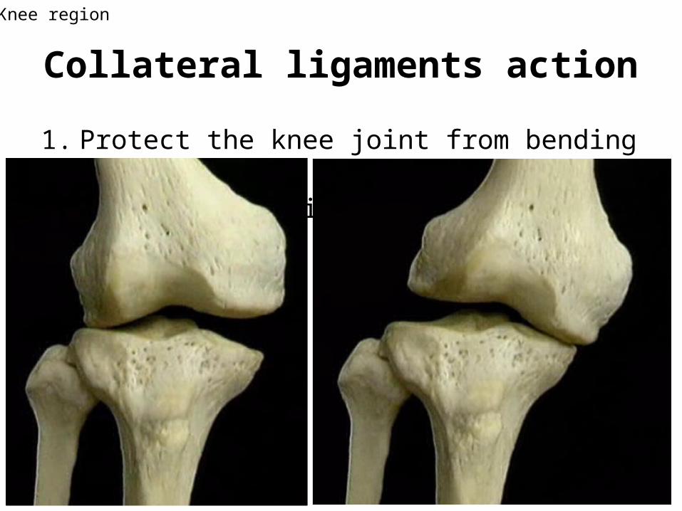
Collateral ligaments action
1. Protect the knee joint from bending side to side. 2. Helps the locking mechanism
Knee region
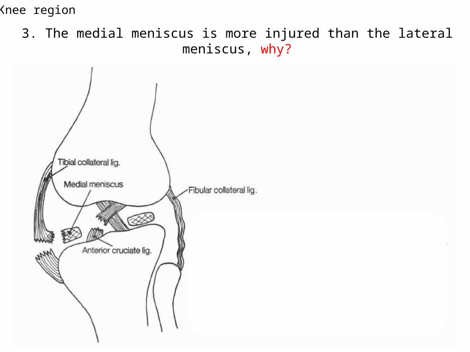
3. The medial meniscus is more injured than the lateral meniscus, why?
Knee region
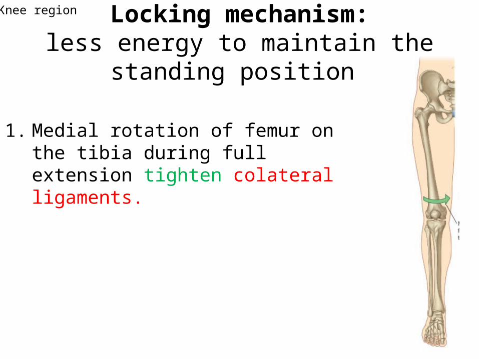
Locking mechanism:less energy to maintain the standing
position
1. Medial rotation of femur on the tibia during full extension tighten colateral ligaments.
Knee region

Locking mechanism (2)
• Joint surfaces become larger and more stable in extension.
Knee region
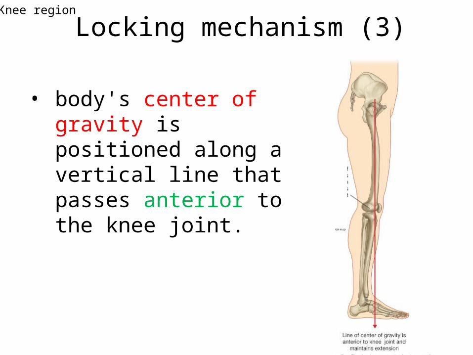
Locking mechanism (3)
• body's center of gravity is positioned along a vertical line that passes anterior to the knee joint.
Knee region
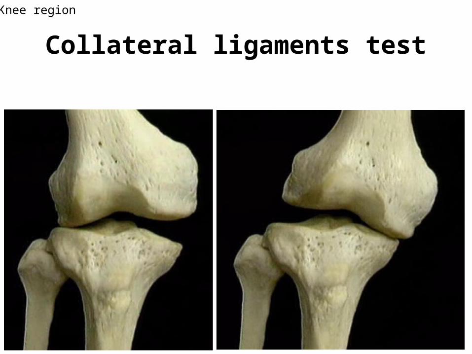
Collateral ligaments testKnee region
26
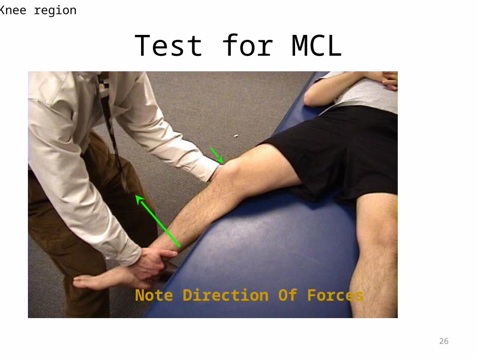
Test for MCL
Note Direction Of Forces
Knee region
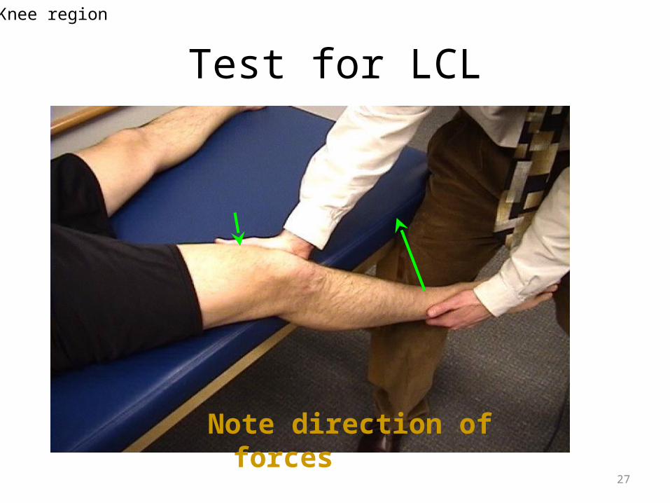
27
Test for LCL
Note direction of forces
Knee region
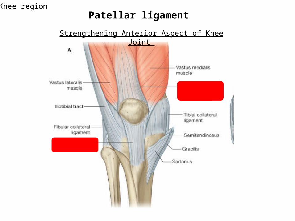
Patellar ligament
Strengthening Anterior Aspect of Knee Joint
Knee region
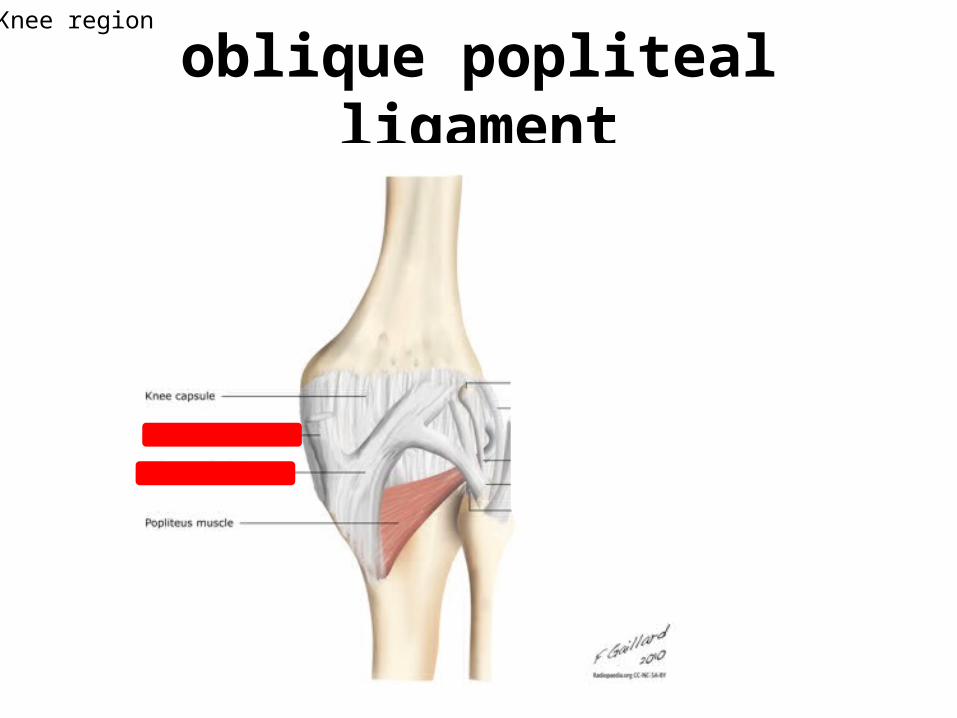
oblique popliteal ligamentKnee region
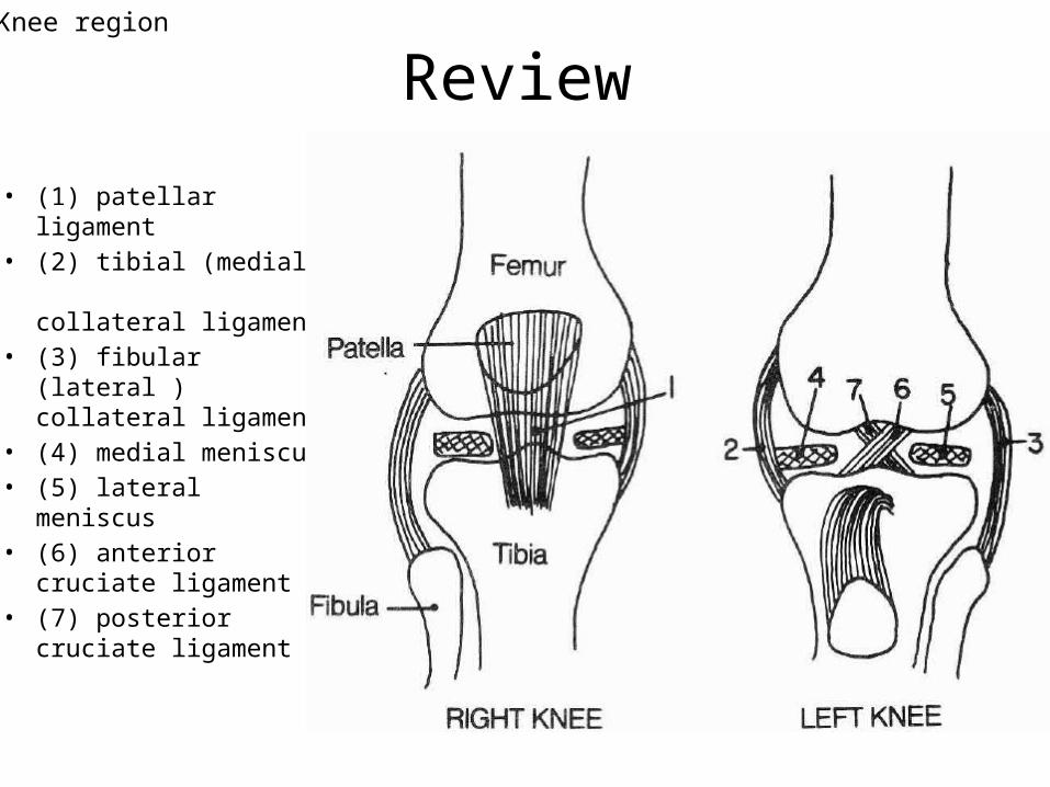
Review
• (1) patellar ligament • (2) tibial (medial)
collateral ligament• (3) fibular (lateral )
collateral ligament• (4) medial meniscus • (5) lateral meniscus• (6) anterior cruciate
ligament • (7) posterior cruciate
ligament
Knee region
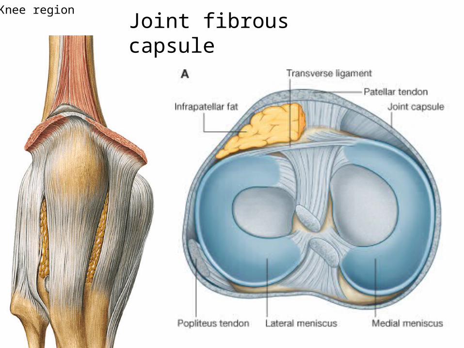
Joint fibrous capsuleKnee region
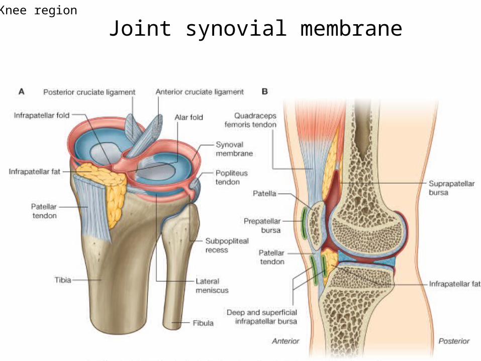
Joint synovial membraneKnee region
Knee region
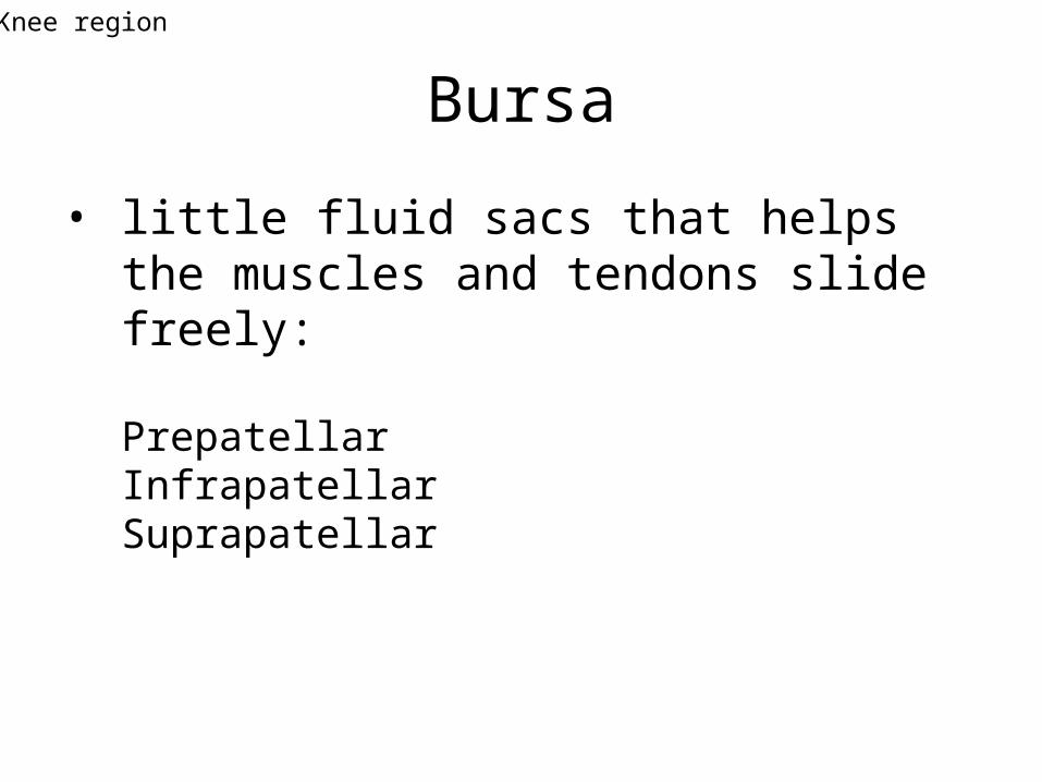
Bursa
• little fluid sacs that helps the muscles and tendons slide freely:
PrepatellarInfrapatellarSuprapatellar
Knee region
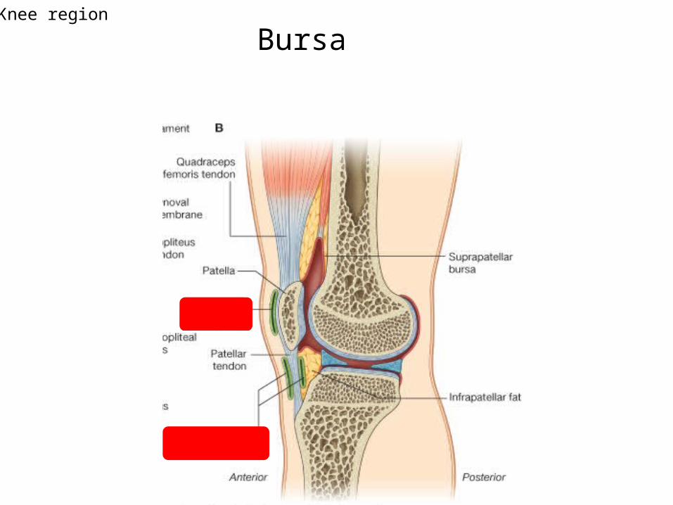
BursaKnee region
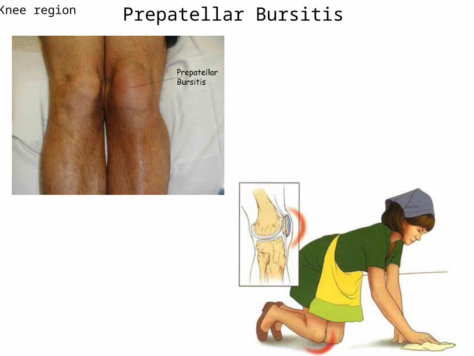
Prepatellar BursitisKnee region
Ankle region
• Ankle joint (talocrural joint)• Subtalar joint (ST J.)• Talocalcaneonavicular joint (TCN J.)
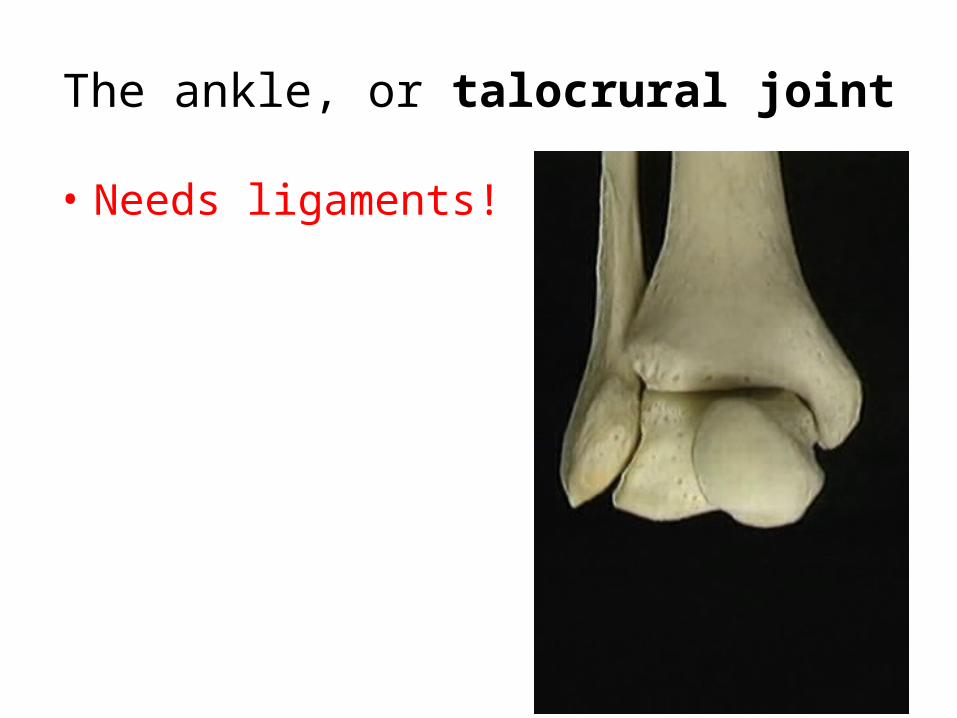
The ankle, or talocrural joint
• Needs ligaments!
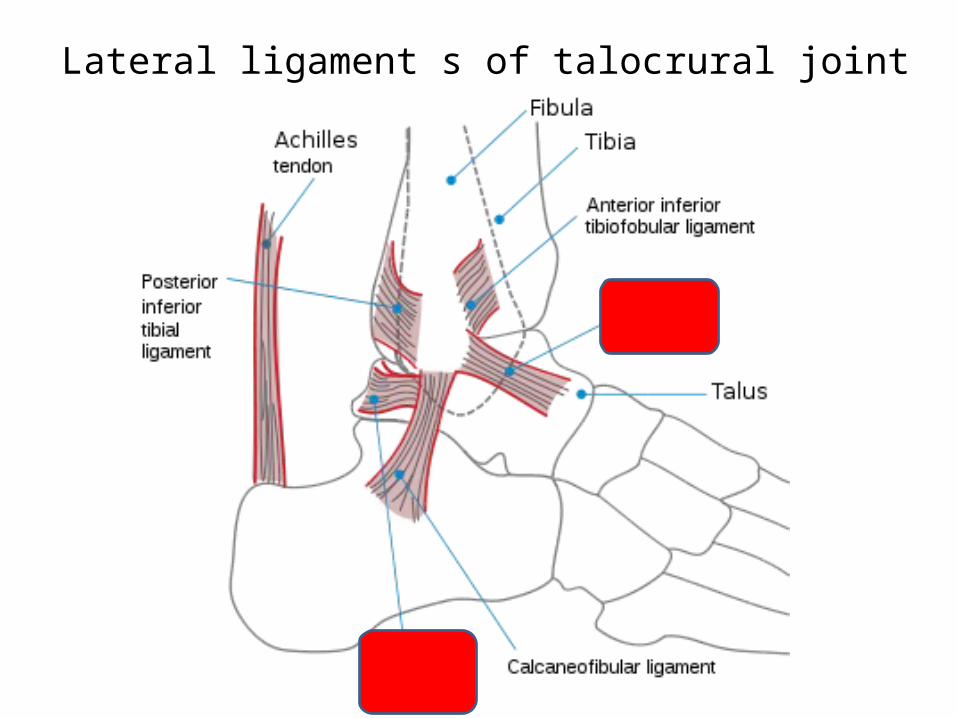
Lateral ligament s of talocrural joint
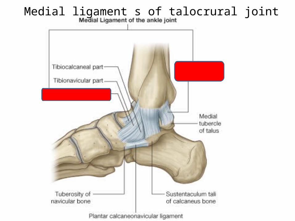
Medial ligament s of talocrural joint
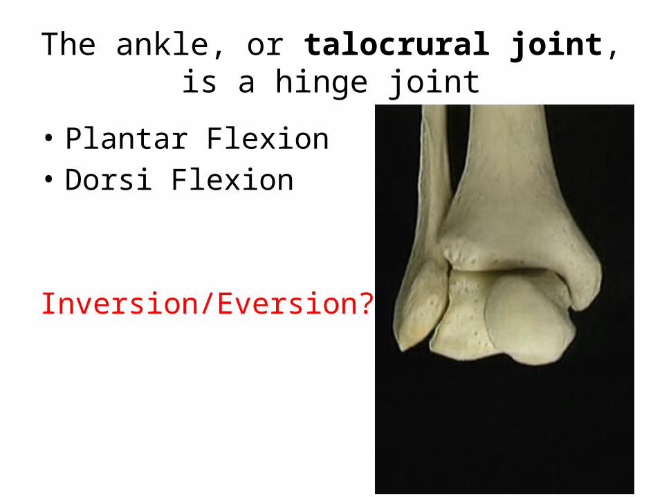
The ankle, or talocrural joint, is a hinge joint
• Plantar Flexion• Dorsi Flexion
Inversion/Eversion?
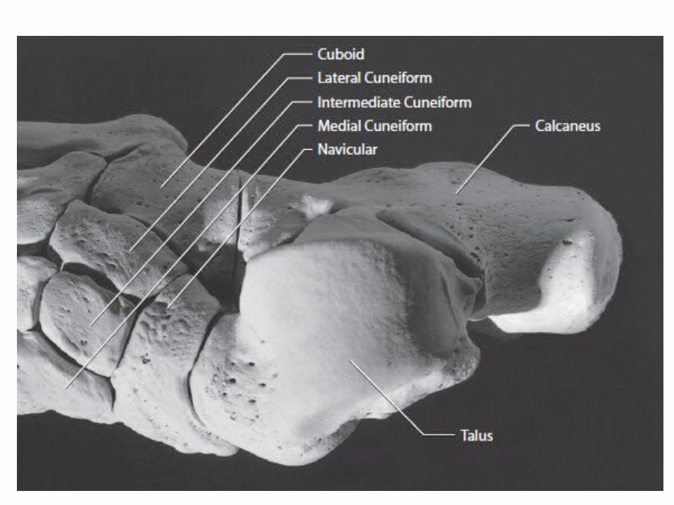
Intertarsal joint:
• Subtalar joint (ST J.)• Talocalcaneonavicular joint (TCN J.)• Calcaneocuboid (small rotation)• Naviculoconeiforms (almost no movement)
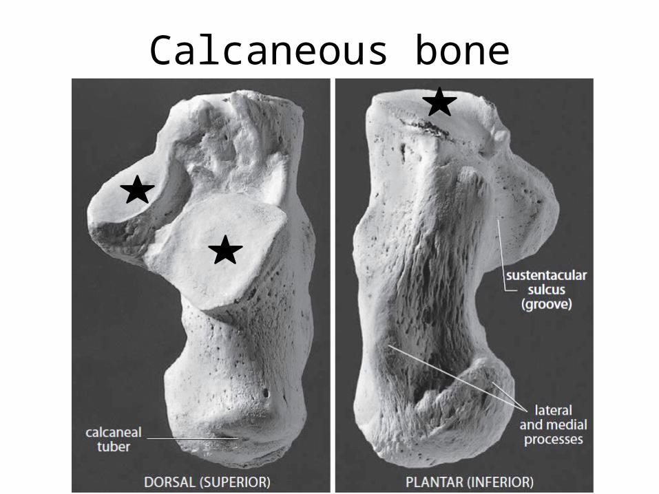
Calcaneous bone
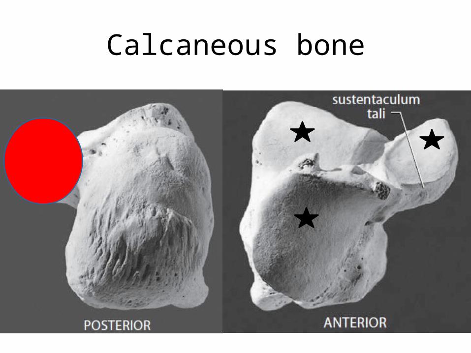
Calcaneous bone
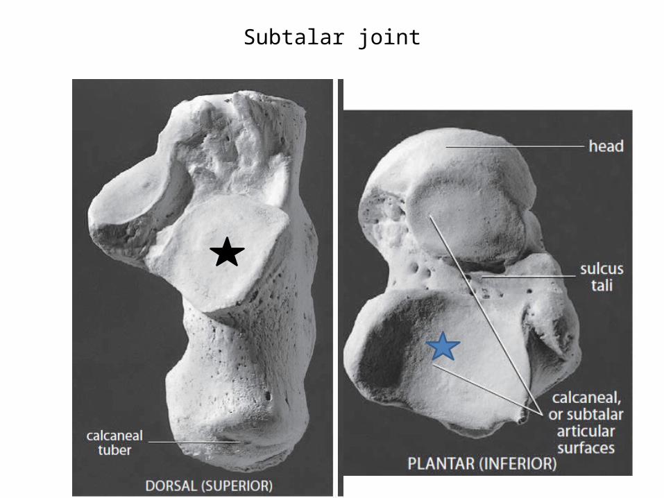
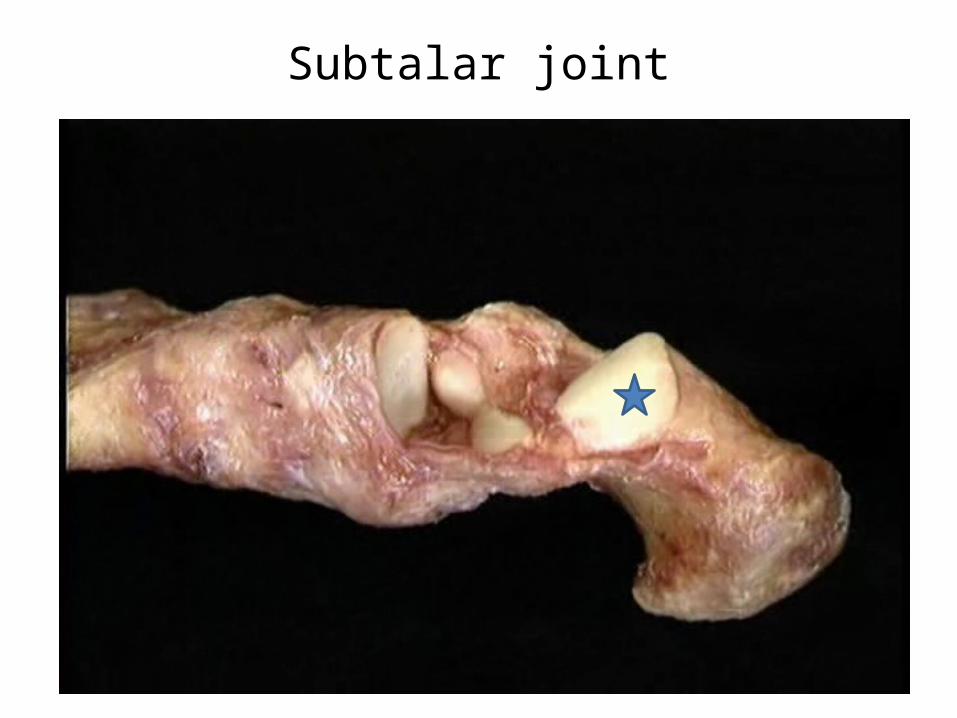
Subtalar joint
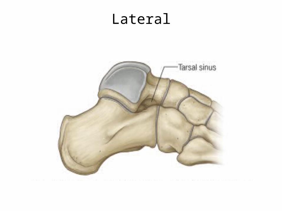
Lateral
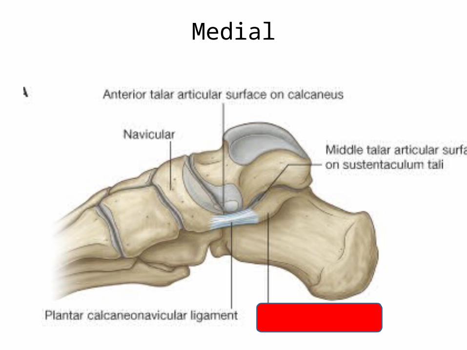
Medial
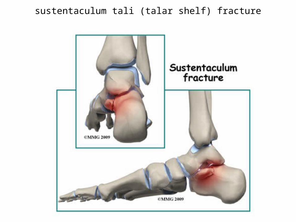
sustentaculum tali (talar shelf) fracture
Subtalar joint
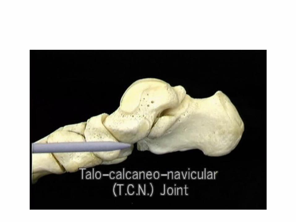
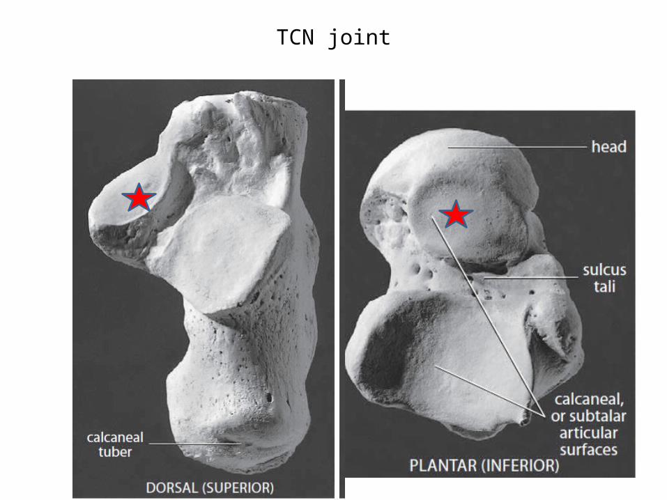
TCN joint
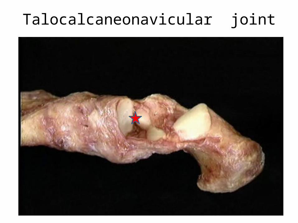
Talocalcaneonavicular joint
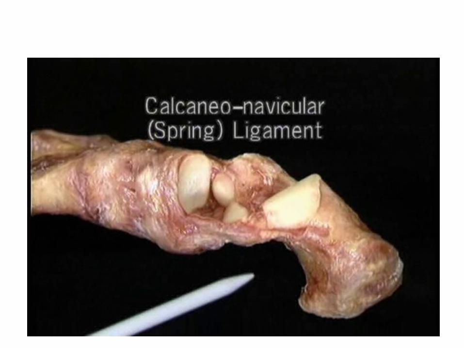
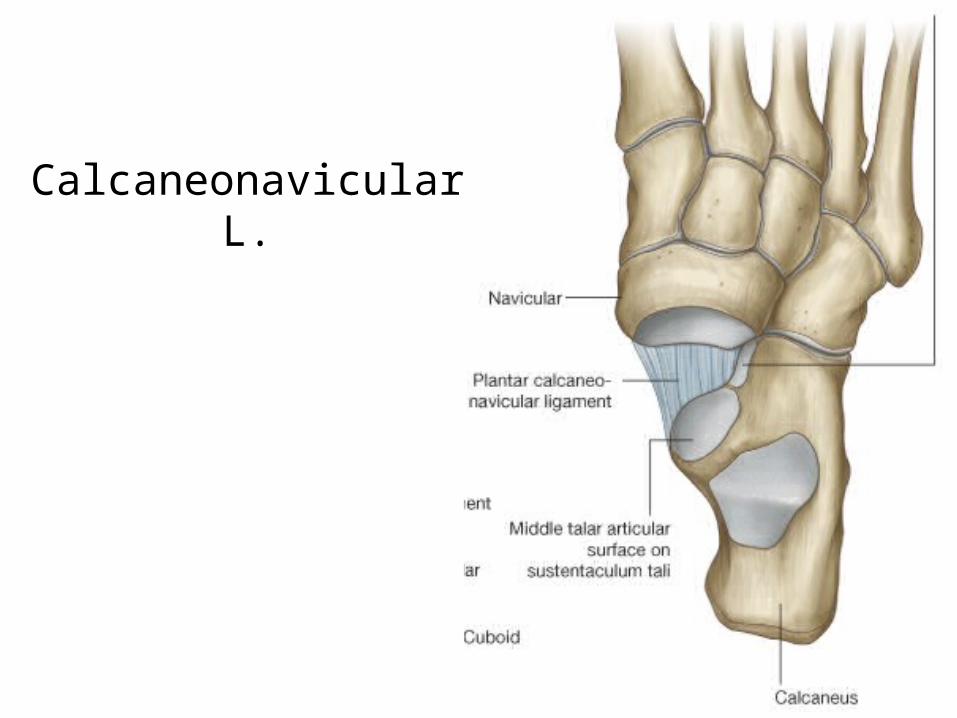
Calcaneonavicular L.
Other ligaments
• Calcaneofibular• Deltoid• Talocalcaneal
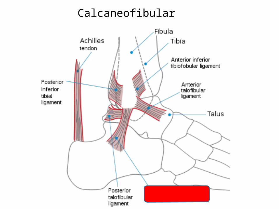
Calcaneofibular
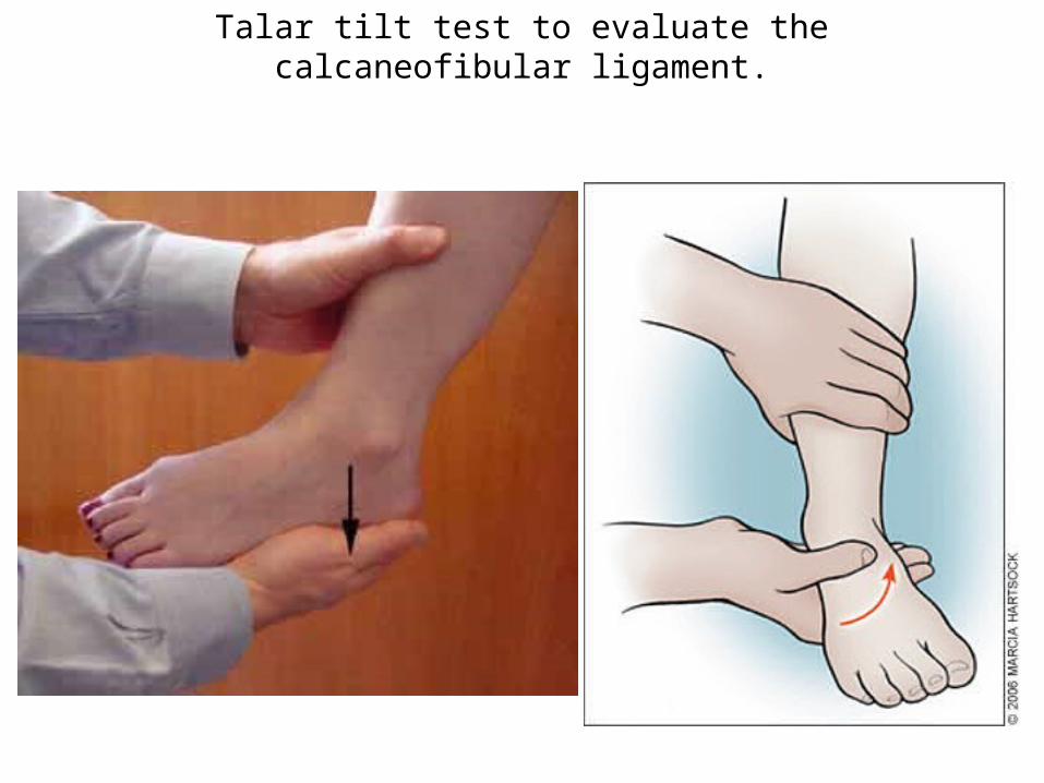
Talar tilt test to evaluate the calcaneofibular ligament.
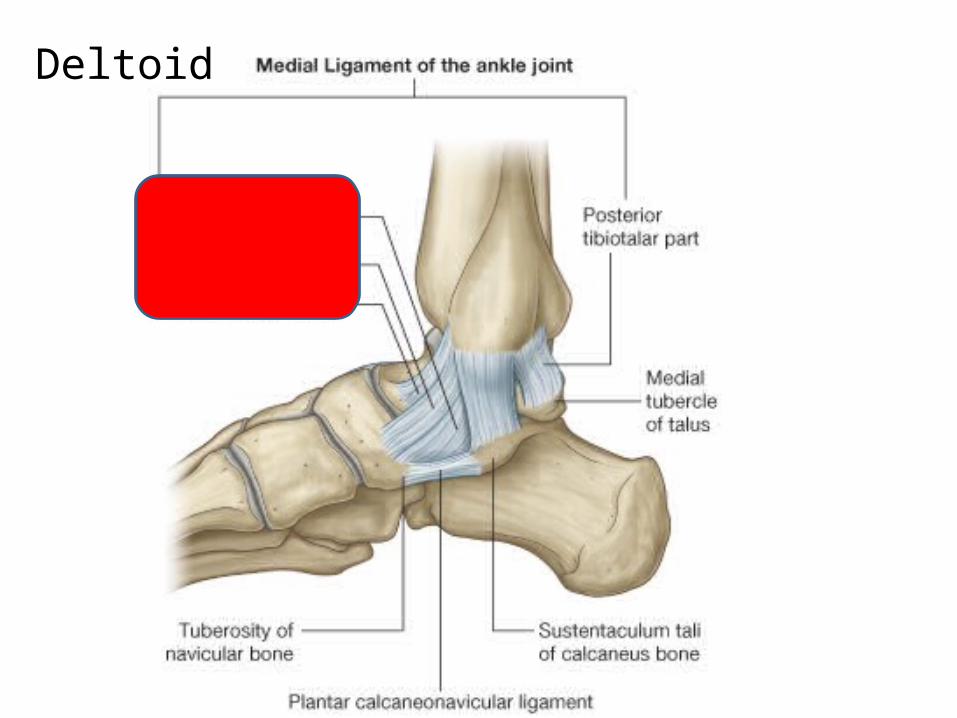
Deltoid
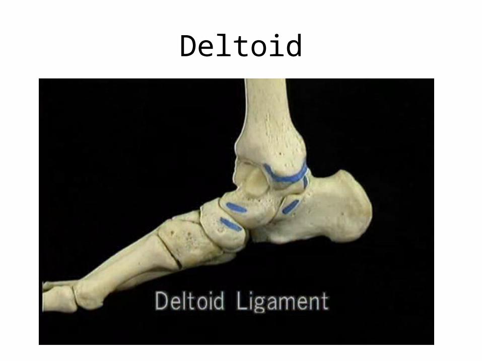
Deltoid
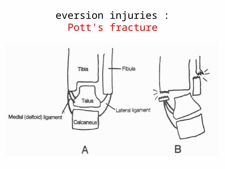
eversion injuries :Pott's fracture
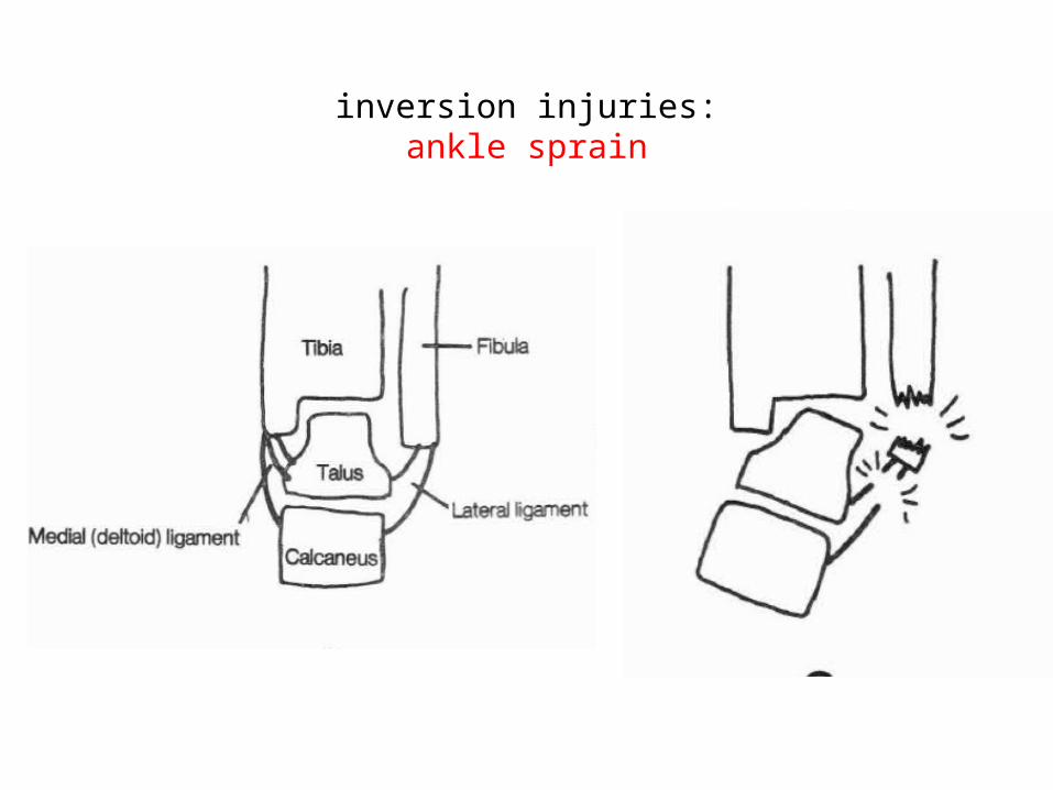
inversion injuries: ankle sprain
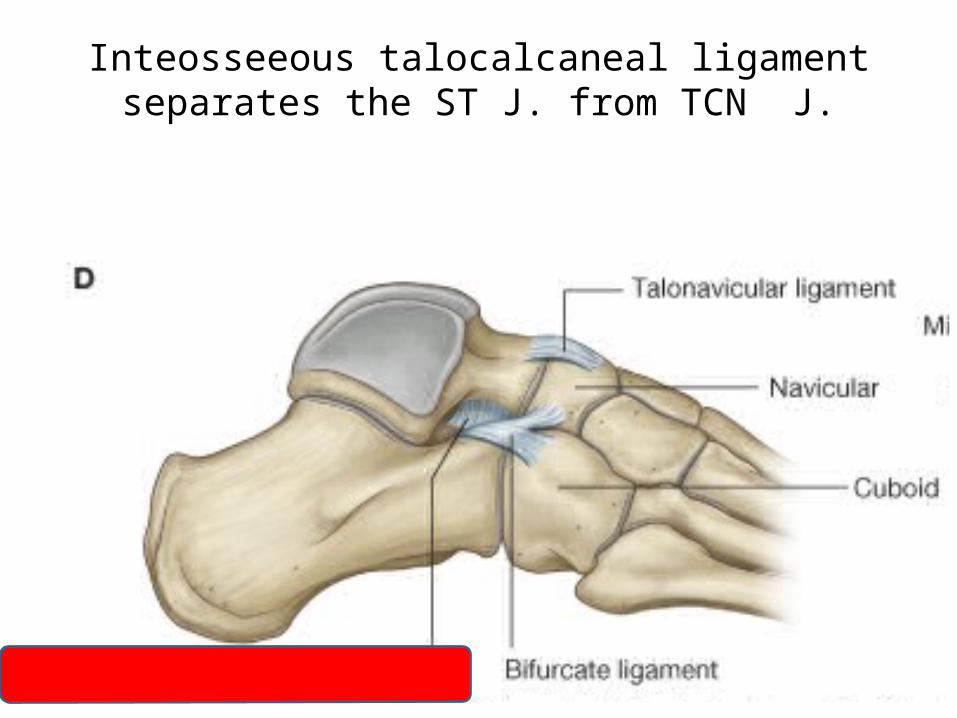
Inteosseeous talocalcaneal ligament separates the ST J. from TCN J.