Joint and Skeletal Pathology 2019
55
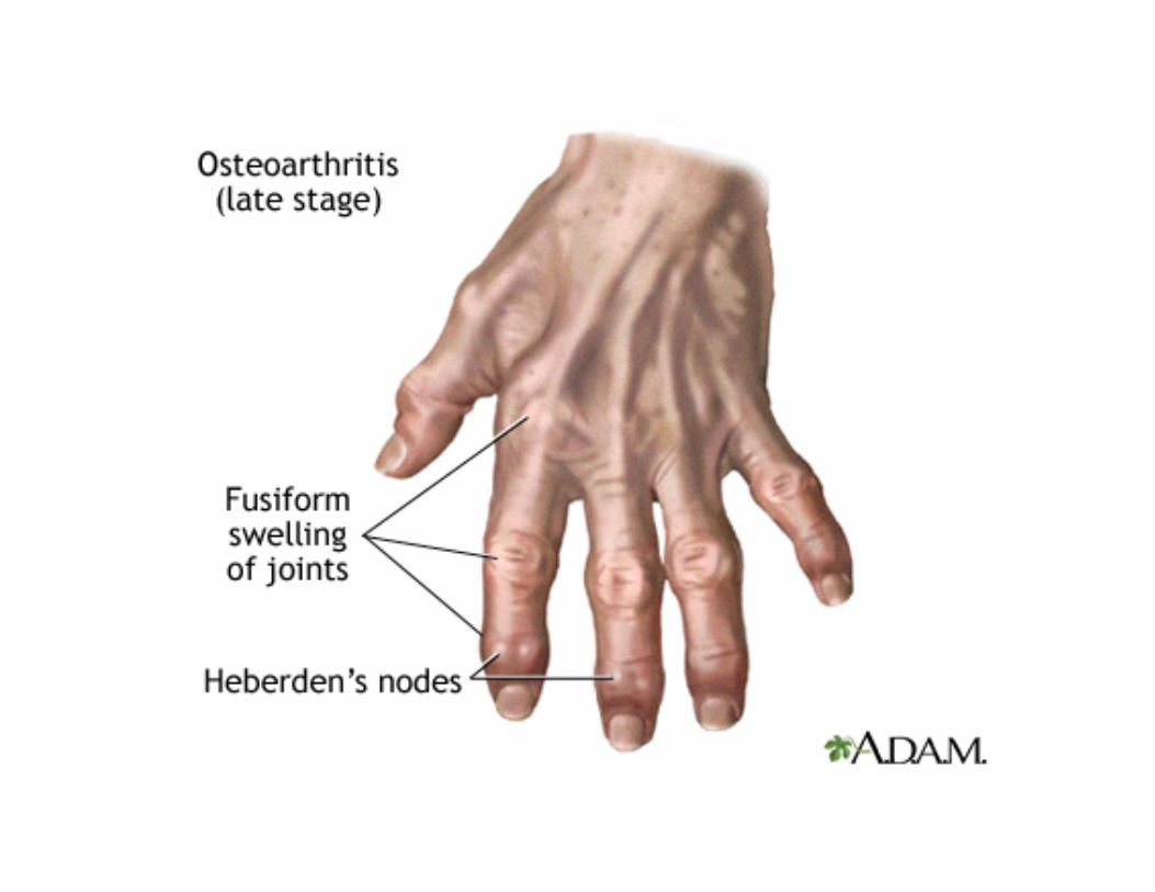
Osteoarthritis • Most common joint disease – Slow progressive degeneration of articular cartilage of weight bearing joints – Subchondral bony thickening and bony overgrowths (osteophytes; “spurs”); knobby protrusions at the margins of the distal interphalangeal joints à nontender Haberden’s nodes
Transcript of Joint and Skeletal Pathology 2019
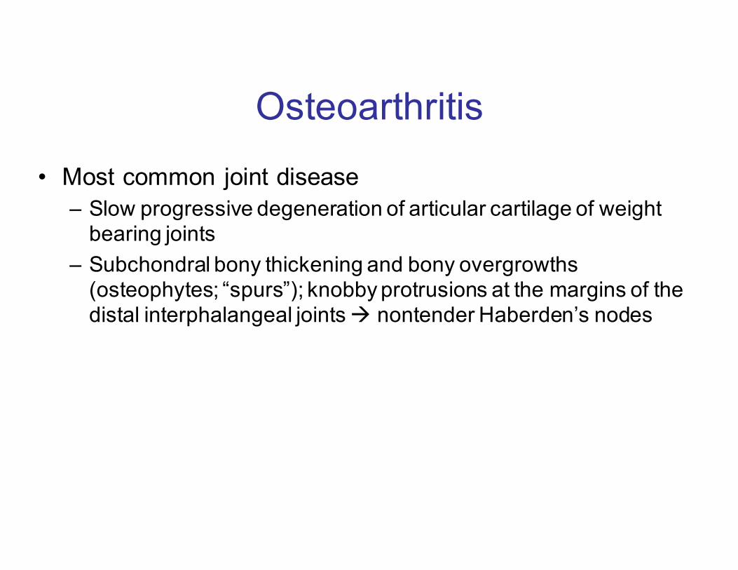
Osteoarthritis
• Most common joint disease– Slow progressive degeneration of articular cartilage of weight
bearing joints– Subchondral bony thickening and bony overgrowths
(osteophytes; “spurs”); knobby protrusions at the margins of the distal interphalangeal joints à nontender Haberden’s nodes
Osteoarthritis
• Primary: defect in cartilage, not an inflammatory disease– Men in midlife, somewhat later in women– 80% of those over 70 years; nonlinear association
• Secondary: appears at any age in a previously damaged or congenitally abnormal joint (trauma, crystal deposits, infection)
• Knees, hips, cervical and lumbar spine
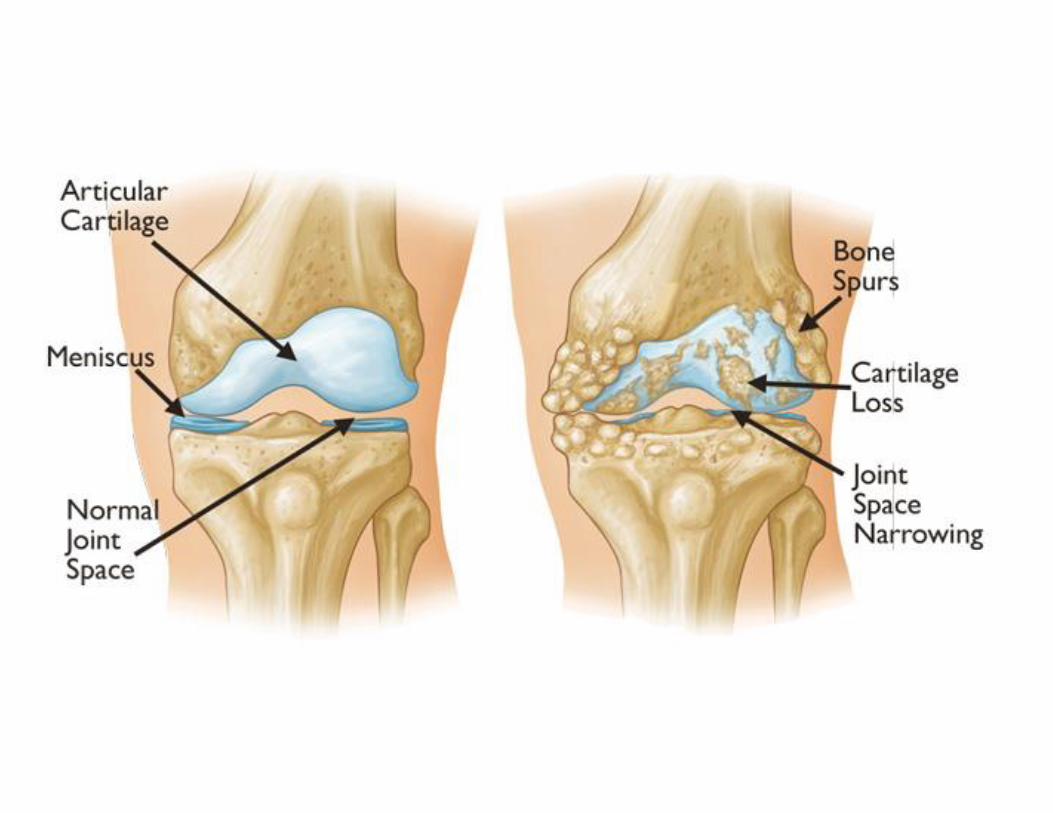
Osteoarthritis• Loss of proteoglycans and areas of decreased number of
chondrocytes alternating with areas of proliferating chondrocytes with matrix basophilia
• Narrowing of joint space (loss of disk)• Increased thickness of subchondral bone
– Fissures, pitting and flaking of cartilage with exposure of bone (eburnated bone)
• Subchondral bone cysts• Inflammation of the synovium• Loose bodies in the joint


Rheumatoid arthritis• Systemic chronic inflammatory disease affecting the
synovium (IT IS A SYNOVITIS THAT LEADS TO DESTRUCTION AND ANKYLOSIS OF AFFECTED JOINTS)
• Autoimmune disease; 1% world prevalence• 3:1 women (3rd-4th decade)• Diarthrodial joints bilaterally• Remissions and exacerbations• Heredity; EBV(?)• HLA-Dw4 haplotype and related B-cell alloantigen

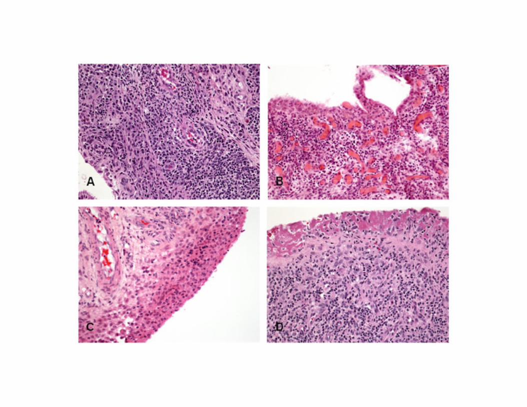
Rheumatoid arthritis• Starts from the proximal joints of hands and feet then
wrists, elbows, ankles, and knees• Villous hypertrophy of the synovium• Hyperplasia of the synoviocytes• Intense lymphoplasmacytic and histiocytic infiltrates• The synovium forms a cloak (pannus) that fills the in the
joint space• Destructive enzymes and cytokines, and the pannus,
destroy the articular surfaces• Fibrous and bony ankylosis
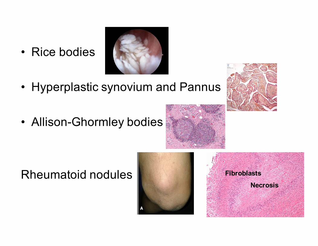
• Rice bodies
• Hyperplastic synovium and Pannus
• Allison-Ghormley bodies
Rheumatoid nodulesNecrosis
Fibroblasts
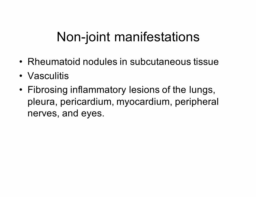
Non-joint manifestations
• Rheumatoid nodules in subcutaneous tissue• Vasculitis• Fibrosing inflammatory lesions of the lungs,
pleura, pericardium, myocardium, peripheral nerves, and eyes.
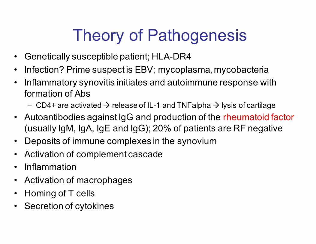
Theory of Pathogenesis• Genetically susceptible patient; HLA-DR4• Infection? Prime suspect is EBV; mycoplasma, mycobacteria• Inflammatory synovitis initiates and autoimmune response with
formation of Abs– CD4+ are activated à release of IL-1 and TNFalpha à lysis of cartilage
• Autoantibodies against IgG and production of the rheumatoid factor(usually IgM, IgA, IgE and IgG); 20% of patients are RF negative
• Deposits of immune complexes in the synovium• Activation of complement cascade• Inflammation• Activation of macrophages• Homing of T cells• Secretion of cytokines


Spondyloarthropathy
• Used to be a type of RA• NOW comprises a group of diseases
– Ankylosing spondylitis• Vertebral column & sacroileac joints, young men
– Reactive arthritis (Reiter syndrome)• Polyarthritis, conjunctivitis, non-gonococcal urethritis, oral
lesions– Psoriatic arthritis– Arthritis and inflammatory bowel disease
(enteropathic arthritis)• Crohn’dz, ulcerative colitis

Reactive arthritis

Gout• Hyperuricemia
– It is necessary for gout, but only a few fraction of hyperuricemic people develop gout
• Idiopathic • Predisposing factors: alcohol, obesity• Most cases occur in men; Almost never in women
before menopause• Attacks of acute arthritis triggered by crystallization
of urates in joints• Asymptomatic intervals• Eventual development of chronic tophaceous gout
and arthritis

Primary gout
• Hyperuricemia in the absence of other disease– Asymptomatic hyperuricemia can precede gout
• Impaired secretion by kidneys

Secondary gout
• Hematopoietic pathologic conditions– Leukemias– Lymphomas
• After chemotherapy• Alcoholism

Clinical features
• Acute gouty arthritis– Painful– Involves one joint initially, then polyarticular– Podagra (painful, red metatarsophalangeal joint)
• Tophaceous gout– Development of tophi
• Chalky, cheesy, yellow-white, pasty deposits of monosodium urate crystals
– Helix and antihelix of ear– Achilles tendon

Gout• Pathology
– Formation of granulomas with needle-shaped crystals• Renal failure, urate stones• Treatment
– Colchicine• Prophylactic
– Probenecid & sulfinpyrazone• Interfere with urate resorption
– Allopurinol• Inhibitor of enzyme that converts the xanthine and
hypoxanthine to uric acid
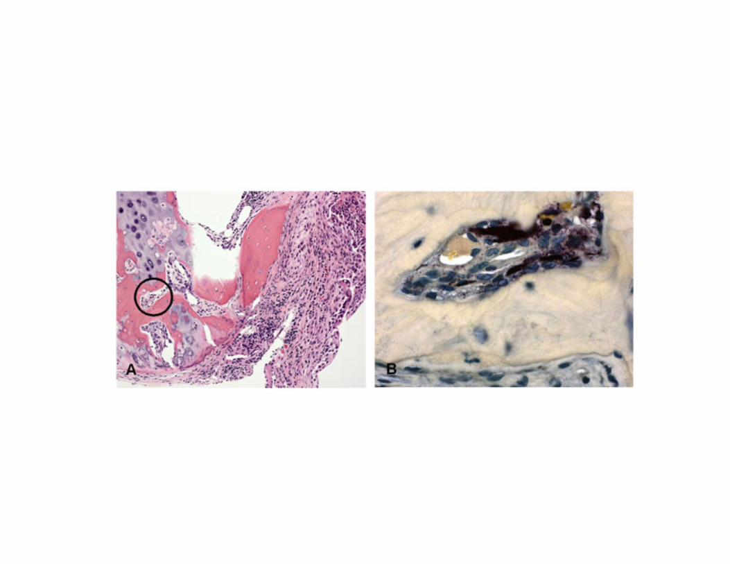
Pseudogout
• Chodrocalcinosis• Calcium pyrophosphate crystals deposits in joints• Older individuals• No gender, race predilection• 30-60% prevalence
– There is a hereditary form• Can cause significant joint damage
– Knees, wrists, elbows, shoulders, ankles
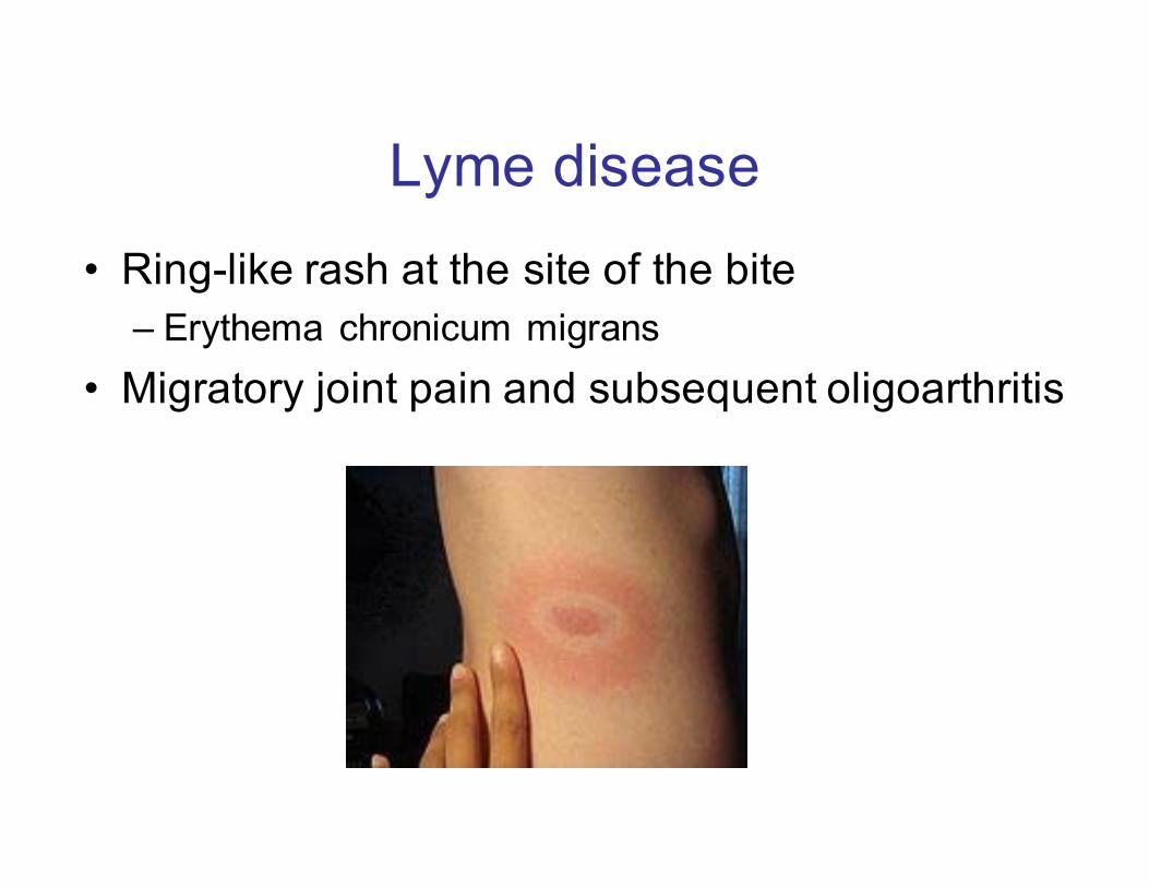
Lyme disease• Ring-like rash at the site of the bite
– Erythema chronicum migrans• Migratory joint pain and subsequent oligoarthritis
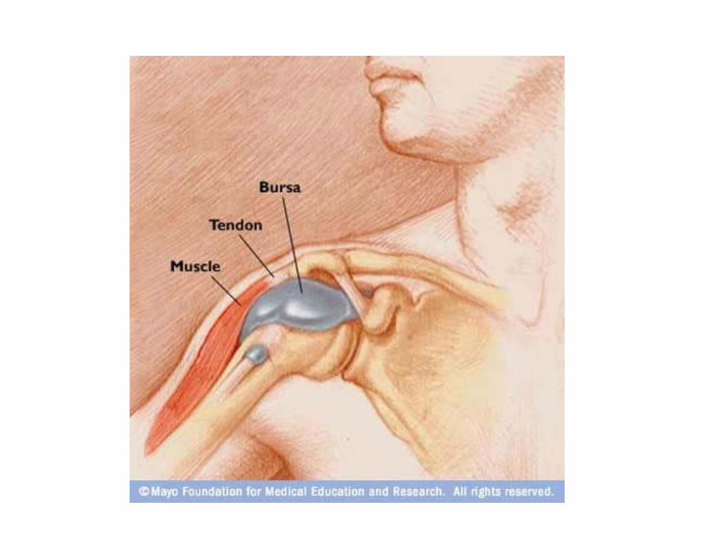
Bursitis
• Inflammation of the bursa– elbow, shoulder, knee
• Fibrous thickening of the bursa wall• Tendency to double-fault in tennis and develop
a bad slide in golf
Tumors and Tumor-like Conditions
• Ganglion cyst: Wrist; connective tissue cyst; near the joint capsule or the tendon sheath
• Synovial cyst: herniation of synovium through the joint capsule (Baker cyst; popliteal fossa)
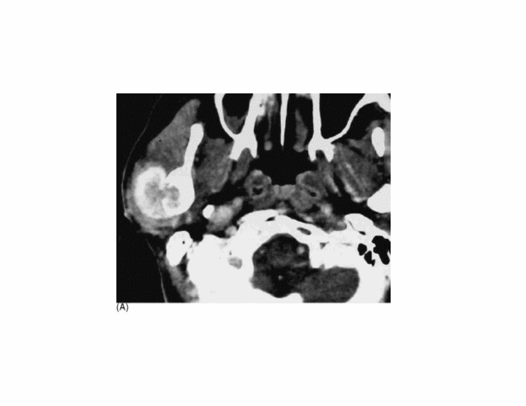
• Pigmented villonodular tenosynovitis– Knee, hip, ankle, pain
• Giant cell tumor of tendon sheath– Painless mass; wrist; Most common soft tissue
tumor of the hand

Synovial Chondromatosis
• 18-75 years (median 45)• More frequent in women in contrast with other
joints• Pain (82%) , swelling (65%), combination (50%),
crepitus• X-ray: Loose irregular radiopaque bodies• Treatment: Removal of loose bodies, some
surgeons do total synovectomy
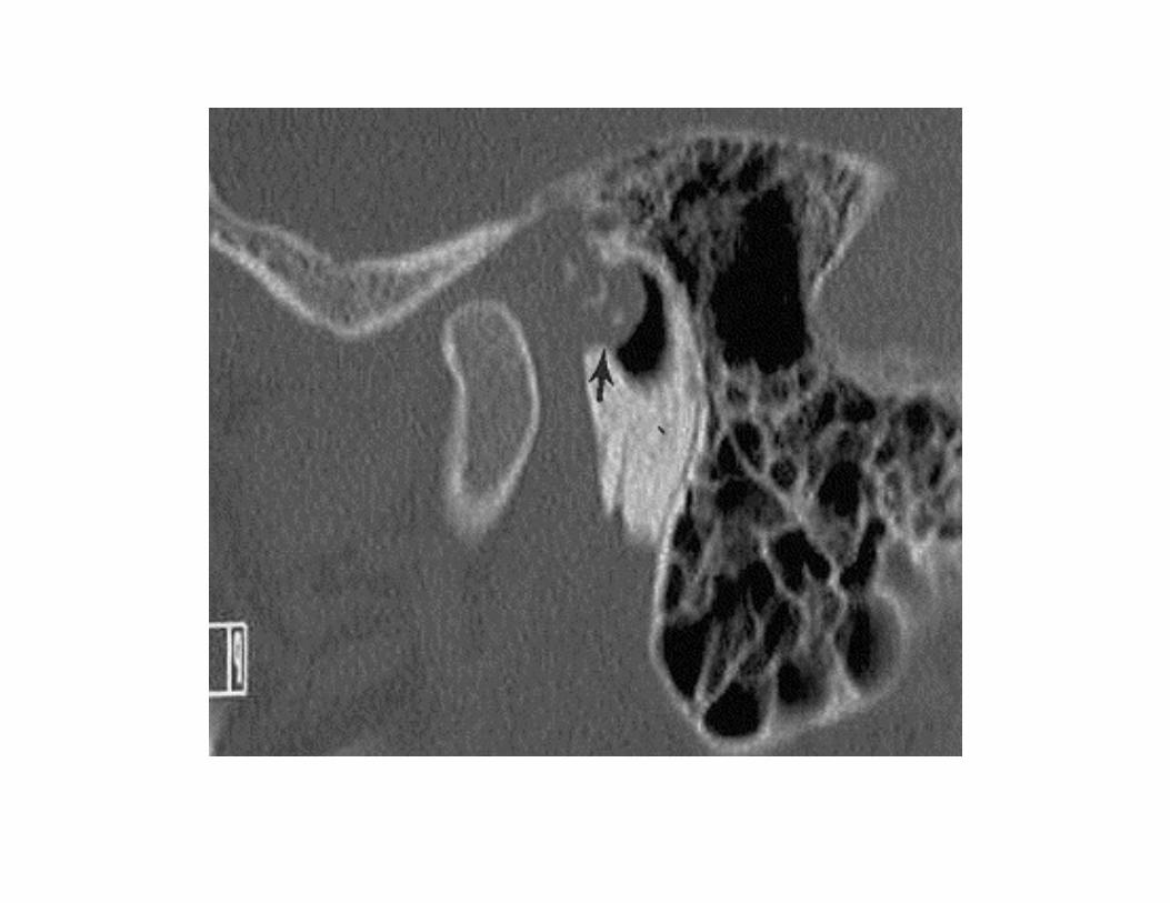

Osteochondroma
• Most frequent neoplasm of bone• Cartilage capped bone projection• Metaphysis• Condylar is rare; coronoid even rarer• Median age ~40 for condylar• ~2F:M
Pigmented villonodular synovitis
• Uncommon• 10-70 years• Patients are rarely younger than 30• Average age 43.7• Slight female predominance• Swelling, pain• Bone erosion• Diffuse or localized; diffuse can be very aggressive• Inflammation, giant cells, hemosiderin, regular mitoses
Ganglion and synovial cyst
• Confusion with the name• Ganglion is a pseudocyst: myxoid degeneration;
does not have synovial lining• Synovial cyst: lined by cuboidal to flattened cells• Preauricular swelling, pain

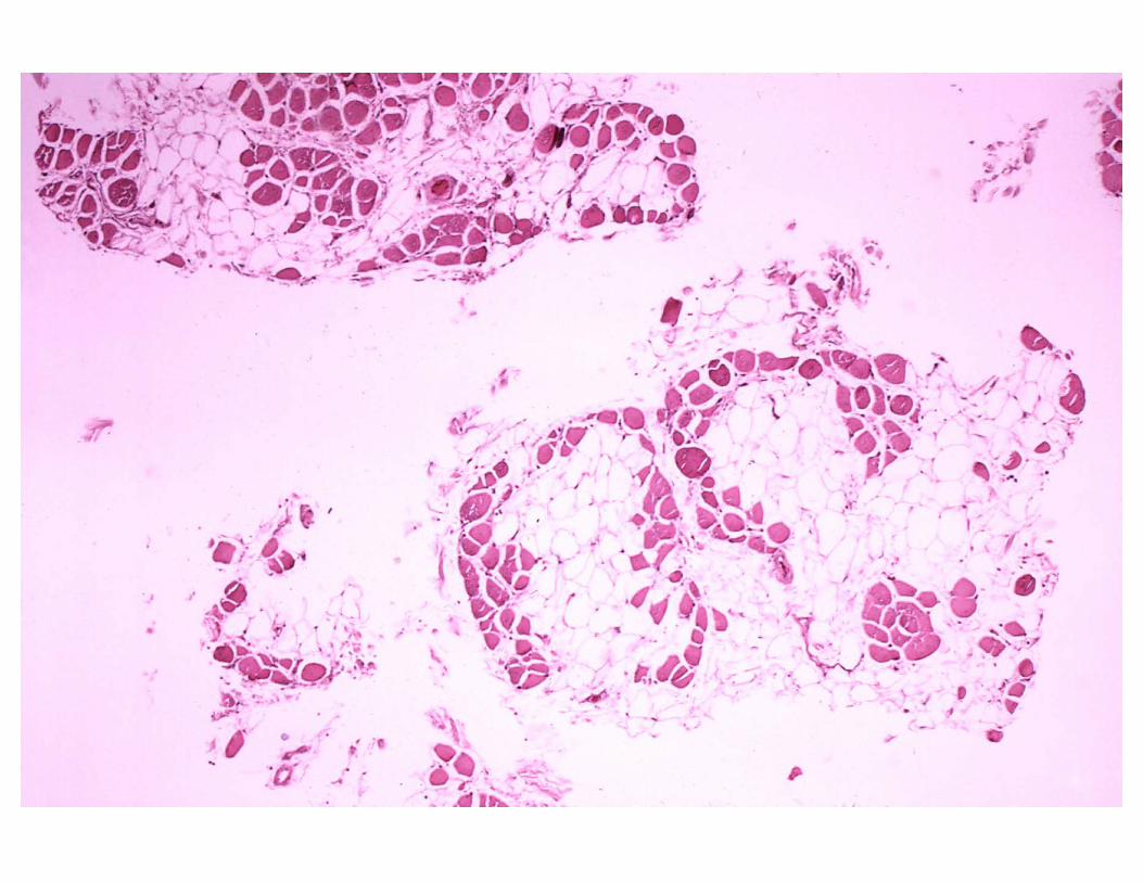
Skeletal Muscle Pathology

Duchene muscular dystrophy• X-linked• Pelvic and shoulder girdles• Deletion of gene that encodes dystrophin (DMD)
– Dystrophin is made in the heart and skeletal muscle– Also in neurons in the hippocampus
• Degeneration of muscles, impaired repair, fibrosis, fibrofatty deposits
• Elevated serum creatinine kinase• Steroid treatment• Death form respiratory insufficiency, cardiac arrhythmia,
10-15 years of age wheel chair-bound

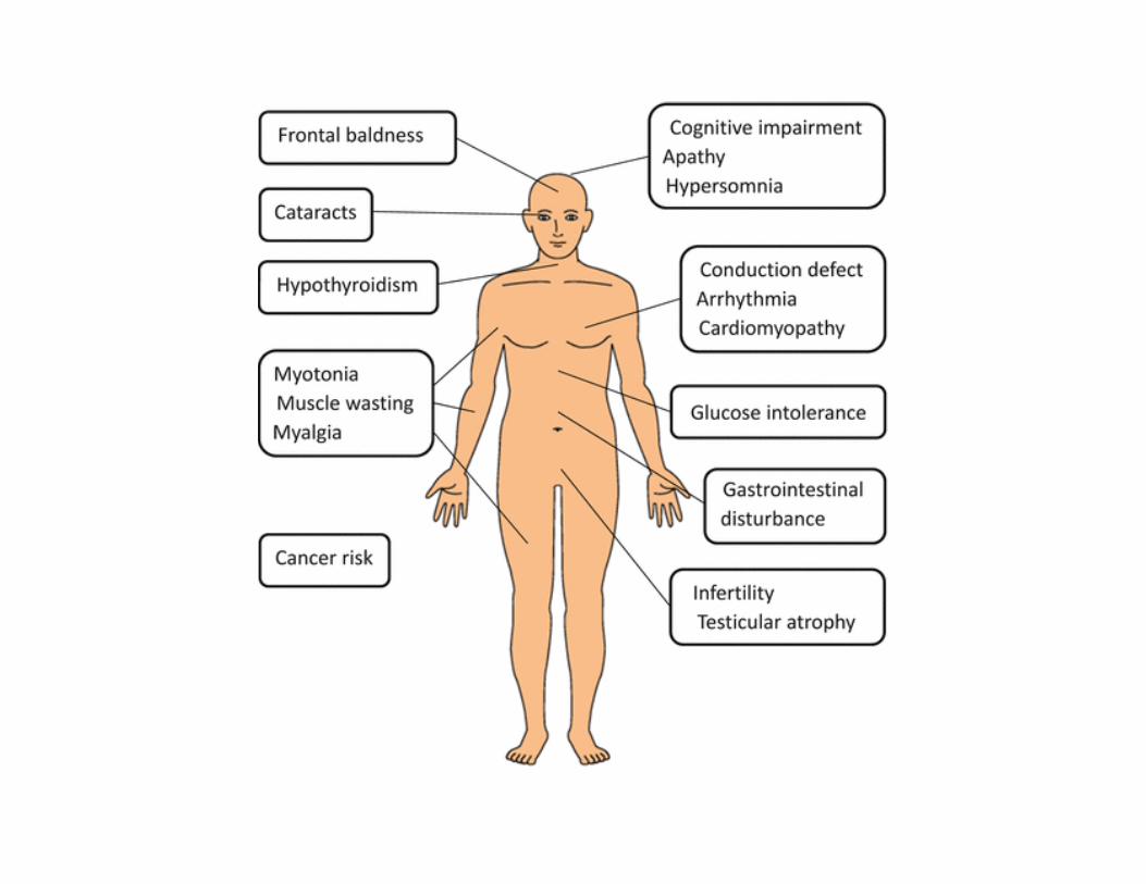
Myotonic dystrophy
• AD• Most common form of adult MD • Sustained muscular contractions and rigidity• Progressive muscle weakness and wasting• Chromosome 19• Atrophy of type I and hypertrophy of type II fibers• Anticipation
– Earlier age of onset and increased severity in successive generations
Myotonic dystrophy
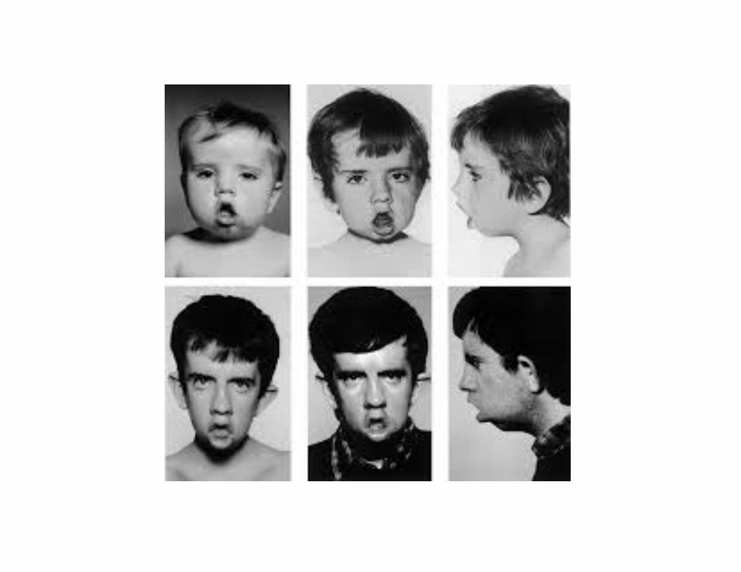
• Three clinical groups– Congenital– Adult: facial and jaw muscles, ptosis– Late: minimal symptoms
Autoimmune Myopathies• Dermatomyositis
– Complement mediated cytotoxic Abs against microvasculature of muscle
– Distinctive skin rash• Face and eyelids and on knuckles, elbows, knees, chest and back• The rash, which can be itchy and painful• It is often the first sign of dermatomyositis.
– Muscle weakness• Muscles closest to the trunk, such as those in your hips, thighs,
shoulders, upper arms and neck– Inflammatory myopathy
Dermatomyositis - Complications• Dysphagia
– Muscles in your esophagus are affected– Weight loss and malnutrition– Aspiration pneumonia
• Shortness of breath• Increased risk for cancer
– Cervix, lungs, pancreas, breasts, ovaries and gastrointestinal tract
– Raynaud’s phenomenon• Cold and numb toes, fingers, ears and redness of the skin
• Calcium deposits– Muscles and skin (tumor calcinosis)

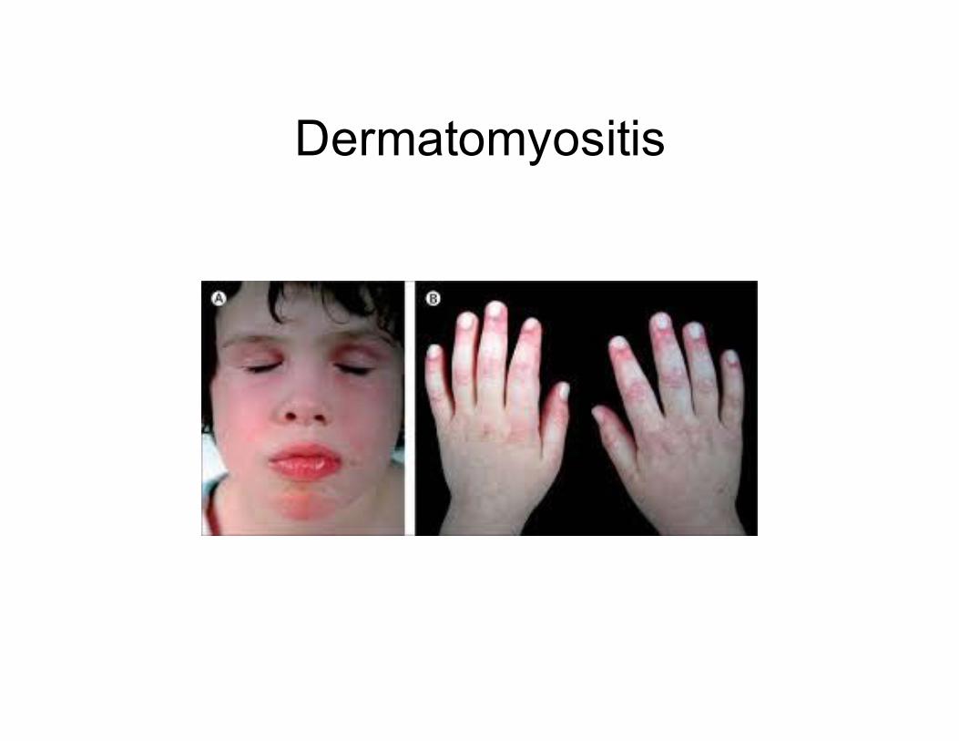
Dermatomyositis
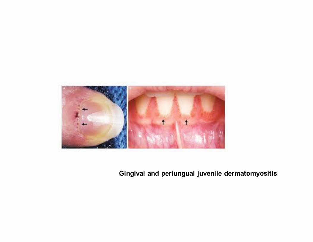
Gingival and periungual juvenile dermatomyositis
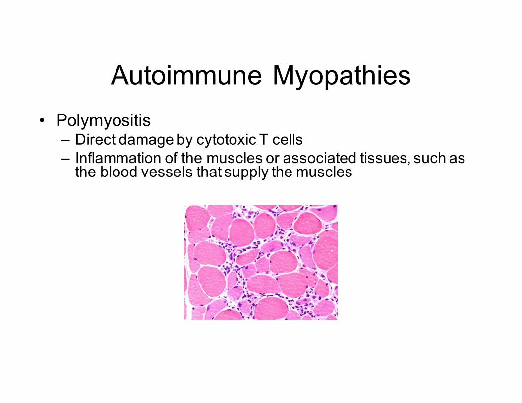
Autoimmune Myopathies• Polymyositis
– Direct damage by cytotoxic T cells– Inflammation of the muscles or associated tissues, such as
the blood vessels that supply the muscles

Autoimmune Myopathies• Myasthenia Gravis (serious muscle weakness)
– Muscular fatigability caused by circulating Abs to acetylcholine receptor at the myoneural junction
– Extraocular muscles, swelling muscles, extremities– Patients can develop other autoimmune diseases– 40% patients have thymoma– 75% of remaining thymic hyperplasia– Removal of thymus can be curative

Polyarteritis Nodosa
• Men• Small and medium size arteries• Vasculitis• Decreased blood supply to organs• Implicated
– Hepatitis B (~30%)– Sulfa drugs, penicillin


Temporal Arteritis
• Inflammation of large arteries• Temporal artery and other arteries• Headache, visula changes• Confirmatory biopsy• If untreated can lead to blindness