JM:ethyl Ethyl Ketone Peroxide in Dim.ethyl Phthalate Toxicology Program Toxicity Report Series...
83
National Toxicology Program Toxicity Report Series Number 18 NTP Technical Report on Toxicity Studies of JM:ethyl Ethyl Ketone Peroxide (CAS No. 1338-23-4) in Dim.ethyl Phthalate (CAS No. 131-11-3) (45:55) Administered Topically to F344/N Rats and B6C3F 1 Mice Errol Zeiger, PhD, Study Scientist National Toxicology Program Post Office Box 12233 Research Triangle Park, NC 27709 NIH Publication 93-3341 February 1993 United States Department of Health and Human Services Public Health Service National Institutes of Health
Transcript of JM:ethyl Ethyl Ketone Peroxide in Dim.ethyl Phthalate Toxicology Program Toxicity Report Series...
National Toxicology Program Toxicity Report Series
Number 18
NTP Technical Report on Toxicity Studies of
JM:ethyl Ethyl Ketone Peroxide (CAS No. 1338-23-4)
in Dim.ethyl Phthalate (CAS No. 131-11-3)
(45:55)
Administered Topically to F344/N Rats and B6C3F1 Mice
Errol Zeiger, PhD, Study Scientist National Toxicology Program
Post Office Box 12233 Research Triangle Park, NC 27709
NIH Publication 93-3341 February 1993
United States Department of Health and Human Services Public Health Service
National Institutes of Health
Foreword
The National Toxicology Program (NTP) is made up of four charter agencies of the United States Department of Health and Human Services (DHHS):
• the National Cancer Institute (NCI) of the National Institutes of Health; • the National Institute of Environmental Health Sciences (NIEHS) of the National Institutes
of Health; • the National Center for Toxicological Research (NC'IR) ofthe Food and Drug Administration;
and • the National Institute for Occupational Safety and. Health (NIOSH) ofthe Centers for Disease
Control. In July 1981, the Carcinogenesis Bioassay Testing Program was transferred from NCI to NIEHS. NTP coordinates the relevant Public Health Service programs, staff, and resources that are concerned with basic and applied research and with biological assay development and validation.
NTP d1~velops, evaluates, and disseminates scientific information about potentially toxic and hazardous chemicals. This knowledge is used for protecting the health of the American people and for the primary prevention of disease.
To carry out its mission, NTP designs and conducts studies to characterize and evaluate the toxicologic potential of selected chemicals in laboratory animals (usually two species, rats and mice). Chemicals selected for NTP toxicology studies are chosen primarily on the bases of human exposure, level of production, and chemical structure. Selection per se is not an indicator of a chemical's toxic potential.
The studies described in this toxicity study report were performed under the direction of NIEHS and were conducted in compliance with NTP laboratory health and safety requirements. These studie:s met or exceeded all applicable federal, state, and local health and safety regulations. Animal care and use were in accord and compliance with the Public Health Service Policy on Huma:ne Care and Use of Animals.
Single copies of this report are available without charge, while supplies last, from the NTP Public Infonnation Office (telephone number 919/541-3991).
NTP Public Information Office NIEHS
Post Office Box 12233 Research Triangle Park, NC 27709

National Toxicology Program Toxicity Report Series
Number 18
NTP Technical Report on Toxicity Studies of
JM:ethyl Ethyl Ketone Peroxide (CAS No. 1338-23-4)
in Dim.ethyl Phthalate (CAS No. 131-11-3)
(45:55)
Administered Topically to F344/N Rats and B6C3F1 Mice
Errol Zeiger, PhD, Study Scientist National Toxicology Program
Post Office Box 12233 Research Triangle Park, NC 27709
NIH Publication 93-3341 February 1993
United States Department of Health and Human Services Public Health Service
National Institutes of Health


2 METHYL ETHYL KETONE PEROXIDE, NTP TOXICITY REPORT NUMBER 18
CONTRIBUTORS This NTP report on the toxicity studies of methyl ethyl ketone peroxide is based primarily on 2-week studies that took place in October 1986 and 13-week studies that began in July 1987 and ended in October 1987 at Hazleton Laboratories America, Inc., Rockville, MD.
National Toxicology Program Evaluated experiment, interpreted results, and reportedfmdings
Errol Zeiger, PhD, Study Scientist
Kamal M. Abdo, PhD John R. Bucher, PhD Leo T. Burka, PhD Rajendra S. Chhabra, PhD Michael P. Dieter, PhD Michael R. Elwell, DVM, PhD Joel Mahler, DVM Robert R. Maronpot, DVM H.B. Matthews, PhD Morrow B. Thompson, DVM, PhD Gregory S. Travlos, DVM
Coordinated report preparation
Jane M. Lambert. BS Edison Mcintyre, BS Kristine L. Witt, MS
Oak Ridge Associated Universities
NTP Pathology Working Group Evaluated slides and prepared pathology report
John C. Seely, DVM, Chairperson Pathco
Michael R. Elwell, DVM, PhD National Toxicology Program
Joel R. Leininger, DVM, PhD National Toxicology Program
Margarita M. McDonald, DVM, PhD National Toxicology Program
A.W. Macklin, DVM, PhD Burroughs Wellcome Research Laboratories
John Peckham, DVM, MS, PhD Experimental Pathology Laboratories. Inc
Hazleton Laboratories America, Inc Principal contributors
Gary W. Wolfe, PhD, Principal Investigator
Samuel V. Machotka II, DVM Michael R. Moore, PhD Ricardo Quander, DVM
Experimental Pathology Laboratories, Inc Provided pathology quality assessment
John Peckham, DVM, MS, PhD
Environmental Health Research and Testing, Inc Provided spenn morphology and vaginal cytology evaluation
Dushant K. Gulati, PhD Susan Russell, BA
Analytical Sciences, Inc Provided statistical analyses
Steven Seilkop, MS Janet Teague, MS Richard Morris, MS
Biotechnlcal Services, Inc Provided toxicity report preparation
Janet L. Elledge, BA, Principal Investigator
Chad J. Fitz, MA Paula C. Higginson, BA Jennifer P. Rector, MAP
3 METHYL ETHYL KETONE PEROXIDE, NTP TOXICITY REPORT NUMBER 18
TABLE OF CONTENTS
ABSTRACT........................................................................................................5
PEER REVIEW PANEL ........................................................................................8
SUMMARY OF PEER REVIEW COMMENTS ...............................................................9
INTRODUCTION ................................................................................................ 11
Physical Properties, Production, Use, and Exposure ..............................................11 Toxicity ................................................................................................................. 12 Study Rationale and Design ............................................................................... 16
MATERIALS AND METHODS .............................................................................. 17 Procurement and Characterization of Methyl Ethyl Ketone Peroxide and Dimethyl Phthalate ...................................... 17 Dose Formulations ................................................................................................. 18 Toxicity Study Designs .......................................................................................... 18 Genetic Toxicity Studies ....................................................................................... 21 Statistical Methods.................................................................................................26 Quality Assurance ............................................................................................... 28
RESULTS ........................................................................................................ 29 2-Week Dermal Study in F344/N Rats ..................................................................29 13-Week Dermal Study in F344/N Rats ................................................................ 32 2-Week Dermal Study in B6C3F1 Mice ................................................................... 37 13-Week Dermal Study in B6C3F1 Mice .................................................................40 Genetic Toxicity Studies ......................................................................................44
DISCUSSION...................................................................................................47
REFERENCES ................................................................................................. 51
TABLES
Table 1 Experimental Design and Materials and Methods in the 2-Week and 13-Week Dermal Studies ..................................24
Table 2 Survival and Weight Gain of F344/N Rats in the 2-Week Dermal Study of Methyl Ethyl Ketone Peroxide ......... 29
Table 3 Liver and Thymus Weights of F344/N Rats Administered Methyl Ethyl Ketone Peroxide Topically for 2 Weeks ..................................................................... 30
Table 4 Survival and Weight Gain of F344/N Rats in the 13-Week Dermal Study of Methyl Ethyl Ketone Peroxide .................................................... 32
4 METHYL ETHYL KETONE PEROXIDE, NTP TOXICITY REPORT NUMBER 18
TABLES (CONTINUED) Table 5 Incidence and Severity of Treatment-Related Lesions
in F344/N Rats Administered Methyl Ethyl Ketone Peroxide Topically for 13 Weeks ....................................................... 36
Table 6 Survival and Weight Gain of B6C3F1 Mice in the 2-Week Dermal Study of Methyl Ethyl Ketone Peroxide .................................................... 37
Table 7 Liver and Thymus Weights of B6C3F1 Mice Administered Methyl Ethyl Ketone Peroxide Topically for 2 Weeks .................................................................... 38
Table 8 Survival and Weight Gain of B6C3F1 Mice in the 13-Week Dermal Study of Methyl Ethyl Ketone Peroxide ................................................... 40
Table 9 Spleen and Body Weights of B6C3F1 Mice Administered Methyl Ethyl Ketone Peroxide Topically for 13 Weeks ...................... 42
Table 10 Incidence and Severity of Treatment-Related Lesions in B6C3F1 Mice Administered Methyl Ethyl Ketone Peroxide Topically for 13 Weeks....................................................... 44
FIGURES Figure 1 Body Weights of F344/N Rats Exposed
to Methyl Ethyl Ketone Peroxide by Topical Administration for 13 Weeks ......................................... 33
Figure 2 Body Weights of B6C3F1 Mice Exposed to Methyl Ethyl Ketone Peroxide by Topical Administration for 13 Weeks ......................................... 41
APPENDICES Appendix A Organ Weights and Organ-Weight-to-Body-Weight Ratios ....................... A-l
Appendix B Reproductive Tissue Evaluations and Estrous Cycle Characterization............................................ B-1
Appendix C Genetic Toxicology ............................................................................ C-1
5 METHYL ETHYL KETONE PEROXIDE, NTP TOXICITY REPORT NuMBER 18
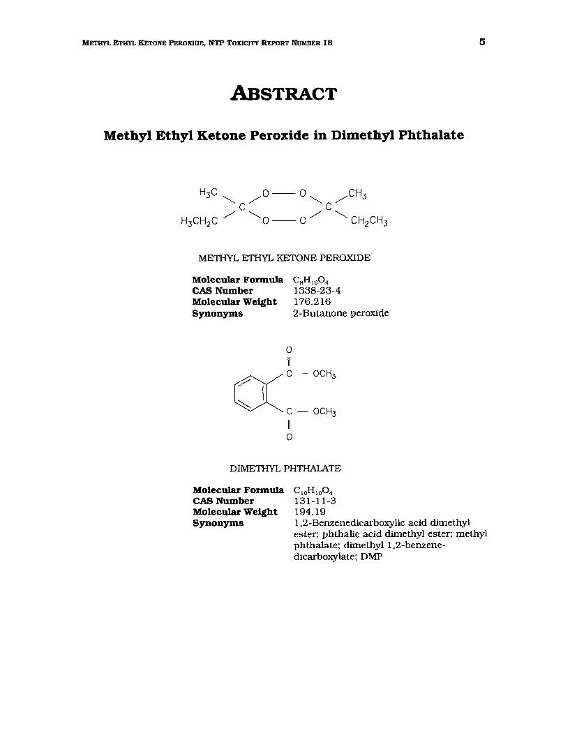
ABSTRACT
Methyl Ethyl Ketone Peroxide in Dimethyl Phthalate
METIIYL ETHYL KETONE PEROXIDE
Molecular Formula CsH1604 CAS Number 1338-23-4 Molecular Weight 176.216 Synonyms 2-Butanone peroxide
0 II
((C-OCH3
C- OCH3
II 0
DIMETHYL PHTHALATE
Molecular Formula C 10H 100 4
CAS Number 131-11-3 Molecular Weight 194.19 Synonyms 1,2-Benzenedicarboxylic acid dimethyl
ester; phthalic acid dimethyl ester; methyl phthalate; dimethyl 1,2-benzenedicarboxylate; DMP
6 METHYL ETHYL KETONE PEROXIDE, NTP TOXICITY REPORT NUMBER 18
Methyl ethyl ketone peroxide (MEKP) is an unstable organic peroxide used in the
manufacture of acrylic resins, as a hardening agent for fiberglass-reinforced plastics, and
as a curing agent for unsaturated polyester resins. It is commercially available as a 40%
to 60% solution in dimethyl phthalate (DMP). Because exposure to MEKP is typically
through dermal contact, 2-week and 13-week toxicity studies were conducted by topical
application of MEKP in DMP (45:55 w /w) to the clipped dorsal region of male and female
Fischer 344/N rats and B6C3F1 mice. Animals were evaluated for histopathology and for
reproductive endpoints. In vitro genetic toxicity studies of MEKP included assessments of
mutagenicity in Salmonella typhimurium and in mouse lymphoma L5178Y cells and
analysis of chromosomal aberrations and sister chromatid exchanges in Chinese hamster
ovary cells. In addition, the peripheral blood of mice from the 13-week study was
evaluated in the micronucleus assay.
In the 2-week studies, groups of 5 animals of each species and sex were administered
MEKP in DMP for 5 days per week at doses of 50.6, 101.3, 202.5, 405, and 810 mg/kg
body weight per day for rats and 112.5, 225, 450, 900, and 1800 mg/kg body weight per
day for mice. Control groups received DMP or no treatment. No rats died during the
studies, but at least 1 mouse in each group receiving MEKP died. Body weight gains of
rats decreased with increasing doses ofMEKP; body weight gains of mice were not affected
by treatment. The primary effects of topical administration of MEKP in both rats and mice
were an extensive coagulative necrosis of the epidermis and dermis, variable degrees of
inflammation ofthe adnexa, and epidermal regeneration and hyperplasia at the application
site. Lesions considered secondary to the dermal lesions included increased hematopoiesis
in the spleen in rats and mice and increased myeloid hyperplasia of the bone marrow in
mice, primarily at the higher doses. Mice showed a marked, dose-related increase in liver
weight.
In the 13-week dermal studies, groups of 10 rats and 10 mice of each sex were
administered MEKP in DMP for 5 days per week at doses of 1.07, 3.57, 10.7, 35.7, and
10,. mg/rat and 0.357, 1.19, 3.57, 11.9, and 35.7 mg/mouse. All high-dose mice,
3 high-dose female rats, and 1 female mouse in the 11.9 mg/animal group died or were
sacrificed during the first week of the studies. Skin lesions similar to those seen in the
2-week studies were judged of sufficient severity to warrant early termination of surviving
7 METHYL ETHYL KETONE PEROXIDE, NTP TOXICITY REPORT NUMBER 18
rats and mice in the 2 highest dose groups. All rats and mice in the remaining dose
groups sunri.ved to the end of the studies, and weight gains were generally lower with
increasing doses of MEKP. Skin lesions at the application site for the remaining animals
(rats and mice) in the 10.7 mg/rat and 3.57 mg/mouse dose groups involved a spectrum
of necrosis, inflammation, and acanthosis {epidermal hyperplasia). Lesions in the lower
dose groups were limited to acanthosis and hyperkeratosis in rats (1.07 and 3.57 mg/rat)
and acanthosis in mice {0.357 and 1.19 mg/mouse). While splenic and bone marrow
lesions similar to those described in the 2-week studies were seen in animals that died
early in the 13-week studies and in the rats and mice that showed ulcerative or necrotic
injury, no other systemic changes were noted in animals that did not show ulcerative skin
lesions.
In genetic toxicity studies, MEKP in DMP (45:55 w/w) was not mutagenic in
Salmonella typhimuriwn strains TA100, TA1535, TA1537, or TA98, with or without
S9 activation. A positive response was obtained in the mouse lymphoma assay for
induction oftrifluorothymidine resistance in L5178Y cells without S9. In cytogenetic tests
with Chinese hamster ovary cells, MEKP induced sister chromatid exchanges and
chromosomal aberrations, with and without S9. No increase in the frequency of
micronucleated erythrocytes was obsexved in peripheral blood samples obtained from male
and female mice at the termination of the 13-week toxicity study.
In summary, topical administration of MEKP in DMP resulted in a spectrum of necrotic,
inflammatory, and regenerative skin lesions limited to the application site. Histopathologic
changes in the spleen and bone marrow were also seen in rats and mice with ulcerative
skin lesions, and were considered a secondary response. A no-obsexved-adverse-effect
level {NOAEL) for histopathologic skin lesions could not be determined from these studies,
as lesions were obsexved with administration of daily doses as low as 1.07 mg for rats and
0.357 mg for mice.

8 METHYL ETHYL KETONE PEROXIDE, NTP TOXICITY REPORT NUMBER 18
PEER REVIEW PANEL
The members of the Peer Review Panel who evaluated the draft report on the toxicity studies ofmethyl ethyl ketone peroxide on June 24, 1992 are listed below. Panel members serve as independent scientists, not as representatives of any institution, company, or governmental agency. In this capacity, panel members determine if the design and conditions of these NTP studies are appropriate and ensure that this toxicity study report presents the experimental results and conclusions fully and clearly.
Gary P. Carlson, PhD. Chair Department of Pharmacology and Toxicology Purdue University West Lafayette, IN
Paul T. Bailey, PhD Environmental and Health Sciences Laboratory. Mobil Oil Corporation Princeton, NJ
Louis S. Beliczky*, MS. MPH Department of Industrial Hygiene United Rubber Workers Intemational Union Akron. OH
Kowetha A Davidson, PhD Health and Safety Research Division Oak Ridge National Laboratory Oak Ridge, TN
Harold Davis, DVM, PhD, Principal Reviewer School of Aerospace Medicine Brooks Air Force Base, TX
Jay I. Goodman, PhD Department of Pharmacology and Toxicology Michigan State University East Lansing. MI
David W. Hayden, DVM, PhD, Principal Reviewer Department of Veterinary Pathobiology College of Veterinary Medicine University of Minnesota St. Paul, MN
*Unable to attend
Curtis D. Klaassen*, PhD Department of Pharmacology and Toxicology University of Kansas Medical Center Kansas City. KS
Daniel S. Longnecker*. MD Department of Pathology Dartmouth Medical School Hanover, NH
Barbara McKnight, PhD Department of Biostatistics University of Washington Seattle, WA
Ellen K. Silbergeld, PhD University of Maryland Medical School Baltimore, MD
Matthew J. van Zwieten, DVM, PhD Department of Safety Assessment Merck, Sharpe & Dohme Research Laboratories West Point, PA
Lauren Zeise, PhD California Department of Health Services/RCHAS Berkeley. CA
9 METHYL ETHYL KETONE PEROXIDE, NTP TOXICITY REPORT NUMBER 18
SUMMARY OF PEER REVIEW COMMENTS
On June 24, 1992, the Technical Reports Review Subcommittee of the Board of Scientific
Counselors for the National Toxicology Program Met in Research Triangle Park, NC, to
review the draft technical report on toxicity studies ofmethyl ethyl ketone peroxide (MEKP)
in dimethyl phthalate.
Dr. Errol Zeiger, NIEHS, introduced the short-term toxicity studies of MEKP by reviewing
the uses of the chemical, study rationale, experimental design, and results.
Dr. Davis, a principal reviewer, said that this was a well-written and concise report. He
asked that the abstract clarify which of the high-dose mice died and which were killed
moribund because of extensive skin lesions, and also asked that the bone marrow lesions
seen in rats be mentioned in the abstract. He also asked for clarification of statements
alluding to the significant potential for human exposure to MEKP. Dr. Zeiger agreed to
modifY the abstract as requested. He stated that clear potential for human exposure to
MEKP exists based on use patterns, and because MEKP is one of the more reactive
peroxides, it was considered a good candidate for study.
Dr. Hayden, a second principal reviewer, thought the study appeared well done, although
the 2 highest doses selected for mice were excessive, as was stated in the report.
Dr. Silbergeld did not know whether peroxides have been demonstrated to be neurotoxins,
but wondered whether the clinical examinations of the animals were sufficient to rule out
such a possibility. Dr. R.A. Griesemer, NIEHS, replied that cageside observations are just
stage 1 of a 3-tiered approach to neurotoxicity testing, and that a decision to proceed to
the next tier is made according to the presence of abnormal clinical signs and the presence
of possible dose-related histologic lesions.
After discussion of editorial matters, Dr. Carlson accepted the report on behalf of the peer
review panel.

10 METHYL ETHYL KETONE PEROXIDE, NTP TOXICITY REPORT NUMBER 18
11 METHYL ETHYL KETONE PEROXIDE, NTP TOXICITY REPORT NUMBER 18
INTRODUCTION
Physical Properties, Production, Use, and Exposure
Methyl ethyl ketone peroxide (MEKP) is a colorless liquid with a flash point of 125° to
200°F. The pure chemical is an unstable peroxide, capable of releasing molecular oxygen.
It is shock, sunlight, and heat sensitive, and undergoes explosive decomposition at 230°F.
Commercial MEKP contains a mixture of peroxide, hydroperoxide, and active oxygen in
dimethyl phthalate (DMP), which is used as a diluent to prevent decomposition and
explosion of MEKP. Because of the high reactivity of MEKP, it is available only as a 40%
to 60% solution in dimethyl phthalate or other phthalates. MEKP can undergo
spontaneous ignition or decomposition if mixed with readily oxidizable organic or
flammable materials or chemical reactants.
MEKP is used in the manufacture of acrylic resins, as a hardening agent for fiberglass
reinforced plastics, and as a curing agent for unsaturated polyester resins (Hawley, 1981).
It acts through the formation offree radicals that catalyze the polymerization of the plastic
monomer.
It was estimated that in the United States, 4.09 x 10⁵ kg ofMEKP were produced in 1979
and 2.68 x 10⁶ kg were produced in 1982 (SRI, 1989). An estimated 20,000 workers may
have been exposed to MEKP in 1974 (NOHS, 1974) and 25,800 workers, including
6,500 women, were potentially exposed in 1983 (NIOSH, 1990). Workplace exposure to
MEKP typically arises in one of 2 ways (Pumell et al., 1979). Workers are exposed to
MEKP in the manufacture of decorative and structural furniture parts made of styrene
polymer plastic; in this operation, a solution of styrene and MEKP is poured into molds
and allowed to cure before being removed by hand (NIOSH, 1974). In the production of
fiberglass-reinforced polyester resin hulls for boats, it was reported that workers were
spraying the hulls with polyester resins containing free styrene monomer and MEKP
(Brigham and Landrigan, 1985; NIOSH, 1988).
MEKP is regulated as an indirect food additive by the U.S. Food and Drug Administration.
It is permitted as a catalyst in the production of resins, to be used at levels not to exceed
12 METHYL ETHYL KETONE PEROXIDE, NTP TOXICITY REPORT NUMBER 18
2% in the finished resin (21 CFR 177.2420). The Occupational Safety and Health
Administration (OSHA) has established a ceiling concentration ofMEKP of0.2 ppm (about
1.5 mg/m3 }. and a permissible exposure limit (PEL) ceiling of 0. 7 ppm (about 5 mg/m3
) in
the work environment (ACGIH, 1990; 29 CFR 1910.1000).
Toxicity
HUMAN EFFECI'S
In the work environment, human exposure to MEKP is by inhalation of, and dermal
exposure to, aerosolized MEKP during the spraying procedure used in some manufacturing
processes, or by dermal exposure to the liquid substance. MEKP is highly iriitating and
corrosive to skin and mucous membranes. A number of cases in which people accidentally
or deliberately ingested MEKP solutions, occasionally with fatal results, have been
reported.
Symptoms of acute MEKP poisoning by ingestion have included gastrointestinal bleeding,
abdominal burns, necrosis, perforation of the stomach, stricture of the esophagus, severe
metabolic acidosis, rapid hepatic failure, rhabdomyolysis, and respiratory insufficiency
(Deisher, 1958; Burger and Chandor, 1971; Wojdyla et al., 1979; Mittleman et aL. 1986;
Karhunen et al., 1990). Temporary cardiac arrest (Karhunen et al., 1990) and toxic
myocarditis (Dines and Shipman, 1962) have also been reported. An autopsy of 1 victim
showed massive periportal hepatic necrosis accompanied by atypical pseudoductular
proliferation. The proliferating cells exhibited atypia and mitoses (Karhunen et al., 1990).
The toxic oral dose of MEKP in dimethyl phthalate was estimated to be 50 to 100 mL
(Wojdyla et al., 1979).
Corneal injury was seen in individuals with accidental single exposure to MEKP.
Significant chronic involvement was seen only in the areas of the corneosclerallimbus and
cornea. It was noted that delayed MEKP keratitis resembles delayed mustard gas keratitis
by its slow progression, exacerbations and remissions, corneal hypoesthesia, and similar
corneal changes. By analogy to mustard gas, the effects of MEKP may be the result of its
alteration of corneal macromolecules to produce new antigens. This would result in an
13 METHYL ETHYL KETONE PEROXIDE, NTP TOXICITY REPORT NUMBER 18
autoimmune response directed at the cornea that could lead to the obseiVed delayed
keratitis (Fraunfelder et al., 1990).
ANIMAL TOXICI1Y
A single intraperitoneal injection ofMEKP in rats led to prostration, followed by death; the
LD50 was estimated to be 65 mg/kg. The LD50 for a single oral gavage dose in rats was
484 mg/kg. Inhalation of MEKP for 4 hours produced an LC50 of 200 ppm for rats and
170 ppm for mice. Pathologic findings included hyperemia of the lungs, with petechial
hemorrhages on the lung surface in some animals and gross hemorrhages in others.
Nasal porphyrin exudate occurred occasionally in acute intraperitoneal and inhalation
studies (Floyd and Stokinger, 1958). In rats injected with about one-fifth the LD50 (Le.,
15 mg MEKP/kg of body weight) 3 times per week for 7 weeks, the liver was mildly
damaged and showed depletion of glycogen, but showed no dissociation ofliver cells (Floyd
and Stokinger, 1958).
In rabbit skin tests. a single administration of MEKP to shaved skin resulted in erythema.
edema, and vesiculation within 2 or 3 days. The maximal nonirritating concentration was
estimated to be 1.5% peroxide (Floyd and Stokinger, 1958). No histopathology was
performed in these studies. The serum albumin/globulin ratio in rabbits increased over
a 2-week period following 3 cutaneous applications of MEKP per week, but a similar
increase was also seen in animals treated with the diluent, dimethyl phthalate, alone
(Floyd and Stokinger, 1958). In eye tests in rabbits, the maximal nonirritating
concentration was estimated to be 0.6% peroxide. However, it was noted that none of the
organic peroxides tested caused irritation when washed from the eyes within 4 seconds
after application (Floyd and Stokinger, 1958).
A number of studies have related the toxicity of MEKP to its peroxidizing ability in vivo.
These studies have also addressed the mechanisms by which toxicity is produced and the
mitigation of MEKP toxicity by antioxidants. MEKP has been described as a more potent
in vivo lipid-peroxidizing agent than cumene or t-butyl hydroperoxide (Litov et al., 1981).
and was the most toxic of 4 organic peroxides tested when administered by a number of
routes (Floyd and Stokinger, 1958). Pretreatment of rats with vitamin E prior to MEKP
administration has been shown to reduce the extent of lipid peroxidation in the animal,
14 METHYL ETHYL KETONE PEROXIDE, NTP TOXICITY REPORT NUMBER 18
with a concurrent decrease in toxicity (Litov et al., 1981). The protective effect of oral
vitamin E against lipid peroxidation has been measured in MEKP-treated animals by the
reduction in exhaled pentane. which is formed from the decomposition of ro6-unsaturated
fatty acid hydroperoxides (Litov et aL, 1981; Herschberger and Tappel, 1982). The plasma
vitamin E and liver glutathione (GSH) levels of rats were significantly decreased following
treatment with MEKP; liver vitamin E levels were less affected. Treatment with dimethyl
phthalate alone did not significantly affect vitamin E levels (Warren and Reed, 1991).
Other antioxidants, such as selenium, vitamin C, and methionine (alone or in
combination). were less effective in reducing lipid peroxidation than vitamin E (Litov et aL,
1981).
There are conflicting reports regarding the effects of vitamin E on MEKP-induced damage
in the brain of rats. Summerfield and Tappel ( 1984) showed a protective effect of dietary
vitamin E against DNA crosslinking and protein-DNA crosslinking produced by MEKP.
Chaudiere et al. (1988) found no differences in malonaldehyde levels in the brain of
neonatal rats maintained on vitamin E-deficient or sufficient diets and treated with MEKP
by intraperitoneal injection. The only difference seen was a small decrease in
GSH-reductase activity in the brain of vitamin E-supplemented rats.
MEKP is a substrate for and irreversibly inhibits the activity of microsomal NADH- and
NADPH-peroxidase in rats. It also binds to cytochrome P450 and inhibits the activity of
tetramethylphenylenediamine peroxidase and aminopyrine demethylase (Ando and Tappel,
1985a,b). Administration ofvitamin E prior to MEKP injection provided protection against
the inhibition of cytochrome P450 , NADH- and NADPH-peroxidase, and aminopyrine
demethylase activities, but did not affect the inhibition of tetramethylphenylenediamine
peroxidase activity (Ando and Tappel, 1985a,b). The induction of these enzymes by
phenobarbital prior to treatment with MEKP decreased the level of inhibition by MEKP
(Ando and Tappel, 1985a,b). The inhibition of NADH- and NADPH-peroxidase activities
was competitive at low concentrations of MEKP. The Vmax values of these enzymes for
MEKP were 33 and 10 nmol NADPH oxidized/min/rug protein, respectively. The
corresponding K.n values were 0.022 and 0.012 mM, respectively (Ando and Tappel,
1985b). The effects of MEKP on these microsomal enzymes increased as a function of
incubation time and were believed to have been a consequence of the lipid-peroxidizing
15 METHYL ETHYL KETONE PEROXIDE, NTP TOXICITY REPORT NUMBER 18
ability of MEKP. This was supported by the demonstration, in vitro, that MEKP generated
more lipid peroxidation products in microsomes from vitamin E-deficient rats than in
microsomes from vitamin E-supplemented rats (Ando and Tappel, 1985a).
Glutathione-S-transferase and GSH-peroxidase activities were not affected by MEKP, either
in the presence or absence of supplemental vitamin E (Condell and Tappel, 1983; Ando
and Tappel, 1985a,b). GSH-peroxidase, which contains selenocysteine at its active site,
is more resistant to peroxidative damage than are the sulfhydryl-containing enzymes
(Condell and Tappel, 1983).
REPRODUCTIVE TOXICITY
Korhonen et aL (1983, 1984) reported that MEKP, administered into the air chamber, was
toxic to 3-day chicken embryos, as indicated by increased incidences of dead and
malformed embryos. The median effective dose (ED50) was 0.19 pmole MEKPI egg.
TuMORIGENICI1Y
MEKP (50% in dibutyl phthalate), applied in acetone twice weekly at 10 pg/mouse, showed
weak tumor-promoting activity on the skin of male and female hairless mice irradiated
with UVB. Dibutyl phthalate without MEKP had no effect. The tumor-promoting activity
of MEKP was enhanced by topical treatment with diethyl maleate, which depletes
intracellular glutathione, suggesting that lipid peroxidation may play a role in tumor
promotion (Logani et al., 1984).
Kolin and Falk (1963) reported that MEKP induced malignant lymphomas in C57B1 mice.
The treatment route and regimen, and the corresponding spontaneous tumor incidences,
were not reported.
GENETIC TOXIC11Y
MEKP (1 to 333 pg/plate) dissolved in dimethyl sulfoxide was not mutagenic in Salmonella
typhimurium strains TAIOO, TA1535, TA1537, or TA98 when tested in a preincubation
protocol with and without induced rat and hamster liver S9 (Mortelmans et al., 1986).
Low-level mutagenic responses in both plate and preincubation test protocols were shown
16 METHYL ETHYL KETONE PEROXIDE, NTP TOXICITY REPORT NUMBER 18
inS. typhimurium strain TA102, which is reported to be sensitive to oxidative mutagens
(Levin et aL, 1984). The authors did not indicate in this report whether liver S9 was
required. It was reported in an abstract, without supportirlg data, that MEKP in dimethyl
phthalate induced a "slight increase" in sister chromatid exchanges in Chinese hamster
ovary cells with and without S9 (Janrentaus et al., 1984). The toxicity of MEKP to the cells
was reduced 2- to 3-fold in the presence of S9.
In rats injected intraperitoneally with MEKP, DNA interstrand crosslinks and DNA-protein
crosslinks were induced in the brain. Pretreatment of the rats with vitamin E reduced the
numbers of both types of crosslinks, presumably by acting as a scavenger of free radicals
produced by MEKP (Summerfield and Tappel, 1984).
Study Rationale and Design
MEKP was nominated by the National Cancer Institute (NCI) for toxicity and
carcinogenicity testing because of a lack of knowledge of its carcinogenic potential and
because of its potential for human exposure. Topical application was chosen as the route
ofadministration for 2-week and 13-week toxicity studies in F344/N rats and B6C3F1 mice
because this is the route by which workers are typically exposed. The studies performed
included reproductive system and histopathologic evaluations. MEKP was also evaluated
for mutagenicity in S. typhimwi.um and in mouse lymphoma L51 78Y cells, for induction
of sister chromatid exchanges and chromosomal aberrations in Chinese hamster ovary
cells, and for induction of micronuclei in red blood cells in mice in the 13-week studies.
17 METHYL ETHYL KETONE PEROXIDE, NTP TOXICITY REPORT NUMBER 18
MATERIALS AND METHODS
Procurement and Characterization of Methyl Ethyl Ketone Peroxide and Dimethyl Phthalate
Methyl ethyl ketone peroxide (MEKP; CAS Number 1338-23-4), was obtained in 1 lot
(Lot 124-4230) from Witco Chemical Corporation (Richmond, CA) as a 45% w/w solution
in dimethyl phthalate (DMP; CAS Number 131-11-3). The same lot ofMEKP was used for
the 2-week and 13-week studies. Two lots of the DMP vehicle were also obtained from
Witco; Lot 124-4230 was used in the 2-week studies, and Lots 124-423G and 15334-FMD
were used in the 13-week studies.
Identity, purity, and stability analyses were conducted at Midwest Research Institute
(Kansas City, MO). The study chemical, a clear, colorless. viscous liquid, was identified
as methyl ethyl ketone peroxide in DMP by infrared, ultraviolet/visible, and nuclear
magnetic resonance spectroscopy. The spectra were consistent with a mixture of MEKP
and DMP and with available literature references for DMP. Cumulative analytical data,
based on spectroscopy, functional group titration, and thin-layer chromatography,
indicated a purity of approximately 45% MEKP. Quantitation based on percent of total
integration in NMR spectroscopy indicated 44.51% MEKP; lN/visible spectroscopy based
on the absorbance at 275 nm indicated the presence of 55% DMP; and iodometric titration
of peroxide functional groups indicated a purity of 49.8 ± 0.5% MEKP, equivalent to
9.05 ± 0.09% active oxygen.
Stability studies indicated that 45% MEKP in DMP was stable after 3 hours of exposure
to air and normal room lighting. The material was stored in its original plastic containers
in the dark at approximately 5°C. At the study laboratory, subsequent chemical
reanalyses by iodometric titration and nuclear magnetic resonance spectroscopy revealed
consistent purity levels for the bulk chemical relative to the reference standard during
these studies.
Identity, purity, and stability analyses were also conducted on both lots of DMP used in
these studies. Cumulative analytical data based on spectroscopy, elemental analyses, Karl
18 METHYL ETHYL KETONE PEROXIDE, NTP TOXICITY REPORT NUMBER 18
Fischer water analysis, functional group titration, thin layer chromatography, and gas
liquid chromatography, indicated a purity of at least 98%. Periodic reanalysis of DMP
during the studies revealed no degradation. The DMP vehicle was stored at 5°C.
Dose Formulations
The test material was a 45% solution (w/w) of MEKP in DMP. It was applied without
further dilution at different volumes in the 2-week studies. In the 13-week studies, dose
formulations were prepared by diluting the bulk material with DMP to achieve the desired
dose levels. Dose formulations were stored in the dark at 5°C. Results of analyses of dose
formulations by functional group titration, before and after administration to animals in
the 13-week studies, were within 10% of theoretical values.
Toxicity Study Designs
Male and female F344/N rats and B6C3F1 mice used in these studies were produced
under strict barrier conditions at Taconic Farms, Germantown, NY. Rats and mice were
shipped to the study laboratory (Hazleton Laboratories America, Inc.) at approximately
3 weeks of age (13-week study in mice) to 4 weeks of age (2-week studies and 13-week
study in rats). quarantined for 13 to 18 days, and then placed on study at about 6 weeks
of age. Blood samples were collected from 5 untreated control animals per sex and species
at the start and termination of the 13-week studies. The sera were analyzed for viral
antibody titers; data from 5 viral screenings performed in rats and 12 screenings
performed in mice (Boorman et al., 1986; Rao et al., 1989a,b) showed no positive antibody
titers. Additional details conceming study design and performance are described in
Table 1.
Rats and mice were housed individually for all studies. Animal room temperatures ranged
from 68°F to 75°F; relative humidity ranged from 31% to 72% with 12 to more than
20 fresh air changes per hour. Fluorescent light was provided for 12 hours per day. Feed
and water were available ad libitum.
In the 2-week study in rats, doses of 0 (untreated and DMP controls), 50.6, 101.3, 202.5,
405, or 810 mg MEKP/kg body weight were applied topically to the clipped dorsal skin.
19 METHYL ETHYL KETONE PEROXIDE, NTP TOXICITY REPORT NUMBER 18
In the 2-week study in mice, MEKP in DMP (45:55 w/w) was administered topically to the
clipped dorsal skin at doses of 0 (untreated and DMP controls). 112.5, 225, 450, 900, or
1800 mg/kg body weight. For both 2-week studies, groups of 5 animals per sex per
species were weighed individually and dosed on the basis of group mean body weights.
MEKP was applied at different dose volumes to attain the different dose levels. Dose
volumes were adjusted weekly based on changes in group mean body weights. The vehicle
control group was treated with DMP alone at a volume equivalent to 0.4 times the volume
ofMEKP applied to the highest dose group so as to be equivalent to the DMP concentration
in MEKP. In addition, an untreated control group was clipped and handled in the same
manner as the DMP control and MEKP groups, but was not dosed. The test article was
administered once a day for 5 days per week for 2 weeks, plus 2 consecutive dose days
before terminal sacrifice.
Doses for the 13-week studies were based on the results of the 2-week studies. In the
13-week studies, MEKP in DMP (45:55 w /w) was diluted with DMP to achieve 0.3, 1.0, 3.0.
10.0, and 30.0% (w/w) solutions. Groups of 10 animals per sex per species were dosed
at a volume of 0.3 mL per rat and 0.1 mL per mouse. MEKP was administered topically
to the clipped dorsal skin of rats at doses of 0 (untreated and DMP controls), 1.07, 3.57,
10.7. 35.7. or 107 mg/animal. MEKP was also administered topically to the clipped dorsal
skin of mice at doses of 0 (untreated and DMP controls). 0.357, 1.19, 3.57, 11.9, or
35.7 mg/animal. DMP controls were dosed with 0.3 mL DMP per rat and 0.1 mL DMP per
mouse; untreated controls were clipped and handled, but were not dosed. The test article
was administered once a day for 5 days per week, except holidays, for a total of 13 weeks.
plus 2 consecutive dose days before terminal sacrifice.
Complete necropsies were performed on all animals. The liver. thymus, right kidney. right
testis, heart, brain, lungs, and spleen (13-week studies only) from animals killed at the end
of the studies were weighed prior to fixation. Organs and tissues were examined for gross
lesions and fixed in 10% neutral buffered formalin. Tissues to be examined
microscopically were trimmed, embedded in paraffin, sectioned. and stained with
hematoxylin and eosin. Complete histopathologic examinations of protocol-required
tissues were performed on all control animals. all animals that died early. all animals in
the highest dose group with at least 60% survivors at the time of sacrifice. plus all animals
20 METHYL ETHYL KETONE PEROXIDE, NTP TOXICITY REPORT NUMBER 18
in the higher dose groups inclusive of early deaths and survivors. Selected tissues and
gross lesions were examined microscopically in animals from lower dose groups until a
no-effect level was determined. Because a no-effect level was not seen following treatment
with MEKP. tissues from all dose groups were examined. Selected tissues examined, and
those required by the protocol to be examined, are listed in Table 1.
Upon completion of the laboratory pathologist's histologic evaluation, the slides, paraffin
blocks, and residual wet tissues were sent to the NTP Archives for inventory, slide/block
match, and wet tissue audit. The slides, individual animal data records, and pathology
tables were sent to an independent pathology laboratory where quality assessment was
performed. The results were reviewed and evaluated by the NTP Pathology Working Group
(PWG); the final diagnoses represent a consensus of contractor pathologists and the PWG.
Details of these review procedures have been described by Maronpot and Boorman ( 1982)
and Boorman et al. (1985).
Vaginal cytology and sperm morphology evaluations were performed on rats dosed with the
DMP vehicle or with 1.07, 3.57, or 10.7 mg MEKP/animal and on mice dosed with the
DMP vehicle or with 0.357, 1.19, or 3.57 mg MEKP/animal during the 13-week studies.
Methods were those described by Morrissey et aL (1988). Briefly, for 7 days prior to
sacrifice, the vaginal vaults of 10 females of each species and dose group were lavaged,
and the aspirated lavage fluid and cells were stained with Toluidine Blue. Relative
numbers ofleukocytes, nucleated epithelial cells, and large squamous epithelial cells were
determined and used to ascertain estrous cycle stage (i.e., diestrus, proestrus, estrus, or
metestrus).
Sperm motility was evaluated at necropsy in the following manner. The right testis and
epididymis were weighed. The tail of the epididymis (cauda epididymis) was then removed
from the epididymal body (corpus epididymis) and weighed. Tyrode's buffer (mice) or egg
yolk (rats) was applied to slides, and a small incision was made at the distal border of the
cauda epididymis. The sperm effluxing from the incision were dispersed in the buffer on
the slides and the numbers ofmotile and nonmotile spermatozoa were counted for 5 fields
per slide.
21 METHYL ETHYL KETONE PEROXIDE, NTP TOXICITY REPORT NUMBER 18
Following completion of sperm motility estimates, each right cauda epididymis was placed
in buffered 0.9% saline solution. Cauda were finely minced, and the tissue was incubated
in the saline solution and then heat fixed at 65°C. Sperm density was then determined
microscopically with the aid of a hemacytometer.
Peripheral blood smears for determination ofmicronuclei were prepared from all untreated,
DMP control, and MEKP-treated mice, although slides prepared from mice in the 11.9 and
35.7 mg/animal groups were not evaluated. Details of this procedure are described in the
following section.
Genetic Toxicity Studies
SALMONEllA 1YPHIMURIUM MUTAGENICI1Y TEST PROTOCOL
Mutagenicity studies of MEKP in Salmonella typhimuriwn were conducted as reported by
Mortelmans et aL (1986). MEKP in DMP (45:55) was sent to the laboratory as a coded
aliquot. It was diluted in dimethyl sulfoxide and incubated for 20 minutes at 37°C with
the S. typhimwium tester strains (TA98, TAlOO, TA1535, or TA1537) either in buffer or
S9 mix (9000 x g liver homogenate supernatant from Aroclor 1254-induced male
Sprague-Dawley rat or Syrian hamster liver, and cofactors). Top agar supplemented with
l-histidine and d-biotin was added, and the contents of the tubes were mixed and poured
onto the surfaces of minimal glucose agar plates. Histidine-independent mutant colonies
arising on these plates were counted following incubation for 2 days at 37°C. Each trial
consisted of triplicate plates of concurrent positive and negative controls and of at least
5 doses of MEKP; the high dose was limited by toxicity. All assays were repeated.
MOUSE LYMPHOMA MUTAGENICI1Y TEST PROTOCOL
The experimental protocol is presented in detail by Myhr et aL (1985). MEKP in DMP
(45:55) was supplied as a coded aliquot. The highest dose used was determined by
toxicity. Mouse lymphoma L5178Y cells were maintained at 37°C as suspension cultures
in supplemented Fischer's medium; normal cycling time was about 10 hours. To reduce
the number of spontaneously occurring trifluorothymidine-resistant cells, subcultures
were exposed to medium containing THMG (thymidine, hypoxanthine, methotrexate, and
22 METHYL ETHYL KETONE PEROXIDE, NTP TOXICITY REPORT NUMBER 18
glycine) for I day, to THG for I day. and to normal medium for 3 to 5 days. For cloning,
horse serum content was increased and Noble agar was added.
All treatment levels and controls within an experiment were replicated. Treated cultures
contained 6 x I06 cells in 10 mL of medium. Incubation with MEKP (diluted in ethanol}
was for 4 hours, then the medium plus MEKP was removed; the cells were resuspended
in fresh medium and incubated for an additional2 days to allow expression of the mutant
phenotype. Log phase growth was maintained. After the expression period, 3 x I 06 cells
were plated in medium and soft agar supplemented with trifluorothymidine (TFT) for
selection of TIT-resistant (TK-1-) cells. In addition, 600 cells were plated in nonselective
medium and soft agar to determine cloning efficiency. Plates were incubated at 37°C in
5% C02 for I 0 to I2 days.
CHINESE HAMSTER OVARY CELL CYTOGENETICS PROTOCOLS
Testing was performed as reported by Galloway et al. (1987). MEKP in DMP (45:55) was
supplied as a coded aliquot. It was diluted in dimethyl sulfoxide and was tested in
cultured Chinese hamster ovary (CHO} cells for induction of sister chromatid exchanges
(SCEs) and chromosomal aberrations (Abs), both in the presence and in the absence of
Aroclor I254-induced male Sprague-Dawley rat liver S9 and cofactor mix. Cultures were
handled under gold lights to prevent photolysis of bromodeoxyuridine-substituted DNA
Each test consisted of concurrent solvent and positive controls. and of at least 3 doses of
MEKP; the high dose was limited by toxicity.
In the SCE test without S9, CHO cells were incubated for 26 hours with MEKP in McCoy's
5A medium supplemented with fetal bovine serum, l-glutamine, and antibiotics.
Bromodeoxyuridine (BrdU) was added 2 hours after culture initiation. After 26 hours, the
medium containing MEKP was removed and replaced with fresh medium plus BrdU and
Colcemid. Incubation was continued for an additional 2 to 3 hours. Cells were then
harvested by mitotic shake-off, fixed, and stained with Hoechst 33258 and Giemsa. In the
SCE test with S9, cells were incubated with MEKP, serum-free medium, and S9 mix for
2 hours. The medium was removed and replaced with medium containing BrdU and no
MEKP, and incubation proceeded for an additional26 hours, with Colcemid present for the
final 2 hours. Harvesting and staining were the same as for cells treated without S9.
23 METHYL ETHYL KETONE PEROXIDE, NTP TOXICITY REPORT NUMBER 18
In the chromosome aberration (Abs) test without S9, cells were incubated in McCoy's
5A medium with MEKP for 10 hours; Colcemid was added and incubation continued for
2 hours. The cells were then harvested by mitotic shake-off. fiXed, and stained with
Giemsa. For the Abs test with S9, cells were treated with MEKP and S9 mix for 2 hours,
after which the treatment medium was removed and the cells incubated for 8 to 10 hours
in fresh medium; Colcemid was present for the final 2 hours of this period. Cells were
harvested in the same manner as for the treatment without S9.
Cells were selected for scoring on the basis of good morphology and completeness of
karyotype (21 ± 2 chromosomes). All slides were scored blind and those from a single test
were read by the same person. For the SCE test, 50 second-division metaphase cells were
scored for frequency of SCEs per cell from each dose level; 100 first-division metaphase
cells were scored at each dose level for the Abs test. Classes of aberrations recorded
included simple (breaks and terminal deletions), complex (rearrangements and
translocations), and other (pulverized cells, despiralized chromosomes, and cells containing
10 or more aberrations).
MOUSE PERIPHERAL BLOOD MICRONUCLEUS TEST PROTOCOL
A detailed discussion of this assay is presented in MacGregor et al. (1983). At the
conclusion of the 13-week toxicity study, blood was obtained by cardiac puncture of
anesthetized mice, and smears were immediately prepared and fixed in absolute methanol.
The methanol-fiXed slides were stained with a chromatin-specific fluorescent dye mixture
of Hoechst 33258/pyronin Y (MacGregor et aL, 1983) and were coded. Slides were
scanned using a semi-automated image analysis system to determine the frequency of
micronuclei in 10,000 normochromatic exythrocytes (NCEs) and 2000 polychromatic
exythrocytes (PCEs) for each of 10 males and 10 females per dose group. The criteria of
Schmid (1976) were used in defining micronuclei, with the additional requirement that
micronuclei exhibit the characteristic fluorescent emissions ofDNA (blue with 360 nm, and
orange with 540 nm UV illumination). The percentage ofPCEs among the total exythrocyte
population was also determined.
24 METHYL ETHYL KETONE PEROXIDE, NTP TOXICITY REPORT NUMBER 18
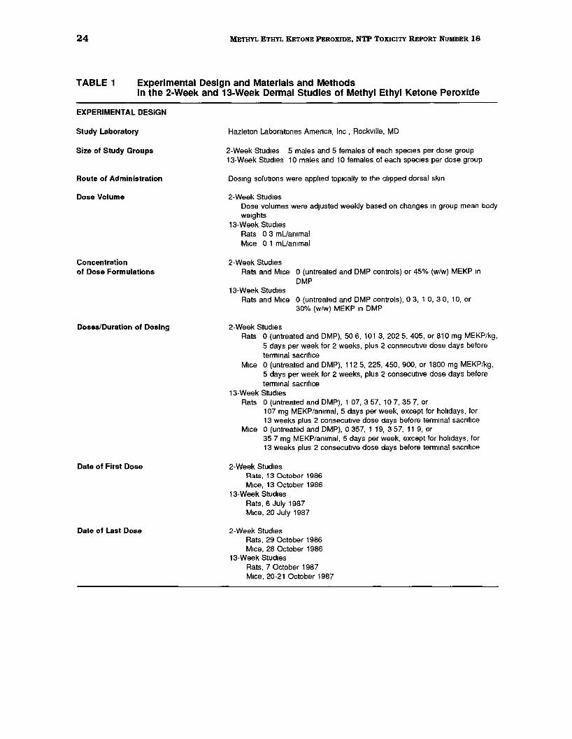
TABLE 1 Experimental Design and Materials and Methods in the 2-Week and 13-Week Dermal Studies of Methyl Ethyl Ketone Peroxide
EXPERIMENTAL DESIGN
Study Laboratory
Size of Study Groups
Route of Administration
Dose Volume
Concentration of Dose Formulations
Doses/Duration of Dosing
Date of First Dose
Date of Last Dose
Hazleton Laboratones Amenca, Inc , Rockville, MD
2-Week Studies 5 males and 5 females of each spec1es per dose group 13-Week Studies 10 mates and 10 females of each spec1es per dose group
Dos1ng solutions were applied topically to the clipped dorsal skm
2-Week Stud1es Dose volumes were adJUSted weekly based on changes 1n group mean body we1ghts
13-Week Studies Rats 0 3 mUammal M1ce 0 1 mUan1mal
2-Week Studies Rats and Mice 0 (untreated and DMP controls) or 45% (w/w) MEKP 1n
DMP 13-Week Studies
Rats and Mice 0 (untreated and DMP controls), 0 3, 1 0, 3 0, 10, or 30% (w/w) MEKP 1n DMP
2-Week Studies Rats 0 (untreated and DMP), 50 6, 101 3, 202 5, 405, or 810 mg MEKP/kg,
5 days per week for 2 weeks, plus 2 consecutive dose days before terminal sacnf1ce
M1ce 0 (untreated and DMP), 112 5, 225, 450, 900, or 1800 mg MEKP/kg, 5 days per week for 2 weeks, plus 2 consecutive dose days before termmal sacnf1ce
13-Week Studies Rats 0 (untreated and DMP), 1 07, 3 57, 10 7, 35 7, or
107 mg MEKP/ammat, 5 days per week, except for holidays, for 13 weeks plus 2 consecutive dose days before termmal sacnf1ce
M1ce 0 (untreated and DMP), 0 357, 1 19, 3 57, 11 9, or 35 7 mg MEKP/ammal, 5 days per week, except for holidays, for 13 weeks plus 2 consecutive dose days before termmal sacnf1ce
2-Week Studies Rats, 13 October 1986 M1ce, 13 October 1986
13-Week Studies Rats, 6 July 1987 M1ce, 20 July 1987
2-Week Stud1es Rats, 29 October 1986 M1ce, 28 October 1986
13-Week Studies Rats, 7 October 1987 M1ce, 20-21 October 1987
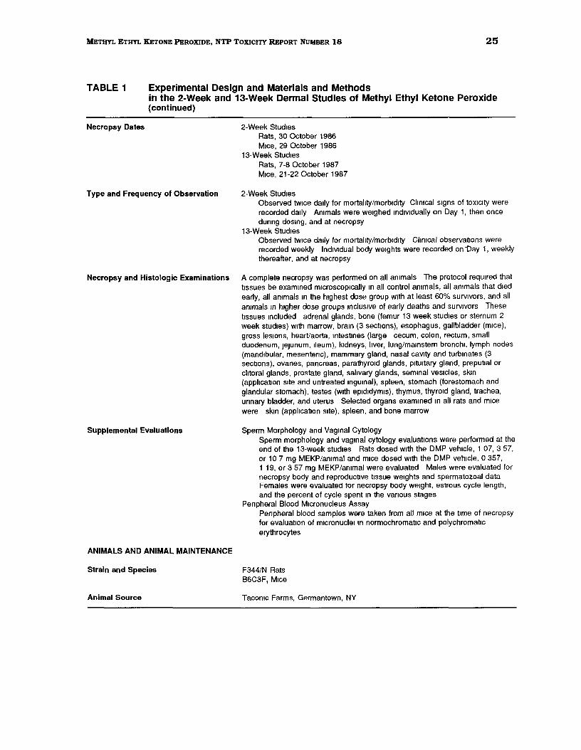
25METHYL ETHYL KETONE PEROXIDE, NTP TOXICITY REPORT NUMBER 18
TABLE 1 Experimental Design and Materials and Methods in the 2-Week and 13-Week Dermal Studies of Methyl Ethyl Ketone Peroxide (continued)
Necropsy Dates 2-Week Stud1es Rats, 30 October 1986 M1ce, 29 October 1986
13-Week Stud1es Rats, 7-8 October 1987 M1ce, 21-22 October 1987
Type and Frequency of Observation 2-Week Stud1es Observed tw1ce dally for mortality/morbidity Climcal s1gns of toXICity were recorded da1ly Ammals were we1ghed md1v1dually on Day 1, then once dunng dosmg, and at necropsy
13-Week Studies Observed tw1ce da1ly for mortality/morbidity Climcal observations were recorded weekly IndiVIdual body we1ghts were recorded on -oay 1, weekly thereafter, and at necropsy
Necropsy and Histologic Examinations A complete necropsy was performed on all ammals The protocol requ1red that t1ssues be examined m1croscop1cally 1n all control ammals, all ammals that d1ed early, all an1mals 1n the highest dose group w1th at least 60% surv1vors, and all ammals 1n higher dose groups 1nclus1ve of early deaths and surv1vors These t1ssues Included adrenal glands, bone (femur 13 week studies or sternum 2 week studies) w1th marrow, bram (3 sect1ons), esophagus, gallbladder (m1ce), gross les1ons, heart/aorta, mtestmes (large cecum, colon, rectum, small duodenum, JeJunum, Ileum), kidneys, liver, lung/mamstem bronchi, lymph nodes (mandibular, mesentenc), mammary gland, nasal caVIty and turbinates (3 sections), ovanes, pancreas, parathyroid glands, pitUitary gland, preputial or clitoral glands, prostate gland, salivary glands, semmal ves1cles, skm (application s1te and untreated mgUinal), spleen, stomach (forestomach and glandular stomach), testes (w1th epididymis), thymus, thyroid gland, trachea, unnary bladder, and uterus Selected organs exam1ned m all rats and m1ce were sk1n (application s1te), spleen, and bone marrow
Supplemental Evaluations Sperm Morphology and Vagmal Cytology Sperm morphology and vag1nal cytology evaluations were performed at the end of the 13-week stud1es Rats dosed w1th the DMP veh1cle, 1 07, 3 57, or 10 7 mg MEKP/ammal and m1ce dosed w1th the DMP vehicle, 0 357, 1 19, or 3 57 mg MEKP/ammal were evaluated Males were evaluated for necropsy body and reproductive t1ssue we1ghts and spermatozoal data Females were evaluated for necropsy body we1ght, estrous cycle length, and the percent of cycle spent 1n the vanous stages
Penpheral Blood Micronucleus Assay Penpheral blood samples were taken from all m1ce at the t1me of necropsy for evaluation of m1cronucle1 m normochromat1c and polychromatic erythrocytes
ANIMALS AND ANIMAL MAINTENANCE
Strain and Species F344/N Rats 86C3F, M1ce
Animal Source Tacomc Farms, Germantown, NY
26 METHYL ETHYL KETONE PEROXIDE, NTP TOXICITY REPORT NUMBER 18
TABLE 1 Experimental Design and Materials and Methods In the 2-Week and 13-Week Dermal Studies of Methyl Ethyl Ketone Peroxide (continued)
Time Held Before Study
Age When Study Began
Age When Killed
Method of Animal Distribution
Diet
Animal Room Environment
2-Week Stud1es Rats, 13 days, M1ce, 14 days 13 Week Stud1es Rats, 13 days, M1ce, 18 days
2-Week Stud1es Rats, 6 weeks, M1ce, 6 weeks 13-Week Stud1es Rats, 6 weeks, M1ce, 6 weeks
2-Week Stud1es Rats, 8 weeks, M1ce, 8 weeks 13 Week Stud1es Rats, 19 weeks, M1ce, 19 weeks
Ammals were we1ghed and randomized us1ng a computer program
NIH-07 Open Formula Pellets (Ze1gler Brothers, Inc, Gardners, PA) and tap water available ad libitum
Rats and m1ce were housed md1v1dually for all stud1es Temperature ranged from 68°F to 75°F, relative humidity ranged from 31% to 72% With 12 to more than 20 a1r changes per hour Fluorescent hght was prov1ded for 12 hours per day
Statistical Methods
ANALYSIS OF CONTINUOUS VARIABLES
Two approacheswere employed to assess the significance ofpairwise comparisons between
dosed and control groups in the analysis of continuous variables_ Organ and body weight
data, which are approximately normally distributed, were analyzed using the parametric
multiple comparisons procedures ofWilliams (1971, 1972) and Dunnett (1955). Data that
typically have skewed distributions were analyzed using the nonparametric multiple
comparisons methods of Shirley (1977) or Dunn (1964). Jonckheere's test (Jonckheere,
1954) was used to assess the significance of dose-response trends and to determine
whether a trend-sensitive test (Williams, Shirley} was more appropriate for pairwise
comparisons than a test capable of detecting departures from monotonic dose response
(Dunnett, Dunn). If the P-value from Jonckheere's test was greater than or equal to 0.10,
Dunn's or Dunnett's test was used rather than Shirley's or Williams' test.
The outlier test of Dixon and Massey (1951) was employed to detect extreme values. No
value selected by the outlier test was eliminated unless it was at least twice the next
largest value or at most half of the next smallest value. The extreme values chosen by the
statistical test were subject to approval by NTP personnel. In addition, values mdicated
27 METHYL ETHYL KETONE PEROXIDE, NTP TOXICITY REPORT NUMBER 18
by the laboratory report as being inadequate due to technical problems were eliminated
from the analysis.
ANALYSIS OF VAGINAL CITOLOGY DATA
Because the data are proportions (the proportion of the observation period that an animal
was in a given estrous state). an arcsine transformation was used to bring the data into
closer conformance with normality assumptions. Treatment effects were investigated by
applying a multivariate analysis of variance (Morrison, 1976) to the transformed data to
test for simultaneous equality of measurements across dose levels.
ANALYSIS OF MUTAGENICITY IN SALMONELLA 1YPHIMURIUM
A positive response in the SalmoneUa typhimurium test was defined as a reproducible.
dose-related increase in histidine-independent (revertant) colonies in any
1 strain/activation combination. An equivocal response was defined as an increase in
revertants that was not dose related, not reproducible, or not of sufficient magnitude to
support a determination of mutagenicity. A negative response was obtained when no
increase in revertant colonies was observed following chemical treatment. There was no
minimum percentage or fold increase required for a chemical to be judged positive or
weakly positive.
ANALYSIS OF MOUSE LYMPHOMA MUTAGENICI1Y DATA
Minimum criteria for accepting an experiment as valid and a detailed description of the
statistical analysis and data evaluation are presented in Caspary et aL (1988). Data were
evaluated statistically for both trend and peak responses. Both responses had to be
significant (P::;0.05) for a chemical to be considered capable of inducing TIT-resistance.
A single significant response led to a "questionable" conclusion. and the absence of both
a trend and a peak response resulted in a "negative" call.
ANALYSIS OF CHO CELL CITOGENETICS DATA
For the SCE data. statistical analyses were conducted on the slopes of the dose-response
curves (Galloway et a1., 1987). An SCE frequency 20% above the concurrent solvent
control value was chosen as a statistically conservative positive response. The probability
28 METHYL ETHYL 'KETONE PEROXIDE, NTP TOXICITY REPORT NUMBER 18
of this level of difference occurring by chance at 1 dose point is less than 0.01; the
probability for such a chance occurrence at 2 dose points is less than 0.00 l. If
only l dose was increased by at least 20% over the solvent control, it was considered weak
evidence for a positive response; increases in at least 2 doses resulted in a determination
of positive. A statistically significant trend (P:::::0.05) in the absence of any responses
reaching 20% above background led to a call of equivocal.
Chromosomal aberration data are presented as percentage of cells with aberrations; both
the dose-response cmve and individual dose points were statistically analyzed (Galloway
et al., 1987). For a single trial, a statistically significant (P:::-:;0.05) difference for l dose point
and a significant trend (P:::::O.Ol5) were considered weak evidence for a positive response;
significant differences for 2 or more doses indicated the trial was positive.
ANALYSIS OF MOUSE PERIPHERAL BLOOD MICRONUCLEUS DATA
Log transformation of the normochromatic erythrocyte (NCE) data, and testing for
normality by the Shapiro-Wilk test, and for heterogeneity of variance by Cochran's test,
were performed before statistical analyses. The frequency of micronucleated cells among
NCEs was analyzed by an analysis of variance using the SAS GLM procedure. The NCE
data for each dose group were compared with data from the concurrent DMP control using
Student's t-test. The frequency ofmicronucleated cells among polychromatic erythrocytes
(PCEs) was analyzed by the Cochran-Armitage trend test, and individual dose groups were
compared to the concurrent DMP control by Kastenbaum-Bowman's (1970) binomial test.
The percentage PCE among total erythrocytes was analyzed by an analysis ofvariance on
ranks (classed by sex) and individual dose groups were compared with the concurrent
DMP control using a t-test on ranks.
Quality Assurance
The animal studies of MEKP were performed in compliance with the United States FDA
Good Laboratory Practices regulations (21 CFR 58). The Quality Assurance Unit of
Hazleton Laboratories America, Inc. performed audits and inspections of protocols,
procedures, data, and reports throughout the course of the studies. The operations of the
Quality Assurance Unit were monitored by the NTP.
29 METHYL ETHYL KETONE PEROXIDE, NTP TOXICITY REPORT NUMBER 18
RESULTS
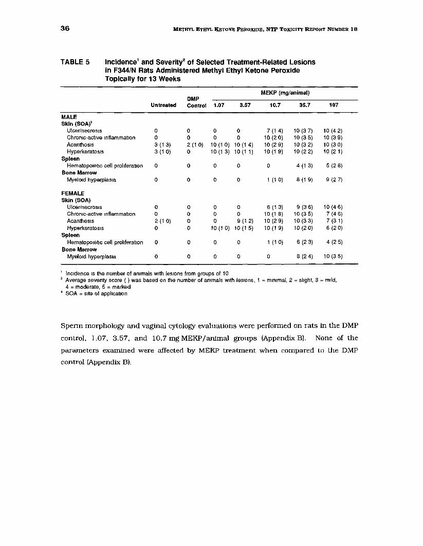
2-Week Dermal Study in F344/N Rats
All rats treated with methyl ethyl ketone peroxide (MEKP) for 2 weeks survived to the end
of the study (Table 2). There was a dose-dependent decrease in the mean body weight gain
compared to the dimethyl phthalate (DMP) control group for all MEKP-treated male rats
and for female rats in the 2 highest (405 and 810 mg/kg) dose groups. A dose-related
decrease in mean final body weights also occurred in rats given MEKP.
TABLE 2 Survival and Weight Gain of F344/N Rats in the 2-Week Dermal Study of Methyl Ethyl Ketone Peroxide
Mean Body Weight (grams) Final Weight Relative to DMP
Dose (mg/kg) Survival' Initial Final Change2 Controls (%)3
MALE Untreated 5/5 125 200 75 102 DMP Control 5/5 127 196 69
506 5/5 128 187 59 96 101 3 5/5 124 176 53 90 202 5 5/5 136 184 48 94 405 5/5 129 177 49 90 810 5/5 134 162 28 83
FEMALE Untreated 5/5 112 145 33 99 DMP Control 5/5 115 147 32
50 6 5/5 112 144 32 98 101 3 5/5 114 141 27 96 202 5 5/5 112 141 30 96 405 5/5 114 136 22 93 810 5/5 113 135 21 92
' Number surv1v1ng at 18 days/number of ammals per dose group 2 Mean we1ght change of the ammals m each dose group 3 (Dosed group mean/DMP control group mean) x 100
Changes in organ weights that were considered to be possibly biologically significant were
limited to decreases in absolute and/or relative thymus weights in males receiving 10 1.3 to
810 mgMEKP/kg and in females receiving 202.5 to 810 mgMEKP/kg, and a trend toward
mild increases in relative liver weights in MEKP-treated male and female rats when
30 METHYL ETHYL KETONE PEROXIDE, NTP TOXICITY REPORT NUMBER 18
compared to the DMP controls (statistically significant in females receiving 202.5 to
810 mg/kg) (Table 3).
TABLE 3 Liver and Thymus Weights of F344/N Rats Administered Methyl Ethyl Ketone Peroxide Topically for 2 Weeks1
MEKP (mg/kg)
DMP Untreated Control 50.6 101.3 202.5 405 810
MALE Necropsy body we1ght 211 206 194 185 189 184* 165**
L1ver weight 11 344 11 234 10 632 10 384 11 050 10 642 9 880 Relat1ve liver we1ght 53 83 54 37 55 16 56 00 58 33 57 87 59 78
Thymus we1ght 0468 0479 0 401 0 348*2 0 374* 0 358** 0 265** Relat1ve thymus we1ght 2 22 2 33 2 07 1 852 1 98 1 94 1 61**
FEMALE Necropsy body we1ght 150 151 146 145 141* 138** 135**
L1ver we1ght 6690 7 260 7 608 7 236 7622 7 514 8 082 Relat1ve liver we1ght 44 57* 4802 5218 4992 54 14** 54 so·· 59 70**
Thymus we1ght 0350 0370 0361 0345 0 291* 0305* 0 273** Relative thymus we1ght 2 33 2 44 2 46 2 39 2 06 2 21 2 03*
Organ we1ghts and body we1ghts are g1ven 1n grams, relatiVe organ we1ghts (organ we1ght-to-body-we1ght rat1os) are g1ven as mg organ we1ghtlg body we1ght, n=5 except where noted, DMP = dimethyl phthalate n=4 S1gn1flcantly different (P$0 OS) from the DMP control group by Williams' test
** Significantly different (P$0 01) from the DMP control group by Williams' test
At necropsy, gross lesions associated with compound administration were obsexved at the
site of application of MEKP to the skin. The skin was thickened and indurated in all rats
in all MEKP-treated groups. A scab-like crust was also present over the application site
in some animals in the 405 and 810 mg/kg groups. An enlarged spleen was noted in
1 male and in 1 female rat receiving 810 mg MEKP/kg.
Gross lesions at the application site were correlated with MEKP-related dermal/epidermal
necrosis and associated inflammatory reactions. The typical histologic presentation at all
MEKP dose levels was extensive coagulative necrosis of the epidermis and dermis as well
as the adnexa. The necrotic skin generally formed a superficial coagulum; the viable
tissue underneath exhibited variable degrees of regeneration and inflammation.
31 METHYL ETHYL KETONE PEROXIDE, NTP TOXICITY REPORT NUMBER 18
Regeneration was characterized by incomplete to complete bridging of denuded areas by
epithelium, as well as by the consistent presence of epidermal hyperplasia (acanthosis) at
the margins of ulcerated areas. Inflammation was usually marked and consisted primarily
of neutrophilic and serous exudate superficially, and of fibrovascular proliferation
("granulation tissue") with mixed leukocyte infiltration in the deep dermis and subcutis.
The severity of the necro-inflammatory lesions was moderate to marked at all MEKP dose
levels, with a tendency for somewhat more extensive involvement at the higher dose levels.
MEKP-related changes were also present in the spleen. Increased hematopoietic cell
proliferation relative to DMP and untreated controls and congestion of the red pulp were
obsetved in all MEKP-treated rats. Increased hemosiderin deposition and focal infiltration
of neutrophils and histiocytic cells into the splenic capsule were also obsetved in the
spleen, primarily at the higher dose levels.
32 METHYL ETHYL KETONE PEROXIDE, NTP TOXICITY REPORT NUMBER 18
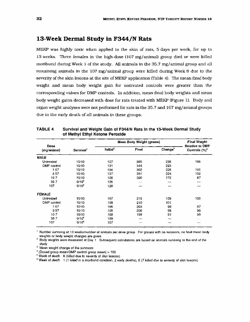
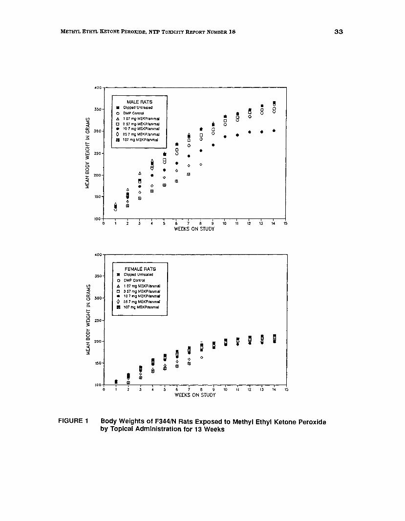
13-Week Dermal Study in F344/N Rats
MEKP was highly toxic when applied to the skin of rats, 5 days per week, for up to
13 weeks. Three females in the high-dose (107 mg/animal) group died or were killed
moribund during Week 1 of the study. All animals in the 35.7 mg/animal group and all
remaining animals in the 107 mg/animal group were killed during Week 8 due to the
severity of the skin lesions at the site of MEKP application (Table 4). The mean final body
weight and mean body weight gain for untreated controls were greater than the
corresponding values for DMP controls. In addition, mean final body weights and mean
body weight gains decreased with dose for rats treated with MEKP (Figure 1). Body and
organ weight analyses were not performed for rats in the 35.7 and 107 mg/animal groups
due to the early death of all animals in these groups.
TABLE 4 Survival and Weight Gain of F344/N Rats in the 13-Week Dermal Study of Methyl Ethyl Ketone Peroxide
Dose (mg/animal) Survival1
Mean Body Weight (grams)
Change3
Final Weight Relative to DMP
Controls (%)4 lnitial2 Final
MALE Untreated DMP control
1 07 3 57
10 7 35 7
107
FEMALE Untreated DMP control
1 07
3 57 10 7 35 7
107
10/10 10/10 10/10 10/10 10/10 0/105
0/105
10/10 10/10 10/10 10/10 10/10 0/105
0/106
127 121 134 127 128 126 128
107 109 106 108 108 109 107
365 344 362 351 300
215 210 203 206 199
238 223 228 224 172
109 101 98 98 91
106
105 102 87
103
97 98 95
1 Number surv1v1ng at 13 weeks/number of ammals per dose group For groups w1th no surviVors, no f1nal mean body we1ghts or body we1ght changes are g1ven
2 Body Weights were measured at Day 1 Subsequent calculations are based on ammals survJvmg to the end of the study
3 Mean we1ght change of the survJvors 4 (Dosed group mean/DMP control group mean) x 100 5 Week of death 8 (killed due to seventy of sk1n les1ons) 6 Week of death 1 (1 krlled m a monbund condrtron, 2 early deaths), 8 (7 krlled due to seventy of skrn lesrons)
(/) ::::;; <a:: <.:l
~ t-J: <.:l w 3: >0 0 ID z < w :::;;
(/):::;; <0:: <.:l
~ ,.... J: <.:l w 3: >0 0 m z < w :::;;
400
zso
300
250
200
150
100
400
350
300
250
200
150
100
0
MALE RATS
• Clipped Untreated
• 10 7 mQ MEKP/an1maJ
0 DMP Control
.b. 1 07 m; MEKPlan1mal
c ~ 57 mg MEKPian1maJ
0 ~57 mg MEKP/an1mal
Ill 107 mg MEKPian1mal
•Ill 8 e •
e h. • ¢
¢ til A •
1810I ~ ¢
~ cil
2 3 5"
FEMALE RATS
• Clipped Untreated
0 OMP Control
.b. 1 07 mg MEKPian1mal
0 ~ 57 mg MEKPian1mal e 10 7 mg MEKPian1mal
0 35 7 mg MEKP/an1mal
Ill 107 mg MEKPiammal
I = I ® ~
' ~ m
I til
!!IIll 0~ 0 8
Q 0* 8*
i 0 0
• •0Ill •0 • • • 0
• 0
•0
•0 0 •• 0 ¢
0
lil
til
6 7 8 9 10 11 12 13 14 15
WEEKS ON STUDY
I a ; I ! I I; ¢
= <> ¢
181181
0 3 5 6 7 8 9 10 II 12 t3 14 15
WEEKS ON STUDY
33 METHYL ETHYL KETONE PEROXIDE, NTP TOXICITY REPORT NUMBER 18
FIGURE 1 Body Weights of F344/N Rats Exposed to Methyl Ethyl Ketone Peroxide by Topical Administration for 13 Weeks
34 METHYL ETHYL KETONE PEROXIDE, NTP TOXICITY REPORT NUMBER 18
There were statistically significant increases in the relative weights of several organs,
primarily in males in the 10.7 mg/animal group (Appendix A). These were considered
secondary to the lower body weight gains in these animals and not reflective of a primary
chemical effect. There were no changes in absolute or relative organ weights that were
considered treatment related. However. the relative liver weights ofthe D MP control female
rats showed greater variability, and the mean was somewhat greater than those of the
untreated controls or of the MEKP-treated groups.
Necropsy observations showed thickened, crusty skin at the site of MEKP application,
particularly in the 35.7 and 107 mg dose group animals that were sacrificed early.
Additional observations in many of the early-death rats receiving 107 mg/animal were
"scar tissue" at the application site and enlarged spleens. In those animals that survived
for 13 weeks, significant necropsy observations were limited to thick, crusty skin at the
application site in several animals from the 10.7 mg/animal group.
Microscopically, MEKP treatment was associated with a spectrum of necrotic,
inflammatory, and regenerative skin lesions at' the application site; the severity of the
lesions increased progressively with dose. Dermal and epidermal necrosis was the primary
lesion at dose levels of 10.7 mg/animal and greater. Necrosis varied in severity from
diffuse involvement of both dermis and epidermis at higher concentrations to focal
ulcerations of the epidermis, which occurred more frequently in the 10.7 mg/animal
group. An associated inflammatory reaction consisted of both surface exudation and
dermal inflammation. Typically. a coagulum of surface exudate containing serous fluid
and neutrophils was seen overlying denuded or regenerative areas. with necrotic skin
tissue admixed in more severe cases. The dermal reaction was subjacent to ulcerated,
sloughed. or regenerated epidermis and was collectively termed "chronic-active" to be
inclusive of varied patterns, including primarily neutrophilic, fibrovascular ("granulation
tissue"). granulomatous, or fibrotic reactions. Epidermal hyperplasia (acanthosis) and
hyperkeratosis were consistently present at the margins of ulcerated areas in the higher
dose groups (10.7 to 107 mg/animal). In the 1.07 and 3.57 mg/animalgroups, acanthosis
and/or hyperkeratosis were the only skin lesions evident at the application site; these
lesions were seen in all animals in these dose groups. Minimal acanthosis and
hyperkeratosis were seen in some untreated and DMP control rats.
35 METHYL ETHYL KETONE PEROXIDE, NTP TOXICITY REPORT NUMBER 18
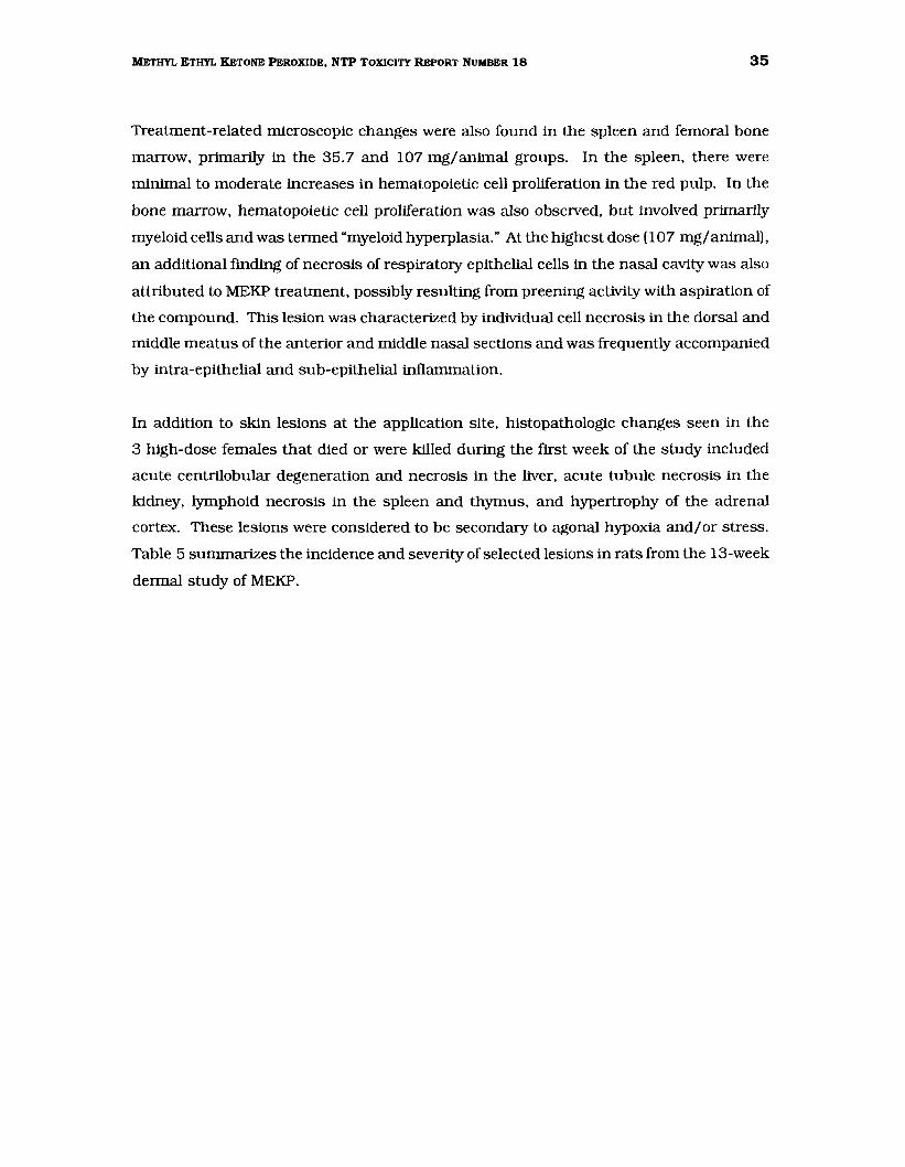
Treatment-related microscopic changes were also found in the spleen and femoral bone
marrow, primarily in the 35.7 and 107 mg/animal groups. In the spleen, there were
minimal to moderate increases in hematopoietic cell proliferation in the red pulp. In the
bone marrow, hematopoietic cell proliferation was also observed, but involved primarily
myeloid cells and was termed "myeloid hyperplasia." At the highest dose (107 mg/animal),
an additional finding of necrosis of respiratory epithelial cells in the nasal cavity was also
attributed to MEKP treatment, possibly resulting from preening activity with aspiration of
the compound. This lesion was characterized by individual cell necrosis in the dorsal and
middle meatus of the anterior and middle nasal sections and was frequently accompanied
by intra-epithelial and sub-epithelial inflammation.
In addition to skin lesions at the application site, histopathologic changes seen in the
3 high-dose females that died or were killed during the first week of the study included
acute centrilobular degeneration and necrosis in the liver, acute tubule necrosis in the
kidney, lymphoid necrosis in the spleen and thymus, and hypertrophy of the adrenal
cortex. These lesions were considered to be secondary to agonal hypoxia and/or stress.
Table 5 summarizes the incidence and severity of selected lesions in rats from the 13-week
dermal study of MEKP.
36 METHYL ETHYL KETONE PEROXIDE, NTP TOXICITY REPORT NUMBER 18
TABLE 5 lncidence1 and Severlty2 of Selected Treatment-Related Lesions in F344/N Rats Administered Methyl Ethyl Ketone Peroxide Topically for 13 Weeks
MEKP (mg/animal) DMP
Untreated Control 1.07 3.57 10.7 35.7 107
MALE Skin (SOA)3
Ulcer/necros1s 0 0 0 0 7 (1 4) 10 (3 7) 10 (4 2) Chromc-act1ve 1nflammat1on 0 0 0 0 10 (2 0) 10 (3 5) 10 (3 9) Acanthosis 3 (1 3) 2 (1 0) 10 (1 0) 10 (1 4) 10 (2 9) 10 (3 2) 10 (3 0) Hyperkeratosis 3 (1 0) 0 10 (1 3) 10 (1 1) 10 (1 9) 10 (2 2) 10 (2 1)
Spleen Hematopo1et1c cell proliferation 0 0 0 0 0 4 (1 3) 5 (2 8)
Bone Marrow Myeloid hyperplasia 0 0 0 0 1 (1 0) 8 (1 9) 9 (2 7)
FEMALE Skin (SOA)
Ulcer/necrosis 0 0 0 0 6 (1 3) 9 (3 6) 10 (4 6) Chromc-act1ve 1nflammat1on 0 0 0 0 10 (1 8) 10 (3 5) 7 (4 6) Acanthosis 2 (1 0) 0 0 9 (1 2) 10 (2 9) 10 (3 3) 7 (3 1) Hyperkeratosis 0 0 10 {1 0) 10 {1 5) 10 {1 9} 10 {2 0} 6 {2 0}
Spleen Hematopo1et1c cell proliferation 0 0 0 0 1 (1 0) 6 (2 3} 4 (2 5}
Bone Marrow Myeloid hyperplasia 0 0 0 0 0 8 (2 4) 10 (3 5)
' Incidence 1s the number of ammals w1th les1ons from groups of 10 2 Average seventy score ( ) was based on the number of ammals With les1ons, 1 = m1mmal, 2 =slight, 3 = m1ld,
4 = moderate, 5 = marked 4 SOA = s1te of apphcat1on
Sperm morphology and vaginal cytology evaluations were performed on rats in the DMP
control, 1.07, 3.57, and 10.7 mg MEKP/animal groups (Appendix B). None of the
paraiileters exanlined were affected by MEKP treatment when compared to the DMP
control (Appendix B).
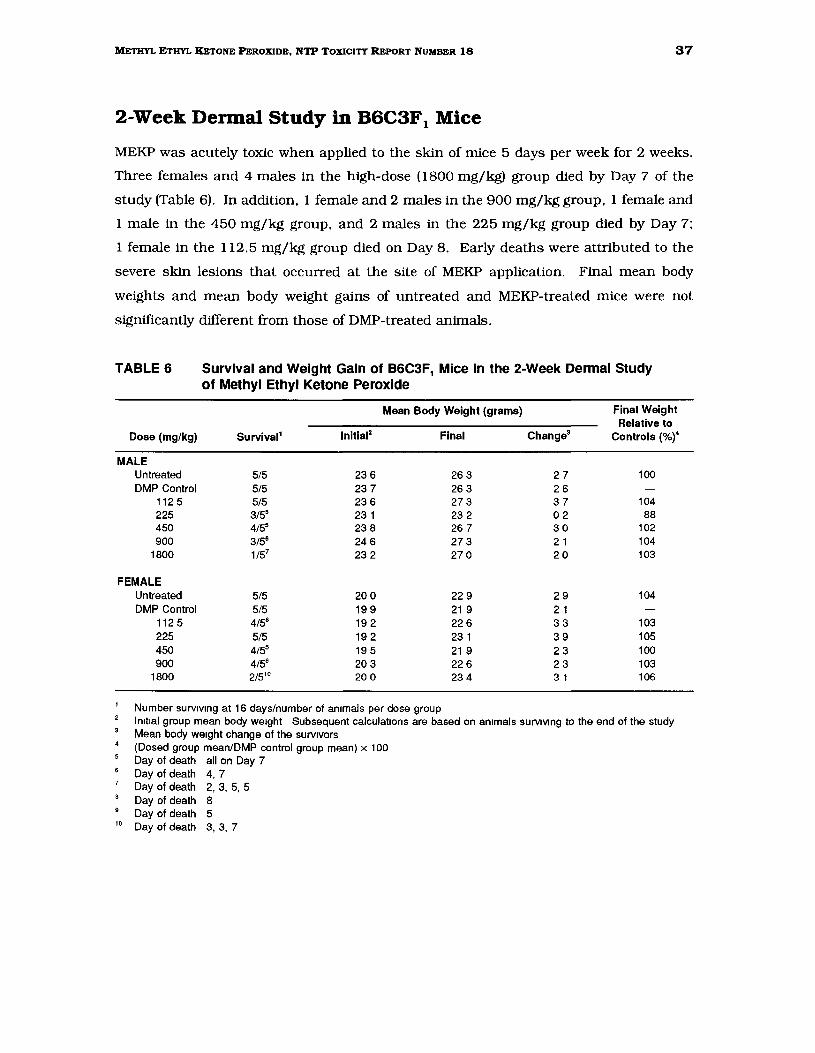
37 METHYL ETHYL KETONE PEROXIDE, NTP TOXICITY REPORT NUMBER 18
2-Week Dermal Study in B6C3F1 Mice
MEKP was acutely toxic when applied to the skin of mice 5 days per week for 2 weeks.
Three females and 4 males in the high-dose (1800 mg/kg) group died by Day 7 of the
study (Table 6). In addition, 1 female and 2 males in the 900 mg/kg group, 1 female and
1 male in the 450 mg/kg group, and 2 males in the 225 mg/kg group died by Day 7;
1 female in the 112.5 mg/kg group died on Day 8. Early deaths were attributed to the
severe skin lesions that occurred at the site of MEKP application. Final mean body
weights and mean body weight gains of untreated and MEKP-treated mice were not
significantly different from those of DMP-treated animals.
TABLE 6 Survival and Weight Gain of B6C3F1 Mice In the 2-Week Dermal Study of Methyl Ethyl Ketone Peroxide
Mean Body Weight (grams) Final Weight Relative to
Dose (mg/kg) Survival' lnitial2 Final Change3 Controls (%t
MALE Untreated 5/5 23 6 26 3 27 100 DMP Control 5/5 23 7 26 3 26
112 5 5/5 23 6 27 3 37 104 225 3/55 23 1 23 2 02 88 450 4/55 23 8 26 7 30 102 900 3t5• 24 6 27 3 2 1 104
1800 1/57 23 2 270 20 103
FEMALE Untreated 5/5 20 0 22 9 29 104 DMP Control 5/5 19 9 21 9 2 1
112 5 4/58 19 2 22 6 33 103 225 5/5 19 2 23 1 39 105 450 4/55 19 5 21 9 23 100 900 4/59 20 3 22 6 23 103
1800 2/510 20 0 23 4 3 1 106
Number surv1v1ng at 16 days/number of ammals per dose group ln1t1al group mean body we1ght Subsequent calculations are based on ammals SUrviVIng to the end of the study Mean body we1ght change of the surv1vors (Dosed group mean/DMP control group mean) x 100 Day of death all on Day 7 Day of death 4, 7 Day of death 2, 3, 5, 5 Day of death 8 Day of death 5
10 Day of death 3, 3, 7
38 METHYL ETHYL KETONE PEROXIDE, NTP TOXICITY REPORT NUMBER 18
Changes in liver and thymus weights were related to chemical treatment (Table 7).
Significant increases in absolute and relative liver weights were noted for male and female
mice treated with MEKP. In addition, the absolute and relative liver weights of DMP
controls were greater than those of the untreated control group. Both absolute and
relative thymus weights were significantly lower than DMP control values for females in
the 3 highest dose groups (450 to 1800 mg/kg); males in the 225 and 900 mg/kg dose
groups also had lower thymus weights than DMP control males, but the differences were
not statistically significant.
TABLE 7 Liver and Thymus Weights of B6C3F, Mice Administered Methyl Ethyl Ketone Peroxide Topically for 2 Weeks1
MEKP (mg/kg) DMP
Untreated Control 112.5 225 450 900 1800
MALE n 5 5 5 3 4 3 Necropsy body we1ght 26 7 26 5 270 21 7 24 9 23 0 21 7
L1ver we1ght 1 284 1 376 1 504 1 607* 1 878** 2 233** 2 410 Relat1ve liver we1ght 48 11* 51 85 55 73 63 09* 68 85** 80 44** 88 93
Thymus we1ght 0048 0049 0 051 0 027" 0054 0 024 0 060 Relat1ve thymus we1ght 1 80 1 86 1 91 1 01 2 2 01 0 85 2 21
FEMALE n 5 5 4 5 4 4 2 Necropsy body we1ght 22 7 23 2 20 4 24 2 22 6 22 1 18 9
L1ver we1ght 1 120* 1 268 1 375 1 552 1 590 1 778** 1 710* Relative liver we1ght 49 22** 5457 61 01* 64 20** 67 99** 76 24** 76 32**
Thymus we1ght 0079 0 084 0 065 0068 0 049* 0 053* 0 054* Relative thymus we1ght 348 3 62 2 87 2 81 2 02** 2 23** 2 41*
Organ we1ghts and body we1ghts are g1ven 1n grams, relat1ve organ we1ghts (organ-we1ght-to-body we1ght rat1os) are g1ven as mg organ we1ghVg body we1ght n=2 S1gn1flcantly different (P$0 05) from the DMP control group by Williams' test
** Significantly different (P$0 01) from the DMP control group by Williams' test
Gross skin lesions at the application site were seen in all MEKP-treated mice at necropsy.
The skin was thickened, crusty, hardened, and, in some animals, sloughed. Splemc
enlargement was noted in l or more animals in most MEKP treatment groups.
39 METHYL ETHYL KETONE PEROXIDE, NTP TOXICITY REPORT NUMBER 18
Microscopically, MEKP application was associated with necrotic and inflammatory lesions
of the skin and underlying tissue at the application site in all dose groups. Coagulative
necrosis of the skin was the primary lesion; it was graded as minimal to marked in
severity. Superficial exudation of neutrophils and proteinaceous fluid invariably
accompanied the necrotic tissue. Proliferation offibrovascular ("granulation") tissue was
variably present and was seen more frequently in mice surviving to the end of the study.
Other histologic findings attributed to MEKP treatment, but believed to be secondary to
the skin inflammation and necrosis, were minimal to moderate hematopoietic cell
proliferation in the spleen at all dose levels and mild to marked myeloid hyperplasia of the
sternebra} bone marrow, which was most evident in females in the 900 and 1800 mg/kg
groups. There were no microscopic findings in the liver to account for the increased liver
weights.
Gross and microscopic findings in animals that died early were generally similar to those
present in mice that survived to the end of the study. However, lymphoid cell depletion
and necrosis in the thymus were additional lesions noted upon microscopic examination
of mice that died early.
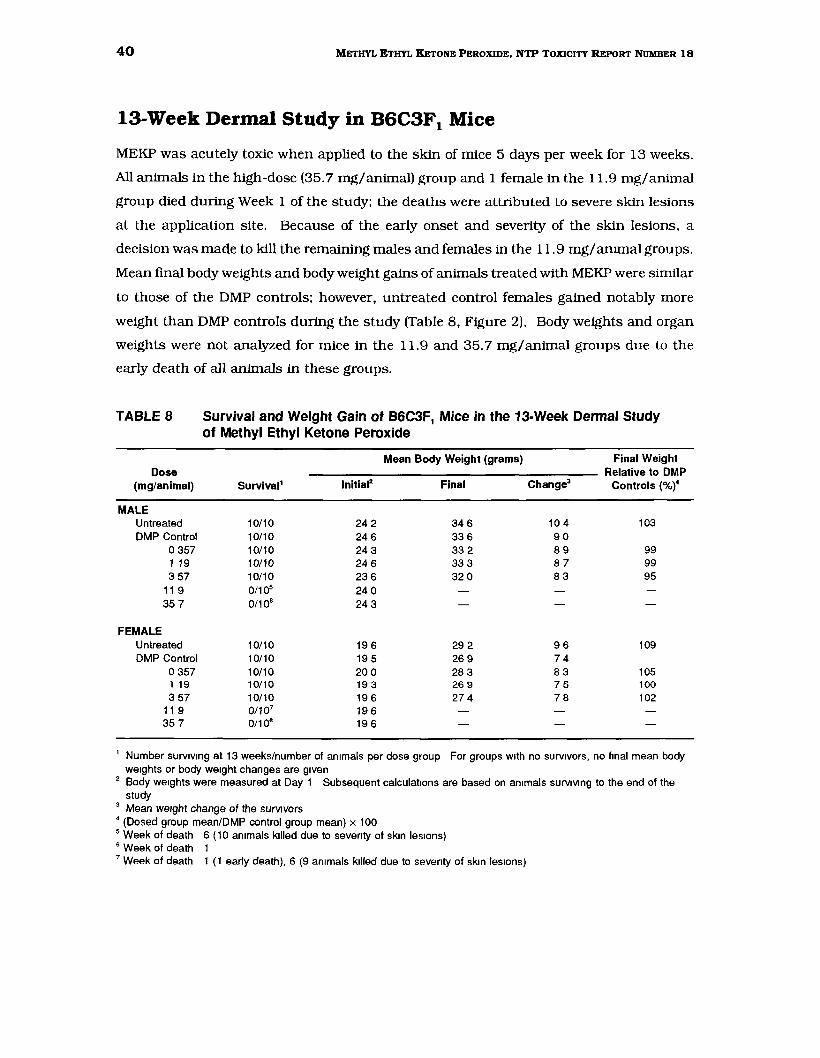
40 METHYL ETHYL KETONE PEROXIDE, NTP TOXICITY REPORT NUMBER 18
13-Week Dermal Study in B6C3F1 Mice
MEKP was acutely toxic when applied to the skin of mice 5 days per week for 13 weeks.
All animals in the high-dose (35. 7 mg/animal) group and 1 female in the 11.9 mg/animal
group died during Week l of the study; the deaths were attributed to severe skin lesions
at the application site. Because of the early onset and severity of the skin lesions, a
decision was made to kill the remaining males and females in the 11.9 mg/anunal groups.
Mean final body weights and body weight gains of animals treated with MEKP were siinilar
to those of the DMP controls; however, untreated control females gained notably more
weight than DMP controls during the study (Table 8, Figure 2). Body weights and organ
weights were not analyzed for mice in the 11.9 and 35.7 mg/animal groups due to the
early death of all animals in these groups.
TABLE 8 Survival and Weight Gain of B6C3F1 Mice in the 13-Week Dermal Study of Methyl Ethyl Ketone Peroxide
Mean Body Weight (grams) Final Weight Dose Relative to DMP
(mg/animal) Survival' lnitial2 Final Change3 Controls (%t
MALE Untreated 10/10 24 2 346 10 4 103 DMP Control 10/10 24 6 33 6 90
0 357 10/10 24 3 33 2 89 99 1 19 10/10 24 6 333 87 99 3 57 10/10 23 6 32 0 83 95
11 9 0/105 24 0 35 7 0/106 24 3
FEMALE Untreated 10/10 19 6 29 2 96 109 DMP Control 10/10 19 5 26 9 74
0 357 10/10 20 0 28 3 83 105 1 19 10/10 19 3 26 9 75 100 3 57 10/10 19 6 27 4 78 102
11 9 0/107 19 6 35 7 0/106 19 6
1 Number surv1v1ng at 13 weeks/number of an1mals per dose group For groups With no survivors, no final mean body we1ghts or body we1ght changes are g1ven
2 Body we1ghts were measured at Day 1 Subsequent calculations are based on ammals SUrviVIng to the end of the study
3 Mean we1ght change of the surv~vors 4 (Dosed group mean/DMP control group mean) x 100 5 Week of death 6 (10 ammals killed due to seventy of skm les1ons) 6 Week of death 1 7 Week of death 1 (1 early death), 6 (9 ammals killed due to seventy of skm les1ons)
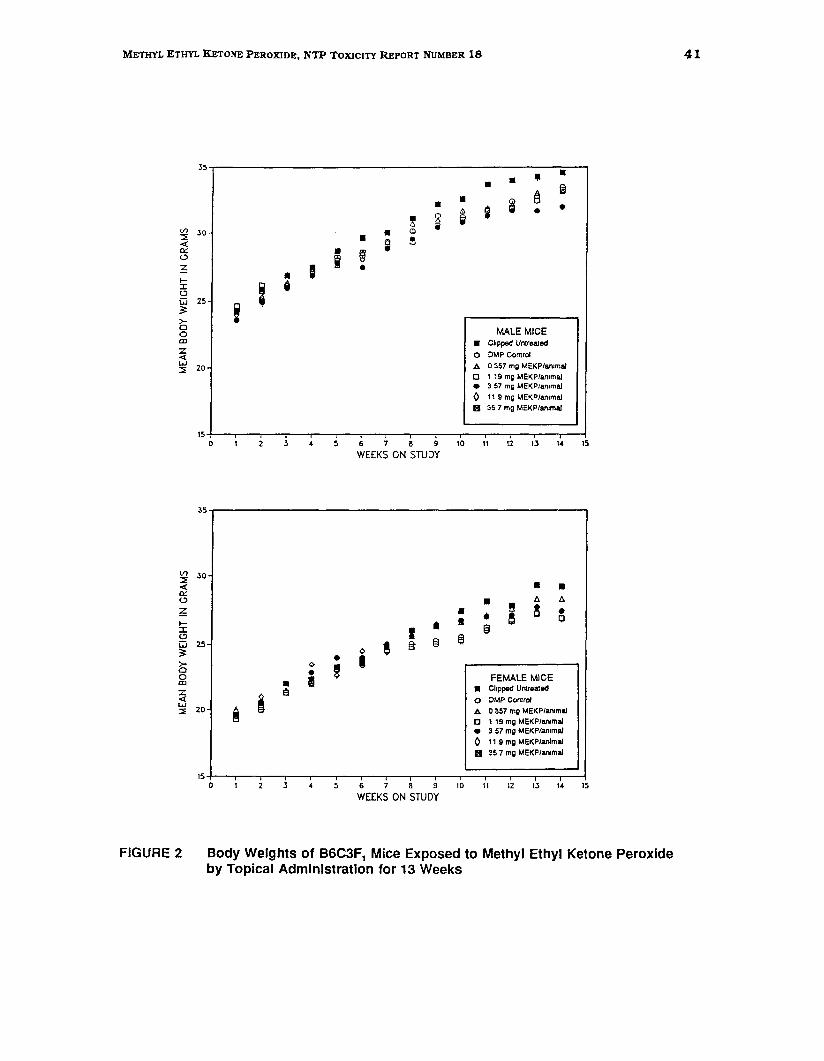
• •
41 METHYL ETHYL KETONE PEROXIDE, NTP TOXICITY REPORT NUMBER 18
35
V> :::0 <( 0:: (.;)
~ 1:I: (.;)
w 3: >0 0 ID
z <( w :::>
30
25
zo
i •
I • I •;
• ~ •
•i
~ 0 e
• 2 li
• &l
~
15
•• Ill• fa ~i •i •
MALE MICE • Clipped Untreated
•
0 DMP Control
A 0 357 mg MEKP/an1mal
0 119 mg MEKP/an1mal 3 57 mg MEKP/ammal
0 11 9 mg MEKP/an1mal
II 35 7 mg MEKP/an1mal
0 2 3 5 6 7 8 9 10 11 12 13 14 15
WEEKS ON STUDY
35,-------------------------------------------------------,
Vl 30:::0 <( 0:: (.;) t:. A
• • I! ! •0 ~ • • e1:I: • § (.;) I e §" § tiJw 25
• I 03:
b 0
0 I• ! ID •z 1!1<( w i:::> 20 ~
15 0 3 4 5 6 7 8 9 10
WEEKS ON STUDY
FEMALE MICE
• Chppeo' Umreared
•
0 DMP Control
A 0 357 mg MEKP/an1mal
0 119 mg MEKP/an1mal 3 57 mg MEKP/an1mal
0 11 9 mg MEKP/anlmal
II 35 7 mg MEKP/an1rnal
11 12 13 14 15
FIGURE 2 Body Weights of B6C3F1 Mice Exposed to Methyl Ethyl Ketone Peroxide by Topical Administration for 13 Weeks

42 METHYL ETHYL KETONE PEROXIDE, NTP TOXICITY REPORT NUMBER 18
Changes in spleen weights of males and females treated with 3.57 mg MEKP/animal were
considered related to treatment (Appendix A). Both absolute and relative spleen weights
were significantly increased in these groups when compared to the DMP controls.
However, the mean spleen weights of both male and female untreated control groups were
greater than those of the DMP vehicle controls, which complicated interpretation of the
MEKP effect (Table 9).
TABLE 9 Spleen and Body Weights of B6C3F1 Mice Administered Methyl Ethyl Ketone Peroxide Topically for 13 Weeks1
MEKP {mg/animal)
Untreated DMP Control 0.357 1.19 3.57
MALE Necropsy body we1ght 35 2 338 32 9 32 2 31 8*
Spleen we1ght 0077 0 067 0 072 0068 0 078** Relative spleen we1ght 2 23 1 99 218 2 11 2 45**
FEMALE Necropsy body we1ght 29 7** 26 7 28 5 26 8 27 3
Spleen we1ght 0 099** 0 078 0084 0083 0 100** Relative spleen we1ght 335 2 94 2 99 310 3 67**
Organ we1ghts and body weights are g1ven 1n grams, relative organ we1ghts (organ-weight-to-body we1ght ratJOs) are g1ven as mg organ we1ghVg body we1ght, n"'10 We1ghts not g1ven 1n 11 9 and 35 7 mg/ammal groups due to 100% mortality 1n these groups S1gmflcantly different (P!>O 05) from the DMP control group by Williams' or Dunnett's test
•• S1gmflcantly different (P!>O 01) from the DMP control group by Williams' or Dunnett's test
At necropsy, gross lesions were present at the application site in most MEKP treatment
groups. High-dose mice (35. 7 mg/animal), which died during Week 1, exhibited thickened
skin with accumulation of subcutaneous fluid (edema) at the application site. Necropsy
of mice in the 11.9 mg/animal group that were killed early due to severe skin lesions at
the application site confirmed the presence of thickened skin with adherent crusts.
Scaliness at the application site was observed in most mice in the 1.19 and
3.57 mg/animal groups. Splenic enlargement in several females from the 11.9 mg/animal
(early sacrifice) group was also considered to be treatment related.
Microscopic lesions attributable to MEKP treatment were found at the application site; in
general, these lesions were characterized by severe necrotic and exudative changes at the
43 METHYL ETHYL KETONE PEROXIDE, NTP TOXICITY REPORT NUMBER 18
higher dose levels and by mild hyperplastic changes at lower dose levels. In the animals
that died during the first week, skin at the application site exhibited coagulative necrosis
involving both epidermis and dermis, with minimal to moderate serosuppurative exudation
in the dermis and subcutis. At the 3.57 and 11.9 mg MEKP/animallevels, smaller areas
of dermal/epidermal coagulative necrosis were intermingled with more focal ulcerations,
in which necrosis was confined to the epidermis. Epidermal hyperplasia (acanthosis) was
present at the margins of ulcerated/necrotic areas except in those mice that died during
Week 1. Minimal to mild acanthosis was the only significant microscopic finding in the
0.357 and 1.19 mg MEKP/animal groups; this finding was also present in several of the
DMP control mice. Inflammation in the animals that survived past the first week was
characterized by superficial neutrophilic exudate, which commonly mixed with necrotic
debris to form crusts covering ulcerated areas, as well as by deeper dermal fibrovascular
proliferation ("granulation tissue") beneath denuded skin.
The spleen and femoral bone marrow also exhibited effects considered secondary to the
skin inflammation and necrosis. In the spleen, increased hematopoietic cell proliferation
was present in all mice that died or that were killed before the end of the study (11.9 and
35.7 mg/animal groups). An additional finding in the spleen was the presence of
increased amounts of golden-brown, intracellular pigment, presumably hemosiderin, in
the red pulp of most mice in the 3.57 and 11.9 mg/animal groups. Hematopoietic cell
hyperplasia, primarily of the myeloid series, was a common finding in the bone marrow in
the 3.57, 11.9, and 35.7 mg/animal groups and was present in mice that died early and
in those that survived to the end of the study. The following lesions, which were attributed
to stress and/or moribund condition, were found only in mice that died during the first
week: lymphoid necrosis and depletion in the spleen and thymus; centrilobular
coagulative necrosis of the liver; cytoplasmic alteration of myocardial cells; and
seminiferous tubule degeneration with syncytial cell formation in the testis. Table 10
summarizes the incidence and severity ofselected lesions in mice from the 13-week dermal
study of MEKP.
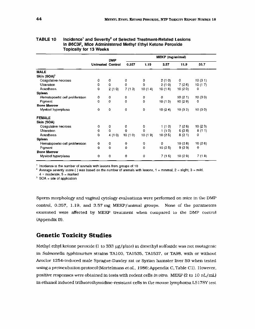
44 METHYL ETHYL KETONE PEROXIDE, NTP TOXICITY REPORT NUMBER 18
TABLE 10 lncidence1 and Severity2 of Selected Treatment-Related Lesions in B6C3F1 Mice Administered Methyl Ethyl Ketone Peroxide Topically for 13 Weeks
MEKP (mg/animal) DMP
Untreated Control 0.357 1.19 3.57 11.9 35.7
MALE Skin (SOA)3
Coagulative necrosis 0 0 0 0 2 (1 0) 0 10 (3 1) Ulceration 0 0 0 0 2 (1 0) 7 (2 6) 10 (1 7) Acanthosis 0 2 (1 0) 7 (1 3) 10 (1 4) 10 (1 8) 10 (2 0) 0
Spleen Hematopo1et1c cell proliferation 0 0 0 0 0 10 (2 1) 10 (3 0) P1gment 0 0 0 0 10 (1 3) 10 (2 9) 0
Bone Marrow Myeloid hyperplasia 0 0 0 0 10 (2 4) 10 (3 2) 10 (3 0)
FEMALE Skin (SOA)
Coagulative necrosis 0 0 0 0 1 (1 0) 7 (2 6) 10 (2 3) Ulceration 0 0 0 0 1 (1 0) 6 (2 8) 8 (1 1) Acanthosis 0 4 (1 0) 10 (1 0) 10 (1 9) 10 (2 5) 8 (2 1) 0
Spleen Hematopoietic cell proliferation 0 0 0 0 0 10 (2 8) 10 (2 6) P1gment 0 0 0 0 10 (2 5) 9 (2 9) 0
Bone Marrow Myeloid hyperplasia 0 0 0 0 7 (1 6) 10 (2 9) 7 (1 8)
1 Incidence 1s the number of ammals w1th les1ons from groups of 10 2 Average seventy score ( ) was based on the number of ammals With les1ons, 1 = m1mmal, 2 = slight, 3 = m1ld,
4 = moderate, 5 = marked 3 SOA = s1te of application
Sperm morphology and vaginal cytology evaluations were performed on mice in the DMP
control, 0.357, 1.19, and 3.57 mg MEKPI animal groups. None of the parameters
exammed were affected by MEKP treatment when compared to the DMP control
(Appendix B).
Genetic Toxicity Studies
Methyl ethyl ketone peroxide (1 to 333 pg/plate) in dimethyl sulfoxide was not mutagenic
in Salmonella typhimurium strains TA100, TA1535, TA1537, or TA98, with or without
Aroclor 1254-induced male Sprague-Dawley rat or Syrian hamster liver S9 when tested
using a preincubation protocol (Mortelmans et aL. 1986; Appendix C. Table C 1). However,
positive responses were obtained in tests with rodent cells in vitro. MEKP (2 to 10 nL/mL)
in ethanol induced trifluorothymidine-resistant cells in the mouse lymphoma L5178Y test
45 METHYL ETHYL KETONE PEROXIDE, NTP TOXICITY REPORT NUMBER 18
in the absence of S9 activation (Table C2). The chemical was not tested with S9 in this
assay. In cytogenetic tests with Chinese hamster ovary cells, MEKP in dimethyl sulfoxide
induced sister chromatid exchanges (SCEs) (Table C3) and chromosomal aberrations (Abs)
(Table C4) at doses up to 50 pg/mL with and withoutAroclor 1254-induced male Sprague
Dawley rat liver S9. In the SCE test, a clearly positive, dose-related response was obtained
in the absence of S9; with S9, a positive response was seen only at the highest nonlethal
dose tested (50 pg/mL). Similarly, in the Abs test with S9, a positive response was seen
only at the highest dose tested, which also produced observable toxicity.
The vehicle, dimethyl phthalate (DMP), at a purity of 99%, was not mutagenic in
S. typhimurium when tested at doses up to 333 pg/plate under the same conditions as
MEKP (Zeiger et aL, 1985). There was no induction of chromosomal aberrations in CHO
cells at doses up to 1500 pg/mL without S9 and up to 5100 pg/mL with S9 (Loveday
et aL, 1990). SCEs were induced at doses of 1240 to 2960 pg/mL in the presence of S9;
DMP did not induce SCEs without S9 when tested at doses up to 1510 pg/mL (Loveday
et aL, 1990).
Despite the positive chromosomal aberrations test results with MEKP in rodent cells in
vitro, no increase was observed in the frequency of micronucleated erythrocytes in
peripheral blood samples obtained from male and female mice at the end of the 13-week
dermal toxicity study (Table C5). In addition, no alterations of the PCE/NCE ratio were
seen, which implies that no overt toxicity was produced in the bone marrow cells by topical
application of0.357 to 3.57 mg MEKP/animal.
Although the pattern of genetic toxicity responses with MEKP is similar to that seen with
DMP, the positive SCE results obtained with MEKP in DMP do not appear to be a result
of the high concentration of DMP. SCEs were induced by MEKP in DMP at doses of 10 and
15 pg/mL (equivalent to 5.5 and 8.25 pg/mL DMP) (Appendix C; Table C3), whereas no
effect was seen at doses below 1240 pg/mL of DMP alone (Loveday et al., 1990).

46 METHYL ETHYL KETONE PEROXIDE, NTP TOXICITY REPORT NUMBER 18
47 METHYL ETHYL KETONE PEROXIDE, NTP TOXICITY REPORT NUMBER 18
DISCUSSION
MEKP is a potent oxidizing agent that has been reported to induce localized necrotic effects
in animals following topical or parenteral administration and in humans following
accidental exposure. The toxic effects of MEKP in DMP administered topically to rats and
mice were primarily limited to inflammation and necrosis at the application site.
In the 2-week studies, MEKP at doses of up to 810 mg/kg body weight in rats and
1800 mg/kgbodyweight in mice, applied daily, caused dose-related increases in epidermal
and dermal necrosis and inflammation, with some evidence of epidermal regeneration in
animals that survived to the end of the studies. Lesions considered secondary to the
dermal injury included hematopoietic cell proliferation in the spleen in rats and mice and
myeloid hyperplasia of the sternebra! bone marrow in mice. Treated mice also showed a
marked increase in liver weight, and decreased thymus weights were generally noted in
treated rats and mice. The dermal lesions were sufficiently severe to result in the death
of some mice, but the pattern of deaths was not clearly dose related. Likewise, the pattern
of weight gains of mice did not show a strong dose response. These 2 factors, plus the
evidence for epidermal regeneration despite continued dosing, led to the selection of doses
for the 13-week studies that were, in retrospect, excessive.
The high doses used for the 13-week studies (107 mg MEKP/rat and 35.7 mg
MEKP/mouse) were nearly the same as the high doses (given on a mg/kg body weight
basis) in the 2-week studies. However, because the range of doses for the 13-week studies
was wider and included doses that were lower than those administered in the 2-week
studies, the highest dose groups that contained survivors in the 13-week studies
(10.7 mg/rat and 3.57 mg/mouse) received doses similar to those administered to the
low-dose groups in the shorter studies. The dermal lesions produced in the 2 studies were
similar. Necrosis was severe in rats and mice in the 2 highest dose groups (35.7 and
107 mg/rat, 11.9 and 35.7 mg/mouse) in the 13-week studies, resulting in the death of
all high-dose mice, 2 high-dose female rats, and 1 female mouse in the 11.9 mg/animal
group. The severe necrosis necessitated the early termination of the surviving animals in
these dose groups. A no-observed-adverse-effect level (NOAEL) was not reached, with
48 METHYL ETHYL KETONE PEROXIDE, NTP TOXICITY REPORT NUMBER 18
acanthosis at the application site occurring in mice given doses as low as 0.357 mg and
acanthosis and hyperkeratosis occurring in rats given as little as 1.07 mg. The highest
dose groups of rats and mice allowed to continue to the end of the studies (10.7 mg/rat
and 3.57 mg/mouse) showed focal ulcerations, inflammation, and fibrovascular
proliferation at the application site. Thus, these studies were considered adequate for the
assessment of the potential for systemic toxicity under conditions of repeated dermal
contact both on ulcerated and nonulcerated skin.
Evidence for systemic injury under these conditions was limited. As in the 2-week studies,
hematopoietic cell proliferation was noted in the spleen, and myeloid hyperplasia occurred
in the bone marrow, primarily in animals that were killed early in the studies; both were
considered secondary responses to the dermal lesions. Organ weight changes in the
13-week studies were not marked, and the substantial and unexplained increases in liver
weights noted in mice in the 2-week studies were not clearly evident following the
prolonged but relatively low-dose exposure of the 13-week studies.
The results of these studies suggest that significant amounts of MEKP in DMP do not
become systemically available and that, as expected, the primary toxicity associated with
contact with these chemicals is limited to the application site. The results of these studies
are in general agreement with study results obtained for other members of this class of
peroxides and are supported by findings of the clinical studies of humans exposed to
MEKP by ingestion or by contact with the eyes or skin.
MEKP is one of a limited number of peroxides and hydroperoxides that have been tested
for mutagenicity by the NTP; MEKP was not mutagenic inS. typhimwium strains TA100.
TA1535, TA1537, or TA98, with or without metabolic activation. One would expect that
these chemicals would yield reactive oxygen moieties that would be mutagenic in
Salmonella. Hydrogen peroxide is a weak mutagen in Salmonella (Abu-Shakra and Zeiger,
1990), and mixed results have been reported for other peroxides and hydroperoxides
(Haworth et aL. 1983; Mortelmans et al., 1986; Zeiger. et al., 1988). Additionally. where
such peroxides have been reported as mutagenic in Salmonella, the response has usually
been weak. Approximately half of the compounds in this class ofchemicals that have been
tested by the NTP have been mutagenic in Salmonella.
49 METHYL ETHYL KETONE PEROXIDE, NTP TOXICITY REPORT NUMBER 18
It is known from early studies performed by Ames and his coworkers (McCann et al., 1975)
and from the more recent studies carried out by the NTP (Ashby and Tennant, 1988, 1991;
Zeiger et al., 1990) that a positive response in the Salmonella mutagenicity test has a high
(approximately 89%) positive predictivity for rodent carcinogenicity. However,
nonmutagenicity in Salmonella is not predictive of noncarcinogenicity in rodents, and the
Salmonella test is not responsive to approximately half of the chemicals found to be
carcinogenic in rodents. The predictivity of the S. typhimurium test also varies with
chemical class (Ashby and Tennant, 1991; Zeiger, 1987). Unfortunately, too few peroxides
and hydroperoxides have been adequately tested in both the S. typhimurium and rodent
carcinogenesis assays to draw any conclusions of the predictive value of S. typhimurium
results for this class of chemicals. The predictive value of rodent bone marrow
cytogenetics tests for carcinogenesis is under investigation, so it is not yet possible to draw
any conclusions from the negative micronucleus test.
MEKP showed weak tumor-promoting activity when applied topically to the skin of mice
that had been UV-irradiated (Logani et al., 1984). The MEKP dose used in that study,
10 pg/mouse, was much lower than the low dose of 357 pg/mouse used in the present
study. In general, the human exposure occurring in the workplace is through splashing,
thereby affecting the skin. Presumably, because ofthe irritating properties of MEKP, it is
immediately removed by washing. Therefore, it is not likely that sustained exposure of the
type needed to promote skin tumor risk factors, as occurs with sunlight or with other
chemicals, would occur with MEKP.

50 METHYL ETHYL KETONE PEROXIDE, NTP TOXICITY REPORT NUMBER 18
51 METHYL ETHYL KETONE PEROXIDE, NTP TOXICITY REPORT NUMBER 18
REFERENCES
ABu-SHAKRA, A., AND ZEIGER, E. (1990). Effects of SalmoneUa genotypes and testing
protocols on H202-induced mutation. Mutagenesis 5, 469-473.
AMERICAN CONFERENCE OF GOVERNMENTAL INDUSTRIAL HYGIENISTS (ACGIH) (1990). Guide to
Occupational Exposure Values.
ANDo, M., AND TAPPEL, A. L. (1985a). Effect of dietary vitamin Eon methyl ethyl ketone
peroxide damage to microsomal cytochrome P-450 peroxidase. Chem.-BioL Interact.
55, 317-326.
ANDO, M., AND TAPPEL, A. L. (1985b). Methyl ethyl ketone peroxide damage to cytochrome
P-450 peroxidase activities. Toxicol. Appl. Pharmacal. 81, 517-524.
ASHBY, J., AND TENNANT, R. W. (1988). Chemical structure, Salmonella mutagenicity and
extent of carcinogenicity as indicators of genotoxic carcinogenesis among
222 chemicals tested in rodents by the US NCI/NTP. Mutat. Res. 204, 17-155.
AsHBY, J., AND TENNANT, R. W. (1991). Definitive relationships among chemical structure,
carcinogenicity and mutagenicity for 301 chemicals tested by the U.S. NTP. Mutat.
Res. 257, 229-306.
BOORMAN, G. A., MONTGOMERY, C. A., JR., EUSTIS, S. L., WOLFE, M. J., MCCONNELL, E. E.,
AND HARDISTY, J. F. (1985). Quality assurance in pathology for rodent carcinogenicity
studies. In Handbook ofCarcinogen Testing (H. A. Milman and E. K. Weisburger, Eds.).
pp. 345-357. Noyes Publications, Park Ridge, NJ.
52 METHYL ETHYL KETONE PEROXIDE, NTP TOXICITY REPORT NUMBER 18
BOORMAN, G. A, HICKMAN, R. L., DAVIS, G. W., RHODES, L. S., WHITE, N. W., GRIFFIN, T. A,
MAYO, J., AND HAMM, T. E., JR. (1986). Serological titers to murine viruses in 90-day
and 2-year studies. In Complications ofViral and Mycoplasmal Injections in Rodents to
Toxicology Research and Testing (T.E. Hamm, Jr., Ed.). pp. 11-23. Hemisphere,
New York.
BRIGHAM, C. R., AND LANDRIGAN, P. J. (1985). Safety and health in boat building and repair.
Am. J. Ind. Med. 8, 169-182.
BURGER, L. M., AND CHANDOR, S. B. (1971}. Fatal ingestion of plastic resin catalyst. Arc!L
Environ. Health 23, 402-404.
CASPARY, W. J., LEE, Y. J., POULTON, S., MYHR, B. C., MITCHELL, A D., AND RUDD, C. J.
( 1988}. Evaluation of the L51 78Y mouse lymphoma cell mutagenesis assay:
Quality-control guidelines and response categories. Environ. Mol. Mutagen. 12 (Suppl.
13}, 19-36.
CODE OF FEDERAL REGULATIONS (CFR) 21, Part 58. Good Laboratory Practice for Nonclinical
Laboratory Studies.
CODE OF FEDERAL REGULATIONS (CFR) 21, Part 177, Section 2420. Indirect Food Additives:
Polymers. Subpart C. Substances for Use Only as Components ofArticles Intended for
Repeated Use. Polyester resins, cross-linked.
CODE OF FEDERAL REGULATIONS (CFR) 29, Part 1910, Section 1000. Occupational Safety
and Health Standards. Subpart Z, Toxic and Hazardous Substances. Air
contaminants.
CHAUDIERE, J., CLEMENT, M., GERARD, D., AND BOURRE, J. M. (1988). Brain alterations
induced by vitamin E deficiency and intoxication with methyl ethyl ketone peroxide.
Neurotoxicology 9, 173-179.
53 METHYL ETHYL KETONE PEROXIDE, NTP TOXICITY REPORT NUMBER 18
CoNDELL, R. A.. AND TAPPEL, A. L. (1983). Evidence for suitability ofglutathione peroxidase
as a protective enzyme: Studies of oxidative damage. renaturation. and proteolysis.
Arch. Biochem. Biophys. 223, 407-416.
DEISHER, J. B. (1958). Poisoning with liquid plastic catalyst. NW Med. 57, 46.
DINES, D. E .• AND SHIPMAN, K. (1962). Toxic myocarditis- Report of a case arising from
ingestion of liquid plastic catalyst. Angiology 13, 297-302.
DIXON, W. J., AND MASSEY, F. J., JR. (1951). Introduction to Statistical Analysis, 1st ed.,
pp. 145-147. McGraw-Hill Book Company. New York.
DUNN, 0. J. (1964). Multiple comparisons using rank sums. Technometrics 6, 241-252.
DUNNETr, C. W. (1955). A multiple comparison procedure for comparing several treatments
with a control. J. Am Stat. Assoc. 50. 1096-1121.
FLOYD, E. P .. AND STOKINGER, H. E. (1958). Toxicity studies of certain organic peroxides
and hydroperoxides. Am. Ind. Hyg. Assoc. J. 19, 205-212.
FRAUNFELDER, F. T., COSTER, D. J., DREW, R., AND FRAUNFELDER, F. W. (1990). Ocular
injury induced by methyl ethyl ketone peroxide. Am J. OphthalmoL 110. 635-640.
GALLOWAY, S. M., ARMSTRONG, M. J .. REUBEN, C .. COLMAN, S .. BROWN, B .. CANNON, C ..
BLOOM, A D., NAKAMURA, F .. AHMED, M., DUK, S .. RIMPO, J .. MARGOLIN, B. H., RESNICK,
M. A. ANDERSON, B.• AND ZEIGER, E. (1987). Chromosome aberrations and sister
chromatid exchanges in Chinese hamster ovary cells: Evaluations of 108 chemicals.
Environ. Mol. Mutagen. 10 (Suppl. 10). 1-175.
HAWLEY, G. G. (ED.) (1981). The Condensed Chemical Dictionary, lOth ed., p. 679.
Van Nostrand Reinhold Co.• New York.
54 METHYL ETHYL KETONE PEROXIDE, NTP TOXICITY REPORT NUMBER 18
HAWORTH, S., LAWLOR, T., MORTELMANS, K., SPECK, W., AND ZEIGER, E. (1983). Salmonella
mutagenicity test results for 250 chemicals. Environ. Mutagen. 5 (Suppl. 1), 1-142.
HERSCHBERGER, L. A, AND TAPPEL, A L. (1982). Effect of vitamin Eon pentane exhaled by
rats treated with methyl ethyl ketone peroxide. Lipids 17, 686-691.
JARVENTAUS, H., NORPPA, H., LINNAINMAA, K., AND SORSA, M. (1984). SCE induction in CHO
cells by peroxides used in the plastic industry. Mutat. Res. 130, 249. (Abstr.)
JONCKHEERE, A R. (1954). A distribution-free k-sample test against ordered alternatives.
Biometrika 41, 133-145.
KARHUNEN, P. J., OJANPERA, 1., LALU, K., AND VUORI, E. (1990). Peripheral zonal hepatic
necrosis caused by accidental ingestion of methyl ethyl ketone peroxide. Hum. Exp.
Toxicol. 9, 197-200.
KASTENBAUM, M. A, AND BOWMAN, K. 0. (1970). Tables for determining the statistical
significance of mutation frequencies. Mutat. Res. 9, 527-549.
KORHONEN, A, HEMMINKI, K., AND VAINIO, H. (1983). Toxicity of rubber chemicals towards
three-day chicken embryos. Scand. J. Work Environ. Health 9, 115-119.
KORHONEN, A, HEMMINKI, K., AND VAINIO, H. (1984). Embryotoxic effects of eight organic
peroxides and hydrogen peroxide on three-day chicken embryos. Environ. Res. 33,
54-61.
KOTIN, P., AND FALK, H. L. (1963). Organic peroxides, hydrogen peroxide, epoxides, and
neoplasia. Radiat. Res. Suppl. 3. 193-211.
LEVIN, D. E., HOLLSTEIN, M., CHRISTMAN, M. F., AND AMES, B. N. (1984). Detection of
oxidative mutagens with a new Salmonella tester strain (TA102). Methods Enzymol.
105, 249-254.
55 METHYL ETHYL KETONE PEROXIDE, NTP TOXICITY REPORT NUMBER 18
LITOV, R. E., MATTHEWS, L. C., AND TAPPEL, A. L. (1981). Vitamin E protection against in
vivo lipid peroxidation initiated in rats by methyl ethyl ketone peroxide as monitored
by pentane. Toxicol. AppL Phannacol. 59, 96-106.
LoGAN!, M. K., SAMBUCO, C. P., FORBES, P. D., AND DAVIES, R. E. (1984). Skin-tumour
promoting activity of methyl ethyl ketone peroxide-a potent lipid-peroxidizing agent.
Food Chem. Toxicol. 22, 879-882.
LoVEDAY, K. S., ANDERSON, B. E., RESNICK, M.A., AND ZEIGER, E. (1990). Chromosome
aberration and sister chromatid exchange tests in Chinese hamster ovary cells in vitro.
V: Results with 46 chemicals. Environ. Mol. Mutagen. 16, 272-303.
MACGREGOR, J. T., WEHR, C. M., AND LANGLOIS, R. G. (1983). A simple fluorescent staining
procedure for micronuclei and RNA in erythrocytes using Hoechst 33258 and
pyronin Y. Mutat. Res. 120, 269-275.
MARONPOT, R. R., AND BOORMAN, G. A. (1982). Interpretation of rodent hepatocellular
proliferative alterations and hepatocellular tumors in chemical safety assessment.
Toxicol. Pathol. 10, 71-80.
MCCANN, J., CHOI, E., YAMASAKI, E., AND AMES, B. N. (1975). Detection of carcinogens as
mutagens in the SalmoneUa/microsome test: Assay of 300 chemicals. Proc. Nat.
Acad. Sci. 72, 5135-5139.
MITTLEMAN, R. E., ROMIG, L.A., AND GRESSMANN, E. (1986). Suicide by ingestion of methyl
ethyl ketone peroxide. J. Forensic ScL 31, 312-320.
MORRISON, D. F. (1976). Multivariate Statistical Methods, pp. 170-179. McGraw-Hill Book
Company, New York.
56 METHYL ETHYL KETONE PEROXIDE, NTP TOXICITY REPORT NUMBER 18
MORRISSEY, R. E., SCHWETZ, B. A., LAMB, J. C., IV, Ross, M.D., TEAGUE, J. L., AND MORRIS,
R. W. (1988). Evaluation of rodent sperm, vaginal cytology, and reproductive organ
weight data from National Toxicology Program 13-week studies. Ftmdam Appl. Toxicol.
11' 343-358.
MORTELMANS, K., HAWORTH, S., LAWLOR, T., SPECK, W., TAINER, B., AND ZEIGER, E. (1986).
Salmonella mutagenicity tests: II. Results from the testing of 270 chemicals. Environ.
Mutagen. 8 (Suppl. 7). 1-119.
MYHR, B., BOWERS, L., AND CASPARY, W. J. (1985). Assays for the induction of gene
mutations at the thymidine kinase locus in L5178Y mouse lymphoma cells in culture.
Prog. Mutat. Res. 5, 555-568.
NATIONAL INSTITUTE FOR OcCUPATIONAL SAFETY AND HEALTH (NIOSH) (1974). Health Hazard
Evaluation Determination Report No. 73-124-127. Cincinnati, OH.
NATIONAL INSTITUTE FOR OCCUPATIONAL SAFETY AND HEALTH (NIOSH) (1988). Health Hazard
Report No. HETA-86-441-1913. Cincinnati, OH.
NATIONAL INSTITUTE FOR OCCUPATIONAL SAFETY AND HEALTH (NIOSH) (1990). National
Occupational Exposure SuiVey (1981 to 1983), unpublished provisional data as of
July 1, 1990. Cincinnati, OH.
NATIONAL OccuPATIONAL HAzARD SuRVEY (NOHS) ( 1974). Hazardous Substances Data Bank.
National Library of Medicine Computerized Literature Retrieval Services.
PURNELL, C. J., MARTIN, G. L., AND WOLFSON, H. (1979). The determination of airborne
methyl ethyl ketone peroxide in the glass reinforced plastics industry. Ann. Occup.
Hyg. 22, 383-387.
RAo, G. N., HASEMAN, J. K., AND EDMONDSON, J. (1989a). Influence of viral infections on
body weight, survival, and tumor prevalence in Fischer 344/NCr rats on two-year
studies. Lab. Anim. ScL 39, 389-393.
57 METHYL ETHYL KETONE PEROXIDE, NTP TOXICITY REPORT NUMBER 18
RAo, G. N., PIEGORSCH, W. W., CRAWFORD, D. D., EDMONDSON, J., AND HASEMAN, J. K.
(1989b). Influence of viral infections on body weight, survival, and tumor prevalence
of B6C3F1 (C57BL/6N x C3H/HeN) mice in carcinogenicity studies. Fundam. Appl.
Toxicol. 13, 156-164.
SCHMID, W. ( 1976). The micronucleus test for cytogenetic analysis. In Chemical Mutagens.
Principles and Methods for Their Detection, Vol 4 (A Hollaender, Ed.), pp. 31-53.
Plenum Press. New York.
SHIRLEY, E. (1977). A non-parametric equivalent of Williams' test for contrasting
increasing dose levels of a treatment. Biometrics 33, 386-389.
STANFORD RESEARCH INSTITUTE (SRI) (1989). Directory ofAmerican Chemical Producers,
p. 785. Menlo Park, CA.
SUMMERFIELD, F. W., AND TAPPEL, A. L. (1984). Vitamin E protects against methyl ethyl
ketone peroxide-induced peroxidative damage to rat brain DNA Mutat. Res. 126,
113-120.
WARREN, D. L., AND REED, D. J. (1991). Modification of hepatic vitamin E stores in vivo.
I. Alterations in plasma and liver vitamin E content by methyl ethyl ketone peroxide.
Arch. Biochem. Biophys. 285, 45-52.
WILLIAMS, D. A (1971). A test for differences between treatment means when several dose
levels are compared with a zero dose control. Biometrics 27. 103-117.
WILLIAMS, D. A (1972). The comparison of several dose levels with a zero dose control.
Biometrics 28, 519-531.
WOJDYLA, Z., PACH, J., AND KOLODZIEJ, J. (1979). Acute poisoning with Ketonox [in Polish).
Arch. Med. Sadowej KryminoL 29, 199-205.
58 METHYL ETHYL KETONE PEROXIDE, NTP TOXICITY REPORT NUMBER 18
ZEIGER, E. (1987). Carcinogenicity of mutagens: Predictive capability of the
Salmonella mutagenesis assay for rodent carcinogenicity. Cancer Res. 47, 1287-1296.
ZEIGER, E., HAWORTI-1, S., MORTELMANS, K., AND SPECK, W. (1985). Mutagenicity testing of
di(2-ethylhexyl)phthalate and related chemicals in SalmoneUa. Environ. Mutagen. 7,
213-232.
ZEIGER, E., ANDERSON, B., HAWORTH, S., LAWLOR, T., AND MORTELMANS, K. (1988).
Salmonella mutagenicity tests: N. Results from the testing of 300 chemicals. Environ.
MoL Mutagen. 11 (Suppl. 12). 1-158.
ZEIGER, E., HASEMAN, J. K., SHELBY, M. D., MARGOLIN, B. H., AND TENNANT, R. W. (1990).
Evaluation of four in vitro genetic toxicity tests for predicting rodent carcinogenicity:
Confirmation of earlier results with 41 additional chemicals. Environ. Mol. Mutagen.
16 (Suppl. 18). 1-14.
A-1 METHYL ETHYL KETONE PEROXIDE, NTP TOXICITY REPORT NUMBER 18
APPENDIX A
Organ Weights and Organ-Weight-to-Body-Weight Ratios
Table AI Organ Weights and Organ-Weight-to-Body-Weight Ratios for Rats in the 13-Week Dermal Study of Methyl Ethyl Ketone Peroxide .................. A-2
Table A2 Organ Weights and Organ-Weight-to-Body Weight Ratios for Mice in the 13-Week Dermal Study of Methyl Ethyl Ketone Peroxide .................. A-4
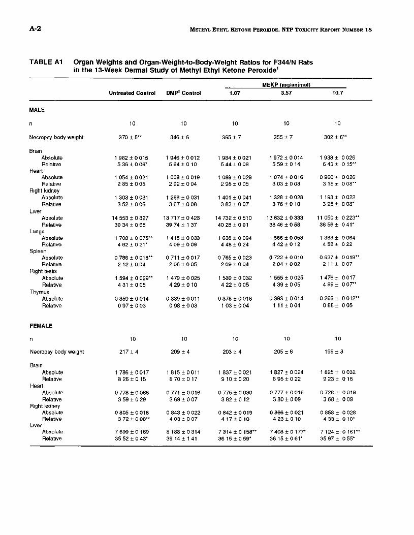
A-2 METHYL ETHYL KETONE PEROXIDE, NTP TOXICITY REPORT NUMBER 18
TABLE A1 Organ Weights and Organ-Weight-to-Body-Weight Ratios for F344/N Rats in the 13-Week Dermal Study of Methyl Ethyl Ketone Peroxide1
MEKP {mg/animal}
Untreated Control DMP2 Control 1.07 3.57 10.7
MALE
n 10 10 10 10 10
Necropsy body we1ght 370 ± 5** 346 ± 6 365 ± 7 355 ± 7 302 ± 6**
Bra1n Absolute 1 982 ± 0 015 1946 ± 0012 1 984 ± 0 021 1 972 ± 0 014 1 938 ± 0 026 Relative 5 36 ± 0 06* 564±010 5 44 ± 0 08 5 59± 014 6 43± 0 15**
Heart Absolute 1 054 ± 0 021 1 008 ± 0 019 1 088 ± 0 029 1 074 ± 0 016 0 960 ± 0 026 RelatiVe 2 85 ± 0 05 2 92 ± 004 2 98 ± 0 05 3 03 ± 0 03 3 18± 0 08**
R1ght kidney Absolute 1 303 ± 0 031 1 268 ± 0 031 1401 ±0041 1 328 ± 0 028 1193 ± 0022 Relative 3 52± 006 3 67 ± 008 3 83 ± 0 07 3 76 ± 010 3 95 ± 0 08*
L1ver Absolute 14 553 ± 0 327 13 717 ± 0 423 14 732 ± 0 510 13 632 ± 0 333 11 050 ± 0 223** Relative 39 34 ± 065 39 74 ± 1 37 40 28 ± 0 91 38 46 ± 0 58 36 56± 0 41*
Lungs Absolute 1 708 ± 0 075** 1415±0033 1638±0094 1 566 ± 0 053 1 383 ± 0064 Relative 462±021* 4 09 ± 009 4 48 ± 0 24 4 42 ± 012 4 58± 0 22
Spleen Absolute 0 786 ± 0 018** 0711±0017 0 765 ± 0 023 0 722 ± 0 010 0 637 ± 0 019** Relative 2 12 ± 0 04 2 06 ± 0 05 2 09 ± 0 04 2 04 ± 0 02 211 ± 007
R1ght testiS Absolute 1 594 ± 0 029** 1479 ± 0025 1 539 ± 0 032 1 555 ± 0 025 1 476 ± 0 017 Relative 4 31 ± 0 05 429±010 4 22 ± 0 05 4 39 ± 0 05 489 ± oor
Thymus Absolute 0 359 ± 0014 0 339 ± 0 011 0 378 ± 0 018 0393±0014 0266± 0012** RelatiVe 0 97± 003 0 98 ± 0 03 1 03 ± 004 1 11 ± 0 04 0 88 ± 0 05
FEMALE
n 10 10 10 10 10
Necropsy body we1ght 217 ± 4 209 ± 4 203 ± 4 205 ± 6 198 ± 3
Bra1n Absolute 1786±0017 1815±0011 1 837 ± 0 021 1 827 ± 0 024 1 825 ± 0 032 Relative 826±015 8 70 ± 017 9 10 ± 0 20 8 95 ± 0 22 9 23 ± 016
Heart Absolute 0778±0066 0 771 ± 0016 0 775 ± 0 030 0 777 ± 0 016 0 728 ± 0 019 Relative 3 59± 0 29 3 69 ± 007 3 82 ± 0 12 380±009 368± 0 09
R1ght kidney Absolute 0 805 ± 0 018 0 843 ± 0022 0 842 ± 0 019 0 866 ± 0 021 0858 ± 0 028 Relative 3 72 ± o08** 4 03 ± 007 417±010 4 23 ± 010 433 ± 0 10*
Ltver Absolute 7 699 ± 0 169 8188 ± 0314 7 314 ± 0 158** 7 408 ± 0 177* 7124 ± 0 161** Relative 35 52± 0 43* 39 14 ± 1 41 36 15 ± 0 59* 36 15 ± 0 61* 35 97 ± 0 55*
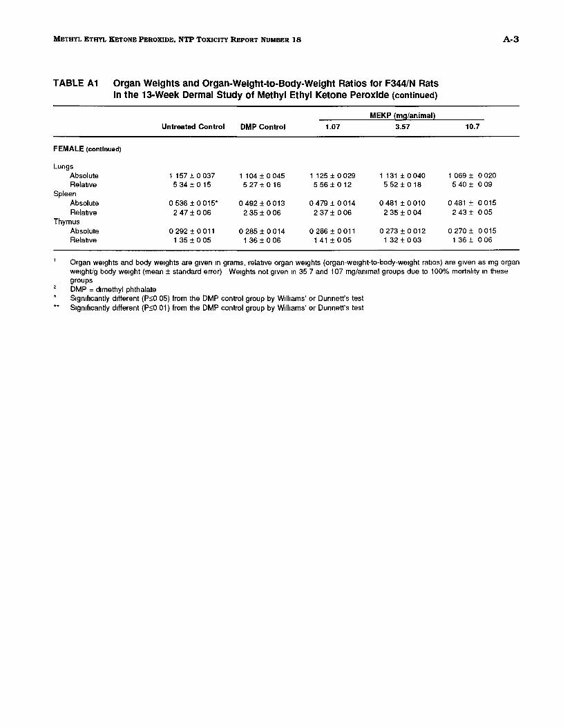
A-3 METHYL ETHYL KETONE PEROXIDE, NTP TOXICITY REPORT NUMBER 18
TABLE A1 Organ Weights and Organ-Weight-to-Body-Weight Ratios for F344/N Rats in the 13-Week Dermal Study of Methyl Ethyl Ketone Peroxide (continued)
MEKP {mg/animal}
10.7Untreated Control DMP Control 1.07 3.57
FEMALE (continued)
Lungs Absolute 1 157 ± 0 037 1 104 ± 0 045 1 125 ± 0 029 1131±0040 1 069 ± 0020 Relat1ve 534±015 5 27 ± 016 5 56± 012 5 52± 018 540 ± 0 09
Spleen Absolute 0 536 ± 0 015* 0 492 ± 0013 0479±0014 0 481 ± 0 010 0481 ± 0 015 Relat1ve 2 47 ± 0 06 235±006 2 37 ± 0 06 2 35 ± 004 2 43± 005
Thymus Absolute 0 292 ± 0 011 0 285 ± 0 014 0 286 ± 0 011 0 273 ± 0 012 0 270 ± 0 015 Relative 1 35 ± 0 05 136±006 1 41 ± 0 05 1 32 ± 0 03 1 36 ± 0 06
Organ we1ghts and body we1ghts are g1ven 1n grams, relat1ve organ we1ghts (organ-we1ght·to-body-we1ght rat1os) are g1ven as mg organ we1ght/g body we1ght (mean± standard error) We1ghts not g1ven 1n 35 7 and 107 mg/ammal groups due to 100% mortality 1n these groups DMP = dimethyl phthalate S1gmhcantly different (P:>O 05) from the DMP control group by Williams' or Dunnett's test S1gmhcantly different (P:>O 01) from the DMP control group by Williams' or Dunnett's test
A-4 METHYL ETHYL KETONE PEROXIDE, NTP TOXICITY REPORT NUMBER 18
TABLE A2 Organ Weights and Organ-Weight-to-Body Weight Ratios for B6C3F, Mice in the 13-Week Dermal Study of Methyl Ethyl Ketone Peroxide'
MEKP (mg/animal}
Untreated Control DMP2 Control 0.357 1.19 3.57
MALE
n 10 10 10 10 10
Necropsy body we1ght 35 2 ± 09 338±08 32 9 ± 0 6 32 2 ± 03 31 8 ± 0 5*
Bra1n Absolute 0443±0004 0 447 ± 0005 0 453 ± 0 006 0 443 ± 0005 0 452 ± 0 004 Relative 1268±039 1331 ±036 13 79 ± 0 28 13 75 ± 0 12 1423± 015*
Heart Absolute 0154±0003 0158±0004 0 152 ± 0 002 0156±0005 0149 ± 0 002 Relat1ve 4 39 ± 0 09 4 70 ± 018 4 62 ± 0 08 484±014 4 69 ± 007
R1ght kidney Absolute 0 322 ± 0009 0304±0009 0311 ±0010 0 310 ± 0 009 0304 ± 0007 Relative 921 ±037 9 02 ± 0 21 9 45 ± 0 27 9 62 ± 0 26 9 56± 019
L1ver Absolute 1 687 ± 0 045 1 617 ± 0 039 1648±0039 1 495 ± 0 013* 1 613 ± 0 035 Relat1ve 4796±045 47 94 ± 0 81 50 09 ± 1 07 46 41 ± 0 36 50 72 ± 094
Lungs Absolute 0182±0006 0180±0008 0199±0013 0186 ± 0009 0175 ± 0010 Relahve 5 21 ± 0 23 5 39 ± 035 604±036 5 78 ± 030 5 51± 034
Spleen Absolute 0 077 ± 0005 0 067 ± 0 002 0 072 ± 0004 0 068 ± 0 002 0 078 ± 0 002** Relat1ve 2 23 ± 0 20 1 99 ± 0 07 2 18 ± 0 09 2 11 ± 0 05 245± 0 06**
R1ght testiS Absolute 0 119 ± 0 002 0116±0003 0119±0003 0115±0003 0 115 ± 0002 Relative 340±010 344±011 3 61 ± 0 07 356±009 3 63± 004
Thymus Absolute 0042±0003 0 041 ± 0 002 0041 ±0003 0 037 ± 0 002 0 043 ± 0004 Relative 1 20 ± 0 09 1 22 ± 007 1 25 ± 0 08 116±006 1 37 ± 0 15
FEMALE
n 10 10 10 10 10
Necropsy body we1ght 29 7 ± 0 5** 26 7± 0 5 28 5 ± 1 4 268±05 27 3 ± 0 3
Bra1n Absolute 0468±0004 0463±0004 0456 ± 0005 0463±0004 0466 ± 0004 Relative 1581±028** 17 42 ± 0 32 1629±071 1731±031 17 11 ± 017
Heart Absolute 0 137 ± 0 004 0 137 ± 0 005 0134±0003 0 133 ± 0 003 0145 ± 0005 Relat1ve 461 ±009* 514±019 4 75 ± 014 496±008 532±016
R1ght kidney Absolute 0 214 ± 0 005 0 222 ± 0 004 0 237 ± 0005 0 235 ± 0007 0 242 ± 0009* Relative 7 23 ± 0 20** 8 37± 0 29 843±030 8 77± 0 23 8 88± 032
L1ver Absolute 1 332 ± 0 023* 1442 ± 0034 1 298 ± 0 044* 1 308 ± 0 028 1 533 ± 0046 Relative 44 97 ± 0 87** 54 33 ± 1 95 46 02 ± 1 70** 48 81 ± 0 78* 56 24 ± 1 55
A-5 METHYL ETHYL KETONE PEROXIDE, NTP ToXICITY REPORT NUMBER 18
TABLE A2 Organ Weights and Organ-Weight-to-Body Weight Ratios for B6C3F, Mice in the 13-Week Dermal Study of Methyl Ethyl Ketone Peroxide (continued)
MEKP (mg/animal}
3.57Untreated Control DMP Control 0.357 1.19
FEMALE (continued)
Lungs Absolute 0195±0010 0 187±0009 0230±0017* 0192±0011 0196 ± 0008 Relat1ve 6 57± 0 35 7 01 ± 0 27 8 12 ± 0 55 715±036 720± 0 31
Spleen Absolute 0 099 ± 0 006** 0 078 ± 0002 0 084 ± 0003 0 083 ± 0003 0 100 ± 0 003** Relat1ve 3 35 ± 0 22 294±009 299±013 310±008 367± 0 09**
Thymus Absolute 0054±0004 0050±0004 0 055 ± 0003 0049±0002 0051 ± 0 003 Relative 180±013 188±013 196±013 1 83 ± 0 08 1 88 ± 009
Organ we1ghts and body we1ghts are g1ven 1n grams, relative organ we1ghts (organ-we1ght-to-body-we1ght rat1os) are g1ven as mg organ we1ght/g body we1ght (mean± standard error) We1ghts not g1ven 1n 11 9 and 35 7 mg/an1mal dose groups due to 100% mortality 1n
these groups DMP = d1metyhl phthalate S1gn1f1cantly different (P::>O 05) from the DMP control group by Williams' or Dunnett's test S1gn1f1cantly different (P::>O 01) from the DMP control group by Williams' or Dunnett's test

A-6 METHYL ETHYL KETONE PEROXIDE, NTP TOXICITY REPORT NUMBER 18
B-1 METHYL ETHYL KETONE PEROXIDE, NTP TOXICITY REPORT NUMBER 18
APPENDIX B
Reproductive Tissue Evaluations and Estrous Cycle Characterization
Table Bl Summary of Reproductive Tissue Evaluations in Male F344/N Rats in the 13-Week Dermal Study of Methyl Ethyl Ketone Peroxide .................. B-2
Table B2 Summary of Estrous Cycle Characterization in Female F344/N Rats in the 13-Week Dermal Study of Methyl Ethyl Ketone Peroxide .................. B-2
Table B3 Summary of Reproductive Tissue Evaluations in Male B6C3F1 Mice in the 13-Week Dermal Study of Methyl Ethyl Ketone Peroxide .................. B-3
Table B4 Summary of Estrous Cycle Characterization in Female B6C3F1 Mice in the 13-Week Dermal Study of Methyl Ethyl Ketone Peroxide .................. B-3
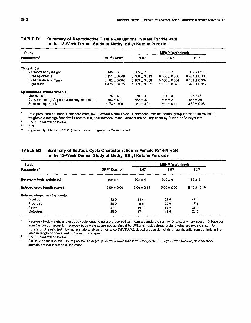
B-2 METHYL ETHYL KETONE PEROXIDE, NTP TOXICITY REPORT NUMBER 18
TABLE B1 Summary of Reproductive Tissue Evaluations in Male F344/N Rats in the 13-Week Dermal Study of Methyl Ethyl Ketone Peroxide
Study MEKP {mg/animaQ
Parameters1 DMP2 Control 1.07 3.57 10.7
Weights (g) Necropsy body we1ght 346 ± 6 365 ± 7 355 ± 7 302 ± 6** R1ght ep1d1dym1s 0 451 ± 0 009 0466±0013 0466±0008 0 454 ± 0 006 R1ght cauda ep1d1dym1s 0162±0004 0163 ± 0006 0160±0004 0161±0007 R1ght test1s 1479 ± 0025 1 539 ± 0032 1 555 ± 0 025 1 476 ± 0 017
Spermatozoal measurements Mot1hty (%) 75± 4 79 ± 3 74 ± 3 84 ± 2' Concentrabon (106/g cauda epididymal tissue) 553 ± 42 652 ± 37 506 ± 27 586 ± 30 Abnormal sperm (%) 0 74 ± 0 09 0 87± 008 0 62 ± 0 11 0 60 ± 008
Data presented as mean± standard error, n=10, except where noted Differences from the control group for reproductive tissue we1ghts are not s1gn1flcant by Dunnett's test, spermatozoal measurements are not s1gmhcant by Dunn's or Shirley's test DMP = dimethyl phthalate
.. n=9 S1gn1f1cantly different (PsO 01) from the control group by Wilham's test
TABLE B2 Summary of Estrous Cycle Characterization in Female F344/N Rats in the 13-Week Dermal Study of Methyl Ethyl Ketone Peroxide
Study MEKP {mg/animal}
Parameters1 DMP2 Control 1.07 3.57 10.7
Necropsy body weight (g) 209 ± 4 203 ± 4 205 ± 6 198 ± 3
Estrous cycle length (days) 500±000 5 00 ± 0173 500±000 510± 010
Estrous stages as % of cycle D1estrus 32 9 386 28 6 41 4 Proestrus 200 86 200 17 1 Estrus 271 35 7 32 9 21 4 Metestrus 20 0 17 1 18 6 20 0
Necropsy body we1ght and estrous cycle length data are presented as mean± standard error, n=10, except where noted Differences from the control group for necropsy body we1ghts are not s1gn1ficant by W1ll1ams' test, estrous cycle lengths are not s1gn1flcant by Dunn's or Shirley's test By mulbvanate analysis of vanance (MANOVA). dosed groups do not differ s1gmflcantly from controls 1n the relative length of t1me spent 1n the estrous stages DMP = dimethyl phthalate For 1/10 ammals 1n the 1 07 mg/an1mal dose group, estrous cycle length was longer than 7 days or was unclear, data for these ammals are not Included 1n the mean

B-3 METHYL ETHYL KETONE PEROXIDE, NTP TOXICITY REPORT NUMBER 18
TABLE B3 Summary of Reproductive Tissue Evaluations in Male B6C3F, Mice in the 13-Week Dermal Study of Methyl Ethyl Ketone Peroxide
Study MEKP (mg/animal}
Parameters1 DMP2 Control 0.357 1.19 3.57
Weights (g) Necropsy body we1ght 338±08 32 9 ± 0 6 32 2 ± 0 3 31 8 ± o5* R1ght ep1d1dym1s 0 047 ± 0002 0 046 ± 0004 0 044 ± 0 001 0 044 ± 0 002 R1ght cauda ep1d1dym1s 0 016 ± 0 001 0 014 ± 0001 0 015 ± 0 001 0016±0001 R1ght test1s 0116±0003 0119±0003 0 115±0003 0 115 ± 0 002
Spermatozoal measurements Mot1hty (%) 86± 2 84 ± 2 87 ± 1 83± 2 Concentration (106/g cauda epididymal tissue) 1233 ± 143 1308 ± 106 1387 ± 83 1404 ± 128 Abnormal sperm (%) 146±017 1 24 ± 0 12 1 g2 ± 0 26 126±012
Data presented as mean± standard error, n=10 Reproductive t1ssue we1ghts are not s1gn1f1cant by Dunnett's test, spermatozoal measurements are not s1gn1f1cant by Dunn's test DMP = dimethyl phthalate Significantly different (P~O 05) from the control group by Wilham's test
TABLE B4 Summary of Estrous Cycle Characterization in Female B6C3F1 Mice in the 13-Week Dermal Study of Methyl Ethyl Ketone Peroxide
Study MEKP (mg/animal}
Parameters1 DMP2 Control 0.357 1.19 3.57
Necropsy body weight (g) 267±05 28 5 ± 1 4 26 8 ± 0 5 27 3 ± 0 3
Estrous cycle length (days) 4 60 ± 016 4 67 ± 0 1"tl 4 33 ± 0 243 430±015
Estrous stages as % of cycle Diestrus 25 7 18 6 271 22 9 Proestrus 28 6 25 7 28 6 24 3 Estrus 21 4 35 7 28 6 35 7 Metestrus 24 3 20 0 15 7 17 1
Necropsy body we1ght and estrous cycle length data are presented as mean± standard error, n=10, except where noted Differences from the control group for necropsy body weights are not s1gmf1cant by Dunnett's test, estrous cycle lengths are not s1gmflcant by Dunn's or Shirley's test By mult1vanate analys1s of vanance (MANOVA), dosed groups do not differ s1gn1hcantly from controls 1n the relat1ve length of t1me spent 1n the estrous stages DMP = dimethyl phthalate For 1/10 an1mals 1n the 0 357 and 1 19 mg/ammal dose groups, estrous cycle length was longer than 7 days or was unclear, data for these an1mals are not mcluded 1n the mean

B-4 METHYL ETHYL KETONE PEROXIDE, NTP TOXICITY REPORT NUMBER 18
C-1 METHYL ETHYL KETONE PEROXIDE, NTP TOXICITY REPORT NUMBER 18
APPENDIX C
Genetic Toxicology Table Cl Mutagenicity of Methyl Ethyl Ketone Peroxide in Salmonella typhimurium ........ C-2
Table C2 Induction of Trifluorothymidine Resistance in Mouse Lymphoma L5178Y Cells by Methyl Ethyl Ketone Peroxide . . . . . . . . . . . . . . . . . . . . . . . . . . . . . . . . . . . . . . C-4
Table CS Induction of Sister Chromatid Exchanges in Chinese Hamster Ovary Cells by Methyl Ethyl Ketone Peroxide . . . . . . . . . . . . . . . . . . . . . . . . . . . . . . . . . . . . . . C-6
Table C4 Induction of Chromosomal Aberrations in Chinese Hamster Ovary Cells by Methyl Ethyl Ketone Peroxide . . . . . . . . . . . . . . . . . . . . . . . . . . . . . . . . . . . . . . C-8
Table C5 Frequency of Micronuclei in Peripheral Blood Erythrocytes of B6C3F1 Mice Treated with Methyl Ethyl Ketone Peroxide for 13 Weeks .......................... C-9
C-2 METHYL ETHYL KETONE PEROXIDE, NTP TOXICITY REPORT NUMBER 18
TABLE C1 Mutagenicity of Methyl Ethyl Ketone Peroxide in Salmonella typhimurium1
Revertants/plate2
Strain Dose ·S9 +10% hamster S9 +10% rat S9 (!Jglplate) Trial1 Trial2 Trial1 Trial 2 Trial1 Trial 2
TA100 0 170 ± 26 111±07 143 ± 4 8 122±64 137 ± 6 0 118 ± 86 1 135 ± 9 8 121 ± 2 3 3 135 ±58 115 ± 9 6 150 ± 2 1 123 ± 81 157 ± 3 2 134±44
10 136± 122 136 ± 71 138 ± 2 3 126 ± 7 8 139 ± 6 7 128 ± 1 5 33 157±103 130 ± 4 1 136 ± 4 7 123±112 165 ± 7 9 131 ± 8 7
100 146 ± 7 2 134 ± 8 7 136 ± 3 5 123 ± 4 7 153± 135 149 ± 1 5 333 142 ± 16 8 136±104 97 ± 23 4 190 ± 4 6
Tnal summary Negat1ve Negative Negative NegatiVe Negative Equivocal Pos1t1ve control" 365±47 313±156 1,603 ± 66 1 1,015 ± 201 682 ± 17 3 675 ± 35 5
TA1535 0 26± 64 27 ± 1 2 10 ± 23 11 ± 3 3 18 ± 3 2 12 ± 20 1 23 ± 2 2 27± 09 3 26±53 24 ± 00 12 ± 1 9 8 ± 26 17 ± 19 14 ± 1 2
10 24± 26 29± 09 9±26 8±25 15 ± 2 1 12 ± 1 7 33 18 ± 3 5 25 ± 61 12 ± 24 7±06 16 ± 20 12 ± 23
100 16 ± 09 14 ± 1 0 10 ± 1 7 7±23 18 ± 1 3 11 ± 1 2 333 12 ± 1 9 8 ± 26 8±30 10 ± 1 2
Tnal summary Negat1ve Negative Negative Negative Negative Negative Pos1t1ve control 359±125 319±123 369±178 336±287 198 ± 8 7 151 ± 1 8
TA1537 0 5±12 6±06 4 ± 09 6±03 14 ± 1 9 11 ± 23 1 7±15 5±03 3 5±03 7±15 12 ± 20 11 ± 2 4 13 ± 2 0 12 ± 1 5
10 7±10 5±03 11 ± 1 5 6±18 11 ± 1 8 14 ± 1 5 33 7±00 8±12 11 ± 2 2 5±09 14 ± 1 5 10 ± 1 2
100 6±17 4 ± o3• 6±09 5±09 11 ± 39 10 ± 1 7 333 9±19 6 ± o3• 11 ± 4 4 11 ± 25
Tnal summary Negative Negative Negat1ve Negat1ve Negative Negat1ve Pos1t1ve control 147±187 74± 84 589±20 313 ± 20 5 161 ± 153 159 ± 19 0
TA98 0 19 ± 1 5 18 ± 35 31 ± 35 29 ± 4 7 49 ± 41 29± 15 1 20± 20 19 ± 1 0 3 21 ± 3 6 23± 46 28 ± 40 25 ± 77 41 ±59 32± 23
10 17 ± 09 24± 26 31 ± 32 27± 1 5 37± 23 25± 20 33 13 ± 2 3 23 ± 1 3 36± 57 32 ± 1 9 32± 3 7 27 ± 3 6
100 27 ± 1 2 1 ± 0 74 30 ± 28 33 ± 27 42± 38 29 ± 34 333 34± 35 32 ± 1 3 31 ±53 24± 4 7
Tnal summary Negative Negat1ve Negative Negat1ve Negative Negative Pos1t1ve control 591 ± 76 8 520 ± 30 7 1'171 ± 136 5 764 ± 34 1 502 ± 49 5 522 ± 44 2
C-3 METHYL ETHYL KETONE PEROXIDE, NTP TOXICITY REPORT NUMBER 18
TABLE C1 Mutagenicity of Methyl Ethyl Ketone Peroxide In Salmonella typhlmurlum (continued)
' Study performed at SRI International The detailed protocol and these data are presented 1n Mortelmans eta/ (1986) Cells and methyl ethyl ketone peroxide or solvent (dimethylsulfoxide) were Incubated m the absence of exogenous metabolic act1vat1on (-S9) or w1th Aroclor 1254-mduced S9 from male Synan hamster liver or male Sprague-Dawley rat liver H1gh dose was limited by tOXICity 0 ).lg/plate dose 1s the solvent control
2 Revertants are presented as mean ± the standard error from 3 plates 3 The pos1t1ve controls m the absence of metabolic act1vabon were 4-nltro-o-phenylenedlamme (TA98), sod1um az1de (TA100 and TA1535),
and 9 ammoacnd1ne (TA 1537) The pos1bve control for metabolic act1vat1on w1th all strams was 2-am1noanthracene 4 Slight tOXICity
C-4 METHYL ETHYL KETONE PEROXIDE, NTP TOXICITY REPORT NUMBER 18
TABLE C2 Induction of Trifluorothymidine Resistance in Mouse Lymphoma L517BY Cells by Methyl Ethyl Ketone Peroxide1
Compound Concentration Cloning
Efficiency (%}
Relative Total Growth
(%}
Mutant Count
Mutant Fraction2
Average Mutant
Fraction
-S9 Trial1
Ethanol 104 50 65 78
128 57
103 113
100 44 69 71
32 29 35 31 32
Methyl methanesulfonate ().tg/ml) 5 57
52 63
41 59 66
505 448 417
295 284 219 266*
Methyl ethyl ketone peroxide (nUmL) 2 72
40 39
60 44 38
136 123 111
63 103 94 87*
3 57 29 35
51 25 40
88 88 88
52 99 83 78*
4 30 46 61
39 32 60
143 273 385
158 200 211 190*
6 39 66 70
55 38 75
229 390 319
198 197 152 182*
8 61 59
34 19
408 396
222 223 223*
10 84 65
29 23
481 495
191 254 222*
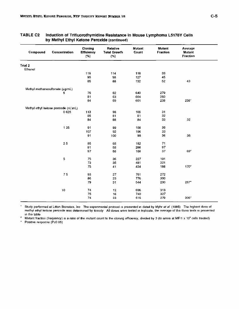
C-5 METHYL ETHYL KETONE PEROXIDE, NTP TOXICITY REPORT NUMBER 18
TABLE C2 Induction of Trifluorothymidine Resistance in Mouse Lymphoma L5178Y Cells by Methyl Ethyl Ketone Peroxide (continued)
Cloning Relative Mutant Mutant Average Compound Concentration Efficiency Total Growth Count Fraction Mutant
(%) (%) Fraction
Trial2 Ethanol
118 114 118 33 95 98 127 45 85 88 132 52 43
Methyl methanesulfonate (Jlg/ml) 5 76 62 640 279
81 63 604 250 84 69 601 238 256*
Methyl ethyl ketone peroxide (nL\mL) 0 625 113 98 106 31
85 81 81 32 84 88 84 33 32
1 25 91 99 108 39 107 92 106 33
91 100 98 36 36
25 85 65 182 71 91 58 266 97 97 86 108 37 69*
5 75 36 227 101 73 35 481 221 75 41 424 188 170*
75 93 27 761 272 86 23 776 300 79 31 544 230 267*
10 74 12 696 313 75 16 740 327 74 23 616 279 306*
1 Study performed at L1tton B1onetJcs, Inc The expenmental protocol1s presented 1n detail by Myhr eta/ (1985) The h1ghest dose of methyl ethyl ketone perox1de was determined by tox1c1ty All doses were tested 1n tnphcate, the average of the three tests 1s presented 1n the table
2 x 10⁶ . Mutant fraction (frequency) IS a rat1o of the mutant count to the clomng eff1c1ency, d1v1ded by 3 (to arnve at MF/1 cells treated) Pos1t1ve response (PsO 05)
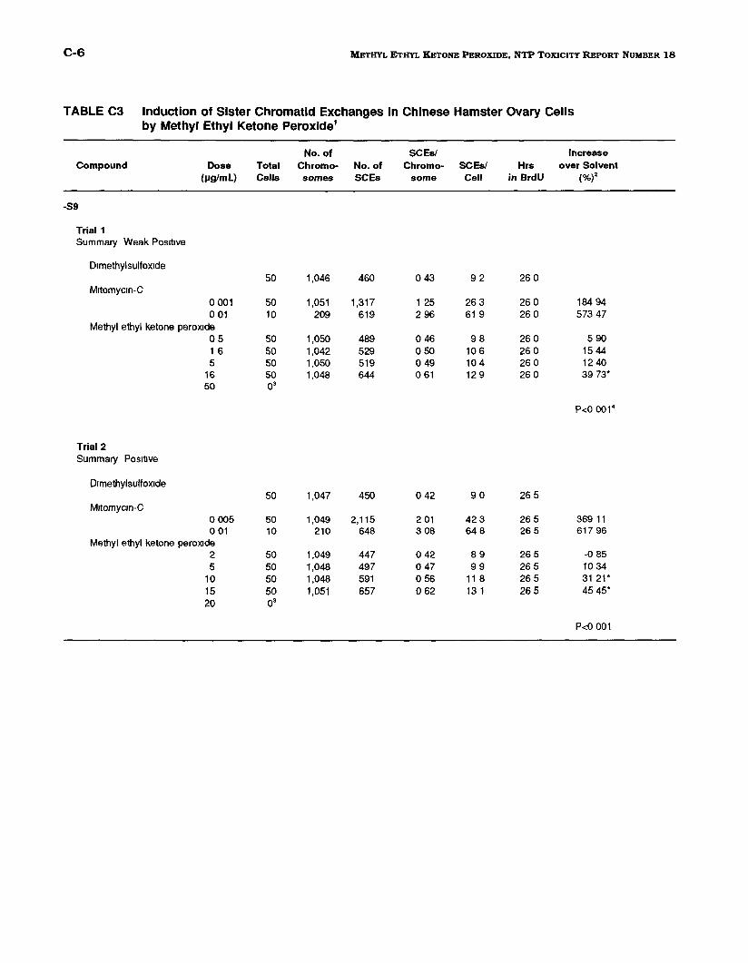
C-6 METHYL ETHYL KEToNE PEROXIDE, NTP TOXICITY REPORT NUMBER 18
TABLE C3 Induction of Sister Chromatid Exchanges in Chinese Hamster Ovary Cells by Methyl Ethyl Ketone Peroxide1
No. of SCEsf Increase Compound Dose Total Chromo- No. of Chromo- SCEs/ Hrs over Solvent
(pg/ml) Cells somes SCEs some Cell in BrdU (%)2
-S9
Trial1 Summary Weak Pos1bve
Dimethylsulfoxide 50 1,046 460 0 43 92 26 0
M1tomyc1n-C 0 001 50 1,051 1,317 1 25 26 3 26 0 184 94 0 01 10 209 619 2 96 61 9 26 0 573 47
Methyl ethyl ketone perox1de 05 50 1,050 489 0 46 98 26 0 5 90 1 6 50 1,042 529 0 50 10 6 26 0 15 44 5 50 1,050 519 0 49 10 4 26 0 12 40
16 50
50 o•
1,048 644 0 61 12 9 26 0 39 73*
P<O 001 4
Trial 2 Summary Pos1bve
Dimethylsulfoxide 50 1,047 450 0 42 90 26 5
M1tomycm-C 0 005 50 1,049 2,115 2 01 423 26 5 369 11 0 01 10 210 648 3 08 64 8 26 5 617 96
Methyl ethyl ketone peroxide 2 50 1,049 447 0 42 89 26 5 -0 85 5 50 1,048 497 0 47 99 26 5 10 34
10 50 1,048 591 0 56 11 8 26 5 31 21* 15 20
50 o•
1,051 657 0 62 13 1 26 5 45 45*
P<O 001
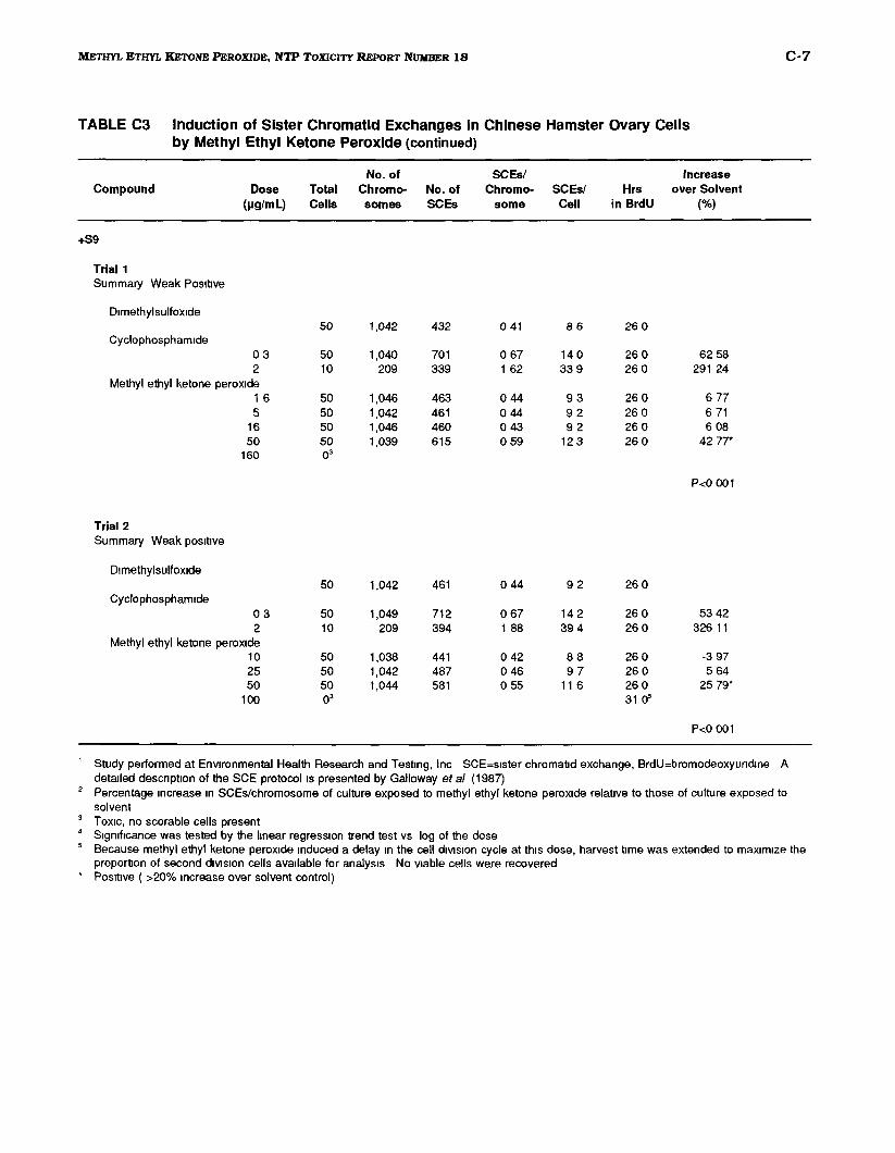
C-7 METHYL ETHYL KETONE PEROXIDE, NTP TOXICITY REPORT NUMBER 18
TABLE C3 Induction of Sister Chromatid Exchanges in Chinese Hamster Ovary Cells by Methyl Ethyl Ketone Peroxide (continued)
No. of SCEs/ Increase Compound Dose Total Chromo· No. of Chromo- SCEs/ Hrs over Solvent
(IJg/ml) Cells somes SCEs some Cell in BrdU (%)
+S9
Trial1 Summary Weak Positive
D1methylsulfox1de 50 1,042 432 0 41 86 260
Cyclophosphamide 03 50 1,040 701 0 67 14 0 260 62 58 2 10 209 339 1 62 339 26 0 291 24
Methyl ethyl ketone peroxide 1 6 50 1,046 463 0 44 93 26 0 677 5 50 1,042 461 0 44 92 26 0 6 71
16 50 1,046 460 0 43 92 26 0 6 08 50 50 1,039 615 059 12 3 26 0 42 77*
160 03
P<O 001
Trial2 Summary Weak pos1t1ve
Dimethylsulfoxide 50 1,042 461 044 92 26 0
Cyclophosphamide 03 50 1,049 712 0 67 14 2 26 0 53 42 2 10 209 394 1 88 39 4 26 0 326 11
Methyl ethyl ketone peroxide 10 50 1,038 441 0 42 88 26 0 -3 97 25 50 1,042 487 0 46 97 26 0 5 64 50 50 1,044 581 0 55 11 6 26 0 25 79*
100 03 31 o"
P<O 001
1 Study performed at EnVIronmental Health Research and Test1ng, Inc SCE=slster chromatid exchange, BrdU=bromodeoxyundlne A detailed descnpt1on of the SCE protocol Is presented by Galloway eta/ (1987)
2 Percentage Increase 1n SCEs/chromosome of culture exposed to methyl ethyl ketone perox1de relat1ve to those of culture exposed to solvent
3 Tox1c, no scorable cells present 4 S1gmflcance was tested by the hnear regression trend test vs log of the dose
. 5 Because methyl ethyl ketone perox1de Induced a delay 1n the cell diVISion cycle at th1s dose, harvest t1me was extended to max1m1ze the
proportion of second diVISion cells available for analys1s No v1able cells were recovered Pos1t1ve ( >20% Increase over solvent control)
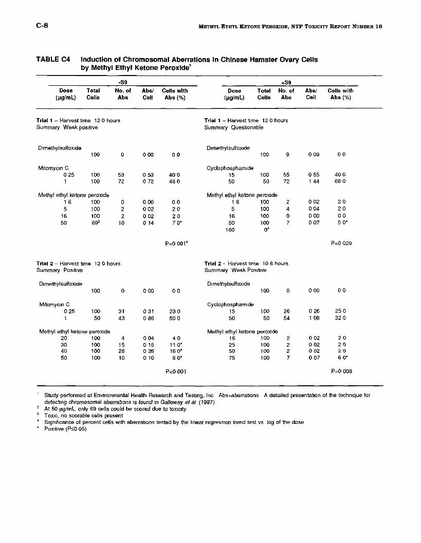
C-8 METHYL ETHYL KETONE PEROXIDE, NTP TOXICITY REPORT NUMBER 18
TABLE C4 Induction of Chromosomal Aberrations in Chinese Hamster Ovary Cells by Methyl Ethyl Ketone Peroxide1
-S9 +59
Dose (IJg/ml)
Total Cells
No. of Abs
Abs/ Cell
Cells with Abs (%)
Dose (IJg/ml)
Total Cells
No. of Abs
Abs/ Cell
Cells with Abs (%)
Trial 1 Harvest t1me 12 0 hours Summary Weak pos1t1ve
Dimethylsulfoxide 100 0
Mitomycin C 0 25 100 53 1 100 72
Methyl ethyl ketone peroxide 1 6 100 0 5 100 2
16 100 2 50 692 10
Trial 2 Harvest t1me 12 0 hours Summary Pos1t1ve
Dimethylsulfoxide 100 0
M1tomyc1n C 0 25 100 31 1 50 43
Methyl ethyl ketone peroxide 20 100 4 30 100 15 40 100 26 50 100 10
0 00
0 53 0 72
0 00 0 02 0 02 0 14
0 00
0 31 0 86
0 04 0 15 0 26 0 10
00
400 48 0
00 20 20 7 0*
P<O 001 4
00
230 50 0
40 11 0* 16 0* 8 0*
P<O 001
Trial 1 Harvest t1me 12 0 hours Summary Questionable
Dimethylsulfoxide 100 9
Cyclophosphamide 15 100 55 50 50 72
Methyl ethyl ketone perox1de 1 6 100 2 5 100 4
16 100 0 50 100 7
160 03
Trial 2- Harvest t1me 10 8 hours Summary Weak Pos1t1ve
Dimethylsulfoxide 100 0
Cyclophosphamide 15 100 26 50 50 54
Methyl ethyl ketone perox1de 16 100 2 25 100 2 50 100 2 75 100 7
0 09
0 55 1 44
0 02 0 04 0 00 0 07
0 00
0 26 1 08
0 02 0 02 0 02 0 07
00
40 0 680
20 20 00 50*
P=O 029
00
25 0 32 0
20 20 20 6 0*
P=O 008
1
2
3
4
*
Study performed at EnVIronmental Health Research and Test1ng, Inc Abs=aberratlons detecting chromosomal aberra!1:ms 1s found 1n Galloway eta/ {1987) At 50 J.lglmL, only 69 cells could be scored due to toXICity Tox1c, no scorable cells present S1gmflcance of percent cells w1th aberrations tested by the linear regression trend test vs Pos1t1ve (Ps;O 05)
A detailed presentation of the techmque for
log of the dose
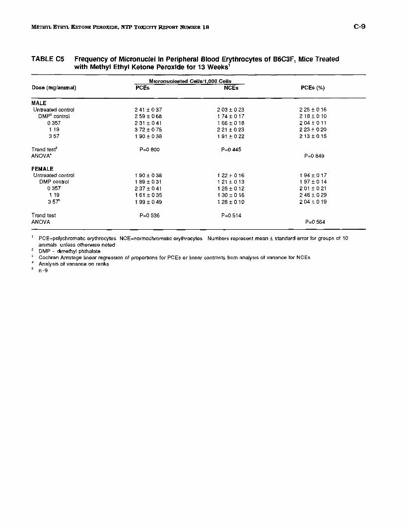
C-9 METHYL ETHYL KETONE PEROXIDE, NTP TOXICITY REPORT NUMBER 18
TABLE C5 Frequency of Micronuclei in Peripheral Blood Erythrocytes of B6C3F, Mice Treated with Methyl Ethyl Ketone Peroxide for 13 Weeks1
Mrcronucleated Cells/1 ,000 Cells Dose (mg/ammal) PCEs NCEs PCEs (%)
MALE Untreated control
DMP2 control 0 357 1 19 3 57
Trend test' ANOVA4
FEMALE Untreated control
DMP control 0 357 1 19 35"t
Trend test ANOVA
2 41 ± 0 37 2 59± 0 68 231±041 3 72 ± 0 75 190±038
P=O 800
1 90 ± 0 38 1 89 ± 0 31 2 37 ± 0 41 1 61 ± 0 35 1 99 ± 0 49
P=O 536
2 03 ± 0 23 174±017 1 66 ± 0 18 2 21 ± 0 23 1 91 ± 0 22
P=O 445
1 22 ± 0 16 121±013 1 25 ± 0 12 1 30 ± 0 16 1 28 ± 0 10
2 25 ± 0 16 218±010 2 04 ± 0 11 2 23 ± 0 20 213±015
P=O 849
1 94 ± 0 17 197±014 2 01 ± 0 21 2 46 ± 0 29 2 04 ± 0 19
P=O 564
1 PCE=polychromatlc erythrocytes NCE=normochromatlc erythrocytes Numbers represent mean± standard error for groups of 10 ammals unless otherw1se noted
2 DMP = dimethyl phthalate 3 Cochran Armitage linear regress1on of proportions for PCEs or linear contrasts from analysis of vanance for NCEs • Analysis of vanance on ranks 5 n=9

C-10 METHYL ETHYL KETONE PEROXIDE, NTP TOXICITY REPORT NUMBER 18
NTP TECHNICAL REPORTS ON TOXICITY STUDIES PRINTED AS OF DECEMBER 1992
Toxicity Report Number
2
3
4
5
6
7
8
9
10
11
12
13
14
15
16
17
19
20
21
22
23
Chemical
Hexachloro-1,3-butadiene
n-Hexane
Acetone
1,2-Dlchloroethane
Cobalt Sulfate Heptahydrate
Pentachlorobenzene
1,2,4,5-Tetrachlorobenzene
D & C Yellow No. 11
a-Cresol m-Cresol p-Creso1
Ethy1benzene
Antimony Potassium Tartrate
Castor Oil
Trinitrofluorenone
p-Chloro-a,a,a-Trtfluorotoluene
t-Butyl Perbenzoate
Glyphosate
Black Newsprint Ink
Formic Acid
Diethanolam1ne
2-Hydroxy-4-Methoxybenzophenone
N, N-Dimethylformamide
o-Nitrotoluene m-Nltrotoluene p-Nltrotoluene
Route of Exposure
Dosed Feed
Inhalation
Drinking Water
Drinking Water, Gavage
Inhalation
Dosed Feed
Dosed Feed
Dosed Feed
Dosed Feed
Inhalation
Drinking Water, l.P. Inject.
Dosed Feed
Dermal, Dosed Feed
Gavage (corn oil, a -CD)
Gavage
Dosed Feed
De~
Inhalation
,Drinking Water, Dermal
Dosed Feed, Drinking Water
Inhalation
Dosed Feed
Publication Number
91-3120
91-3121
91-3122
91-3123
91-3124
91-3125
91-3126
91-3127
92-3128
92-3129
92-3130
92-3131
92-3132
92-3133
92-3134
92-3135
92-3340
92-3342
92-3343
92-3344
93-3345
92-3346


![Copper-based nanocatalysts for 2-butanol … · rial for the production of methyl ethyl ketone peroxide [1,2]. As per 2012, the global production of MEK reached 1,141 thousand metric](https://static.fdocuments.in/doc/165x107/5ac5d2f07f8b9a5c558d99d0/copper-based-nanocatalysts-for-2-butanol-for-the-production-of-methyl-ethyl.jpg)