
jhghjgf hkfgfgf uyuiyoui gfyyty
9
1559 DEVELOPMENT AND DISEASE RESEARCH ARTICLE INTRODUCTION Over the past two decades there has been rapid progress in understanding the molecular mechanisms for neuronal subtype fate determi nation. Ma ny of the init ial disco veries were made in Drosophila, with more-recent studies including other model organisms such as C. elegans, zebrafish and chick. In the last decade, these studies have been extended to mammals, for which, particularly in the spinal cord and retina (Da sen et al., 2005; Livesey and Cepko, 2001; Shirasaki and Pfaff, 2002), further advances have occurred. Despite these advances, and the tremendous relevance to human neuropsychiatric illnesses, there has been relatively little progress in cracking the ‘molecular codes’ for specifying neuronal subtypes of the mammalian forebrain (Schuurmans et al., 2004; Zhao et al., 2003). Beyond the issues of function al pleiotropy and redundan cy, a key reason for this lag is the lengthy delay between the final cell cycle, when much of the crucial specification-relat ed signaling is likely to occur (McConnell and Kaznowski, 1991; Xu et al., 2005), and the appearance of the mature neuronal phenotype. This delay dictates that the readout of any manipulation of cell fate in progenitor cells may only be assessable weeks later, in the context of many potential confounding influences of that manipulation on neuronal development. In this paper we present a transfection/transplantation method for studying the genetic control of neuronal fate determination in the developing mammalian forebrain. Recent evidence indicates that, at least in rodents and ferrets, the large majority of cortical interneurons originate in the subcortical telencephalon (for a review, see Wonders and Anderson, 2006). Despite advances in determining the origins, migratory pathways and the regulation of interneuron migration, little is known about the specification of distinct subgroups of cortical interneurons. Roughly 70% of cortical interneurons can be divided into two practically non- overlapping groups based on their expression of the calcium-binding proteins parvalbumin (PV) or neuropeptide somatostatin (SST) (Gonchar and Burkhalter, 1997; Tamamaki et al., 2003). Both of these subgroups appear to originate mainly within the medial ganglionic eminence (MGE) (Butt et al., 2005; Valcanis and Tan, 2003; Wichterle et al., 2001; Xu et al., 2004), where their fate determi nation depend s on the transcri ption factors NKX2 .1 and LHX6 (Liodis et al., 2007; Xu et al., 2004). Expre ssion of SST and PV matures postnatally, with PV expression only beginning in rodent cortex during the second postnatal week (Alcantara et al., 1996). Nkx2.1 is expressed in the MGE and preoptic region of the pallidal telencephalon (Sussel et al., 1999) (see Fig. S1 in the supplementary material). Over 90% of S-phase cells expres s Nkx2.1 in both the ventricular and subventricular zones of the MGE (Xu et al., 2005). These progenitors produce oligodendrocytes and interneurons of the striatum and cerebral cortex (Kessaris et al., 2006; Marin and Rubenstein, 2001) and projection neurons of the globus pallidus (Xu et al., 2008). As cells migrate out of the MGE, Nkx2.1 expression is downregulated in cortical interneurons but is maintained in subgroups of striatal interneurons postnatally (Marin et al., 2000). As they exit the proliferative zone, most MGE-derived cells begin to express LHX6 (Grigoriou et al., 1998), a homeodomain -containing transcription factor that is not detectable in the telencephalon of Nkx2.1-null embryos (Sussel et al., 1999). Lhx6 continues to be expressed in many interneurons, including most of those that go on to expr ess PV or SST, as they migrate to and then differentiate within the cerebral cortex (see Fig. S1 in the supplementary material) (Cobos et al., 2005; Fogarty et al., 2007; Gong et al., 2003; Lavdas et al., 1999; Liodis et al., 2007). Here we use genetic gain- and loss-of-function mani pulations in mouse embryonic slice cultures, followed by transplantation into cortical environments in vitro and in vivo, to further examine the role of Nkx2.1 in the specificatio n of MGE-deriv ed (PV- or SST- expressing) cortical interne uron subgroups. We demonstrate that the LIM-homeodomain transcription factor gene Lhx6 is activated by and appears to be a direct target of NKX2.1. Like Nkx2.1 itself, Lhx6 is sufficient to rescue both neurochemical and morphological aspects of Nkx2.1 –/– MGE-derived interneurons. In addition, at least for the specification of the SST-expressing phenotype, Lhx6 is required around the time of cell cycle exit and not postnatally in mature NKX2.1 specifies cortical interneuron fate by activating Lhx6 T onggong Du, Qing Xu, Polloneal J. Ocbina and Stewart A. Anderson* In the ventral telencephalon, the medial ganglionic eminence (MGE) is a major source of cortical interneurons. Exp ression of the transcription factor NKX2.1 in the MGE is required for the spec ification of two major subgroups of cortical interneurons – those that express parvalbumin (PV) or somatosta tin (SST) – but direct targets of NKX2.1 remain to be esta blished. We find that electroporation of Nkx2.1 cDNA into the ventral telencephalon of slice cultures fromNkx2.1 –/– mouse embryos, followed by transplantation into neonatal cortex to permit postnatal analysis of their fate, rescues the loss of PV- and SST-expre ssing cells. The LIM-homeobox gene Lhx6 is induced by this rescue experiment, and gain- and loss-of-function studies suggest that Lhx6 is necessary and sufficient to rescue these and other interneuron phenotypes in cells transplanted from Nkx2.1 –/– slices. Finally, NKX2.1 protein binds a highly conserved sequence in theLhx6 promoter, and this sequence appears to mediate the direct activation of Lhx6 by NKX2.1. The slice transfection and transplantation methods employed here are beginning to uncover embryonic mechanisms for specifying neuronal fates that only become definable postnatally. KEY WORDS: Cell fate determination, GABA, Medial ganglionic eminence, Nkx2.1, Parvalbumin, Somatostatin, Mouse Developmen t 135, 1559-1567 (2008) doi:10.1242/dev .015123 Department of Psychiatry, and Department of Neurology and Neuroscience, Weill Medical College of Cornell Universit y, 1300 Y ork Avenue, Box 244, New York, NY 10021, USA. *Author for correspond ence (e-mail: SAA2007@med.cornell.edu) Accepted 2 February 2008
-
Upload
chytoudis-harrys -
Category
Documents
-
view
215 -
download
0
Transcript of jhghjgf hkfgfgf uyuiyoui gfyyty
8/14/2019 jhghjgf hkfgfgf uyuiyoui gfyyty
http://slidepdf.com/reader/full/jhghjgf-hkfgfgf-uyuiyoui-gfyyty 1/9
1559DEVELOPMENT AND DISEASE RESEARCH ARTICLE
INTRODUCTIONOver the past two decades there has been rapid progress in
understanding the molecular mechanisms for neuronal subtype fate
determination. Many of the initial discoveries were made in
Drosophila, with more-recent studies including other model
organisms such as C. elegans, zebrafish and chick. In the last decade,
these studies have been extended to mammals, for which,
particularly in the spinal cord and retina (Dasen et al., 2005; Livesey
and Cepko, 2001; Shirasaki and Pfaff, 2002), further advances have
occurred. Despite these advances, and the tremendous relevance to
human neuropsychiatric illnesses, there has been relatively little
progress in cracking the ‘molecular codes’ for specifying neuronal
subtypes of the mammalian forebrain (Schuurmans et al., 2004;
Zhao et al., 2003).
Beyond the issues of functional pleiotropy and redundancy, a keyreason for this lag is the lengthy delay between the final cell cycle,
when much of the crucial specification-related signaling is likely to
occur (McConnell and Kaznowski, 1991; Xu et al., 2005), and the
appearance of the mature neuronal phenotype. This delay dictates
that the readout of any manipulation of cell fate in progenitor cells
may only be assessable weeks later, in the context of many potential
confounding influences of that manipulation on neuronal
development. In this paper we present a transfection/transplantation
method for studying the genetic control of neuronal fate
determination in the developing mammalian forebrain.
Recent evidence indicates that, at least in rodents and ferrets, the
large majority of cortical interneurons originate in the subcortical
telencephalon (for a review, see Wonders and Anderson, 2006).
Despite advances in determining the origins, migratory pathwaysand the regulation of interneuron migration, little is known about the
specification of distinct subgroups of cortical interneurons. Roughly
70% of cortical interneurons can be divided into two practically non-
overlapping groups based on their expression of the calcium-binding
proteins parvalbumin (PV) or neuropeptide somatostatin (SST)(Gonchar and Burkhalter, 1997; Tamamaki et al., 2003). Both of
these subgroups appear to originate mainly within the medial
ganglionic eminence (MGE) (Butt et al., 2005; Valcanis and Tan,
2003; Wichterle et al., 2001; Xu et al., 2004), where their fate
determination depends on the transcription factors NKX2.1 and
LHX6 (Liodis et al., 2007; Xu et al., 2004). Expression of SST and
PV matures postnatally, with PV expression only beginning in
rodent cortex during the second postnatal week (Alcantara et al.,
1996).
Nkx2.1 is expressed in the MGE and preoptic region of the
pallidal telencephalon (Sussel et al., 1999) (see Fig. S1 in the
supplementary material). Over 90% of S-phase cells express Nkx2.1
in both the ventricular and subventricular zones of the MGE (Xu et
al., 2005). These progenitors produce oligodendrocytes andinterneurons of the striatum and cerebral cortex (Kessaris et al.,
2006; Marin and Rubenstein, 2001) and projection neurons of the
globus pallidus (Xu et al., 2008). As cells migrate out of the MGE,
Nkx2.1 expression is downregulated in cortical interneurons but is
maintained in subgroups of striatal interneurons postnatally (Marin
et al., 2000). As they exit the proliferative zone, most MGE-derived
cells begin to express LHX6 (Grigoriou et al., 1998), a
homeodomain-containing transcription factor that is not detectable
in the telencephalon of Nkx2.1-null embryos (Sussel et al., 1999).
Lhx6 continues to be expressed in many interneurons, including
most of those that go on to express PV or SST, as they migrate to and
then differentiate within the cerebral cortex (see Fig. S1 in the
supplementary material) (Cobos et al., 2005; Fogarty et al., 2007;
Gong et al., 2003; Lavdas et al., 1999; Liodis et al., 2007).Here we use genetic gain- and loss-of-function manipulations in
mouse embryonic slice cultures, followed by transplantation into
cortical environments in vitro and in vivo, to further examine the
role of Nkx2.1 in the specification of MGE-derived (PV- or SST-
expressing) cortical interneuron subgroups. We demonstrate that the
LIM-homeodomain transcription factor gene Lhx6 is activated by
and appears to be a direct target of NKX2.1. Like Nkx2.1 itself, Lhx6
is sufficient to rescue both neurochemical and morphological aspects
of Nkx2.1 –/– MGE-derived interneurons. In addition, at least for the
specification of the SST-expressing phenotype, Lhx6 is required
around the time of cell cycle exit and not postnatally in mature
NKX2.1 specifies cortical interneuron fate by activating Lhx6
Tonggong Du, Qing Xu, Polloneal J. Ocbina and Stewart A. Anderson*
In the ventral telencephalon, the medial ganglionic eminence (MGE) is a major source of cortical interneurons. Expression of the
transcription factor NKX2.1 in the MGE is required for the specification of two major subgroups of cortical interneurons – those thatexpress parvalbumin (PV) or somatostatin (SST) – but direct targets of NKX2.1 remain to be established. We find that
electroporation ofNkx2.1 cDNA into the ventral telencephalon of slice cultures fromNkx2.1–/– mouse embryos, followed by
transplantation into neonatal cortex to permit postnatal analysis of their fate, rescues the loss of PV- and SST-expressing cells. The
LIM-homeobox gene Lhx6 is induced by this rescue experiment, and gain- and loss-of-function studies suggest thatLhx6 is necessary
and sufficient to rescue these and other interneuron phenotypes in cells transplanted fromNkx2.1–/– slices. Finally, NKX2.1 protein
binds a highly conserved sequence in theLhx6 promoter, and this sequence appears to mediate the direct activation ofLhx6 by
NKX2.1. The slice transfection and transplantation methods employed here are beginning to uncover embryonic mechanisms for
specifying neuronal fates that only become definable postnatally.
KEY WORDS: Cell fate determination, GABA, Medial ganglionic eminence, Nkx2.1, Parvalbumin, Somatostatin, Mouse
Development 135, 1559-1567 (2008) doi:10.1242/dev.015123
Department of Psychiatry, and Department of Neurology and Neuroscience,Weill Medical College of Cornell University, 1300 York Avenue, Box 244, New York,NY 10021, USA.
*Author for correspondence (e-mail: [email protected])
Accepted 2 February 2008

8/14/2019 jhghjgf hkfgfgf uyuiyoui gfyyty
http://slidepdf.com/reader/full/jhghjgf-hkfgfgf-uyuiyoui-gfyyty 2/9
1560
interneurons. These results and the system presented lay important
groundwork for further studies on the transcriptional regulation of
interneuron fate in the mammalian forebrain.
MATERIALS AND METHODSAnimals
Nkx2.1-null mice (Kimura et al., 1996) on a CD1 background (Xu et al.,
2004) and non-transgenic CD1 strain mice were used. All animal procedures
were undertaken according to the guidelines of the Institutional Animal Careand Use Committee at the Weill Cornell Medical College.
Gene constructs, slice electroporation and transplantation
Slice electroporation (EP) was conducted as described (Stuhmer et al., 2002;
Xu et al., 2005) using vectors concentrated with Endotoxin-free DNA
Maxiprep Kits (Qiagen). Full-length cDNAs for Nkx2.1 [from John
Rubenstein (UCSF, San Francisco, CA) and Oscar Marin (Universidad
Miguel Hernández, Alicante, Spain)] and Lhx6 (from Vassilis Pachnis,
MRC, London, UK) were cloned into pCAG-IRES-GFP (from Connie
Cepko, Harvard Medical College, Boston, MA) and their expression
confirmed by immunofluorescence (see Fig. 1 for NKX2.1). VP16-Nkx2.1
is a gift from Parvis Minoo (Li et al., 2002). The Nkx2.1homeodomain point
mutant (Val45Phe) (Krude et al., 2002) was generated using the QuikChange
Site-Directed Mutagenesis Kit (Stratagene, La Jolla, CA) and subcloned into
the pCAGGS vector to produce pNkx2.1HD.
To generate the Lhx6 RNAi vector, a sequence encoding a small hairpin(sh) targeting the distal end of exon 2 (sense strand, 5 -GTCAGACG-
CAGAGGCCTTGGCCATGGCCAAGGCCTCTGCGTCTGACTTTTTT;
antisense strand, 5-agcttAAAAAAGTCAGACGCAGAGGCCTTGG-
CCATGGCCAAGGCCTCTGCGTCTGACggcc; lowercase letters indicate
nucleotides used in cloning; underlined nucleotides indicate loop region)
was inserted into the ApaI and HindIII sites in pSilencer 1.0 (a gift from
Yang Shi) (Sui et al., 2002). This shRNAi species has previously been
demonstrated to greatly reduce LHX6 abundance in MGE-derived cells
and to reduce interneuron migration to the cortex (Alifragis et al., 2004).
To enable visualization of the RNAi-transfected cells with a single
plasmid, the pU6-shLhx6 construct was then inserted into the SpeI site in
pCAG-IRES-GFP (shLhx6-GFP). The Scramble RNAi sequence
(Ambion) was also subcloned into the same site in the pCAG-IRES-GFP
vector. To ensure that nearly all Nkx2.1-transfected cells also received the
RNAi vector, 2 mg/ml of this vector and 1 mg/ml of pNkx2.1 weremicroinjected into the slice for EP. This procedure results in over 90% co-
labeling of NKX2.1 and GFP in transplanted cells (data not shown) (see
also Stuhmer et al., 2002).
For transplantation studies, 12-16 hours after EP GFP epifluorescence
was imaged, then the regions of the MGE with the highest densities of GFP+
cells were dissected out, mechanically dissociated, and counted on a
hemocytometer under epifluorescence to calculate the percentage of all cells
that were GFP+ (usually 10-15%). In the case of slices from Nkx2.1-null
embryos, in which a morphologically identifiable MGE does not exist
(Sussel et al., 1999), tissue was targeted for EP and dissection from the same
approximate dorsal-ventral level as the MGE of control slices, as described
(Xu et al., 2004).
For in vitro transplants, the feeder cells were prepared from neonatal
cortex as described (Xu et al., 2004; Xu et al., 2005) and the transfected cells
were plated at a density of 1000 GFP+ cells per well of a 16-well glasschamber slide (36 mm2; Lab-Tek). For in vivo transplants, cells were
injected into S1 cortex 1 mm below the pial surface of cold-anaesthetized
neonatal pups (day of birth or P1), using a microinjector (Nanoinject II,
Drummond). Per injection site, 10,000-20,000 cells were placed per
hemisphere to obtain at least 1000 GFP+ cells per transplant. At P30 the
brains were removed, fixed with 4% paraformaldehyde, and sectioned on a
vibratome at 50 m.
RT-PCR
Slices from Nkx2.1 nulls were electroporated with pNkx2.1-IRES-GFP or
pGFP control (n=3). After 12 hours the MGE-like regions were dissociated
and sorted by FACS (Vantage, Becton-Dickinson) producing a yield of
3000-5000 cells. Total RNA was purified (RNeasy Kit, Qiagen) and
subjected to reverse transcription (Omniscript reverse transcriptase, Qiagen)
and PCR (HotStar Kit, Qiagen). Primers included Lhx6 (5-TGATG-
GCCCAGCCAG and 5-GTCCATCTTGCAGTAGATC; 422 bp product),
Nkx2.1 (5-AACAGCGGCCATGCAGCAGCAC and 5-CCATGTTCT-
TGCTCACGTCC; 315 bp) and -actin (5-GAGCTGCCTGACGGCC-
AGGT and 5-TACTCCTGCTTGCTGATCCA; 364 bp).
Immunodetection
Immunofluorescence labeling of cells in dissociated cultures was conducted
as described (Xu et al., 2004), and labeling of antigens in postnatal brainsections was conducted floating. Primary antibodies used included GFP
(rabbit or chick; Molecular Probes), GABA (rabbit; Sigma), LHX6 [rabbit, a
gift from Vassilis Pachnis (Lavdas et al., 1999)], neuropeptide Y (rabbit;
Immunostar), NKX2.1 (mouse; Lab Vision), somatostatin (rat; Chemicon),
parvalbumin (mouse; Chemicon), calretinin (rabbit; Chemicon), Kv3.1
(rabbit, a gift from Bernardo Rudy, New York University, NY) and PCNA
(mouse IgG; Novocastra). Alexa line secondary antibodies (Molecular
Probes) were used.
Lhx6 promoter analyses
Phylogenetic sequence comparisons of the Lhx6 locus were performed using
the ECR browser (http://www.dcode.org) sequence alignment and
visualization tool (Ovcharenko et al., 2004b).
rVISTA (http://rvista.dcode.org/) (Loots and Ovcharenko, 2004;
Ovcharenko et al., 2004a) was used to identify potential transcription factor
binding sites (http://zpicture.dcode.org/).Chromatin immunoprecipitation was conducted on E12.5 MGE samples
as per the manufacturer’s instructions (Upstate, 17-295), using a mouse anti-
NKX2.1 monoclonal antibody (Lab Vision). A 119 bp PCR fragment of the
Lhx6 promoter that includes a consensus NKX2.1 binding sequence at
position –240 bp relative to the putative translational start site was identified
using primers 5-AGTCCTAACTTTGTAGTG and 5-TTTCCCCCTCAG-
AGGCTTG.
To generate Lhx6 reporter constructs, a 2.1 kb fragment of 5 Lhx6
genomic region (Fig. 5) was cloned from BAC RP23-2D16 by PCR [5-
ACTAGT(SpeI)CAGCCTTTAGAAGCTGGTGC and 5-TCTAGA( XbaI)-
CCCTGGCTGGGCCATC]. This fragment was inserted in place of the
CAG promoter in pCAG-IRES-GFP to produce p5-Lhx6-IRES-GFP.
Site directed mutagenesis (using the oligo sequence 5-CCCTCTCCC-
TGCACTTAACCCGTGATCGCTTAGTTCCTTTTGCAATCCAAGCC;
QuikChange Site-directed Mutagenesis Kit, Stratagene) was then used toremove the putative NKX2.1 binding domain (GCTCTTGAAGTA) from
–239 to –250 nt.
RESULTSExpression of Nkx2.1 cDNA in Nkx2.1
–/– slicesrescues interneuron fateOur previous work has shown that progenitors from the MGE-like
region of Nkx2.1 –/– slices (MGE*, Fig. 1B,G; see Fig. S2 in the
supplementary material; Materials and methods), cultured on
dissociated cells from neonatal cortex, fail to differentiate into the
SST- or PV-expressing interneuron phenotype (Xu et al., 2004). To
determine whether rescued expression of Nkx2.1 in the ventral
telencephalon of slices from Nkx2.1 –/– mutants can rescue the PV or
SST fate of these cells, telencephalic slices were prepared from Nkx2.1 –/– mouse embryos at embryonic day (E) 12.5, and an
expression vector, pNkx2.1-GFP, was introduced into the MGE* by
electroporation (Fig. 1 and see Fig. S2 in the supplementary
material). After 1 day in vitro (DIV), subregions of the MGE* with
the highest proportion of transfected cells were dissected,
dissociated, plated over a feeder culture made from neonatal cortex
and maintained for 14-28DIV (see Fig. S2 in the supplementary
material). In other experiments, cells were transplanted directly into
the cortical plate of the somatosensory cortex of neonatal pups and
then examined in tissue sections after 30 days (Fig. 1). After
fixation, the fates of cells with neuronal morphology (the vast
RESEARCH ARTICLE Development 135 (8)

8/14/2019 jhghjgf hkfgfgf uyuiyoui gfyyty
http://slidepdf.com/reader/full/jhghjgf-hkfgfgf-uyuiyoui-gfyyty 3/9
majority of surviving cells from donors at this age of E12.5+1DIV)
were determined by immunofluorescence for GFP and the given
interneuron subgroup marker. Similar transplantations of cells
directly from the MGE of transgenic mice into neonatal cortex havebeen shown to give rise to neurons with neurochemical and
physiological characteristics of inhibitory interneurons (Alvarez-
Dolado et al., 2006; Cobos et al., 2005).
Consistent with previous results using in vitro transplants from
Nkx2.1 nulls (Xu et al., 2004), Nkx2.1 –/– cells transfected with
control vector almost never gave rise to PV+, SST+ or NPY+
interneurons (Table 1). In marked contrast, transfection with Nkx2.1
cDNA resulted in substantial rescue of PV or SST expression (Table
1, Fig. 1I-L). In addition, neurons expressing NPY, a neuropeptide
that shares about 50% co-labeling with SST in mouse cortex, were
also rescued (Fig. 1M,N). Similar results were obtained whether
cells were plated onto a cortical feeder layer or transplanted into the
neonatal cortical plate in vivo (Table 1, Fig. 1 and see Fig. S2 in the
supplementary material).
Lhx6 is induced when Nkx2.1 is expressed in theNkx2.1–/– MGE*The ability of Nkx2.1 expression to rescue neurochemical aspects of
the Nkx2.1 –/– interneuron phenotype provides an opportunity toidentify downstream effectors of cortical interneuron fate
determination. A lead candidate for such an effector is LHX6, a
LIM-homeodomain-containing transcription factor that is expressed
in most MGE-derived interneurons from around the time of their
final cell cycle through their maturity in the postnatal cerebral cortex
(Cobos et al., 2005; Fogarty et al., 2007; Gong et al., 2003; Lavdas
et al., 1999; Liodis et al., 2007) (see Fig. S1 in the supplementary
material); LHX6 is not detectable in the MGE* of Nkx2.1-null
embryos (Sussel et al., 1999). Indeed, RT-PCR detected the
induction of Lhx6 expression in the MGE* of Nkx2.1-null embryos
transfected with Nkx2.1 (Fig. 2A).
To further confirm that Lhx6 can be induced in Nkx2.1 –/– MGE*
progenitors after transfection with pNkx2.1-GFP, rescue- and
control-transfected neurons were assessed after in vivo
1561RESEARCH ARTICLENKX2.1 controls interneuron fate via Lhx6
Fig. 1. Transfection of Nkx2.1–/–
slices with Nkx2.1 cDNA results inrescue of interneuron phenotypes.(A-D) Schematic showing sliceelectroporation and transplantationparadigm. (A) The Nkx2.1 domain isshown in the MGE of an E12.5 wild-type (wt) mouse embryo slice. TheMGE*, a region that expresses a
truncated Nkx2.1 transcript (Sussel etal., 1999), is shown in the slice from anNkx2.1 mutant embryo. (B) This MGE*region is targeted for electroporation,and after 1 day in vitro (DIV) the regionis dissected out, dissociated andtransplanted (C,D) directly into theneocortex of neonatal pups (as in H-N),or plated onto a high-density culture ofneonatal cortical cells [as in Xu et al. (Xuet al., 2004); see Fig. S2 in thesupplementary material]. (E,F) Coronalsections of a slice from an E12.5 wtembryo that was electroporated withpNkx2.1-GFP, maintained 1DIV, then
fixed and examined for GFPfluorescence (E) and NKX2.1immunolabeling (F). The right-hand,electroporated side of the slice has extensive ectopic NKX2.1 expression, whereas only native NKX2.1 expression is seen on the left-hand side of theslice (arrow in F). (G) A slice from an Nkx2.1–/– embryo was electroporated with pNkx2.1-GFP. After 1DIV, cells from the MGE* (outlined in white)were transplanted into the cortical plate of a neonatal pup and then examined at postnatal day 30 (P30) in 40 m coronal sections.(H) Transplanted GFP-expressing cells scattered through the medial cortex. (I-N) Examples of co-labeling for GFP and parvalbumin (PV; I,J),somatostatin (SST; K,L), and neuropeptide Y (NPY; M,N). In control experiments with pGFP vector, almost no cells expressing any of these markersare detected after transplantation of Nkx2.1–/– MGE* progenitors (Table 1). MGE, medial ganglionic eminence; LGE, lateral ganglionic eminence;Ctx, cerebral cortex. Scale bars: 100 m in G,H.
Table 1. Transfection and transplantation of the MGE-like region in Nkx2.1–/– slices with Nkx2.1 or Lhx6 cDNAs rescues theexpression of MGE-derived interneuron markers
PV SST NPY
cDNA, transplantation Control Rescue Control Rescue Control Rescue
Nkx2.1, in vitro (n=4) 0.4±0.2% (272) 17.0±3.1% (177) 0.6±05% (895) 16.9±4.9% (1802) 0.0±0.0% (1620) 15.1±1.7% (1803)Nkx2.1, in vivo (n=3) 0.0±0.0% (134) 12.7±4.0% (157) 0.0±0.0% (161) 27.6±11.4% (257) 0.0±0.0% (164) 7.1±0.1% (154)Lhx6 , in vivo (n=3) 0.0±0.0% (167) 17.5±3.5% (464) 0.0±0.0% (159) 21.9±5.1% (521) 0.0±0.0% (106) 8.5±0.8% (284)
The numbers refer to the percentage of transplanted cells with neuronal morphologies that co-label with the given marker (with total counted neuronsshown in parentheses).
8/14/2019 jhghjgf hkfgfgf uyuiyoui gfyyty
http://slidepdf.com/reader/full/jhghjgf-hkfgfgf-uyuiyoui-gfyyty 4/9
1562
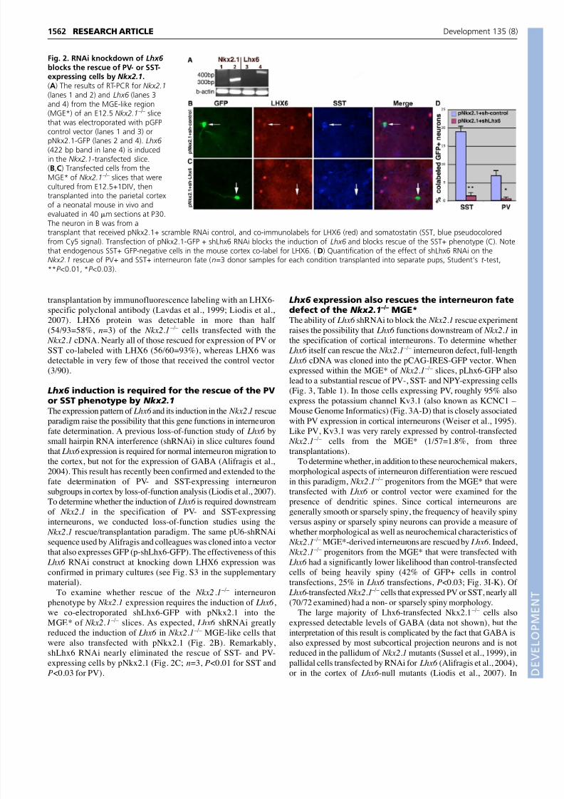
transplantation by immunofluorescence labeling with an LHX6-
specific polyclonal antibody (Lavdas et al., 1999; Liodis et al.,
2007). LHX6 protein was detectable in more than half
(54/93=58%, n=3) of the Nkx2.1 –/– cells transfected with the
Nkx2.1 cDNA. Nearly all of those rescued for expression of PV or
SST co-labeled with LHX6 (56/60=93%), whereas LHX6 was
detectable in very few of those that received the control vector
(3/90).
Lhx6 induction is required for the rescue of the PVor SST phenotype by Nkx2.1
The expression pattern of Lhx6 and its induction in the Nkx2.1 rescue
paradigm raise the possibility that this gene functions in interneuronfate determination. A previous loss-of-function study of Lhx6 by
small hairpin RNA interference (shRNAi) in slice cultures found
that Lhx6 expression is required for normal interneuron migration to
the cortex, but not for the expression of GABA (Alifragis et al.,
2004). This result has recently been confirmed and extended to the
fate determination of PV- and SST-expressing interneuron
subgroups in cortex by loss-of-function analysis (Liodis et al., 2007).
To determine whether the induction of Lhx6 is required downstream
of Nkx2.1 in the specification of PV- and SST-expressing
interneurons, we conducted loss-of-function studies using the
Nkx2.1 rescue/transplantation paradigm. The same pU6-shRNAi
sequence used by Alifragis and colleagues was cloned into a vector
that also expresses GFP (p-shLhx6-GFP). The effectiveness of this
Lhx6 RNAi construct at knocking down LHX6 expression wasconfirmed in primary cultures (see Fig. S3 in the supplementary
material).
To examine whether rescue of the Nkx2.1 –/– interneuron
phenotype by Nkx2.1 expression requires the induction of Lhx6 ,
we co-electroporated shLhx6-GFP with pNkx2.1 into the
MGE* of Nkx2.1 –/– slices. As expected, Lhx6 shRNAi greatly
reduced the induction of Lhx6 in Nkx2.1 –/– MGE-like cells that
were also transfected with pNkx2.1 (Fig. 2B). Remarkably,
shLhx6 RNAi nearly eliminated the rescue of SST- and PV-
expressing cells by pNkx2.1 (Fig. 2C; n=3, P<0.01 for SST and
P<0.03 for PV).
Lhx6 expression also rescues the interneuron fatedefect of the Nkx2.1
–/– MGE*The ability of Lhx6 shRNAi to block the Nkx2.1 rescue experiment
raises the possibility that Lhx6 functions downstream of Nkx2.1 in
the specification of cortical interneurons. To determine whether
Lhx6 itself can rescue the Nkx2.1 –/– interneuron defect, full-length
Lhx6 cDNA was cloned into the pCAG-IRES-GFP vector. When
expressed within the MGE* of Nkx2.1 –/– slices, pLhx6-GFP also
lead to a substantial rescue of PV-, SST- and NPY-expressing cells
(Fig. 3, Table 1). In those cells expressing PV, roughly 95% also
express the potassium channel Kv3.1 (also known as KCNC1 –
Mouse Genome Informatics) (Fig. 3A-D) that is closely associated
with PV expression in cortical interneurons (Weiser et al., 1995).Like PV, Kv3.1 was very rarely expressed by control-transfected
Nkx2.1 –/– cells from the MGE* (1/57=1.8%, from three
transplantations).
To determine whether, in addition to these neurochemical makers,
morphological aspects of interneuron differentiation were rescued
in this paradigm, Nkx2.1 –/– progenitors from the MGE* that were
transfected with Lhx6 or control vector were examined for the
presence of dendritic spines. Since cortical interneurons are
generally smooth or sparsely spiny, the frequency of heavily spiny
versus aspiny or sparsely spiny neurons can provide a measure of
whether morphological as well as neurochemical characteristics of
Nkx2.1 –/– MGE*-derived interneurons are rescued by Lhx6 . Indeed,
Nkx2.1 –/– progenitors from the MGE* that were transfected with
Lhx6 had a significantly lower likelihood than control-transfectedcells of being heavily spiny (42% of GFP+ cells in control
transfections, 25% in Lhx6 transfections, P<0.03; Fig. 3I-K). Of
Lhx6 -transfected Nkx2.1 –/– cells that expressed PV or SST, nearly all
(70/72 examined) had a non- or sparsely spiny morphology.
The large majority of Lhx6-transfected Nkx2.1 –/– cells also
expressed detectable levels of GABA (data not shown), but the
interpretation of this result is complicated by the fact that GABA is
also expressed by most subcortical projection neurons and is not
reduced in the pallidum of Nkx2.1 mutants (Sussel et al., 1999), in
pallidal cells transfected by RNAi for Lhx6 (Alifragis et al., 2004),
or in the cortex of Lhx6 -null mutants (Liodis et al., 2007). In
RESEARCH ARTICLE Development 135 (8)
Fig. 2. RNAi knockdown of Lhx6 blocks the rescue of PV- or SST-expressing cells by Nkx2.1.(A) The results of RT-PCR for Nkx2.1(lanes 1 and 2) and Lhx6 (lanes 3and 4) from the MGE-like region(MGE*) of an E12.5 Nkx2.1–/– slicethat was electroporated with pGFPcontrol vector (lanes 1 and 3) or
pNkx2.1-GFP (lanes 2 and 4). Lhx6(422 bp band in lane 4) is inducedin the Nkx2.1-transfected slice.(B,C) Transfected cells from theMGE* of Nkx2.1–/– slices that werecultured from E12.5+1DIV, thentransplanted into the parietal cortexof a neonatal mouse in vivo andevaluated in 40 m sections at P30.The neuron in B was from atransplant that received pNkx2.1+ scramble RNAi control, and co-immunolabels for LHX6 (red) and somatostatin (SST, blue pseudocoloredfrom Cy5 signal). Transfection of pNkx2.1-GFP + shLhx6 RNAi blocks the induction of Lhx6 and blocks rescue of the SST+ phenotype (C). Notethat endogenous SST+ GFP-negative cells in the mouse cortex co-label for LHX6. ( D) Quantification of the effect of shLhx6 RNAi on theNkx2.1 rescue of PV+ and SST+ interneuron fate (n=3 donor samples for each condition transplanted into separate pups, Student’s t -test,**P <0.01, *P <0.03).

8/14/2019 jhghjgf hkfgfgf uyuiyoui gfyyty
http://slidepdf.com/reader/full/jhghjgf-hkfgfgf-uyuiyoui-gfyyty 5/9
summary, these results suggest that Lhx6 directs both neurochemical
and morphological aspects of MGE-derived interneuron fate,
independently of the expression of GABA.
NKX2.1 appears to directly activate Lhx6
expression in the MGEThe requirement for Nkx2.1 for expression of Lhx6 (Sussel et al.,
1999), and the induction of Lhx6 in the Nkx2.1 rescue experiment(Fig. 2), raise the question of whether NKX2.1 directly activates
Lhx6 expression. Whereas NKX2.2 regulates neuronal fate in the
ventral spinal cord by transcriptional repression (Muhr et al.,
2001), NKX2.1 is known to directly activate the transcription of
target genes in the lung and thyroid (Li et al., 2000; Mizuno et al.,
1991; Tell et al., 1998). To examine this issue we first compared
the mouse, human, chicken, fugu and frog sequences over
approximately 10 kb of genomic DNA 5 to the predicted Lhx6
start site. There are multiple regions of high homology,
particularly within 500 bp of the putative translational start site
(Fig. 4A). One of these regions includes a consensus NK2 family
binding sequence [T(T/C)AAGT(A/G)(G/C)TT] (Watada et al.,
2000) located at –240 bp (Fig. 4B). To determine whether
NKX2.1 binds this region, chromatin immunoprecipitation wasconducted on lysates of MGE from E12.5 embryos. PCR on the
DNA pulled down using an anti-NKX2.1 monoclonal antibody
(see Materials and methods) indicated that a 119 bp fragment
including the above sequence appears to bind NKX2.1 in vivo
(Fig. 4C).
To determine whether this sequence promotes the transcription
of Lhx6 within the Nkx2.1 expression domain, an IRES-GFP
construct was cloned into the 3 end of a 2.1 kb fragment of the
Lhx6 promoter (p5-Lhx6-GFP). Electroporation (EP) of this
construct into the MGE of E13.5 slices resulted in robust expression
of GFP (Fig. 5A-C; n=5). By contrast, little expression was
apparent upon EP into either the dorsal midline of wild-type
embryos (Fig. 5A-C; n=5), or into the lateral ganglionic eminence
(LGE) or cortex (Fig. 6A-C). Consistent with the requirement for
NKX2.1 to drive the expression of Lhx6 , no GFP expression was
seen after EP into the MGE-like region of Nkx2.1 –/– slices (Fig. 5D-
F; n=5). However, Co-EP of p5-Lhx6-GFP together with an
Nkx2.1 expression vector restored GFP expression in the MGE-like
region of Nkx2.1 –/– slices (Fig. 5G-I; n=5). Similarly, Co-EP of p5-Lhx6-GFP together with an Nkx2.1 expression vector was able to
drive p5-Lhx6-GFP expression in the LGE and cortex of wild-type
slices (Fig. 6D-F; n=5).
The above results suggest that expression of this Lhx6 promoter
fragment in the telencephalon requires the presence of Nkx2.1. To
determine whether the NKX2.1 binding sequence in the promoter
fragment of p5-Lhx6-GFP is necessary for Lhx6 expression within
the MGE, this sequence was removed from the reporter construct,
generating p5-Lhx6-GFP. EP of p5-Lhx6-GFP into the MGE
of wild-type embryos resulted in very limited expression of GFP
(Fig. 5J-L; n=5). Since ectopic expression of Nkx2.1 was able to
drive expression of pLhx6-GFP in the LGE and cortex (Fig. 6D-F),
and this effect was nearly eliminated when the NKX2.1 consensus
binding sequence is removed from the reporter construct (Fig. 6J-L), we next tested whether the DNA-binding region of NKX2.1 is
required for this effect. A point mutation that is associated with a
hereditary movement disorder in humans (Krude et al., 2002),
resulting in a Val45Phe alteration in the homeodomain, was
introduced into the Nkx2.1 expression vector (pNkx2.1HD). This
mutation greatly reduced the ability of NKX2.1 to bind to its
consensus target sequence. Co-EP of pNkx2.1HD together with
pLhx6-GFP into the LGE resulted in minimal activation of the
reporter (Fig. 6, compare G-I with D-F). Finally, a vector containing
the VP16 transcriptional activation sequence fused to Nkx2.1 was
tested (VP16Nkx2.1) (Li et al., 2002). VP16Nkx2.1 strongly
1563RESEARCH ARTICLENKX2.1 controls interneuron fate via Lhx6
Fig. 3. Lhx6 expression can rescueinterneuron phenotypes intransplanted cells from Nkx2.1–/–
MGE*. pLhx6-GFP was electroporatedinto the MGE-like region of E12.5Nkx2.1–/– slices, then after 1DIV thetransfected regions were dissociated andtransplanted into the cortex of neonatalpups. Shown are coronal sections
through a P30 mouse that had receivedthe transplantation into the cortical plateat P1. (A-H) Examples of co-labeling forGFP together with Kv3.1 andparvalbumin (PV; A-D), somatostatin(SST; E,F), and neuropeptide Y (NPY;G,H). In control experiments with pGFPvector, few cells expressing thesemarkers are detected aftertransplantation of Nkx2.1–/– MGE-likeprogenitors (see text and Table 1).(I,J) Transfected neurons (I, pGFP control;
j, pLhx6-GFP) photographed at highermagnification to reveal dendritic spines.Insets show the boxed regions at higher
magnification. (K) The frequency ofheavily spiny neurons is significantlylower in the Nkx2.1–/– MGE* cells transfected with Lhx6 than in controls (41.9% versus 24.7%, n=3, *P <0.03). In addition, those Nkx2.1–/– cells‘rescued’ for expression of PV or SST by Lhx6 are nearly all non- or sparsely spiny. These results suggest that Lhx6 can act downstream of Nkx2.1 todirect some aspects of both the neurochemical and morphological fates of MGE-derived cortical interneurons.

8/14/2019 jhghjgf hkfgfgf uyuiyoui gfyyty
http://slidepdf.com/reader/full/jhghjgf-hkfgfgf-uyuiyoui-gfyyty 6/9
1564
activated the pLhx6-GFP reporter, suggesting that NKX2.1 does not
indirectly activate Lhx6 transcription by repressing the expression
of an intermediate gene.
DISCUSSIONDespite its likely relevance to neuropsychiatric disease, progress in
revealing the molecular control of cell fate determination in the
mammalian telencephalon has been slow. In this paper we
demonstrate that the homeodomain transcription factor NKX2.1 actsto specify neurochemical and morphological aspects of cortical/
striatal interneuron fate by directly activating the LIM-homeodomain
gene Lhx6 . The combined use of slice EP and transplantation of
transfected cells should be applicable to the study of other
characteristics of interneuron subgroups, and possibly to other
neurons whose defining characteristics are achieved long after crucial
fate-determining events have occurred during embryogenesis.
Nkx2.1-null mice fail to generate normal MGE tissue (Sussel et
al., 1999) and are unable to generate cortical interneurons expressing
PV or SST (Xu et al., 2004), distinct subgroups that are known to
originate primarily from the MGE (Wonders and Anderson, 2006).
By transfecting Nkx2.1 back into the MGE-like region of Nkx2.1 –/–
slices, culturing the slice for 24 hours and then transplanting the
transfected cells into the cortex of neonatal pups, both the PV andthe SST phenotypes can be rescued in vivo (Fig. 1, Table 1). Parallel
experiments in which the Nkx2.1-transfected cells are cultured on a
feeder layer of dissociated neonatal cortex also show substantial
rescue of these neurochemical phenotypes, whereas the expression
of these phenotypes in Nkx2.1-null cells transfected with the control
plasmid is almost non-existent (Table 1; see Fig. S2 in the
supplementary material).
In addition to the neurochemical phenotype, two additional
lines of evidence suggest that the rescued cells are interneuron-
like. First, they display morphological features of interneurons,
including curved terminals characteristic of large PV+ and
smaller SST+ basket cells, and aspiny or sparsely spiny dendrites.
Second, more than 90% of the Nkx2.1 –/– neurons rescued for
the PV or SST phenotype are immunopositive for LHX6,
a transcription factor expressed in most MGE-derived
interneurons of the striatum and cortex from around the timethat they exit the cell cycle through maturity (Cobos et al.,
2005; Fogarty et al., 2007; Gong et al., 2003; Lavdas et al.,
1999; Liodis et al., 2007) (see Fig. S1 in the supplementary
material).
Lhx6 specifies interneuron fates downstream ofNkx2.1
The expression patterns of Nkx2.1 and Lhx6 (see Fig. S1 in the
supplementary material), the loss of Lhx6 expression in Nkx2.1nulls
(Sussel et al., 1999), and the induction of Lhx6 by Nkx2.1 (Figs 2, 5,
6), raise the possibility that Lhx6 functions directly downstream of
Nkx2.1 in the specification of PV and SST interneuron fate. Co-
transfection of the Nkx2.1 –/– MGE-like region in slices with
expression vectors for both Nkx2.1 and pLhx6-shRNAi produces adramatic reduction in the frequency of PV+ and SST+ phenotypes
(Fig. 2). This result suggests that Lhx6 expression is required for the
acquisition of these phenotypes, a notion that is strongly supported
by the cortical interneuron phenotype observed in Lhx6 nulls in
which GABA expression in cortex is grossly normal but the number
of PV-or SST-expressing interneurons is very dramatically reduced
(Liodis et al., 2007).
RESEARCH ARTICLE Development 135 (8)
Fig. 4. Binding of NKX2.1 to aconserved consensus bindingsequence in the Lhx6 promoter.(A) Evolutionarily conserved region(ECR) visualization of the LHX6 genelocus in the human genome. Theconservation profile of the humansequence in comparison with themouse, chicken, frog and fugugenomes is shown. In the model, theLhx6 loci of the given species aredisplayed on the horizontal axis andthe vertical axis represents thepercentage of base-pair identity(from 50 to 100%) between thegiven species and human. Thehorizontal bar above each speciesprovides an overview of thedistribution of ECRs. A conservedalignment is blue if it overlaps with a
coding exon. Yellow, untranslatedregion; pink, intron (although mostof the pink region 5 to the Lhx6
translation start site in fact appearsto be an intergenic region, see NCBIsequence data); red, intergenicregion. The green bars at the bottomindicate repetitive elements in the sequence. (B) rVISTA analysis revealed a conserved NKX2.1/TITF1 binding site 240 bp from the translation startsite. The arrowheads indicate the locations of the PCR primers used to clone a 2.1 kb promoter fragment of genomic DNA (see Materials andmethods and Figs 5, 6). (C) The NKX2.1 consensus binding sequence is located within a 119 bp PCR product that was used to probe chromatinimmunoprecipitation results on E12.5 MGE. IgG, control mouse IgG; Nkx2.1 Ab, mouse anti-NKX2.1; Input, control PCR on a crosslinked,sonicated MGE sample; left, molecular weight marker. These results suggest that NKX2.1 binds the Lhx6 promoter at a highly conserved NKX2.1consensus binding sequence in vivo.

8/14/2019 jhghjgf hkfgfgf uyuiyoui gfyyty
http://slidepdf.com/reader/full/jhghjgf-hkfgfgf-uyuiyoui-gfyyty 7/9
The loss-of-function evidence raises the question of whether Lhx6
is not only required for acquisition of the PV+ and SST+
phenotypes, but is also sufficient to restore this phenotype in the Nkx2.1-null context. Indeed, expression of Lhx6 cDNA within the
MGE-like region of Nkx2.1 nulls also results in a substantial
restoration of these phenotypes (Fig. 2, Table 1). The apparent
rescue extends beyond PV and SST because most of the ‘rescued’
PV-expressing cells also express the Kv3.1 potassium channel. In
addition, in contrast to controls, Lhx6 -rescued PV- and SST-
expressing cells are nearly all aspiny or sparsely spiny, suggesting
that Lhx6 promotes both morphological and neurochemical aspects
of interneuron fate (Fig. 3). The control of multiple aspects of MGE-
derived interneuron characteristics suggests that Lhx6 might
function to drive multiple transcriptional cascades to direct the
specification of several subgroups of this telencephalic neuronal
subclass.
NKX2.1 appears to directly activate Lhx6
expressionThe results presented above suggest that NKX2.1 functions to
specify interneuron subgroups in the MGE largely or entirely by
activating Lhx6 transcription. Unlike the prominent repressor
functions of other Nkx family members in directing cell fate within
the ventral spinal cord (Vallstedt et al., 2001), NKX2.1 directly
activates target genes in the thyroid and lung (Liu et al., 2002;
Mizuno et al., 1991; Moya et al., 2006), although these targets are
not known to include LIM-homeodomain transcription factors.
Comparative genomic sequence and transcription factor binding site
analyses reveal a highly conserved consensus NKX2.1 binding
sequence about 240 bp from the Lhx6 translation initiation site (Fig.
4), and this site is present in a 119 bp fragment identified bychromatin immunoprecipitation. In addition, a 2.1 kb fragment
upstream of the translation initiation site drives expression of GFP
reporter specifically in Nkx2.1-expressing regions of wild-type slices
(Figs 5, 6). This expression is lost in Nkx2.1-null slices, can be
rescued by exogenous addition of Nkx2.1 to the Nkx2.1-null slices,
and is abolished when the NKX2.1 binding sequence is removed
from the reporter (Fig. 5). Moreover, the ability of ectopic Nkx2.1 to
drive the Lhx6 reporter construct in the LGE or cortex (Fig. 6) is
abolished by a point mutation in the homeodomain that has
previously been shown to greatly diminish the ability of NKX2.1 to
bind its target DNA sequence (Krude et al., 2002). Although
establishment of the definitive role played by the identified NKX2.1
binding sequence requires in vivo confirmation, taken together these
results suggest that NKX2.1 drives specification of the SST+ andPV+ phenotypes via the direct activation of Lhx6 .
Role of Lhx6 in cortical interneuron specificationInterestingly, ectopic expression of either Lhx6 or Nkx2.1 in the
ventral half of the E12.5 LGE, which normally gives rise primarily
to medium spiny neurons of the striatum (Stenman et al., 2003), does
not produce PV+ or SST+ neurons (T.D. and S.A.A., unpublished).
This result suggests that, consistent with the residual expression of
a truncated Nkx2.1 transcript within the MGE-like region of Nkx2.1
nulls (Sussel et al., 1999), the MGE* is molecularly distinct from the
LGE proper despite the presence of ventricular zone, subventricular
1565RESEARCH ARTICLENKX2.1 controls interneuron fate via Lhx6
Fig. 5. Nkx2.1 activates the expressionof an Lhx6 promoter reporter. Shownare examples of coronal, telencephalic slicesat E13.5+1DIV that were electroporatedwith the constructs indicated.(A-C) Constitutively expressing pCAG-DsRed2 (A, pDsRed2) was introduced into awt mouse embryo slice together with areporter construct that contains 2.1 kb of
the Lhx6 promoter region placed 5 toIRES-GFP (p5-Lhx6-GFP, B). The mergedimage in C shows that the reporterconstruct is detectable in the ventral,Nkx2.1-expressing region (arrowheads) andnot in the electroporated region of themedial cortex (arrow). (D-F) In markedcontrast to B and C, electroporation of p5-Lhx6-GFP into the MGE-like region (MGE*)of this slice from an Nkx2.1 null results inno reporter expression (E,F). (G-I) However,the expression of p5-Lhx6-GFP is rescuedin an Nkx2.1-null slice by the addition ofexogenous Nkx2.1 (red signal in G and I isNKX2.1 immunofluorescence). (J-L) A wild-
type slice electroporated with a mutatedreporter construct in which only theNKX2.1 consensus binding sequence hasbeen deleted (p5--Lhx6-GFP; seeMaterials and methods). Minimalexpression of GFP is detected in the MGEwith this construct (K-L). n=at least fiveexperiments for each result. MGE, medialganglionic eminence; LGE, lateralganglionic eminence; Ctx, cerebral cortex.Scale bar: 200 m in A for A-L.

8/14/2019 jhghjgf hkfgfgf uyuiyoui gfyyty
http://slidepdf.com/reader/full/jhghjgf-hkfgfgf-uyuiyoui-gfyyty 8/9
1566
zone and mantle zone gene expression that is normally restricted to
the LGE (Sussel et al., 1999). The absence or presence of such a
factor would supply competence to attain a PV+ or SST+ phenotype
in response to Lhx6 expression despite the absence of Nkx2.1.
Although these results indicate that Nkx2.1 function, as it pertains
to some crucial aspects of cortical interneuron specification, acts via
the activation of Lhx6 , they do not address the extent to which other
aspects of Nkx2.1 function in the MGE depend on Lhx6 . For
example, Nkx2.1 is also required for the expression of Lhx7 (also
known as Lhx8 – Mouse Genome Informatics) (Sussel et al., 1999),
and both genes are required for the specification of most cholinergic
neurons of the basal forebrain (Fragkouli et al., 2005; Marin et al.,2000; Sussel et al., 1999; Zhao et al., 2003). However, although the
MGE or the underlying preoptic/anterior endopeduncular region
gives rise to cholinergic interneurons of the striatum (Marin et al.,
2000), cholinergic phenotypes are not seen in transplants of these
regions into cortex (T.D. and S.A.A., unpublished results), such that
a role for Lhx6 in the specification of these cells was not tested in
this study. Interestingly, although essentially all cholinergic
interneurons of the striatum derive from Nkx2.1-expressing
progenitors, a bipolar-morphology Nkx2.1-lineage-negative
cholinergic interneuron has recently been described in mouse
neocortex (Xu et al., 2008).
Our previous work showed that sonic hedgehog signaling during
the age range of neurogenesis is required to maintain Nkx2.1
expression within, and interneuron generation by, cycling
progenitors of the MGE (Xu et al., 2005). This paper extends that
work in suggesting that Nkx2.1 specifies PV+ or SST+ interneuron
subgroups and other neurochemical, as well as morphological,
aspects of MGE-derived interneuron fates by directly activating
Lhx6 . Several lines of evidence suggest that progenitors giving rise
to these subgroups might be partially segregated on the dorsal-
ventral axis of the MGE (Flames et al., 2007; Fogarty et al., 2007;
Ghanem et al., 2007; Wonders et al., 2008). As Lhx6 itself does not
appear to be differentially expressed along the dorsal-ventral axis of the MGE, a key remaining question is how Lhx6 function is
modified to differentially specify the MGE-derived interneuron
subgroups of the cerebral cortex.
We thank Vassilis Pachnis for full-length Lhx6 cDNA, anti-LHX6 polyclonalantibody and for communicating then-unpublished data on the Lhx6-nullphenotype, and John Rubenstein, Oscar Marin, Yang Shi, Parvis Minoo andConnie Cepko for plasmids. This work was supported by grants to S.A.A. fromthe NIMH, the EJLB Foundation and NARSAD.
Supplementary materialSupplementary material for this article is available athttp://dev.biologists.org/cgi/content/full/135/8/1559/DC1
RESEARCH ARTICLE Development 135 (8)
Fig. 6. Ectopic activation of Lhx6-GFPreporter expression by Nkx2.1. (A-C) Inmouse embryo slice cultures maintained fromE13.5+1DIV, no expression of GFP is detectedwhen p5-Lhx6-GFP is introduced into the wtLGE (arrowhead) or lateral cortex (arrow).(D-F) Ectopically expressed NKX2.1 drivesLhx6-reporter expression in these regions.(G-I) This activation is not present in response
to ectopic expression of an altered NKX2.1construct containing a missense pointmutation in the homeodomain that abrogatesits ability to bind DNA (Krude et al., 2002).Note that the red signal in G and I is NKX2.1immunofluorescence that is not affected bythe point mutation. (J-L) Co-electroporationof pNkx2.1 and the Lhx6 promoter reporterconstruct that lacks the NKX2.1 consensusbinding sequence (p5-Lhx6-GFP) results inlittle expression of GFP. (M-O) As with Nkx2.1cDNA, fusion of the VP16 activator domain toNkx2.1 strongly induces the reporter GFPexpression in wt LGE or cerebral cortex. In thiscase, there is reduced detection of the altered
NKX2.1 protein by immunofluorescence(M,O). MGE, medial ganglionic eminence;LGE, lateral ganglionic eminence; Ctx,cerebral cortex. Scale bar: 200 m in A forA-O.

8/14/2019 jhghjgf hkfgfgf uyuiyoui gfyyty
http://slidepdf.com/reader/full/jhghjgf-hkfgfgf-uyuiyoui-gfyyty 9/9
ReferencesAlcantara, S., de Lecea, L., Del Rio, J. A., Ferrer, I. and Soriano, E. (1996).
Transient colocalization of parvalbumin and calbindin D28k in the postnatalcerebral cortex: evidence for a phenotypic shift in developing nonpyramidalneurons. Eur. J. Neurosci. 8, 1329-1339.
Alifragis, P., Liapi, A. and Parnavelas, J. G. (2004). Lhx6 regulates the migrationof cortical interneurons from the ventral telencephalon but does not specifytheir GABA phenotype. J. Neurosci. 24, 5643-5648.
Alvarez-Dolado, M., Calcagnotto, M. E., Karkar, K. M., Southwell, D. G.,Jones-Davis, D. M., Estrada, R. C., Rubenstein, J. L., Alvarez-Buylla, A. and
Baraban, S. C. (2006). Cortical inhibition modified by embryonic neuralprecursors grafted into the postnatal brain. J. Neurosci. 26, 7380-7389.Butt, S. J., Fuccillo, M., Nery, S., Noctor, S., Kriegstein, A., Corbin, J. G. and
Fishell, G. (2005). The temporal and spatial origins of cortical interneuronspredict their physiological subtype. Neuron 48, 591-604.
Cobos, I., Calcagnotto, M. E., Vilaythong, A. J., Thwin, M. T., Noebels, J. L.,Baraban, S. C. and Rubenstein, J. L. (2005). Mice lacking Dlx1 show subtype-specific loss of interneurons, reduced inhibition and epilepsy. Nat. Neurosci. 8,1059-1068.
Dasen, J. S., Tice, B. C., Brenner-Morton, S. and Jessell, T. M. (2005). A Hoxregulatory network establishes motor neuron pool identity and target-muscleconnectivity. Cell 123, 477-491.
Flames, N., Pla, R., Gelman, D. M., Rubenstein, J. L., Puelles, L. and Marin, O.(2007). Delineation of multiple subpallial progenitor domains by thecombinatorial expression of transcriptional codes. J. Neurosci. 27, 9682-9695.
Fogarty, M., Grist, M., Gelman, D., Marin, O., Pachnis, V. and Kessaris, N.(2007). Spatial genetic patterning of the embryonic neuroepithelium generatesGABAergic interneuron diversity in the adult cortex. J. Neurosci.27, 10935-10946.
Fragkouli, A., Hearn, C., Errington, M., Cooke, S., Grigoriou, M., Bliss, T.,Stylianopoulou, F. and Pachnis, V. (2005). Loss of forebrain cholinergicneurons and impairment in spatial learning and memory in LHX7-deficient mice.Eur. J. Neurosci. 21, 2923-2938.
Ghanem, N., Yu, M., Long, J., Hatch, G., Rubenstein, J. L. and Ekker, M.(2007). Distinct cis-regulatory elements from the Dlx1/Dlx2 locus mark differentprogenitor cell populations in the ganglionic eminences and different subtypesof adult cortical interneurons. J. Neurosci. 27, 5012-5022.
Gonchar, Y. and Burkhalter, A. (1997). Three distinct families of GABAergicneurons in rat visual cortex. Cereb. Cortex 7, 347-358.
Gong, S., Zheng, C., Doughty, M. L., Losos, K., Didkovsky, N., Schambra, U.B., Nowak, N. J., Joyner, A., Leblanc, G., Hatten, M. E. et al. (2003). A geneexpression atlas of the central nervous system based on bacterial artificialchromosomes. Nature 425, 917-925.
Grigoriou, M., Tucker, A. S., Sharpe, P. T. and Pachnis, V. (1998). Expressionand regulation of Lhx6 and Lhx7, a novel subfamily of LIM homeodomainencoding genes, suggests a role in mammalian head development.Development 125, 2063-2074.
Kessaris, N., Fogarty, M., Iannarelli, P., Grist, M., Wegner, M. andRichardson, W. D. (2006). Competing waves of oligodendrocytes in theforebrain and postnatal elimination of an embryonic lineage. Nat. Neurosci. 9,173-179.
Kimura, S., Hara, Y., Pineau, T., Fernandez-Salguero, P., Fox, C. H., Ward, J.M. and Gonzalez, F. J. (1996). The T/ebp null mouse: thyroid-specific enhancer-binding protein is essential for the organogenesis of the thyroid, lung, ventralforebrain, and pituitary. Genes Dev. 10, 60-69.
Krude, H., Schutz, B., Biebermann, H., von Moers, A., Schnabel, D., Neitzel,H., Tonnies, H., Weise, D., Lafferty, A., Schwarz, S. et al. (2002).Choreoathetosis, hypothyroidism, and pulmonary alterations due to humanNKX2-1 haploinsufficiency. J. Clin. Invest. 109, 475-480.
Lavdas, A. A., Grigoriou, M., Pachnis, V. and Parnavelas, J. G. (1999). Themedial ganglionic eminence gives rise to a population of early neurons in thedeveloping cerebral cortex. J. Neurosci. 19, 7881-7888.
Li, C., Cai, J., Pan, Q. and Minoo, P. (2000). Two functionally distinct forms ofNKX2.1 protein are expressed in the pulmonary epithelium. Biochem. Biophys.Res. Commun. 270, 462-468.
Li, C., Zhu, N. L., Tan, R. C., Ballard, P. L., Derynck, R. and Minoo, P. (2002).Transforming growth factor-beta inhibits pulmonary surfactant protein B genetranscription through SMAD3 interactions with NKX2.1 and HNF-3 transcriptionfactors. J. Biol. Chem. 277, 38399-38408.
Liodis, P., Denaxa, M., Grigoriou, M., Akufo-Addo, C., Yanagawa, Y. andPachnis, V. (2007). Lhx6 activity is required for the normal migration andspecification of cortical interneuron subtypes. J. Neurosci. 27, 3078-3089.
Liu, C., Glasser, S. W., Wan, H. and Whitsett, J. A. (2002). GATA-6 and thyroidtranscription factor-1 directly interact and regulate surfactant protein-C geneexpression. J. Biol. Chem. 277, 4519-4525.
Livesey, F. J. and Cepko, C. L. (2001). Vertebrate neural cell-fate determination:lessons from the retina. Nat. Rev. Neurosci. 2, 109-118.
Loots, G. G. and Ovcharenko, I. (2004). rVISTA 2.0: evolutionary analysis oftranscription factor binding sites. Nucleic Acids Res. 32, W217-W221.
Marin, O. and Rubenstein, J. L. (2001). A long, remarkable journey: Tangentialmigration in the telencephalon. Nat. Rev. Neurosci. 2, 780-790.
Marin, O., Anderson, S. A. and Rubenstein, J. L. (2000). Origin and molecularspecification of striatal interneurons. J. Neurosci. 20, 6063-6076.
McConnell, S. K. and Kaznowski, C. E. (1991). Cell cycle dependence of laminardetermination in developing neocortex. Science 254, 282-285.
Mizuno, K., Gonzalez, F. J. and Kimura, S. (1991). Thyroid-specific enhancer-binding protein (T/EBP): cDNA cloning, functional characterization, and structuralidentity with thyroid transcription factor TTF-1. Mol. Cell. Biol. 11, 4927-4933.
Moya, C. M., de Nanclares, G. P., Castano, L., Potau, N., Bilbao, J. R.,Carrascosa, A., Bargada, M., Coya, R., Martul, P., Vicens-Calvet, E. et al.(2006). Functional study of a novel single deletion in the Titf1/Nkx2.1 homeobox
gene that produces congenital hypothyroidism and benign chorea but notpulmonary distress. J. Clin. Endocrinol. Metab. 91, 1832-1841.Muhr, J., Andersson, E., Persson, M., Jessell, T. M. and Ericson, J. (2001).
Groucho-mediated transcriptional repression establishes progenitor cell patternand neuronal fate in the ventral neural tube. Cell 104, 861-873.
Ovcharenko, I., Loots, G. G., Hardison, R. C., Miller, W. and Stubbs, L.(2004a). zPicture: dynamic alignment and visualization tool for analyzingconservation profiles. Genome Res. 14, 472-477.
Ovcharenko, I., Nobrega, M. A., Loots, G. G. and Stubbs, L. (2004b). ECRBrowser: a tool for visualizing and accessing data from comparisons of multiplevertebrate genomes. Nucleic Acids Res. 32, W280-W286.
Schuurmans, C., Armant, O., Nieto, M., Stenman, J. M., Britz, O., Klenin, N.,Brown, C., Langevin, L. M., Seibt, J., Tang, H. et al. (2004). Sequentialphases of cortical specification involve Neurogenin-dependent and -independentpathways. EMBO J. 23, 2892-2902.
Shirasaki, R. and Pfaff, S. L. (2002). Transcriptional codes and the control ofneuronal identity. Annu. Rev. Neurosci. 25, 251-281.
Stenman, J., Toresson, H. and Campbell, K. (2003). Identification of two distinct
progenitor populations in the lateral ganglionic eminence: implications forstriatal and olfactory bulb neurogenesis. J. Neurosci. 23, 167-174.Stuhmer, T., Anderson, S. A., Ekker, M. and Rubenstein, J. L. (2002). Ectopic
expression of the Dlx genes induces glutamic acid decarboxylase and Dlxexpression. Development 129, 245-252.
Sui, G., Soohoo, C., Affar, El B., Gay, F., Shi, Y., Forrester, W. C. and Shi, Y.(2002). A DNA vector-based RNAi technology to suppress gene expression inmammalian cells. Proc. Natl. Acad. Sci. USA 99, 5515-5520.
Sussel, L., Marin, O., Kimura, S. and Rubenstein, J. L. (1999). Loss of Nkx2.1homeobox gene function results in a ventral to dorsal molecular respecificationwithin the basal telencephalon: evidence for a transformation of the palliduminto the striatum. Development 126, 3359-3370.
Tamamaki, N., Yanagawa, Y., Tomioka, R., Miyazaki, J., Obata, K. andKaneko, T. (2003). Green fluorescent protein expression and colocalization withcalretinin, parvalbumin, and somatostatin in the GAD67-GFP knock-in mouse. J.Comp. Neurol. 467, 60-79.
Tell, G., Perrone, L., Fabbro, D., Pellizzari, L., Pucillo, C., De Felice, M.,Acquaviva, R., Formisano, S. and Damante, G. (1998). Structural and functional
properties of the N transcriptional activation domain of thyroid transcription factor-1: similarities with the acidic activation domains. Biochem. J. 329, 395-403.Valcanis, H. and Tan, S. S. (2003). Layer specification of transplanted
interneurons in developing mouse neocortex. J. Neurosci. 23, 5113-5122.Vallstedt, A., Muhr, J., Pattyn, A., Pierani, A., Mendelsohn, M., Sander, M.,
Jessell, T. M. and Ericson, J. (2001). Different levels of repressor activity assignredundant and specific roles to Nkx6 genes in motor neuron and interneuronspecification. Neuron 31, 743-755.
Watada, H., Mirmira, R. G., Kalamaras, J. and German, M. S. (2000).Intramolecular control of transcriptional activity by the NK2-specific domain inNK-2 homeodomain proteins. Proc. Natl. Acad. Sci. USA 97, 9443-9448.
Weiser, M., Bueno, E., Sekirnjak, C., Martone, M. E., Baker, H., Hillman, D.,Chen, S., Thornhill, W., Ellisman, M. and Rudy, B. (1995). The potassiumchannel subunit KV3.1b is localized to somatic and axonal membranes ofspecific populations of CNS neurons. J. Neurosci. 15, 4298-4314.
Wichterle, H., Turnbull, D. H., Nery, S., Fishell, G. and Alvarez-Buylla, A.(2001). In utero fate mapping reveals distinct migratory pathways and fates ofneurons born in the mammalian basal forebrain. Development 128, 3759-3771.
Wonders, C. P. and Anderson, S. A. (2006). The origin and specification ofcortical interneurons. Nat. Rev. Neurosci . 7, 687-696.Wonders, C., Taylor, L., Welagan, J., Mbata, I., Xiang, J. and Anderson, S.
(2008). A spatial bias for the origins of interneuron subgroups within the medialganglionic eminence. Dev. Biol . 314, 127-136.
Xu, Q., Cobos, I., De La Cruz, E., Rubenstein, J. L. and Anderson, S. A. (2004).Origins of cortical interneuron subtypes. J. Neurosci. 24, 2612-2622.
Xu, Q., Wonders, C. P. and Anderson, S. A. (2005). Sonic hedgehog maintainsthe identity of cortical interneuron progenitors in the ventral telencephalon.Development 132, 4987-4998.
Xu, Q., Tam, M. and Anderson, S. A. (2008). Fate mapping Nkx2.1-lineage cellsin the mouse telencephalon. J. Comp. Neurol. 506, 16-29.
Zhao, Y., Marin, O., Hermesz, E., Powell, A., Flames, N., Palkovits, M.,Rubenstein, J. L. and Westphal, H. (2003). The LIM-homeobox gene Lhx8 isrequired for the development of many cholinergic neurons in the mouseforebrain. Proc. Natl. Acad. Sci. USA 100, 9005-9010.
1567RESEARCH ARTICLENKX2.1 controls interneuron fate via Lhx6


