
Jemds.com Original Research Article singh 2.pdf · carcinoma and 1 case (6.66%) due to...
6
Jemds.com Original Research Article J. Evolution Med. Dent. Sci./eISSN- 2278-4802, pISSN- 2278-4748/ Vol. 5/ Issue 86/ Oct. 27, 2016 Page 6389 A CLINICOPATHOLOGICAL STUDY OF CERVICAL LYMPHADENOPATHY Sanjenbam Shyamchand Singh 1 , Sougrakpam Robindro Singh 2 1 Assistant Professor, Department of Surgery, JNIMS, Imphal, Manipur. 2 Associate Professor, Department of Surgery, JNIMS, Imphal, Manipur. ABSTRACT BACKGROUND In our country, cervical lymphadenopathy is a common manifestation where the incidence of tuberculosis and oral cancer is very high. Therefore, the proper and early diagnosis of lymphadenopathy is very important for starting of early and effective treatment. 1. To conduct a clinicopathological study of cervical lymphadenopathy with regard to age, sex, clinical mode of presentation and location of the lymph nodes. 2. To evaluate the accuracy of the Fine Needle Aspiration Cytology (FNAC) and histopathology. MATERIALS AND METHODS Fifty patients of both sexes above 15 years of age with cervical lymphadenopathy who attended in Regional Institute of Medical Science Hospital from November 2002 to November 2004 were studied prospectively. However, those patients with acute lymphadenitis who have already recovered after antibiotic treatment and did not require a diagnostic biopsy were excluded from the study. Detailed history taken and clinical examination conducted. The distribution of age, sex, mode of presentation and location of the cervical lymph nodes were recorded and whenever required routine investigations like complete haemogram, urine analysis, chest x-ray, Mantoux test were performed. Fine Needle Aspiration Cytology (FNAC) was carried out according to the method of Franzen and Links (1983) followed by Excision biopsy in all the cases. These investigations were performed either as an outpatient procedure or as an inpatient procedure if already admitted in the ward. RESULTS In the present study, 35 cases (70%) were benign lesions and 15 cases (30%) were malignant lesions. Number of males with benign lesion were more. In benign lesion cases, 22 (62.8%) were due to tubercular lymphadenitis and 13 (37.1%) were due to Non-specific Lymphadenitis whereas in malignant lesion cases, 8 (53.33%) were due to adenocarcinoma, 6 were (40%) due to squamous cell carcinoma and 1 case (6.66%) due to Non-Hodgkin’s Lymphoma. Benign lesions were more common in the age group of 26–35 years (42.85%) whereas malignant lesions were more common in the age group of 46-55 years (40%). Neck swelling is the commonest presentation. Jugulodigastric group of lymph nodes were commonly involved in both benign and malignant conditions. Majority of the cases were successfully aspirated at first attempt. The results of FNAC were correlated with histology. Histopathology revealed 35 cases to be benign and 15 cases to be malignant while the cytology revealed 30 cases benign (85.5%) and 13 cases malignant (86.6%). Overall accuracy rate in cytology was 86%. The accuracy rates for benign and malignant groups were 85.71% and 86.66% respectively. CONCLUSIONS Fine Needle Aspiration Cytology (FNAC) is a rapid, simple, safe, painless and cost effective procedure for early diagnosis and initiating better treatment of cervical lymphadenopathy without hospitalisation. However, aspiration cytology is not the substitute for histopathological examination but it is one of the weapons to be used to hit the diagnostic target. KEYWORDS Fine Needle Aspiration Cytology (FNAC), Cervical Lymphadenopathy, Clinicopathological. HOW TO CITE THIS ARTICLE: Singh SS, Singh SR. A clinicopathological study of cervical lymphadenopathy. J. Evolution Med. Dent. Sci. 2016;5(86):6389-6394, DOI: 10.14260/jemds/2016/1445 BACKGROUND Lymph nodes are peripheral lymphoid organs. The lymphadenopathy is a disease condition requiring further investigation with respect to its aetiology and diagnostic procedure. There are approximately 800 lymph nodes in the body. No fewer than 300 of them lie in the neck which are involved in the various pathological conditions. 1,2 Financial or Other, Competing Interest: None. Submission 22-09-2016, Peer Review 13-10-2016, Acceptance 22-10-2016, Published 26-10-2016. Corresponding Author: Dr. S. Robindro Singh, Singjamei Sougrakpam Leikai, Imphal West-795008, Manipur. E-mail: [email protected], [email protected] DOI: 10.14260/jemds/2016/1445 Cervical lymphadenopathy is defined as cervical lymph nodal tissue measuring more than 1 cm in diameter. 3 Cervical lymphadenopathy is the commonest cause of a lump in the neck. It is usually secondary to acute infection. It can be present as an isolated feature or as a part of generalised lymphadenopathy. Disease affecting cervical lymph nodes are of varying severity starting from simple curable infection to difficult incurable malignant disease. The analysis of lymph node enlargement in the neck is not an easy task and the diagnosis of the condition is a problem because most of the diseases resemble each other. Improper diagnosis and treatment may convert a potentially curable disease into an incurable one. A swelling in the neck region can be a diagnostic challenge. The unfortunate patient has to undergo detailed investigative procedure which may be time consuming, traumatic and expensive. For cost effective, less traumatic, and
Transcript of Jemds.com Original Research Article singh 2.pdf · carcinoma and 1 case (6.66%) due to...

Jemds.com Original Research Article
J. Evolution Med. Dent. Sci./eISSN- 2278-4802, pISSN- 2278-4748/ Vol. 5/ Issue 86/ Oct. 27, 2016 Page 6389
A CLINICOPATHOLOGICAL STUDY OF CERVICAL LYMPHADENOPATHY Sanjenbam Shyamchand Singh1, Sougrakpam Robindro Singh2 1Assistant Professor, Department of Surgery, JNIMS, Imphal, Manipur. 2Associate Professor, Department of Surgery, JNIMS, Imphal, Manipur.
ABSTRACT
BACKGROUND
In our country, cervical lymphadenopathy is a common manifestation where the incidence of tuberculosis and oral cancer is very
high. Therefore, the proper and early diagnosis of lymphadenopathy is very important for starting of early and effective treatment.
1. To conduct a clinicopathological study of cervical lymphadenopathy with regard to age, sex, clinical mode of presentation and
location of the lymph nodes.
2. To evaluate the accuracy of the Fine Needle Aspiration Cytology (FNAC) and histopathology.
MATERIALS AND METHODS
Fifty patients of both sexes above 15 years of age with cervical lymphadenopathy who attended in Regional Institute of Medical
Science Hospital from November 2002 to November 2004 were studied prospectively. However, those patients with acute
lymphadenitis who have already recovered after antibiotic treatment and did not require a diagnostic biopsy were excluded from
the study. Detailed history taken and clinical examination conducted. The distribution of age, sex, mode of presentation and location
of the cervical lymph nodes were recorded and whenever required routine investigations like complete haemogram, urine analysis,
chest x-ray, Mantoux test were performed. Fine Needle Aspiration Cytology (FNAC) was carried out according to the method of
Franzen and Links (1983) followed by Excision biopsy in all the cases. These investigations were performed either as an outpatient
procedure or as an inpatient procedure if already admitted in the ward.
RESULTS
In the present study, 35 cases (70%) were benign lesions and 15 cases (30%) were malignant lesions. Number of males with benign
lesion were more. In benign lesion cases, 22 (62.8%) were due to tubercular lymphadenitis and 13 (37.1%) were due to Non-specific
Lymphadenitis whereas in malignant lesion cases, 8 (53.33%) were due to adenocarcinoma, 6 were (40%) due to squamous cell
carcinoma and 1 case (6.66%) due to Non-Hodgkin’s Lymphoma. Benign lesions were more common in the age group of 26–35 years
(42.85%) whereas malignant lesions were more common in the age group of 46-55 years (40%). Neck swelling is the commonest
presentation. Jugulodigastric group of lymph nodes were commonly involved in both benign and malignant conditions. Majority of
the cases were successfully aspirated at first attempt. The results of FNAC were correlated with histology. Histopathology revealed
35 cases to be benign and 15 cases to be malignant while the cytology revealed 30 cases benign (85.5%) and 13 cases malignant
(86.6%). Overall accuracy rate in cytology was 86%. The accuracy rates for benign and malignant groups were 85.71% and 86.66%
respectively.
CONCLUSIONS
Fine Needle Aspiration Cytology (FNAC) is a rapid, simple, safe, painless and cost effective procedure for early diagnosis and initiating
better treatment of cervical lymphadenopathy without hospitalisation. However, aspiration cytology is not the substitute for
histopathological examination but it is one of the weapons to be used to hit the diagnostic target.
KEYWORDS Fine Needle Aspiration Cytology (FNAC), Cervical Lymphadenopathy, Clinicopathological.
HOW TO CITE THIS ARTICLE: Singh SS, Singh SR. A clinicopathological study of cervical lymphadenopathy. J. Evolution Med. Dent. Sci. 2016;5(86):6389-6394, DOI: 10.14260/jemds/2016/1445
BACKGROUND
Lymph nodes are peripheral lymphoid organs. The
lymphadenopathy is a disease condition requiring further
investigation with respect to its aetiology and diagnostic
procedure. There are approximately 800 lymph nodes in the
body. No fewer than 300 of them lie in the neck which are
involved in the various pathological conditions.1,2
Financial or Other, Competing Interest: None. Submission 22-09-2016, Peer Review 13-10-2016, Acceptance 22-10-2016, Published 26-10-2016. Corresponding Author: Dr. S. Robindro Singh, Singjamei Sougrakpam Leikai, Imphal West-795008, Manipur. E-mail: [email protected], [email protected] DOI: 10.14260/jemds/2016/1445
Cervical lymphadenopathy is defined as cervical lymph
nodal tissue measuring more than 1 cm in diameter.3 Cervical
lymphadenopathy is the commonest cause of a lump in the
neck. It is usually secondary to acute infection. It can be
present as an isolated feature or as a part of generalised
lymphadenopathy. Disease affecting cervical lymph nodes are
of varying severity starting from simple curable infection to
difficult incurable malignant disease. The analysis of lymph
node enlargement in the neck is not an easy task and the
diagnosis of the condition is a problem because most of the
diseases resemble each other. Improper diagnosis and
treatment may convert a potentially curable disease into an
incurable one. A swelling in the neck region can be a diagnostic
challenge. The unfortunate patient has to undergo detailed
investigative procedure which may be time consuming,
traumatic and expensive. For cost effective, less traumatic, and
Jemds.com Original Research Article
J. Evolution Med. Dent. Sci./eISSN- 2278-4802, pISSN- 2278-4748/ Vol. 5/ Issue 86/ Oct. 27, 2016 Page 6390
accurate diagnosis of cervical lymphadenopathy, FNAC has an
immense potential as reported in different leading serial
publications. FNAC is an easy, minimally invasive, rapid and
valuable diagnostic tool for the evaluation of cervical
lymphadenopathy.4,5
Aims and Objective
1. To conduct a clinicopathological study of cervical
lymphadenopathy with regard to age, sex, clinical mode of
presentation and location of the lymph nodes.
2. To evaluate the accuracy of the Fine Needle Aspiration
Cytology (FNAC) and histopathology.
MATERIALS AND METHODS
Fifty patients of both sexes above 15 years of age with cervical
lymphadenopathy who attended in RIMS Hospital during the
period from November 2002 to November 2004 were studied
prospectively. Those patients with acute lymphadenitis who
have already recovered after antibiotic treatment and did not
require a diagnostic biopsy were excluded from the study.
Detailed history and clinical examination were taken. The
distribution of age, sex and location of the cervical
lymphadenopathy were recorded and whenever required
routine investigations like complete haemogram, urine
analysis, chest x-ray, Mantoux test were performed. Fine
Needle Aspiration Cytology (FNAC) was carried out according
to the method of Franzen and Links (1983) followed by
Excision biopsy in all the cases. These investigations were
performed either as an outpatient procedure or as an inpatient
procedure if already admitted in the ward.
RESULTS
Fifty patients above 15 years of age have been taken for the
study and diagnosis of the cases were proven
histopathologically. The results and observations are in co-
operated with tables and charts.
In the present study, 35 cases (70%) were benign lesions
and 15 cases (30%) were malignant lesions as shown in table
I. In benign lesion cases, 22 (62.8%) were due to tubercular
lymphadenitis and 13 (37.1%) were due to Non-specific
Lymphadenitis whereas in malignant lesion, 8 (53.33%) due
to adenocarcinoma, 6 (40%) due to squamous cell carcinoma
and 1 case (6.66%) due to Non-Hodgkin’s Lymphoma as
shown in table II.
Out of 50 cases studied, there were 23 males (65.71%) and
12 females (34.24%) with benign lesions whereas with
malignant lesions, there were 9 males (60%) and 6 females
(40%) as shown in table III.
It was observed that benign lesions were more common in
the age group of 26–35 years (42.85%) which was followed by
the age group of 15–25 years (28.57%) whereas malignant
lesions were more common in the age group of 46-55 years
(40%) as shown in table IV.
The prominent clinical features observed in this study both
for benign and malignant lesion patients were neck swelling
(100%), fever (42.85% and 26.66%), cough and expectoration
(28.57% and 13.33%), nasal problems (17.14% and 6.66%),
neck pain (11.42% and 20%), difficulty in swallowing (0% and
20%) and lump with abdominal pain (0% and 13.33%) which
were shown in table V.
Enlargement of Jugulodigastric lymph nodes was highest
in both benign and malignant conditions with the percentage
of 51.42% and 33.33% respectively as shown in table VI.
Bilateral involvement of nodes were found in 7 (14.00%)
cases and that of unilateral involvement on right side was 17
(34%) cases and on left side was 26 (52%) cases. It was
observed that unilateral involvement nodes were more
common with 86.00% (43 cases) as shown in table VII.
During aspiration, majority of the cases were successfully
done at first attempt in 40 cases (80%) whereas in 10 cases
(20%) second attempt of aspiration was required as shown in
table VIII. The nature of aspiration was blood in 4 cases, scanty
material in 3 cases whereas in 43 cases aspirate gives some
diagnosis.
The results of FNAC were correlated with histology.
Histopathology revealed 35 cases (70%) to be benign and 15
cases (30%) to be malignant while the cytology revealed 30
cases were benign (85.71%) and 13 cases were malignant
(86.6%) as shown in table IX. Overall accuracy rate in cytology
was 86%.
The complications came across during the study by FNAC
were minimal in comparison to open biopsy.
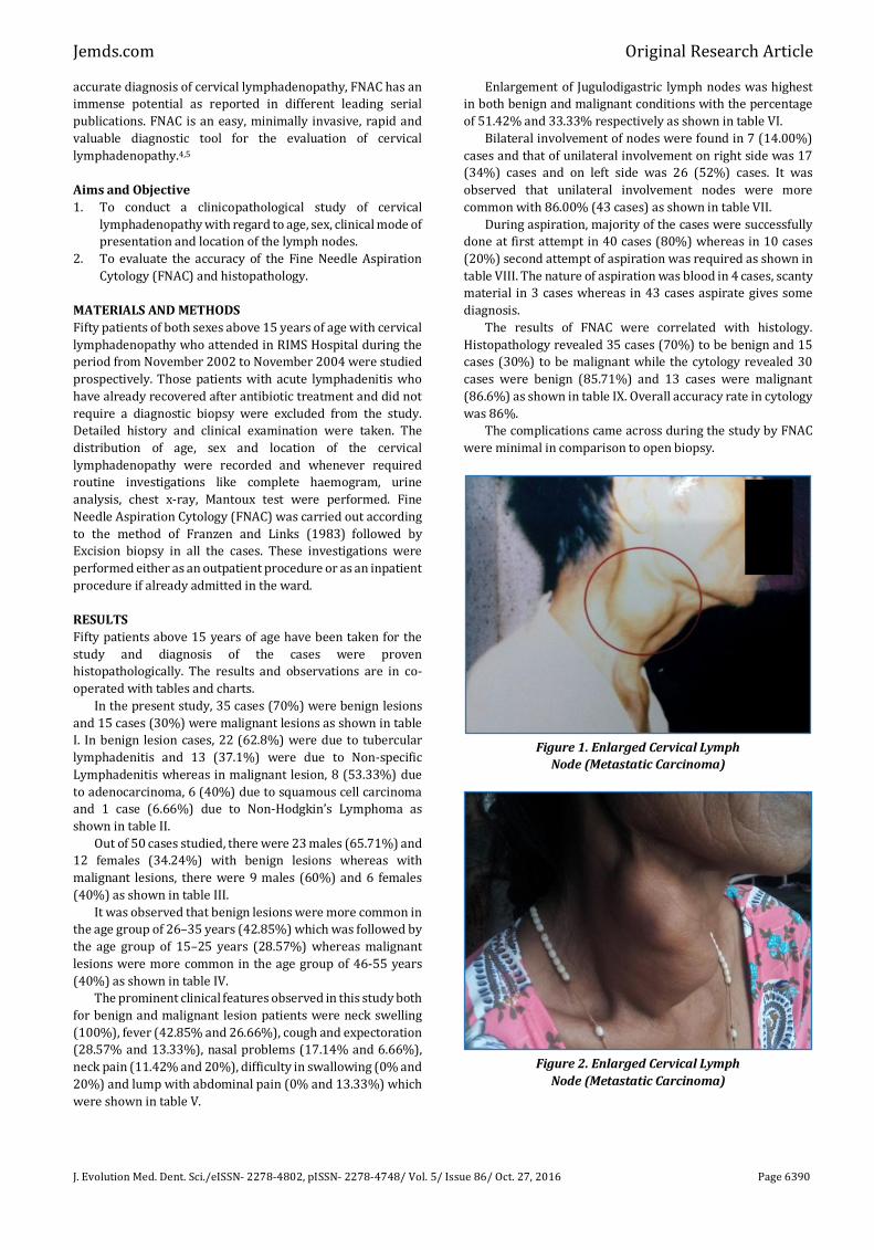
Figure 1. Enlarged Cervical Lymph
Node (Metastatic Carcinoma)
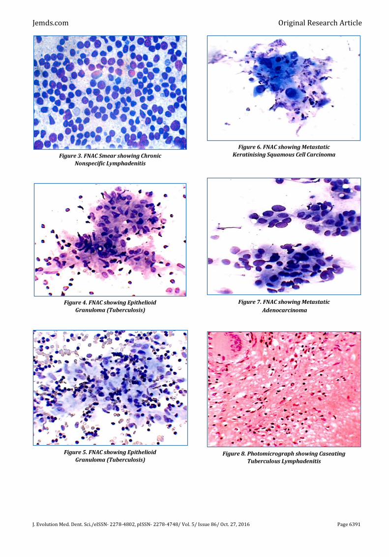
Figure 2. Enlarged Cervical Lymph
Node (Metastatic Carcinoma)
Jemds.com Original Research Article
J. Evolution Med. Dent. Sci./eISSN- 2278-4802, pISSN- 2278-4748/ Vol. 5/ Issue 86/ Oct. 27, 2016 Page 6391
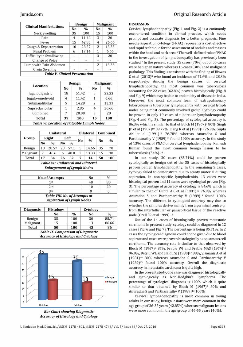
Figure 3. FNAC Smear showing Chronic
Nonspecific Lymphadenitis
Figure 4. FNAC showing Epithelioid
Granuloma (Tuberculosis)
Figure 5. FNAC showing Epithelioid
Granuloma (Tuberculosis)
Figure 6. FNAC showing Metastatic
Keratinising Squamous Cell Carcinoma
Figure 7. FNAC showing Metastatic
Adenocarcinoma
Figure 8. Photomicrograph showing Caseating
Tuberculous Lymphadenitis

Jemds.com Original Research Article
J. Evolution Med. Dent. Sci./eISSN- 2278-4802, pISSN- 2278-4748/ Vol. 5/ Issue 86/ Oct. 27, 2016 Page 6392
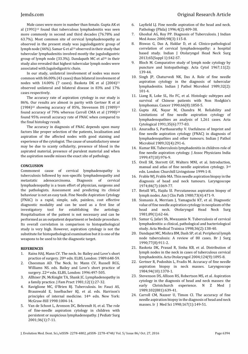
Figure 9. Photomicrograph showing Massive
Caseating Tuberculous Lymphadenitis
Group Type of
Lesions
No. of
Cases Percentage
I Benign 35 70%
II Malignant 15 30%
Total 50 100%
Table I. Distribution of Cases
Proven Histopathologically
Lesions Diagnosis Histology Cytology
No % No %
Benign Tubercular 22 62.8 19 63.3
Non-Specific 13 37.1 11 36.6
Malignant
Squamous Cell
Carcinoma 6 40 4 30.7
Adenocarcinoma 8 53.33 8 61.5
Non-Hodgkin’s
Lymphoma 1 6.66 1 7.6
Table II. Cytology and Histopathology
Correlation of Benign and Malignant Lesions
Bar Chart showing Cytology and
Histology Correlation of Lesions
Sex Benign Malignant
No % No %
Male 23 65.71 9 60
Female 12 34.28 6 40
Total 35 100 15 100
Table III. Sex Distribution
Bar Chart showing Sex Distribution
Age Group
In Years
Benign Malignant Overall
No % No % No %
15-25 10 28.57 0 --- 10 20
26-35 15 42.85 2 13.33 17 34
36-45 8 22.85 4 26.66 12 24
46-55 2 5.71 6 40 8 16
Above 55 --- --- 3 20 3 6
Total 35 100 15 100 50 100
Table IV. Age Distribution
Bar Chart showing Age Distribution
Jemds.com Original Research Article
J. Evolution Med. Dent. Sci./eISSN- 2278-4802, pISSN- 2278-4748/ Vol. 5/ Issue 86/ Oct. 27, 2016 Page 6393
Clinical Manifestations Benign Malignant
No % No % Neck Swelling 35 100 15 100
Pain 4 11.42 3 20 Fever 15 42.85 4 26.66
Cough & Expectoration 10 28.57 2 13.33 Nasal Problem 6 17.14 1 6.66
Difficulty in Swallowing - - 3 20 Change of Voice - - - -
Lump with Pain Abdomen - - 2 13.33 Groin Swelling - - - -
Table V. Clinical Presentation
Location Benign Malignant
No % No %
Jugulodigastric 18 51.42 5 33.33
Jugulo-omohyoid 4 11.42 1 6.66
Submandibular 5 14.28 2 13.33
Supraclavicular 1 2.85 4 26.66
Combined 7 20.00 3 20
Total 35 100 15 100
Table VI. Location of Palpable Lymph Nodes
Group
Unilateral Bilateral Combined
Right Left No % No %
No % No %
Benign 10 28.57 20 57.1 5 14.66 35 70
Malignant 7 46.6 6 40.0 2 13.33 15 30
Total 17 34 26 52 7 14 50 100
Table VII. Unilateral and Bilateral
Enlargement of Lymph Nodes
No. of Attempts No % 1st 40 80 2nd 10 20 3rd 0 0
Table VIII. No. of Attempts at Aspiration of Lymph Nodes
Diagnosis Histology Cytology No % No %
Benign Malignant
35 15
100 100
30 13
85.71 86.66
Total 50 100 43 86 Table IX. Comparison of Diagnostic Accuracy of Histology and Cytology
Bar Chart showing Diagnostic
Accuracy of Histology and Cytology
DISCUSSION
Cervical lymphadenopathy (Fig. 1 and Fig. 2) is a commonly
encountered condition in clinical practice, which needs
prompt and accurate diagnosis for a better prognosis. Fine
needle aspiration cytology (FNAC) represents a cost effective
and rapid technique for the assessment of nodules and masses
within the head and neck area.6 The well–defined role of FNAC
in the investigation of lymphadenopathy has previously been
studied.7 In the present study, 35 cases (70%) out of 50 cases
were benign in nature whereas 15 cases (30%) had malignant
pathology. This finding is consistent with the finding of Biswas
G et al (2013)8 who found an incidence of 71.6% and 28.3%
respectively. Among the benign causes of cervical
lymphadenopathy, the most common was tuberculosis
accounting for 22 cases (62.8%) proven histologically (Fig. 8
and Fig. 9) which may be due to endemicity of disease in India.
Moreover, the most common form of extrapulmonary
tuberculosis is tubercular lymphadenitis with cervical lymph
nodes being most commonly involved group. Cytology could
be proven in only 19 cases of tubercular lymphadenopathy
(Fig. 4 and Fig. 5). The percentage of cytological accuracy is
86.3% which is similar to that of Bolch M (1967)9 80%, Singh
JP et al (1987)10 89.77%, Liang R et al (1990)11 76.9%, Gupta
AK et al (1991)12 76.78% whereas Anuradha S and
Parthasarathy V (1989)13 found 100% accuracy. In the study
of 1396 cases of FNAC of cervical lymphadenopathy, Ramesh
Kumar found the most common benign lesion to be
tuberculosis (54%).14
In our study, 30 cases (85.71%) could be proven
cytologically as benign out of the 35 cases of histologically
proven benign lymphadenopathy. In the remaining 5 cases,
cytology failed to demonstrate due to scanty material during
aspiration. In non-specific lymphadenitis, 13 cases were
histological proven and 11 cases were cytological proven (Fig.
3). The percentage of accuracy of cytology is 84.6% which is
similar to that of Gupta AK et al (1991)12 76.9% whereas
Anuradha S and Partharsarathy V (1989)13 found 100%
accuracy. The different in cytological accuracy may due to
whether the samples derive mainly from a germinal centre or
from the interfollicular or paracortical tissue of the reactive
node (Orell SR et al 1999).15
Out of the 14 cases of histologically proven metastatic
carcinoma in present study, cytology could be diagnosed in 12
cases (Fig. 6 and Fig. 7). The percentage is being 85.71%. In 2
cases the cytological diagnosis could not be given due to blood
aspirate and cases were proven histologically as squamous cell
carcinoma. The accuracy rate is similar to that observed by
Bloch M (1967)9 87%, Frable WJ and Frable MAS (1974)16
96.8%, Betsill WL and Haldu SI (1980)17 89%, Sismanis A et al
(1981)18 80% whereas Anuradha S and Parthasarathy V
(1989)13 found 100% accuracy. Overall the diagnostic
accuracy in metastatic carcinoma is quite high.
In the present study, one case was diagnosed histologically
and cytologically as Non-Hodgkin’s Lymphoma. The
percentage of cytological diagnosis is 100% which is quite
similar to that obtained by Bloch M (1967)9 80% and
Anuradha S and Parthasarathy V (1989)13 100%.
Cervical lymphadenopathy is most common in young
adults. In our study, benign lesions were more common in the
age group of 26-35 years (42.85%) whereas malignant lesions
were more common in the age group of 46-55 years (40%).
Jemds.com Original Research Article
J. Evolution Med. Dent. Sci./eISSN- 2278-4802, pISSN- 2278-4748/ Vol. 5/ Issue 86/ Oct. 27, 2016 Page 6394
Male cases were more in number than female. Gupta AK et
al (1991)12 found that tuberculous lymphadenitis was seen
more commonly in second and third decades (76.78% and
63.7%). Most common site of cervical lymphadenopathy as
observed in the present study was jugulodigastric group of
lymph node (46%). Samar G et al19 observed in their study that
tubercular lymphadenitis involved mostly the jugulodigastric
group of lymph node (33.3%). Dandapath MC et al20 in their
study also revealed that highest tubercular lymph nodes were
associated with Jugulodigastric chain.
In our study, unilateral involvement of nodes was more
common with 86.00% (43 cases) than bilateral involvement of
nodes with 14.00% (7 cases). Baskota DK et al (2004)21
observed unilateral and bilateral disease in 83% and 17%
cases respectively.
The accuracy rate of aspiration cytology in our study is
86%. Our results are almost in parity with Gertner R et al
(1984)22 showing accuracy of 85%, Stevenson DS (1989)23
found accuracy of 79% whereas Carroll CMA et al (1998)24
found 95% overall accuracy rate of FNAC when compared to
the final histology result.
The accuracy in diagnosis of FNAC depends upon many
factors like proper selection of the patients, localisation and
aspiration of the affected nodes with good staining and
experience of the cytologist. The cause of unsatisfactory smear
may be due to scanty cellularity, presence of blood in the
aspirated material, presence of purulent material and when
the aspiration needle misses the exact site of pathology.
CONCLUSION
Commonest cause of cervical lymphadenopathy is
tuberculosis followed by non-specific lymphadenopathy and
metastatic adenocarcinoma. Diagnosis of cervical
lymphadenopathy is a team effort of physician, surgeons and
the pathologists. Assessment and predicting its clinical
behaviour is not an easy task. Fine Needle Aspiration Cytology
(FNAC) is a rapid, simple, safe, painless, cost effective
diagnostic modality and can be used as a first line of
investigatory tool for establishing the aetiology.
Hospitalisation of the patient is not necessary and can be
performed as an outpatient department or bedside procedure.
Its overall correlation in comparison to histopathological
study is very high. However, aspiration cytology is not the
substitute for histopathological examination but it is one of the
weapons to be used to hit the diagnostic target.
REFERENCES
1. Rains HAJ, Mann CV. The neck. In: Bailey and Love’s short practice of surgery. 20th edn. ELBS, London: 1989:648-59.
2. Cheesman AD. The Neck. In: Mann CV, Russell RCG, Williams NS. eds. Bailey and Love’s short practice of
surgery. 22nd edn. ELBS, London: 1996:497-505. 3. Allhiser JN, McKnight TA, Shank JC. Lymphadenopathy in
a family practice. J Fam Pract 1981;12(1):27-32. 4. Raviglione MC, O’Brien RJ. Tuberculosis. In: Fauci AS,
Braunwald E, Isselbacher KJ, et al. eds. Harrison’s
principles of internal medicine. 14th edn. New York: McGraw-Hill 1998:1004-14.
5. Van de Schoot L, Aronson DC, Behrendt H, et al. The role of fine-needle aspiration cytology in children with
persistent or suspicious lymphadenopathy. J Pediatr Surg 2001;36(1):7-11.
6. Layfield LJ. Fine needle aspiration of the head and neck. Pathology (Phila) 1996;4(2):409-38.
7. Ghoshal AG, Roy PP. Diagnosis of Tuberculosis. J Indian Med Assoc 2000;98(3):115-8.
8. Biswas G, Das A, Haldar D, et al. Clinico-pathological correlation of cervical lymphadenopathy: a hospital based study. Indian J Otolaryngol Head Neck Surg
2013;65(Suppl 1):S42-S7. 9. Bloch M. Comparative study of lymph node cytology by
puncture and histopathology. Acta Cytol 1967;11(2): 139-44.
10. Singh JP, Chaturvedi NK, Das A. Role of fine needle aspiration cytology in the diagnosis of tubercular
lymphadenitis. Indian J Pathol Microbiol 1989;32(2): 101-4.
11. Liang R, Loke SL, Ho FC, et al. Histologic subtypes and
survival of Chinese patients with Non Hodgkin’s lymphomas. Cancer 1990;66(8):1850-5.
12. Gupta AK, Nayar M, Chandra M. Reliability and Limitations of fine needle aspiration cytology of
lymphadenopathies an analysis of 1,261 cases. Acta Cytological 1991;35(6):777-83.
13. Anuradha S, Parthasarathy V. Usefulness of Imprint and fine needle aspiration cytology (FNAC) in diagnosis of lymphadenopathies and other tumours. Indian J Pathol
Microbiol 1989;32(4):291-6. 14. Kumar RK. Tuberculosis lymphadenitis in children-role of
fine needle aspiration cytology. J Assoc Physicians India 1999;47(10):976-9.
15. Orell SR, Sterrett GF, Walters MNI, et al. Introduction, manual and atlas of fine needle aspiration cytology. 3rd edn. London: Churchill Livingstone 1999:1-8.
16. Frable WJ, Frable MA. Thin needle aspiration biopsy in the diagnosis of head and neck tumours. Laryngoscope
1974;84(7):1069-77. 17. Betsill WL, Hajdu SI. Percutaneous aspiration biopsy of
lymph nodes. Am J Clin Path 1980;73(4):471-9. 18. Sismanis A, Merriam J, Yamaguchi KT, et al. Diagnostic
value of fine needle aspiration cytology in neoplasm of the head and neck. Otolaryngol Head Neck Surg 1981;89(1):62-66.
19. Samar G, Jafari Sh, Mwazamie N. Tuberculosis of cervical lymphadenitis: a clinical, pathological and bacteriological
study. Acta Medical Trainica 1998;36(2):138-40. 20. Dandapat MC, Mishra BM, Dash SP, et al. Peripheral lymph
node tuberculosis: A review of 80 cases. Br J Surg 1990;77(8):911-2.
21. Baskota DK, Prasad R, Sinha KB, et al. Distribution of lymph nodes in the neck in cases of tuberculous cervical lymphadenitis. Acta Otolaryngol 2004;124(9):1095-8.
22. Gertner R, Podoshin L, Fradis M. Accuracy of fine needle aspiration biopsy in neck masses. Laryngoscope
1984;94(10):1370-1. 23. Stevenson DS, Allison RS, Robertson MS, et al. Aspiration
cytology in the diagnosis of head and neck masses: the early Christchurch experience. N Z Med J
1989;102(881):639-41. 24. Carroll CM, Nazeer U, Timon CI. The accuracy of fine
needle aspiration biopsy in the diagnosis of head and neck
masses. Ir J Med Sci 1998;167(1):149-51.


