Jeffrey Kalish, David Gillespie, Marc Schermerhorn, Daniel ... · Marc Schermerhorn, Daniel...
34
Ultrasound Guidance during Arterial Access for Peripheral Vascular Intervention: A VSGNE Quality Improvement Project On behalf of the Vascular Study Group of New England Jeffrey Kalish, David Gillespie, Marc Schermerhorn, Daniel Bertges, Chris Healey, Paul Bloch
Transcript of Jeffrey Kalish, David Gillespie, Marc Schermerhorn, Daniel ... · Marc Schermerhorn, Daniel...
Ultrasound Guidance during Arterial Access for Peripheral Vascular
Intervention:A VSGNE Quality Improvement Project
On behalf of the Vascular Study Group of New England
Jeffrey Kalish, David Gillespie,Marc Schermerhorn, Daniel Bertges,
Chris Healey, Paul Bloch

Introduction• Local vascular complication = most
frequent adverse outcome from femoral puncture– Groin hematoma– Pseudoaneurysm– Retroperitoneal hematoma– Vessel thrombosis– Arteriovenous fistula
(Koreny M, JAMA 2004;291:350-7)(Tavris DR, J Invasive Cardiol 2012;24:328-34)

Landmarks• Inguinal skin crease = unreliable marker
for puncture– Bifurcation of common femoral artery (CFA) is
cephalad to this crease ~75% of the time• Lechner, Cardiovasc Intervent Radiol 1988
• Fluoroscopy and bony landmarks ↓access site complications vs. palpation– CFA overlies femoral head 92% of the time
• Garrett, Catheter Cardiovasc Interv 2005

Cephalad
Upper Half
Lower HalfCaudal
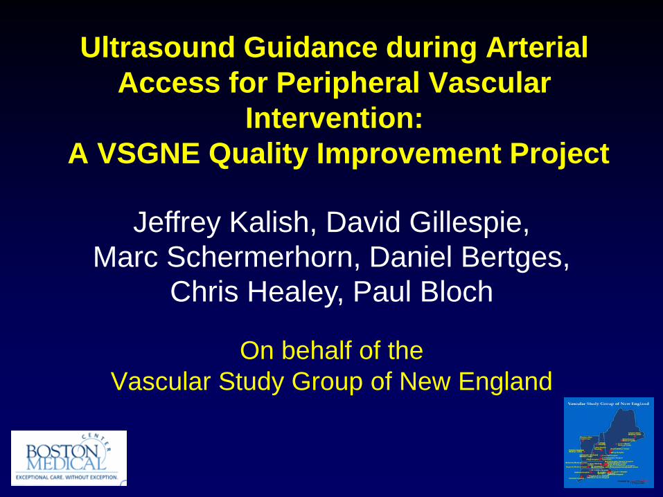
FAUST ResultsFemoral Artery Access with Ultrasound Trial
Fluoroscopy Ultrasound p-valueCFA cannulation 83.3% 86.4% 0.17Sheath in EIA 4.9% 6.6% 0.25Sheath distal to CFA bifurcation
11.8% 7.0% < 0.01Number of attempts
3.0 ± 3.2 1.3 ± 0.9 < 0.001First-pass success 46.4% 82.7% < 0.001Mean time to insertion (sec)
213 ± 194 185 ± 175 < 0.02Hematoma > 5cm 2.2% 0.6% 0.03Any complication 3.4% 1.4% 0.04

FAUST Learning Curve(CFA Cannulation)
83.3%82.4%
83.3%
86.4%
87.6%
80.0%
82.0%
84.0%
86.0%
88.0%
90.0%
Fluoroscopy 4-6 7-10 11-15 15+
Ultrasound Procedures

Published VSGNE Study
1. Utilize the VSGNE database to identify variables associated with groin hematoma after peripheral vascular intervention (PVI)
2. Examine routine vs. selective use of ultrasound guidance in relation to hematoma rates
Kalish J, Eslami M, Gillespie D, et al. J Vasc Surg 2015;epub

Definitions of Hematoma
• includes pseudoaneurysms
• Minor = required compression or observation
• Moderate = required transfusion or thrombin injection
• Major = required operation

Sample Selection• 7359 PVI performed in 6108 patients
– January 2010 to January 2014– 159 interventionalists from 26 academic and
community medical centers– 1 to 239 PVI per interventionalist– 26 to 1043 PVI per medical center
• Percutaneous femoral access– Occlusive disease (excludes aneurysms)– Overall groin hematoma rate = 4.5%
(moderate/major = 0.8%)

Routine vs. Selective Users
• Outcomes of interventionalists based on routine or selective use of ultrasound– 114 interventionalists with ≥ 10 PVI
procedures– Unadjusted and adjusted analyses
• 31 Routine Users (≥ 80%)
• 83 Selective Users (< 80%)
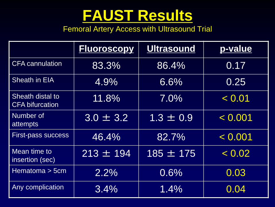
Ultrasound Use

Multivariate Logistic RegressionVariable Odds Ratio 95% CI P-value
Age > 80 2.45 1.31-4.57 0.005
Sheath Size > 6 French 1.62 1.24-2.11 <0.001
Female Gender 1.54 1.22-1.94 <0.001
Bilateral Femoral Access 1.44 1.08-1.93 0.013
Closure Device 0.47 0.37-0.61 <0.001
Timing (Jan-Jun) 0.73 0.58-0.92 0.007
Routine Ultrasound (≥ 80%) 0.74 0.57-0.95 0.02

Updated VSGNE Data• PVI Dataset (Jan 2010 – Feb 2015)
– Femoral access only
• 27 Centers– Hematoma range 0-13%
• 25 Centers with ≥ 10 procedures– Hematoma range 1-13%

0%
2%
4%
6%
8%
10%
12%
14%
0% 10% 20% 30% 40% 50% 60% 70% 80% 90% 100%
Perc
ent H
emat
oma
Percent Usage of Ultrasound
VSGNE Centers
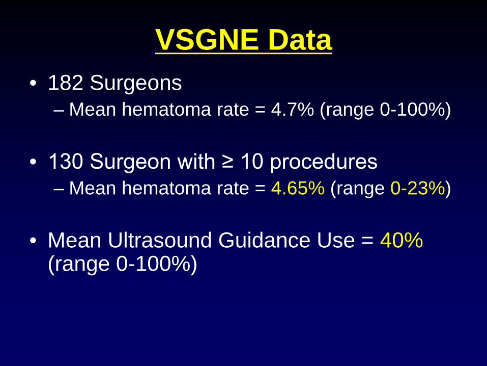
VSGNE Data• 182 Surgeons
– Mean hematoma rate = 4.7% (range 0-100%)
• 130 Surgeon with ≥ 10 procedures– Mean hematoma rate = 4.65% (range 0-23%)
• Mean Ultrasound Guidance Use = 40%(range 0-100%)

VSGNE Data• 23 Routine Users (≥ 80%)
– Mean hematoma rate = 3.7%
• 107 Selective Users (< 80%)– Mean hematoma rate = 4.9%
• VSGNE members given personalized Quality Reports– Hematoma Rate and Ultrasound Guidance

0%
5%
10%
15%
20%
25%
0% 10% 20% 30% 40% 50% 60% 70% 80% 90% 100%
Perc
ent H
emat
oma
Percent Usage of Ultrasound
VSGNE Surgeons
How To Use Ultrasound Guidance

Transverse View
CFV
CFAGSV

Longitudinal View
SFA
PFA
CFA

Step 1: Survey

Step 2: Spread

Step 3: Puncture

Step 3: Puncture

Step 4: Wire

Percutaneous EVAR

CPT Code 76937• Ultrasound guidance for vascular access
requiring ultrasound evaluation of potential access sites, documentation of selected vessel patency, concurrent real-time ultrasound visualization of vascular needle entry, with permanent recording and reporting

Documentation• “Ultrasound guidance was used to access the
right common femoral artery using the micropuncture technique after infiltration of 1% lidocaine for local anesthesia. Ultrasound documented patency of the common femoral artery and vein, as well as the exact location for puncture. Images were stored in PACS for documentation purposes.”
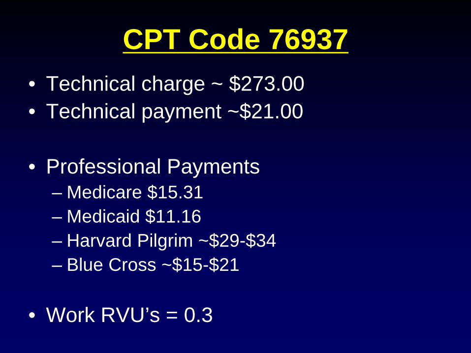
CPT Code 76937• Technical charge ~ $273.00• Technical payment ~$21.00
• Professional Payments– Medicare $15.31– Medicaid $11.16– Harvard Pilgrim ~$29-$34– Blue Cross ~$15-$21
• Work RVU’s = 0.3

Barriers to Adoption1. No U/S machine easily accessible
2. Cost to purchase and maintain U/S machine
3. Time for set-up and access
4. Lack of training
5. Learning curve

Summary• Puncture over upper half of femoral head
is safest access location
• Cephalad punctures ↑ complication rates and should be avoided even if access appears in CFA by ultrasound
• Caudal punctures acceptable if ultrasound identifies needle placement cephalad to femoral bifurcation

Summary• Operator interpretation of images crucial to ↓ complications
• Learning curve necessary to perfect the technique
• Ultrasound ↓ complications from routine percutaneous femoral access, and possibly with “Preclose” for large sheaths in EVAR

Conclusions• Many important risk factors that predict
hematoma formation after femoral arterial access are not modifiable.
• Appropriate use of smaller sheaths, closure devices, and routine ultrasound guidance may potentially protect against hematoma formation.
• Routine use of ultrasound guidance may decrease the risk for hematoma formation for both modifiable and non-modifiable patient/procedural characteristics.
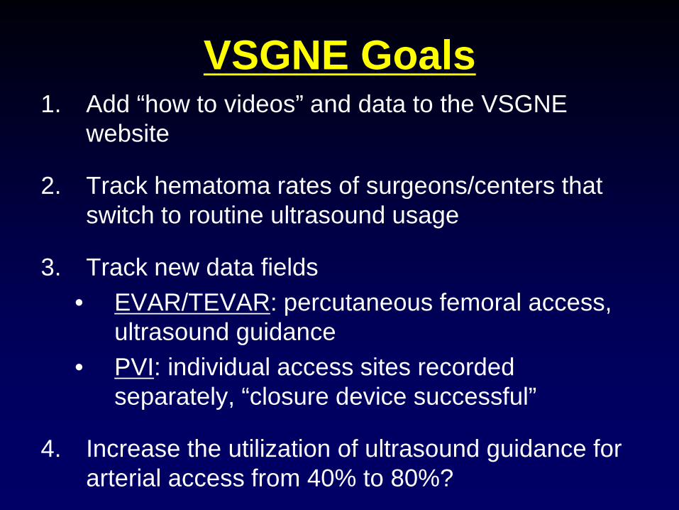
VSGNE Goals1. Add “how to videos” and data to the VSGNE
website
2. Track hematoma rates of surgeons/centers that switch to routine ultrasound usage
3. Track new data fields• EVAR/TEVAR: percutaneous femoral access,
ultrasound guidance• PVI: individual access sites recorded
separately, “closure device successful”
4. Increase the utilization of ultrasound guidance for arterial access from 40% to 80%?