


JBC Papers in Press. Published on January 6, 2010 as ... · 1 A NOVEL MBL/FICOLIN ASSOCIATED...
21
1 A NOVEL MBL/FICOLIN ASSOCIATED PROTEIN IS HIGHLY EXPRESSED IN HEART AND SKELETAL MUSCLE TISSUES AND INHIBITS COMPLEMENT ACTIVATION* Mikkel-Ole Skjoedt 1,2¶ , Tina Hummelshoj 1¶ , Yaseelan Palarasah 2 , Christian Honore 1 , Claus Koch 2 , Karsten Skjodt 2 and Peter Garred 1 1 Laboratory of Molecular Medicine, Department of Clinical Immunology, Sect 7631, Rigshospitalet, Faculty of Health Sciences, University Hospital of Copenhagen, Denmark 2 Research Unit of Immunology and Microbiology, Institute of Medical Biology, Faculty of Health Science, University of Southern Denmark, Odense, Denmark Running title: A novel complement inhibitory serum protein ¶ Mikkel-Ole Skjoedt and Tina Hummelshoj should be considered as joint first authors Address correspondence to: Dr. Peter Garred, Department of Clinical Immunology, Section 7631, Rigshospitalet, Blegdamsvej 9, DK 2100 Copenhagen, Denmark Telephone +45 35457631, Fax +45 35398766, E-mail: [email protected] The human lectin complement pathway involves circulating complexes consisting of mannose-binding lectin (MBL) or three ficolins (-1, -2 and -3) in association with three serine proteases MASPs (-1, -2 and -3) and a non-enzymatic sMAP. MASP-1 and MASP-3 (MASP1 isoform 1 and 2, respectively) are splice variants of the MASP1 gene, while MASP-2 and sMAP are splice variants of the MASP2 gene. We have identified a novel serum protein of 45kDa that is associated with MBL and the ficolins. This protein is named MBL/Ficolin associated protein 1 (MAP-1 corresponding to MASP1 isoform 3). The transcript generating MAP-1 (MASP1_v3) contains exon 1-8 and a novel exon encoding an in-frame stop codon. The corresponding protein lacks the serine protease domains, but contains most of the common heavy chain of MASP-1 and MASP-3. Additionally MAP-1 contains 17 unique C-terminal amino acids. By use of qPCR and MAP-1 specific immunohistochemistry we found that MAP-1 is highly expressed in myocardial and skeletal muscle tissues as well as in liver hepatocytes with a different expression profile from that observed for MASP-1 and MASP-3. MAP-1 co-precipitated from human serum with MBL, Ficolin-2 and Ficolin-3 and recombinant MAP-1 was able to inhibit complement C4 deposition via both the Ficolin-3 and MBL pathway. In conclusion we have identified a novel 45kDa serum protein derived from the MASP1 gene, which is highly expressed in striated muscle tissues. It is found in complex with MBL and ficolins and may function as a potent inhibitor of the complement system in vivo. Activation of the complement system is accomplished via three different initiation pathways: The alternative pathway, the classical pathway, and the lectin pathway (1). In humans, four recognition molecules of the lectin pathway have been described: mannose-binding lectin (MBL), Ficolin-1 (also called M-ficolin), Ficolin- 2 (also called L-ficolin) and Ficolin-3 (also called H-ficolin or Hakata antigen) (2). MBL and the ficolins bind structures on different classes of microorganisms and are involved in sequestration and removal of dying host cells (2,3). Three MBL/Ficolin associated serine proteases have been described so far (MASP-1, MASP-2 and MASP-3) and a protein lacking a serine protease domain named sMAP or MAp19 (4). Present consensus places MASP-2 as the main initiator of the lectin complement pathway by cleaving C4 and C2 to form the C4b2a complex leading to further downstream complement activation (5). Although the functions of the other MASPs are poorly understood, MASP-1 appears to play a role as an amplifier of complement activation (6,7). Additionally, MASP-1 is able to cleave fibrinogen to fibrin, while MASP-2 is able to generate active thrombin by cleavage of prothrombin (8,9). No conclusive biological function has yet been attributed to MASP-3 and sMAP. http://www.jbc.org/cgi/doi/10.1074/jbc.M109.065805 The latest version is at JBC Papers in Press. Published on January 6, 2010 as Manuscript M109.065805 Copyright 2010 by The American Society for Biochemistry and Molecular Biology, Inc. by guest on August 26, 2019 http://www.jbc.org/ Downloaded from
Transcript of JBC Papers in Press. Published on January 6, 2010 as ... · 1 A NOVEL MBL/FICOLIN ASSOCIATED...
1
A NOVEL MBL/FICOLIN ASSOCIATED PROTEIN IS HIGHLY EXPRESSED IN HEART AND SKELETAL MUSCLE TISSUES AND INHIBITS COMPLEMENT ACTIVATION*
Mikkel-Ole Skjoedt1,2¶ , Tina Hummelshoj1¶, Yaseelan Palarasah2, Christian Honore1, Claus Koch2, Karsten Skjodt2 and Peter Garred1
1Laboratory of Molecular Medicine, Department of Clinical Immunology, Sect 7631, Rigshospitalet,
Faculty of Health Sciences, University Hospital of Copenhagen, Denmark 2Research Unit of Immunology and Microbiology, Institute of Medical Biology, Faculty of Health
Science, University of Southern Denmark, Odense, Denmark Running title: A novel complement inhibitory serum protein
¶Mikkel-Ole Skjoedt and Tina Hummelshoj should be considered as joint first authors Address correspondence to: Dr. Peter Garred, Department of Clinical Immunology, Section 7631,
Rigshospitalet, Blegdamsvej 9, DK 2100 Copenhagen, Denmark Telephone +45 35457631, Fax +45 35398766, E-mail: [email protected]
The human lectin complement pathway involves circulating complexes consisting of mannose-binding lectin (MBL) or three ficolins (-1, -2 and -3) in association with three serine proteases MASPs (-1, -2 and -3) and a non-enzymatic sMAP. MASP-1 and MASP-3 (MASP1 isoform 1 and 2, respectively) are splice variants of the MASP1 gene, while MASP-2 and sMAP are splice variants of the MASP2 gene. We have identified a novel serum protein of 45kDa that is associated with MBL and the ficolins. This protein is named MBL/Ficolin associated protein 1 (MAP-1 corresponding to MASP1 isoform 3). The transcript generating MAP-1 (MASP1_v3) contains exon 1-8 and a novel exon encoding an in-frame stop codon. The corresponding protein lacks the serine protease domains, but contains most of the common heavy chain of MASP-1 and MASP-3. Additionally MAP-1 contains 17 unique C-terminal amino acids. By use of qPCR and MAP-1 specific immunohistochemistry we found that MAP-1 is highly expressed in myocardial and skeletal muscle tissues as well as in liver hepatocytes with a different expression profile from that observed for MASP-1 and MASP-3. MAP-1 co-precipitated from human serum with MBL, Ficolin-2 and Ficolin-3 and recombinant MAP-1 was able to inhibit complement C4 deposition via both the Ficolin-3 and MBL pathway. In conclusion we have identified a novel 45kDa serum protein derived from the MASP1 gene, which is highly expressed in striated muscle tissues. It is found in complex
with MBL and ficolins and may function as a potent inhibitor of the complement system in vivo. Activation of the complement system is accomplished via three different initiation pathways: The alternative pathway, the classical pathway, and the lectin pathway (1). In humans, four recognition molecules of the lectin pathway have been described: mannose-binding lectin (MBL), Ficolin-1 (also called M-ficolin), Ficolin-2 (also called L-ficolin) and Ficolin-3 (also called H-ficolin or Hakata antigen) (2). MBL and the ficolins bind structures on different classes of microorganisms and are involved in sequestration and removal of dying host cells (2,3). Three MBL/Ficolin associated serine proteases have been described so far (MASP-1, MASP-2 and MASP-3) and a protein lacking a serine protease domain named sMAP or MAp19 (4). Present consensus places MASP-2 as the main initiator of the lectin complement pathway by cleaving C4 and C2 to form the C4b2a complex leading to further downstream complement activation (5). Although the functions of the other MASPs are poorly understood, MASP-1 appears to play a role as an amplifier of complement activation (6,7). Additionally, MASP-1 is able to cleave fibrinogen to fibrin, while MASP-2 is able to generate active thrombin by cleavage of prothrombin (8,9). No conclusive biological function has yet been attributed to MASP-3 and sMAP.
http://www.jbc.org/cgi/doi/10.1074/jbc.M109.065805The latest version is at JBC Papers in Press. Published on January 6, 2010 as Manuscript M109.065805
Copyright 2010 by The American Society for Biochemistry and Molecular Biology, Inc.
by guest on August 26, 2019
http://ww
w.jbc.org/
Dow
nloaded from

2
MASP-1 and MASP-2 were originally named after their association with MBL (5,10). Subsequently, MASP-3 and sMAP (also named Map19) were identified as alternative splicing variants of the MASP1 and MASP2 genes, respectively. However, the names of the MASPs are confusing since they do not correspond to the actual gene name. According to the nomenclature suggested by the human genome organisation (HUGO) the proteins should have been named MASP1 isoform 1 (MASP-1), MASP1 isoform 2 (MASP-3), MASP2 isoform 1 (MASP-2) and MASP2 isoform 2 (sMAP or Map19). These named relates to the splice variants MASP1_v1, MASP1_v2, MASP2_v1 and MASP2_v2, respectively. However, the terms MASP-1, MASP-2 and MASP-3 and sMAP are commonly used in the literature. The MASP1 gene is located on chromosome 3q27-q28. MASP-1 (MASP1 isoform 1) and MASP-3 (MASP1 isoform 2) contain an identical heavy chain except for 15 C-terminal residues. The heavy chain is comprised of two C1r/C1s, Urchin-EGF, Bone morphogenetic protein (CUB) domains separated by an Epidermal Growth Factor (EGF) domain and followed by two complement control protein domains (CCPs). The light chain contains the serine protease domain, which is different for MASP-1 and MASP-3 (11). MASP-1 is primarily expressed in the liver, while a more broad tissue expression pattern is observed for MASP-3 (12). The MASP2 gene is located on chromosome 1p36-p36.2. MASP-2 (MASP2 isoform 1) has a similar modular structure as MASP-1 and MASP-3 containing a heavy chain of two CUB domains, an EGF domain and two CCP domains. The light chain is a MASP-2 unique serine protease domain, while sMAP (MASP2 isoform 2) is a truncated form lacking the serine protease domain and a major part of the heavy chain. Expression of the two MASP2 isoforms is predominantly localized to the liver. In this report we describe the identification of a novel differential spliced variant (MASP1_v3) of the MASP1 gene that is found as a serum protein with a molecular weight of 45 kDa. This protein lacks the second CCP domain and the entire serine protease domain, but contains 17 unique C-terminal amino acid residues. The protein is
found in association with MBL and ficolins and down regulates activation of complement via the lectin pathway. We propose to use the term MBL/Ficolin associated protein 1 (MAP-1) in addition to MASP1 isoform 3. This will indicate that the protein is not a serine protease, is derived from the MASP1 gene and will not be mistaken as MASP-3.
EXPERIMENTAL PROCEDURES
Alignment of the MASP1 gene and its splice variants-The National Centre for Biotechnology Information (NCBI) Entrez databases were searched for alternative transcript sequences of MASP1. Three splice variants were identified with the accession numbers NM001879, NM139125, and NM001031849 corresponding to MASP1_v1, MASP1_v2, and MASP1_v3, respectively. Sequences were aligned using BioEdit Software (v7.0.9.0). Real-time quantitative PCR analysis-Commercially available normalized human tissue cDNA panels (Clontech) were investigated for MASP1_v1, MASP1_v2, and MASP1_v3 expression by real-time quantitative PCR (qPCR) analysis on a Stratagene Mx2005 platform. Taqman probes were labelled with 5´-FAM reporter dye and 3´-TAMRA quencher dye. The reactions were performed with 1×Taqman Universal PCR Master Mix, 0.4 µM of each primer and 0.25 µM Taqman probe. The cycling parameters were 2min50°C, 10min95°C, 45cycles (15sec95°C, 1min60°C). All samples were measured in duplicates in three individual experiments on different plates using 0.7 ng cDNA in a total volume of 10 µl. The amplifications were normalized to BCR, GAPDH and beta-actin. Data were corrected for differences in amplification efficiencies. The relative gene expression was calculated with reference to MASP1_v3 expression in heart (100*2^(deltaCt(MASP1_v3heart) – deltaCt(x)), where x is any of the other tissues). DNA sequencing of the MASP1_v3 exon 9 in 100 healthy individuals-Sequencing of the exon 9 of the MASP1 gene spanning from position +44,083 to +44,431 relative to the translation ATG start site (NCBI accession number NC_000003), was performed on genomic DNA templates from 100
by guest on August 26, 2019
http://ww
w.jbc.org/
Dow
nloaded from

3
healthy Caucasian individuals. The fragment was amplified by using a single primer set (table 1), where the forward primers contained a 5´-T7 sequence (5´-ttatacgactcacta-3´). PCR was performed in 20-µl volumes containing 2 ng liver cDNA (Clontech), 0.25 µM of each primer, 2.5 mM MgCl2, 0.2 mM dNTP, 50 mM KCl, 10 mM Tris·HCl, pH 8.4, and 0.4 units of Platinum Taq DNA polymerase (Invitrogen) and run at the following cycling parameters: 2min94°C, 15 cycles (30sec94°C, 60sec64°C, 60sec72°C), 15 cycles (30sec94°C, 60sec58°C, 60sec72°C), 5min72°C. The products were sequenced in the forward direction using the ABI BigDye cycle sequencing terminator kit (Applied Biosystems) according to the manufacturer’s protocol using 5´-biotinylated T7-sequence primers. Sequence reactions were purified on a PyroMark Vacuum Prep Workstation (Biotage) using streptavidin beads (GenoVision) and analyzed on an ABI Prism 3100 Genetic Analyzer (Applied Biosystems). Recombinant proteins-Expression of rMASP-1, rMASP-3, rMAP-1 (MASP1 isoform 1, 2 and 3, respectively), rMASP-2 (MASP2 isoform 1), rMBL and rFicolin-3 in CHO cells was carried out as described elsewhere (13,14). Generation of monoclonal and polyclonal antibodies-BALB/c×NMRI mice and rabbits were immunized subcutaneously three times with 25 µg of a MAP-1 specific peptide (CKKNEIDLESELKSEQVTE) coupled to diphtheria toxoid adsorbed to Al(OH)3, and mixed in 1:1 ratio with Freund’s incomplete adjuvant. The peptide contains the 17 C-terminal amino acids and additional two natural N-terminal residues lysine and cysteine (the latter used for coupling). The coupling was conducted using the EMCS coupling reagents (Pierce) according to the manufacturer’s recommendations. The fusion and selection of the antibodies was done essentially as described previously (15). Selected monoclonal antibodies were purified from culture supernatant by protein A affinity chromatography using the Äkta FPLC system according to the manufacturer’s instructions (GE Healthcare). The monoclonal antibody (mAb) 20C4 was used for immunoprecipitation, immunoaffinity purification and immunoblotting. The mAb
12B11 was used for the immunohistochemistry analysis. Rabbit polyclonal antibodies (pAb) to MAP-1 were affinity purified using a column coupled with human serum albumin to which the 17-residue MAP-1 specific peptide had been conjugated. ELISA specificity-Enzyme-linked immunosorbent assay (ELISA) was used to validate the specificity of the MAP-1 antibodies. The rMASP-1, -3 and rMAP-1 was immobilized to Maxisorp ELISA plates (Nunc, Denmark) in two-fold serial dilutions starting at 5 µg/ml. After washing/blocking in PBS, 0.05% Tween, a mAb to MAP-1 (mAb 20C4) or an antibody that reacts with the common heavy chain of MASP-1 and -3 (mAb 8B3) was applied at 5 µg/ml. Secondary detection was performed with HRP-Rabbit anti mouse IgG (PO260, Dako, Denmark) diluted 1:1000 in PBS, 0.05% Tween20. SDS-PAGE and immunoblotting-Electrophoresis was performed on 10% or 4–12% (w/v) Bis-Tris Polyacrylamide-gels with discontinuous buffers using the NuPAGE system (Invitrogen) as recommended by the manufacturer. Immunoblotting was performed using polyvinylidene difluoride membranes (PVDF-HyBond, GE Healthcare), 2 µg/ml of primary mAbs and secondary visualization by HRP conjugated streptavidin (P0397, Dako) diluted 1:1500 or HRP-Rabbit anti mouse IgG (PO260, Dako) diluted 1:1000 in PBS, 0.05% Tween20. The membranes were developed with 3-amino-9-ethylcarbazole (Sigma-Aldrich) (0.04% in acetone) and 0.015% H2O2 in 50 mM sodium acetate buffer pH 5. Immunoprecipitation-Immunoprecipitation of MAP-1 from serum was performed with the MAP-1 specific mAb 20C4 or mAb 8B3 (a monoclonal antibody reacting against the common heavy chain of MASP-1 and MASP-3) as a positive control. Additionally an IgG mAb antibody (IgG1κ) with no known specificity was applied as a negative control. A total of 10 µg of mAb 20C4, 8B3 or IgG1κ was allowed to bind to sheep anti mouse IgG Dynabeads (M-280, cat. 112.02D, Dynal/Invitrogen). After a washing step the beads were applied to a pool of normal human serum (diluted 1:1 in TBS) and incubated
by guest on August 26, 2019
http://ww
w.jbc.org/
Dow
nloaded from

4
end over end for 1 hour at 4°C. After the final washing steps and magnetic separation the beads were boiled in SDS loading buffer and subjected to SDS-PAGE and immunoblotting probed with biotinylated antibodies to MAP-1, MBL or Ficolin-3. The same precipitation procedure as described above was performed with mAbs to MBL (Hyb 131-11, Bioporto), Ficolin-2 (FCN219) (16) and Ficolin-3 (FCN334) (17). To compensate for differences in serum concentrations of MBL, Ficolin-2 and -3, we precipitated from 1 ml, 300 µl and 100 µl serum, respectively. Samples were analyzed by SDS-PAGE and immunoblotting, probed with a biotinylated antibody to MAP-1. Immunohistochemistry-CHO cells expressing rMAP-1 were grown in culture flasks in RPMI-1640 with 10% fetal calf serum. Cells were harvested at 80-90% confluence and fixed for 24h in 4% formaldehyde-PBS and subsequently embedded in paraffin. Six different human liver tissues and samples from two different myocardial tissues, two skeletal muscle tissues and two samples obtained from human aorta were also fixed and paraffin embedded as described above. Sections of 5 µm slices were obtained with a Leitz Wetzlar microtome and placed on glass slides and stored at 4°C until assayed. Pre-treatments and analysis were performed as described previously (18). Primary antibodies against MAP-1 were the mAb 12B11 or the affinity purified pAb MAP-1 both diluted to 5 µg/ml. Isotype antibody controls were applied to the tissues at the same concentration. Secondary antibody was EnVision™ antibody (HRP-anti mouse or HRP-anti rabbit, Dako). Analysis of staining patterns was conducted under a Leica DMLB2 microscope. Complement activation assay-The influence of MAP-1 on the MBL and Ficolin-3 mediated complement factor C4 deposition was assessed essentially as described previously (19,20). Briefly, mannan (MBL ligand) (Sigma-Aldrich M7504) or acetylated bovine serum albumin (AcBSA) (Ficolin-3 ligand) was immobilized to Maxisorp ELISA plates (Nunc) at 10 µg/ml. After washing, rMBL or rFicolin-3 (0.4 µg/ml) was added and incubated for 1.5 hour. The rMAP-1 or rMASP-2 was applied for 1 hour in
two-fold serial dilutions in the first dimension followed by incubation for 45 min at 37°C with serial dilutions of serum deficient of MBL (21) or Ficolin-3 (20) in the second dimension. The C4 deposition was measured using a polyclonal antibody to C4c (Q0369, Dako). In addition we assessed the displacement of MASP-2 with MAP-1 using a pure system. The rMASP-2 was pre-incubated for 45 min at 20°C in serial dilutions in the first dimension on an rMBL/mannan matrix as described above followed by incubation with dilutions of rMAP-1 in the second dimension for 45 min at 20°C. Purified C4 (Quidel) was added at a concentration of 1 µg/ml and incubated for 45 min at 37°C. Detection was conducted as above.
RESULTS
Alignment of the transcript variants of MASP1-We searched the NCBI Entrez database for alternative transcript sequences of MASP1 and identified a novel mRNA sequence (MASP1_v3). The corresponding protein “MASP-1 isoform 3” was named MAP-1 for MBL/Ficolin associated protein 1. Alignment analysis of the nucleotide sequences of the different MASP1 transcript variants (fig. 1 and fig. 2) demonstrated that MASP1_v3 only shares exon 1 to exon 8 with the other two splice variants of MASP1. These exons encode most of the common heavy chain. However, due to an extra exon (exon 9) containing a stop codon, this molecule does not have a serine protease domain (fig. 2). The corresponding MAP-1 protein sequence encodes 380 amino acids whereas MASP-1 and MASP-3 contains 699 and 728 amino acids, respectively (fig. 1). By alignment analysis of the deduced amino acid sequences we found that MAP-1 lacks the second CCP domain and the entire serine protease domain, but contains 17 unique C-terminal amino acid residues (fig. 1). We calculated the theoretical molecular mass based on the deduced amino acid sequence to be 43.6 kDa. Tissue distribution of human MASP1_v1, MASP1_v2 and MASP1_v3 mRNA using real-time quantitative PCR analysis-The expression of MASP1_v1, MASP1_v2, and MASP1_v3 transcripts in tissues were investigated by end
by guest on August 26, 2019
http://ww
w.jbc.org/
Dow
nloaded from

5
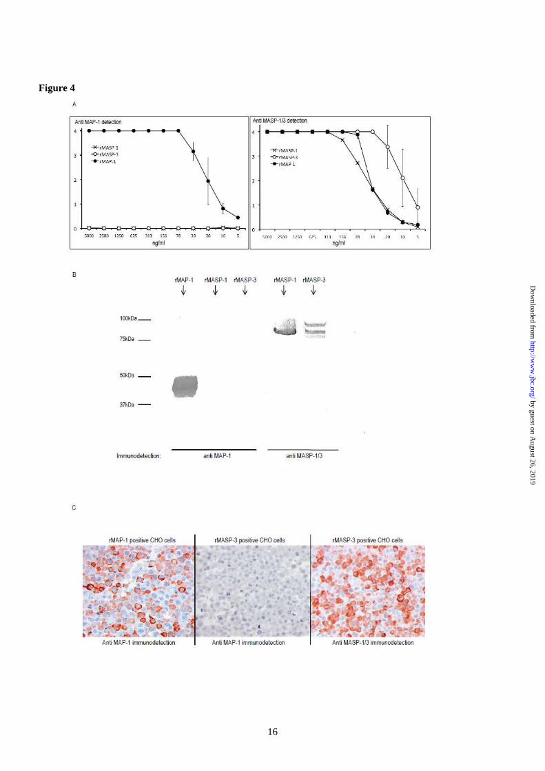
point PCR analysis on cDNA panels from Clontech (data not shown). High mRNA levels of all three transcript variants were detected in the liver. Furthermore, MASP1_v3 was strongly expressed in heart tissue and lower mRNA level of MASP1_v3 was detected in skeletal muscle, brain, colon, prostate, and small intestine. No detectable mRNA levels were detected in bone marrow, cecum, duodenum, oesophagus, jejunum, kidney, lung, lymph, ovary, pancreas, placenta, spleen, stomach, testis, thymus, or tonsil. Based on the initial end point PCR analysis, tissues with MASP1_v1, MASP1_v2 or MASP1_v3 expression were selected for further investigations using a new independent cDNA library. For quantification we used real-time quantitative PCR analysis (qPCR). Confirming the previous observations that high levels of MASP1_v1 and MASP1_v2 mRNA mainly was found in liver tissue (fig. 3), but a minor extra-hepatic expression of MASP1_v2 was also detected in all the investigated tissues, corresponding to approximately 1-5% of that observed in the liver (data not shown). The mRNA concentration of MASP1_v3 was higher in liver tissue compared to MASP1_v1 and MASP1_v2 (fig. 3). However, of particular interest was the finding that the MASP1_v3 mRNA level was expressed about two times higher in skeletal muscle and about four times higher in the heart tissue compared with the relative mRNA concentration in the liver. DNA sequencing of the MASP1 exon 9 in 100 individuals-To investigate a possible genetic variation of the specific exon of the MASP1_v3 transcript, we sequenced the MASP1_exon 9 in 100 healthy Caucasian individuals. No genetic variations were observed in this exon (data not shown). Antibody specificities-We used enzyme-linked immunosorbent assay (ELISA), immunoblotting and immunohistochemistry to validate the specificities of the antibodies used. Fig. 4A, left side, shows the reaction pattern of anti MAP-1 mAb 20C4 to rMASP-1, rMASP-3 and rMAP-1 immobilized to an ELISA plate in serial dilutions. No cross-reactivity to rMASP-1 or rMASP-3 was observed. An antibody to the
common heavy chain of MASP-1 and MASP-3 was used as a positive control (4A, right side). Additionally no cross-reactivity of mAb 20C4 to rMASP-1 and rMASP-3 was observed in SDS-PAGE/immunoblotting (fig. 4B). CHO cells expressing either rMASP-3 or rMAP-1 were fixed in formaldehyde and embedded in paraffin and used to screen for specificity in immunohistochemistry. The anti MAP-1 antibody showed a positive staining of the MAP-1 positive cells and no cross-reactivity to the MASP-3 cells was evident (fig. 4C). The anti MASP-1/3 mAb 8B3 served as a control for the immunohistochemical staining of the MASP-3 positive cells. Tissue localization of MAP-1-Immunohistochemistry was used to confirm the qPCR analysis and to identify specific cells and tissue areas positive for MAP-1. We analyzed samples from different individuals (two myocardial samples, two skeletal muscle samples, six liver samples and two aortic samples). Strong staining with the anti MAP-1 monoclonal antibody was observed in myocardium/cardiac muscle fibres, skeletal muscle fibres and liver hepatocytes, but no staining was seen in aortic tissue (fig. 5, left panel). This staining pattern was further validated using polyclonal anti-MAP-1 antibodies (data not shown). Difference in the inter-individual staining intensity of the tissues was observed (data not shown). No staining was evident when the tissues were analyzed with isotype control antibodies (fig. 5, right panel). MAP-1 co-precipitates with Ficolin-2, Ficolin-3 and MBL-To investigate a possible association between MAP-1 and MBL and Ficolin-3 we precipitated serum complexes using both an antibody to MAP-1 (mAb 20C4) and an antibody against the common heavy chain of MASP-1 and MASP-3 (mAb 8B3). An IgG1κ isotype antibody served as negative control. The precipitates were subsequently analyzed by reduced SDS-PAGE and immunoblotting detected with biotinylated antibodies to MAP-1, MBL and Ficolin-3, respectively. We observed pronounced Ficolin-3 co-precipitation bands but weaker bands were also seen with MBL (fig. 6A). The samples were not probed with antibodies against Ficolin-2 since they were not applicable for reduced
by guest on August 26, 2019
http://ww
w.jbc.org/
Dow
nloaded from
6
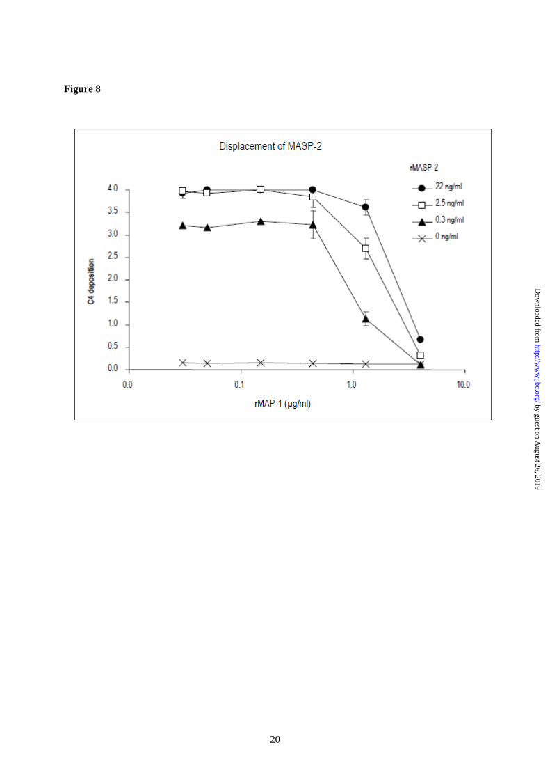
immunoblotting. We then reversed the immunoprecipitation using mAbs against MBL, Ficolin-2 and Ficolin-3 to precipitate serum complexes from 1 ml, 300 µl and 100 µl serum, respectively. The different serum volumes were chosen to adjust for differences in the serum concentration of MBL (2 µg/ml), Ficolin-2 (5 µg/ml) and Ficolin-3 (20 µg/ml), respectively. The samples were subsequently analyzed by immunoblotting probed with a biotinylated antibody to MAP-1. Distinct MAP-1 bands were observed in the precipitates from Ficolin-2 and Ficolin-3 and a slightly weaker band was apparent in the MBL precipitate (fig. 6B, left side). Immunoprecipitated serum MAP-1 and rMAP-1 served as positive controls (fig. 6B, right side). No MAP-1 band was evident when the IgG1κ isotype antibody was used as precipitation mAb. MAP-1 inhibits complement activity of the lectin pathway-The rMBL (13) and rFicolin-3 (14) were used to reconstitute the complement activity in serum deficient of MBL (21) or Ficolin-3 (20), respectively. Mannan and acetylated BSA served as ligands for MBL and Ficolin-3. Both rMBL and rFicolin-3 were able to initiate C4 deposition in the MBL and Ficolin-3 deficient sera, respectively (fig. 7A and 7D). Preincubation of rMBL or rFicolin-3 with serial dilutions of rMASP-2 resulted in a strong dose dependent enhancement of the C4 deposition via both the MBL and Ficolin-3 activation pathways (fig. 7B and 7E), whereas preincubation of rMBL or rFicolin-3 with rMAP-1 resulted in a pronounced dose dependent inhibition of the C4 deposition via both pathways (fig. 7C and 7F). In addition we addressed a possible displacement of MASP-2 with MAP-1 using a system of pure components comprising only of rMBL, rMASP-2, rMAP-1 and purified C4. The rMASP-2 was pre-incubated with mannan/rMBL complexes in serial dilutions. Thereafter, rMAP-1 was added in varying concentrations followed by addition of purified C4. Application of rMAP-1 to the system clearly reduced the levels of C4 deposition (fig. 8).
DISCUSSION We discovered an unidentified protein band with an apparent molecular mass of 45 kDa when ficolins or MBL were precipitated from human serum and subsequently subjected to immunoblotting probed with antibodies against the common heavy chain of the MASP-1 and MASP-3 (MASP1 isoform 1 and 2, respectively) (data not shown). This prompted us to search the databases for alternative transcript sequences of the MASP1 gene. A novel MASP1 derived transcript was found containing exons 1-8 that encodes most of the common heavy chain shared with MASP1_v1 and MASP1_v2. This transcript did not contain exons 10-18 encoding the second CCP domain and both serine protease domains of MASP1 and MASP3 (fig. 2). However, the transcript contained an until now unidentified exon (exon 9) encoding 17 unique C-terminal amino acid residues and an in-frame a stop codon. The transcript was named MASP1_v3 and the corresponding protein MAP-1 (MASP1 isoform 3). Performing end point PCR analysis on cDNA libraries we found that the MASP1_v3 transcript was expressed in different tissues including liver, brain, colon, prostate, heart and skeletal muscles. This was different from the expression of MASP1_v1 and MASP1_v2, which were mainly expressed in the liver. Based on the initial results, tissues with positive signal of MASP1_v1, MASP1_v2 or MASP1_v3 transcripts were selected for further quantitative expression analysis on a new cDNA library. mRNA levels of MASP1_v1 and MASP1_v2 were mainly found in liver tissue. Nevertheless, minor extra-hepatic expression of MASP1_v2 was also detected in all the investigated tissues, corresponding to approximately to 1-5% of that observed in the liver. The MASP1_v3 mRNA level was higher in liver tissue compared to MASP1_v1 and MASP1_v2. Surprisingly the highest concentration of MASP1_v3 mRNA was found in heart tissue followed by skeletal muscle tissue indicating that striated muscle tissue is a major source of MASP1_v3. We have previously observed some differences in the expression pattern between MASP1_v1 and MASP1_v2 suggesting that a differential genetic regulation of these two splicing forms may exist (12).
by guest on August 26, 2019
http://ww
w.jbc.org/
Dow
nloaded from
7
Alternative splicing is widely accepted as an important mechanism for generating genetic diversity and regulation of gene expression (22). Alternative promoter usage has also been correlated with downstream alternative splicing events, differential expression levels and different tissue specificity even in the absence of protein isoforms (23). Alternative pre-mRNA splicing and the differential inclusion or exclusion of portions of a nascent transcript into the final protein-coding mRNA is widely recognized to be a ubiquitous mechanism for controlling protein expression (22). Differences in the activities and/or specificities of splicing factors and regulators of transcription may thus well explain the different mRNA levels and tissue distribution of MASP1_v3 compared with the MASP1_v1 and MASP1_v2 variants. MASP1_v3 transcription could also be initiated by a separate MASP1_v3 promoter in analogy with what has been observed for the MBL2 gene encoding the MBL protein sequence, which is initiated by alternative promoters (12,24). However, none of these theories are mutually exclusive. Future studies will hopefully reveal the molecular origin behind the different expression pattern of the different MASP1 transcripts. In order to characterize MAP-1, we generated both monoclonal and mono-specific polyclonal antibodies raised against a peptide comprising the 17 unique C-terminal amino acids of MAP-1. When using MAP-1 specific antibodies in a panel of paraffin embedded tissues of myocardial, skeletal muscle, liver and aortic tissue we could clearly demonstrate that MAP-1 is ubiquitously present in myocardial myocyte fibrils, while a more restricted patchy staining pattern was observed in skeletal muscle fibrils. The latter could indicate that MAP-1 is induced by muscle contractions. By contrast no MAP-1 staining could be observed in aortic tissue indicating that this isoform is expressed in striated muscle tissue and not in smooth muscle tissue, which are indeed fundamentally different in terms of structure, function, excitation-contraction coupling, and mechanisms of contraction. We have shown that MAP-1 is present in serum as a 45 kDa protein in complex with MBL, Ficolin-2, and Ficolin-3 based on different
immunoprecipitation experiments, which were visualized by SDS-PAGE and immunoblotting. These experiments clearly demonstrate that MAP-1 is a novel serum protein associated with the lectin pathway initiator molecules. Recently, crystal structures of the interaction between MASP-1 and MASP-3 with MBL and the ficolins revealed that the CUB1-EGF-CUB2 domains are responsible for the binding of MASP-1 and MASP-3 to MBL and ficolin (25). Therefore it is reasonable to assume that these domains are also responsible for the interactions between MAP-1 and MBL or ficolin. Furthermore, the main initiator of the lectin complement pathway, MASP-2 has similar CUB1-EGF-CUB2 domains that also interacts with MBL and ficolin (26,27). The serine protease domain of MASP-2 is responsible for the cleavage of C4 and C2 leading to further downstream complement activation (5). Thus, we hypothesized that MAP-1 could have a potentially regulatory role in the lectin complement pathway. When complexes of rMAP1 with rMBL and rFicolin-3 was incubated with MBL- or Ficolin-3-deficient serum, respectively it was clear that rMAP-1 exert a strong dose dependent inhibitory effect on the complement C4 deposition, while recombinant rMASP-2 augmented this deposition. To further investigate whether rMAP-1 could displace rMASP-2 we used a serum free system employing recombinant MBL, MASP-2, MAP-1, and purified C4. The rMAP-1 was able to almost completely inhibit the C4 deposition in spite of the fact that it was added in sequence after MASP-2, thus indicating that it could displace rMASP-2 from rMBL. This is in agreement with results demonstrating that the same amino acid residues in MBL are essential not only for MASP-1 and MASP-3 binding, but also MASP-2 binding (28). Taken together these results indicate that MAP-1 functions as a potent systemic regulator of the complement system in vivo together with C1 inhibitor and C4 binding protein. The presence of MAP-1 in striated muscle tissues may also indicate that MAP-1 could function as a local inhibitor of complement activation. Because of the tissue expression pattern, we cannot exclude the possibility that MAP-1 may have other functions outside the complement system yet to be clarified.
by guest on August 26, 2019
http://ww
w.jbc.org/
Dow
nloaded from
8
In conclusion we describe the identification of a novel differential spliced gene product of the MASP1 gene named MAP-1 (or MASP1 isoform 3), which is found as a serum protein with a molecular mass of 45 kDa. MAP-1 lacks the second CCP domain and the entire serine protease domain, but contains 17 unique C-
terminal amino acid residues. MAP-1 is found in association with MBL and ficolins and is highly expressed in myocardial and skeletal muscle tissues and also in liver hepatocytes. MAP-1 inhibited complement deposition by MBL and Ficolin-3 and appears to be a novel regulator of complement activation via the lectin pathway.
REFERENCES
1. Walport, M. J. (2001) N Engl J Med 344, 1058-1066 2. Endo, Y., Matsushita, M., and Fujita, T. (2007) Immunobiology 212, 371-379 3. Turner, M. W. (2003) Mol. Immunol. 40, 423-429 4. Fujita, T. (2002) Nat. Rev. Immunol. 2, 346-353 5. Thiel, S., Vorup-Jensen, T., Stover, C. M., Schwaeble, W., Laursen, S. B., Poulsen, K., Willis,
A. C., Eggleton, A. C., Hansen, S., Holmskov, U., Reid, KB. M., and Jensenius, J. C. (1997) Nature 386, 506-510
6. Chen, C. B. and Wallis, R. (2004) J Biol. Chem. 279, 26058-26065 7. Takahashi, M., Iwaki, D., Kanno, K., Ishida, Y., Xiong, J., Matsushita, M., Endo, Y., Miura, S.,
Ishii, N., Sugamura, K., and Fujita, T. (2008) J Immunol 180, 6132-6138 8. Krarup, A., Gulla, K. C., Gal, P., Hajela, K., and Sim, R. B. (2008) Biochim. Biophys. Acta
1784, 1294-1300 9. Krarup, A., Wallis, R., Presanis, J. S., Gal, P., and Sim, R. B. (2007) PLoS. ONE. 2, e623 10. Matsushita, M. and Fujita, T. (1992) J. Exp. Med. 176, 1497-1503 11. Takahashi, M., Mori, S., Shigeta, S., and Fujita, T. (2007) Adv. Exp. Med. Biol. 598, 93-104 12. Seyfarth, J., Garred, P., and Madsen, H. O. (2006) Mol. Immunol. 43, 963-971 13. Larsen, F., Madsen, H. O., Sim, R. B., Koch, C., and Garred, P. (2004) J. Biol. Chem. 279,
21302-21311 14. Hummelshoj, T., Munthe-Fog, L., Madsen, H. O., Sim, R. B., and Garred, P. (2008) Mol.
Immunol. 45, 1623-1632 15. Skjoedt, M. O., Palarasah, Y., Rasmussen, K., Vitved, L., Salomonsen, J., Kliem, A., Hansen,
S., Koch, C., and Skjodt, K. (2010) Dev. Comp. Immunol. 34, 59-68 16. Hummelshoj, T., Thielens, N. M., Madsen, H. O., Arlaud, G. J., Sim, R. B., and Garred, P.
(2007) Mol. Immunol. 44, 401-411 17. Munthe-Fog, L., Hummelshoj, T., Ma, Y. J., Hansen, B. E., Koch, C., Madsen, H. O., Skjodt,
K., and Garred, P. (2008) Mol. Immunol. 45, 2660-2666 18. Madsen, J., Kliem, A., Tornoe, I., Skjodt, K., Koch, C., and Holmskov, U. (2000) J. Immunol.
164, 5866-5870 19. Garred, P., Larsen, F., Seyfarth, J., Fujita, R., and Madsen, H. O. (2006) Genes Immun. 7, 85-94 20. Munthe-Fog, L., Hummelshoj, T., Honore, C., Madsen, H. O., Permin, H., and Garred, P.
(2009) N Engl J Med 360, 2637-2644 21. Garred, P., Larsen, F., Madsen, H. O., and Koch, C. (2003) Mol. Immunol. 40, 73-84 22. House, A. E. and Lynch, K. W. (2008) J Biol Chem 283, 1217-1221 23. Landry, J. R., Mager, D. L., and Wilhelm, B. T. (2003) Trends Genet 19, 640-648 24. Naito, H., Ikeda, A., Hasegawa, K., Oka, S., Uemura, K., Kawasaki, N., and Kawasaki, T.
(1999) J. Biochem. (Tokyo) 126, 1004-1012 25. Teillet, F., Gaboriaud, C., Lacroix, M., Martin, L., Arlaud, G. J., and Thielens, N. M. (2008) J
Biol Chem 283, 25715-25724 26. Thielens, N. M., Cseh, S., Thiel, S., Vorup-Jensen, T., Rossi, V., Jensenius, J. C., and Arlaud,
G. J. (2001) J Immunol 166, 5068-5077
by guest on August 26, 2019
http://ww
w.jbc.org/
Dow
nloaded from

9
27. Girija, U. V., Dodds, A. W., Roscher, S., Reid, K. B., and Wallis, R. (2007) J Immunol 179, 455-462
28. Teillet, F., Lacroix, M., Thiel, S., Weilguny, D., Agger, T., Arlaud, G. J., and Thielens, N. M. (2007) J Immunol 178, 5710-5716
FOOTNOTES * The authors wish to thank Vibeke Witved and Anette Kliem for technical assistance. This work was supported by Rigshospitalet, The Capital Region of Denmark, The Novo Nordisk Research Foundation, The Lundbeck Foundation, The John and Birthe Meyer Foundation and The Carlsberg Foundation. The abbreviations used are: MBL, mannose-binding lectin; MASP, MBL/Ficolin-associated serine protease; CCP, complement control protein; CUB, C1r/C1s, Urchin-EGF, Bone morphogenetic protein; EGF, Epidermal Growth Factor; sMAP/Map19, small MBL-associated protein/19-kDa MBL-associated protein; MAP-1, MBL/Ficolin-associated protein (MASP1 isoform 3); BCR, breakpoint cluster region; GAPDH, glyceraldehyde-3-phosphate dehydrogenase; mAb, monoclonal antibody: pAb, polyclonal antibody.
by guest on August 26, 2019
http://ww
w.jbc.org/
Dow
nloaded from
10
FIGURE LEGENDS
FIGURE 1: Alignment of the isoforms of MASP1. The deduced amino acid sequences of MASP-1 (MASP1 isoform 1), MASP-3 (MASP1 isoform 2), and MAP-1 (MASP1 isoform 3) were aligned using the BioEdit Software. Dots represent identical amino acids. MASP-1 and MASP-3 contain different C-terminal serine protease domains whereas MAP-1 does not contain a serine protease domain. Instead the protein contains 17 unique amino acids in the C-terminal end. FIGURE 2: Alternative splicing of the MASP1 gene. MASP1_v1 is generated by splicing out of 9 and exon 12, which both contain a stop codon sequence (marked with black boxes). The MASP1_v1 sequence contains a stop codon in exon 18. The MASP1_v2 is generated by splicing out of exon 9 and terminated by the stop codon in exon 12. The MASP1_v3 is generated if exon 9 is not spliced out. The MAP-1 protein contains the exons encoding the two CUB domains, the EGF domain and the first CCP domain together with the unique exon 9 domain. FIGURE 3: Quantitative tissue distribution of MASP1_v1, MASP1_v2, and MASP1_v3. The tissue distributions of the MASP1_v1, MASP1_v2, and MASP1_v3 transcripts were investigated using real-time quantitative PCR analysis (qPCR). A very high expression of MASP1_v3 was detected in the heart and skeletal muscle tissues. The MASP1_v3 expression was higher in liver compared to MASP1_v1 and MASP1_v2. The transcription was normalized to the mean of BCR, GAPDH, and beta-actin housekeeping genes. The relative expression of the splice variants of MASP1 was calculated with the MASP1_v3 expression in the heart as reference (index 100). Data were corrected for differences in amplification efficiencies. The qPCR system was performed trice in duplicates. FIGURE 4: Antibody specificities. (A) rMASP-1, rMASP-3 and rMAP-1 (MASP1 isoform 1, 2 and 3, respectively) were immobilized in two-fold serial dilutions on Maxisorp plates at a starting concentration of 5 µg/ml. Immunodetection was performed with anti MAP-1 (mAb 20C4), left side, and with anti MASP-1/3 (mAb 8B3), right side. Immunodetection values are given as OD490-650nm. Error bars indicate two times the standard deviation of double determinations. (B) SDS-PAGE/electroblotting of rMASP-1, rMASP-3 and rMAP-1. Immunodetection was performed with anti MAP-1 (mAb 20C4), left side, and anti MASP-1/3 (mAb 8B3), right side. (C) Immunohistochemical analysis of formaldehyde fixed CHO cells expressing rMASP-3 or rMAP-1. Immunodetection was performed with anti MAP-1 (mAb 12B11), left and middle section, and with anti MASP-1/3 (mAb 8B3), right section. FIGURE 5: Immunohistochemical analysis of MAP-1 (MASP1 isoform 3) tissue localization. Left panel shows staining with the specific mAb 12B11 to MAP-1. Right panel shows the isotype control staining with a non-related IgG1k mAb. (A-B): Myocardium, (C-D): Skeletal muscle, (E-F): Liver tissue, (G-H): Aortic tissue. Bottom right corner bar indicates 50 µm on all slides. FIGURE 6: Immunoprecipitation of MAP-1 (MASP1 isoform 3) and MASP-1/3 (MASP-1 isoform 1 and 2) serum complexes. (A) MAP-1 and MASP-1 and -3 were immunoprecipitated from serum using anti MAP-1 (mAb 20C4) and anti MASP-1/3 (mAb 8B3). Serum immunoprecipitation with an IgG1κ isotype antibody served as a negative control. Reduced samples were applied to SDS-PAGE and immunoblotting detected with biotinylated mAbs to Ficolin-3 (FCN313), MBL (Hyb 131-1) or MAP-1 (mAb 20C4). (B) Immunoprecipitation with mAbs to MBL (Hyb 131-11), Ficolin-2 (FCN219) and Ficolin-3 (FCN334) from 1 ml, 300 µl and 100 µl serum, respectively (Left side). Positive controls were MAP-1
by guest on August 26, 2019
http://ww
w.jbc.org/
Dow
nloaded from

11
precipitated from 100 µl serum and rMAP-1 from 1 ml culture supernatant using mAb 20C4 (right side). Immunoprecipitation from 1 ml serum with an IgG1κ isotype antibody served as negative control. The samples were analyzed by immunoblotting probed with a biotinylated antibody to MAP-1. FIGURE 7: Influence of rMASP-2 (MASP2 isoform 1) and rMAP-1 (MASP1 isoform 3) on the MBL and Ficolin-3 mediated complement C4 deposition. Polystyrene plates were coated with either mannan followed by 400 ng/ml rMBL or AcBSA followed by 400 ng/ml rFicolin-3. MBL- or Ficolin-3-deficient serum was added in dilutions from 0.01% to 2.5%. C4 depositions were measured using a polyclonal antibody to C4 and are given as OD490-650nm values. Error bars indicate two times the standard deviation of double determinations. Approximated concentrations of rMBL, rFicolin-3, rMAP-1 and rMASP-2 are given in the figure labels. Panels A-C show MBL dependent C4 deposition on mannan, while panels D-F show Ficolin-3 dependent C4 deposition on AcBSA. (A) C4 deposition on a mannan coated surface incubating rMBL at 400ng/ml before addition of MBL deficient serum in different dilutions. The control was without addition of rMBL. (B) Dose dependent enhancing effect of rMASP-2 on the rMBL mediated C4 deposition. A total of 400 ng/ml of rMBL was pre-incubated with serial dilutions of rMASP-2 prior to the application of MBL deficient serum as described in experimental procedures. (C) Dose dependent inhibitory effect of rMAP-1 on the rMBL mediated C4 deposition. A total of 400 ng/ml of rMBL was pre-incubated with serial dilutions of rMAP-1 prior to the application of MBL deficient serum as described in experimental procedures. (D) C4 deposition on an AcBSA coated surface incubating rFicolin-3 at 400 ng/ml before addition of Ficolin-3 deficient serum in different dilutions. The control was without addition of rFicolin-3. (E) Dose dependent enhancing effect of rMASP-2 on the rFicolin-3 mediated C4 deposition. A total of 400 ng/ml of rFicolin-3 was pre-incubated with serial dilutions of rMASP-2 prior to the application of Ficolin-3 deficient serum as described in experimental procedures. (F) Dose dependent inhibitory effect of rMAP-1 on the rFicolin-3 mediated C4 deposition. A total of 400 ng/ml of rFicolin-3 was pre-incubated with serial dilutions of rMAP-1 prior to the application of Ficolin-3 deficient serum as described in experimental procedures. FIGURE 8: Influence of rMASP-2 (MASP2 isoform 1) and rMAP-1 (MASP1 isoform 3) on the complement C4 deposition in a pure system. A total of 400 ng/ml of rMBL was applied to a mannan surface followed by incubation for 45 min with serial dilutions of rMASP-2 in the first dimension. Afterwards serial dilutions of rMAP-1 were applied in the second dimension followed by application of purified C4 at 1 µg/ml. The C4 depositions (given as OD490-650nm values) were measured with a polyclonal antibody to C4. Error bars indicate two times the standard deviation of double determinations. Approximated concentrations of rMAP-1 and rMASP-2 are given in the figure labels.
by guest on August 26, 2019
http://ww
w.jbc.org/
Dow
nloaded from

12
Table 1
Oligonucleotides utilized for PCR amplification
Oligonucleotide (5´-3´) used for PCR amplification of human cDNA
cDNA Forward Reverse
MASP1_v1 GCACCCAGAGCCACAGTG GCCTTCCAGTGTGTGGGC
MASP1_v2 GCACCCAGAGCCACAGTG GCCTTCCAGAGTGTGGTCA
MASP1_v3 GCACCCAGAGCCACAGTG CGATCTGGAGAGCGAACTC
Oligonucleotides (5´-3´) used for qPCR analysis of human cDNA
cDNA Forward Probe Reverse
MASP1_v1 CAAGATGCTCAACAATAACACAGGT TATATACCTGTTCTGCCCAAGGAGTCTGGATGA GCTGCCCATTCAGGTGTGAC
MASP1_v2 CAAGATGCTCAACAATAACACAGGT TATATACCTGTTCTGCCCAAGGAGTCTGGATGA GGAAGAGGCCAGGCTCAGC
MASP1_v3 AGGCTACAAAGTGCTGAAGGAT TCTGAAGGATGGGACGTGGAGTAAC CACTCTGTCACTTGCTCTGAC
BCR CCTTCGACGTCAATAACAAGGAT TCCATCTCGCTCATCATCACCGACA CCTGCGATGGCGTTCAC
beta-actin TaqMan Gene Expression Assays (Applied Biosystems, ACTB_Hs_00242273_A1)
GAPDH TaqMan Gene Expression Assays (Applied Biosystems, GAPDH_Hs_99999905_A1)
Oligonucleotides (5´-3´) used for sequencing of human genomic DNA
cDNA Forward Reverse
MASP1_v3 CTGTTCTTCACACTGGCTG CTGCTGAGATCATGTTGTTC
by guest on August 26, 2019
http://ww
w.jbc.org/
Dow
nloaded from
Koch, Karsten Skjodt and Peter GarredMikkel-Ole Skjoedt, Tina Hummelshoj, Yaseelan Palarasah, Christian Honore, Claus
muscle tissues and inhibits complement activationA novel MBL/ficolin associated protein is highly expressed in heart and skeletal
published online January 6, 2010J. Biol. Chem.
10.1074/jbc.M109.065805Access the most updated version of this article at doi:
Alerts:
When a correction for this article is posted•
When this article is cited•
to choose from all of JBC's e-mail alertsClick here
by guest on August 26, 2019
http://ww
w.jbc.org/
Dow
nloaded from