JBC Papers in Press. Published on December 7, 2011 as ... · Tryptamine -gallic acid hybrid against...
29
Tryptamine-gallic acid hybrid against NSAID induced gastropathy 1 Tryptamine-gallic Acid Hybrid Prevents Non-steroidal Anti-inflammatory Drug-induced gastropathy: Correction of Mitochondrial Dysfunction and Inhibition of Apoptosis in Gastric Mucosal Cells* Chinmay Pal 1 , Samik Bindu 1 , Sumanta Dey 1 , Athar Alam 1 , Manish Goyal 1 , Mohd. Shameel Iqbal 1 , Souvik Sarkar 1 , Rahul Kumar 1 , Kamal Krishna Halder 2 , Mita Chatterjee Debnath 2 , Susanta Adhikari 3 , and Uday Bandyopadhyay 1 1 From the Division of Infectious Diseases and Immunology, Indian Institute of Chemical Biology, 4 Raja S. C. Mullick Road, Jadavpur, Kolkata-700032, India 2 Nuclear Medicine Division, Indian Institute of Chemical Biology, 4 Raja S. C. Mullick Road, Jadavpur, Kolkata-700032, India 3 Department of Chemistry, University of Calcutta, 92, A. P. C. Road, Kolkata-700 009, West Bengal, India *Running head: Tryptamine-gallic acid hybrid against NSAID induced gastropathy To whom correspondence should be addressed: Uday Bandyopadhyay, Division of Infectious Diseases and Immunology, Indian Institute of Chemical Biology 4 Raja S. C. Mullick Road, Jadavpur, Kolkata 700032, West Bengal, India. Fax: 91-33-24730284; E-mail: [email protected] Keywords: Small molecule; gastropathy; mitochondria; apoptosis; oxidative stress; dysfunction; radio labelling. Background: Non-steroidal anti-inflammatory drugs (NSAIDs) induce gastropathy by promoting mitochondrial pathology, oxidative stress and apoptosis in gastric mucosal cell. Results: We have synthesized SEGA (3a), a tryptamine-gallic acid hybrid, which prevents NSAID-induced gastropathy by preventing mitochondrial oxidative stress, dysfunction and apoptosis. Conclusion: SEGA (3a) bears an immense therapeutic potential against NSAID-induced gastropathy. Significance: This novel molecule is a significant addition in the discovery of gastroprotective drug. SUMMARY We have investigated the gastroprotective effect of SEGA (3a), a newly synthesized tryptamine-gallic acid hybrid molecule against non-steroidal anti-inflammatory drug (NSAID)-induced gastropathy with mechanistic details. SEGA (3a) prevents indomethacin (NSAID)-induced mitochondrial oxidative stress (MOS) and dysfunctions in gastric mucosal cells, which play a pathogenic role in inducing gastropathy. SEGA (3a) offers this mitoprotective effect by scavenging of mitochondrial superoxide anion (O 2 •- ) and intra-mitochondrial free iron released as a result of MOS. SEGA (3a), in vivo blocks indomethacin-mediated MOS as evident from the inhibition of indomethacin-induced mitochondrial protein carbonyl formation, lipid peroxidation and thiol depletion. SEGA (3a) corrects indomethacin-mediated mitochondrial dysfunction in vivo by restoring defective electron transport chain function, collapse of transmembrane potential and loss of dehydrogenase activity. SEGA (3a) not only corrects mitochondrial dysfunction but also inhbits the activation of mitochondrial pathway of apoptosis by indomethacin. SEGA (3a) inhibits indomethacin-induced down regulation of Bcl-2 and up-regulation of Bax genes in gastric mucosa. SEGA (3a) also inhbits indometacin-induced activation of caspase-9 and caspase-3 in gastric mucosa. Besides gastroprotective effect against NSAID, SEGA http://www.jbc.org/cgi/doi/10.1074/jbc.M111.307199 The latest version is at JBC Papers in Press. Published on December 7, 2011 as Manuscript M111.307199 Copyright 2011 by The American Society for Biochemistry and Molecular Biology, Inc. by guest on June 2, 2018 http://www.jbc.org/ Downloaded from
Transcript of JBC Papers in Press. Published on December 7, 2011 as ... · Tryptamine -gallic acid hybrid against...

Tryptamine-gallic acid hybrid against NSAID induced gastropathy
1
Tryptamine-gallic Acid Hybrid Prevents Non-steroidal Anti-inflammatory Drug-induced gastropathy: Correction of Mitochondrial Dysfunction and Inhibition of Apoptosis in Gastric Mucosal Cells*
Chinmay Pal1, Samik Bindu1, Sumanta Dey1, Athar Alam1, Manish Goyal1, Mohd. Shameel Iqbal1, Souvik Sarkar1, Rahul Kumar1, Kamal Krishna Halder2, Mita Chatterjee Debnath2,
Susanta Adhikari 3, and Uday Bandyopadhyay1
1From the Division of Infectious Diseases and Immunology, Indian Institute of Chemical Biology, 4 Raja S. C. Mullick Road, Jadavpur, Kolkata-700032, India
2Nuclear Medicine Division, Indian Institute of Chemical Biology, 4 Raja S. C. Mullick Road, Jadavpur, Kolkata-700032, India
3Department of Chemistry, University of Calcutta, 92, A. P. C. Road, Kolkata-700 009, West Bengal, India
*Running head: Tryptamine-gallic acid hybrid against NSAID induced gastropathy
To whom correspondence should be addressed: Uday Bandyopadhyay, Division of Infectious Diseases and Immunology, Indian Institute of Chemical Biology 4 Raja S. C. Mullick Road, Jadavpur, Kolkata 700032, West Bengal, India. Fax: 91-33-24730284; E-mail: [email protected] Keywords: Small molecule; gastropathy; mitochondria; apoptosis; oxidative stress; dysfunction; radio labelling. Background: Non-steroidal anti-inflammatory drugs (NSAIDs) induce gastropathy by promoting mitochondrial pathology, oxidative stress and apoptosis in gastric mucosal cell. Results: We have synthesized SEGA (3a), a tryptamine-gallic acid hybrid, which prevents NSAID-induced gastropathy by preventing mitochondrial oxidative stress, dysfunction and apoptosis. Conclusion: SEGA (3a) bears an immense therapeutic potential against NSAID-induced gastropathy. Significance: This novel molecule is a significant addition in the discovery of gastroprotective drug. SUMMARY We have investigated the gastroprotective effect of SEGA (3a), a newly synthesized tryptamine-gallic acid hybrid molecule against non-steroidal anti-inflammatory drug (NSAID)-induced gastropathy with mechanistic details. SEGA (3a) prevents indomethacin (NSAID)-induced mitochondrial oxidative stress (MOS) and dysfunctions in gastric mucosal cells, which
play a pathogenic role in inducing gastropathy. SEGA (3a) offers this mitoprotective effect by scavenging of mitochondrial superoxide anion (O2
•-) and intra-mitochondrial free iron released as a result of MOS. SEGA (3a), in vivo blocks indomethacin-mediated MOS as evident from the inhibition of indomethacin-induced mitochondrial protein carbonyl formation, lipid peroxidation and thiol depletion. SEGA (3a) corrects indomethacin-mediated mitochondrial dysfunction in vivo by restoring defective electron transport chain function, collapse of transmembrane potential and loss of dehydrogenase activity. SEGA (3a) not only corrects mitochondrial dysfunction but also inhbits the activation of mitochondrial pathway of apoptosis by indomethacin. SEGA (3a) inhibits indomethacin-induced down regulation of Bcl-2 and up-regulation of Bax genes in gastric mucosa. SEGA (3a) also inhbits indometacin-induced activation of caspase-9 and caspase-3 in gastric mucosa. Besides gastroprotective effect against NSAID, SEGA
http://www.jbc.org/cgi/doi/10.1074/jbc.M111.307199The latest version is at JBC Papers in Press. Published on December 7, 2011 as Manuscript M111.307199
Copyright 2011 by The American Society for Biochemistry and Molecular Biology, Inc.
by guest on June 2, 2018http://w
ww
.jbc.org/D
ownloaded from

Tryptamine-gallic acid hybrid against NSAID induced gastropathy
2
(3a) also expedites the healing of already damaged gastric mucosa. Radio labelled [99mTc labeled SEGA (3a)] tracer studies confirm that SEGA (3a) enters into mitochondria of gastric mucosal cell in vivo and it is quite stable in serum. Thus, SEGA (3a)bears an immense potential to be a novel gastroprotective agent against NSAID-induced gastropathy. NSAIDs are the most popular drugs commonly used throughout the world for the treatment of pain, inflammation, rheumatic disorders, and osteoarthritis (1-2). Approximately, 30 million patients consume NSAIDs on a daily basis (1). But NSAIDs have limitations; it induces gastropathy and approximately 107,000 patients are hospitalized every year due to NSAID-related gastrointestinal complications (3). Extensive studies have established that besides acid secretion, there are other important factors, such as gastric mucosal blood flow, mucus-bicarbonate secretion, antioxidant level, reactive oxygen species (ROS), mitochondrial oxidative stress (MOS), apoptosis and mucosal cell renewal, involved in the pathogenesis of gastroduodenal injury (4-11). It is well established that the major cause of NSAID-induced gastropathy is the development of oxidative stress in gastric mucosa due to the excess generation of ROS. It has also been documented that ROS induces gastropathy through the induction of gastric mucosal cell apoptosis (4-6,12). NSAID acts as an inhibitory uncoupler in human mitochondria (13). Indomethacin (NSAID) interacts with the complex I of electron transport chain and results in the leakage of electron, which in turn leads to the formation of superoxide anion radical (O2
•-) (14). The dismutation of mitochondrial O2
•- by superoxide dismutase (SOD) leads to the formation of hydrogen peroxide (H2O2) (15-16) and H2O2 further reacting with O2
•- generates highly reactive hydroxyl radical (.OH) through Haber-Weiss reaction (6). The excess O2
•- if not dismutated, offers toxic insult by oxidatively damaging and inactivating mitochondrial aconitase and resulting in the release of iron from its Fe-S cluster (6,17). Again, the released iron in presence of H2O2 generates .OH through Fenton’s reaction (17). Heme oxygenase 1 (HO-
1) may also generate free iron by catabolizing excess free heme. HO-1 translocates to mitochondria and decreases intra-mitochondrial free heme accumulated during gastric injury by NSAID. Excess free heme and over-activity of HO-1 inside mitochondria may favor the accumulation of free iron surplusing ferritin sequestration (18). Free iron overload in cells has been shown to be associated with the development and progression of several pathological conditions(19-21). Intra-mitochondrial free iron and ROS lead to MOS and consequent dysfunction (19-23)(24-25). MOS disrupts cellular integrity and promotes cell death (5,26-27), which ultimately leads to organ damage. The over-production of ROS develops mitochondrial pathology (22,24,28-29), as indicated by the defect in electron transport chain and ATP synthesis, opening of mitochondrial permeability transition pore (MPTP), fall in transmembrane potential (∆ψm), oxidative damage of mitochondrial DNA, proteins and phospholipids (30) and finally the activation of mitochondrial pathway of apoptosis (6,31). Thus, mitochondrial dysfunction triggers mitochondrial pathway of apoptosis (6,32-38). Mitochondrial dysfunction and concurrent apoptosis play an important role in NSAID induced gastropathy (4,6-7,12). Hence, it is clear that the molecule which will prevent MOS and consequent mitochondrial dysfunction will be effective against NSAIDs induced gastropathy. The aim of the present study is to design a small molecule, which will correct NSAID induced mitochondrial pathology, apoptosis and gastropathy. Here, we report the designing of tryptamine-gallic acid hybrid molecule, SEGA (3a), which prevented NSAID- induced mitochondrial pathology, apoptosis and gastropathy by blocking MOS, chelating intra-mitochondrial free iron and correcting mitochondrial pathology entering into mitochondria. EXPERIMENTAL PROCEDURES Indomethacin, thiobarbituric acid (TBA), 5,5
/-
dithiobis-nitrobenzoic acid (DTNB), 2,2-Diphenyl-1-Picrylhydrazyl (DPPH), dimethyl sulfoxide (DMSO), albumin, collagenase type I, hyaluronidase, paraformaldehyde and caspase-3
by guest on June 2, 2018http://w
ww
.jbc.org/D
ownloaded from

Tryptamine-gallic acid hybrid against NSAID induced gastropathy
3
assay kit,4-hydroxy cinnamic acid were obtained from Sigma (St Luis, MO, USA). Serotonin was purchased from Alfa aesar. 2,4,6-Tris (2-pyridyl)-s-triazine (TPTZ) was obtained from Acros Organics (Geel, Belgium). Gallic acid and indole -3 acetic acid were procured from SRL, India. Fetal bovine serum was obtained from Gibco, Invitrogen(CA, USA). JC1 (5,5/,6,6/-tetrachloro-1,1/,3,3/-tetraethylbenzimidazolcarbocyanine iodide) was procured from Molecular Probes (Eugene, OR, USA) and caspase 9 assay kit was bought from Biovision (Mountain view, CA, USA). MitoSOX, Mitotracker Red and Phen Green SK were purchased from Invitrogen. Mitochondria isolation kit was purchased from Biochain Institute (Hayward, CA, USA). Dead-End Colorimetric TUNEL Assay Kit was purchased from Promega (USA) and APO-BrdU™ TUNEL Assay Kit was purchased from Invitrogen (Carlsbad, CA, USA). Anti- active caspase-3 antibody was purchased from Cell Signaling Technology (USA). All other reagents were of analytical grade purity. General N-(3-Dimethylaminopropyl)-N′-ethylcarbodiimide hydrochloride (EDC) coupling procedure for formation of esters or amide- To a solution of 3, 4, 5-tris (benzyloxy) benzoic acid/ 4-hydroxy cinnamic acid/ Indole-3-aceticacid (1 equv., 12 mmol), amines (hydrochloride)/ alcohol (1.2 equiv., 14.4 mmol), HOBT (1 equiv.,12 mmol, for amines) and Et3N (6 equiv., 72 mmol, for amines) / DMAP (1 equiv.,12 mmol, for alcohols) in DMF, EDC hydrochloride (1.2 equiv., 14.4 mmol) was added at 0ºC. Then the reaction mixture was stirred at room temperature for overnight until completion of reaction, monitored by TLC. After that reaction mixture was quenched by addition of ice cold H2O, and extracted with ethyl acetate. The combined organic phase was washed with brine and dried over Na2SO4. The organic phase was then reduced under vacuo; the concentrated ethyl acetate extracts were chromatographed over a silica gel column. General procedure of de-benzylation (Hydrogenolysis)-A mixture of compound and 10 % Pd/C (catalytic) in methanol-chloroform mixture (5:1) (10 mL) was hydrogenated at 40 psi for 2 h and was filtered through celite, after removal of catalyst. The filtrate was evaporated
in vacuo to dryness, to give the product. The details of material and methods for synthesis are described separately (see the supplemental). Determination of in vitro antioxidant property by following ferric reducing antioxidant power (FRAP)-The assay was performed in a 96-well microplate as described earlier (4). FRAP reagent was prepared by mixing of 10 mL acetate buffer (200 mM, pH-3.6), 1 mL of 2,4,6-tris (2-pyridyl)-s-triazine (TPTZ) solution (10 mM in 40 mM HCl) and 1 mL of ferric chloride solution (20 mM ) in distilled water. The solution was kept for 1 h in a water bath at 370C. In a 96-well microplate, 25 μL of the compounds under investigation dissolved (in methanol or water) at different concentrations in the range 1-100 μM were placed in triplicate and freshly prepared FRAP solution (175 μL) was added to this sample. Absorbance was monitored at 595 nm at different time intervals up to 150 min in a micro plate reader. Absorbance of 175 μL FRAP solution and 25 μL methanol or water mixture was taken which was subtracted from the absorbance of the samples at each time interval to calculate the absorbance change (ΔA). The FRAP value at time interval t (FRAP valuet) was calculated according to the formula
FRAP valuet (M) = (ΔatT/ ΔatFe2+) X 10-5
where ΔatT is the absorbance change after the time interval t (6 min) relative to the tested tryptamine derivatives at the concentration of 10 μM and ΔatFe2+ is the absorbance change at the same time interval relative to ferrous sulphate at the same concentration (4). Free radical scavenging activity by following 2, 2-Diphenyl-1-picrylhydrazyl (DPPH) radical assay - DPPH is a stable free radical and shows absorbance at 517 nm. Antioxidant molecules scavenge the DPPH radical by donating hydrogen as visualized by discoloration of the DPPH radical from purple to yellow (4,39). The assay system contained 1 mL of compounds under investigation dissolved (in methanol or water) at different concentrations in the range 10-100 μM and 4 mL of DPPH (0.15 mM) in methanol (80 % in water v/v) and mixed well. It was allowed to stand for 30 min at room temperature away from light. Ascorbic acid and gallic acid were used as positive control. The
by guest on June 2, 2018http://w
ww
.jbc.org/D
ownloaded from

Tryptamine-gallic acid hybrid against NSAID induced gastropathy
4
absorbance of the solution was measured at 517 nm. Iron chelating activity in vitro- The assay system has a total volume of 1 mL containing Fe (II) (10 μM) in 20 mM phosphate buffer, pH 7.4. SEGA (3a) at different concentrations (500 nM - 100 µM) and TPTZ (20 μM solution) were added to the Fe (II) solutions in small volumes to the sample cuvette with concomitant addition of same volume of DMSO to the reference cuvette [SEGA (3a) was dissolved in DMSO]. For control group, assay system is same without SEGA (3a). Desferrioxamine, a well-known iron chelator was used as a positive control. Iron chelating ability of SEGA (3a) at different concentrations was monitored by recording the absorbance of the Fe (II)-TPTZ complex immediately after each addition in the quartz cells of 1 cm light-path in a Perkin Elmer Lamda 15 UV/VIS spectrophotometer at 25 ± 1 0C. The contents were mixed well before the spectrum was recorded. Animals and indomethacin (NSAID)-induced gastric damage (gastropathy)- All the in vivo studies were done in accordance with the institute animal ethical committee guidelines. Sprague-Dawley rats (180-220 gm) were used for this study. Each group (control or experimental) of animals was maintained at 24 ± 2 0C with 12 h light and dark cycle. The animals were fasted for 24 h before the start of experiments to avoid food-induced increased acid secretion and its effect on gastric lesions. The rats were provided with water ad libitum. Gastric mucosal lesion was induced by indomethacin as described (5). Briefly, all the animals were divided into control, indomethacin treated and drug pretreated indomethacin treated groups. Oral administration of indomethacin at a dose of 48 mg.kg-1 b.w. was given to the fasted animals to induce gastric injury. In the drug-pretreated groups, the animals were given SEGA (3a) [50, 20, 10, 5, 3, 1 mg.kg-1 b.w.], intraperitoneally (i.p.) 30 min prior indomethacin treatment. Control group received vehicle only. After 4 h of indomethacin treatment, the animals were sacrificed under proper euthanasia and stomachs were collected. The severity of mucosal lesions was scored as injury index (40) according to the following scale : 0 = no pathology; 1 = a small injury (1–2 mm); 2 = a medium injury (3–4 mm); 4 = a large
injury (5–6 mm) and 8 = a larger injury (> 6 mm). The sum of the total scores in each group of rats divided by the number of the animals was expressed as the mean injury index (4-6). For the healing study, gastric mucosal injury was first induced with indomethacin treatment at a dose of 48 mg.kg-1 b.w. After 4 h of the induction of mucosal injury, some of the animals were divided into two different groups (n = 6), i.e., auto healing and SEGA (3a)-induced healing. This time point was referred as ‘0’ h of healing. At this point of time, in the SEGA (3a) -induced healing group, SEGA (3a) was administered (i.p) at the dose of 50 mg.kg-1 b.w. (this dose was selected from the dose-response curve). The animals, which were not treated with SEGA (3a) and received only indomethacin, served as the auto healing group. Starting from 0 h, the stomach was dissected out from all groups at the intervals of 2 h, 4 h, 8 h and 24 h respectively for measuring injury index as described (6,12) and histological studies. Histological study- Stomach tissue from control, and experimental groups were washed for a number of times with phosphate buffered saline (PBS, pH 7.4) and fixed in 10% buffered formalin for 12 h at 25°C. The fixed tissues were then dehydrated and embedded in paraffin for preparing semi-thin sections (4). Microtome was used to prepare the semi-thin sections which were then taken over poly-L-lysine coated glass slide for hematoxylin -eosin staining. The stained sections were observed under microscope (Leica DM-2500, Germany) and were documented by high-resolution digital camera. Soret spectroscopy to detect hemoglobin released in stomach during mucosal injury- After opening the stomach, gastric mucosal tissues from control and experimental groups were washed with PBS (pH 7.4). This PBS solution was collected and clarified by centrifugation at 12000 × g for 20 min. The clarified PBS solution was monitored to detect hemoglobin by recording Soret absorbance immediately in a quartz cells of 1 cm light-path in a Perkin Elmer Lamda 15 UV/VIS spectrophotometer at 25 ± 1 0C (41-42). Gastric mucosal cell culture- Gastric mucosal cells were isolated and cultured as described earlier (4). Mucosa from rat stomach was scraped in Hank’s Balanced Salt Solution
by guest on June 2, 2018http://w
ww
.jbc.org/D
ownloaded from

Tryptamine-gallic acid hybrid against NSAID induced gastropathy
5
(HBSS) (pH 7.4) containing 100 U/mL penicillin, 100 U/mL streptomycin and 10µg/mL gentamycin. The mucosa was then minced and suspended in HBSS (pH 7.4), containing 0.05 % hyaluronidase and 0.1 % collagenase type I. The suspension was incubated for 30 min at 37 °C in 5 % CO2 environment with shaking and then filtered through a sterile nylon mesh. The filtrate was centrifuged at 600 × g for 5 min; the cell pellet was washed with HBSS (pH 7.4) and further centrifuged. The pellet was incubated in 5 mL of Ham’s F-12 media in T25 flask supplemented with 10 % foetal bovine serum (FBS) and 100 U/mL penicillin, 100 U/mL streptomycin and 10µg/mL gentamycin. Cells were cultured at 37 °C with 5 % CO2 and grown to ~90 % confluence before treatment. 90% of the cells, obtained following this protocol, possessed epithelial characteristics. For all in vitro experiments, cultured cells were first divided into three groups control, indomethacin treated and SEGA (3a) pretreated indomethacin treated in 12 well plates with 106 cells per well. Each well of the ‘indomethacin group’ was treated indomethacin, each well of the ‘indomethacin plus SEGA (3a) group’ was treated with SEGA (3a) 30 min prior to indomethacin treatment and the control was treated with only vehicle. Measurement of intra-mitochondrial superoxide anion (O2
•-) in gastric mucosal cells- Mitochondrial O2
•- was detected by fluorescence microscopy using a specific dye MitoSOX. Equal number of gastric mucosal primary cultured cells from control and experimental groups were used for the detection of intra-mitochondrial O2
•- using MitoSOX, a superoxide-sensitive fluorescence probe following the protocol as described in the product catalogue (6,43). Cells were stained with the fluorescent probe in HBSS (pH 7.4) and incubated for 15 min at 37 °C in the dark. After the incubation, cells were washed with HBSS three times and used for fluorescence microscopy (Leica, DM-2500, Leica Microsystems). Staining of MitoSOX was visualized using Red filter. Measurement of intra-mitochondrial free iron in gastric mucosal cells- Equal number of gastric mucosal primary cultured cells from control and experimental groups were used for free iron localization using Phen Green SK, an iron-
sensitive fluorescence probe following the protocol as described in the product catalogue. Cells were first incubated with Phen Green SK (20 μM) for 15 min at 37 °C in the dark. After the incubation, cells were washed with HBSS and used for fluorescence microscopy (Leica, DM-2500, Leica Microsystems). The florescence of Phen Green SK was visualized using green filter (6). Isolation of mitochondria- Mitochondria were isolated and purified by following the protocol as reported earlier (18,44) In brief, the scrapped gastric mucosa from control, indomethacin (48 mg.kg-1 b.w.) treated and SEGA (3a) (50 mg.kg-1 b.w.) pre-treated indomethacin treated were minced and homogenized in isolation buffer followed by centrifugation at 600 g for 10 min to remove the cell debris and nuclear pellet. This was further centrifuged at 12000 g for 15 min to get the crude mitochondrial pellet. A 25% to 50% Percoll density gradient was prepared. Over the 25% Percoll, the crude mitochondrial pellet (resuspended in cold 15% Percoll solution) was layered. It was further centrifuged at 30,000g at 4°C for 30 min to obtain pure mitochondria at the interface between the Percoll (25-50%) layers. The mitochondria were isolated from interface. Further, they were washed with isolation buffer and centrifuged at 16,700g at 4°C for 10 min. The supernatant was discarded and 10 mg mL-1 fatty-acid-free BSA was added and mixed. Afterwards, the mixture was centrifuged at 6,900g at 4°C for 10 min. The purified mitochondria (pellet) were resuspended in storage buffer. Mitochondrial protein content was determined by using the method of Lowry (45). Measurement of mitochondrial oxidative stress (MOS)- Isolated mitochondria from stomach tissue of control, indomethacin treated (48 mg.kg-1 b.w.), SEGA (3a) pretreated (50 mg.kg-1 b.w.) indomethacin treated (48 mg.kg-1 b.w.) rats were used to detect MOS. MOS was measured as described earlier through the quantification of total thiol, lipid peroxidation products and protein carbonyl in mitochondria. In brief, thiol content was measured by its reaction with 5-5’- dithionitrobenzoic acid (DTNB) to yield the yellow chromophore of TNB (thionitrobenzoic acid), which was measured at 412 nm. Mitochondrial lipid peroxidation was assayed by adding 1 mL of the mitochondrial fraction in 0.9
by guest on June 2, 2018http://w
ww
.jbc.org/D
ownloaded from

Tryptamine-gallic acid hybrid against NSAID induced gastropathy
6
% normal saline to 2 mL of TBA-TCA mixture (0.375 % w/v and 15 % w/v, respectively) in 0.25 N HCl and was mixed and boiled for 15 min. The samples were then cooled and after centrifugation, the absorbance of the supernatant was read at 535 nm. Tetraethoxypropane was used as standard. Protein carbonyl was measured by following the standard colorimetric method that measures the binding of dinitrophenylhydrazine (DNP) to the carbonyl group and was quantified by taking the absorbance at 362 nm (4,43). Assessment of mitochondrial respiratory function by following mitochondrial oxygen consumption- Mitochondrial oxygen consumption was measured using a Clark-type electrode in a Liquid-Phase Oxygen Measurement System (Oxygraph, Hansatech, Norfolk, UK) (46). Complex I (Stage 3) mediated oxygen consumption was initiated by the incorporation of glutamate and malate (5 mM each) to 1 mL of respiratory medium (250 mM sucrose, 5 mM KH2PO4, 5 mM MgCl2, 0.1 mM EDTA and 0.1% BSA in 20 mM HEPES, pH 7.2). Basal respiration (state 2) was measured following the addition of mitochondrial suspension. Addition of ADP (1 mM) marks the initiation of State 3 respiration. State 4 respiration was recorded in absence of ADP. Respiratory control ratio (RCR) was obtained from the ratio of state 3 respiration (nmoles of O2 consumed) and state 4 respiration (nmoles of O2 consumed) (18,47). Measurement of mitochondrial transmembrane potential (∆Ψm)- Isolated mitochondria from stomach tissue of rat were used to detect mitochondrial transmembrane potential (∆Ψm). Equal amount of mitochondria (25 µg) from control and experimental groups were taken in 100 µL of JC-1 assay buffer (100 mM MOPS, pH 7.5, containing 550 mM KCl, 50 mM ATP, 50 mM MgCl2, 50 mM sodium succinate, 5 mM EGTA) and were incubated in dark with JC1 (300 nM) for 15 min at 25 °C. The fluorescence of each sample was measured in a Hitachi F-7000 fluorescence spectrofluorimeter (excitation 490 nm, emission 530 nm for JC-1 monomer and emission 590 nm for JC-1 aggregates). ∆Ψm was expressed as fluorescence ratio of 590 nm /530 nm (4,43). Measurement of mitochondrial dehydrogenase activity- Mitochondrial metabolic function was
studied by observing the ability of mitochondrial dehydrogenases to reduce MTT [3-(4,5-dimethylthiazol-2-yl)-2,5-diphenyl tetrazolium bromide] into formazan dye (4). Equal number of gastric mucosal primary cultured cells were taken (106) in each well of a 12 well plate and were divided into control and experimental groups. After incubating for 16 h, cells were dissociated and centrifuged at 500×g for 5 min and the supernatant was then discarded. Cell pellet was reconstituted in fresh cell culture media. Equal number of cells (105 cells) in a final volume of 100 µL cell culture media from each group was then taken in 96 well plate in triplicates. MTT (0.1 % final concentration) solution was added to each well of both control and experimental groups, mixed well and incubated for 3 h at 37 °C in a CO2 incubator. After the incubation, 100 µL of MTT solubilization solution containing 10 % Triton X-100 plus 0.1 N HCl in anhydrous isopropanol was added to solubilize the insoluble formazan crystals at the bottom of the well. The MTT reduction (absorbance of formazan dye) was measured at 570 nm. Assay of caspase-9 and caspase-3 activities-Caspase-9 and caspse-3 activities were measured using commercially available kit (Biovision, Mountain View, CA, USA and Sigma respectively). In brief, stomach tissue from control, indomethacin (48 mg.kg-1 b.w.) treated, SEGA (3a) pretreated (5, 10, 50 mg.kg-1 b.w.) indomethacin treated rats were minced and homogenized in caspase lysis buffer (provided with the respective kits). The homogenate was centrifuged at 16,000 × g for 15 min. For caspase-9 assay, the supernatant was collected, containing equal amount of protein for each sample and mixed with 50 µL of 2× reaction buffer (provided with the kit). This was followed by addition of the substrates: LEHD-pNA (200 µM final concentration) for caspase-9. In case of caspase-3, 5 µL of the supernatant was taken together with 1x reaction buffer and 10 µl of substrate (provided with the kit): Ac-DEDV-pNA (200 µM final concentration). The mixture was incubated at 37 °C for 1 h and absorbance was taken at 405 nm (4,43). RT-PCR for pro-apoptotic and anti-apoptotic genes- Equal amounts of stomach tissue (30 mg) from control, indomethacin treated (48 mg.kg-1 b.w.), SEGA (3a) pretreated (50 mg.kg-1 b.w.)
by guest on June 2, 2018http://w
ww
.jbc.org/D
ownloaded from

Tryptamine-gallic acid hybrid against NSAID induced gastropathy
7
indomethacin treated (48 mg.kg-1 b.w.) rats were used for total RNA isolation using a commercially available kit (RNeasy kit, Qiagen, Germany). RNA (2 µg) was used to prepare cDNA using oligo(dT)18. Equal amounts of cDNA was used for PCR amplification using specific forward and reverse primers of Bcl-2, Bax, and actin. The PCR-amplified products were resolved in 2% agarose gel and documented in a gel-doc system (Alpha Infotech). The intensity of each band was quantified with densitometric software (Lab Image Beta version, Kapelan GmbH, Germany). The intensity of each band was normalized with that of actin (6). Terminal deoxynucleotidyl transferase dUTP nick end labelling (TUNEL) Assay in vitro in cultured gastric mucosal cells and in vivo in rat gastric mucosa- In vitro apoptosis was detected in the cultured gastric mucosal cells using a commercially available APO-BrdU™ TUNEL Assay Kit (Invitrogen). In brief, cultured cells were first divided into control, indomethacin treated and SEGA (3a) pretreated indomethacin treated groups. After 16 h of incubation, the cells were washed with PBS twice and fixed with 1 % paraformaldehyde in PBS (pH 7.4), followed by treatment with 70 % ethanol in ice. The cells were then loaded with DNA labelling solution, containing terminal deoxynucleotidyl transferase (TdT). Cells were then stained with Alexa Fluor® 488 dye–labeled antiBrdU antibody. The cells were finally stained with propidium iodide (PI) solution containing RNase A. The cells were then visualized under fluorescence microscope (Leica, DM-2500, Leica Microsystems GmbH, Wetzlar, Germany) using appropriate filters for Alexa Fluor 488 and PI (4,6,43). For the detection of in vivo, the gastric mucosa from control, indomethacin treated and SEGA (3a) pretreated indomethacin treated rats were collected. Then these tissues were fixed in 10% buffered formalin for 12 h at 25°C and processed as described in histological studies section. The semi-thin sections (5 µm) were used for TUNEL staining using commercially available kit (Promega, USA). Immunohistochemical studies- The semi-thin sections (5 µm) of mucosal tissues from control and experimental groups were deparaffinized in xylene and rehydrated in graded ethanol. After
antigen retrieval, slides were then rinsed in water and washed twice with tris-buffered saline (TBS) (pH 7.4)plus Triton X-100 (0.025%) with gentle agitation. The sections were then blocked with 1% BSA in TBS for 2 h at 25ºC. Primary anti- active caspase-3 was added at a dilution of 1:500 to the sections and kept at 4ºC overnight. Next day, the sections were rinsed twice for 5 min in TBS plus Triton X-100 (0.025%) with gentle agitation. To block the endogenous peroxidases the slides were incubated with 0.3% H2O2. Then, the slides were incubated with HRP-labelled anti-rabbit IgG secondary anti-rat antibody at a dilution of 1:1000 in 1% BSA in TBS for 2 h. Finally, the slides were rinsed thrice in TBS. Slides were stained with diaminobenzidine (DAB) and counterstained with hematoxylin. The slides were viewed under 10x objective of a Leica DM 2500 (Leica Microsystems, GmbH, Wetzlar, Germany). Radiolabelling of SEGA (3a)- SEGA (3a) was labelled with 99mTc by standard stannous reduction method (48) as per the equation given below. Nitrogen-purged water was used for the preparation of aqueous 99mTcO4
- solution and stannous chloride solution. Briefly, aqueous 99mTcO4
- (2mci/mL) was mixed with 0.03 mL of freshly prepared stannous chloride solution (1 mg/mL) at pH 3.2 and further mixed with 1mL of SEGA (3a) solution (3 mg/mL). The mixture was incubated separately for 20 min at room temperature (30º C) Reduction SEGA (3a) + 99mTcO4
- 99mTc labeled SEGA (3a) (Sn ++) The effect of stannous chloride on the labelling efficiency at different concentrations was studied to find the optimum concentration needed for maximum labelling. After adding SEGA (3a) to the mixture of 99mTcO4
- and SnCl2 adjusted at the optimum pH (pH 3.2), the solution was incubated for various time periods to see the effect of incubation time on the yield of labelling. The extent of labelling of SEGA (3a) was determined by ascending thin layer chromatography (TLC) using 2.5x10 cm silica gel strips as stationary phase and either acetone, methanol or brine solution as mobile phase. The test sample (incubation mixture) (2–3µL) was applied 1 cm from the base of the TLC plate and dried at room temperature. The plates were then
by guest on June 2, 2018http://w
ww
.jbc.org/D
ownloaded from
Tryptamine-gallic acid hybrid against NSAID induced gastropathy
8
developed in appropriate solvent systems. Acetone is used for the determination of free pertechnetate, whereas either methanol or brine solution is used for the determination of radio colloid. After developing, the plates were dried and the distribution of radioactivity was determined by cutting the portion of the strips and counting it in a gamma scintillation counter (Electronic Corporation of India, Model LV4755, Hyderabad, India) at 140 keV). Transchelation with diethylene triamine pentaacetic acid (DTPA) - This study was performed to check the stability and strength of binding of 99mTc with the SEGA (3a). Radio labelled preparations of 0.5 mL were challenged against three different concentrations (10, 30 and 50 mM) of DTPA in 0.9 % saline by incubating at 37 ºC for 2 h. The effect of DTPA on labelling was measured by TLC on silica gel plate using normal saline and acetone as mobile phase that allowed the separation of free pertechnetate and DTPA-chelate (Rf = 0.9) from that of the 99mTc-labeled SEGA (3a) which remains at the point of application (Rf = 0) Determination of mitochondrial uptake of SEGA (3a)- Mitochondrial uptake were carried out according to the following method. All animal experiments were carried out in compliance with the relevant national laws relating to the conduct of animal experimentation and with the approval of institutes animal ethics committee. Sprague-Dawley rats (180-220 gm) were used for this study. The animals were fasted for 24 h before the start of experiments as described above. After 24 h fasting, all the rats were well hydrated by i.p. administration of saline (0.9%, 2 mL) for 1 h. After another 1 h, the 99mTc-chelate SEGA (3a) in a total volume 0.03 mL (5–8 µCi) was administered through i.p. route in each rat. After 30 min, indomethacin (48 mg kg-1 b.w.) was administrated orally in each rat. The rats were sacrificed at 4 h post injection. Mitochondria of stomach mucosa were isolated as described above. Mitochondrial protein was estimated and the radioactivity of 99mTc-chelate SEGA (3a) was counted in a gamma scintillation counter against suitably diluted aliquots of the injected solution as standard. The data were expressed as percent dose per mg mitochondrial protein. Stability studies-The stability of 99mTc-labeled SEGA (3a) was determined in vitro using 0.9% sodium chloride and serum (from rat) by
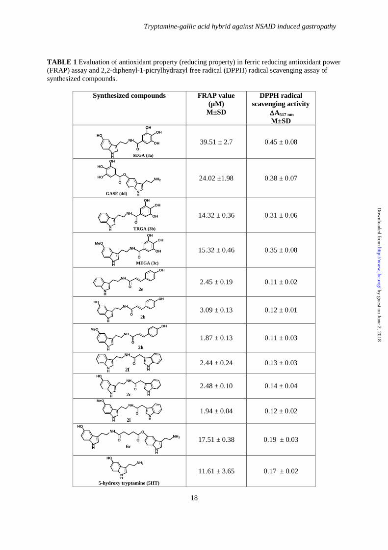
ascending TLC. The labelled complex (0.5 mL) was mixed with 1.5 mL of normal saline or rat serum and incubated at 37ºC. The samples were withdrawn at regular intervals up to 24 h, monitored by TLC and analyzed in gamma counter. Statistical Analysis- All data were presented as mean ± SEM. The level of significance was determined by unpaired student’s t-test with one-way ANOVA as applied. p value ≤ 0.05 was considered as significant. RESULTS Synthesis of tryptamine-gallic acid hybrid molecule- A small molecule having iron chelating property and capability to prevent MOS entering into mitochondria is necessary to protect gastric mucosa against NSAID-induced gastric mucosal injury or gastropathy. To design such a small molecule, we began the synthesis using 5’-hydroxy tryptamine (5HT), a hydrophobic amine that enters inside mitochondria (49). But, 5HT is toxic at high concentration (50). In contrast, free amine and hydroxyl groups of 5HT offer a scope to conjugate a powerful antioxidant bearing iron chelating property to make a non-toxic antioxidant hybrid molecule retaining mitochondrial penetration. Gallic acid (GA), a natural polyphenol and antioxidant (4) possesses iron chelating property (4,51) and that’s why we selected it to make a hybrid molecule with 5HT. The strategy might give double benefits because the conjugation of GA with 5HT is expected to enhance the bioavailability of GA in body fluid (lack of bioavailability is a common problem of bioactive polyphenol) or 5HT may be detoxified by GA through toxic group protection. 5HT was conjugated with GA through an amide linkage to synthesize SEGA (3a) and through an ester linkage to synthesize GASE (4d) (Fig. 1) (Supplemental data, scheme S1, S2). Both conjugates were tested first for their antioxidant property in vitro by following ferric reducing antioxidant power (FRAP) and 2, 2-diphenyl-1-picrylhydrazyl (DPPH) free radical scavenging activity. The FRAP assay is based on the measurement of the ability of a substance to reduce Fe (III) to Fe (II); the greater the reducing ability, the better the antioxidant property. Antioxidants reduce the colourless Fe (III)–
by guest on June 2, 2018http://w
ww
.jbc.org/D
ownloaded from

Tryptamine-gallic acid hybrid against NSAID induced gastropathy
9
TPTZ to a blue Fe (II)–TPTZ complex, which results in an increase in the absorbance at 595 nm, giving a FRAP value. A higher FRAP value indicates a greater reducing (i.e., antioxidant property) ability of the compound. FRAP values at 6 min was calculated from the equation described above. At 6 min, the absorbance change takes place abruptly due to reduction of Fe (III) into Fe (II). The results clearly indicate that SEGA (3a) shows a reducing ability [Fe (III) to Fe (II)] that is much better than GASE (4d) (Table 1). In DPPH assay, the decrease of absorbance is correlated with the antioxidant potency of the compounds. Greater the decrease in absorbance, higher is the DPPH scavenging potency i.e. the antioxidant potency of different synthesized compounds. The results indicate that SEGA (3a) also shows greater DPPH scavenging potency compared to GASE (4d) (Table 1). These results indicate that when 5HT is conjugated with GA through amide linkage appears to be more effective than when conjugated by ester linkage. Our next objective was to synthesize different types of tryptamine-antioxidant conjugates through amide linkage using other antioxidants replacing GA and evaluate their activities for comparative efficacy. We replaced GA by 4-hydroxy cinnamic acid and indole-3-acetic acid to synthesize other tryptamine-antioxidant conjugates such as 2b and 2c respectively (Fig 1). These compounds were synthesized by successive condensation of 5HT with 4-hydroxy cinnamic acid and indole-3-acetic acid respectively through amide linkage (Supplement data Scheme S1). Next, we searched to find out whether 5HT is the best possible tryptamine for our purpose. We replaced 5HT with other tryptamines to synthesize several other tryptamine-antioxidant conjugates such as TRGA (3b), MEGA (3c), 2f, 2h, and 2i by successive condensation of tryptamine, 5-methoxytryptamine with GA, 4-hydroxy cinnamic acid and indole-3-acetic acid respectively (Fig. 1) (Supplement data Scheme S3). 5HT itself showed little antioxidant activity in vitro (Table 1). We were interested to investigate whether in SEGA (3a), 5HT has any individual antioxidant property. To explore this, we have synthesized dimer of 5HT (Fig. 1) (Supplement data Scheme S4). Now antioxidant potencies were evaluated of all the synthesized
compounds by FRAP and DPPH free radical scavenging assays in vitro. For the preliminary screening of antioxidant activity, all the synthesized compounds were taken at high concentration (100 µM). From the above results it is evident that SEGA (3a) shows antioxidant property in FRAP as well as DPPH assays (Table 1). We were interested to check the antioxidant property of SEGA (3a) at different concentrations. Results indicate that SEGA (3a) shows excellent antioxidant property in FRAP assay as well as in DPPH scavenging assay in a concentration-dependent fashion (data not shown). Now, we tested whether SEGA (3a) could chelate free iron in vitro. The iron chelating property was performed by TPTZ assay. Fe (II) solution in presence of TPTZ gives a broad peak at 595 nm (Fig. 2). This peak was obtained due to Fe (II)-TPTZ complex formation. When SEGA (3a) was added to the Fe (II) solution, no such broad peak was observed after the addition of TPTZ solution (Fig. 2). Thus, from this experiment, it is evident that SEGA (3a) chelates free iron in vitro. Now, SEGA (3a), because of its maximum antioxidant potential and iron chelating property was subjected for further detailed biological evaluation and mechanistic studies on NSAID induced gastropathy SEGA (3a) prevents indomethacin (NSAID)-induced gastric mucosal damage- We tested whether SEGA (3a) could protect indomethacin (an NSAID)- induced MOS-mediated mitochondrial pathology and gastropathy in vivo. SEGA (3a) protected gastric mucosa from indomethacin-induced gastric injury in a dose-dependent manner (ED50 = 6.9 mg.kg-1 b.w.) as evident from gastric injury index (Fig. 3A). For rapid visualization of the protective effect of SEGA (3a), we are presenting the real morphological data opening the stomach interior. From the morphology, it is very clear that SEGA (3a) protected injury and oozing out of blood (appeared black due to oxidation of released hemoglobin under acidic environment) in indomethacin exposed rat gastric mucosa (Fig. 3B). Gastroprotective effect of SEGA (3a) was also verified by following the changes in microscopic structure of the actual histology of the gastric mucosa. SEGA (3a) restored normal architecture of gastric mucosa from indomethacin-induced increased gastric mucosal
by guest on June 2, 2018http://w
ww
.jbc.org/D
ownloaded from

Tryptamine-gallic acid hybrid against NSAID induced gastropathy
10
cell death and cell shedding in the superficial mucosa (Fig. 3C). Excessive gastric mucosal injury by NSAID leads to the release of blood in stomach. In indomethacin- treated rat, a sharp Soret peak (417 nm) was observed indicating the presence of hemoglobin (Hb) in the stomach due to mucosal injury. But in case of SEGA (3a) pretreatment, we did not find any Soret peak. The data further confirmed the gastroprotective effect of SEGA (3a) (Fig. 3D). SEGA (3a) scavenges intra-mitochondrial O2
•- , chelates intra-mitochondrial free iron and prevents mitochondrial oxidative stress (MOS)- Intra-mitochondrial generation of ROS and subsequent oxidative stress play a critical role in NSAID-induced gastric injury. Since SEGA (3a) protects gastric mucosa from NSAID-induced damage, we tested ROS scavenging activity of SEGA (3a). Mitochondrial O2
•- is precursor of ROS and mitochondrial free iron played an important role in the generation of ROS and development of MOS (6). Thus, the effect of SEGA (3a) on indomethacin-induced generation of mitochondrial O2
•- and free iron was evaluated. The generation of O2
•- and free iron was induced in cultured gastric epithelial cell by indomethacin (Fig. 4A, B). Mitochondrial O2
•- was measured by MitoSOX, a mitochodria-specific fluorescence indicator (52). MitoSOX is a derivative of hydroethydium and due to the cationic property this dye accumulates in huge amount within the mitochondria in response to negative membrane potential. O2
•- -derived oxidation product of MitoSOX has a distinct excitation wavelength at 396 nm and emission wavelength at 510 nm (6,52). Indomethacin stimulated intra-mitochondrial generation of O2
•- but pretreatment with SEGA (3a) significantly inhibited the generation of O2
•- as revealed from decreased fluorescence of O2
•- -derived oxidation product of MitoSOX (Fig. 4A). Mitochondrial free iron was measured by Phen Green SK, a specific fluorescent probe used to assay chelatable iron (Fig. 4B). Mitochondria of gastric mucosal cells were tagged by mitotraker red, a specific fluorescent probe for mitochondria. From the experiment, it is evident that indomethacin treatment resulted in increased intra-mitochondrial free iron accumulation but pretreatment with SEGA (3a) significantly inhibited indomethacin-mediated free iron
accumulation as revealed from decreased fluorescence of Phen Green SK (Fig. 4B). Mitochondrial O2
•- and free iron are responsible for MOS (6). SEGA (3a) by scavenging O2
•- and free iron protected mitochondria from MOS and restored the mitochondrial functions in gastric mucosal cells during indomethacin-induced gastropathy (Fig. 4C). SEGA (3a) significantly prevented indomethacin-induced mitochondrial lipid peroxidation, thiol depletion and protein carbonyl formation (Fig. 4C), which are the markers for MOS. SEGA (3a) corrects indomethacin-induced mitochondrial dysfunction- Since SEGA (3a) scavenges intra-mitochondrial O2
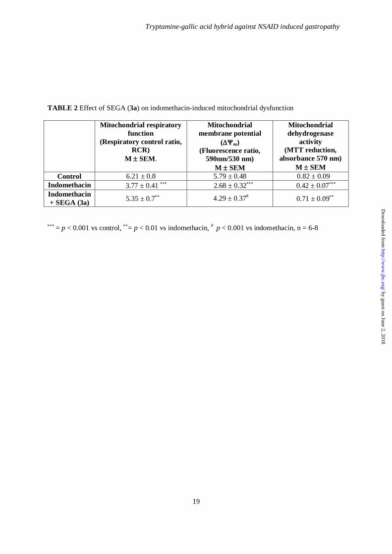
•-, chelates intra-mitochodrial free iron as well as prevents MOS, therefore we were interested to find out whether SEGA (3a) could prevent mitochondrial pathology or dysfunction. Fall of mitochondrial respiratory control ratio (RCR), collapse of mitochondrial transmembrane potential (∆ψm) and loss of dehydrogenase activity are hallmarks or indicators for mitochondrial pathology or dysfunction. The functional integrity of mitochondria in presence or absence of SEGA (3a) in gastric mucosal cell after indomethacin treatment was investigated. Mitochondria from indomethacin–treated gastric mucosa showed severe inhibition of complex-I mediated state 3 (in presence of ADP) respiration and mild inhibition of state 4 (in absence of ADP) respiration. As a consequence, the respiratory control ratio (RCR, the ratio of state 3 and state 4 respiration) was significantly decreased indicating impairment of mitochondrial respiration. Interestingly, administration of SEGA (3a) restored altered RCR value (Table 2). SEGA (3a) prevented indomethacin -induced loss of ∆ψm. Indomethacin showed about 40% decrease of ∆ψm, as measured by the ratio of fluorescence values measured at 590 nm (JC-1 aggregate) and 530 nm (JC-1 monomer). SEGA (3a) pretreatment restored indomethacin -induced fall of ∆ψm almost to the control level (Table 2). SEGA (3a) protected the mitochondrial dehydrogenase from indomethacin-induced inactivation. Indomethacin significantly inhibited mitochondrial dehydrogenases activity (as measured by MTT reduction) in gastric mucosal cells, indicating functional impairment of mitochondria, but pretreatment with SEGA (3a)
by guest on June 2, 2018http://w
ww
.jbc.org/D
ownloaded from
Tryptamine-gallic acid hybrid against NSAID induced gastropathy
11
under similar conditions restored the loss of mitochondrial dehydrogenase activity (Table 2). SEGA (3a) prevents the activation of mitochondrial pathway of apoptosis- The activation of mitochondrial pathway of apoptosis in gastric mucosal cell is a consequence of indomethacin-induced MOS. Since SEGA (3a) prevents MOS as well as mitochondrial dysfunction, SEGA (3a) should protect mitochondrial pathway of apoptosis in gastric mucosal cell by indomethacin. The data indicate that pretreatment with SEGA (3a) significantly attenuated indomethacin-induced activation of casapse-9 (marker of mitochondrial pathway apoptosis) (Fig. 5A) as well as caspase-3 (general marker for apoptosis) (Fig. 5B) in gastric mucosa in a dose dependent manner. Indomethacin stimulated about 2-fold activation of caspase-9 in the gastric mucosal cells (Fig. 5A). Pretreatment with SEGA (3a) significantly attenuated the activation of casapse-9 and brought the activity almost close to the normal level (Fig. 5A). Indomethacin activated caspase-3 by more than 2-fold in the gastric mucosal cells (Fig. 5B). Pretreatment with SEGA (3a) significantly blocked the activation of caspase-3 (Fig. 5B). Bcl-2 and Bax play a critical role in the mitochondrial pathway of apoptosis. Indomethacin was found to down-regulate the expression of antiapoptotic Bcl-2 and up-regulate the expression of Bax compared to control (Fig. 5C, D, E). SEGA (3a) pretreatment was found to block indomethacin -induced up-regulation of Bcl-2 as well as down-regulation of Bax (Fig. 5C, D, E). The antiapoptotic effect of SEGA (3a) was further tested by following DNA fragmentation performing terminal deoxynucleotidyl transferase dUTP nick end labeling (TUNEL) assay in gastric mucosal cell in vitro (Fig. 6A) and in vivo in presence of indomethacin (Fig. 6B). In control cells, absence of green signal (DNA fragmentation was indicated by green fluorescence of Alexa Fluor 488) indicated no apoptotic DNA fragmentation. However, in indomethacin-treated gastric mucosal cells, green fluorescence was prominent, which was co-localized with propidium iodide (PI)- stained nuclei indicating apoptotic DNA fragmentation. In SEGA (3a) pretreated cell, the intensity as well as total number of cells showing green fluorescence were very less compared to indomethacin treated
cells (Fig. 6A). SEGA (3a) pretreatment also significantly prevented indomethacin-induced gastric mucosal apoptosis in vivo in mucosal tissue (Fig. 6B). The TUNEL positive cells (dark brown staining, indicated by arrow) were abundant in the gastric mucosal tissue in presence of indomethacin, whereas the TUNEL positive cells were decreased significantly in SEGA (3a) pretreated indomethacin-treated group (Fig. 6B). Anti apoptotic effect of SEGA (3a) was further confirmed by immunohistochemistry using anti-active caspase-3 antibody (Fig. 6C). Active caspase-3 immunolabelled mucosal cells (dark brown staining, indicated by arrow) were found after indomethacin treatment. But SEGA (3a) pretreatment significantly decreased the active caspase-3-immunolabelled cells indicating the antiapoptotic role of SEGA (3a) (Fig. 6C). Thus, the data indicate that SEGA (3a) prevents indomethacin-induced gastric mucosal cell apoptosis. SEGA (3a) accelerates healing of indomethacin-induced damaged gastric mucosa -Prevention of MOS and mitochondrial pathway of apoptosis expedites the healing process (5). Since SEGA (3a) prevents both MOS as well as apoptosis, thus we are interested to find out whether SEGA (3a) could accelerate the healing of indomehacin-induced already damaged gastric mucosa. Interestingly, in addition to the gastroprotective effect, SEGA (3a) also accelerated healing of already injured mucosa by indomethacin (Fig 7). Although auto-healing takes place in case of damaged mucosa but SEGA (3a) treatment accelerates the healing process. SEGA (3a)-induced healing of gastric mucosal injury was checked by histological analysis (Fig. 7). At 4 h, 8 h and 20 h, the mucosa shows gastric injury with injury index of 52, 28 and 20 respectively whereas after treatment with SEGA (3a) damage of gastric mucosa was gradually repaired as evident from injury index (II) of 14, 8 and 0 respectively. At 20 h SEGA (3a) completely restored normal architecture of gastric mucosa where as in case of indomethacin group there was significant injury. The results indicate that mucosa shows a time dependent auto-healing of the indomethacin-induced gastric damage in absence of SEGA (3a). But, SEGA (3a) treatment significantly expedites healing with the progress
by guest on June 2, 2018http://w
ww
.jbc.org/D
ownloaded from

Tryptamine-gallic acid hybrid against NSAID induced gastropathy
12
of time as evident from the restoration of gastric mucosa (Fig. 7). In indomethacin-treated animal, the auto-healing at 4 h was negligible as evident from the distorted mucosal histology but SEGA (3a) treatment restored healthy mucosal architecture at 4 h with almost complete restoration at 20 h (Fig. 7). Quantitation of SEGA (3a) entered inside mitochondria- Since SEGA (3a) scavenged intra-mitochondrial O2
•-, chelated intra-mitochondrial iron and prevented MOS, therefore, we were interested to quantitate how much of the administered SEGA (3a) entered inside mitochondria under in vivo condition. For this purpose, SEGA (3a) was radiolabelled with 99mTc isotope as reported (48) and administered to rat. The data indicated that 0.05 % of the administered dose of SEGA (3a) entered into per mg mitochondria of gastric mucosal tissue (Fig. 7A). The stability or the structural integrity of SEGA (3a) in physiological saline as well as in serum was checked. SEGA (3a) was found to be very stable at 370C (Fig. 7B). DISCUSSION The present study describes the designing and synthesis of a small molecule, tryptamine-gallic acid hybrid [SEGA (3a)], which prevents NSAID-induced mitochondrial pathology, apoptosis as well as gastropathy by blocking MOS through scavenging of intra-mitochondrial O2
•- and free iron and correcting mitochondrial dysfunction. Mitochondria is a potential subcellular therapeutic drug-target against NSAID induced gastropathy since it produces O2
•- and free iron, which play an important role in triggering this pathological condition. A mitochondria targeted molecule is required for this purpose. This molecule must be small, lipophilic and ROS scavenger in nature. Moreover, free iron is known to generate ROS through Fenton’s reaction. Thus, iron chelating property would be an additional advantage in controlling oxidative stress. All these criteria were considered while designing the molecule. Lot of antioxidants and iron chelators are reported, but none of them can satisfy all the above criteria. Thus, a new molecule is essential, which will satisfy all these criteria in preventing NSAID-induced gastropathy. Keeping this in mind, we have synthesized a series of tryptamine-antioxidant
hybrid molecules. GA when conjugated with 5HT through amide linkage shows greater activity both in vitro as well as in vivo. Thus all other tryptamine –antioxidant hybrid molecules were generated through the amide linkage. For the structure -activity relationship (SAR) studies, we synthesized different tryptamine-antioxidant derivatives. Since SEGA (3a) appears to be the most active among all the tryptamine-antioxidant conjugates, it is suggested that the presence of the 5-hydroxy group in the indole moiety of SEGA (3a) plays an important role for its gastroprotective activity. When the 5-hydroxy group in the indole moiety of SEGA (3a) was replaced by hydrogen and methoxy group in the molecules TRGA (3b) and MEGA (3c), the activity is decreased. Indomethacin is selected as the representative NSAID over others because it is the most frequently used NSAID in gastrointestinal toxicity studies in experimental animals (13). The dose of indomethacin was selected as 5 mM and 48 mg kg-1 b.w. for in vitro and in vivo studies as reported earlier (4,6-7). The role of MOS and consequent apoptosis behind NSAID-induced gastric mucosal injury is already well established and is considered to be the major player in the acid independent (5) and COX-independent pathway of NSAID-mediated gastric injury (53-54). Indomethacin with its acidic carboxyl group (pKa = 4.5) and lipid solubility has been found to damage both rat and human mitochondria (13). Moreover, indomethacin enhances mitochondrial ROS, which disrupts mitochondrial function (6). Since the gastroprotective effect of SEGA (3a) is dependent on its iron chelating and free radical scavenging properties, we compared the gastroprotective effect of SEGA (3a) with that of the standard iron chelating agent desferrioxamine (ED50 =100 mg.kg-1 b.w.) and free radical scavenging agents gallic acid (ED50 =18.9 mg.kg-1 b.w.), vitamin E (ED50 = 45 mg.kg-1 b.w.), Phenyl-N-tert-Butylnitrone (PBN) (ED50 =100 mg.kg-1 b.w.) and quercetin (ED50 =125 mg.kg-1 b.w.) (4,55-56). We found that SEGA (3a) (ED50 = 6.9 mg.kg-1 b.w.) is much more effective than these compounds. The gastroprotective efficacy (ED50) of SEGA (3a) was also compared with those of ranitidine (histamine H2-receptor antagonist), omeprazole and lansoprazole (proton pump inhibitors), the three most commonly used gastroprotective
by guest on June 2, 2018http://w
ww
.jbc.org/D
ownloaded from

Tryptamine-gallic acid hybrid against NSAID induced gastropathy
13
agents. The gastroprotective potency (ED50 = 6.9 mg.kg-1 b.w.) of SEGA (3a) in protecting indomethacin-induced gastric mucosal injury was found to be superior to ranitidine (ED50 = 13.5 mg.kg-1 b.w.) (55) but inferior to omeprazole (ED50 = 5 mg.kg-1 b.w.) (7) and lansoprazole (ED50 = 5.4 mg.kg-1 b.w.)(5). Although proton pump inhibitors (PPIs) are effective at a very low dose against NSAID-induced gastropathy (5), they have some adverse effects like diarrhoea (57), linear mucosal defects and friable mucosa associated with collagenous colitis (58-59), subacute cutaneous lupus erythematosus (60), leydig cell tumors (61), acute nephritis (62), myopathy including polymyositis (63), and anaphylactic reactions (64). PPIs are reported to be associated with an increased risk of bacterial infection and related diseases (65-66). Moreover PPIs exacerbate NSAID-induced small intestinal injury through induction of dysbiosis (67). SEGA (3a) has several advantages over the commercially available antioxidants, iron chelator and known gastroprotective agents. It chelates intra-mitochondrial free iron and scavenges ROS entering into mitochondria. Although, it appears that the dose of SEGA (3a) (50 mg.kg-1 b.w.) selected for rat against indomethacin-induced
gastropathy is high but this dose is only 8 mg.kg-
1 b.w. when converted to the human equivalent dose as described by US Food and Drug Administration (FDA) (68). We propose the whole gastroprotective mechanism of SEGA (3a) through a schematic representation (Fig. 9). Indomethacin interacts with the complex I of electron transport chain and results in the leakage of electron, which in turn leads to the generation of ROS. SEGA (3a) enters into mitochondria and scavenges generated ROS and prevents MOS by attenuating mitochondrial protein oxidation, lipid peroxidation and depletion of thiol. Iron is released from Fe-S cluster of aconitase due to ROS-mediated damage and further aggravates oxidative stress by producing hydroxyl radical. SEGA (3a) also scavenges the released intra-mitochondrial iron. SEGA (3a) prevents mitochondrial pathway of apoptosis by preventing indomethacin-induced activation of caspase 9 and caspase 3 and downregulation of Bcl-2 (antiapoptotic gene) and upregulation of Bax (proapoptotic gene). In conclusion, SEGA (3a) is a novel small molecule, which protects gastric mucosa against NSAID-induced MOS-mediated gastric injury.
REFERENCES
1. Regula, J., Butruk, E., Dekkers, C. P., de Boer, S. Y., Raps, D., Simon, L., Terjung, A., Thomas, K. B., Luhmann, R., and Fischer, R. (2006) Am. J. Gastroenterol. 101, 1747-1755
2. Thompson, M., and Percy, J. S. (1966) Br. Med. J. 1, 80-83 3. Becker, J. C., Domschke, W., and Pohle, T. (2004) Br. J. Clin. Pharmacol. 58, 587-600 4. Pal, C., Bindu, S., Dey, S., Alam, A., Goyal, M., Iqbal, M. S., Maity, P., Adhikari, S. S., and
Bandyopadhyay, U. (2010) Free Radic. Biol. Med. 49, 258-267 5. Maity, P., Bindu, S., Choubey, V., Alam, A., Mitra, K., Goyal, M., Dey, S., Guha, M., Pal, C.,
and Bandyopadhyay, U. (2008) J. Biol. Chem. 283, 14391-14401 6. Maity, P., Bindu, S., Dey, S., Goyal, M., Alam, A., Pal, C., Mitra, K., and Bandyopadhyay, U.
(2009) J. Biol. Chem. 284, 3058-3068 7. Biswas, K., Bandyopadhyay, U., Chattopadhyay, I., Varadaraj, A., Ali, E., and Banerjee, R.
K. (2003) J. Biol. Chem. 278, 10993-11001 8. Wolfe, M. M., and Soll, A. H. (1988) N. Engl. J. Med. 319, 1707-1715 9. Szabo, I., and Tarnawski, A. S. (2000) J. Physiol. Pharmacol. 51, 3-15 10. Wallace, J. L., and Granger, D. N. (1996) Faseb J. 10, 731-740 11. Kwiecien, S., Brzozowski, T., and Konturek, S. J. (2002) J. Physiol. Pharmacol. 53, 39-50 12. Maity, P., Bindu, S., Dey, S., Goyal, M., Alam, A., Pal, C., Reiter, R., and Bandyopadhyay,
U. (2009) J. Pineal. Res. 46, 314-323 13. Jacob, M., Bjarnason, I., Rafi, S., Wrigglesworth, J., and Simpson, R. J. (2001) Aliment.
Pharmacol. Ther. 15, 1837-1842
by guest on June 2, 2018http://w
ww
.jbc.org/D
ownloaded from
Tryptamine-gallic acid hybrid against NSAID induced gastropathy
14
14. Demaurex, N., and Scorrano, L. (2009) Nat. Neurosci. 12, 819-820 15. Turrens, J. F. (1997) Biosci. Rep. 17, 3-8 16. Turrens, J. F. (2003) J. Physiol. 552, 335-344 17. Fariss, M. W., Chan, C. B., Patel, M., Van Houten, B., and Orrenius, S. (2005) Mol. Interv. 5,
94-111 18. Bindu, S., Pal, C., Dey, S., Goyal, M., Alam, A., Iqbal, M. S., Dutta, S., Sarkar, S., Kumar,
R., Maity, P., and Bandyopadhyay, U. (2011) J. Biol. Chem. 286, 39387-39402 19. Ceccarelli, D., Gallesi, D., Giovannini, F., Ferrali, M., and Masini, A. (1995) Biochem.
Biophys. Res. Commun. 209, 53-59 20. Britton, R. S., Ramm, G. A., Olynyk, J., Singh, R., O'Neill, R., and Bacon, B. R. (1994) Adv.
Exp. Med. Biol. 356, 239-253 21. Fraga, C. G., and Oteiza, P. I. (2002) Toxicology 180, 23-32 22. Liang, L. P., Jarrett, S. G., and Patel, M. (2008) J. Neurosci. 28, 11550-11556 23. Link, G., Saada, A., Pinson, A., Konijn, A. M., and Hershko, C. (1998) J. Lab Clin. Med. 131,
466-474 24. Ling, Y. H., Liebes, L., Zou, Y., and Perez-Soler, R. (2003) J. Biol. Chem. 278, 33714-33723 25. Zimmerman, M. C., and Zucker, I. H. (2009) Hypertension 53, 112-114 26. Gonzalez-Flecha, B., Cutrin, J. C., and Boveris, A. (1993) J. Clin. Invest. 91, 456-464 27. Droge, W. (2002) Physiol. Rev. 82, 47-95 28. Schapira, A. H. (2006) Lancet 368, 70-82 29. Lin, M. T., and Beal, M. F. (2006) Nature 443, 787-795 30. Jing, X. B., Cai, X. B., Hu, H., Chen, S. Z., Chen, B. M., and Cai, J. Y. (2007) Biochem. Cell
Biol. 85, 265-271 31. Orrenius, S., Gogvadze, V., and Zhivotovsky, B. (2007) Annu. Rev. Pharmacol. Toxicol. 47,
143-183 32. Malik, F., Kumar, A., Bhushan, S., Khan, S., Bhatia, A., Suri, K. A., Qazi, G. N., and Singh,
J. (2007) Apoptosis 12, 2115-2133 33. Parish, R., and Petersen, K. F. (2005) Curr. Diab. Rep. 5, 177-183 34. Carelli, V., Ross-Cisneros, F. N., and Sadun, A. A. (2004) Prog. Retin. Eye Res. 23, 53-89 35. Naumann, M., Reiners, K., Gold, R., Schindler, R., Paulus, W., Klopstock, T., and
Reichmann, H. (1995) Acta. Neuropathol. 89, 152-157 36. Ren, J., Pulakat, L., Whaley-Connell, A., and Sowers, J. R. (2010) J. Mol. Med. 88, 993-1001 37. Serviddio, G., Bellanti, F., Sastre, J., Vendemiale, G., and Altomare, E. (2010) Curr. Med.
Chem. 17, 2325-2337 38. Ko, J. K., Ma, J. J., Chow, J. Y., Ma, L., and Cho, C. H. (1998) Free Radic. Biol. Med. 24,
1007-1014 39. Ben Farhat, M., Jordan, M. J., Chaouech-Hamada, R., Landoulsi, A., and Sotomayor, J. A.
(2009) J. Agric. Food Chem. 57, 10349-10356 40. Chattopadhyay, I., Nandi, B., Chatterjee, R., Biswas, K., Bandyopadhyay, U., and Banerjee,
R. K. (2004) Inflammopharmacology 12, 153-176 41. Pal, C., Kundu, M. K., Bandyopadhyay, U., and Adhikari, S. (2011) Bioorg. Med. Chem. Lett.
21, 3563-3567 42. Kumar, S., Das, S. K., Dey, S., Maity, P., Guha, M., Choubey, V., Panda, G., and
Bandyopadhyay, U. (2008) Antimicrob. Agents Chemother. 52, 705-715 43. Dey, S., Guha, M., Alam, A., Goyal, M., Bindu, S., Pal, C., Maity, P., Mitra, K., and
Bandyopadhyay, U. (2009) Free Radic. Biol. Med. 46, 271-281 44. Sims, N. R., and Anderson, M. F. (2008) Nat. Protoc. 3, 1228-1239 45. Lowry, O. H., Rosebrough, N. J., Farr, A. L., and Randall, R. J. (1951) J. Biol. Chem. 193,
265-275 46. Rousou, A. J., Ericsson, M., Federman, M., Levitsky, S., and McCully, J. D. (2004) Am. J.
Physiol. Heart Circ. Physiol. 287, H1967-1976 47. Frezza, C., Cipolat, S., and Scorrano, L. (2007) Nat. Protoc. 2, 287-295
by guest on June 2, 2018http://w
ww
.jbc.org/D
ownloaded from

Tryptamine-gallic acid hybrid against NSAID induced gastropathy
15
48. Halder, K. K., Mandal, B., Debnath, M. C., Bera, H., Ghosh, L. K., and Gupta, B. K. (2008) J. Drug Target 16, 311-320
49. Basu, B., Desai, R., Balaji, J., Chaerkady, R., Sriram, V., Maiti, S., and Panicker, M. M. (2008) Reproduction 135, 657-669
50. Mulac, D., and Humpf, H. U. (2011) Toxicology 282, 112-121 51. A.E. Fazary, M. T., Y.H.Ju,. (2009) J. Chem. Eng. Data 54, 35-42 52. Robinson, K. M., Janes, M. S., Pehar, M., Monette, J. S., Ross, M. F., Hagen, T. M., Murphy,
M. P., and Beckman, J. S. (2006) Proc. Natl. Acad. Sci. U S A 103, 15038-15043 53. Musumba, C., Pritchard, D. M., and Pirmohamed, M. (2009) Aliment. Pharmacol. Ther. 30,
517-531 54. Fornai, M., Colucci, R., Antonioli, L., Awwad, O., Ugolini, C., Tuccori, M., Fulceri, F.,
Natale, G., Basolo, F., and Blandizzi, C. (2011) Pharmacol. Res. 63, 59-67 55. Bandyopadhyay, U., Biswas, K., Chatterjee, R., Bandyopadhyay, D., Chattopadhyay, I.,
Ganguly, C. K., Chakraborty, T., Bhattacharya, K., and Banerjee, R. K. (2002) Life Sci. 71, 2845-2865
56. Alarcon de la Lastra, C., Martin, M. J., and Motilva, V. (1994) Pharmacology 48, 56-62 57. Mukherjee, S. (2003) J. Gastroenterol. Hepatol. 18, 602-603 58. Umeno, J., Matsumoto, T., Nakamura, S., Jo, Y., Yada, S., Hirakawa, K., Yoshimura, R.,
Yamagata, H., Kudo, T., Hirano, A., Gushima, M., Yao, T., Nakashima, Y., and Iida, M. (2008) Gastrointest. Endosc. 67, 1185-1191
59. Yusuke, H., Jun, T., Naotaka, M., Yuichi, T., Yutaka, E., and Kazuaki, I. (2009) Endoscopy 41 Suppl 2, E281-282
60. Panting, K. J., Pinto, M., and Ellison, J. (2009) Clin. Exp. Dermatol. 34, 733-734 61. Fort, F. L., Miyajima, H., Ando, T., Suzuki, T., Yamamoto, M., Hamashima, T., Sato, S.,
Kitazaki, T., Mahony, M. C., and Hodgen, G. D. (1995) Fundam. Appl. Toxicol. 26, 191-202 62. Jose, J., Saravu, K., Khera, K., Jimmy, B., and Shastry, B. A. (2008) J. Pak. Med. Assoc. 58,
206-207 63. Clark, D. W., and Strandell, J. (2006) Eur. J. Clin. Pharmacol. 62, 473-479 64. Gonzalez, P., Soriano, V., Lopez, P., and Niveiro, E. (2002) Allergol. Immunopathol. (Madr)
30, 342-343 65. Dial, S., Delaney, J. A., Barkun, A. N., and Suissa, S. (2005) JAMA 294, 2989-2995 66. Laheij, R. J., Sturkenboom, M. C., Hassing, R. J., Dieleman, J., Stricker, B. H., and Jansen, J.
B. (2004) JAMA 292, 1955-1960 67. Wallace, J. L., Syer, S., Denou, E., De Palma, G., Vong, L., McKnight, W., Jury, J., Bolla,
M., Bercik, P., Collins, S. M., Verdu, E., and Ongini, E. (2011) Gastroenterology 141, 1314-1322
68. Reagan-Shaw, S., Nihal, M., and Ahmad, N. (2008) Faseb J. 22, 659-661 FOOTNOTES We thank Council of Scientific and Industrial Research (CSIR), New Delhi and University Grants Commission (UGC) New Delhi, for providing grants to carry out the work. The abbreviations used are: NSAID, non-steroidal anti-inflammatory drugs; ROS, reactive oxygen species; MOS, mitochondrial oxidative stress; RCR, respiratory control ratio; JC-1, 5,5/,6,6/-tetrachloro-1,1/,3,3/-tetraethylbenzimidazolcarbocyanine iodide; TUNEL, terminal deoxynucleotidyl transferase dUTP nick end labeling; PPI, Proton pump inhibitor
by guest on June 2, 2018http://w
ww
.jbc.org/D
ownloaded from

Tryptamine-gallic acid hybrid against NSAID induced gastropathy
16
FIGURE LEGENDS FIGURE 1. General scheme for the synthesis of tryptamine derivatives. FIGURE 2. Iron chelating activity of SEGA (3a) in vitro. Spectroscopic analysis for SEGA (3a)-Fe (II) interaction at different concentrations of SEGA (3a) (a, 500 nM; b, 1 μM; c, 10 μM; d, 50 μM; e, 100 μM ). Detail descriptions were given under ‘EXPERIMENTAL PROCEDURES’. FIGURE 3. SEGA (3a) prevents indomethacin-induced gastropathy. (A) Protection of indomethacin - induced gastric mucosal injury by SEGA (3a) at different doses as measured by injury index (* = p<0.001 vs. indomethacin, n = 6). (B) Morphology of gastric mucosa from control, indomethacin (48 mg.kg-1 b.w.) -treated and SEGA (3a) [50 mg.kg-1 b.w.] pretreated indomethacin-treated rats. Arrow indicates damage in the gastric mucosa. (C) Haematoxylin-eosin staining of gastric mucosal sections from indomethacin-treated and indomethacin treated-SEGA (3a)-pretreated rats. Arrow indicates injury in the gastric mucosa. (D) Detection of hemoglobin in gastric washing of control, indomethacin-treated and SEGA (3a) pretreated indomethacin-treated rats as measured by Soret spectroscopy. Detail descriptions were given under ‘EXPERIMENTAL PROCEDURES’. FIGURE 4. SEGA (3a) scavenges indomethacin-induced intra-mitochondrial O2
•- , free iron in gastric mucosal cell and prevents mitochondrial oxidative stress (MOS) (A) SEGA (3a)[50 µM] scavenges intra mitochondrial O2
•- generated by indomethacin (5 mM) in gastric mucosal cells. Mitochondrial generation of O2
•- was detected by MitoSOX red staining (red color). (B) SEGA (3a) [50 µM] chelates intra-mitochondrial free iron in vitro in cultured gastric mucosal cells. Mitochondrial generation of free iron was detected by Phen Green SK staining (green color). (C) SEGA (3a) [50 mg.kg-1 b.w.] prevents indomethacin-induced formation of protein carbonyl, peroxidation of lipid and depletion of thiol content in mitochondria (*** = p<0.001 vs control, ### = p<0.001 vs. indomethacin, n = 5). Detail descriptions were given under ‘EXPERIMENTAL PROCEDURES’. FIGURE 5. SEGA (3a) prevents the activation of mitochondrial pathway of apoptosis (A) Dose dependent inhibition of indomethacin-induced of caspase-9 in gastric mucosa of rat by SEGA (3a). (B) Dose dependent inhibition of indomethacin-induced of caspase-3 in gastric mucosa of rat by SEGA (3a) [5, 10, 50 mg.kg-1 b.w.].Data were presented as mean ± SEM (*** = p<0.001 vs control, ### = p<0.001, ** = p<0.01 vs. indomethacin, n = 6). (C) SEGA (3a) [50 mg.kg-1 b.w.] inhibits down-regulation of Bcl-2 and up-regulation of Bax by indomethacin in gastric mucosa as measured by RT-PCR. Actin was used as an internal control. (D) Densitometric analysis of Bcl-2 expression. (E) Densitometric analysis of Bax expression. Detail descriptions were given under ‘EXPERIMENTAL PROCEDURES’. Data were presented as mean ± SEM (*** = p<0.001 vs control, ### = p<0.001 vs. indomethacin, n = 5). FIGURE 6. SEGA (3a) prevents indomethacin-induced apoptosis in vitro and in vivo (A) SEGA (3a) [50 µM] inhibits indomethacin (5 mM)-induced apoptosis in vitro in primary gastric mucosal cells in culture as measured by TUNEL assay. The first column shows nuclei stained (red) with propidium iodide (PI), the second column shows apoptotic DNA stained with Alexa Fluor 488 (green fluorescence) and third column shows the merged pictures of first (PI) and second (Alexa Fluor 488) column. (B) SEGA (3a) [50 mg.kg-1 b.w.] inhibits indomethacin -induced apoptosis in vivo in gastric mucosal cells as measured by TUNEL assay in mucosal tissue. TUNEL assay shows that indomethacin triggers apoptosis (deep brown staining showing apoptotic DNA fragmentation, indicated by arrows) of gastric mucosal cells and SEGA (3a) blocks indomethacin-induced gastric mucosal cell apoptosis. (C) Immnuhistochemical staining of mucosal tissue with the anti-active
by guest on June 2, 2018http://w
ww
.jbc.org/D
ownloaded from

Tryptamine-gallic acid hybrid against NSAID induced gastropathy
17
caspase-3 antibody (deep brown staining showing apoptotic cells indicated by arrows). Detail descriptions were given under ‘EXPERIMENTAL PROCEDURES’. FIGURE 7. Effect of SEGA (3a) on healing of indomethacin-induced gastric mucosal injury. Haematoxylin and eosin staining of gastric mucosal section of control, indomethacin and indomethacin + SEGA (3a) [50 mg.kg-1 b.w.] treated groups at different time points. ‘0 h’ = control. II = Injury index. Arrow indicates mucosal injury. FIGURE 8. Mitochondrial uptake of SEGA (3a) (A) Uptake of 99mTc-labeled SEGA (3a) into the mitochondria of gastric mucosal cells 4 hours after intra-peritoneal injection (i.p). Mitochondrial uptake of SEGA (3a) was expressed as % of dose/mg of mitochondrial protein (B) Stability studies of 99mTc-labeled SEGA (3a) in physiological saline and serum in vitro at 37°C. Detail descriptions were given under ‘EXPERIMENTAL PROCEDURES’. FIGURE 9. Scheme showing indomethacin (NSAID) interacts with the complex I of electron transport chain and results in the leakage of electron in mitochondria leads to the formation of superoxide anion radical (O2
•-), which leads to the generation of reactive oxygen species (ROS). Increased ROS develops mitochondrial oxidative stress (MOS) by oxidizing protein and lipid including cardiolipin and protein thiol. Iron (Fe++) is released from Fe-S cluster of aconitase due to ROS-mediated damage and further aggravates oxidative stress by producing hydroxyl radical (.OH). The MOS results in mitochondrial dysfunction or pathology and activation of the mitochondrial pathway of apoptosis, which plays a pathogenic role for gastropathy. Tryptamine – gallic acid hybrid molecule, SEGA (3a) enters inside the mitochondria and prevents NSAID- induced gastropathy.
by guest on June 2, 2018http://w
ww
.jbc.org/D
ownloaded from
Tryptamine-gallic acid hybrid against NSAID induced gastropathy
18
TABLE 1 Evaluation of antioxidant property (reducing property) in ferric reducing antioxidant power (FRAP) assay and 2,2-diphenyl-1-picrylhydrazyl free radical (DPPH) radical scavenging assay of synthesized compounds.
Synthesized compounds FRAP value (µM)
M±SD
DPPH radical scavenging activity
∆A517 nm M±SD
O
NH
NH
OHOH
OH
SEGA (3a)
HO
39.51 ± 2.7 0.45 ± 0.08
NH2
NH
O
O
OHHO
HO
GASE (4d)
24.02 ±1.98 0.38 ± 0.07
O
NH
NH
OHOH
OH
TRGA (3b)
14.32 ± 0.36 0.31 ± 0.06
O
NH
NH
OHOH
OH
MEGA (3c)
MeO
15.32 ± 0.46 0.35 ± 0.08
O
NH
NH
OH
2e
2.45 ± 0.19 0.11 ± 0.02
O
NH
NH
OH
2b
HO
3.09 ± 0.13 0.12 ± 0.01
O
NH
NH
OH
2h
MeO
1.87 ± 0.13 0.11 ± 0.03
O
NH
NH 2f
NH
2.44 ± 0.24 0.13 ± 0.03
O
NH
NH 2c
HO
NH
2.48 ± 0.10 0.14 ± 0.04
O
NH
NH 2i
MeO
NH
1.94 ± 0.04 0.12 ± 0.02
NH2
NH
O
OO
NH
NH
HO
6c
17.51 ± 0.38 0.19 ± 0.03
5-hydroxy tryptamine (5HT)
NH2
NH
HO
11.61 ± 3.65 0.17 ± 0.02
by guest on June 2, 2018http://w
ww
.jbc.org/D
ownloaded from
Tryptamine-gallic acid hybrid against NSAID induced gastropathy
19
TABLE 2 Effect of SEGA (3a) on indomethacin-induced mitochondrial dysfunction
Mitochondrial respiratory function
(Respiratory control ratio, RCR)
M ± SEM.
Mitochondrial membrane potential
(∆Ψm) (Fluorescence ratio,
590nm/530 nm) M ± SEM
Mitochondrial dehydrogenase
activity (MTT reduction,
absorbance 570 nm) M ± SEM
Control 6.21 ± 0.8 5.79 ± 0.48 0.82 ± 0.09 Indomethacin 3.77 ± 0.41 ∗∗∗ 2.68 ± 0.32∗∗∗ 0.42 ± 0.07∗∗∗ Indomethacin + SEGA (3a) 5.35 ± 0.7∗∗ 4.29 ± 0.37# 0.71 ± 0.09∗∗
∗∗∗ = p < 0.001 vs control, ∗∗= p < 0.01 vs indomethacin, # p < 0.001 vs indomethacin, n = 6-8
by guest on June 2, 2018http://w
ww
.jbc.org/D
ownloaded from
Susanta Adhikari and Uday BandyopadhyayIqbal, Souvik Sarkar, Rahul Kumar, Kamal Krishna Halder, Mita Chatterjee Debnath,
Chinmay Pal, Samik Bindu, Sumanta Dey, Athar Alam, Manish Goyal, Mohd. Shameelof apoptosis in gastric mucosal cells
drug-induced gastropathy: Correction of mitochondrial dysfunction and inhibition Tryptamine-gallic acid hybrid prevents non-steroidal anti-inflammatory
published online December 7, 2011J. Biol. Chem.
10.1074/jbc.M111.307199Access the most updated version of this article at doi:
Alerts:
When a correction for this article is posted•
When this article is cited•
to choose from all of JBC's e-mail alertsClick here
Supplemental material:
http://www.jbc.org/content/suppl/2011/12/07/M111.307199.DC1
by guest on June 2, 2018http://w
ww
.jbc.org/D
ownloaded from