JB Accepts, published online ahead of print on 15 July...
41
1 Roles of the redox active disulfide, and histidine residues forming a catalytic dyad, in reactions 2 catalyzed by 2-Ketopropyl-Coenzyme M Oxidoreductase/Carboxylase 3 4 Running title: Mechanism of 2-ketopropyl-CoM carboxylation 5 6 Melissa A. Kofoed 1 , David A. Wampler 1 † , Arti S. Pandey 2 ‡ John W. Peters 2 , and Scott A. 7 Ensign 1 * 8 9 1 Department of Chemistry and Biochemistry, Utah State University, Logan, Utah 84322 and 10 2 Department of Chemistry and Biochemistry and Astrobiology Biogeocatalysis Research Center, 11 Montana State University, Bozeman, Montana 59717 12 13 * Corresponding Author. Mailing address: Department of Chemistry and Biochemistry, Utah 14 State University, 0300 Old Main Hill, Logan, UT 84322 Phone: (435) 797-3969. FAX: (435) 15 797-3390. E-mail: [email protected] 16 17 † Present address: University of Texas Health Science Center at San Antonio, San Antonio, TX 18 78229 19 ‡ Present address: Department of Biochemistry, Kathmandu Medical College and Teaching 20 Hospital, Duwakot, Bhaktapur, Nepal 21 22 23 Copyright © 2011, American Society for Microbiology and/or the Listed Authors/Institutions. All Rights Reserved. J. Bacteriol. doi:10.1128/JB.05231-11 JB Accepts, published online ahead of print on 15 July 2011 on June 6, 2019 by guest http://jb.asm.org/ Downloaded from
Transcript of JB Accepts, published online ahead of print on 15 July...

1
Roles of the redox active disulfide, and histidine residues forming a catalytic dyad, in reactions 2
catalyzed by 2-Ketopropyl-Coenzyme M Oxidoreductase/Carboxylase 3
4
Running title: Mechanism of 2-ketopropyl-CoM carboxylation 5
6
Melissa A. Kofoed1, David A. Wampler1† , Arti S. Pandey2‡ John W. Peters2, and Scott A. 7
Ensign1* 8
9
1Department of Chemistry and Biochemistry, Utah State University, Logan, Utah 84322 and 10
2Department of Chemistry and Biochemistry and Astrobiology Biogeocatalysis Research Center, 11
Montana State University, Bozeman, Montana 59717 12
13
* Corresponding Author. Mailing address: Department of Chemistry and Biochemistry, Utah 14
State University, 0300 Old Main Hill, Logan, UT 84322 Phone: (435) 797-3969. FAX: (435) 15
797-3390. E-mail: [email protected] 16
17
† Present address: University of Texas Health Science Center at San Antonio, San Antonio, TX 18
78229 19
‡ Present address: Department of Biochemistry, Kathmandu Medical College and Teaching 20
Hospital, Duwakot, Bhaktapur, Nepal 21
22
23
Copyright © 2011, American Society for Microbiology and/or the Listed Authors/Institutions. All Rights Reserved.J. Bacteriol. doi:10.1128/JB.05231-11 JB Accepts, published online ahead of print on 15 July 2011
on June 6, 2019 by guesthttp://jb.asm
.org/D
ownloaded from

2
ABSTRACT 24
NADPH:2-ketopropyl-coenzyme M oxidoreductase/carboxylase (2-KPCC), an atypical 25
member of the disulfide oxidoreductase (DSOR) family of enzymes, catalyzes the reductive 26
cleavage and carboxylation of 2-ketopropyl-coenzyme M (2-(2-ketopropylthio)ethanesulfonate; 27
2-KPC) to form acetoacetate and coenzyme M (CoM) in the bacterial pathway of propylene 28
metabolism. Structural studies of 2-KPCC from Xanthobacter autotrophicus strain Py2 have 29
revealed a distinctive active site architecture that includes a putative catalytic triad consisting of 30
two histidine residues hydrogen bonded to an ordered water molecule proposed to stabilize 31
enolacetone formed from dithiol-mediated 2-KPC thioether bond cleavage. Site-directed mutants 32
of 2-KPCC were constructed to test the tenets of the mechanism proposed from studies of the 33
native enzyme. Mutagenesis of the interchange thiol of 2-KPCC (C82A) abolished all redox-34
dependent reactions of 2-KPCC (2-KPC carboxylation or protonation). The air-oxidized C82A 35
mutant, as well as wild-type 2-KPCC exhibited the characteristic charge transfer absorbance 36
seen in site-directed variants of other DSOR enzymes, but with a pKa value for C87 (8.8) four 37
units higher (i.e. four orders of magnitude less acidic) than for the flavin thiol of canonical 38
DSOR enzymes. The same higher pKa value was observed in native 2-KPCC when the 39
interchange thiol was alkylated by the CoM analog 2-bromoethanesulfonate. Mutagenesis of the 40
flavin thiol (C87A) also resulted in an inactive enzyme for steady state redox-dependent 41
reactions, but this variant catalyzed a single turnover reaction producing a 0.8:1 ratio of 42
product:enzyme. Mutagenesis of the histidine proximal to the ordered water (H137A) led to 43
nearly complete loss of redox-dependent 2-KPCC reactions, while mutagenesis of the distal 44
histidine (H84A) reduced these activities by 58-76%. A redox-independent reaction of 2-KPCC 45
(acetoacetate decarboxylation) was not decreased for any of the aforementioned site-directed 46
on June 6, 2019 by guesthttp://jb.asm
.org/D
ownloaded from
3
mutants. These results are interpreted and rationalized in terms of a mechanism of catalysis for 2-47
KPCC employing a unique hydrophobic active site architecture promoting thioether bond 48
cleavage and enolacetone formation not seen for other DSOR enzymes. 49
50
51
on June 6, 2019 by guesthttp://jb.asm
.org/D
ownloaded from
4
52
INTRODUCTION 53
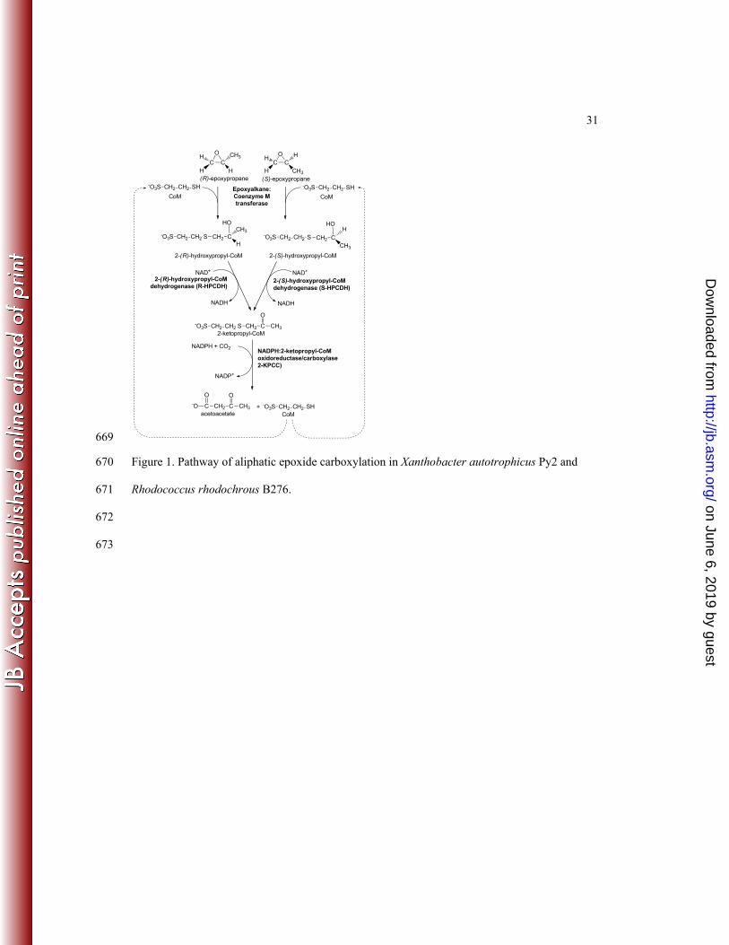
The bacterial metabolism of gaseous propylene by the proteobacterium Xanthobacter 54
autotrophicus Py2 and the actinomycete Rhodococcus rhodochrous B276 is initiated by the 55
insertion of a single oxygen atom into the olefin bond of propylene, forming (R)- and (S)-56
epoxypropane (19, 26, 34, 39). These epoxypropane enantiomers are further metabolized by a 57
three-step linear pathway that uses four enzymes and the atypical cofactor coenzyme M (CoM) 58
(2-mercaptoethanesulfonic acid) to catalyze the net carboxylation of epoxypropane to form 59
acetoacetate as shown in Figure 1 (1, 2, 4, 5, 18, 24). NADPH:2-ketopropyl-coenzyme M 60
oxidoreductase/carboxylase (2-KPCC) is the CO2-fixing enzyme of this pathway, catalyzing the 61
reductive cleavage and carboxylation of 2-ketopropyl-coenzyme M (2-ketopropyl-CoM; 2-(2-62
ketopropylthio)ethanesulfonate; 2-KPC) with the stoichiometry shown in Equation 1 (1, 17, 18): 63
2-KPC + CO2 + NADPH acetoacetate + NADP+ + CoM (1) 64
2-KPCC is the only known carboxylase that is a member of the disulfide oxidoreductase (DSOR) 65
family of enzymes, and, accordingly, employs a mechanistic strategy for organic substrate 66
carboxylation not seen in any other known enzyme (10, 18). All members of the DSOR family 67
follow the general mechanistic strategy shown in Figure 2A for the steps leading to the reduction 68
of a cysteine disulfide bond, where the cysteine residues proximal and distal to FAD are termed 69
the flavin and interchange thiols, respectively (28). For the DSOR enzymes catalyzing the 70
reduction of an oxidized substrate containing a disulfide bond such as glutathione reductase, the 71
interchange thiol attacks one of the sulfur atoms of the disulfide, leading to disulfide bond 72
cleavage, reduction of one thiol of the substrate, and formation of a mixed disulfide between the 73
on June 6, 2019 by guesthttp://jb.asm
.org/D
ownloaded from
5
interchange thiol and the second substrate thiol (28) (Figure 2B). Reformation of the oxidized 74
cysteine pair leads to the reduction and release of the second substrate thiol. 75
As shown in Figure 2C, 2-KPCC catalyzes the reduction of a thioether rather than a 76
disulfide bond, a feature not seen in any other known DSOR enzyme. Mechanistic (10) and 77
structural (27, 29, 30) studies have provided evidence for a reaction mechanism where thioether 78
bond cleavage results in the formation of a mixed disulfide between CoM and the interchange 79
thiol with formation of the enolacetone anion (Figure 2C). The enolacetone anion then 80
undergoes carboxylation to form the product acetoacetate, while the formation of the oxidized 81
cysteine pair results in the release of free CoM (Figure 2C). 82
It is of interest to determine the unique features of 2-KPCC relative to other DSOR 83
enzymes that allow the enzyme to catalyze the unique reactions of thioether bond cleavage, and 84
enolacetone anion formation, stabilization, and carboxylation. Significant insights into these 85
features have come from x-ray crystallographic structures of 2-KPCC determined for various 86
states, including the substrate-free enzyme (27), a 2-KPC-bound form of the enzyme (27), a form 87
of the enzyme where the mixed disulfide of CoM was trapped (30), and a form in which the 88
substrate CO2 is bound (29). Collectively, the structural characterization of 2-KPCC revealed a 89
unique active site architecture relative to other DSOR enzymes, where substrate binding induces 90
a conformational change creating a hydrophobic pocket that encapsulates the substrate 2-KPC 91
(22, 27). Of relevance to the question of how 2-KPCC stabilizes the enolacetone anion for 92
subsequent carboxylation, a hydrogen bonding network was identified in the 2-KPC-bound 93
enzyme consisting of an ordered water molecule hydrogen bonded to both the carbonyl oxygen 94
of 2-KPC and two histidine residues (H137 and H84) (Figure 3A). It has been proposed that this 95
hydrogen bonding network is responsible for stabilizing enolacetone formed from thioether bond 96
on June 6, 2019 by guesthttp://jb.asm
.org/D
ownloaded from
6
cleavage as the interchange thiol attacks the thioether bond as shown in Figure 3B (22, 27). An 97
additional novel feature of the active site of 2-KPCC is a pair of methionine residues (M140 and 98
M361) which flank the substrate 2-KPC. These methionine residues are not present in other 99
DSOR enzymes and may contribute to the unique environment promoting enolacetone 100
stabilization and attack on CO2. 101
The construction of site-directed substitutions of 2-KPCC would allow the tenets of this 102
proposed mechanism to be tested directly. To date, however, all attempts to express 2-KPCC in 103
an active state in a heterologous system have failed, in spite of the relative ease with which the 104
other enzymes of the epoxypropane carboxylation pathway have been expressed and purified 105
(11, 24, 33). In the present paper, we describe an expression system that solves the problems 106
encountered previously and allows 2-KPCC to be expressed in a fully active and soluble state. 107
Site-directed mutants were constructed in the redox active disulfide, the histidine dyad, and one 108
of the flanking methionine residues, and the effects of these mutations on various reactions 109
catalyzed by 2-KPCC were determined in order to gain further insights into their catalytic roles. 110
111
MATERIALS AND METHODS 112
Materials. Commercially available compounds used were of analytical grade and 113
purchased from either Sigma-Aldrich Chemicals or Fisher Scientific. 2-(2-Keto-114
propylthio)ethanesulfonate (2-KPC) was synthesized as described previously (1). All 115
oligonucleotides were purchased from Integrated DNA Technologies. 116
Purification of native 2-KPCC from of Xanthobacter autotrophicus strain Py2. 117
Native 2-KPCC was purified from propylene grown X. autotrophicus Py2 as described 118
previously (3). 119
on June 6, 2019 by guesthttp://jb.asm
.org/D
ownloaded from
7
Plasmid construction of recombinant 2-KPCC. Total genomic DNA was isolated from 120
propylene-grown cells of X. autotrophicus strain Py2 as described previously (24). The gene 121
encoding 2-KPCC (xecC) was amplified by PCR using the primers 5'-122
CACCGTGAAAGTCTGGAACGCCC-3' and 5'-TCACAGGCTCACCAGATTCT-3' following 123
the Failsafe PCR® protocol (Epicentre Biotechnologies, Madison, WI). PCR products were then 124
ligated into the pBAD Directional TOPO® vector (Invitrogen) following the manufacturer's 125
protocol to generate plasmid pDW1 which was then transformed into E. coli One Shot® Top10 126
(Invitrogen) cells. pDW1 was isolated and the insert sequence confirmed by DNA sequencing at 127
the Center for Intergrated BioSystems, Utah State University. 128
Site-Directed Mutagenesis. Site-directed mutagenesis of pDW1 was carried out 129
utilizing the Quikchange® Site-Directed Mutagenesis Kit (Stratagene) according to the 130
manufacturer’s protocols. The sequences of the primer pairs used to create the desired mutations 131
are as follows: C82A, 5'-TCC TGG GCG GCT CGG CCC CGC ACA ATG 132
CGT-3' and 5'-AAG GAC CCG CCG AGC CGG GGC GTG TTA CGC-3'; C87A 5'-GTG CCC 133
GCA CAA TGC GGC CGT GCC GCA CCA TAT GTT-3' and 5'-GAA CAG ATG GTG CGG 134
CAC GGC CGC ATT GTG CGG GCA-3'; H137A, 5’-GAA GTT CAT GAT GCC GGC CGG 135
GCC GTT GCG CC-3’ and CGC AAC GGC CCG GCC GGC ATCATG AAC TTC CA-3’; 136
H84A, 5’-CAC GCA CGC ATT GGC CGG GCA CGA GCC GCC-3’ and 5’-CCG CCG AGC 137
ACG GGC CGG TTA CGC ACG CAC-3’; M140A, 5’-CGG CCC GCA CGG CAT CGC GAA 138
CTT CCA GTC CAA GG-3’ and 5’-CCT TGG ACT GGA AGT TCG CGA TGC CGT GCG 139
GGC CG-3’. Mutations were confirmed by primer extension sequencing at SeqWright DNA 140
Technology Services (Houston, TX). 141
on June 6, 2019 by guesthttp://jb.asm
.org/D
ownloaded from

8
Growth Media. E. coli Top10 cells were grown in Luria-Bertani Rich (LB-Rich) broth 142
containing ampicillin (100 μg/mL). The LB-Rich media contained the following components per 143
liter: 20 g of tryptone, 15 g of yeast extract, 2 g of K2HPO4, 1 g of KH2PO4, and 8 g of NaCl. 144
Growth of Bacteria. All bacteria were grown at 37˚C unless otherwise stated. E. coli 145
Top10 cells that had been transformed with the pDW1 or corresponding mutant plasmid were 146
plated and grown overnight. A single colony from this plate was used to grow a 25-mL liquid 147
culture to an A600 of 0.6 for preparation of 25% glycerol (v/v) stocks that were stored at -80˚C 148
until use. For use, cells from a frozen stock were inoculated into 125 mL of LB-rich medium 149
and grown to an A600 between 0.6 and 1.0 as measured on a Shimadzu UV160U 150
spectrophotometer. This culture was used as the inoculum for a 15 L capacity microferm 151
fermentor (New Brunswick Scientific) containing 12 L of LB-Rich media supplemented with 152
riboflavin (15 mg/L) and antifoam A (0.005% v/v). Cells were allowed to grow at 37˚C with 153
agitation at 400 rpm and forced aeration to an A600 between 0.6 and 1.0. At this time, the 154
temperature was reduced to 30˚C, arabinose was added to 0.02% and the cells were allowed to 155
grow at this temperature for 6 h. Cells were concentrated using a tangential flow filtration 156
system (Millipore) and pelleted by centrifugation. Cell paste was drop frozen in liquid nitrogen 157
and stored at -80˚C. 158
Purification of recombinant 2-KPCC. Cell paste was resuspended in 3 volumes of 159
buffer A (50 mM Tris, 1 mM DTT, 0.1 mM EDTA, 5% glycerol v/v, prepared and buffered to 160
pH 7.4 at 4˚C) with DNase I (0.03 mg/mL) and lysozyme (0.03 mg/mL) and thawed at 30˚C with 161
shaking. All subsequent treatments were performed either on ice or at 4˚C. Cell suspension was 162
passed three times through a French pressure cell at a pressure of 1.1 x 10⁵ kPa and clarified by 163
centrifugation (184,000 relative centrifugal force for 30 minutes at 4˚C). Clarified cell extract 164
on June 6, 2019 by guesthttp://jb.asm
.org/D
ownloaded from
9
was applied to a 0.5 x 5.0 cm column of Ni-NTA Superflow (Pharmacia Biotech) at 7.0 mL/min. 165
The column was then washed with 4 column volumes of buffer A and the bound sample was 166
eluted with a 15 column volume gradient from 0-400 mM imidazole. The purification was 167
followed using SDS-PAGE analysis. Appropriate fractions were pooled and (NH4)2SO4 was 168
added to 800 mM. The solution was incubated at 4˚C with gentle stirring and then applied to a 169
2.6 x 5.5 cm column of phenyl sepharose that had been preequilibrated with buffer A + 800 mM 170
(NH4)2SO4 (buffer C). The column was then washed with 4 column volumes of buffer C and 171
bound protein was eluted with a 15 column volume gradient from 0-100% buffer A followed by 172
an additional five column volumes buffer A. Appropriate fractions were pooled and 173
concentrated by ultrafiltration using a YM30 membrane (Amicon). 174
For removal of the Histidine-patch (HP) thioredoxin leader acquired from the pBAD 175
Directional TOPO® vector, protein samples prepared as above were incubated with enterokinase 176
(0.1 U EKMax, Invitrogen) at 4˚C for 24 hours. The protein was then reloaded onto the Ni-NTA 177
Superflow column. Appropriate fractions were pooled, concentrated by ultrafiltration and frozen 178
dropwise in liquid nitrogen for storage at -80°C. 179
Protein concentrations were determined using a modified biuret assay (9). 2-KPCC 180
concentrations were also determined by using the previously determined extinction coefficient 181
(є450 of 11828 M-1•cm- 1) (3). 182
SDS-PAGE and Immunoblotting Procedures. SDS-PAGE (12% T) was performed 183
following the Laemmli procedure (25). Electrophoresed proteins were visualized by staining 184
with Coomassie blue R-250. The apparent molecular masses of polypeptides were determined 185
by comparison with Rf values of standard proteins. Immunoblot analysis was conducted by 186
electrophoretically transferring proteins from an SDS-polyacrylamide gel onto a polyvinylidene 187
on June 6, 2019 by guesthttp://jb.asm
.org/D
ownloaded from

10
difluoride membrane. The membrane was incubated with polyclonal antiserum raised against 188
purified 2-KPCC from X. autotrophicus strain Py2. Cross-reacting proteins were visualized 189
using horseradish peroxidase conjugated to goat anti-rabbit immunoglobin G (Promega). 190
Coupled spectophotometric assay for 2-KPCC carboxylation activity. A continuous 191
spectrophotometric assay described previously (8) was utilized that couples acetoacetate 192
production by 2-KPCC to acetoacetate reduction and NADH oxidation by β-hydroxybutyrate 193
dehydrogenase (β-HBDH). This assay relies on the fact that DTT can be used as an alternate 194
reductant for 2-KPCC in place of NADPH while NADH cannot, so that loss of absorption at A340 195
due to NADH oxidation by β-HBDH as acetoacetate is reduced can be measured according to 196
equations 2 and 3 (8): 197
2-KPC + CO2 + DTTred acetoacetate + DTTox + CoM (2) 198
acetoacetate + NADH + H+ β-hydroxybutyrate + NAD+ (3) 199
By including a large excess of highly active (260 U/mg, where one unit is defined as one 200
μmol/min) β-HBDH in assays with a fixed concentration of NADH, steady state rates and 201
kinetic parameters can be measured that are not possible in assays where NADPH is consumed 202
directly by 2-KPCC in stoichiometric proportions to 2-KPC. 203
Assays were conducted in 2 mL anaerobic quartz cuvettes that contained a total reaction 204
volume of 1 mL. Assays contained either 0.125 or 0.25 mg 2-KPCC, 0.345 mg (90 units) β-205
HBDH, 10 mM DTT, 0.2 mM NADH and 60 mM carbonate species (added as 33.5 mM CO2 gas 206
plus 26.5 mM KHCO3) in 100 mM Tris buffer, with the pH adjusted to 7.4 at 30°C. Reactions 207
were allowed to equilibrate to 30°C and assays were initiated by the addition of 2.5 μmol 2-KPC. 208
Continuous spectrophotometric assay for 2-KPC formation (reverse reaction). As 209
described previously (10), 2-KPCC catalyzes the reverse of the physiologically important 210
on June 6, 2019 by guesthttp://jb.asm
.org/D
ownloaded from

11
forward reaction shown in Equation 1, a reaction that is dependent on NADP+ as the oxidant as 211
shown in Equation 4: 212
acetoacetate + NADP+ + CoM 2-KPC + CO2 + NADPH (4) 213
Assays were conducted in 2 mL anaerobic quartz cuvettes with a total reaction volume 214
of 1 mL. Each assay contained 0.125 mg 2-KPCC, 5 mM NADP+, 5 mM CoM, and 100 mM 215
acetoacetate in 100 mM Tris buffer, pH 7.4. Reactions were allowed to equilibrate to 30°C and 216
assays were initiated by the addition of acetoacetate. Rates of reaction were measured by 217
monitoring the increase in absorbance (A340) associated with the production of NADPH in a 218
Shimadzu UV160U spectrophotometer containing a water-jacketed cell holder for temperature 219
control. 220
2-Ketopropyl-CoM protonation assays. In the absence of CO2, 2-KPCC catalyzes the 221
cleavage and protonation of 2-KPC to form acetone, using either NADPH or DTT as reductant, 222
at rates about 25% of the rate of 2-KPC carboxylation as shown using DTT as reductant in 223
equation 4 (10): 224
2-KPC + H+ + DTTred acetone+ DTTox + CoM (5) 225
Assays for acetone formation were performed essentially as described (10). Assays were 226
conducted in 9 mL sealed serum vials with a total reaction volume of 1 mL. Each assay 227
contained either 0.125 or 0.25 mg 2-KPCC and 10 mM DTT in 100 mM Tris buffer, pH 7.4. 228
Assay vials were incubated in a 30°C shaking water bath and assays were initiated by the 229
addition of 2.5 μmol 2-KPC. Acetone formation was quantified as a function of time by 230
removing samples and analyzing acetone by gas chromatography as described previously (13). 231
Single turn-over protonation assay. Assays were performed in sealed 3 mL serum vials 232
with a total reaction volume of 1 mL. Each assay contained 2-KPCC (0, 57, or 144 μM) in 100 233
on June 6, 2019 by guesthttp://jb.asm
.org/D
ownloaded from

12
mM Tris buffer, pH 7.4. Assay vials were incubated in a 30˚C shaking water bath (200 cycles 234
min-1) and assays were initiated by addition of 2.5 mM 2-KPC. After 10 min., the vials were 235
transferred to a 65˚C static water bath and incubated for 10 min to allow denaturation of the 236
protein and volatilization of acetone released. Acetone was quantified by gas chromatography as 237
described above. 238
Acetoacetate decarboxylation assay. Assays were performed as described previously 239
(10). Briefly, assays were conducted in 9 mL sealed serum vials with a total reaction volume of 240
1 mL. Vials were depleted of carbonate species by including a KOH-containing trap as 241
described (13). Each assay contained 250 mM acetoacetate and either 0 or 5 mM CoM in 100 242
mM Tris buffer, pH 7.4. Assay vials were incubated in a 30°C shaking water bath and assays 243
were initiated by addition of 0.125 mg 2-KPCC. Acetone was quantified by gas chromatography 244
as described above. 245
Preincubation of 2-KPCC with 2-KPC. For assays that used 2-KPCC preincubated 246
with 2-KPC, 2-KPCC and 2-KPC were incubated together for 5 minutes at 30°C in GTP buffer 247
(200 mM each of glycine, Tris and sodium phosphate), pH 10. Following incubation, 2-KPCC 248
was desalted by loading onto a 2.3 x 5 cm column of Sephadex G-25 PD-10 that had been pre-249
equilibrated with 50 mM Tris buffer pH 7.4. 250
Cys 87-flavin charge transfer. All spectra were obtained at room temperature in a 251
quartz ultramicro (120 μl) cuvette with a 1 cm light path. Aliquoted GTP buffer was adjusted to 252
the desired pH and added to 2-KPCC for a final volume of 130 μl. Spectra were then obtained 253
using a Shimadzu UV160U UV-Vis recording spectrophotometer interfaced with a PC running 254
PC160 Personal Spectroscopy Software Version 1.4. The reference cell contained protein-free 255
on June 6, 2019 by guesthttp://jb.asm
.org/D
ownloaded from
13
GTP buffer. The actual pH of each solution used to obtain a spectrum was determined by mixing 256
an appropriate amount of protein free buffer A with the each of the GTP buffers. 257
UV/visible spectral analysis of native 2-KPCC. Samples of native 2-KPCC (3.6 258
mg/ml) were incubated anoxically in the presence of 10 mM DTT and 10 mM BES for four 259
hours as described previously (8). The samples were then desalted using prepacked columns of 260
Sephadex G-25 (Pharmacia, PD-10) equilibrated in 50 mM Tris buffer, pH 7.4. Desalted 261
samples were air oxidized for 30 minutes. Desalted 2-KPCC was mixed with GTP buffer as 262
described above. 263
Data Analysis. Kinetic constants (Km and Vmax) were calculated by fitting initial rate data 264
to the Michaelis-Menten equation using the methods described by Cleland (14) using the 265
software SIGMAPLOT. pKa values were calculated by fitting A555 versus pH data to a four 266
parameter sigmoidal equation, also using SIGMAPLOT. 267
268
RESULTS 269
Cloning, expression, purification and characterization of recombinant-2-KPCC (r-270
2-KPCC). All previous attempts to express 2-KPCC in a recombinant form resulted in insoluble 271
and consequently inactive protein. To increase the solubility and translation efficiency of 2-272
KPCC, a vector was chosen that included a thioredoxin (HP) fusion tag (38). Clarified cell 273
extract from cultures of E. coli Top10 cells that had been transformed with pDW1 and induced 274
with an optimized concentration of L-(+)-arabinose revealed high soluble expression of 2-KPCC 275
when subjected to SDS-PAGE and immunoblot analyses (Figure 4). Recombinant 2-KPCC (r-2-276
KPCC) purified by two steps (Ni2+ affinity and phenyl sepharose columns) was >90% 277
homogeneous, and migrated with the expected molecular mass of 68 kDa, (57 kDa for the 278
on June 6, 2019 by guesthttp://jb.asm
.org/D
ownloaded from

14
enzyme plus 11 kDa for the fusion tag) (Figure 4, lane 4). Cleavage of the HP tag using 279
enterokinase resulted in the expected shift in migration to 57 kDa for 2-KPCC (Figure 4, lanes 5 280
and 6). R-2-KPCC samples containing and lacking the HP tag had the characteristic UV/visible 281
absorption spectrum expected for members of the DSOR family. A comparison of the 282
absorption spectra of native 2-KPCC and r-2-KPCC (both with and without the HP tag) revealed 283
that all three enzymes contained 1 mol FAD: mol protein monomer, demonstrating that the 284
recombinant expression system results in incorporation of FAD into each active site. 285
Purified 2-KPCC containing the HP tag from different enzyme preparations exhibited 286
specific activities consistently in the range of 50-55 nmol acetone produced/min/mg when 287
assayed according to Equation 4. This activity is in the same range as measured for acetone 288
formation with preparations of native 2-KPCC under identical assay conditions (note that rates of 289
2-KPC carboxylation and protonation when using DTT are 60-65% of the corresponding rates 290
when NADPH is used as the reductant, and that the rate of 2-KPC protonation is about 25% of 291
the rate of 2-KPC carboxylation) (8, 10). Removal of the HP tag resulted in no change in 2-292
KPCC activity. Since 2-KPCC preparations containing and lacking the HP tag were both fully 293
active, contained a full complement of FAD, and migrated on gel filtration columns at the 294
expected positions (i.e. as dimers), subsequent experiments used the form of 2-KPCC retaining 295
the HP tag to avoid the laborious, time consuming and expensive steps required to remove the 296
tag for large preparations of enzyme. 297
Kinetic parameters for r-2-KPCC-catalyzed substrate carboxylation were determined 298
using the recently developed continuous spectrophotometric assay described in the methods 299
(Equations 2 and 3) by using saturating concentrations of CO2 and DTT and varying the 300
concentration of 2-KPC, as was previously done for native 2-KPCC using a discontinuous assay 301
on June 6, 2019 by guesthttp://jb.asm
.org/D
ownloaded from

15
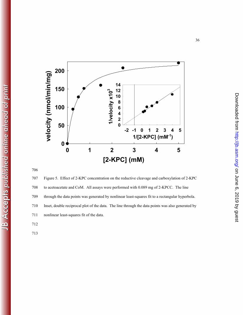
that relies on measuring the incorporation of 14CO2 into acetoacetate (10). As shown in Figure 5, 302
the enzyme conformed to Michaelis-Menten kinetics, providing the following apparent kinetic 303
parameters: Km = 0.42 ± 0.08 mM 2-KPC and Vmax = 233 ± 12 nmol acetoacetate formed•min-304
1•mg-1formed, which corresponds to a kcat of 13 min-1. By comparison, the apparent Km and Vmax 305
values for native 2-KPCC determined previously by the discontinuous assay were 0.63 mM and 306
412 nmol/min/mg, respectively (10). The difference in Vmax to that reported here can be 307
attributed to the use of DTT as the reductant in the discontinuous assay, which results in rates 308
about 60% of the rates with the physiological reductant NADPH (8, 10). 309
Redox-dependent activities for mutants in the redox active disulfide (C87 and C82). 310
Cys87 and Cys82 have been identified by sequence analysis (36) and structural characterization 311
(27) as the flavin and interchange thiols, respectively, of 2-KPCC (see Figure 3). Mutation of 312
either cysteine residue to alanine resulted in complete loss of steady-state 2-KPC carboxylation 313
(Equations 1 and 2) and protonation (Equation 5) activities, regardless of whether DTT or 314
NADPH was used as the reductant in the assays (Table 1). Likewise, no activity was seen when 315
the alanine-substituted enzymes were assayed in the reverse direction for 2-KPC production from 316
acetoacetate (Equation 4). 317
It is noteworthy that the C87A variant was inactive for the forward steady-state reactions 318
where DTT was used as the reductant in place of the physiologically important NADPH, since, 319
conceivably, this enzyme might be capable of reacting to form the mixed disulfide of C82 and 320
CoM (see Figure 3B), which could then be reduced by DTT to allow further turnovers. To test 321
the possibility that the C87A variant can undergo a single turnover, which would not be detected 322
using the steady state activity assays, a high concentration of the enzyme was incubated with the 323
substrate 2-KPC and in the absence of reductant for a fixed time, and the amount of acetone that 324
on June 6, 2019 by guesthttp://jb.asm
.org/D
ownloaded from

16
was produced was then determined. When 57 nmol or 144 nmol of 2-KPC were incubated in 325
this fashion, 87 and 220 nmol of acetone were produced, respectively, once equilibrium was 326
established. Since 2-KPCC is a dimer, this corresponds to an average production of 0.76 mol 327
acetone per enzyme active site for both concentrations of enzyme assayed in this fashion, 328
suggesting that C82 is active for a single turnover to cleave the thioether bond of 2-KPC to 329
release enolacetone which undergoes protonation to acetone. This reaction presumably occurs 330
by formation of the C82-CoM mixed disulfide. As a control, this experiment was repeated with 331
the C82A variant and no acetone was observed within the detection limit of the assay. The 332
addition of DTT to assays of the C87A variant did not increase the amount of acetone 333
production. 334
UV/visible spectral properties of r-2-KPCC variants. A distinguishing feature of 335
DSOR enzymes is an interaction between the oxidized flavin and the deprotonated flavin thiol 336
when the enzyme is in the two electron reduced state (Figure 2A, product of reaction (iiiA)) (37). 337
This interaction results in a charge-transfer complex that alters the spectral features of FAD 338
through a characteristic increase in absorbance above 525 nm and a decrease in absorbance at the 339
maximal absorbance of 450 nm (37). The charge transfer complex can be observed in one of 340
several ways for DSOR enzymes: (a) by titration of the enzyme under anoxic conditions to the 341
two electron reduced state, (b) by rapid-scanning spectrophotometric techniques where the 342
development of the charge transfer can be observed transiently for single turnover catalysis, (c) 343
by site-directed mutagenesis of the interchange cysteine, in which case no disulfide can form and 344
the oxidized enzyme exhibits the characteristic charge transfer, and (d) by specific alkylation of 345
the interchange thiol, which has the same effect as (c). Methods (c) and (d) have routinely been 346
used to determine the pKa values for the flavin thiol of DSOR enzymes (7, 32). 347
on June 6, 2019 by guesthttp://jb.asm
.org/D
ownloaded from

17
In order to characterize the interaction between C87 and FAD in 2-KPCC, UV/visible 348
spectra were recorded for air-oxidized samples of native r-2-KPCC and the alanine substitutions 349
in the interchange (C82) and flavin (C87) cysteine residues over a range of pH values from 5 to 350
11. The spectra of the native recombinant and C87A proteins were indistiguishable from that of 351
the spectrum of oxidized native 2-KPCC, and no change in the spectra were observed over the 352
range of pH values from 5 to 11 (data not shown). This is as expected, since for air oxidized 353
native r-2-KPCC, C82 and C87 will be in the disulfide form, and C87A has no flavin thiol, and 354
hence no charge transfer can develop. As shown in representative spectra for C82A at different 355
pH values in Figure 6, the characteristic charge transfer due to interaction of the thiolate of C87 356
with FAD only developed at pH values substantially higher than physiological pH, reaching a 357
maximum value at pH 11. This is in stark contrast to other DSOR enzymes where the flavin 358
cysteine is largely in the deprotonated form in the microenvironment of the active sites of these 359
enzymes at pH 7 since the microscopic pKa values are perturbed from the value of 8.2 for free 360
cysteine to values around 5 (7, 15, 31, 32). The absorbance at 555 nm was plotted vs. pH for a 361
range of pH values to determine the pKa value for the flavin cysteine (C87) in C82A (Figure 7, 362
solid circles). A fit of this data provided a pKa of 8.76 ± 0.09, which is fully four units higher 363
(four orders of magnitude less acidic) than that determined for the classical DSOR enzymes 364
glutathione reductase (7) and mercuric reductase (32). 365
Recently, the CoM analog 2-bromoethanesulfonate was shown to be a highly specific 366
affinity label for 2-KPCC that specifically alkylates the interchange thiol (C82) of 2-KPCC (8). 367
As shown in Figure 7 (open triangles), the charge transfer absorbance of BES-modified 2-KPCC 368
developed in a similar fashion to that of C82A with increasing pH, but to a lower maximal value 369
than for the mutant. In spite of the lower change in total absorbance, a fit of the titration data for 370
on June 6, 2019 by guesthttp://jb.asm
.org/D
ownloaded from
18
BES-modified 2-KPCC yielded a pKa value that is identical within experimental error to that 371
seen for the C82A mutant (8.74 ± 0.13). 372
Redox-dependent Reactions of H84A and H137A 2-KPCC. The structures of 2-KPCC 373
show the presence of a ‘catalytic dyad” of histidine molecules (H137 and H84) that are hydrogen 374
bonded to an ordered water molecule that is in turn within hydrogen bonding distance of the 375
carbonyl oxygen of 2-KPC in the 2-KPC-bound structure (Figure 3) (27). This unique pair of 376
histidine residues is not seen in any other member of the DSOR family, and has been proposed to 377
stabilize enolacetone formed from 2-KPC thioether bond cleavage (27) (Figure 3B). 378
In order to test the proposed roles of H137 and H84 in the redox-dependent reactions of 379
2-KPCC, the residues were individually mutated to alanine residues and the effects on specific 380
activities for 2-KPC carboxylation (Equation 2), 2-KPC protonation (Equation 5), and 2-KPC 381
formation from acetoacetate (Equation 4) were determined. As shown in Table 1, mutagenesis 382
of the histidine proximal to the ordered water (H137) resulted in drastically reduced activities 383
(92-100% loss of activity) for the three redox-dependent activities of 2-KPCC. Mutagenesis of 384
the distal histidine resulted in more moderate losses of activity (58-76%). 385
Effect of mutations in the redox active disulfide and catalytic dyad on acetoacetate 386
decarboxylase activity of 2-KPCC. 2-KPCC was previously shown to catalyze a reaction that 387
does not involve redox chemistry: the decarboxylation of acetoacetate to produce acetone and 388
CO2 according to Equation 6 (10): 389
acetoacetate + H+ acetone + CO2 Equation 6 390
Acetoacetate decarboxylase activity was very low when catalyzed by 2-KPCC alone, but was 391
stimulated 77-fold upon the addition of a saturating (5 mM) concentration of CoM (10). 392
on June 6, 2019 by guesthttp://jb.asm
.org/D
ownloaded from
19
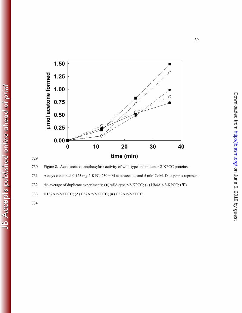
The rate of CoM-dependent acetoacetate decarboxylation by native r-2-KPCC was found 393
to be comparable to that reported for native 2-KPCC previously (10). As shown 394
in Figure 8, the rates of CoM-dependent acetoacetate decarboxylation by H137A and H84A were 395
comparable to that of the wild-type enzyme, while C82A and C87A exhibited rates that were 396
somewhat higher than the native enzyme. As for native 2-KPCC, acetoacetate decarboxylase 397
activity in each of the site-directed variants was approximately 70-80-fold lower when CoM was 398
not included in the assays. 399
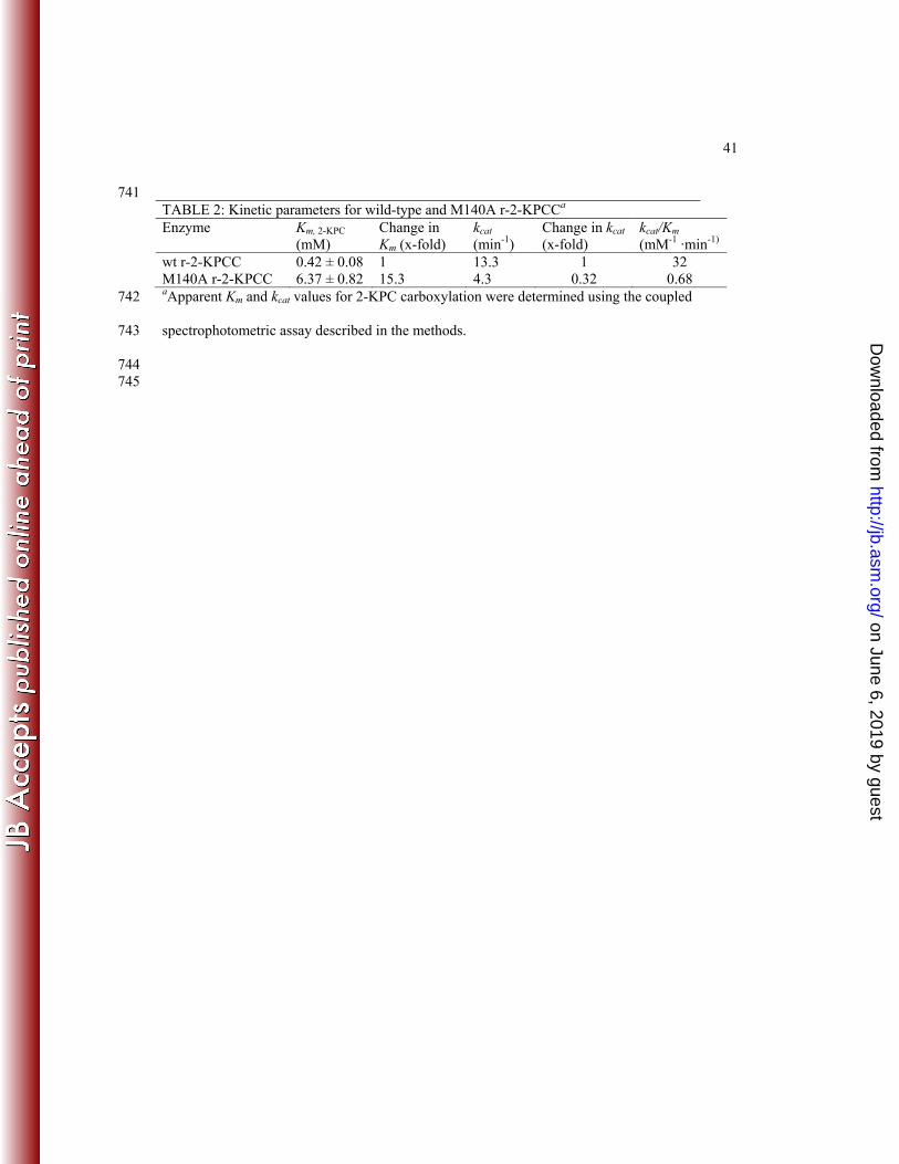
Kinetic characterization of a mutation in one of the methonines flanking 2-KPC. As 400
shown in Figure 3A, a distinguishing feature of 2-KPCC is the presence of two methionine 401
residues (M140 and M361) that flank the substrate 2-KPCC. An alanine substitution was 402
successfully created in M140, but, unfortunately, attempts to make and express a corresponding 403
substitution in M361 were unsuccessful. As shown in Table 2, the M140A substitution resulted 404
in an enzyme with a 15-fold higher Km for the substrate 2-KPC and a 3-fold lower kcat, resulting 405
in an overall catalytic efficiency for 2-KPC carboxylation that is 47-fold lower than for the 406
native enzyme. 407
DISCUSSION 408
In the present work, 2-KPCC has for the first time been expressed in a heterologous 409
system. The biochemical and kinetic properties of r-2-KPCC containing the HP tag show that it 410
is essentially identical to that of native 2-KPCC, and thus suitable for construction of, and 411
comparative studies with, site-directed variants. The characterization of five of these site-412
directed variants has allowed key tenets of the mechanism of thioether bond cleavage and 413
substrate carboxylation proposed from mechanistic (8, 10) and structural (27, 29, 30) studies to 414
be tested. 415
on June 6, 2019 by guesthttp://jb.asm
.org/D
ownloaded from

20
Catalytic roles and unique properties of the redox active disulfide of 2-KPCC. As 416
expected, the redox active cysteines of 2-KPCC are crucial for all steady-state reactions requiring 417
thioether bond cleavage or formation (Equations 1, 2, 4 and 5), consistent with the key 418
intermediacy of the Cys82-CoM mixed disulfide in these reactions (Figure 3B). The CoM-419
Cys82 mixed disulfide has previously been captured for native 2-KPCC by incubating the 420
enzyme in the presence of 20 mM CoM prior to crystallization (30). The C87A substituted 2-421
KPCC catalyzed the single turnover production of acetone from 2-KPC, demonstrating that the 422
interchange thiol (C82) is still capable of thioether bond cleavage, and presumably CoM-Cys82 423
mixed disulfide formation, in the absence of the flavin thiol. The fact that the addition of DTT 424
did not lead to an increase in acetone production in these assays indicates that DTT is not 425
capable of directly reducing the CoM-C82 mixed disulfide, as by doing so C82 would be re-426
reduced for additional rounds of catalysis. 427
These observations provide insights into how DTT mediates the redox-dependent 428
reactions of 2-KPCC in place of NADPH. As noted above, the rates of DTT-dependent 2-KPC 429
protonation or carboxylation are about 60% of the rates observed when NADPH is used as 430
reductant. It is presumed that DTT reduces the oxidized disulfide of C82 and C87 directly, i.e. 431
without the intermediacy of FAD. Incubation of air-oxidized 2-KPCC in the absence of 432
substrates results in reduction of the redox active disulfide, as evidenced by the ability of the 433
DTT-reduced enzyme to undergo stoichiometric alkylation on C82 by the CoM analog BES (8). 434
Prolonged incubation of 2-KPCC with DTT results in bleaching of the flavin of 2-KPCC, but this 435
occurs too slowly (on the order of 30 minutes to an hour) to be a relevant pathway for electron 436
transfer under steady state conditions. In any event, the results presented herein show that DTT is 437
a suitable reductant for 2-KPCC when both redox active thiols are present, but not when C87 has 438
on June 6, 2019 by guesthttp://jb.asm
.org/D
ownloaded from

21
been substituted by alanine. This suggests that DTT does not directly reduce the mixed disulfide 439
of C82 and CoM under steady state turnover conditions but instead reduces the redox active 440
cysteine pair. 441
As shown in Figures 6 and 7, the flavin thiol (C87) is in the fully protonated state at 442
neutral pH for both the alanine substitution in the interchange thiol and for the native enzyme 443
where the interchange thiol is alkylated by ethylsulfonate. C87 has a pKa value fully 4 units 444
higher (four orders of magnitude more basic) than the perturbed value for the flavin thiols of 445
other members of the DSOR family (7, 32). A key piece of structural information that explains 446
this difference is the replacement of a histidine side chain that interacts with and lowers the pKa 447
of the flavin thiol for all other DSOR enzymes (31) by a phenylalanine residue in 2-KPCC (27). 448
It should be noted that DSOR enzymes are dimers, with each active site being at the interface 449
between the two subunits, and hence interacting with residues from the second subunit. The 450
histidine that lowers the pKa of the interchange thiol in conventional DSOR enzymes, and that is 451
replaced by phenylalanine in 2-KPCC, resides on the other subunit of the dimer (Phe501 of 452
subunit 2 in this instance). All of the other active site residues noted up until now reside within 453
the primary subunit forming the active site of 2-KPCC (with two such active sites present on 454
each dimeric enzyme). The replacement of histidine by phenylalanine in the environment of the 455
flavin thiol is thought to help maintain the hydrophobicity of the enzyme active site, and direct 456
the immediate product of thioether bond cleavage (enolacetone) towards carboxylation to form 457
acetoacetate (the physiologically important reaction) rather than protonation to form acetone (a 458
fortuitous side reaction that occurs when CO2 is not present) (10, 27). The prevention of 459
protonation of enolacetone to form acetone, an essentially irreversible reaction, is of paramount 460
importance in 2-KPCC. In stark contrast, for glutathione reductase, a secondary (and major) role 461
on June 6, 2019 by guesthttp://jb.asm
.org/D
ownloaded from

22
of the histidine that lowers the the pKa of the flavin thiol is to protonate the first molecule of 462
glutathione formed upon disulfide bond cleavage to drive the reaction forward and prevent the 463
reverse reaction from occurring (31). 464
The lack of the histidine general base that deprotonates the flavin thiol would be expected 465
to slow the rate of disulfide bond formation by 2-KPCC relative to glutathione reductase 466
(compare steps in Figures 2B and 2C). However, it is likely that a step other than disulfide bond 467
formation, or another step involving oxidation/reduction, is rate limiting for 2-KPCC, which 468
turns over much slower than glutathione reductase. In this context it should be noted that 2-KPC 469
carboxylation is approximately 40% faster when the physiological reductant NADPH is used 470
instead of DTT, indicating that at least one redox step is partially rate limiting. 471
It should be noted that the steady state kcat we report here for 2-KPCC carboxylation 472
(13.3 min-1), is fully 3300-times lower than the turnover number reported for glutathione 473
reduction by glutathione reductase from E. coli (44,000 min-1) (20). The large difference in 474
steady state turnover highlights the more difficult chemistry of thioether bond cleavage and 475
substrate carboxylation relative to disulfide bond reduction catalyzed by classical DSOR 476
enzymes. The turnover numbers for the other enzymes of the epoxypropane carboxylation 477
pathway (Figure 1) are significantly higher (6.5 s-1 for epoxyalkane:CoM transferase, and 25-50 478
s-1 for the R- and S-hydroxypropyl-CoM dehydrogenases (12, 23, 33) ) As a reflection of the low 479
turnover of 2-KPCC,it is expressed at very high levels (~25% of soluble cell protein) in 480
propylene-grown cells of X. autotrophicus Py2 (3, 16). In spite of this high level of expression, 481
2-KPCC has the lowest specific activity of the enzymes of the epoxide carboxylation pathway in 482
cell extracts, with an activity that is identical to the specific activity of epoxypropane 483
consumption by whole cell suspensions of X. autotrophicus Py2 growing with propylene as the 484
on June 6, 2019 by guesthttp://jb.asm
.org/D
ownloaded from

23
carbon source (90 nmol epoxypropane consumed/min/mg (35) ). Clearly, the reaction catalyzed 485
by 2-KPCC is the rate limiting step of the epoxide carboxylation pathway shown in Figure 1. 486
Still, the very high level of expression of 2-KPCC allows X. autotrophicus and R. rhodochrous to 487
grow fairly well (doubling times in exponential phase of about 8 hours) with propylene as the 488
carbon source (4, 35). 489
Role of the catalytic histidine dyad in stabilizing enolacetone in reactions involving 490
thioether bond cleavage or breakage. Site-directed mutagenesis of the histidines proximal 491
(H137) and distal (H84) to the ordered water that interacts with the carbonyl oxygen of bound 2-492
KPC in the crystal structure (Figure 3A) support the hypothesis that these residues are critical in 493
stabilizing enolacetone as an intermediate in reactions that make or break a thioether bond 494
(Equations 1, 2, 4 and 5). As expected, mutation of the proximal histidine resulted in much 495
lower activities than for the distal histidine, indicating that the proximal histdine can still provide 496
some stabilization in the absence of the additional charge relay provided by H84. 497
Acetoacetate decarboxylase activity of 2-KPCC does not involve the redox active 498
disulfide or histidine dyad. The decarboxylation of acetoacetate to acetone and CO2 by 2-499
KPCC must of necessity occur by C-C bond cleavage to form enolacetone, the same high energy 500
intermediate(s) formed in the redox-dependent reactions of 2-KPCC. The fact that the C82A, 501
C87A, H84A, and H137A all retain full (or slightly increased) CoM-dependent acetoacetate 502
decarboxylase activity demonstrates that acetoacetate decarboxylation must be occurring by a 503
fundamentally different mechanism than for the reactions that involve redox chemistry and 504
thioether bond cleavage. At the same time, acetoacetate decarboxylase activity still requires CoM 505
for optimal activity. These results can be rationalized and explained by examining the structural 506
features of the form of 2-KPCC captured in the presence of the mixed disulfide of CoM (30). 507
on June 6, 2019 by guesthttp://jb.asm
.org/D
ownloaded from
24
This structure showed the presence of acetone bound to a distinct binding pocket 4.5 Å from the 508
thiol sulfur of CoM (30). Within this pocket, which consists of residues from the second enzyme 509
subunit, are Gln509, which formed a hydrogen bond with the carbonyl O of acetone, and His506. 510
Based on this structure, it was proposed that this second pocket constitutes a binding pocket for 511
the physiological product acetoacetate (30). By extrapolation, we propose now that this second 512
pocket is the active site for acetoacetate decarboxylation. The stimulatory effect of CoM on 513
acetoacetate decarboxylase activity can be rationalized in one or both of the following ways: (1), 514
the thiol of CoM, when bound to the CoM binding pocket of 2-KPCC, is the proton donor for 515
formation of acetone from enolacetone, i.e. CoM serves as a general acid to promote 516
decarboxylation; and/or (2), binding of CoM induces a conformational change similar to that 517
induced by binding of the substrate 2-KPC that facilitates the binding of acetoacetate within the 518
second binding pocket such that it can undergo decarboxylation. 519
Methionine140 modulates Km and kcat for 2-KPC carboxylation. A distinctive feature 520
of 2-KPCC relative to other DSOR enzymes is the pair of methionine residues that flank the 521
substrate 2-KPC (27) (Figure 3A). The step prior to that catalyzed by 2-KPCC in bacterial 522
epoxide metabolism is catalyzed by a pair of stereoselective dehydrogenases that oxidize the R- 523
and S-enantiomers of hydroxypropyl-CoM (HPC) to 2-KPC (1, 6). Interestingly, the crystal 524
structure solved for R-hydroxypropyl-CoM dehydrogenase (R-HPCDH) in the presence of the 525
product 2-KPC also shows a pair of methionione residues flanking 2-KPC in a fashion similar to 526
that seen in 2-KPCC (21). Additionally, a homology model for the other dehydrogenase, S-527
HPCDH, also shows flanking methionines (22). Thus, methionine residues seem to be a crucial 528
feature in the binding of CoM thioethers during the steps of bacterial epoxide carboxylation. The 529
M140A substituted 2-KPCC was kinetically characterized in the present work and found to 530
on June 6, 2019 by guesthttp://jb.asm
.org/D
ownloaded from
25
exhibit both a marked increase in Km and decrease in kcat (Table 2). Unfortunately, all attempts 531
to make an alanine substitution in the other methionine (M361) were unsuccessful. Although the 532
crystal structures of 2-KPCC and R-HPCDH do not indicate a catalytic role for the flanking 533
methionines, this initial characterization shows that they appear to be crucial for substrate 534
binding and catalysis. 535
Summary. 2-KPCC is distinct from all other known members of the DSOR family in 536
catalyzing thioether bond cleavage and substrate carboxylation. The results of the present work 537
establish the essential role of a novel catalytic dyad that facilitates enolacetone formation and 538
stabilization, and a unique active site environment for the redox active cysteine pair that is unlike 539
that seen in any other DSOR enzyme. The combination of structural biology and site-directed 540
mutagenesis, together with the characterization of redox-dependent and redox-independent 541
reactions, provides a powerful complement for elucidating mechanistic details for this novel 542
carboxylase. 543
544
ACKNOWLEDGMENTS 545
This work was supported by National Institutes of Health grant GM51805 to S.A.E. and by 546
Department of Energy Grant DE-FG02-04ER15563 to J.W.P. 547
548
549
REFERENCES 550
1. Allen, J. R., D. D. Clark, J. G. Krum, and S. A. Ensign. 1999. A role for coenzyme M 551
(2-mercaptoethansulfonic acid) in a bacterial pathway of aliphatic epoxide carboxylation. 552
Proc. Natl. Acad. Sci. U.S.A. 96:8432-8437. 553
on June 6, 2019 by guesthttp://jb.asm
.org/D
ownloaded from
26
2. Allen, J. R., and S. A. Ensign. 1996. Carboxylation of epoxides to ß-keto acids in cell 554
extracts of Xanthobacter strain Py2. J. Bacteriol. 178:1469-1472. 555
3. Allen, J. R., and S. A. Ensign. 1997. Characterization of Three Protein Components 556
Required for Functional Reconstitution of the Epoxide Carboxylase Multienzyme 557
Complex from Xanthobacter strain Py2. J. Bacteriol. 179:3110-3115. 558
4. Allen, J. R., and S. A. Ensign. 1998. Identification and characterization of epoxide 559
carboxylase activity in cell extracts of Nocardia corallina strain B276. J. Bacteriol. 560
180:2072-2078. 561
5. Allen, J. R., and S. A. Ensign. 1997. Purification to Homogeneity and Reconstitution of 562
the Individual Components of the Epoxide Carboxylase Multiprotein Enzyme Complex 563
from Xanthobacter strain Py2. J. Biol. Chem. 272:32121-32128. 564
6. Allen, J. R., and S. A. Ensign. 1999. Two Short-Chain Dehydrogenases Confer 565
Stereoselectivity for Enantiomers of Epoxypropane in the Multiprotein Epoxide 566
Carboxylating Systems of Xanthobacter Strain Py2 and Nocardia corallina B276. 567
Biochemistry 38:247-256. 568
7. Arscott, L. D., C. Thorpe, and C. H. J. Williams. 1981. Glutathione reductase from 569
yeast. Differential reactivity of the nascent thiols in two-electron reduced enzyme and 570
properties of a monoalkylated derivative. Biochemistry 20:1513. 571
8. Boyd, J. M., D. D. Clark, M. A. Kofoed, and S. A. Ensign. 2010. Mechanism of 572
Inhibition of Aliphatic Epoxide Carboxylation by the Coenzyme M Analog 2-573
Bromoethanesulfonate. Journal of Biological Chemistry 285:25232-25242. 574
9. Chromy, V., J. Fischer, and V. Kulhanek. 1974. Re-evaluation of EDTA-chelated 575
biuret reagent. Clin. Chem. 20:1362-1363. 576
on June 6, 2019 by guesthttp://jb.asm
.org/D
ownloaded from
27
10. Clark, D. D., J. R. Allen, and S. A. Ensign. 2000. Characterization of five catalytic 577
activities associated with the NADPH : 2-ketopropyl-coenzyme M [2-(2-578
ketopropylthio)ethanesulfonate] oxidoreductase/carboxylase of the Xanthobacter strain 579
Py2 epoxide carboxylase. Biochemistry 39:1294-1304. 580
11. Clark, D. D., J. M. Boyd, and S. A. Ensign. 2004. The stereoselectivity and catalytic 581
properties of Xanthobacter autotrophicus 2-[(R)-2-Hydroxypropylthio]ethanesulfonate 582
dehydrogenase are controlled by interactions between C-terminal arginine residues and 583
the sulfonate of coenzyme M. Biochemistry 43:6763-71. 584
12. Clark, D. D., and S. A. Ensign. 2002. Characterization of the 2- (R)-2-585
hydroxypropylthio ethane sulfonate dehydrogenase from Xanthobacter strain Py2: 586
product inhibition, pH dependence of kinetic parameters, site-directed mutagenesis, rapid 587
equilibrium inhibition, and chemical modification. Biochemistry 41:2727-2740. 588
13. Clark, D. D., and S. A. Ensign. 1999. Evidence for an inducible nucleotide-dependent 589
acetone carboxylase in Rhodococcus rhodochrous B276. J. Bacteriol. 181:2752-2758. 590
14. Cleland, W. W. 1979. Statistical analysis of enzyme kinetic data. Methods Enzymol. 591
63:103-138. 592
15. Distefano, M. D., K. G. Au, and C. T. Walsh. 1989. Mutagenesis of the redox-active 593
disulfide in mercuric reductase: catalysis by mutant enzymes restricted to flavin redox 594
chemistry. Biochemistry 28:1163-1183. 595
16. Ensign, S. A. 1996. Aliphatic and chlorinated alkenes and epoxides as inducers of alkene 596
monooxygenase and epoxidase activities in Xanthobacter strain Py2. Appl. Environ. 597
Microbiol. 62:61-66. 598
on June 6, 2019 by guesthttp://jb.asm
.org/D
ownloaded from

28
17. Ensign, S. A. 2001. Microbial metabolism of aliphatic alkenes. Biochemistry 40:5845-599
5853. 600
18. Ensign, S. A., and J. R. Allen. 2003. Aliphatic Epoxide Carboxylation. Ann. Rev. 601
Biochem. 72:55-76. 602
19. Gallagher, S. C., R. Cammack, and H. Dalton. 1997. Alkene monooxygenase from 603
Nocardia corallina B-276 is a member of the class of dinuclear iron proteins capable of 604
stereospecific epoxygenation reactions. European Journal of Biochemistry 247:635-641. 605
20. Henderson, G. B., N. J. Murgolo, J. Kuriyan, K. Osapay, D. Kominos, A. Berry, N. 606
S. Scrutton, N. W. Hinchliffe, R. N. Perham, and A. Cerami. 1991. Engineering the 607
substrate specificity of glutathione reductase toward that of trypanothione reduction. 608
Proc. Natl. Acad. Sci. USA 88:8769-8773. 609
21. Krishnakumar, A. M., B. P. Nocek, D. D. Clark, S. A. Ensign, and J. W. Peters. 610
2006. Structural basis for stereoselectivity in the (R)- and (S)-611
hydroxypropylthioethanesulfonate dehydrogenases. Biochemistry 45:8831-40. 612
22. Krishnakumar, A. M., D. Sliwa, J. A. Endrizzi, E. S. Boyd, S. A. Ensign, and J. W. 613
Peters. 2008. Getting a Handle on the Role of Coenzyme M in Alkene Metabolism. 614
Microbiology and Molecular Biology Reviews 72:445. 615
23. Krum, J. G., H. Ellsworth, R. R. Sargeant, G. Rich, and S. A. Ensign. 2002. Kinetic 616
and microcalorimetric analysis of substrate and cofactor interactions in epoxyalkane : 617
CoM transferase, a zinc- dependent epoxidase. Biochemistry 41:5005-5014. 618
24. Krum, J. G., and S. A. Ensign. 2000. Heterologous expression of bacterial epoxyalkane 619
: Coenzyme M transferase and inducible coenzyme M biosynthesis in Xanthobacter 620
strain Py2 and Rhodococcus rhodochrous B276. J. Bacteriol. 182:2629-2634. 621
on June 6, 2019 by guesthttp://jb.asm
.org/D
ownloaded from
29
25. Laemmli, U. K. 1970. Cleavage of structural proteins during the assembly of the head of 622
bacteriophage T4. Nature 227:680-685. 623
26. Miura, A., and H. Dalton. 1995. Purification and characterization of the alkene 624
monooxygenase from Nocardia corallina B-276. Biosci Biotechnol Biochem 59:853-859. 625
27. Nocek, B., S. B. Jang, M. S. Jeong, D. D. Clark, S. A. Ensign, and J. W. Peters. 2002. 626
Structural basis for CO2 fixation by a novel member of the disulfide oxidoreductase 627
family of enzymes: 2-ketopropyl-Coenzyme M Oxidoreductase/ Carboxylase. 628
Biochemistry 41:12907-12913. 629
28. Pai, E. F. 1991. Variations on a theme:the family of FAD-dependent NAD(P)H-630
(disulfide)-oxidoreductases. Curr. Opin. Struct. Biol. 1:796-803. 631
29. Pandey, A. S., D. W. Mulder, S. A. Ensign, and J. W. Peters. 2011. Structural basis for 632
carbon dioxide binding by 2-ketopropyl coenzyme M oxidoreductase/carboxylase. FEBS 633
Letters 585:459-464. 634
30. Pandey, A. S., B. Nocek, D. D. Clark, S. A. Ensign, and J. W. Peters. 2006. 635
Mechanistic implications of the structure of the mixed-disulfide intermediate of the 636
disulfide oxidoreductase, 2-ketopropyl-coenzyme M oxidoreductase/carboxylase. 637
Biochemistry 45:113-120. 638
31. Rietveld, P., L. D. Arscott, A. Berry, N. S. Scrutton, M. P. Deonarain, R. N. Perham, 639
and C. H. Williams Jr. 1994. Reductive and oxidative half-reactions of glutathione 640
reductase from Escherichia coli. Biochemistry 33:13888-13895. 641
32. Schultz, P. G., K. G. Au, and C. T. Walsh. 1985. Directed Mutagenesis of the Redox-642
Active Disulfide in the Flavoenzyme Mercuric Ion Reductase. Biochemistry 24:6840-643
6848. 644
on June 6, 2019 by guesthttp://jb.asm
.org/D
ownloaded from
30
33. Sliwa, D. A., A. M. Krishnakumar, J. W. Peters, and S. A. Ensign. 2010. Molecular 645
Basis for Enantioselectivity in the (R)- and (S)-Hydroxypropylthioethanesulfonate 646
Dehydrogenases, a Unique Pair of Stereoselective Short-Chain 647
Dehydrogenases/Reductases Involved in Aliphatic Epoxide Carboxylation. Biochemistry 648
49:3487-3498. 649
34. Small, F. J., and S. A. Ensign. 1997. Alkene Monooxygenase from Xanthobacter strain 650
Py2: Purification and Characterization of A Four-Component System Central to the 651
Bacterial Metabolism of Aliphatic Alkenes. J. Biol. Chem. 272:24913-24920. 652
35. Small, F. J., and S. A. Ensign. 1995. Carbon Dioxide Fixation in the Metabolism of 653
Propylene and Propylene Oxide by Xanthobacter strain Py2. J. Bacteriol. 177:6170-6175. 654
36. Swaving, J., C. A. Weijers, A. J. van Ooyen, and J. A. M. de Bont. 1995. 655
Complementation of Xanthobacter Py2 mutants defective in epoxyalkane degradation, 656
and expression and nucleotide sequence of the complementing DNA fragment. 657
Microbiology 141:477-484. 658
37. Walsh, C. 1979. Enzymatic Reaction Mechanisms. W. H. Freeman and Co., New York. 659
38. Zhijian, L., E. A. DiBlasio, K. L. Grant, N. W. Warne, E. R. LaVallie, L. A. Collins-660
Racie, M. T. Follettie, M. J. Williams, and J. M. McCoy. 1996. Histidine patch 661
thioredoxins: Mutant forms of thioredoxin with metal chelating affinity that provide for 662
convenient purifications of thioredoxin fusion proteins. J. Biol. Chem. 271:5059-5065. 663
39. Zhou, N. Y., C. K. C. K. Chion, and D. J. Leak. 1996. Cloning and expression of the 664
genes encoding the propene monooxygenase from Xanthobacter, Py2. Appl. Microbiol. 665
Biotechnol. 44:582-588. 666
667
668
on June 6, 2019 by guesthttp://jb.asm
.org/D
ownloaded from
31
CH3C
OC
H
H
H(R)-epoxypropane
HC
OC
CH3
H
H(S)-epoxypropane
S
2-(R)-hydroxypropyl-CoM
C
HO
CH2CH3
HC
HO
CH2H
CH3S
2-(S)-hydroxypropyl-CoM
C
O
CH2 CH3S
NAD+
NADH
NAD+
NADH
C
O
CH2 CH3C
O-O
acetoacetate
Epoxyalkane:Coenzyme Mtransferase
2-(R)-hydroxypropyl-CoMdehydrogenase (R-HPCDH)
2-ketopropyl-CoM
2-(S)-hydroxypropyl-CoMdehydrogenase (S-HPCDH)
NADPH + CO2 NADPH:2-ketopropyl-CoMoxidoreductase/carboxylase2-KPCC)
NADP+
-O3S CH2 CH2 SH -O3S CH2 CH2 SH
-O3S CH2 CH2-O3S CH2 CH2
-O3S CH2 CH2
-O3S CH2 CH2 SH
CoM
+
CoM
CoM
669
Figure 1. Pathway of aliphatic epoxide carboxylation in Xanthobacter autotrophicus Py2 and 670
Rhodococcus rhodochrous B276. 671
672
673
on June 6, 2019 by guesthttp://jb.asm
.org/D
ownloaded from

32
Cys S-
Cys SH
Cys S
Cys S
FADH2FADCys S
Cys S
NADPH + H+ NADP+
FAD
Cys S-
Cys SH
FADCys S-
Cys S
FAD
S-G
FADCys S
Cys S
G-S S-G G-SH G-SH
CoM-S CH2 C
O
CH3
H2C C
O-
CH3
(iA) (iiiA)
(ivB) (vB)
Cys SH
Cys SH
FADCys SH
Cys S
FAD
S-CoM
FADCys S
Cys S
CoM-SH
(ivC) (vC)
CO2
CH2 C
O
CH3C-O
O
(viC)
Cys S
Cys SH
FADH
(iiA) + H+
(A)
(B)
(C)
H+
H+
674
Figure 2. Reactions of members of the DSOR family. (A), steps resulting in the reduction of the 675
redox active cysteine disulfide for all DSOR enzymes; (B), steps resulting in reduction of a 676
substrate with an oxidized disulfide bond, as illustrated for glutathione reductase; (C), steps 677
resulting in reduction of the thioether bond of 2-KPC in 2-KPCC, resulting in the production of 678
enolacetone, which undergoes carboxylation to form acetoacetate. Note that the reduced flavin 679
cysteine is shown in the thiol rather than as the thiolate form for 2-KPCC for reasons described 680
in the paper. 681
682
on June 6, 2019 by guesthttp://jb.asm
.org/D
ownloaded from

33
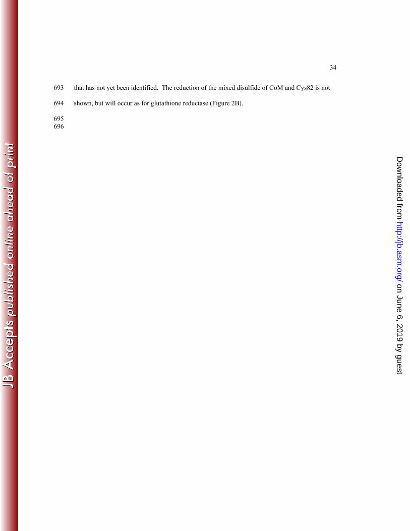
A. 683
B.684
SS
Cys87
Cys82
S
O
SOO
O
2-KPC
FAD
HO
H
His84
His137
H
SS
Cys87
Cys82
S
O-
SOO
O
2-KPC
FAD
HO
H
His84
His137
CO
OC
O
O
SS
Cys87
Cys82
S O
SOO
O
FAD
HO
H
His84
His137
-OO
H H H
685 686 687
Figure 3. Active site architecture and proposed mechanism for 2-KPCC. A. Structure of 2-KPC 688
bound to 2-KPCC highlighting active site residues believed to be key to catalysis (PDB ID: 689
1MO9). B. Proposed mechanism of thioether bond cleavage, enolacetone formation and 690
stabilization, and carboxylation based on the structures solved for 2-KPCC and the results of the 691
present work. The initial abstraction of a proton from C82 may be facilitated by a general base 692
on June 6, 2019 by guesthttp://jb.asm
.org/D
ownloaded from
34
that has not yet been identified. The reduction of the mixed disulfide of CoM and Cys82 is not 693
shown, but will occur as for glutathione reductase (Figure 2B). 694
695 696
on June 6, 2019 by guesthttp://jb.asm
.org/D
ownloaded from

35
697
698 699
Figure 4. SDS-PAGE analysis and western blot of recombinant 2-KPCC. (A) SDS-PAGE gel. 700
Lane: 1, molecular weight standards; 2, cell extract (13.3 μg); 3, first Ni2+ fraction (6.7 μg); 4, 701
phenyl-sepharose fraction (6.2 μg); 5, enterokinase digestion (15 μg); 6, the eluate from the 702
second Ni2+ affinity column (15 μg). (B) Immunoblot prepared blot from an identical SDS-703
PAGE gel using antibodies raised to native 2-KPCC. 704
705
on June 6, 2019 by guesthttp://jb.asm
.org/D
ownloaded from
36
[2-KPC] (mM)0 1 2 3 4 5
velo
city
(nm
ol/m
in/m
g)
0
50
100
150
200
1/[2-KPC] (mM-1)-2 -1 0 1 2 3 4 5
1/ve
loci
ty x
103
02468
101214
706
Figure 5. Effect of 2-KPC concentration on the reductive cleavage and carboxylation of 2-KPC 707
to acetoacetate and CoM. All assays were performed with 0.089 mg of 2-KPCC. The line 708
through the data points was generated by nonlinear least-squares fit to a rectangular hyperbola. 709
Inset, double reciprocal plot of the data. The line through the data points was also generated by 710
nonlinear least-squares fit of the data. 711
712
713
on June 6, 2019 by guesthttp://jb.asm
.org/D
ownloaded from

37
Wavelength (nm)350 400 450 500 550 600 650
Abs
orba
nce
0.00
0.05
0.10
0.15
0.20
0.25
0.30
(i)
(ii)(iii)
(iv)
714
Figure 6. UV/visible absorption spectra of r-2-KPCC C82A mutant at various pH values. 715
Spectra were obtained using 0.15 mg of 2-KPCC diluted to a volume of 130 μl as described in 716
the methods. The spectral noise at ~A420 and ~A535 is due to the ultramicro cuvette that was 717
used. The spectra shown are for pH values of: (i), 11.0; (ii), 9.0; (iii), 7.5; (iv), 5.0. 718
719
720
on June 6, 2019 by guesthttp://jb.asm
.org/D
ownloaded from

38
pH5 6 7 8 9 10 11
incr
ease
in A
555
(x-fo
ld)
1.0
1.5
2.0
2.5
3.0
3.5
4.0
721
Figure 7. Determination of pKa values for the interchange thiol of 2-KPCC. The fold increase in 722
A555 due to formation of charge transfer absorbance between FAD and the thiolate of C87 is 723
plotted vs. pH. The lines were derived from the four parameter sigmoidal curve fit used to 724
calculate pKa values. (●) C82A mutant of r-2-KPCC; (∆) native 2-KPCC alkylated on C82 with 725
BES. 726
727
728
on June 6, 2019 by guesthttp://jb.asm
.org/D
ownloaded from
39
time (min)0 10 20 30 40
μmol
ace
tone
form
ed
0.00
0.25
0.50
0.75
1.00
1.25
1.50
729
Figure 8. Acetoacetate decarboxylase activity of wild-type and mutant r-2-KPCC proteins. 730
Assays contained 0.125 mg 2-KPC, 250 mM acetoacetate, and 5 mM CoM. Data points represent 731
the average of duplicate experiments; (●) wild-type r-2-KPCC; (○) H84A r-2-KPCC; (▼) 732
H137A r-2-KPCC; (∆) C87A r-2-KPCC; (■) C82A r-2-KPCC. 733
734
on June 6, 2019 by guesthttp://jb.asm
.org/D
ownloaded from

40
TABLE 1. Effect of active site mutations on the three redox-dependent activities of 2-KPCCa 735 Enzyme Specific activity (nmol/min/mg) for:
2-KPC protonation
2-KPC Carboxylation
Formation of 2-KPC from acetoacetate
wt r-2-KPCC 53.8 ± 6.4 221 ± 16 47 ± 4
C87A r-2-KPCC NDb ND ND
C82A r-2-KPCC ND ND ND
H84A r-2-KPCC 22.7 ± 0.1 37.0 ± 4.2 12.0 ± 0.3
H137A r-2-KPCC ND 18 ± 1 1.0 ± 0.1
aAll assays were repeated in duplicate. 2-KPC protonation and carboxylation assays were 736
performed using DTT as the reductant, while formation of 2-KPC from acetoacetate was 737
performed using NADP+ as the oxidant. 738
bND, no detectable activity 739
740
on June 6, 2019 by guesthttp://jb.asm
.org/D
ownloaded from
41
741 TABLE 2: Kinetic parameters for wild-type and M140A r-2-KPCCa Enzyme Km, 2-KPC
(mM) Change in Km (x-fold)
kcat (min-1)
Change in kcat (x-fold)
kcat/Km (mM-1 ·min-1)
wt r-2-KPCC 0.42 ± 0.08 1 13.3 1 32 M140A r-2-KPCC 6.37 ± 0.82 15.3 4.3 0.32 0.68 aApparent Km and kcat values for 2-KPC carboxylation were determined using the coupled 742
spectrophotometric assay described in the methods. 743
744 745
on June 6, 2019 by guesthttp://jb.asm
.org/D
ownloaded from


![Downloaded from //jb.asm.org/content/jb/early/2016/09/08/JB.00545-16.full.pdf · ð òô,1752'8&7,21 òõ +rul]rqwdojhqhwudqvihulqedfwh uldlvdpdmruirufhlqwkhdg dswdwlrqwrqryho óì](https://static.fdocuments.in/doc/165x107/5e880168150d351f4b0ea6f0/downloaded-from-jbasmorgcontentjbearly20160908jb00545-16fullpdf.jpg)