Jasna Mihailovic Stanley J. Goldsmith Ronan P. Killeen FDG ...978-3-642-29866-0/1.pdf · Jasna...
14
Jasna Mihailovic Stanley J. Goldsmith Ronan P. Killeen FDG PET/CT in Clinical Oncology
Transcript of Jasna Mihailovic Stanley J. Goldsmith Ronan P. Killeen FDG ...978-3-642-29866-0/1.pdf · Jasna...

Jasna M ihai lovicStanley J . Goldsmith
Ronan P. K i l leen
FDG PET/CT in Clinical Oncology


Jasna MihailovicStanley J. GoldsmithRonan P. Kil leen
FDG PET/CT in Clinical Oncology
Case Based Approach with Teaching Points

Jasna Mihailovic, M.D., Ph.D.Assoc Professor & HeadDepartment of Nuclear MedicineOncology Institute of VojvodinaSremska KamenicaTechnical Faculty “Mihajlo Pupin” Zrenjanin, University of Novi Sad, Serbia
Stanley J. Goldsmith, M.D.Division of Nuclear Medicine & Molecular ImagingNew York-Presbyterian HospitalProfessor, Radiology & MedicineWeill Cornell Medical CollegeNew YorkUSA
Ronan P. Killeen, M.D.Consultant Radiologist and Nuclear Medicine PhysicianSt Vincents University HospitalDublin Ireland
ISBN 978-3-642-29865-3 ISBN 978-3-642-29866-0 (eBook)DOI 10.1007/978-3-642-29866-0Springer Heidelberg New York Dordrecht London
Library of Congress Control Number: 2012950989
© Springer-Verlag Berlin Heidelberg 2012
This work is subject to copyright. All rights are reserved by the Publisher, whether the whole or part of the
material is concerned, speci fi cally the rights of translation, reprinting, reuse of illustrations, recitation, broad-
casting, reproduction on micro fi lms or in any other physical way, and transmission or information storage and
retrieval, electronic adaptation, computer software, or by similar or dissimilar methodology now known or
hereafter developed. Exempted from this legal reservation are brief excerpts in connection with reviews or
scholarly analysis or material supplied speci fi cally for the purpose of being entered and executed on a com-
puter system, for exclusive use by the purchaser of the work. Duplication of this publication or parts thereof is
permitted only under the provisions of the Copyright Law of the Publisher’s location, in its current version, and
permission for use must always be obtained from Springer. Permissions for use may be obtained through
RightsLink at the Copyright Clearance Center. Violations are liable to prosecution under the respective
Copyright Law.
The use of general descriptive names, registered names, trademarks, service marks, etc. in this publication does
not imply , even in the absence of a speci fi c statement, that such names are exempt from the relevant protec-
tive laws and regulations and therefore free for general use.
While the advice and information in this book are believed to be true and accurate at the date of publication,
neither the authors nor the editors nor the publisher can accept any legal responsibility for any errors or omis-
sions that may be made. The publisher makes no warranty, express or implied, with respect to the material
contained herein.
Printed on acid-free paper
Springer is part & Springer Science + Business Media (www.springer.com)

To my son, Aleks, whom I love and appreciate and who has enriched my life despite the time I spend away with patients, medical research and writing this book; and to my parents, Milos and Ljubica who have brought joy and love to my life. Special appreciation and gratitude to my mentor and friend, Professor Dr. Ljubomir Stefanovic, who passed away while this volume was being assembled. Professor Ljubomir Stefanovic created me and supported me throughout my adult life, both professionally and during personal challenges. And to Stanley Goldsmith who gave me the idea of writing this book in the fi rst place.
Jasna Mihailovic
To my mother, Angela, and my late father, John: thank you for aff ording me every opportunity a person could want in life. And to Stanley Goldsmith: thank you for inspiring me and mentoring me in all things molecu-lar and otherwise.
Ronan Patrick Killeen
To my wife, Miriam, and our family who have sacri fi ced much so that I can continue to pursue my dreams.
Stanley J. Goldsmith
Collectively, the editors express appreciation and gratitude to their colleagues and patients who have taught us so much and continue to teach us daily in our practice of nuclear medicine.
Jasna Mihailovic, Ronan Patrick Killeen, and Stanley J. Goldsmith

vii
Foreword
This volume presents a variety of nuclear oncology FDG PET/CT cases which can serve as an introductory text for radiologists and nuclear medicine physicians initially confronting the practice of PET/CT. It is likely that it will also serve medical oncologists who order these procedures and even medical students studying the diagnosis and management of patients with malignancies.
This volume evolved from an experience that the editors had working together in the PET/CT component of a clinical nuclear medicine department in a large academic medical center. Most of the cases included in this collection were accumulated during a 2-month period. Since the majority of clinical referrals are for various indications involving oncology patients, it was decided to limit this collection to that application of FDG PET/CT. The cases illustrate the diverse nature of the clinical issues that confront a nuclear medicine physician in daily practice. They are presented in a clinical context with the limited information provided by referring clinicians. While obviously less than ideal in some instances, the setting illustrates what nuclear medicine physicians have to deal with. To demonstrate another point, the cases are organized according to the variety of clinical indications within nuclear oncology for FDG PET/CT studies. First among these indications is “diagnosis” when the primary malignancy is unknown or when a tumor is identi fi ed incidentally in a study requested for another reason. This demonstrates that even though most cases are accompanied by a clinical diagnosis, the nuclear medicine physi-cian has to be mindful of alternative diagnoses. Most often, however, FDG PET/CT is requested to determine the “extent of disease” which determines staging and often de fi nes a course of therapy. The ef fi cacy of FDG PET/CT for this application depends on the sensitivity of this tech-nique which in turn depends on the technical quality of the study, the amount of FDG accumu-lated, the size and location of the lesion – although in this regard, PET/CT imaging is generally at an advantage compared to SPECT imaging as PET images are created by coincidence imag-ing and are corrected for attenuation and therefore are less in fl uenced by distance from the detectors. Use of PET/CT for con fi rmation of the “response to treatment” depends also on sen-sitivity for detection but also requires knowledge of the natural history of the disease and the physiologic responses associated with tumoricidal therapies. The role of FDG PET/CT in the “follow-up” of the patient who has initially responded to therapy is dependant on sensitivity of the technique as well as the speci fi city of the technique which diff erentiates between residual fi brous tissue at sites of tumor that has been successfully treated and would remain detectable on CT alone from recurrent tumor, the metabolic activity of which is detectable only with FDG PET.
FDG PET/CT of brain tumors is diff erent enough from body imaging to merit a separate section. Consistent with the usual practice in our institution, correlation of the FDG PET/CT with MRI images is provided. MRI images are an essential component of the information necessary for PET/CT interpretation of these problems.
Each case presentation begins with a brief historical setting. In general, the whole body maximum intensity projection [MIP] image is provided separately from the speci fi c sections that most illustrate the signi fi cant fi nding or fi ndings. The most frequently presented are the transaxial sections of the FDG PET distribution alone, the corresponding CT image, and

viii Foreword
the fusion image with the metabolic FDG data superimposed on the CT anatomic map. When followed up, similar sections, transaxial or other, are presented in succession.
Finally, the material is supplemented with “Teaching Points” as if after image interpreta-tion, the case is presented in a clinical conference setting and discussed by a senior nuclear medicine physician.
Stanley J. Goldsmith , M.D. Jasna Mihailovic , M.D., Ph.D.
Ronan Kileen , M.D.
ix
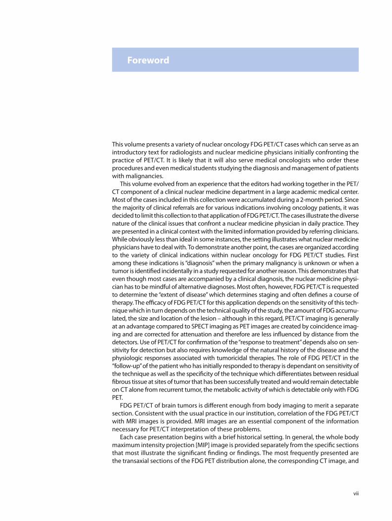
Part I Diagnosis
Chapter 1 Solitary Pulmonary Nodule (SPN), Chest Mass. . . . . . . . . . . . . . . . . . . . . . . . . . . . . 3
Chapter 2 Unknown Primary . . . . . . . . . . . . . . . . . . . . . . . . . . . . . . . . . . . . . . . . . . . . . . . . . . . . . . . 29
Chapter 3 Unexpected Malignancy/Second Primary . . . . . . . . . . . . . . . . . . . . . . . . . . . . . . . . . 41
Part II Extent of Disease
Chapter 4 Head and Neck Carcinoma . . . . . . . . . . . . . . . . . . . . . . . . . . . . . . . . . . . . . . . . . . . . . . . 67
Chapter 5 Lung Carcinoma . . . . . . . . . . . . . . . . . . . . . . . . . . . . . . . . . . . . . . . . . . . . . . . . . . . . . . . . . 73
Chapter 6 Breast Carcinoma. . . . . . . . . . . . . . . . . . . . . . . . . . . . . . . . . . . . . . . . . . . . . . . . . . . . . . . . 89
Chapter 7 Esophageal Carcinoma . . . . . . . . . . . . . . . . . . . . . . . . . . . . . . . . . . . . . . . . . . . . . . . . . . 103
Chapter 8 Colorectal Carcinoma . . . . . . . . . . . . . . . . . . . . . . . . . . . . . . . . . . . . . . . . . . . . . . . . . . . . 109
Chapter 9 Lymphoma. . . . . . . . . . . . . . . . . . . . . . . . . . . . . . . . . . . . . . . . . . . . . . . . . . . . . . . . . . . . . . 123
Chapter 10 Multiple Myeloma . . . . . . . . . . . . . . . . . . . . . . . . . . . . . . . . . . . . . . . . . . . . . . . . . . . . . . . 149
Chapter 11 Gynecological Adenocarcinomata . . . . . . . . . . . . . . . . . . . . . . . . . . . . . . . . . . . . . . . . 165
Chapter 12 Miscellaneous Malignancies. . . . . . . . . . . . . . . . . . . . . . . . . . . . . . . . . . . . . . . . . . . . . . 171
Part III Follow-Up
Chapter 13 Head and Neck . . . . . . . . . . . . . . . . . . . . . . . . . . . . . . . . . . . . . . . . . . . . . . . . . . . . . . . . . . 181
Chapter 14 Lung Carcinoma . . . . . . . . . . . . . . . . . . . . . . . . . . . . . . . . . . . . . . . . . . . . . . . . . . . . . . . . . 185
Chapter 15 Breast Carcinoma. . . . . . . . . . . . . . . . . . . . . . . . . . . . . . . . . . . . . . . . . . . . . . . . . . . . . . . . 199
Chapter 16 Pancreas Carcinoma . . . . . . . . . . . . . . . . . . . . . . . . . . . . . . . . . . . . . . . . . . . . . . . . . . . . . 211
Chapter 17 Colorectal Carcinoma . . . . . . . . . . . . . . . . . . . . . . . . . . . . . . . . . . . . . . . . . . . . . . . . . . . . 217
Chapter 18 Lymphoma. . . . . . . . . . . . . . . . . . . . . . . . . . . . . . . . . . . . . . . . . . . . . . . . . . . . . . . . . . . . . . 233
Contents

x Contents
Chapter 19 Multiple Myeloma . . . . . . . . . . . . . . . . . . . . . . . . . . . . . . . . . . . . . . . . . . . . . . . . . . . . . . . 247
Chapter 20 Thyroid Gland . . . . . . . . . . . . . . . . . . . . . . . . . . . . . . . . . . . . . . . . . . . . . . . . . . . . . . . . . . . 289
Chapter 21 Urologic Tumors Bladder. . . . . . . . . . . . . . . . . . . . . . . . . . . . . . . . . . . . . . . . . . . . . . . . . 305
Chapter 22 Miscellaneous . . . . . . . . . . . . . . . . . . . . . . . . . . . . . . . . . . . . . . . . . . . . . . . . . . . . . . . . . . . 317
Part IV Response to Treatment
Chapter 23 Lymphoma. . . . . . . . . . . . . . . . . . . . . . . . . . . . . . . . . . . . . . . . . . . . . . . . . . . . . . . . . . . . . . 327
Chapter 24 Multiple Myeloma . . . . . . . . . . . . . . . . . . . . . . . . . . . . . . . . . . . . . . . . . . . . . . . . . . . . . . . 367
Chapter 25 Lung. . . . . . . . . . . . . . . . . . . . . . . . . . . . . . . . . . . . . . . . . . . . . . . . . . . . . . . . . . . . . . . . . . . . 379
Chapter 26 Gastrointestinal Tumors . . . . . . . . . . . . . . . . . . . . . . . . . . . . . . . . . . . . . . . . . . . . . . . . . 389
Chapter 27 Breast. . . . . . . . . . . . . . . . . . . . . . . . . . . . . . . . . . . . . . . . . . . . . . . . . . . . . . . . . . . . . . . . . . . 397
Chapter 28 Neuroendocrine Tumor . . . . . . . . . . . . . . . . . . . . . . . . . . . . . . . . . . . . . . . . . . . . . . . . . . 407
Part V Neuro Section
Chapter 29 Brain Tumors . . . . . . . . . . . . . . . . . . . . . . . . . . . . . . . . . . . . . . . . . . . . . . . . . . . . . . . . . . . . 415
References . . . . . . . . . . . . . . . . . . . . . . . . . . . . . . . . . . . . . . . . . . . . . . . . . . . . . . . . . . . . . . . . . . . . . . . . . . 445
Index . . . . . . . . . . . . . . . . . . . . . . . . . . . . . . . . . . . . . . . . . . . . . . . . . . . . . . . . . . . . . . . . . . . . . . . . . . . . . . . 451

xi
Introduction
Without doubt, presently, 18 F- fl uorodeoxyglucose [FDG] PET/CT is the most frequently per-formed positron emission tomography [PET] clinical application used in medicine.
The role of glucose in accelerated tumor metabolism of tumors was initially described by Warburg in the 1920s when he observed the increased anaerobic metabolism of neoplasms. FDG acts as a glucose analog in which one of the hydroxyl groups (molecular weight of 17 and a single negative charge) is replaced by 18 F (atomic weight of 18 and as the fl uoride ion also with a single negative charge). Since a hydroxyl group is removed (deoxy) and replaced by a fl uorine, the molecule is “ fl uorodeoxyglucose”; its molecular weight and electronega-tivity is suf fi ciently close to the glucose molecule to be acceptable to the glucose transport mechanisms of most tissues and tumors. Hence, it is carried into the intracellular environ-ment by the glucose transport mechanism and undergoes enzymatic phosphorylation to fl uorodeoxyglucose-6-phosphate. Fortunately, the enzyme in the next step of glucose metabolism, glucose 1–6 phosphate transferase, does not ef fi ciently recognize the fl uorinated deoxyglucose-6-phosphate, which remains in that form with minimal conver-sion to anything else, nor is it eliminated from the cell until it eventually undergoes break-down. Hence, the FDG serves as a soluble microsphere and is trapped in the cell. The amount of FDG accumulated, however, serves as a marker of the degree of anaerobic glucose metabolism and hence is useful to identify and characterize tumors.
Regardless of whether a normal subject or a patient with a malignancy is imaged, the FDG PET scan will identify normal organ and tissue utilization of glucose. At rest, the brain, principally the gray matter, is the dominant site of glucose utilization. PET imaging of the brain is suf fi ciently sensitive to identify the diff erences in the visual cortex utilization of glucose when the eyes are open or shut, staring at a blank wall versus a checkered pattern or a complex scene. Similar diff erences in the auditory cortex can be characterized depend-ing on the complexity of the sound stimulation.
Since the heart, particularly the left ventricle, is always consuming energy, the left ventricle may be identi fi ed, although in the truly fasting state the left ventricle and the remainder of the myocardium utilizes free fatty acid. After meals, however, or other sources of glucose and subsequent insulin release, plasma free fatty acids decrease dramatically and the myocar-dium switches to glucose metabolism. In PET evaluation of myocardial viability, diff erences in fatty acid and glucose utilization is useful to identify viable hibernating myocardium. Bowel peristalsis will also consume more glucose and consequently accumulate FDG.
Glucose appears in the glomerular fi ltrate. The enzyme-mediated tubular reabsorption of glucose, however, does not handle FDG as a substrate. As a consequence, FDG appears in the urine. In fact, the relatively rapid renal excretion of FDG augments the contrast between the tumor or normal organ glucose accumulation and the background.
It is clear from the above brief review that since tumor uptake of FDG is not insulin dependant, FDG PET imaging is augmented in the fasting state. Likewise, a nonfasting met-abolic state results in an increased fraction of the FDG entering the liver and muscles. Even if the blood glucose level is normalized by physiologic or pharmacologic insulin, a greater fraction of FDG will enter these other sites, making less FDG available for tumor uptake and adversely aff ecting the tumor to background ratio.

xii Introduction
Lastly, physicians utilizing FDG PET/CT must be aware of other causes of FDG accumulation. In fl ammation, either sterile or infectious, is of most concern. In addition to unrelated infections, the in fl ammatory response to therapy, particularly radiation therapy, will be associated with increased FDG accumulation, thus complicating evaluation of the tumor response to therapy.
In oncology, a routine 18 F-FDG acquisition protocol consists of several steps. Blood glu-cose level should be determined prior to FDG administration. Clinical history as to whether or not the patient has diabetes and the medication used to control blood glucose should be reviewed prior to scheduling. Diabetic patients require special preparation which often has to be individualized so as to maintain the blood glucose below (or near) 200 mg/dL without insulin administration within 1–2 h preceding the FDG administration. In many centers, the dose of FDG is based on patient body weight (approximately 5 MBq/kg), whereas in other centers, a 370–555 MBq (10–15 mCi) is used in adults with PET scanners utilizing BGO crystals. Recent advances in PET scanner design and engineering have substi-tuted BGO with more sensitive crystals. In this circumstance, satisfactory images can be obtained with doses below 370 MBq for an adult of 70 kg or more.
Patients should relax and avoid any unnecessary movement for at least 45 min to pre-vent muscular uptake which results in artifacts and possible misinterpretation. The patient should be told not to engage in conversation or chewing particularly if the area of interest involves the head and neck region. Depending on the area of interest, oral contrast may be administered prior to FDG injection as an aid in identifying bowel loops and diff erentiating bowel from masses, lymph nodes, or blood vessels. In some centers, and again depending upon the organs or regions of interest, IV contrast CT may be obtained. In these cases, the IV contrast is usually administered after initially obtaining a CT without contrast to be used for attenuation correction of the PET image data which also is obtained prior to the admin-istration of the IV contrast. The typical image acquisition for body imaging involves scan-ning from the base of the skull to mid thigh. Of course, brain and head and neck FDG PET/CT involves imaging this area (a single-bed position) usually for a longer PET acquisition period than for body PET acquisitions. FDG PET/CT for evaluation of patients with either multiple myeloma or malignant melanoma should include head-to-toe imaging.
Following the completion of the CT scan, PET emission scanning is performed in the caudocranial direction, starting at the pelvis region. With BGO crystal scanners, depending on the axial range and the individual bed position acquisition time, scanning, including transmission and emission scans, can be completed in 30–45 min. Newer instruments with faster crystals and electronics can reduce this to 20 min or less.
The clinical utilization of FDG PET imaging was initially limited to brain imaging as the diameter of the early PET scanner openings could not accommodate larger body parts. Although all tumors did not vigorously concentrate the FDG tracer, Giovanni Di Chiro at the National Institutes of Health in the United States noted the relationship between the “tumor avidity” for FDG and tumor aggressiveness. This observation provided a basis for the strati fi cation of the aggressiveness of therapeutic intervention. Nevertheless, initially between the cost of the instrumentation, the dif fi culty in obtaining FDG, and the limited application for patient management, utilization of FDG PET was limited to only a few cen-ters with access to cyclotrons and PET radiochemistry laboratories.
All of that has changed so that, at the present time, FDG PET/CT plays a role in several aspects of management of the patient with a malignant tumor – in some cases, even in assisting in the original diagnosis, in almost every case contributing to the assessment of the extent of disease, con fi rming response to therapy, playing an essential role in the monitoring of the patient after therapy for early detection of recurrence of disease, and more recently (and still under develop-ment) predicting response to the current course of chemotherapy (and to a limited extent, radiotherapy) early in the course of therapy. In the future, when this latter application is better understood, it is possible that some of the current prolonged therapeutic regimen can be ter-minated earlier. Certainly, in the event that it can be demonstrated that despite a transient clini-cal response to therapy during the regular administration of chemotherapy (or irradiation), if it is possible to predict early relapse, alternative therapeutic options can be considered.

Introduction xiii
To progress from the early days of limited availability and application of FDG PET to the now widely available and utilized status of FDG PET/CT, many components had to be brought together. In retrospect, it is remarkable that such a complex and costly procedure has grown so fast and spread so far. There are probably several versions of the sequence of historical growth of this procedure. From the perspective of the editors, the following sequence seems to be a reasonable reconstruction of the events involved:
Realization that there might be applications of FDG PET imaging in areas of the body • other than limited to the brain. Commercial development of instrumentation that allowed imaging of other body parts • (head and neck, thorax, abdomen, pelvis, extremities). Imaging of these areas and demonstration of tumor utilization of FDG. • Realization by commercial interests of the potential wider demand for cyclotron-pro-• duced 18 F and FDG synthesis, hence increased availability of FDG (although FDG was still quite expensive). Simultaneous academic-industrial development and exploitation of whole body scan-• ning accompanied by growth in computer capabilities that permitted storage of large amounts of data representing the reconstructed transaxial images which could be stored with reconstruction of additional views, that is, coronal and sagittal sections or slices as well as a volume display, currently called maximal intensity projection [MIP].
Even prior to the combination of PET and computed tomography [CT], the MIP and coronal images provided suf fi cient anatomical detail so as to improve the anatomic placement of observations. The imaging community beyond nuclear medicine began to appreciate the images which heretofore had been accessible to a limited group of dedi-cated physicians interested and supportive of this technique. Further increase in the availability of FDG. With increased utilization, competition among • commercial suppliers resulted in decreasing costs of unit doses, further increasing utilization. Finally, the marriage of PET and CT resulted in widespread application and further assess-• ment of the clinical merit of the technique. At the same time, on the other side of the patient is the issue of costs. Increased utiliza-
tion and demand for FDG brought about competition among suppliers and decreased cost for unit doses of FDG, from perhaps $800 per dose initially to less than $150 in some areas. This is still expensive but competitive with costs of contrast agents in general. Equipment costs have not declined as dramatically, in part because the technology employed contin-ues to grow more sophisticated, with faster acquisition times, faster image reconstruction, extraction of more complex data, and most recently, the combination of PET and MRI – a technology with a new set of advantages and challenges.
In the USA, approval of reimbursement for the procedure by government and third-party insurance was necessary because of the considerable cost per examination. The extent of procedures approved proceeded slowly but gradually as the merit of each appli-cation was demonstrated in peer-reviewed medical reports. Ultimately, a national review body, the NOPR (National Oncology Procedure Registry), was formed to collate data on the contribution of PET/CT in oncology to improved patient management and outcome. Analysis of the data accumulated during the fi rst year of the NOPR was published in the Journal of Clinical Oncology several years ago. It demonstrated that in over two-thirds of the cases, FDG PET/CT had a signi fi cant impact on patient management. The monitoring of FDG PET/CT continues, but FDG PET/CT is available for most oncology applications. Throughout Europe, Japan, the Middle East, other Asian countries, South America, and most recently, parts of Africa, FDG PET/CT has become increasingly available and reimbursable.
It is likely that in the near future additional PET tracers will become available. 18 F-choline which is a measure of cell membrane synthesis can serve as a tumor marker because of the incremental rate of tumor cell division and hence would be more tumor speci fi c than FDG as it would diff erentiate between tumor proliferation and in fl ammation. Other tracers that

xiv Introduction
characterize protein synthesis, RNA replication, or tissue oxygenation are being evaluated and developed for more widespread application.
In developing countries, fi nancial resources are very limited. In a country like Serbia, there are only two PET/CT centers for the entire country with a population of 7.5 million people. FDG is imported from neighboring nations and currently costs approximately 540 euros per dose. The charge for the entire examination is 700 euros. There is a plan for the central government to build a cyclotron and PET radiochemistry facility within Serbia. Nevertheless, other problems remain: insuf fi cient physician awareness of the value of FDG PET/CT in the management of patients, particularly oncology patients, and limited fi nancial resources to support access and the costs that would be associated with expansion of utili-zation of this valuable procedure.
By contrast, in New York City, in the borough of Manhattan alone, there are more than 15 PET/CT scanners and 2 cyclotrons and PET radiochemistry facilities within medical centers. In addition, the area is supplied with FDG by 3-5 competing commercial radiopharmacies.
Hopefully, in the near future, the costs of the various components of the examination, particularly the FDG dose, will be further reduced and the merits of FDG PET/CT in the man-agement of the oncology patient will be appreciated by referring physicians, government, and third-party payers so as to make this essential tool more widely available.
Stanley J. Goldsmith, M.D.
Jasna Mihailovic, M.D., Ph.D.Ronan Kileen, M.D.

![Killeen, Thomas B. TOCs/Killeen, Thomas B.toc.pdf · 2012-12-17 · 2 INTERVIEW [Note: This transcript was not edited by Mr. Killeen.] SUMMARY: This history of Thomas B. Killeen focuses](https://static.fdocuments.in/doc/165x107/5fba91d984c64d2537799e12/killeen-thomas-b-tocskilleen-thomas-btocpdf-2012-12-17-2-interview-note.jpg)