Jane Doe DOB: 08/28/1952 Family History - medquestltd.com · Jane Doe DOB: 08/28/1952 ... Diabetes...
25
Jane Doe 1 of 5 Jane Doe DOB: 08/28/1952 Patient is a 56-year-old female. Past Medical History: Hypertension, Heart disease, Non-Q wave myocardial infarction, Diabetes (since 1995), Chronic Obstructive Pulmonary Disease, Acute Respiratory Disease Syndrome, Chronic Renal Insufficiency, Right Upper Extremity Deep Vein Thrombosis, Chronic urinary track infections, Gall Bladder Disease Surgical History: Laparoscopic Cholecystectomy in August 2007, Thoracocentesis of Left Lung, Heart Bypass Surgery in 2007, Pleurodesis in November 2007, Appendectomy and Tubal Ligation in 1968, Tennis Elbow Surgery in 1983. Family History: Not significant. Social History: Patient had history of smoking but quit in August 2007. Does not drink or use drugs. She is married. Medications: Aspirin, Metoprolol, Lasix, Lisinopril, Protonix, Lantus, Simvastatin, Levemir, Bacitracin, Lactinex, Zofran, Morphine, Lortab, Haldol, Zyvox Allergy: Coded Allergies: Amiodarone, Codeine, Hydrocodone, Metronidazole, Penicillin and Quinolones. Uncoded Allergies: EGGS, Lortab DATE PROVIDER OCCURRENCE/TREATMENT PDF REFERENCE 11/25/2007 General Medical Center Peter David, M.D. Admission for Shortness of Breath and Back Pain Patient complained of pain in the right lower chest and upper abdomen. She had shortness of breath which had worsened over the last week. She also had episodes of nausea and vomiting. She also had right- sided back pain, dry cough and some dizziness. Over the last week she had increased swelling of her lower extremities, which was right greater than left. Vein was taken out from the right leg for the previous coronary artery bypass grafting. The previous day she treated herself by taking 80mg of Lasix and said that there had been at least 5 pounds of weight loss since with some improvement in the swelling. Her baseline dose is 40 mg of Lasix every other day Physical Examination: Blood pressure 178/95. On auscultation breath sounds are decreased at bilateral bases particularly at the left base. Chest X-ray showed chronic changes. There was left- sided pleural effusion which appeared to be smaller since she had the video assisted thoracoscopy performed last week. Labs showed increased WBC and blood glucose. Creatinine was highly elevated. Chest X-ray showed extensive infiltrates throughout the left lung with a small left hydropneumothorax. Left pleural-based mass may reflect loculated pleural fluid. 71, 73-74 2166-2169
Transcript of Jane Doe DOB: 08/28/1952 Family History - medquestltd.com · Jane Doe DOB: 08/28/1952 ... Diabetes...

Jane Doe
1 of 5
Jane Doe DOB: 08/28/1952
Patient is a 56-year-old female. Past Medical History: Hypertension, Heart disease, Non-Q wave myocardial infarction, Diabetes (since 1995), Chronic Obstructive Pulmonary Disease, Acute Respiratory Disease Syndrome, Chronic Renal Insufficiency, Right Upper Extremity Deep Vein Thrombosis, Chronic urinary track infections, Gall Bladder Disease Surgical History: Laparoscopic Cholecystectomy in August 2007, Thoracocentesis of Left Lung, Heart Bypass Surgery in 2007, Pleurodesis in November 2007, Appendectomy and Tubal Ligation in 1968, Tennis Elbow Surgery in 1983. Family History: Not significant. Social History: Patient had history of smoking but quit in August 2007. Does not drink or use drugs. She is married. Medications: Aspirin, Metoprolol, Lasix, Lisinopril, Protonix, Lantus, Simvastatin, Levemir, Bacitracin, Lactinex, Zofran, Morphine, Lortab, Haldol, Zyvox Allergy: Coded Allergies: Amiodarone, Codeine, Hydrocodone, Metronidazole, Penicillin and Quinolones. Uncoded Allergies: EGGS, Lortab
DATE PROVIDER OCCURRENCE/TREATMENT PDF REFERENCE 11/25/2007 General Medical
Center Peter David, M.D.
Admission for Shortness of Breath and Back Pain Patient complained of pain in the right lower chest and upper abdomen. She had shortness of breath which had worsened over the last week. She also had episodes of nausea and vomiting. She also had right-sided back pain, dry cough and some dizziness. Over the last week she had increased swelling of her lower extremities, which was right greater than left. Vein was taken out from the right leg for the previous coronary artery bypass grafting. The previous day she treated herself by taking 80mg of Lasix and said that there had been at least 5 pounds of weight loss since with some improvement in the swelling. Her baseline dose is 40 mg of Lasix every other day Physical Examination: Blood pressure 178/95. On auscultation breath sounds are decreased at bilateral bases particularly at the left base. Chest X-ray showed chronic changes. There was left-sided pleural effusion which appeared to be smaller since she had the video assisted thoracoscopy performed last week. Labs showed increased WBC and blood glucose. Creatinine was highly elevated. Chest X-ray showed extensive infiltrates throughout the left lung with a small left hydropneumothorax. Left pleural-based mass may reflect loculated pleural fluid.
71, 73-74 2166-2169
RHilario
Sticky Note
Refer to page 2166
Jane Doe
2 of 5
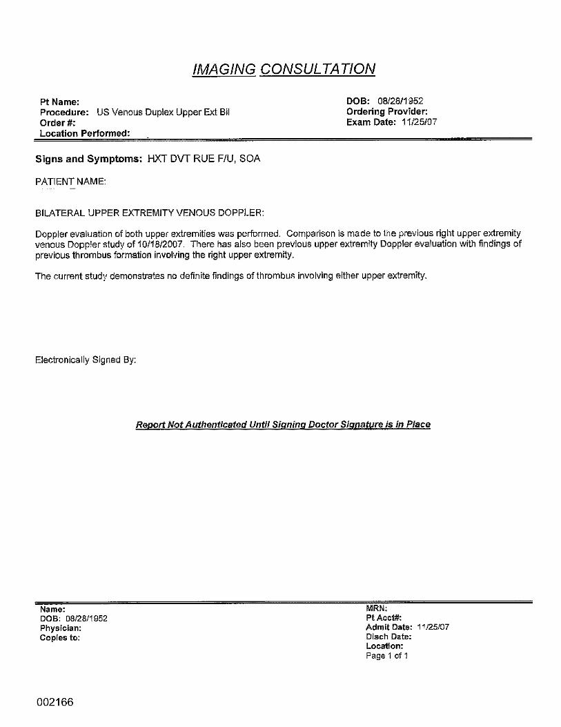
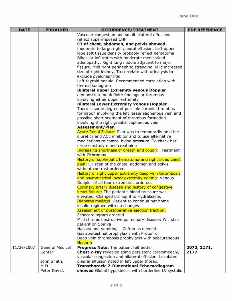
DATE PROVIDER OCCURRENCE/TREATMENT PDF REFERENCE Vascular congestion and small bilateral effusions reflect superimposed CHF CT of chest, abdomen, and pelvis showed moderate to large right pleural effusion. Left upper lobe soft tissue density probably reflect hematoma. Bibasilar infiltrates with moderate mediastinal adenopathy. Right lung nodule adjacent to major fissure. Mild right perinephric stranding. Mild increased size of right kidney. To correlate with urinalysis to exclude pyelonephritis Left thyroid nodule. Recommended correlation with thyroid sonogram Bilateral Upper Extremity venous Doppler demonstrate no definite findings or thrombus involving either upper extremity Bilateral Lower Extremity Venous Doppler There is some degree of possible chronic thrombus formation involving the left lesser saphenous vein and possible short segment of thrombus formation involving the right greater saphenous vein Assessment/Plan Acute Renal Failure: Plan was to temporarily hold her diuretics and ACE inhibitor and to use alternative medications to control blood pressure. To check her urine electrolyte and creatinine Increasing shortness of breath and cough: Treatment with Zithromax History of subhepatic hematoma and right sided chest pain: CT scan of the chest, abdomen and pelvis without contrast ordered History of right upper extremity deep vein thrombosis and asymmetrical lower extremity edema: Venous Doppler of all four extremities ordered. Coronary artery disease and history of congestive heart failure: The patient’s blood pressure was elevated. Changed Lisinopril to Hydralazine. Diabetes mellitus: Patient to continue her home insulin regimen with no changes Assessment of postoperative ejection fraction: Echocardiogram ordered Mild chronic obstructive pulmonary disease: Will start patient on Spiriva Nausea and vomiting – Zofran as needed. Gastrointestinal prophylaxis with Protonix Deep vein thrombosis prophylaxis with subcutaneous Heparin
11/26/2007 General Medical Center John Smith, M.D. Peter David,
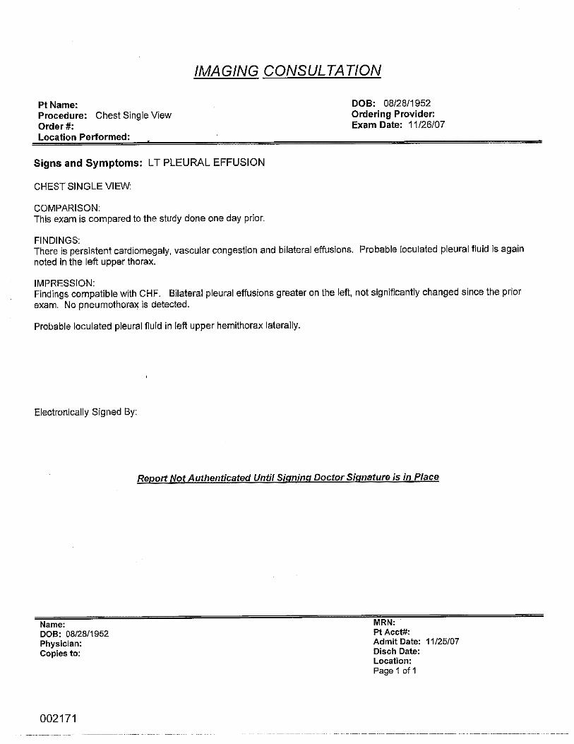
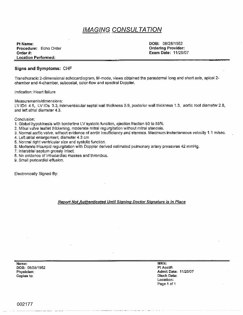
Progress Note: The patient felt better. Chest x-ray revealed some persistent cardiomegaly, vascular congestion and bilateral effusion. Loculated pleural effusion noted in left upper thorax. Transthoracic 2-Dimentional Echocardiogram showed Global hypokinesis with borderline LV systolic
2072, 2171, 2177
Jane Doe
3 of 5
DATE PROVIDER OCCURRENCE/TREATMENT PDF REFERENCE M.D. Harry R. smith, M.D. William Lee, M.D.
function, ejection fraction 50 to 55% Mitral valve leaflet thickening, moderate mitral regurgitation without mitral stenosis Normal aortic valve, without evidence of aortic insufficiency and stenosis Left atrial enlargement, diameter 4.3 cm Normal right ventricular size and systolic function Moderate tricuspid regurgitation with Doppler derived estimated pulmonary artery pressures Interatrial septum grossly intact No evidence of intracardiac masses and thrombus Small pericardial effusion Assessment and Plan Acute renal failure: Planned to hold off her ACE inhibitor, creatinine elevated to 2.1 Chronic obstructive pulmonary disease: patient received supplemental oxygen at 2 litres per nasal cannula and was monitored closely Increased shortness of breath and cough: given Zithromax, and would increase Ceftriaxone 1 gm daily History of subhepatic hematoma and right sided chest pain: CT of abdomen and pelvis showed mild perinephric stranding. Continued to monitor Lower extremity superficial thrombosis: Bilateral lower venous Doppler revealed some degree of chronic thrombus formation Coronary artery disease and history of congestive heart failure: renal function remained elevated and appear to be slightly fluid volume overloaded, continued to monitor Diabetes mellitus: to continue current insulin regimen Nausea and vomiting: the patient was given Zofran Gastrointestinal prophylaxis with Protonix Deep venous thrombosis prophylaxis with subcutaneous heparin C-diff prophylaxis with Lactinex Patient reported itching in various places of her body on administration of Rocephin. Benadryl ordered and to continue with current regimen.
11/27/2007 General Medical Center Peter David, M.D. Harry R. smith, M.D.
Progress Note: The patient had hypoglycemia. Blood glucose was mildly elevated. She had some swelling in lower extremities. Lab revealed elevated creatinine. Chest X-ray showed bilateral atelectasis or infiltrate and CHF. No change in the left upper lateral thoracic pleural based mass probably reflect resolving hematoma Assessment/Plan Acute renal failure: Etiology unclear. The patient had developed renal failure around the time of last admission. Not known the patient had significant hypotension.
2217

Jane Doe
4 of 5
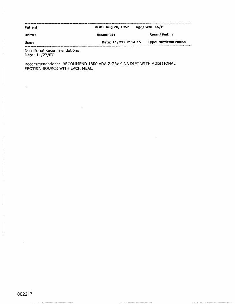
DATE PROVIDER OCCURRENCE/TREATMENT PDF REFERENCE Bilateral pulmonary infiltrates and pleural effusions status post left-sided pleurodesis. The patient’s CT scan showed extensive infiltrates bilaterally particularly on the left side. Planned to keep the patient on broad-spectrum antibiotics. Considered performing a thoracentesis on the right side and remove some fluid from her chest and bronchoscopy. Chronic Obstructive pulmonary disease. History of subhepatic hematoma and right-sided chest pain. Lower extremity superficial thrombosis. Coronary artery disease and history of congestive heart failure. Plan to consider removal of right-sided pleural effusion. Gastrointestinal prophylaxis with Protonix Deep vein thrombosis prophylaxis with subcutaneous Heparin Nutritional Recommendations: 1800 ADA 2 gram NA diet with additional protein source with each meal
11/28/2007 General Medical Center John Smith, M.D. Peter David, M.D. Michael Warren, M.D.
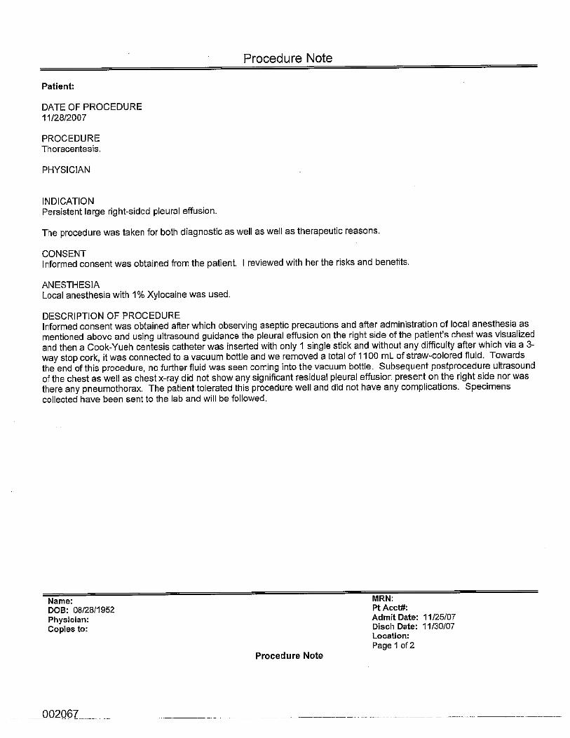
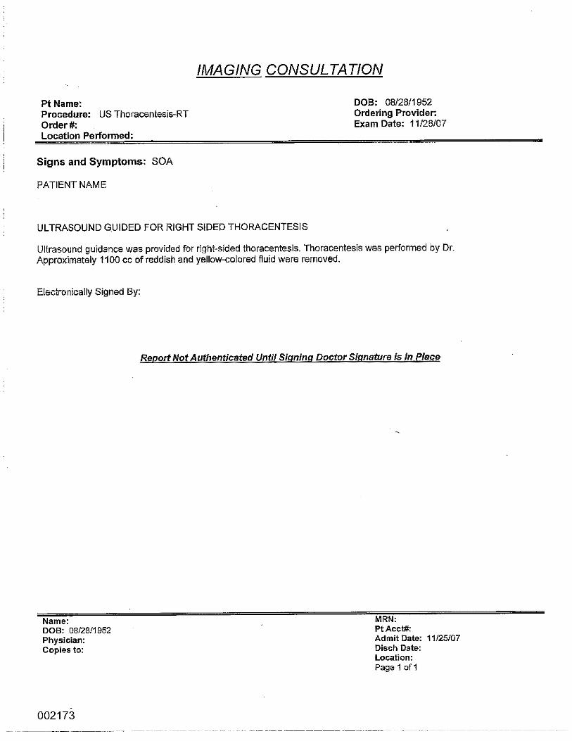
Progress Note: The patient felt better. Procedure: Thoracentesis under local anesthesia Thoracentesis was performed by Dr. Mark, and 1100cc of reddish and yellow-colored fluid was removed. The patient tolerated the procedure and did not have any complications. Specimen were collected and sent to lab Lab report was normal Chest X-ray after status post thoracentesis revealed decrease in left pleural effusion. No pneumothorax Non-Gyn Cytology Report Collected fluid from pleural fluid. A PCP stain ordered. Atypical cells present
2077, 2067, 2136, 2173
11/29/2007 General Medical Center Peter David, M.D.
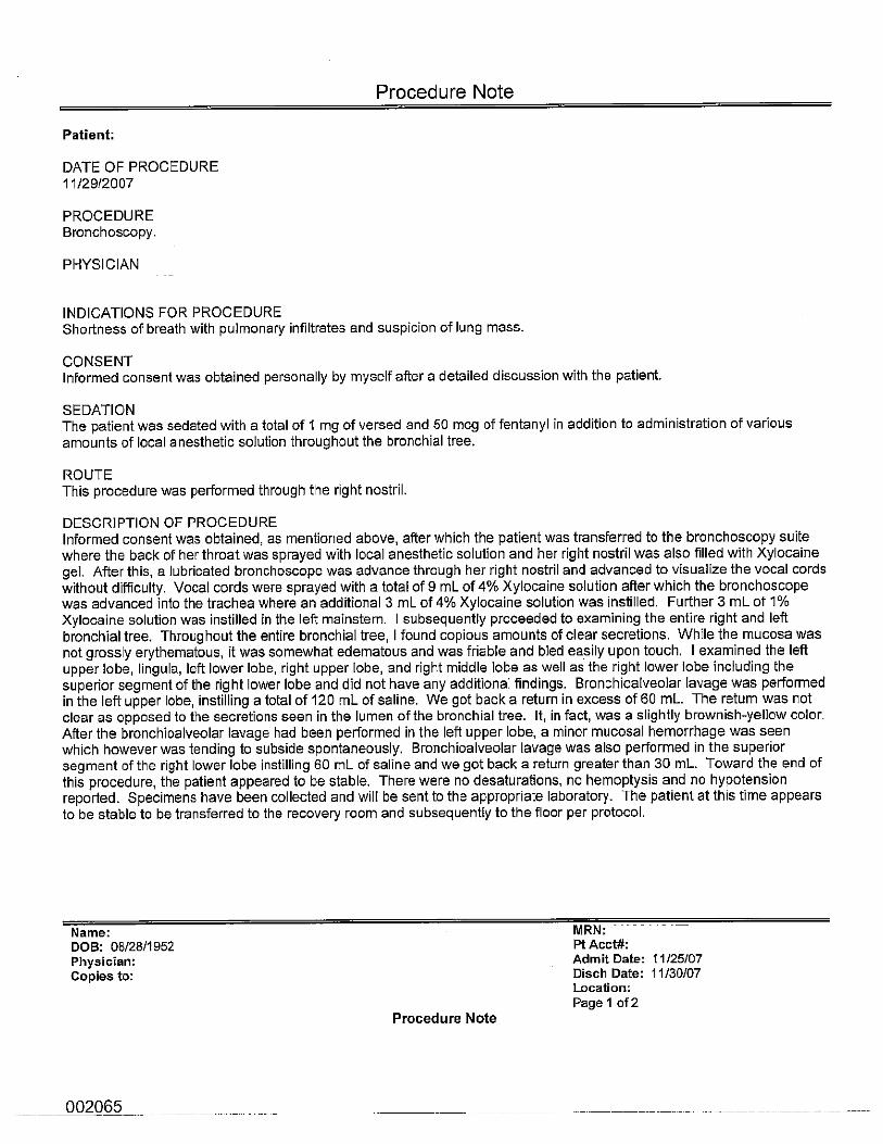
Progress Note: Procedure: Bronchoscopy Specimen were collected and sent to laboratory. Subsequent to bronchoscopy, she had some drowsiness and needed 1-2 litres of oxygen to maintain O2 saturations. Lab showed hemoglobin slightly drifted downward, creatinine elevated. Noted mild hyperglycemia Chest x-ray showed improved right-sided pleural effusion. Stable appearance of chest Assessment and Plan Pulmonary infiltrates with pleural effusion, status post left-sided pleurodesis with suspicion of left-sided lung mass. Continued to follow reports of bronchoscopy and thoracentesis Acute renal failure Lower extremity superficial thrombosis Coronary artery disease and history of congestive heart failure Diabetes mellitus Gastrointestinal prophylaxis Deep vein thrombosis prophylaxis
2065, 2078-2079

Jane Doe
5 of 5
DATE PROVIDER OCCURRENCE/TREATMENT PDF REFERENCE Mycobacteriology showed no mycobacteria recovered, no acid-fast bacilli found Non-Gyn Cytology Report :Specimen Type: Bronchial Washing Result: Negative for malignancy
11/30/2007 General Medical Center John Smith, M.D. Peter David, M.D.
Discharge Summary: Throughout her hospitalization, the patient was supportive. Her electrolytes were managed. Her creatinine came down to 2. She underwent thoracentesis on 11/28/2007 and bronchoscopy and some bronchoalveolar lavage on 11/29/2007. Findings showed copious amount of clear sputum and mucosa was not erythematous but only mildly edematous and friable. ACE inhibitor was discontinued due to her renal failure. She had mild chronic obstructive pulmonary disease and she was on Spiriva. Overall, the patient remained hemodynamically stable and stable from a respiratory standpoint. By the date of discharge she maintained proper saturation in the low 90s on room air. She was afebrile at discharge and was stable for discharge home Lab reports were normal. Sputum culture and urine culture were all normal Venous Doppler revealed some degree of chronic thrombus formation in the lesser saphenous vein and possible short segment thrombus in the right greater saphenous vein, but no definite deep venous thrombosis CT of chest, abdomen, pelvis revealed large pleural effusion. Discharge Diagnoses Acute renal failure Shortness of breath with bilateral pulmonary infiltrates Coronary artery disease Congestive heart failure Type II diabetes mellitus Mild chronic obstructive pulmonary disease Right-sided pleural effusion Left upper lobe lung mass Plan: Advised patient to follow up with Dr. James in 2 weeks. The patient would require a CT of chest on the day of office visit. Ordered CBC, basic metabolic panel and magnesium. To monitor the patient closely as she may need percutaneous biopsy in future.
2050-2052