Jade D. Jamias, MD, FPCP, FPSG,FPSDE National Kidney...
28
Jade D. Jamias, MD, FPCP, FPSG,FPSDE National Kidney and Transplant Institute
Transcript of Jade D. Jamias, MD, FPCP, FPSG,FPSDE National Kidney...

Jade D. Jamias, MD, FPCP, FPSG,FPSDE
National Kidney and Transplant Institute

1. It is a wound-healing response in which extracellular matrix (ECM), or “scar” is accumulated.
2. Myofibroblast-like cells produce hepatic fibrosis regardless of underlying cause.
3. It follows a chronic, not self-limited injury.
4. It occurs earliest in regions where injury is most severe.
Schiff’s Diseases of the Liver, 10th edition

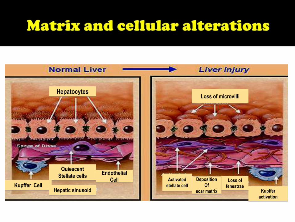
Quiescent
Stellate cells
Kupffer Cell
Endothelial
Cell
Hepatic sinusoid
Hepatocytes
Deposition
Of
scar matrix Kupffer
activation
Activated
stellate cell
Loss of microvilli
Loss of
fenestrae
Quiescent
Stellate cells
Kupffer Cell
Endothelial
Cell
Hepatic sinusoid
Hepatocytes
Deposition
Of
scar matrix Kupffer
activation
Activated
stellate cell
Loss of microvilli
Loss of
fenestrae


Stellate Cell Activation:
Central event in Hepatic Fibrosis

PERPETUATION• Increased cytokine secretion
• Upregulation of tyrosine kinase
receptor
• Accelerated ECM remodelling
INITIATION• Transcriptional events
• Paracrine stimulation
• Early ECM changes
Friedman SL. J Biol Chem 2000;275:2247-2250

Earliest changes from paracrine stimulation (i.e. sinusoidal epithelium, Kupffer cells, hepatocytes and platelets) (1)
Endothelial cells Loss of fenestrations (2,3)
Production of cellular fibronectin (1)
Conversion of TGF-B to profibrogenic form (2,3)
Express proinflammatory molecules (e.g. VEGF, adhesion molecules intercellular adhesion molecule 1) (2,3)
1. Olaso et al. Hepatology 2003;37:674-685
2. LeCoute J, et al. Science2003;299:890-893

Kupffer cells Influx (1)
Matrix synthesis, cell proliferation, release of retinoids (mediators:TGF-B1, reactive O2 intermediates/lipid peroxides)(1)
Hepatocytes Potent source of
fibrogenic lipid peroxides (2,3)
1. Friedman SL. J Clin Invest 2005;115:29-32
2. Paradis V, et al. J Clin Pathol 1997;50:401-406
3. Nieto N, et al. Hepatology 2002;35:62-73

Platelets Potent source of growth
factors (1)
Mediators: PDGF, TGF-B1, epidermal growth factor (EGF)(1)
1. Bachem MG, et al. Jclin Chem Clin Biochem 1989;27:555-565

Net effect: increase in the accumulation of ECM
Proliferation and chemotaxis Increase in the numbers of
collagen-producing cells
More matrix production per cell
Cytokine release Amplify the inflammatory
and fibrogenic responses
Matrix proteases hasten production scar
Schiff’s Diseases of the Liver, 10th edition

Changes in stellate cell behavior
Mediator/s Key Events
1. Proliferation PDGF Induction of PDGF receptors
2. Chemotaxis PDGF, MCP1 Migration of stellate cells toward cytokine chemoattractants
3. Fibrogenesis TGF-B1 Increased matrix production (Collagen type 1); production of other matrix components (fibronectin, proteoglycans)
4. Contractility ET-1, Angiotensin II
Increased expression of cytoskeletal protein; decreased portal blood flow
Schiff’s Diseases of the Liver, 10th edition

Changes in stellate cell behavior
Mediator Events
5. Matrix degradation MMP-2 Quantitative and qualitative changes in matrix protease activity
6. Retinoid loss PDGF Loss of perinuclear retinoid (vitamin A) droplets and acquire a more fibroblastic appearance
7. WBC chemoattractant and Cytokine release
MCP-1 Amplification of inflammatory response by inducing mononuclear and PMN infiltration; production of chemokines (i.e. MCP-1, RANTES, CCR5); expression of toll-like receptorsSchiff’s Diseases of the Liver, 10th edition

Hepatic fibrosis is a wound-healing response characterized by accumulation of ECM.
Hepatic fibrosis follows a chronic, but not self-limited, liver disease.
Activation of hepatic stellate cells is the central event in hepatic fibrosis.
Hepatic stellate cells and related myofibroblasts from intra and extrahepatic sources orchestrate an array of changes including degradation of normal ECM, deposition of scar molecules, vascular and organ contraction , and release of cytokines.

Pathologic increase in portal pressure, in which the pressure gradient between the portal vein and inferior vena cava (portal pressure gradient, PPG) is increased above 5 mm Hg (1-4)
Clinically significant when PPG increases above 10 mm Hg (e.g. formation of varices) or 12mm Hg (e.g. variceal bleeding, ascites)(1-4)
Subclinical portal hypertension : PPG between 6 to 10
mm Hg (1-4)1. Casado M, et al. Gastroenterology 1998;114(6): 1296-1303
2. Rigau J, et al. Gastroenterology 1989;96(3):873-880
3. Viallet A, et al. Gastroenterology 1975;69(6):1297-1300
4. Garcia-Tsao, et al. Hepatology 1985;5(3):419-424

PPG (P) results from interaction between the portal blood flow (F) and resistance to flow (R)
PHT can result from:
1. Increase in vascular resistance (cirrhosis)
2. Increase in portal blood flow
3. Combination of both
P = F x R
Bosch J, et al. Gastenterol Clin North Am 1992;21(1): 1-14.

Consequence of architectural distortion of hepatic circulation by fibrosis (fixed and mechanical)(1)
Thrombosis of medium and large portal and hepatic veins (1)
Active contraction of contractile elements (i.e. smooth muscles, stellate cells and hepatic myofibroblasts) of the liver (dynamic) (2)
1. Wanless IR, et al. Hepatology 1995;21(5):1238-1247
2. Bathal PS, et al. J Hepatol 1985;1:325-329

Va
sod
ilato
rsV
aso
con
stri
cto
rs

Endothelial dysfunction of the hepatic vascular bed (1)
Abrupt post-prandial increase in portal pressure (2)
Impaired response to endothelium-derived vasodilator, acetylcholine (3)
Overactivation of COX-1 pathway with an increased production of vasoconstictor-derived compounds (i.e. TXA2) (4)
1.Gupta TK, et al. Hepatol 1998; 28(4):926-931
2. Bellis L et al. Hepatol 2003;37(2): 378-384
3. Graupera M, et al. J Hepatol 2003; 39(4): 515-521
4. Aleixandre de Artinano M, et al. Pharmacol Res 1999; 40(2):113-114

Observed in the advanced stages of PHT
Result from the marked arteriolar dilatation in the splanchnic organs draining into the portal vein (SPLANCHNIC VASODILATION)

MECHANISMS
Neurogenic
Humoral
Increased levels of vasodilators
Local
CANDIDATE VASODILATORS
1. Glucagon• most evidence
2. Endocannabinoids
3. Nitric oxide
4. Prostaglandins
5. Carbon monoxide

Vasodilator Mechanism/s of action
1. Glucagon vascular smooth muscle relaxation and decreasing its sensitivity to endogenous vasoconstrictors i.e. norepinephrine, angiotensin II and vasopressin. (SOMATOSTATIN and ITS ANALOGUES)
2. Endocannabinoids Increase NO production by activation of endothelial CB1 receptor
3. Nitric oxide
4. Prostaglandins Vascular smooth muscle relaxation by activating adenylate cyclase and augmenting intracellular level of cyclic adenosine monophosphate (CAMP)
5. Carbon monoxide

Portosystemic collateral circulation Peripheral vasodilation and hyperkinetic
circulation Characterized by reduced arterial pressure and
peripheral resistance, and increased plasma volume and cardiac output (CO)
Activation of neurohumoral systems that cause Na retention, increases plasma volume and cardiac index
Expansion of plasma volume Necessary to maintain an increased CO

Architectural Disturbances(fibrosis, scarring, vascular thrombosis etc.)
Functional alterations(contraction of vascular smooth muscle and stellate cells)
Increased hepatic resistance
Portal Hypertension

Architectural Disturbances(fibrosis, scarring, vascular thrombosis etc.)
Functional alterations(contraction of vascular smooth muscle and stellate cells)
Increased hepatic resistance
Portal Hypertension
Splanchnic vasodilatation Effective hypovolemia
Activation of endogenous
vasoactive systems
Collaterals and PSS
Na retentionHypervolemia
Increased cardiac index
Increased portal blood inflow
Schematic diagram of the pathophysiology of portal hypertension

Portal pressure gradient is determined by the product of blood flow and vascular resistance within the portal venous system.
PHT is initiated by an increased resistance to portal blood flow and aggravated by an increased portal venous inflow.
Increased resistance in cirrhosis is due not only to disruption of vascular architecture but also a dynamic component from active contraction of endothelial SM, myofibroblasts and hepatic stellate cells.

Portal inflow is increased by sphlanchnic vasodilation, which is caused by an increase secretion of local endothelial factors and humoral vasodilators.

Thank you for your kind attention !!!


![Welcome! [] · Frederick Alden Jaklitsch Elijah Alexander Jamias ... 90501. If you have any questions, please call St. James Parish Representative, Nancy Cebra, 424.558.8183. Catholic](https://static.fdocuments.in/doc/165x107/5ed0328b940df508d74942bd/welcome-frederick-alden-jaklitsch-elijah-alexander-jamias-90501-if-you.jpg)