Jacobs Journal of Nanomedicine and...
8
OPEN ACCESS Jacobs Journal of Nanomedicine and Nanotechnology Controlled Deposition of Gold and Silver on a Porous Silicone Matrixs Solano-Umaña Victor 1* , Vega-Baudrit, José Roberto 2 1 Hologic Surgical Products, Costa Rica 2 National Nanotechnology Laboratory (LANOTEC-CeNAT-CONARE), San José, Costa Rica *Corresponding author: Dr. Solano-Umaña Victor, Hologic Surgical Products, Senior Chemist, DOCINADE’s Ph.D. student, phone. (506) 24362739, E-mail: [email protected] Received: 05-05-2016 Accepted: 06-08-2016 Published: 07-12-2016 Copyright: © 2016 Victor Research article Cite this article: Solano-Umaña Victor. Controlled Deposition of Gold and Silver on a Porous Silicone Matrix. J J Nanomed Nanotech. 2016, 2(1): 006. Introduction Nanotechnology is an emerging new technology that opens the possibility to create new products in a different science areas with an unimaginable applications, as industrial uses, biology, biotechnology, and medicine [1]. Nanotechnology could be defined as the gate to design, characterize, produce and apply materials, structures, devicesand systems with sizes and shapes at the nanoscale level. It provides especial characteristics that can mimic the biological molecules and their systems, for this reason nanotechnology has a tremen- dous potential use for biomedical applications [2]. The new nanoscience and nanotechnology field applied to health sciences is called Nanomedicine, and its objective is to develop new devices and diagnostic methods to improve the human life quality. Nanomedicine is a branch of nanosci- ence and nanotechnology that would allow the ability to cure diseases in the cellular or molecular habitat [3]. Nanomedi- cine studies nanoscale interactions and it uses devices, sys- tems, and technologies that include nanostructures that can interact at those levels. The common objective from this new applied field (nanomedicine) is to solve medical problems, and improved human healthy, nanomedicine embraces a big number of miniaturized technology [4]. Micro, Meso and Macro porous materials can be manufac- tured using different substances base on the application. Attached to the advantage of porous materials, micro, meso and macro channels surface can be modify with metallic nanoparticles or nanofilms to further improve their proper- ties [5]. Most new researches used different polymers like the silicone to address the requirement of porous materials with surface functionality, where channels and walls play a key for a successful medical applications. The potential of sil- icone polymers implanted in the human body are countless because they can be easily manufactured in different shapes such as fibers, fabrics, films and blocks. Silicone or polysilox- ane is a hybrid compound with chemical formula (R2SiO) n, where R could be methyl, ethyl or phenyl (organic group), attached to an inorganic network of Silicon and Oxygen [6]. Abstract During the last decade and nowadays an enormous research effort has been deployed to the porous materials. Design, pores sizes, shapes, morphology and density are crucial features to increase the surface material, which helps to improve adsorp- tion and absorption properties and helps the interaction of living cells with the porous material. To achieve this goal, the use of coatings with gold and silver nanoparticles or nanofilms can increase the level of biocompatibility or biotoxicity, especially due to their effect on cell adhesion and proliferation. The biocompatibility and other properties obtained for nanoparticles application coatings are related to the particle size. Gold and silver nanoparticles are size controlled when a colloid is form and later this particles are immobilized on a porous silicone matrix, the pH, temperature, concentration, and proportion or ratio between the metal compound and the stabilizer or reducing agent are factors to consider for a gold and silver particles control on the colloid formation, an lather on the immobilization of these particles over a porous silicone matrix to obtain hybrid nanostructures that combine the best prop- erties from porous polymers and metal particles based on the application. Keywords: Gold nanoparticles; Silver nanoparticles; Porous materials; Silicone Matrix; Nanofilm JACOBS PUBLISHERS 1(2):006.
Transcript of Jacobs Journal of Nanomedicine and...

OPEN ACCESS
Jacobs Journal of Nanomedicine and Nanotechnology
Controlled Deposition of Gold and Silver on a Porous Silicone MatrixsSolano-Umaña Victor1*, Vega-Baudrit, José Roberto 2
1Hologic Surgical Products, Costa Rica 2National Nanotechnology Laboratory (LANOTEC-CeNAT-CONARE), San José, Costa Rica
*Corresponding author: Dr. Solano-Umaña Victor, Hologic Surgical Products, Senior Chemist, DOCINADE’s Ph.D. student,
phone. (506) 24362739, E-mail: [email protected]
Received: 05-05-2016
Accepted: 06-08-2016
Published: 07-12-2016
Copyright: © 2016 Victor
Research article
Cite this article: Solano-Umaña Victor. Controlled Deposition of Gold and Silver on a Porous Silicone Matrix. J J Nanomed Nanotech. 2016, 2(1): 006.
Introduction
Nanotechnology is an emerging new technology that opens the possibility to create new products in a different science areas with an unimaginable applications, as industrial uses, biology, biotechnology, and medicine [1]. Nanotechnology could be defined as the gate to design, characterize, produce and apply materials, structures, devicesand systems with sizes and shapes at the nanoscale level. It provides especial characteristics that can mimic the biological molecules and their systems, for this reason nanotechnology has a tremen-dous potential use for biomedical applications [2].
The new nanoscience and nanotechnology field applied to health sciences is called Nanomedicine, and its objective is to develop new devices and diagnostic methods to improve the human life quality. Nanomedicine is a branch of nanosci-ence and nanotechnology that would allow the ability to cure diseases in the cellular or molecular habitat [3]. Nanomedi-cine studies nanoscale interactions and it uses devices, sys-tems, and technologies that include nanostructures that can
interact at those levels. The common objective from this new applied field (nanomedicine) is to solve medical problems, and improved human healthy, nanomedicine embraces a big number of miniaturized technology [4].
Micro, Meso and Macro porous materials can be manufac-tured using different substances base on the application. Attached to the advantage of porous materials, micro, meso and macro channels surface can be modify with metallic nanoparticles or nanofilms to further improve their proper-ties [5]. Most new researches used different polymers like the silicone to address the requirement of porous materials with surface functionality, where channels and walls play a key for a successful medical applications. The potential of sil-icone polymers implanted in the human body are countless because they can be easily manufactured in different shapes such as fibers, fabrics, films and blocks. Silicone or polysilox-ane is a hybrid compound with chemical formula (R2SiO) n, where R could be methyl, ethyl or phenyl (organic group), attached to an inorganic network of Silicon and Oxygen [6].
Abstract
During the last decade and nowadays an enormous research effort has been deployed to the porous materials. Design, pores sizes, shapes, morphology and density are crucial features to increase the surface material, which helps to improve adsorp-tion and absorption properties and helps the interaction of living cells with the porous material. To achieve this goal, the use of coatings with gold and silver nanoparticles or nanofilms can increase the level of biocompatibility or biotoxicity, especially due to their effect on cell adhesion and proliferation. The biocompatibility and other properties obtained for nanoparticles application coatings are related to the particle size.
Gold and silver nanoparticles are size controlled when a colloid is form and later this particles are immobilized on a porous silicone matrix, the pH, temperature, concentration, and proportion or ratio between the metal compound and the stabilizer or reducing agent are factors to consider for a gold and silver particles control on the colloid formation, an lather on the immobilization of these particles over a porous silicone matrix to obtain hybrid nanostructures that combine the best prop-erties from porous polymers and metal particles based on the application.
Keywords: Gold nanoparticles; Silver nanoparticles; Porous materials; Silicone Matrix; Nanofilm
JACOBSPUBLISHERS
1(2):006.1(2):006.

Surface modification can be achieved by applying coatings, or using metal nanoparticles to increase biocompatibility prop-erties, prevent biofilm formation and favor cells adhesion and proliferation. Along with the size pore control, the application of metal nanoparticles improves the adsorption and absorp-tion process, that are very important during vascularization and cells growing [7].
To obtain gold nanoparticle dispersion on a water base solu-tion from a redox chemical reaction of tetrachloroauric acid with a reducing agent is important to control the pH, to en-sure stability and control the reactivity that can achieve the formation of a ruby color red solution. Typical changes in gold nanoparticles size or shape distributions results from a change in pH [8].
Gold nanoparticles with size at nanometers level are of impor-tance in bio-nanotechnology. There are great opportunities to design medical devices and detection methods on nanomedi-cine field, as these materials were used in pre-clinical biomed-ical research to study the biological effects of their application on different materials and medical devices [9]. The effective application of gold and silver nanoparticles on medicine de-pends from the particle size, therefore size particle controlled process is a key on this synthesis [10:11]. Gold and silver nanoparticles offer different physical and chemical properties from the bulk materials, and these properties are related to their size, shape, and surface. Biomedical applications of the gold nanoparticles based on their biocompatibility, depend from the size and shape, combined with surface functionaliza-tion. [12].
Colloidal gold nanoparticles with size controllable distribution and stability could be prepared with the use of amino com-pounds as stabilizer agents. These compounds form covalent bounds that give stability to the nanoparticles. One popular procedure for nanoparticles production is based on the tetra-chloroauric acid reduction by reduced agents, in presence of protective or stabilizer agents as amino or thiol compounds [13]. Nanoparticles stability is always an issue, nanoparticles will always be created with a size distribution of particles, and the changes in size distribution of a colloid over time is a criti-cal interest factor in biomedical applications [14].
Porous gold films have attracted interest over the last ten years due to possible applications, and their used in diverse fields, for example, as electronica, sensors, and solar cells [15]. With the control of different variables that effect the metal particle synthesis is possible to obtain an uniform metal film or a po-rous film both of them are very important and have different applications on industrial, scientific and medical areas, and uniform film or uniform particle deposition on porous silicon matrix have a tremendous potential on implants and scaffolds for biomedical applications.
Jacobs Publishers 2
Cite this article: Solano-Umaña Victor. Controlled Deposition of Gold and Silver on a Porous Silicone Matrix. J J Nanomed Nanotech. 2016, 2(1): 006.
2- Experimental
2.1- Preparation
2.1.1- Chemicals
All chemicals were used as received. Porous Silicon Matrix was prepared in the laboratory with poly-dimethylsiloxane (PDMS) product of Nusil Silicon technology (MED-4860), the porous size are between 100 and 300 micro meters, AgNO3 (99.99%, Sigma-Aldrich), HAuCl4•3H2O (99.99%, Sigma-Al-drich), L-Ascorbic acid (99%, Sigma-Aldrich), Disodium eth-ylenediaminetetra-acetate (EDTA-2Na ) ACS reagent (99.4 %, powder, Sigma-Aldrich), Dextrose monohydrate (USP grade, Sigma-Aldrich), Sodium hydroxide (ACS reagent, ≥97.0%, pellets, Sigma-Aldrich), Ammonium hydroxide solution (ACS reagent, 28.0-30.0%, Sigma-Aldrich), and anhydrous Sodium Sulfite (≥98%, Sigma-Aldrich).
To prepare a chemical solution and perform a silver and gold deposition treatment over the porous silicon matrix, the con-ditions on the following table were followed:
Table 1. Chemical solution parameters.
2.1.2 Silver solution
Based on Table 1, AgNO3 was weighted and dissolved in DI-water and concentrated ammonium hydroxide was added to the aqueous silver nitrate solution. In those cases where a precipitation of silver oxide occurred the addition of more ammonium hydroxide re-dissolved the precipitated silver ox-ide by forming an ammoniac silver complex (Ag(NH3)2]OH), later a EDTA-2Na solution was added, molar ratio 1:1 respect to AgNO3, and the pH was adjusted where required (using am-monia), later the porous silicone matrix was added, dextrose monohydrate solution was added and finally the required tem-perature was reached. The molar ratio from silver nitrate to Dextrose monohydrate was 1:5.
The porous silicone matrix was removed after one hour to reach the required temperature.
Cn/(mol/lt) pH Temp./ᵒC lot Ag Au Ag Au Ag Au
0.0263 0.0026 9 10 35 35 1 0.0263 0.0026 9 10 50 50 2 0.0263 0.0026 10 11 35 35 3 0.0263 0.0026 10 11 50 50 4 0.0263 0.0026 8 8 35 35 5 0.0263 0.0013 9 9 50 50 6 0.0131 0.0013 9 9 50 50 7
1(2):006.1(2):006.
2.1.3 Gold solution
Based on Table 1, HAuCl4•3H2O was weighted and dissolved in DI-water, added to a Sodium Sulfite (Na2SO3) solution, mo-lar ratio from HAuCl4•3H2O to Na2SO3 was 1:10. EDTA was dissolved in DI-water and added to the mixed solution, molar ratio from HAuCl4•3H2O to EDTA was 1:1, the pH was adjust-ed with NaOH, the porous silicone matrix was added and then the L-Ascorbic acid solution was added, molar ratio from HAu-Cl4•3H2O to L-Ascorbic acid was 1:10, which finally reached the required temperature.
The porous silicone matrix was removed after one hour to reach the required temperature.
Several complexing agents have been investigated such as sul-fite, thiosulfate, thiomalate, chloride, and phosphate with the gold source with ascorbic acid, as the reducing agents to made gold nanoparticles. The gold sulfite reduction proceeds follow-ing this equation:
Gold sulfite solution has a relatively high stability, particularly under alkaline conditions, as long as the pH is maintained be-low 10 the solution compatibility and stability is good, most commercial gold sulfite solutions are operated in the pH range from 9 to 10 [16:17].
2.2 Characterization method
2.2.1 Optical inspection
AA visual inspection was performed on a SmartScope Flash 200, model CNC200, serial SVW2003849. SmartScope Flash is a full-featured automatic dimensional measurement system, optical metrology with a large capacity video measuring sys-tem for dimensional verification of manufacturing parts.
2.2.2 Fluorescence X-Ray Spectrophotometer
A thickness measurement from a silver and gold deposition over the porous silicone matrix was performed on x-ray equip-ment. The fluorescence (XRF) spectrometer is an x-ray instru-ment used on non-destructive chemical analyses of different materials and metal plating. XRF is an analytical technique used to determine the elemental composition of materials, it determines the chemistry of a sample by measuring the fluo-rescent ray emitted from a sample when it is excited by x-ray source. Each element present a characteristic fluorescent pattern signs that are unique for that specific element, XRF spectroscopy is used for qualitative and quantitative analysis of material composition and can measure the thickness of the metal film deposit over different surfaces.
Jacobs Publishers 3
Cite this article: Solano-Umaña Victor. Controlled Deposition of Gold and Silver on a Porous Silicone Matrix. J J Nanomed Nanotech. 2016, 2(1): 006.
2.2.2 Scanning electron microscopy (SEM)
SEM images were obtained, using a JEOL JSM-6390LV Scanning Electron Microscope.
3- Results and Discussion
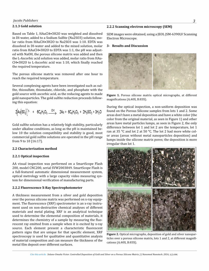
Figure 1. Porous silicone matrix optical micrographs, at different magnifications (A;40X, B:83X).
During the optical inspection, a non-uniform deposition was found on the Porous Silicone samples from lots 1 and 2. Some areas don’t have a metal deposition and have a white color (the color from the original material, as seen in Figure 1) and other areas have metal particles lumps, as seen in Figure 2, the only difference between lot 1 and lot 2 are the temperature, lot 1 ran at 35 ᵒC and lot 2 at 50 ᵒC. The lot 2 had more white col-or areas (areas without metal nanoparticles deposition) and lumps inside the silicone matrix pores; the deposition is more irregular than lot 1.
Figure 2. Optical micrographs, deposition of gold and silver nanopar-ticles over a porous silicone matrix, lots 1 and 2, at different magnifi-cations (A;40X, B:83X).
1(2):006.1(2):006.

TThe samples from lot number 3 and 4 ran at the same metal concentration and temperature than lots 1 and 2, but the pH was increased in one unit, lot 3 corresponds to lot 1 and lot 4 to lot 2, see table 1, in both cases the pH value increase produced a particle agglomeration to form a big lump inside the silicone matrix pores, like a solid rocks inside the holes, also present a white surface areas that are the silicone surface without metal particles deposition. This effect produces the clogging of the pores, as seen in Figure 3, it was confirmed by a rigid consistency on the porous matrix and also after the cutting of the matrix, on the cross section area, the pores were fill-up of solid particles. Consequently to this, at low pH, the ascorbic acid solutions are quite stable, however, as the pH is raised the stability of the ascorbate solution decreases, and the oxidation rate increases, since the pH has an extreme importance of met-als in controlling nanoparticles size [18].
On the silver nanoparticles reaction the pH is also a critical factor. Chemical reduction route to obtain silver colloid with controllable particles sizes ranging from 45 to 380 nm is the reduction of [Ag(NH3)2]+ complex with reducing sugars as dextrose, fructose and sucrose, where ammonia or sodium hy-droxide concentration in the reaction is the key parameter to control the particles size. Also sodium hydroxide could be used to enhance the reaction velocity [19:20:21].
Figure 3. Optical micrographs, deposition of gold and silver nanopar-ticles over a porous silicone matrix, lots 3 and 4, images at different magnification (A:40X, B:83X).
The pH plays an important role in silver nanoparticles (AgNPs) production from silver nitrate reduction with dextrose, and it
Jacobs Publishers 4
Cite this article: Solano-Umaña Victor. Controlled Deposition of Gold and Silver on a Porous Silicone Matrix. J J Nanomed Nanotech. 2016, 2(1): 006.
can be studied using surface plasmon resonance (SPR), and the different SPR behavior is explained in terms of size distribu-tion of AgNPs, if the pH is increased (more ammonia or sodium hydroxide) the particles size increase [22].
Lots 5, 6 and 7 show a better distribution of the metal nanopar-ticles compared to lots 1,2,3, and 4, the deposition is uniform and non-clogged up pores were detected, as seen in Figure 4, 5 and 9.
Figure 4. Optical micrographs, deposition of gold and silver nanopar-ticles over a porous silicone matrix, lots 5 and 6, different magnifica-tion (A;40X, B:83X).
Figure 5. Optical micrographs, deposition of gold and silver particles over a porous silicone matrix, lot 7, different magnification (A;40X, B:83X).
The lots 5, 6 and 7 show a uniform bright color which it is in-dicative from their particle size, and the lot 7 shows a ruby color, on this samples the pores are not clogging and it was confirm by a transversal section cutting and inspection, also
1(2):006.1(2):006.

Jacobs Publishers 5
Cite this article: Solano-Umaña Victor. Controlled Deposition of Gold and Silver on a Porous Silicone Matrix. J J Nanomed Nanotech. 2016, 2(1): 006.
a SEM imagine confirm this, Figure 10 . Noble metal nanopar-ticles like gold presents a characteristic strong color of their colloidal solutions, caused by surface plasmon absorption properties [23].
The results of the optical inspection were validated with a met-al thickness measurement, performed with the Fluorescence X-Ray Spectrophotometer XDLM model, which used WinFTM software (Fisher Thickness Management Software for Win-dows) to control the measurement head of the x-ray and han-dles the evaluation of the signal. Later the thickness values were analyzed with statistical software called Minitab 17 to obtain the boxplot graphs from the different lots.
Figure 6. Thickness of silver nanoparticles deposited over a porous silicone matrix, thickness boxplot.
The silver and the gold thickness from lots 1, 2,3 and 4 have a huge standard deviation, therefore the boxplot shows a huge variability. As seen in Figures 6 and 7, lots 5, 6 and 7 have a lower standard deviation or lower variability than lots 1,2,3 and 4. This effect is due to the pH reduction, but also lot num-ber 7 has the lowest variation (lowest standard deviation), on this lot the metal concentration for silver and gold was reduced by 50%, and this reduction from the initial concentration im-pacts the size distribution of nanoparticles over the porous sil-icone matrix. This difference is seen in lots 5, 6 and 7 detailed in Figure 8. The size of colloidal nanoparticles during the gold reduction could be easily tuned in the nanometer range by ad-justing initial concentration of HAuCl4 solution [24:25]. The agglomeration of nanoparticles from a solution is practically unavoidable in the absence of a stabilizer, stabilization has two approaches: a- steric repulsion caused by surfactants, or oth-er electrostatic (van der Waals) repulsion of organic species bound to nanoparticles’ surface, for this study the EDTA was
us as stabilizer. The growth process of the nanoparticles start as nucleation, experimental evidence suggests, that concentra-tion and temperature become predominant factors determin-ing growth rates from nanoparticles [26]. In Figure 6 the tem-perature effect over the resulting thickness is evidenced when lots 1 and 2, or lots 3 and 4 are compared, as seen in Table 1, the temperature increase on lots 2 and 4 produces an increase on the main silver thickness. Also the variability and standard deviation from these lots increase respect to lot 1 and 3. In Fig-ure 7 the same situation is present for the gold thickness, when lots 3 and 4 are compared, lot 4 has the biggest thickness, stan-dard deviation and variation, however the comparison from lots 1 and 2 does not match this criteria, lot 2 was produced at bigger temperature than lot 1, and on this lot the variation for the gold thickness is smaller than the variation from lot 1, but it is contrary on the silver thickness, as seen in Figure 6 and 7, it could be explained for the non-uniform deposition of the particles on lot 2.
Figure 7. Deposition of gold nanoparticles over a porous silicone matrix, thickness boxplot.
On the chemical reaction, disodium ethylenediaminetetra-ac-etate (EDTA-2Na) that is a hexadentate chelating agent which could be administered intravenously to the human body and makes its way through the blood stream to pull toxic heavy metals because these attach themselves to it, and the attach-ment forms a compound which can be excreted through urine, EDTA-2Na could solvate metal cations, also serve as stabilizing agents once the metal nanoparticles are made. On this study EDTA-2Na ratio respect to metals was constant to avoid this interference on the particle’s size.
inspection, also a SEM imagine confirm this . Noble metal nanoparticles like gold nanoparticles have a characteristic strong color and it is present on their colloidal solutions, the strong color is caused by the surface plasmon absorption properties [23]. The results of the optical inspection were validated with a metal thickness measurement, performed with the Fluorescence X-Ray Spectrophotometer XDLM that used WinFTM Software (Fischer Thickness Management Software for Windows) to control the measurement head of the x-ray and handles the evaluation of the signal. Later the thickness values were analyzed with statistical software Minitab 17 to obtain the boxplot graphs from the different lots.
Boxplot of Ag Thickness
40000
. 30000
(nm
)
Ag
Th
ick
nes
s
20000
10000
0
1 2 3 4 5 6 7
Lot
1(2):006.1(2):006.

Gold nanoparticles can be made from chloroauric acid (HAu-Cl4) through redox chemical reaction with ascorbic acid and the particles can be immobilized on a silicon compound [27]. One way is to use a desired quantity of stable additive agent and ascorbic acid, a weak reducing agent; on the wet chemical synthesis to reduce aqueous HAuCl4 solution. If ascorbic acid content was little, the nanoparticles can grow up because the system will be unstable, more gold atoms move to the surface, the chemical reaction is slower and Au seed can grow up [28]. On these reactions the molar ratio from the metal to reducing agent was constant, 1:5 for silver and 1:10 for gold in order to prevent the reducer concentration effect over the particle’s size.
Gold colloid color changes are visible to the naked eye and are associated to plasmon resonances (SPR) that may occur by changes on particle size, surface chemistry or inter-particles distance [29:30].
Figure 8 shows the different particle size measurement be-tween lots 5, 6 and 7 which is an evidence for the difference color obtained on the silicone porous matrix, shown in Figure 4 and 5. On lot 7 the porous matrices show a ruby color and lot 6 a light golden color, as seen in Figure 9.
Jacobs Publishers 6
Cite this article: Solano-Umaña Victor. Controlled Deposition of Gold and Silver on a Porous Silicone Matrix. J J Nanomed Nanotech. 2016, 2(1): 006.
Figure 10 shows more detail imagine respect Figure 9 (lot 7), the metal film deposited over the porous silicone matrix has a thickness measure in the nanometer order. The silver film thickness is below 150 nm and gold film thickness is below 100 nm, see Figure 8, these measures evidence the deposition or immobilization of gold and silver nanoparticles to form a nanofilm, also Figure 10 shows the pores from the silicone ma-trix, initial pore size from 100 to 300 micro meters free and open. The silver and gold film immobilized over the porous sil-icone matrix do no block the channels.
Conclusions
Nowadays, gold and silver are more than noble metals, they have an enormous potential on different fields such as indus-trial and biomedical.
Based on their nanoparticle’s properties, many applications were developed and more are coming. Applications that will be revolutionize the medicine and strengthen the nanomedi-cine name.
Figure 8. Thickness of nanoparticles deposited over a porous silicone matrix, boxplot for lots 5, 6 and 7, A: silver thickness, B: gold thickness.
Figure 9. Optical micrographs, gold and silver nanoparticles deposited over a porous silicone matrix, lot 5, 6 and 7 pore details.
1(2):006.1(2):006.

Figure 10. SEM image of silver and gold particles deposition over a porous silicone matrix, lot number 7.
The green chemistry is an excellent alternative to prepare nanostructures with potential used on medicine, because these chemicals like dextrose or sodium ascorbate are not tox-ic or dangerous for human health. After the colloid formation or metal (silver, gold) nanoparticles stabilization on water, these particles would be immobilized on a porous silicone ma-trix, and obtain a hybrid nanocomposite, see Figure 9.
Many factors and their interactions are present on the metal nanoparticles synthesis, on this research the pH, temperature and concentration effects on gold and silver particles size were evidenced, but also, the proportion or ratio between the metal compound and the stabilizer or reducing agent affects the par-ticle size. Therefore many factors must be control to obtain a particle’s size reproducibility.
On this research, one goal was to maintain the original porous free of obstructions, and this goal was met with lots number 5, 6 and 7, as show Figure 9 and 10, also base on the thickness measure from these lots (Figure 8) the gold and silver particles deposition do not affect the original pore size (100 to 300 mi-crometers), because on these lots the gold and silver thickness are a few nanometers that do not affect the silicone matrix po-rous size, this is important for a future medical applications because the pore size is related to the cellular growth and the biocompatibility. On the other hand, the samples from lots 3 and 4 could be very useful to explore another application for a porous metal film immobilization (see Figure 3), on silicone matrix holes.
References
1. Logothetidis S. Nanotechnology in Medicine, The Medicine of Tomorrow and Nanomedicine.
2. Jong. W,Hagens. W,Krystek. P,Burger. M,Sips. A et al. Particle size-dependent organ distribution of gold nanoparticles after in-travenous administration, Biomaterials.2008(29):1912-1919.
Jacobs Publishers 7
Cite this article: Solano-Umaña Victor. Controlled Deposition of Gold and Silver on a Porous Silicone Matrix. J J Nanomed Nanotech. 2016, 2(1): 006.
3. Solano-Umaña. V,Vega-Baudrit. J. González-Paz. R. The New Field of the Nanomedicine, International Journal of Applied Science and Technology.2015.5(1): 79-88.
4. Svennersten. K,Larsson. K,Berggren. M, Dahlfors. A.Organ-ic Bioelectronics in nanomedicine, Biochimica et Biophysica Act.2011(1810):276–285
5. Solano-Umaña. V, Vaga-Baudrit. J. Micro, Meso and Macro Porous Material son Medicine, Journal of Biomaterials and Na-nobiotechnology.2015( )6: 247-256.
6. Solano-Umaña. V, Vega-Baudrit. J. Porous Control on Silicone Matrix, International Journal of Recent Scientific Research. (2015).6(9):6290-6295.
7. Solano-Umaña. V, Vega-Baudrit. J. Gold and Silver Nano-technology on Medicine, Journal of Chemistry and Biochemis-try.2015. 3(1):21-33.
8. Sivaraman.S, Kumar.S, Sathanam. V. Room-temperature syn-thesis of gold nanoparticles – Size-control by slow addition, Gold Bulletin.2010.43(4):775-286.
9. Bienert.R, Emmerling. F.Thünemann. A. The size distri-bution of gold standard nanoparticles, Anal Bioanal Chem. 2009,395(6):1651-1660.
10. Hussain. S, Iqbal. M, Mazhar. M. Size control synthesis of starch capped-gold nanoparticles, Journal of Nanoparticles Re-search.2009(11):1383-1391.
11. Ohyma. J,Hitomi.Y,Higuchi.Y, Tanaka. T.Size Controlled Syn-thesis of Golf Nanoparticles by Porphyrin with Four Sulfur At-oms, Top Catalysis Journal.2009(52):852-859.
12. Sau.T,Rogach. A,Döblinger. M, Feldmann. J. One-Step High-Yield Aqueous Synthesis of Size-Tunable Multispiked Gold Nanoparticles. Small Journal.2011.7(15):2188–2194.
13. Feng. Ch, Gu. L, Yang Hu. J, Lu.G.Huang. X. Size-controllable gold nanoparticles stabilized by PDEAEMA-based double hy-drophilic graft copolymer, Polymer.2009(50):3990-3996.
14. Doak .J, Gupta .R., Manivannan .K, Ghosh. K, Kahol. P. Ef-fect of particle size distributions on absorvance spectra of gold nanoparticles, Physca E Journal homepage.2010( 42): 1605-1609.
15. Zhang. R,Olin. H.Porous Gold Films—A Short Review on Re-cent Progress, Materials Journal.2014( 7): 3834-3854.
16. Angstetra A, Jeffrey. M. The Effect of Additives on the Elec-troless Deposition of Gold From a Thiosulfate – Ascorbic Acid Bath, ECS Transactions, 2006,2(3):267-279.
17. Dimitrijevic.S, Vujasinovic. M. Trujic. V. Non-Cianide Elec-
1(2):006.1(2):006.

trolytes for Gold Plating, International Journal of Electrochem-ical Science.2013(8): 6620-6646.
18. Buettner,.G.Jurkiewicz. B. Catalytic, Ascorbate and free Rad-icals: Combinations to Avoid, Radiation Research,.1996(145): 532-541
19. Kvítek. L,Prucek. R, Panáček. A, Novotný. R,Hrbáča. J. The influence of complexing agent concentration on particle size in the process of SERS active silver colloid synthesis , J. Mater. Chem.2005( 15):1099-1105.
20. Wang.H,Qiao. X,Chen. J.Ding. S. Preparation of silver nanoparticles by chemical reduction method, Colloids and Sur-faces.2005.256 (2-3), pp 111-115.
21. Singh.M,Sinha. I, Mandal. R. Role of pH in the green syn-thesis of silver nanoparticles, Materials Letters.2009. 63(3-4):425-427.
22. Mehta. S,Chaudhary. S, Gradzielski, M. Time dependence of nucleation and growth of silver nanoparticles generated by sugar reduction in micellar media, Journal of Colloid and Inter-face Science, 2010.343(2):447-453
23. Moores. A, Goettmann. F. The plasmon band in noble metal nanoparticles: an introduction to theory and applications, New Journal of Chemistry.2006(30): 1121-1132.
24. Liang.X,Wang. Z,Liu.Ch. Size-Controlled Synthesis of Colloi-dal Gold Nanoparticles at Room Temperature Under the Influ-ence of Glow Discharge, Nanoscale Res Lett.2010(5):124-129.
Jacobs Publishers 8
Cite this article: Solano-Umaña Victor. Controlled Deposition of Gold and Silver on a Porous Silicone Matrix. J J Nanomed Nanotech. 2016, 2(1): 006.
25. Anh. N, Phu. D,Duy. N,Du. B,Hien. N. (2010). Synthesis of alginate stabilized gold nanoparticles by ɣ-Irradiantion with controllable size using different Au3+ concentration and seed particles enlargement, Radiation Physics and Chemistry .2010(79):405-408.
26. Cushing. B, Kolesnichenko. V. O’Connor. Ch. Recent Ad-vances in the Liquid-Phase Syntheses of Inorganic Nanoparti-cles, Chem. Rev. 104(9): 3893-3946.
27. Kalimuthu.P, John. S.Size dependent electrocatalytic activ-ity of gold nanoparticles immobilized onto three dimensional sol-gel networks, Journal of Electroanalytical Chemistry.2008( 617):164-170.
28. YunZhi. F,Yukou. D,Ping. Y, JinRu.L,Long. J. Shape-controlled synthesis of highly monodisperse and small size gold nanopar-ticles, Sci China Ser B-Chem.2007.50(4): 494-500.
29. Link.S,Wang Z, El-Sayed. M. Alloy Formation of Gold−Sil-ver Nanoparticles and the Dependence of the Plasmon Ab-sorption on Their Composition, J. Physical. Chemistry.1999. 103(18):3529–3533.
30. Krpetic´.Z,Guerrini. L,Larmour.A. Reglinski. J.Faulds. K. et al. Importance of Nanoparticle Size in Colorimetric and SERS-Based Multimodal Trace Detection of Ni (II) Ions with Func-tional Gold Nanoparticles, Small Journal.2012.8(5):707–714.
1(2):006.