IVC ‘The pro-BNP of ultrasound’ - NSW Agency for … · 2017-10-26 · IVC ‘The pro-BNP of...
90
1 IVC ‘The pro-BNP of ultrasound’ Dr Justin Bowra Critical Care Ultrasound Course
Transcript of IVC ‘The pro-BNP of ultrasound’ - NSW Agency for … · 2017-10-26 · IVC ‘The pro-BNP of...
1
IVC‘The pro-BNP of ultrasound’
Dr Justin BowraCritical Care Ultrasound Course
2
IVC‘More an art than a science’
Dr Justin BowraCritical Care Ultrasound Course
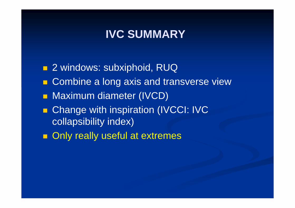
IVC SUMMARY
2 windows: subxiphoid, RUQ Combine a long axis and transverse view Maximum diameter (IVCD) Change with inspiration (IVCCI: IVC
collapsibility index) Only really useful at extremes
TheIVC
The IVC

The inferior vena cava (IVC)
Largest vein in the body. To the anatomical right of aorta Oval, thin walled Breathe in: diameter decreases (opposite if
ventilated) Dehydration: ‘flattens out’. Downstream occlusion (eg tamponade) or
fluid overload (eg CCF): ‘fattens up’.
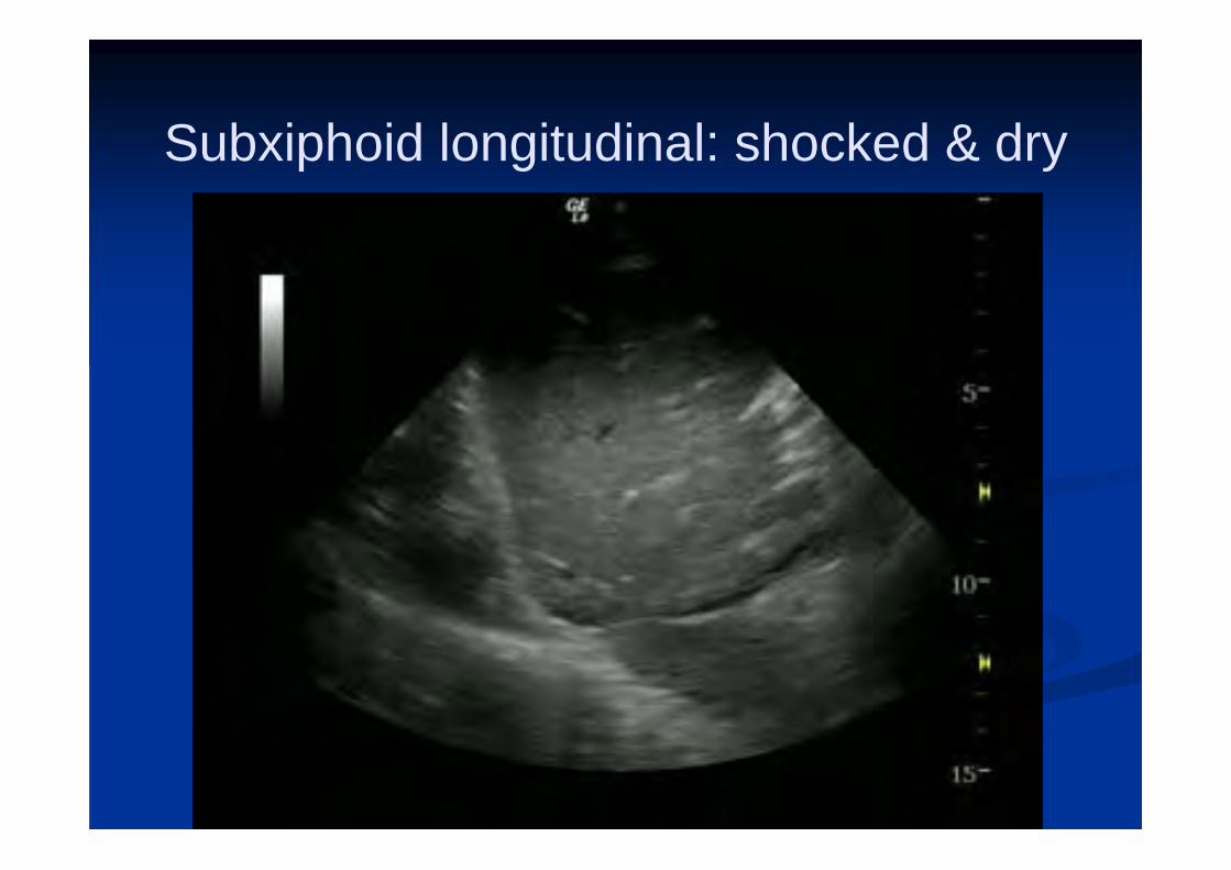
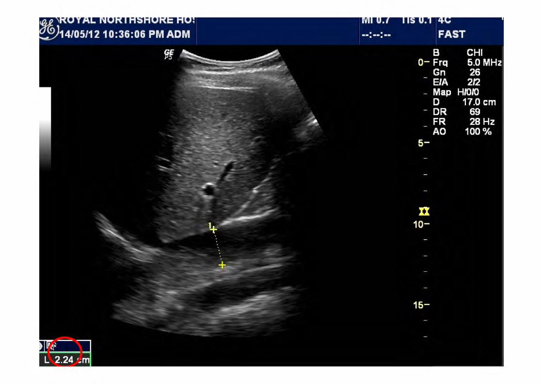
Subxiphoid longitudinal: shocked & dry
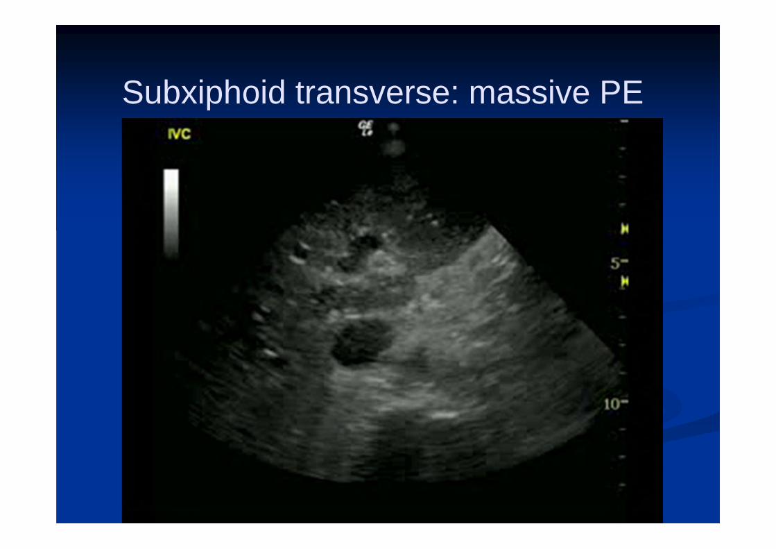
Subxiphoid transverse: massive PE
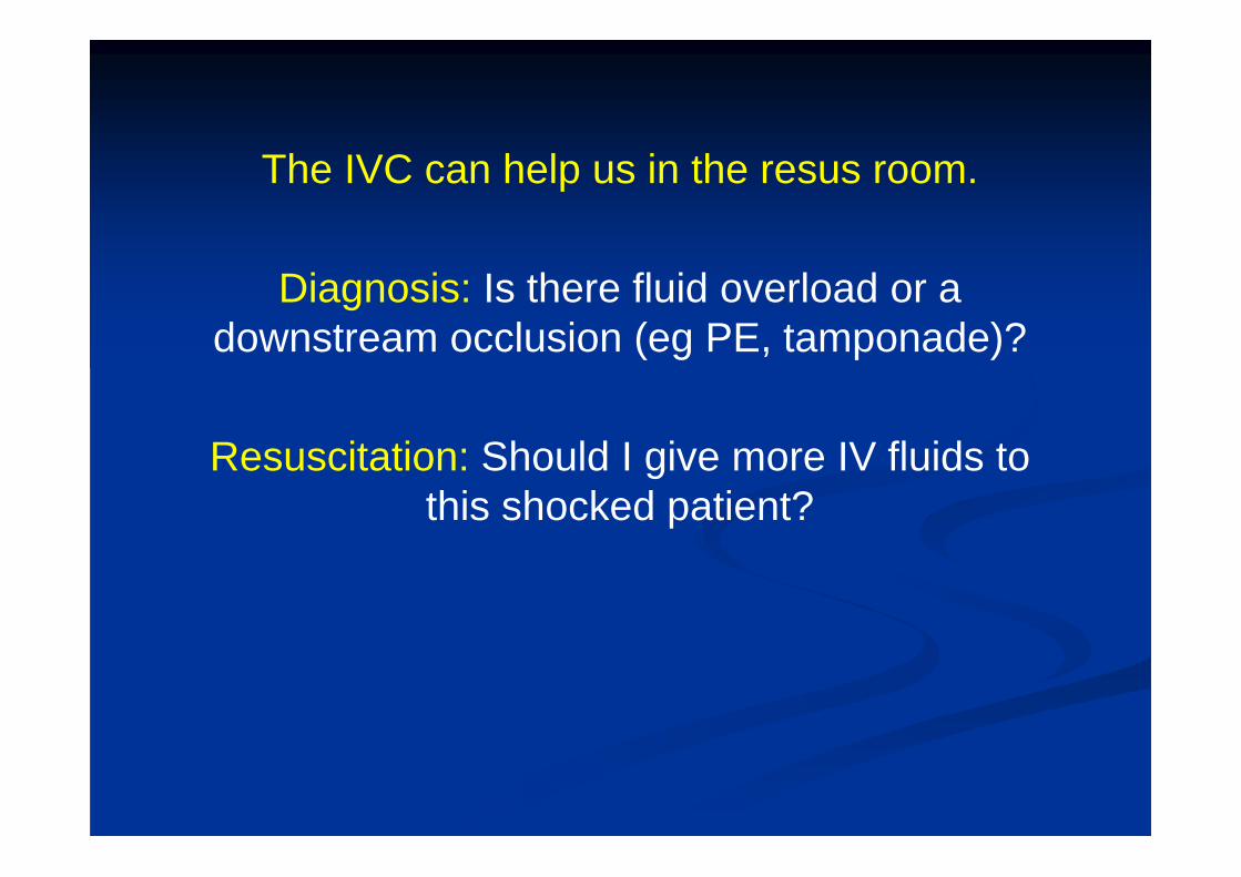
The IVC can help us in the resus room.
Diagnosis: Is there fluid overload or a downstream occlusion (eg PE, tamponade)?
Resuscitation: Should I give more IV fluids to this shocked patient?
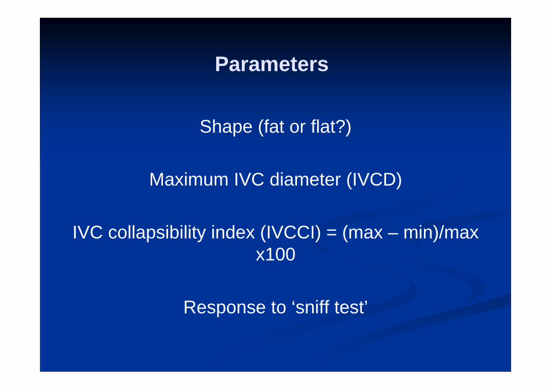
Parameters
Shape (fat or flat?)
Maximum IVC diameter (IVCD)
IVC collapsibility index (IVCCI) = (max – min)/max x100
Response to ‘sniff test’
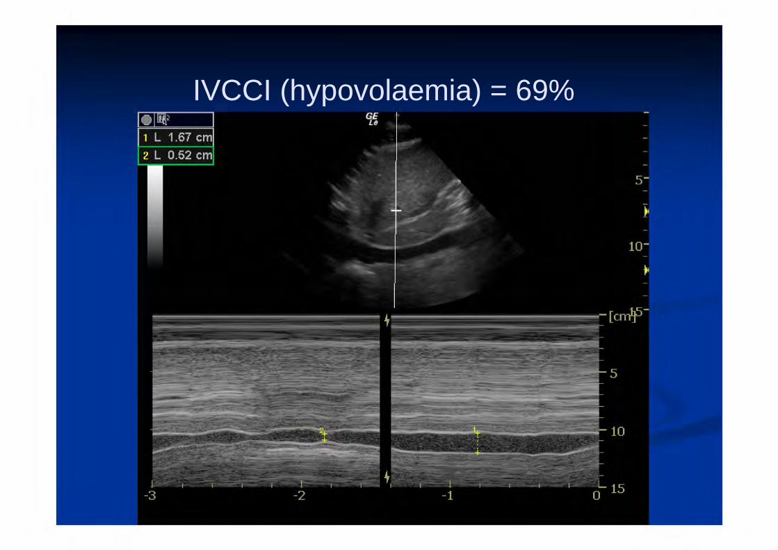
IVCCI (hypovolaemia) = 69%
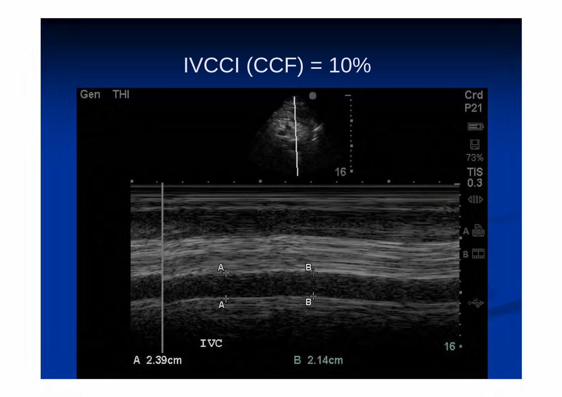
IVCCI (CCF) = 10%

IVC: the good news
CheapEasy to find & measure
NoninvasiveRapid
Repeatable

IVC: the bad news
Poorly validatedOnly useful at extremes
No-one really knows where or how to measure it
How to image the IVC
What probe?What preset?
Where?How?
What probe should we use?No-one knows.
Curved or sector (cardiac) probably OK.
What preset?No-one knows.
Abdo (FAST) probably beats cardiac preset.
Where should we put the probe?
How should we align it?

Thou shalt Place the
probelong axis
In subxiphoidWindow
AboutHere.
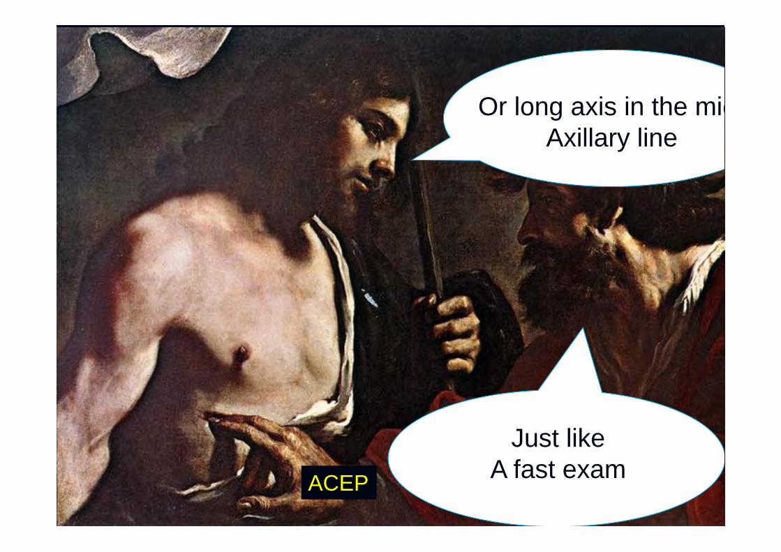
Or long axis in the midAxillary line
Just likeA fast examACEP

Where should we put the probe?
How should we align it?
Where should we put the probe?
How should we align it?
NO-ONE KNOWS!
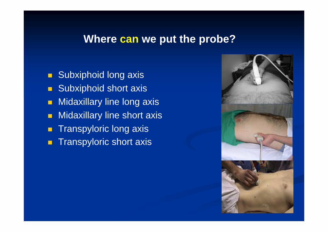
Where can we put the probe?
Subxiphoid long axis Subxiphoid short axis Midaxillary line long axis Midaxillary line short axis Transpyloric long axis Transpyloric short axis
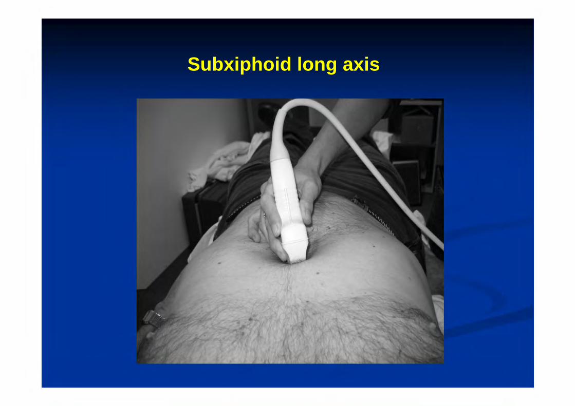
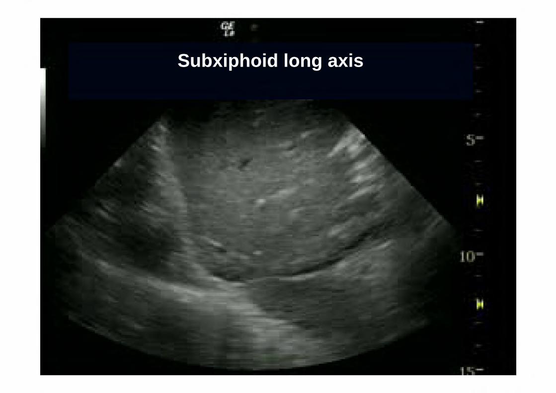
Subxiphoid long axis

Subxiphoid long axis
Most studies & experts measure here Probe sagittal Angled up through the liver Find the right atrium: confirm IVC entering RA Find the hepatic veins entering IVC ‘Hepatic vein confluence’
Subxiphoid long axis
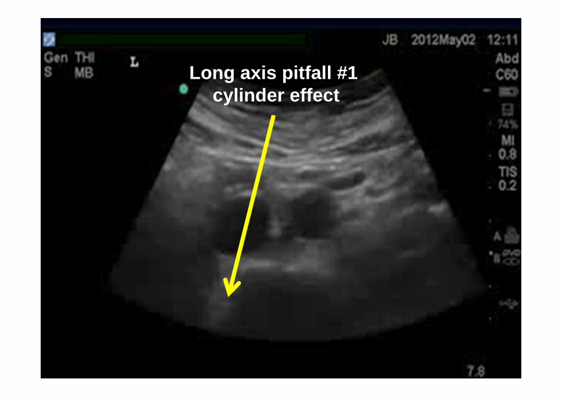
Long axis pitfall #1cylinder effect

Long axis pitfall #1cylinder effect
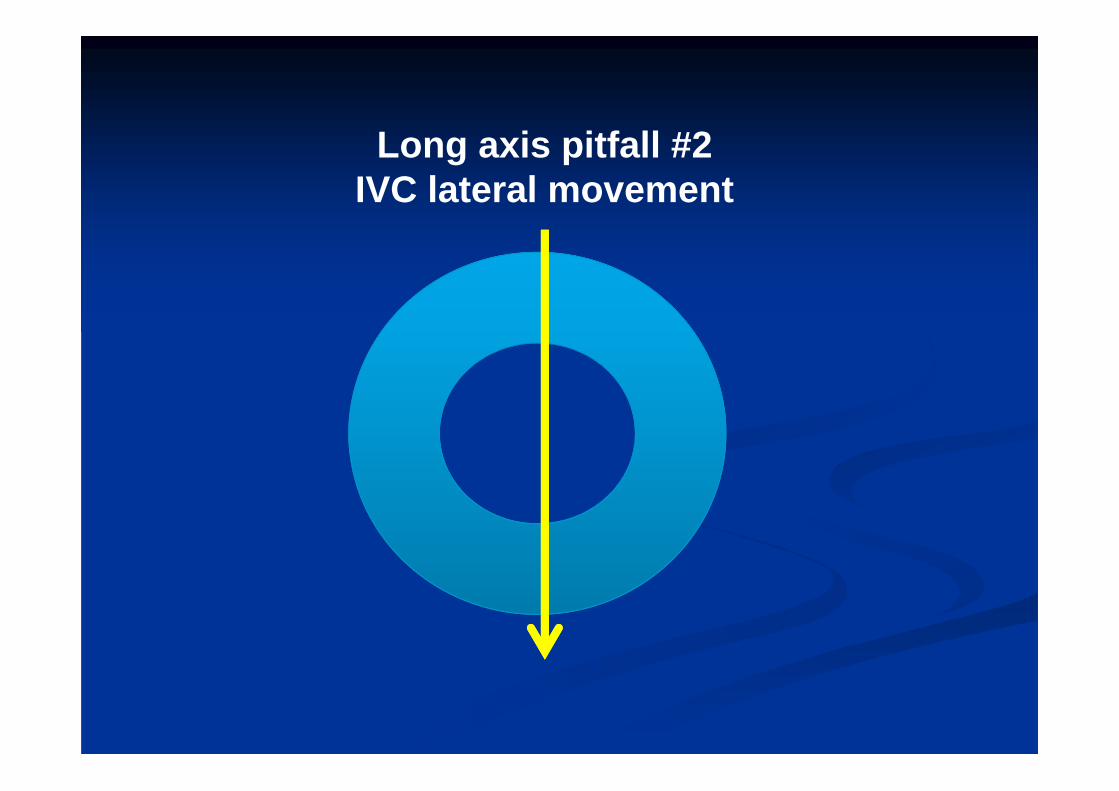
Long axis pitfall #2IVC lateral movement

Subxiphoid short axis
Subxiphoid short axis
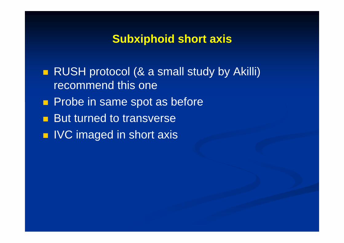
RUSH protocol (& a small study by Akilli) recommend this one
Probe in same spot as before But turned to transverse IVC imaged in short axis

Subxiphoid short axis
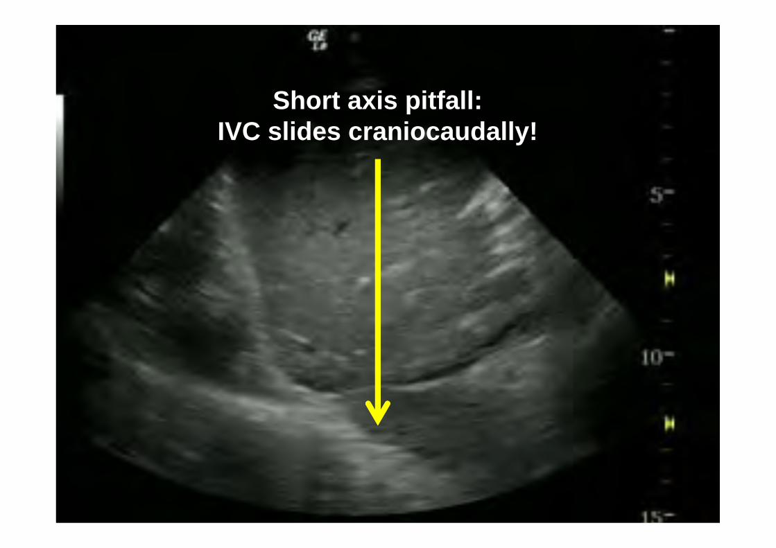
Short axis pitfall:IVC slides craniocaudally!
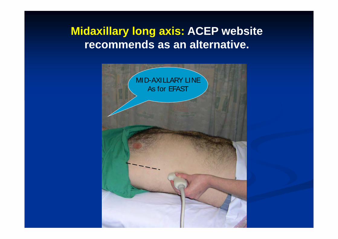
MID-AXILLARY LINEAs for EFAST
Midaxillary long axis: ACEP website recommends as an alternative.
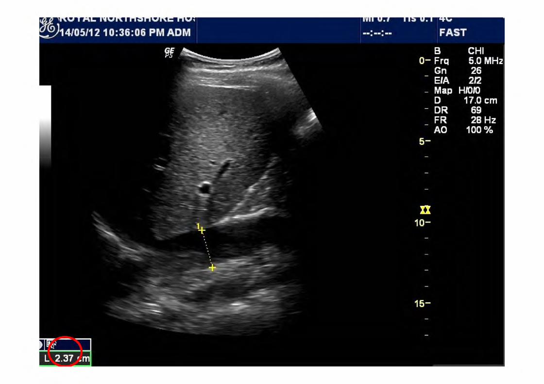
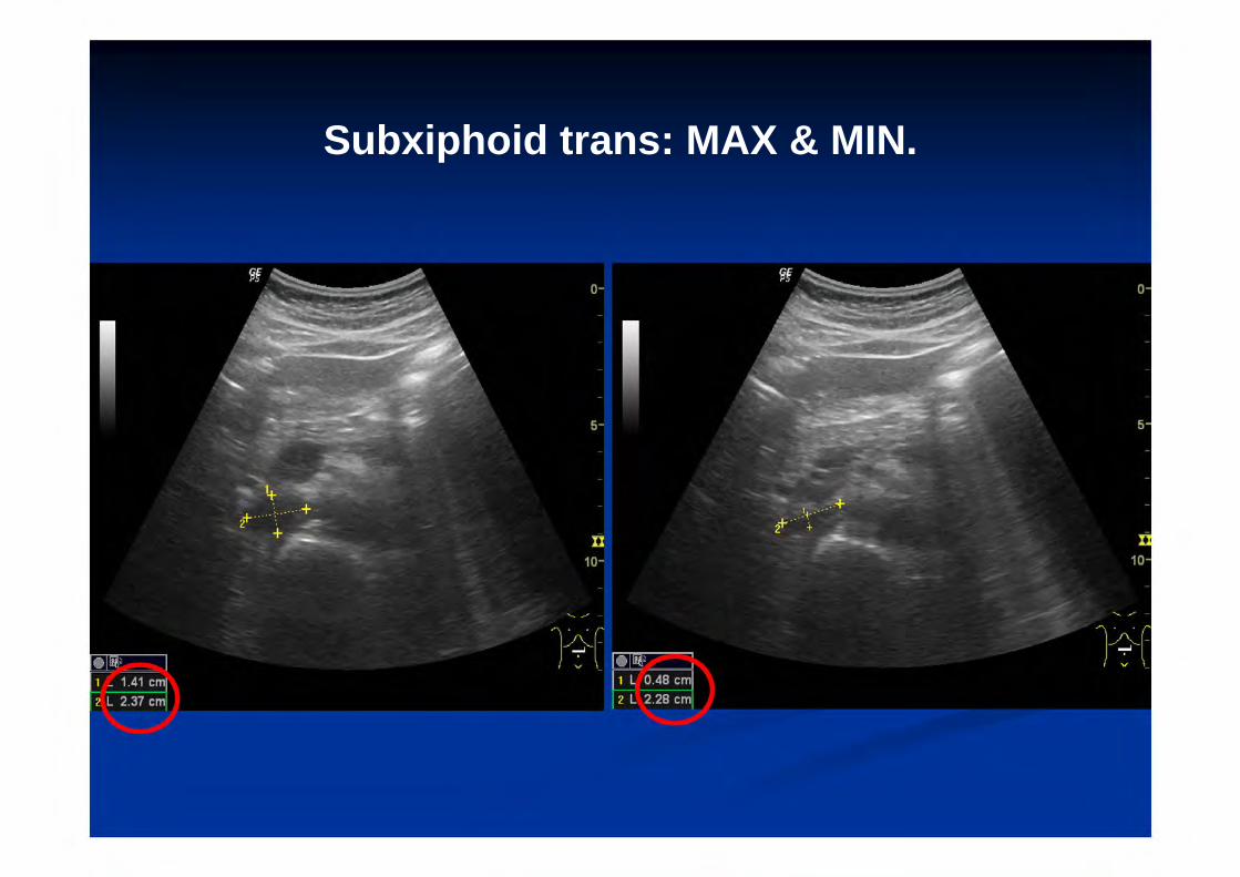
Subxiphoid trans: MAX & MIN.
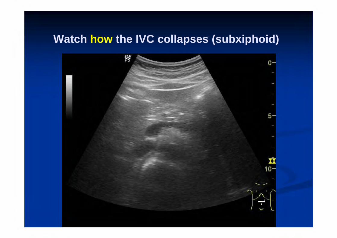
Watch how the IVC collapses (subxiphoid)

Watch how the IVC collapses (RUQ)

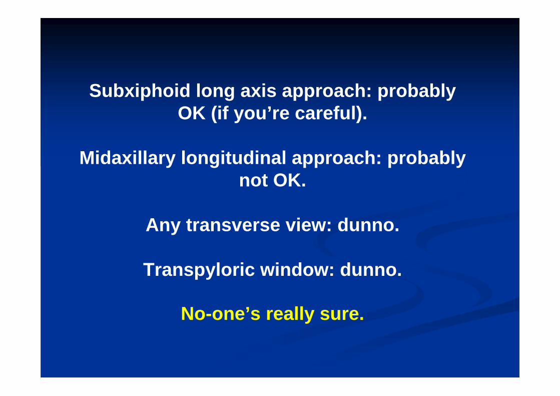
Subxiphoid long axis approach: probably OK (if you’re careful).
Midaxillary longitudinal approach: probably not OK.
Any transverse view: dunno.
Transpyloric window: dunno.
No-one’s really sure.
Where should we measure the IVC?
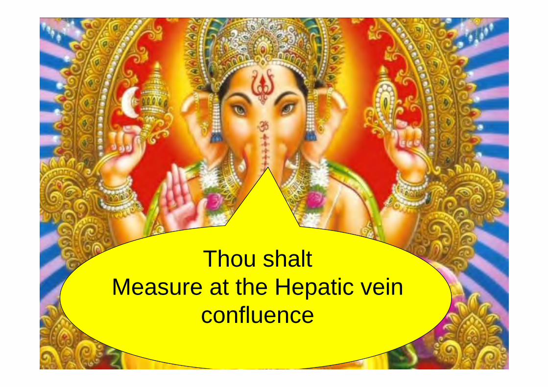
Thou shalt Measure at the Hepatic vein
confluence

Where should we measure the IVC?
No-one knows!
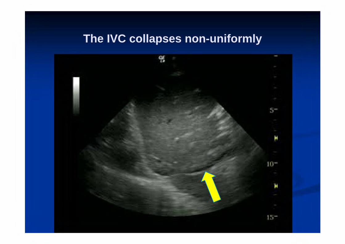
The IVC collapses non-uniformly
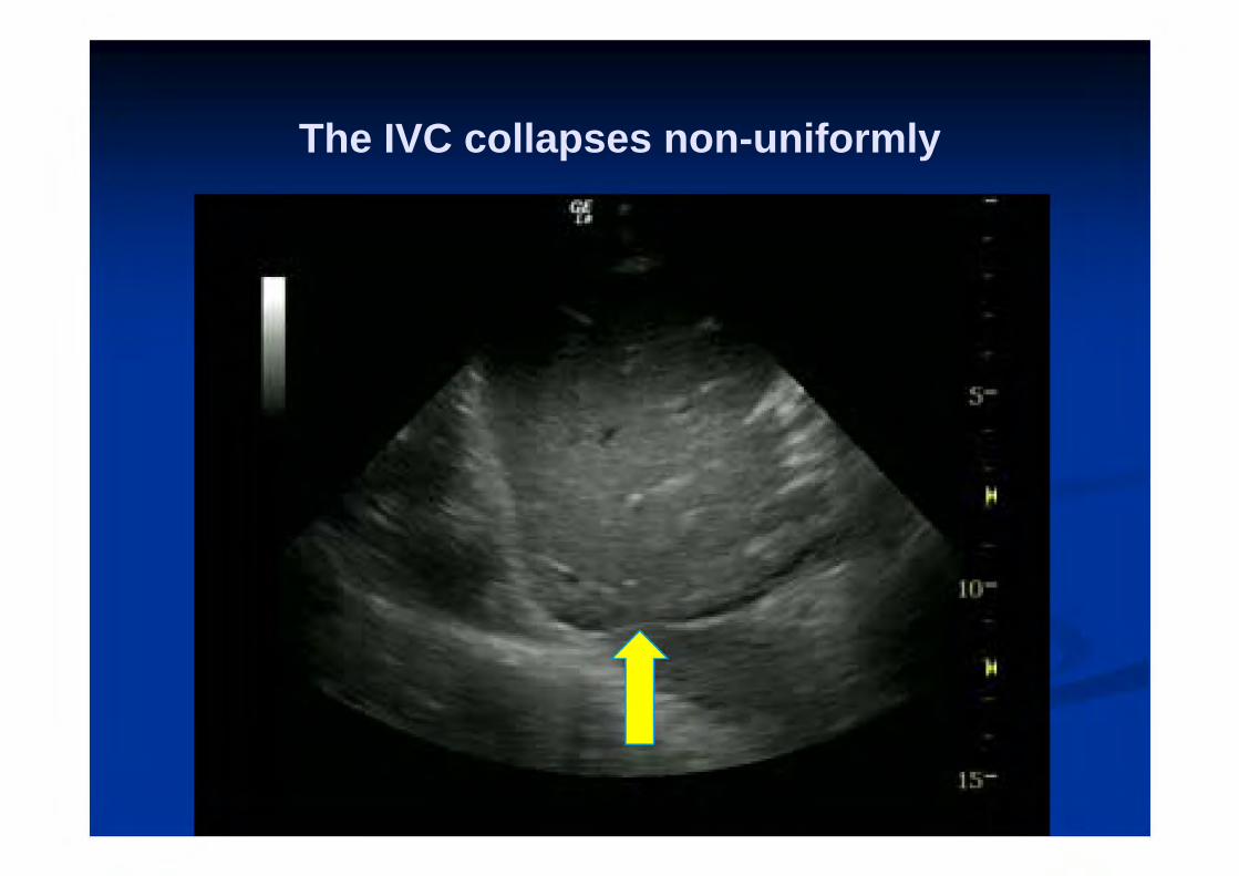
The IVC collapses non-uniformly
Most of us measure at/near the confluence with the hepatic veins.
This is where most of the numbers / guidelines come from.
Beow the liver (eg transpyloric) might be OK, but is probably more prone to probe pressure.
Should I use M-mode?
Should I measure in M-mode?
Lots of fun.Displays max & min diameter on the same image.
Many experts recommend it.
I like it.
But even experienced users can get the angles wrong…

M-mode pitfalls:wrong angle, and IVC moves

Top tip:
When starting out, avoid M-mode.
Should I do a sniff test?

Sniff test (great in healthy volunteers)
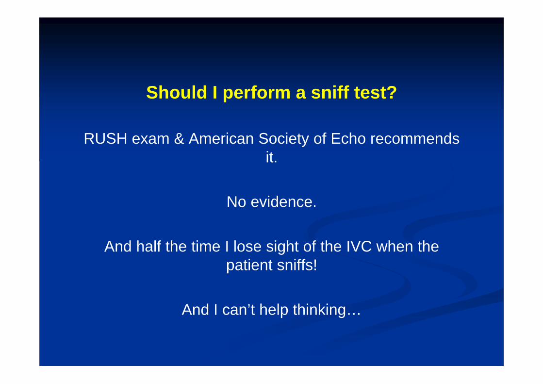
Should I perform a sniff test?
RUSH exam & American Society of Echo recommends it.
No evidence.
And half the time I lose sight of the IVC when the patient sniffs!
And I can’t help thinking…
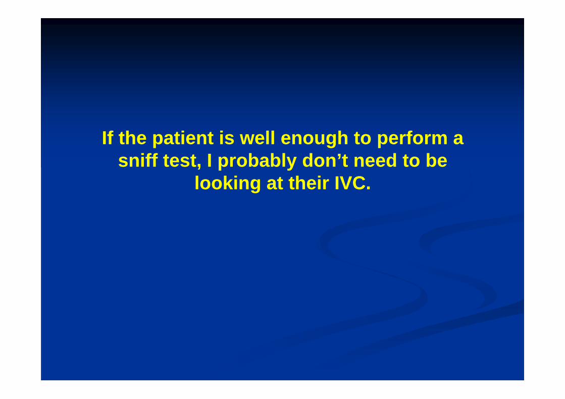
If the patient is well enough to perform a sniff test, I probably don’t need to be
looking at their IVC.

IVC MYTHS

Myth 1:There’s an ivc table
You can use to predict
fluid status
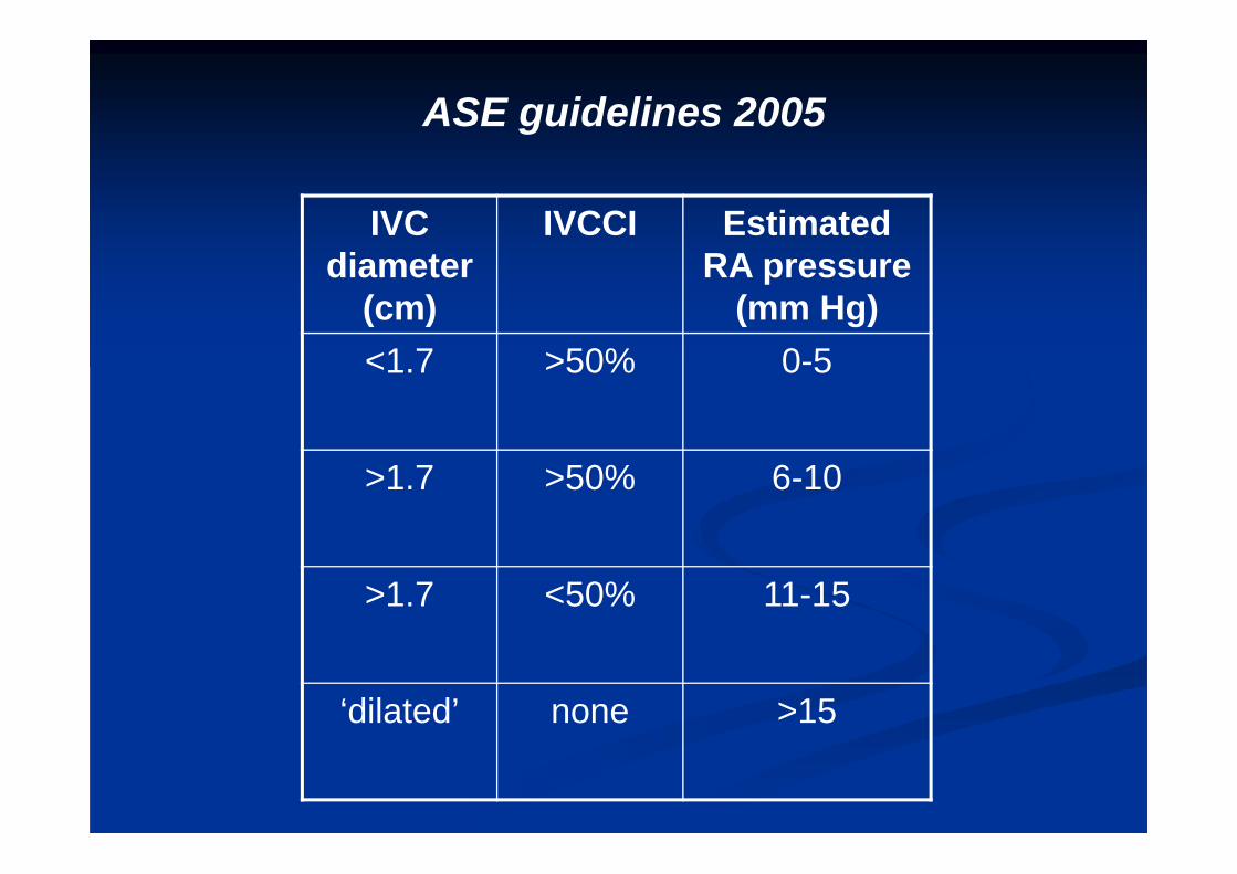
IVC diameter
(cm)
IVCCI Estimated RA pressure
(mm Hg)<1.7 >50% 0-5
>1.7 >50% 6-10
>1.7 <50% 11-15
‘dilated’ none >15
ASE guidelines 2005
Not validated in critically ill patients.
Based on sonographer measurements (which don’t correlate with clinician measurements).
Performed on patients in the left decubitus position.But our patients are either sitting up (SOB) or supine
(shock).
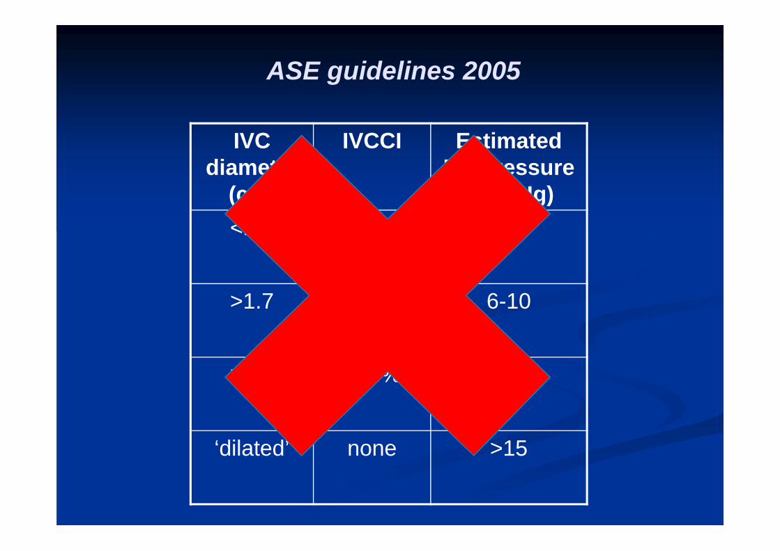
IVC diameter
(cm)
IVCCI Estimated RA pressure
(mm Hg)<1.7 >50% 0-5
>1.7 >50% 6-10
>1.7 <50% 11-15
‘dilated’ none >15
ASE guidelines 2005
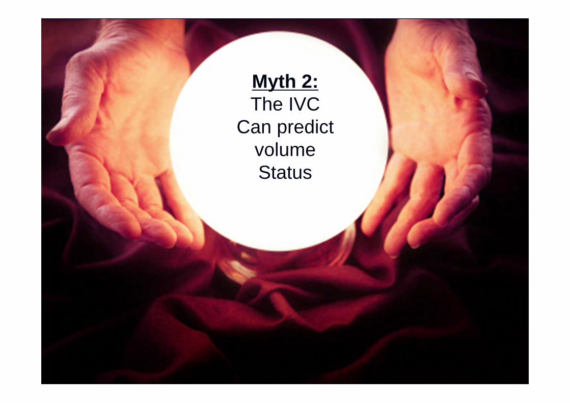
Myth 2:The IVC
Can predictvolume Status
‘The IVC can predict fluid status’
Empty IVC equals empty tank
Full IVC equals full tank
Logical: It makes sense.

Myth 3:IVC ultrasound
Can predictFluid
responsiveness
‘The IVC can predict fluid responsiveness’
Empty IVC IV fluids improved end-organ perfusion
Full IVC IV fluids won’t help
Logical: It makes sense.
These statements are only true at extremes.
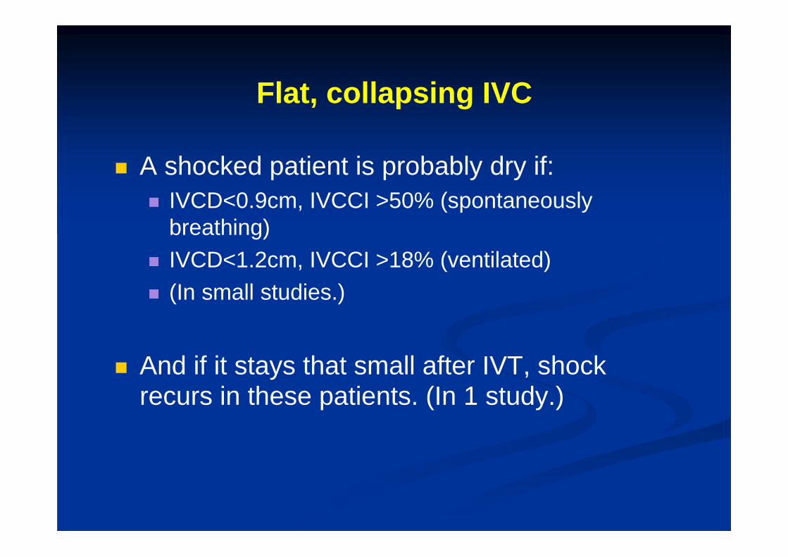
Flat, collapsing IVC
A shocked patient is probably dry if: IVCD<0.9cm, IVCCI >50% (spontaneously
breathing) IVCD<1.2cm, IVCCI >18% (ventilated) (In small studies.)
And if it stays that small after IVT, shock recurs in these patients. (In 1 study.)
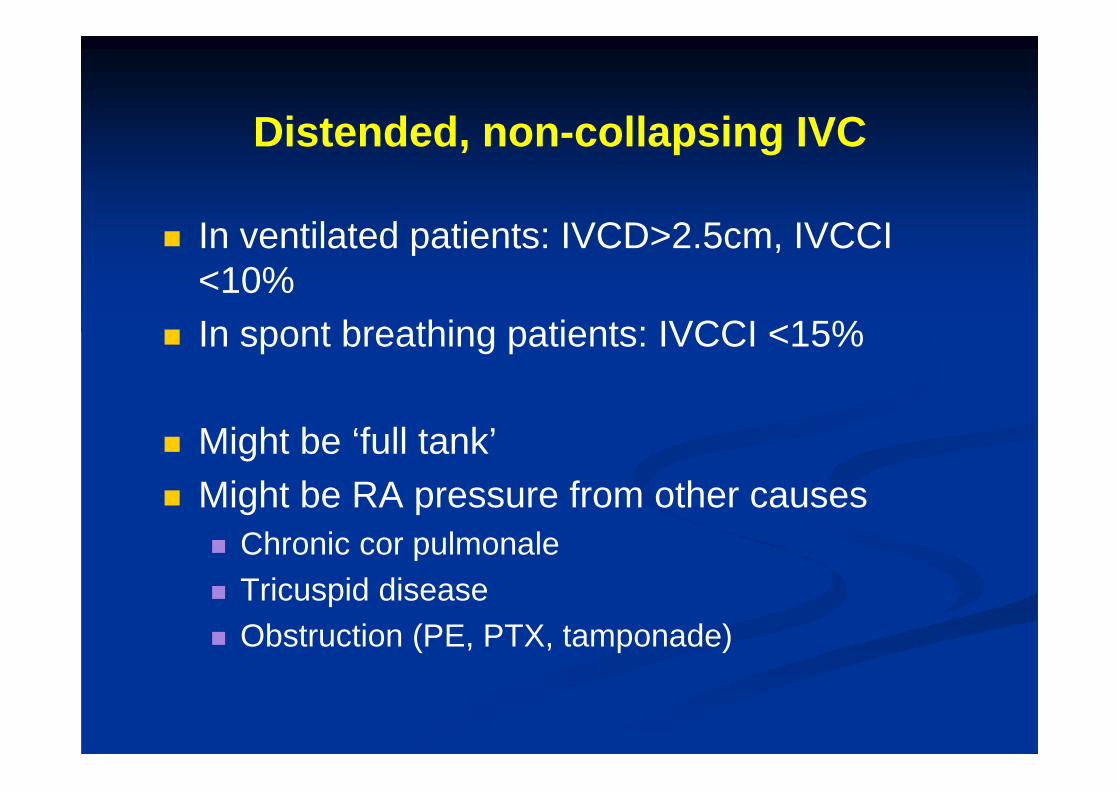
Distended, non-collapsing IVC
In ventilated patients: IVCD>2.5cm, IVCCI <10%
In spont breathing patients: IVCCI <15%
Might be ‘full tank’ Might be RA pressure from other causes
Chronic cor pulmonale Tricuspid disease Obstruction (PE, PTX, tamponade)
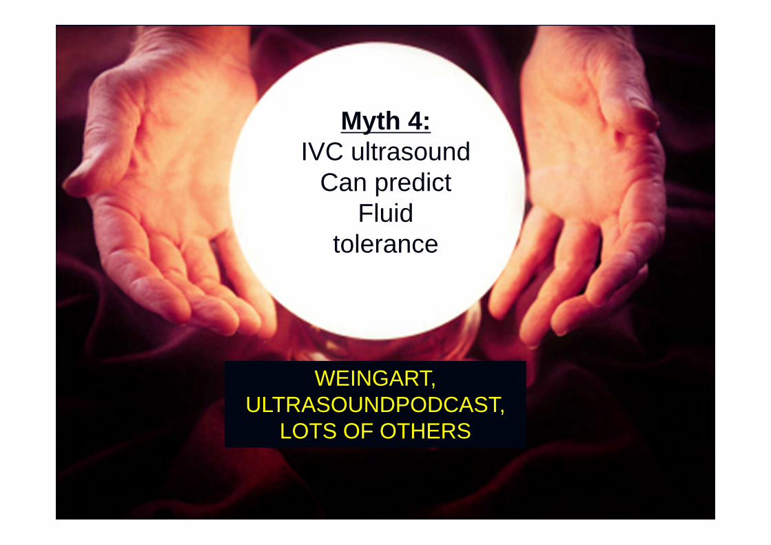
Myth 4:IVC ultrasound
Can predictFluid
tolerance
WEINGART, ULTRASOUNDPODCAST,
LOTS OF OTHERS
Surely it’s SAFE to give fluids if the IVC is flat? And maybe it’s BAD to give fluids if the IVC is
distended?
Well, it seems to make sense. And most of us follow this approach.
Surely it’s SAFE to give fluids if the IVC is flat? And maybe it’s BAD to give fluids if the IVC is
distended?
Well, it seems to make sense. And most of us follow this approach.
But there’s no evidence for these statements.
Surely it’s SAFE to give fluids if the IVC is flat? And maybe it’s BAD to give fluids if the IVC is
distended?
Well, it seems to make sense. And most of us follow this approach.
But there’s no evidence for these statements.
And there’s evidence that IVC is affected by a number of other factors.
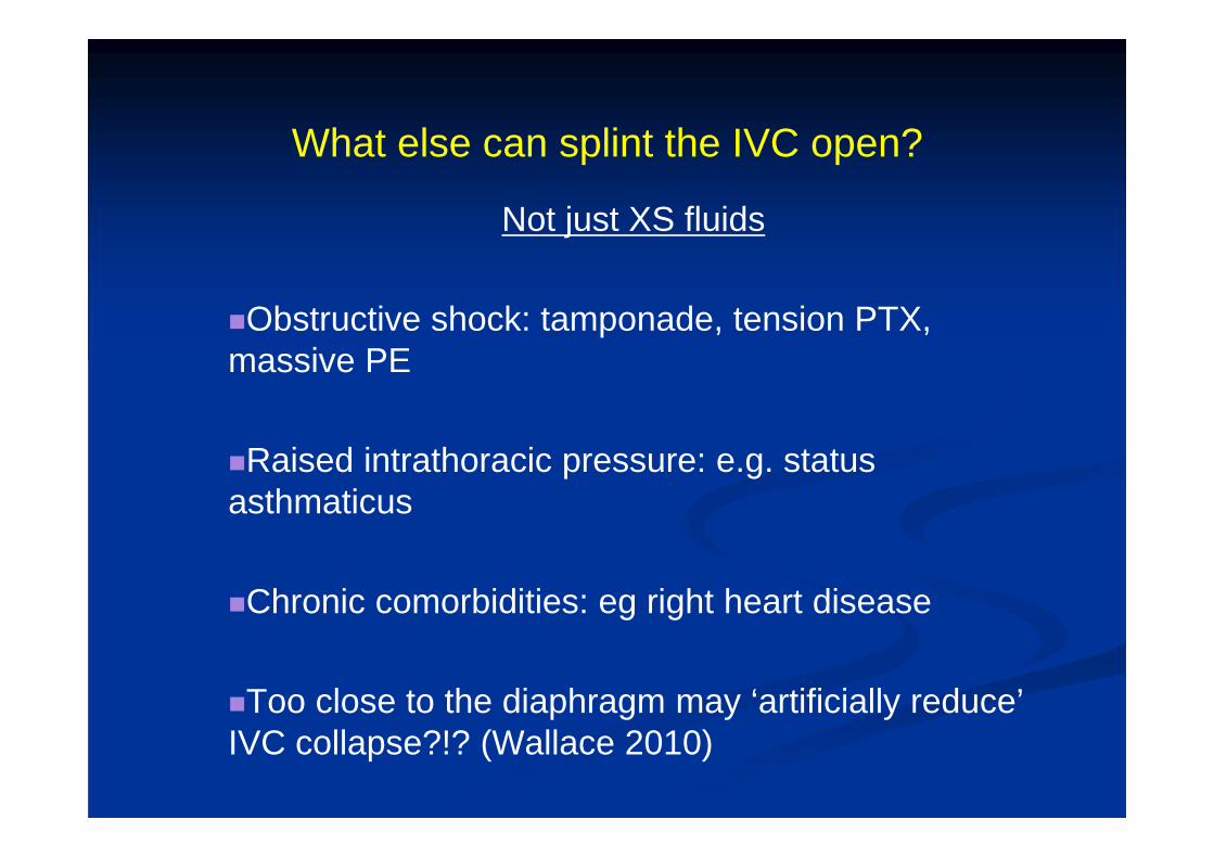
What else can splint the IVC open?
Not just XS fluids
Obstructive shock: tamponade, tension PTX, massive PE
Raised intrathoracic pressure: e.g. status asthmaticus
Chronic comorbidities: eg right heart disease
Too close to the diaphragm may ‘artificially reduce’IVC collapse?!? (Wallace 2010)
What else can cause the IVC to collapse?
1. Ventilation: ‘Diaphragmatic breathing’ (using abdominal wall muscles as well as the chest wall): (Kimura 2011)
2. Raised intra-abdominal pressure (in animal studies: Takata 1990)
3. Even pressure from the probe! (anecdotally)
Just because the IVC collapses, it doesn’t mean it’s safe to give fluids.
Most of us do, but that’s not evidence.
So what can the IVC really tell us?

In shocked patients:IVCD IVCCI Correlation
Spontaneously breathing
<0.9cm >50% Probably empty / fluid responsive
? <15% Probably full & unresponsive
Anything else Dunno
Ventilated <1.2cm >18% Probably empty/ responsive
>2.5cm <10% Probably full/ unresponsive
Or PE/ PTX/ tamponadeOr other stuff that raises CVP
1. IVC just isn’t that precise.
2. CVP isn’t great as a marker of fluid status
Why is this so?
IVC ultrasound: summary
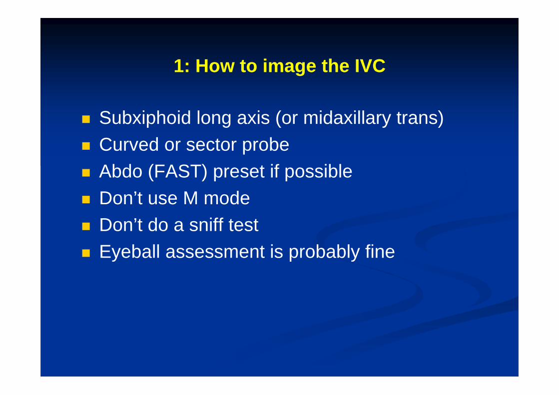
1: How to image the IVC
Subxiphoid long axis (or midaxillary trans) Curved or sector probe Abdo (FAST) preset if possible Don’t use M mode Don’t do a sniff test Eyeball assessment is probably fine
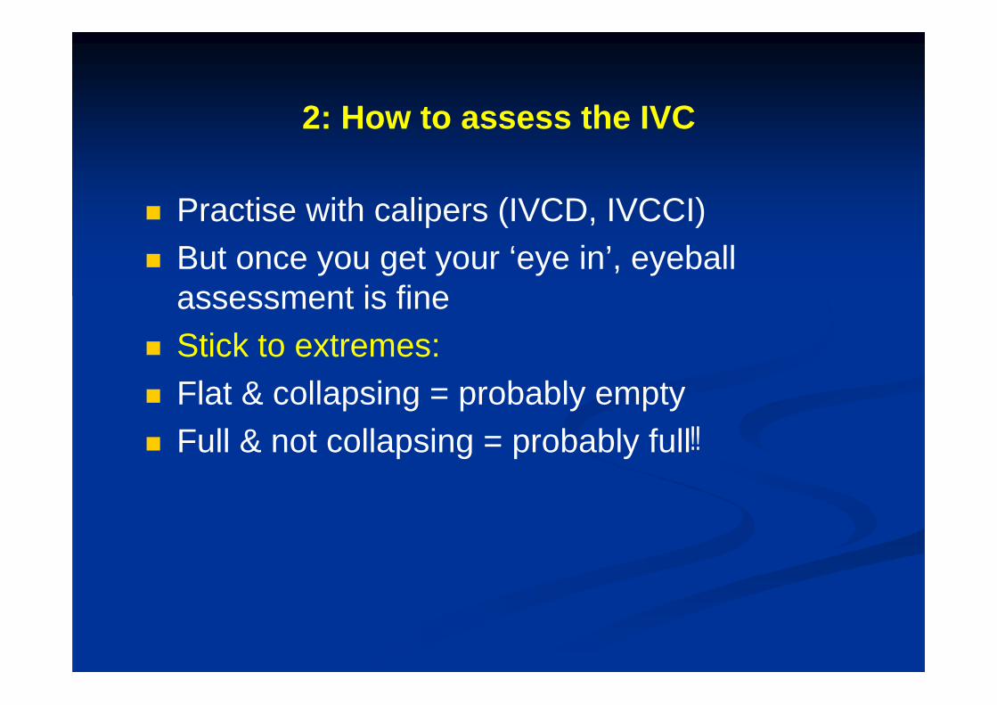
2: How to assess the IVC
Practise with calipers (IVCD, IVCCI) But once you get your ‘eye in’, eyeball
assessment is fine Stick to extremes: Flat & collapsing = probably empty F �ull & not collapsing = probably full
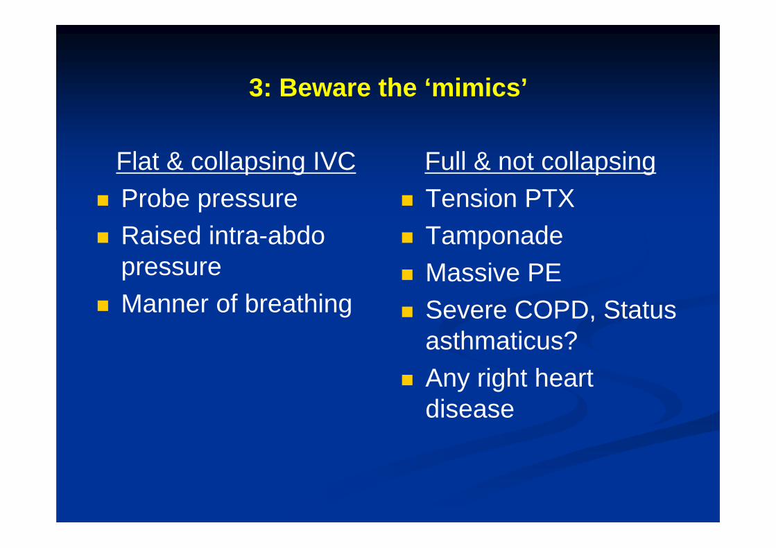
3: Beware the ‘mimics’
Flat & collapsing IVC Probe pressure Raised intra-abdo
pressure Manner of breathing
Full & not collapsing Tension PTX Tamponade Massive PE Severe COPD, Status
asthmaticus? Any right heart
disease

4. Be a doctor
Clinical assessment is always the best Add lung US (wet or dry? PTX? Chunky?) Add basic echo (Tamponade? Massive RV?) And the rest: CXR, ECG, etc etc If US findings don’t match clinical assesment,
turn off the machine
Thanks to
Dr Kylie Baker (for that literature review)Dr Adrian Goudie (for that IVC long axis image)
Drs Mike Blaivas, Matt Dawson, Cliff Reid & Scott Weingart(for their advice & input)

References
ACEP http://www.acep.org/Content.aspx?id=80791 Akilli B, Bayir A et al. Inferior vena cava diameter as a marker of
early hemorrhagic shock: a comparative study. Ulus Travma AcilCerrahi Derg 2010;16(2):113-8.
Baker, K. Review of Bedside Sonography for Guidance of FluidTherapy in the Emergency Department. (unpublished)
Barbier C, Loubières Y, Schmit C, Hayon J, Ricôme JL, Jardin F,Vieillard-Baron A. Respiratory changes in inferior vena cavadiameter are helpful in predicting fluid respon- siveness in ventilatedseptic patients. Intensive Care Med 2004; 30:1740–1746
Blehar DJ, Dickman E, Gaspari R. Identification of congestive heartfailure via respiratory variation of inferior vena cava. Am J Em Med2009;27:71–5.
Blehar et al. Inferior vena cava displacement during respirophasicultrasound imaging. Critical Ultrasound Journal 2012, 4:18
References
Charron C, Caille V, Jardin F, Viellard-Baron A. Echocardiographicmeasurement of fluid responsiveness. Curr Op Crit Care 2006;12(3): 249-54.
Corl K, Napoli A, Gardiner F. Bedside sonographic measurement ofthe inferior vena cava caval index is a poor predictor of fluidresponsiveness in emergency department patients. EmergencyMedicine Australasia (2012) 24, 534–539
Dipti A et al. Role of inferior vena cava diameter in assessment ofvolume status: a meta-analysis. AJEM 2012 (30). 1414 -19.
Feissel M, Michard F, Faller JP, Teboul JL (2004) The respiratoryvariation in inferior vena cava diameter as a guide to fluid therapy.Intensive Care Med 30:1834–1837
Jue J, Chung W, Schiller NB. Does inferior vena cava size predictright atrial pressures in patients receiving mechanical ventilation. JAm Soc Echocardiogr 1992; 5: 613-9.

References
Kimura BJ, Dalugdugan R, Gilcrease GW 3rd, Phan JN, ShowalterBK, Wolfson T. The effect of breathing manner on inferior venacaval diameter. Eur J Echocardiogr. 2011 Feb;12(2):120-3
Kircher B, Himelman R, Schiller N. Noninvasive estimation of rightatrial pressure from the inspiratory collapse of the inferior vena cava.AM J Cardiol 1990; 66: 493-6.
Lang RM, Bierig M, Devereux F et al Recommendations forchamber quantification: a report from the American Society ofEchocardiography’s guidelines and standards committee and thechamber quantification writing group, developed in conjunction withthe European Association of Echocardiography, ad branch of theEuropean Society of Cardiology. J Am Soc Echocardiogr 2005; 18:1440-63.
Lanspa MJ, Grissom CK et al. Applying dynamic parameters topredict hemodynamic response to volume expansion inspontaneously breathing patients with septic shock. Shock 2013.39(2). pp. 155-160
References
Medscape http://www.medscape.com/viewarticle/727097 Moretti R, Pizzi B. Inferior vena cava distensibility as a predictor of
fluid responsiveness in patients with subarachnoid hemorrhage.Neurocrit Care. 2010 Aug;13(1):3-9.
Muller L et al. Respiratory variations of inferior vena cava diameterto predict fluid responsiveness in spontaneously breathing patientswith acute circulatory failure: need for a cautious use. Critical Care2012, 16:R188
Nagdev AD, Merchant RC, Tirado-Gonzalez A, Sisson CA, MurphyMC. Emergency Department Bedside UltrasonographicMeasurement of the Caval Index for Noninvasive Determination ofLow Central Venous Pressure. Ann Emerg Med. 2010Mar;55(3):290-5
Perera P et al. The RUSH Exam 2012: Rapid Ultrasound in Shock in the Evaluation of the Critically Ill Patient. Em Med Clinics. Ultrasound Clin 7 (2012) 255–278
References
Randazzo MR, Snoey ER, Levitt MA, et al. Accuracy of emergencyphysician assessment of left ventricular ejection fraction and centralvenous pressure using echocardiography. Acad Emerg Med2003;10:973–7.
Sefidbakht S, Assadsangabi R, Abbasi HR, Nabavizadeh A.Sonographic measurement of the inferior vena cava as a predictorof shock in trauma patients. Emerg Radiol 2007; 14(3): 181-185.
Stanford University.http://www.stanford.edu/group/ccm_echocardio/cgi-bin/mediawiki/index.php/IVC
Takata M, Wise RA, Robotham JL. Effect of abdominal pressure onvenous return: abdominal vascular zone conditions. J Appl Physiol1990 (69):1961– 1972
ultrasoundpodcast http://www.ultrasoundpodcast.com/?s=ivc )
References
Wallace DJ, Allison M, Stone MB. Inferior vena cava percentagecollapse during resuscitation is affected by the sampling location: anultrasound study in healthy volunteers. Acad Emerg Med2010;17:96–9.
Weekes A, Tassone HM, Tayal VS, Babcok AJ, Norton J.Sonodynamic Comparison of Systolic Blood Pressure to AorticVelocity Time Integral Measurements as a Measure of FluidResponsiveness In Non-Traumatic Symptomatic HypotensiveEmergency Department Patients. Annals of Emergency Medicine2010. Volume 56, Issue 3 Suppl, pS76
Weekes AJ, Tassone HM, Babcock A, et al. Comparison of serialqualitative and quantitative assessments of caval index and leftventricular systolic function during early fluid resuscitation ofhypotensive emergency department patients. Acad Emerg Med2011; 18(9):912-21.
References
Yanagawa Y, Nishi K, Sakamoto T, Okada Y. Early diagnosis ofhypovolemic shock by sonographic measurement of inferior venacava in trauma patients. J Trauma 2005; 58(4): 825-829.
Yanagawa Y, Sakamoto T, Okada Y. Hypovolemic shock evaluatedby sonographic measurement of the inferior vena cava duringresuscitation in trauma patients. J Trauma 2007; 63(6): 1245-1248.