Discrepancy Switches - Control Discrepancy Switches Standard Electrical Diagrams
~ 179 ~
ISSN Print: 2394-7500
ISSN Online: 2394-5869
Impact Factor: 5.2
IJAR 2018; 4(9): 179-185
www.allresearchjournal.com
Received: 06-07-2018
Accepted: 08-08-2018
Dr. Sangeeta Yadav
Department of Orthodontics
and Dentofacial Orthopaedics,
Mahatma Gandhi Dental
College and hospital, Jaipur,
Rajasthan, India
Dr. Jay Prakash Yadav
Department of Human
Anatomy, Govt. Medical
College Churu, Rajasthan,
India
Correspondence
Dr. Sangeeta Yadav
Department of Orthodontics
and Dentofacial Orthopaedics,
Mahatma Gandhi Dental
College and hospital, Jaipur,
Rajasthan, India
Relationship of arch parameters with arch length-
tooth material discrepancy
Dr. Sangeeta Yadav and Dr. Jay Prakash Yadav
Abstract
Dental crowding can be defined as a disparity in the relationship between tooth size and jaw size which
resulting imbrications and rotation of teeth. Eight pairs of study models were used in this study. Each
study model met the following criteria: all permanent teeth with the exception of the molars were
present in both the maxillary and mandibular arches and these was no history of previous orthodontic
treatment.
Keywords: skeletal, Vernier caliper, brass wire, arch Length
1. Introduction
Dental crowding can be defined as a disparity in the relationship between tooth size and jaw
size which result in imbrications and rotation of teeth. Three condition which may predispose
the dental arches to crowding are excessively large teeth, excessively small bones of the jaws
and a combination of large teeth and small jaws [1].
A concept that large teeth are characteristic of modern, civilized man has been advanced by
dental anthropologists, orthodontists and student of developing occlusion. It is a generally
accepted theory that primitive civilizations exhibited a significant degree of wear or attrition,
probably the result of more vigorous mastication of harder foodstuff than is commonly
associated attritional occlusion theory given by Begg².
2. Aims and objectives
To determine the extent to which various dental arch parameters contribute to dental
crowding.
3. Material and methods
Eight pairs of study were used in this study. Each study model met the following criteria: all
permanent teeth with the exception of the third molars were present in both the maxillary and
the mandibular arches. There was no history of previous orthodontic treatment.
These study models were divided into two groups.
Group 1 consisted of forty pairs (20 males and 20 females) of study models with angles class
I normal occlusion having class I skeletal base and little or no crowding. These patients
exhibited a straight profile, normal overjet and overbite and class I canine relationship. The
mean age for this group was 21.28±2.85years (figure-1)
Group 2 consisted of forty pairs (20 males and 20 females) of study models with angles class
I normal occlusion having class I skeletal base and more than 5 mm crowding. The mean age
for this group was 18.25±3.24 years (figure-2)
The following Mearsurement were recorded
1. The largest mesiodistal width of each tooth (except the second and third molars) of each
arch.
2. Buccal and lingual inter-canine width.
3. Buccal and lingual inter-molar width
4. Arch length
5. Arch perimeter of both arches
Digital vernier caliper was used to recorded the measurements. It was calibrated to 0.1
mm (figure 3).
International Journal of Applied Research 2018; 4(9): 179-185

~ 180 ~
International Journal of Applied Research
Measurement of mesiodistal width of tooth
The mesiodistal width of the teeth were measured in the
widest area by caliper calibrated to 0.1 mm. The caliper was
held perpendicular to the long axis of tooth (figure-4)
Measurement of arch diamension
Arch dimension in the canine and molar regions were
buccally and lingually. The buccal arch dimension war
measures 5 mm apical to the mesiodistal centre of the
gingival margin of the canine tooth on one side to the same
point on the contralateral side. On the lingual side, the
distance between midpoint on the cervical region of the
canine on one side was measured to the corresponding point
on the contra-lateral side. The same procedure was
preformed in the molar region. (figure 5).
Measurement of arch perimeter
The arch perimeter is a line drawn from the surface of the
first perimeter molar around the arch over contact points and
incisal edges in a smooth curve to the distal surface of the
first permanent molars on the opposite side. The brass wire
made into a arch, free kinds and in a simulated arch form.
The wire is then straighten and measured with scale. (figure-
6).
Measurement of arch length
To determine the arch length a line was drawn a point
midway to the central incisors perpendicular to the tangent
touching the distal surface of the first permanent molars.
(figure-7)
Fig 1: Normal dentition
Fig 2: Crowded dentition
Fig 3: Digital vernier caliper
Fig 4: Measurement of tooth size using Digital vernier caliper
Fig 5: Measurement of Arch dimension
Fig 6: Measurement of Arch perimeter
Fig 7: Measurement of Arch length using brass wire
4. Result
Table 1: Demographic data
Males Females Mean age
Group-1 20 20 21.82±2.85
Group -2 20 20 18.25±3.24
~ 181 ~
International Journal of Applied Research
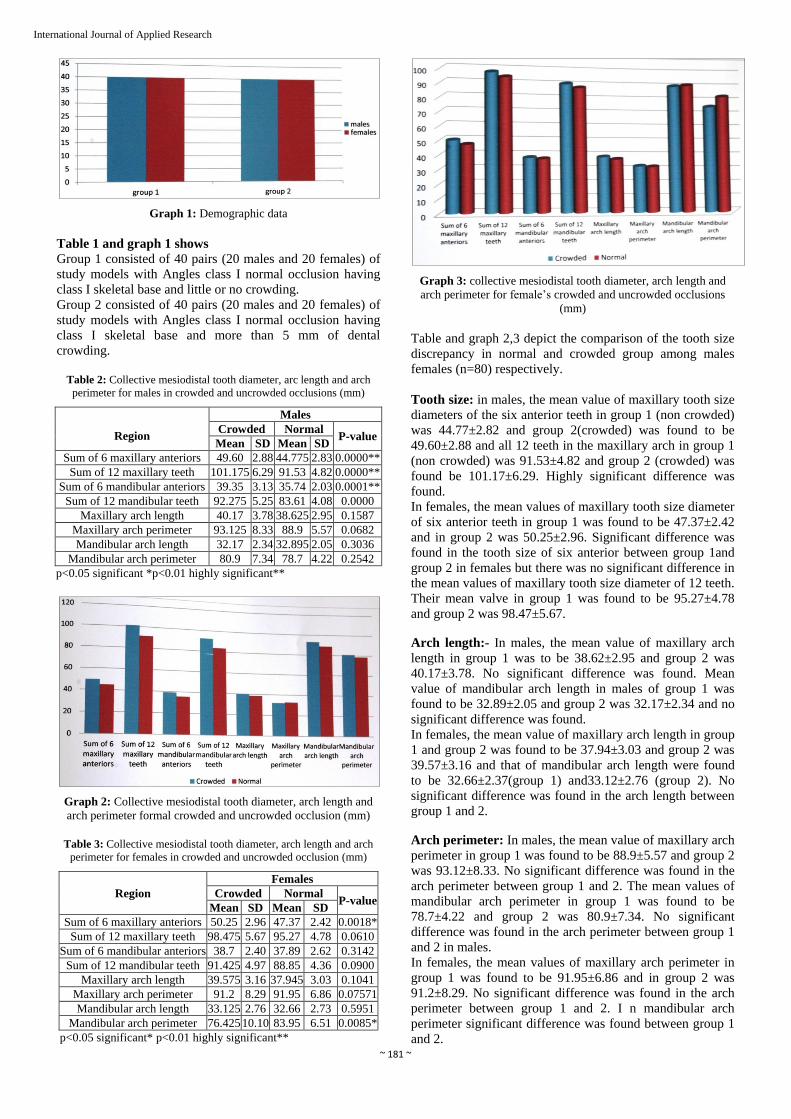
Graph 1: Demographic data
Table 1 and graph 1 shows
Group 1 consisted of 40 pairs (20 males and 20 females) of
study models with Angles class I normal occlusion having
class I skeletal base and little or no crowding.
Group 2 consisted of 40 pairs (20 males and 20 females) of
study models with Angles class I normal occlusion having
class I skeletal base and more than 5 mm of dental
crowding.
Table 2: Collective mesiodistal tooth diameter, arc length and arch
perimeter for males in crowded and uncrowded occlusions (mm)
Region
Males
Crowded Normal P-value
Mean SD Mean SD
Sum of 6 maxillary anteriors 49.60 2.88 44.775 2.83 0.0000**
Sum of 12 maxillary teeth 101.175 6.29 91.53 4.82 0.0000**
Sum of 6 mandibular anteriors 39.35 3.13 35.74 2.03 0.0001**
Sum of 12 mandibular teeth 92.275 5.25 83.61 4.08 0.0000
Maxillary arch length 40.17 3.78 38.625 2.95 0.1587
Maxillary arch perimeter 93.125 8.33 88.9 5.57 0.0682
Mandibular arch length 32.17 2.34 32.895 2.05 0.3036
Mandibular arch perimeter 80.9 7.34 78.7 4.22 0.2542
p<0.05 significant *p<0.01 highly significant**
Graph 2: Collective mesiodistal tooth diameter, arch length and
arch perimeter formal crowded and uncrowded occlusion (mm)
Table 3: Collective mesiodistal tooth diameter, arch length and arch
perimeter for females in crowded and uncrowded occlusion (mm)
Region
Females
Crowded Normal P-value
Mean SD Mean SD
Sum of 6 maxillary anteriors 50.25 2.96 47.37 2.42 0.0018*
Sum of 12 maxillary teeth 98.475 5.67 95.27 4.78 0.0610
Sum of 6 mandibular anteriors 38.7 2.40 37.89 2.62 0.3142
Sum of 12 mandibular teeth 91.425 4.97 88.85 4.36 0.0900
Maxillary arch length 39.575 3.16 37.945 3.03 0.1041
Maxillary arch perimeter 91.2 8.29 91.95 6.86 0.07571
Mandibular arch length 33.125 2.76 32.66 2.73 0.5951
Mandibular arch perimeter 76.425 10.10 83.95 6.51 0.0085*
p<0.05 significant* p<0.01 highly significant**
Graph 3: collective mesiodistal tooth diameter, arch length and
arch perimeter for female’s crowded and uncrowded occlusions
(mm)
Table and graph 2,3 depict the comparison of the tooth size
discrepancy in normal and crowded group among males
females (n=80) respectively.
Tooth size: in males, the mean value of maxillary tooth size
diameters of the six anterior teeth in group 1 (non crowded)
was 44.77±2.82 and group 2(crowded) was found to be
49.60±2.88 and all 12 teeth in the maxillary arch in group 1
(non crowded) was 91.53±4.82 and group 2 (crowded) was
found be 101.17±6.29. Highly significant difference was
found.
In females, the mean values of maxillary tooth size diameter
of six anterior teeth in group 1 was found to be 47.37±2.42
and in group 2 was 50.25±2.96. Significant difference was
found in the tooth size of six anterior between group 1and
group 2 in females but there was no significant difference in
the mean values of maxillary tooth size diameter of 12 teeth.
Their mean valve in group 1 was found to be 95.27±4.78
and group 2 was 98.47±5.67.
Arch length:- In males, the mean value of maxillary arch
length in group 1 was to be 38.62±2.95 and group 2 was
40.17±3.78. No significant difference was found. Mean
value of mandibular arch length in males of group 1 was
found to be 32.89±2.05 and group 2 was 32.17±2.34 and no
significant difference was found.
In females, the mean value of maxillary arch length in group
1 and group 2 was found to be 37.94±3.03 and group 2 was
39.57±3.16 and that of mandibular arch length were found
to be 32.66±2.37(group 1) and33.12±2.76 (group 2). No
significant difference was found in the arch length between
group 1 and 2.
Arch perimeter: In males, the mean value of maxillary arch
perimeter in group 1 was found to be 88.9±5.57 and group 2
was 93.12±8.33. No significant difference was found in the
arch perimeter between group 1 and 2. The mean values of
mandibular arch perimeter in group 1 was found to be
78.7±4.22 and group 2 was 80.9±7.34. No significant
difference was found in the arch perimeter between group 1
and 2 in males.
In females, the mean values of maxillary arch perimeter in
group 1 was found to be 91.95±6.86 and in group 2 was
91.2±8.29. No significant difference was found in the arch
perimeter between group 1 and 2. I n mandibular arch
perimeter significant difference was found between group 1
and 2.

~ 182 ~
International Journal of Applied Research
Tables 4: Mandibular arch dimensions for males in crowded and uncrowded occlusions (mm)
Region
Males
Crowded Normal P value
Mean SD Mean SD
Mandibular intercanine width buccal 28.935 2.23 28.23 1.98 0.2965
Mandibular intercanine width lingual 19.205 2.17 19.455 1.30 0.6619
Mandibular intermolar width buccal 55.2 2.07 55.055 2.67 0.8490
Mandibular intermolar width lingual 30.89 2.20 32.86 2.14 0.0067
P<0.05-significant * P<0.01= highly significant **
Graph 4: Mandibular arch dimension for males in crowded and uncrowded occlusion (mm)
Tables 5: Mandibular arch dimensions for females in crowded and uncrowded occlusions (mm)
Region
Males
Crowded Normal
P value Mean SD Mean SD
Mandibular intercanine width buccal 28.25 2.65 30.82 2.25 0.0021*
Mandibular intercanine width lingual 18.525 3.19 20.835 0.89 0.0050*
Mandibular intermolar width buccal 55.225 2.71 58.22 2.22 0.0005**
Mandibular intermolar width lingual 31.375 2.93 36.125 2.42 0.0000**
P<0.05-significant * P<0.01= highly significant **
Graph 5: Mandibular arch dimensions for females in crowded and uncrowded occlusions (mm)
~ 183 ~
International Journal of Applied Research
Arch dimensions
Mandibular intercanine and intermolar width
In males, the mean value of buccal mandibular intercanine
width in group 1 was to be 28.23±1.98 and group 2 was
28.93±2.23. No significant difference was found between
the two groups. And no significant difference was found of
lingual mandibular intercanine width between group 1 and
2.No significant difference was found in the buccal
mandibular intermolar width between group 1 and 2.But
significant difference was found in lingual side.
In females, highly significant difference found in buccal
and lingual mandibular intercanine width between group 1
and 2. (Table and graph 4,5)
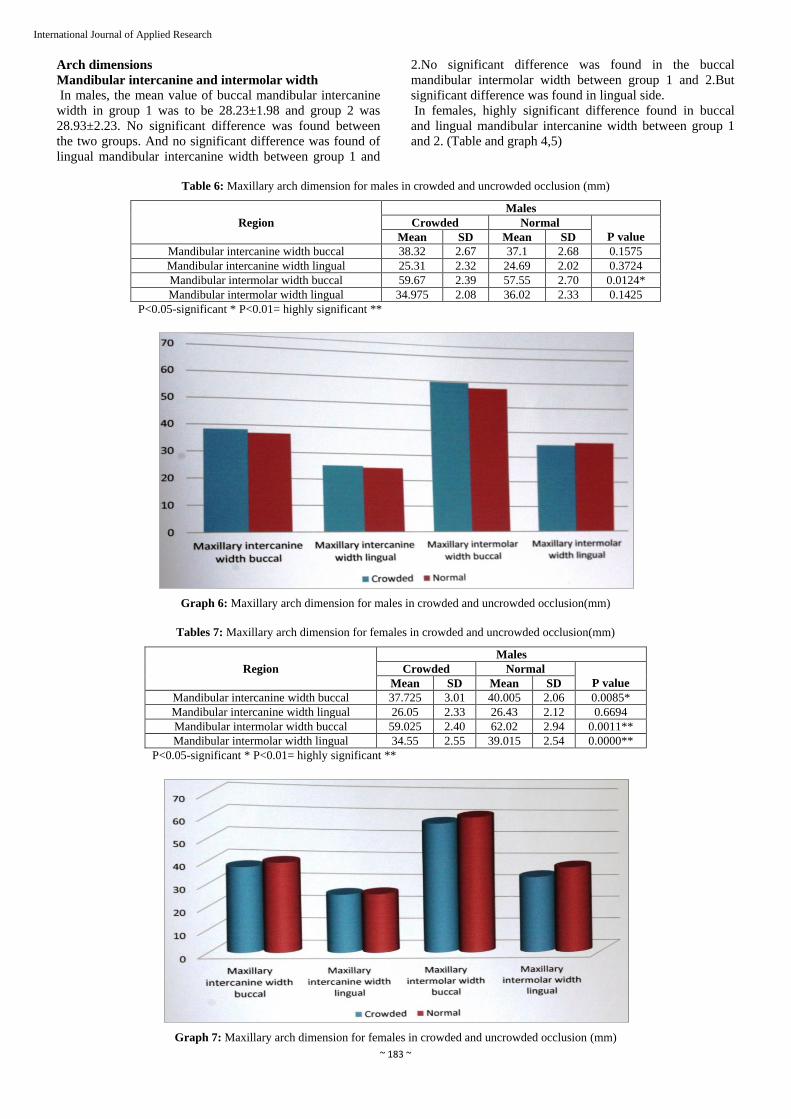
Table 6: Maxillary arch dimension for males in crowded and uncrowded occlusion (mm)
Region
Males
Crowded Normal
P value Mean SD Mean SD
Mandibular intercanine width buccal 38.32 2.67 37.1 2.68 0.1575
Mandibular intercanine width lingual 25.31 2.32 24.69 2.02 0.3724
Mandibular intermolar width buccal 59.67 2.39 57.55 2.70 0.0124*
Mandibular intermolar width lingual 34.975 2.08 36.02 2.33 0.1425
P<0.05-significant * P<0.01= highly significant **
Graph 6: Maxillary arch dimension for males in crowded and uncrowded occlusion(mm)
Tables 7: Maxillary arch dimension for females in crowded and uncrowded occlusion(mm)
Region
Males
Crowded Normal
P value Mean SD Mean SD
Mandibular intercanine width buccal 37.725 3.01 40.005 2.06 0.0085*
Mandibular intercanine width lingual 26.05 2.33 26.43 2.12 0.6694
Mandibular intermolar width buccal 59.025 2.40 62.02 2.94 0.0011**
Mandibular intermolar width lingual 34.55 2.55 39.015 2.54 0.0000**
P<0.05-significant * P<0.01= highly significant **
Graph 7: Maxillary arch dimension for females in crowded and uncrowded occlusion (mm)

~ 184 ~
International Journal of Applied Research
Maxillary intercanine and intermolar width
In males, no significant difference was found in the
maxillary buccal and lingual intercanine width between
group 1 and 2. And no significant difference was found in
the maxillary lingual inter molar width between group 1 and
2 but in buccal side significant difference are occur.
In females, no significant difference was found in the
maxillary buccal and lingual intercanine width between
group 1 and 2. But highly significant difference was found
in the maxillary buccal and lingual intermolar width
between group 1 and 2. (tables and graphs- 6,7)
5. Discussion
Malocclusion has been described as a "disease of
civilization Comuecim and Kaul 1984)"because of its high
prevalence in contemporary Industrialized countries as
compared to historic populations and, even, isolated cultures
that continue to subsist on less-processed diets (Corruccini
1984).
The causes of malocclusion could be classified in two major
categories dental or skeletal. One way of distinguishing
between the two is to compare tooth size and arch
dimensions in Class I non crowded occlusions with Class I
malocclusions exhibiting severe crowding. The results will
reveal the dental or skeletal causes of crowding. This
information will be helpful in treatment
planning prior to orthodontic treatment. Nance described
dental crowding as the difference between the spaces needed
in the dental arch and the space available in that arch that is,
the space discrepancy. Thus, crowding or spacing can be
described as an expression of an altered tooth/tissue ratio or
as a dent alveolar disproportion. The causes of crowding or
spacing are, however, still not fully understood.
In our study, the sample size was selected within an age
group of 15-25 years that was equally distributed in
crowded and non crowed groups (40 samples) and with
equal sex ratio (20 males and 20 female).This was in
accordance with the study done by Doris et al.' and Puri et
al. 2" This age range was chosen such that the subjects were
beyond the active growth phase and therefore had stable
arch width. Also, early adult dentitions have less mutilation
and attrition in most subjects.
In the present study both group had class I skeletal
relationship. However, in the previous studies done by
Howe et al. the skeletal relationship was not included in
factors considered for crowded group. The skeletal
relationship may affect arch malocclusions.
In our study, there was a highly significant difference in
tooth size diameter of all the teeth (maxillary and
mandibular) between the crowded and non crowded groups,
among males. However, in the maxillary arch, though there
was tooth size significant discrepancy in the sum of
maxillary six anterior teeth in diameter was significantly
noted in the two groups, there was no correlation found
between both the groups in the region of maxillary twelve
teeth.
Furthermore, no statistically significant difference was
observed in the mandibular tooth size diameter of six
anterior teeth and twelve teeth between both the groups.
This is in accordance to the studies conducted by
Lundstrom, Doris et al., Chang et al., Fastlitcht and
Lomardi1 was
found that crowding was greater in those individuals with
larger teeth. However, in the studies conducted by Howe et
al Randzie, Fsber, and Gilmore, no significant correlation
was found between the mesiodistal tooth size crowding.
In the evaluation of arch dimension, the most significant
difference was seen in the maxillary and mandibular
intercanine and intermolar (buccal and lingual aspect) arch
width in females and mandibular lingual intermolar and
maxillary buccal intermolar width in males in the
noncrowded and crowded groups. Similar results were
found in the study done by Howe et al., where significant
differences in lingual measurements of lower arch were
noted in both crowded and noncrowded groups. But in the
study done by Possi et al. mandibular arch dimensions, both
in the transverse and longitudinal directions, did not differ
significantly between the uncrowded and crowded groups,
excerpt for the buccal intercanine width which was
significantly greater in the uncrowded group. This may be
due to prominent canine root areas in the uncrowded
dentitions. In the majority of crowded mandibles, the canine
toot rotated and does not show a root prominence in the oral
mucosa.
In the present study there was no significant difference in
cither arch or arch perimeter in both the groups in either
maxilla and mandible. These results were in conformity
with the results evaluated by Poosti M et al. However
studies conducted by Howe et al. found significant
difference in dental arch perimeter measurement for maxilla
between the crowded and non crowded dentitions. Moreover
they revealed that the non crowded arches tended to be
wider and more broadly contoured than did the crowded
arches.
Dental crowding associated with small dental arches rather
than with large teeth, is an important consideration for
treatment techniques which increase dental arch length. This
may be especially relevant in younger patients whose
dentitions are in the deciduous and mixed stages of
development. If such a patient is diagnosed as having dental
crowding and small dental arches, then treatment measures
may include efforts to further jaw development in order to
accommodate the existing tooth mass. This might be
accomplished by early expansion procedures using such
appliances as the rapid palatal expander, the quad helix
appliance, or the Fränkel appliance", alone or in
combination.
Observations made during the course of this study suggests
further investigation. For example, the findings presented
could be interpreted to suggest that compared to dental arch
dimensions, tooth size may be significantly associated with
dental crowding. However, associated differences
between the two groups may have been overlooked.
6. Conclusion
The correct tooth size-arch length relationship between the
maxillary and mandibular teeth is an important factor for
achieving proper interception during the final stages of
orthodontic treatment
The following conclusions can be drawn from the present
study
1. Mesiodistal tooth size was larger in crowded arches as
compared to non-crowded arches in both males and
females.
2. In females, maxillary arch width was found to be
smaller in crowded arches as compared to non crowded
arches.

~ 185 ~
International Journal of Applied Research
3. In males, inter-canine width was not significantly
different in the crowded and non-crowded arches but
maxillary buccal and mandibular lingual inter-molar
width was found to be significantly smaller in the
crowded arches.
References
1. Howe RP, McNamara JA Jr, O'Connor KA. An
examination of dental crowding and its relationship to
tooth size and arch dimension. Am J Orthod. 1983;
83(5):363-73.
2. Hooton EA. Up from the ape. New York: The
Macmillan Company, 1947.
3. Brash JC. The aetiology of irregularity and
malocclusion of the tecth 2nd ed. London: Dental Board
of the United Kingdom, 1956.
4. Barber TK. The crowded arch. J South Calif Dent
Assoc. 1967; 35(5):232-40.
5. Begg PR. Stone Age man's dentition with reference to
anatomically correct occlusion, the etiology of
malocclusion, and a technique for its treatment, Am. J
Orthod. 1954, 40:298-312
6. Beresford JS. Tooth size and class distinction. Dent
Pract Dent Rec. 1969; 20(3):113-20.
7. Lundström A. The aetiology of crowding of the teeth
(based on studies of twins and on morphological
investigations) and its bearing on orthodontic treatment
(expansion or extraction). Eur Orthod Soc [Report],
1951, 176-91.
8. Doris JM, Bernard BW, Kuftinec MM, Stom D. A
biometric study of tooth size and dental crowding, Am J
Orthod. 1981; 79(3):326-36
9. Poosti M, Jalali T. Tooth size and arch dimension in
uncrowded versus crowded Class I malocclusions. J
Contemp Dent Pract. 2007; 8(3):45-52.
10. Mill LF. Arch width, arch length, and tooth size in
youngmales, Angle Orthod. 1964; 34:124-129.
11. Peck S, Peck H. Crown dimensions and mandibular
incisor alignment. Angle Orthod. 1972; 42(2):148-53.
12. Keene A, Engel G. The mandibular dental arch, part IV:
prediction and prevention of lower anterior relapse.
Angle Orthod. 1979; 49:173-80.
13. Radnzic D. Dental crowding and its relationship to
mesiodistal crown diameters and arch dimensions. Am
J Orthod Dentofacial Orthop. 1988; 94(1):50-6
14. Little RM. Mandibular incisor din Am J Orthod. 1984;
86(6):493-502.
15. Little R, An evaluation of changes in mandibular
anterior alignment from 10 to 20 years postretention.
AmJ Orthod Dentofac Orthop. 1988; 93:423-8.