ISSN 1996 - 0808 Volume 12 Number 2 14 January, 2018
29
African Journal of Microbiology Research Volume 12 Number 2 14 January, 2018 ISSN 1996-0808
Transcript of ISSN 1996 - 0808 Volume 12 Number 2 14 January, 2018

African Journal of
Microbiology Research
Volume 12 Number 2 14 January, 2018
ISSN 1996-0808

The African Journal of Microbiology Research (AJMR) is published weekly (one volume per year) by Academic Journals.
provides rapid publication (weekly) of articles in all areas of Microbiology such as: Environmental Microbiology, Clinical Microbiology, Immunology, Virology, Bacteriology, Phycology, Mycology and Parasitology, Protozoology, Microbial Ecology, Probiotics and Prebiotics, Molecular Microbiology, Biotechnology, Food Microbiology, Industrial Microbiology, Cell Physiology, Environmental Biotechnology, Genetics, Enzymology, Molecular and Cellular Biology, Plant Pathology, Entomology, Biomedical Sciences, Botany and Plant Sciences, Soil and Environmental Sciences, Zoology, Endocrinology, Toxicology. The Journal welcomes the submission of manuscripts that meet the general criteria of significance and scientific excellence. Papers will be published shortly after acceptance. All articles are peer-reviewed.
Contact Us
Editorial Office: [email protected]
Help Desk: [email protected]
Website: http://www.academicjournals.org/journal/AJMR
Submit manuscript online http://ms.academicjournals.me/

Editors
Prof. Stefan Schmidt Applied and Environmental Microbiology School of Biochemistry, Genetics and Microbiology University of KwaZulu-Natal Pietermaritzburg, South Africa. Prof. Fukai Bao Department of Microbiology and Immunology Kunming Medical University Kunming, China. Dr. Jianfeng Wu Dept. of Environmental Health Sciences School of Public Health University of Michigan USA. Dr. Ahmet Yilmaz Coban OMU Medical School Department of Medical Microbiology Samsun, Turkey. Dr. Seyed Davar Siadat Pasteur Institute of Iran Pasteur Square, Pasteur Avenue Tehran, Iran. Dr. J. Stefan Rokem The Hebrew University of Jerusalem Department of Microbiology and Molecular Genetics Jerusalem, Israel. Prof. Long-Liu Lin National Chiayi University Chiayi, Taiwan.
Dr. Thaddeus Ezeji Fermentation and Biotechnology Unit Department of Animal Sciences The Ohio State University USA. Dr. Mamadou Gueye MIRCEN/Laboratoire commun de microbiologie IRD-ISRA-UCAD Dakar, Senegal. Dr. Caroline Mary Knox Department of Biochemistry, Microbiology and Biotechnology Rhodes University Grahamstown, South Africa. Dr. Hesham Elsayed Mostafa Genetic Engineering and Biotechnology Research Institute (GEBRI) Mubarak City For Scientific Research Alexandria, Egypt. Dr. Wael Abbas El-Naggar Microbiology Department Faculty of Pharmacy Mansoura University Mansoura, Egypt. Dr. Barakat S.M. Mahmoud Food Safety/Microbiology Experimental Seafood Processing Laboratory Costal Research and Extension Center Mississippi State University Pascagoula, USA. Prof. Mohamed Mahrous Amer Faculty of Veterinary Medicine Department of Poultry Diseases Cairo university Giza, Egypt.

Editors Dr. R. Balaji Raja Department of Biotechnology School of Bioengineering SRM University Chennai, India. Dr. Aly E Abo-Amer Division of Microbiology Botany Department Faculty of Science Sohag University Egypt.
Dr. Haoyu Mao Department of Molecular Genetics and Microbiology College of Medicine University of Florida Florida, USA. Dr. Yongxu Sun Department of Medicinal Chemistry and Biomacromolecules Qiqihar Medical University Heilongjiang P.R. China. Dr. Ramesh Chand Kasana Institute of Himalayan Bioresource Technology Palampur, India. Dr. Pagano Marcela Claudia Department of Biology, Federal University of Ceará - UFC Brazil. Dr. Pongsak Rattanachaikunsopon Department of Biological Science Faculty of Science Ubon Ratchathani University Thailand. Dr. Gokul Shankar Sabesan Microbiology Unit, Faculty of Medicine AIMST University Kedah, Malaysia.
Dr. Kamel Belhamel Faculty of Technology University of Bejaia Algeria. Dr. Sladjana Jevremovic Institute for Biological Research Belgrade, Serbia. Dr. Tamer Edirne Dept. of Family Medicine Univ. of Pamukkale Turkey. Dr. Mohd Fuat ABD Razak Institute for Medical Research Malaysia. Dr. Minglei Wang University of Illinois at Urbana-Champaign USA. Dr. Davide Pacifico Istituto di Virologia Vegetale – CNR Italy. Prof. N. S. Alzoreky Food Science & Nutrition Department College of Agricultural Sciences & Food King Faisal University Saudi Arabia. Dr. Chen Ding College of Material Science and Engineering Hunan University China. Dr. Sivakumar Swaminathan Department of Agronomy College of Agriculture and Life Sciences Iowa State University USA. Dr. Alfredo J. Anceno School of Environment, Resources and Development (SERD) Asian Institute of Technology Thailand. Dr. Iqbal Ahmad Aligarh Muslim University Aligrah, India.

Dr. Juliane Elisa Welke UFRGS – Universidade Federal do Rio Grande do Sul Brazil. Dr. Iheanyi Omezuruike Okonko Department of Virology Faculty of Basic Medical Sciences University of Ibadan Ibadan, Nigeria. Dr. Giuliana Noratto Texas A&M University USA. Dr. Babak Mostafazadeh Shaheed Beheshty University of Medical Sciences Iran. Dr. Mehdi Azami Parasitology & Mycology Department Baghaeei Lab. Isfahan, Iran. Dr. Rafel Socias CITA de Aragón Spain. Dr. Anderson de Souza Sant’Ana University of São Paulo Brazil. Dr. Juliane Elisa Welke UFRGS – Universidade Federal do Rio Grande do Sul Brazil. Dr. Paul Shapshak USF Health Depts. Medicine and Psychiatry & Beh Med. Div. Infect. Disease & Internat Med USA. Dr. Jorge Reinheimer Universidad Nacional del Litoral (Santa Fe) Argentina. Dr. Qin Liu East China University of Science and Technology China. Dr. Samuel K Ameyaw Civista Medical Center USA.
Dr. Xiao-Qing Hu State Key Lab of Food Science and Technology Jiangnan University China. Prof. Branislava Kocic University of Nis School of Medicine Institute for Public Health Nis, Serbia. Prof. Kamal I. Mohamed State University of New York Oswego, USA. Dr. Adriano Cruz Faculty of Food Engineering-FEA University of Campinas (UNICAMP) Brazil. Dr. Mike Agenbag Municipal Health Services, Joe Gqabi, South Africa. Dr. D. V. L. Sarada Department of Biotechnology SRM University Chennai India. Prof. Huaizhi Wang Institute of Hepatopancreatobiliary Surgery of PLA Southwest Hospital Third Military Medical University Chongqing China. Prof. A. O. Bakhiet College of Veterinary Medicine Sudan University of Science and Technology Sudan. Dr. Saba F. Hussain Community, Orthodontics and Peadiatric Dentistry Department Faculty of Dentistry Universiti Teknologi MARA Selangor, Malaysia.

Prof. Zohair I. F. Rahemo Department of Microbiology and Parasitology Clinical Center of Serbia Belgrade, Serbia. Dr. Afework Kassu University of Gondar Ethiopia. Dr. How-Yee Lai Taylor’s University College Malaysia. Dr. Nidheesh Dadheech MS. University of Baroda, Vadodara, India. Dr. Franco Mutinelli Istituto Zooprofilattico Sperimentale delle Venezie Italy. Dr. Chanpen Chanchao Department of Biology, Faculty of Science, Chulalongkorn University Thailand. Dr. Tsuyoshi Kasama Division of Rheumatology, Showa University Japan. Dr. Kuender D. Yang Chang Gung Memorial Hospital Taiwan. Dr. Liane Raluca Stan University Politehnica of Bucharest Department of Organic Chemistry Romania. Dr. Mohammad Feizabadi Tehran University of Medical Sciences Iran. Prof. Ahmed H Mitwalli Medical School King Saud University Riyadh, Saudi Arabia.
Dr. Mazyar Yazdani Department of Biology University of Oslo Blindern, Norway. Dr. Babak Khalili Hadad Department of Biological Sciences Islamic Azad University Roudehen, Iran. Dr. Ehsan Sari Department of Plant Pathology Iranian Research Institute of Plant Protection Tehran, Iran. Dr. Snjezana Zidovec Lepej University Hospital for Infectious Diseases Zagreb, Croatia. Dr. Dilshad Ahmad King Saud University Saudi Arabia. Dr. Adriano Gomes da Cruz University of Campinas (UNICAMP) Brazil Dr. Hsin-Mei Ku Agronomy Dept. NCHU Taichung,Taiwan. Dr. Fereshteh Naderi Islamic Azad University Iran. Dr. Adibe Maxwell Ogochukwu Department of Clinical Pharmacy and Pharmacy Management, University of Nigeria Nsukka, Nigeria. Dr. William M. Shafer Emory University School of Medicine USA. Dr. Michelle Bull CSIRO Food and Nutritional Sciences Australia.

Prof. Márcio Garcia Ribeiro School of Veterinary Medicine and Animal Science- UNESP, Dept. Veterinary Hygiene and Public Health, State of Sao Paulo Brazil. Prof. Sheila Nathan National University of Malaysia (UKM) Malaysia. Prof. Ebiamadon Andi Brisibe University of Calabar, Calabar, Nigeria. Dr. Julie Wang Burnet Institute Australia. Dr. Jean-Marc Chobert INRA- BIA, FIPL France. Dr. Zhilong Yang Laboratory of Viral Diseases National Institute of Allergy and Infectious Diseases, National Institutes of Health USA. Dr. Dele Raheem University of Helsinki Finland. Dr. Biljana Miljkovic-Selimovic School of Medicine, University in Nis, Serbia. Dr. Xinan Jiao Yangzhou University China. Dr. Endang Sri Lestari, MD. Department of Clinical Microbiology, Medical Faculty, Diponegoro University/Dr. Kariadi Teaching Hospital, Semarang Indonesia. Dr. Hojin Shin Pusan National University Hospital South Korea.
Dr. Yi Wang Center for Vector Biology Rutgers University New Brunswick USA. Prof. Natasha Potgieter University of Venda South Africa. Dr. Sonia Arriaga Instituto Potosino de Investigación Científicay Tecnológica/ División de Ciencias Ambientales Mexico. Dr. Armando Gonzalez-Sanchez Universidad Autonoma Metropolitana Cuajimalpa Mexico. Dr. Pradeep Parihar Lovely Professional University Punjab, India. Dr. William H Roldán Department of Medical Microbiology Faculty of Medicine Peru. Dr. Kanzaki, L. I. B. Laboratory of Bioprospection University of Brasilia Brazil. Prof. Philippe Dorchies National Veterinary School of Toulouse, France. Dr. C. Ganesh Kumar Indian Institute of Chemical Technology, Hyderabad India. Dr. Zainab Z. Ismail Dept. of Environmental Engineering University of Baghdad Iraq. Dr. Ary Fernandes Junior Universidade Estadual Paulista (UNESP) Brasil.

Dr. Fangyou Yu The first Affiliated Hospital of Wenzhou Medical College China. Dr. Galba Maria de Campos Takaki Catholic University of Pernambuco Brazil. Dr Kwabena Ofori-Kwakye Department of Pharmaceutics Kwame Nkrumah University of Science & Technology Kumasi, Ghana. Prof. Liesel Brenda Gende Arthropods Laboratory, School of Natural and Exact Sciences, National University of Mar del Plata Buenos Aires, Argentina. Dr. Hare Krishna Central Institute for Arid Horticulture Rajasthan, India. Dr. Sabiha Yusuf Essack Department of Pharmaceutical Sciences University of KwaZulu-Natal South Africa. Dr. Anna Mensuali Life Science Scuola Superiore Sant’Anna Italy. Dr. Ghada Sameh Hafez Hassan Pharmaceutical Chemistry Department Faculty of Pharmacy Mansoura University Egypt.
Dr. Kátia Flávia Fernandes Department of Biochemistry and Molecular Biology Universidade Federal de Goiás Brasil. Dr. Abdel-Hady El-Gilany Department of Public Health & Community Medicine Faculty of Medicine Mansoura University Egypt. Dr. Radhika Gopal Cell and Molecular Biology The Scripps Research Institute San Diego, CA USA. Dr. Mutukumira Tony Institute of Food Nutrition and Human Health Massey University New Zealand. Dr. Habip Gedik Department of Infectious Diseases and Clinical Microbiology Ministry of Health Bakırköy Sadi Konuk Training and Research Hospital Istanbul, Turkey. Dr. Annalisa Serio Faculty of Bioscience and Technology for Food Agriculture and Environment University of Teramo Teramo, Italy.

African Journal of Microbiology Research
Table of Contents: Volume 12 Number 2 14 January, 2018
ARTICLES
Evaluation of antibacterial and phytochemical properties of different spice extracts 27 Suman Upadhyaya, Divya Yadav, Ram Chandra and Naveen Arora Phenotypic detection methods of metallo-β-lactamases -producing Pseudomonas aeruginosa strains isolated in urology ward from Skikda hospital Algeria 38 Fatma Zohra Mellouk and Sameh Meradji

Vol. 12(2), pp. 27-37, 14 January, 2018
DOI: 10.5897/AJMR2017.8731
Article Number: 690AFFE55806
ISSN 1996-0808
Copyright © 2018
Author(s) retain the copyright of this article
http://www.academicjournals.org/AJMR
African Journal of Microbiology Research
Full Length Research Paper
Evaluation of antibacterial and phytochemical properties of different spice extracts
Suman Upadhyaya*, Divya Yadav, Ram Chandra and Naveen Arora
Department of Environmental Microbiology, Babasaheb Bhimrao Ambedkar University (A Central University), Vidya Vihar, Rae Bareli Road, Lucknow, 226025, Uttar Pradesh, India.
Received 5 October, 2017; Accepted 29 December, 2017
Now-a-days majority of world population rely on the plant preparations as medicines to cure diseases, as they are considered safe and as effective as allopathic preparations without any side effects. Spices are plant products having aroma, are mainly used during cooking to impart flavor and taste to the dish and also possess medicinal values. The present study was designed to evaluate the antimicrobial activity of four Indian spices: clove (Syzygium aromaticum), cinnamon (Cinnamomum verum), turmeric (Curcuma longa), and black pepper (Piper nigrum) against Gram positive and Gram negative pathogenic bacteria viz., Staphylococcus aureus (ATCC 25923), Escherichia coli (ATCC 25922), Pseudomonas aeruginosa (ATCC 27853), and Klebsiella pneumoniae (ATCC 70063) using aqueous, 75% ethanol and 75% chloroform extracts. The antibacterial activity of spices extract and spices which are used in powder form was determined by agar well diffusion method and the antimicrobial activity of antibiotics (Clindamycin, Ciprofloxacin, Gentamycin) was measured by disc diffusion method. The clove and cinnamon extracts had exhibited maximum antibacterial property against pathogens; also, turmeric had shown less antimicrobial activity, while black pepper had exhibited moderate activity. Spices (powder) had shown highest antimicrobial activity than spice extracts. Phytochemical screening was carried out on ethanol, chloroform and distilled water extracts of spices for its chemical composition. Qualitative phytochemical analysis of these spice extracts confirm the presence of various phytochemicals like alkaloids, terpanoids, flavonoids, saponins, steroids and tannins. Key words: Antimicrobial activity, Piper nigrum, agar well diffusion method and phytochemicals.
INTRODUCTION
The natural products are more effective with least side effects when compared to commercial antibiotics. Consequently they are used as a substitute medication
for curing various infections. Spices are plant substances that are generally used to enhance flavor, which include leaves (coriander or mint), flower bud (clove), fruits (black
*Corresponding author. E-mail: [email protected]. Tel: +91 522 244 0822, 9936302204. Fax: +91 522 244081.
Author(s) agree that this article remains permanently open access under the terms of the Creative Commons Attribution
License 4.0 International License

28 Afr. J. Microbiol. Res. pepper), bark (cinnamon), and rhizomes (turmeric) (Pavithra et al., 2016). Natural antioxidants such as flavonoids, tannins and phenols are increasingly attracting because they are disease preventing, health promoting and anti-aging substances (Tamizhazhagan et al., 2017). Medicinal plants produce bioactive molecules that have both antibacterial and antifungal activities. Spices have been found to reduce inflammation, protect against infection, helps to detoxify the liver and also protect from cell damage that can lead to rheumatoid arthritis, osteoporosis, heart disease and other degenerative diseases. The term spices refer to aromatic or pungent vegetable substances used for flavouring foods and have several commercial uses according to International Organization for Standardization (ISO). Since ancient times people used spices for preventing food deterioration and pathogenic diseases. Spices have become today as an integral part of our daily diet and many of the spices are widely used to flavour food and beverages for food preservations, medicinal preparations, cosmetics, perfumery, bakery goods and various other products (Shiva Rani et al., 2013). Even today spices are used as an ingredient in drug preparations in Unani, Homeopathy and Ayurveda systems of medicine (Bharath et al., 2016). Phytochemicals are bio-active chemicals of plant origin and are regarded as secondary metabolites because the plant that manufactures them they may have little need for them. They are naturally synthesized in all parts of the plant body; bark, leaves, stem, root, flower, fruits, seeds etc. that is, any part of the plant body may contain active components (Tiwari et al., 2011). Homeopathic medicine has been using spices as one of the chief ingredients in most of their preparations. Phytochemical investigations of the aerial parts of the plants have tartaric acid, acetic acid, citric acid, succinic acid, gums, pectin, sugars, tannins, alkaloids, flavonoids, glycosides and sesquiterpenes (Bharath et al., 2016). Although the primary purpose of spices is to impart flavour and piquancy to food, the medicinal, antimicrobial and antioxidant properties of spices have also been exploited. Major antimicrobial components in clove and cinnamon have been reported to be eugenol and cinnamaldehyde, respectively, which have been given special attention to find their antimicrobial activity against micro-organisms. Spice, cinnamon or Cinnamomum zeylanicum, found in the inner bark of Cinnamon trees, is commonly used in cooking for its aroma, flavor, and taste. Historically, cinnamon has been used by the Egyptians for embalming, most likely due to its antimicrobial properties. Eugenol and cinnamaldehyde are the two major chemical components in cinnamon that are responsible for its health benefits. Eugenol, a phenol compound, inhibits mold and adds flavor and aroma to bakery items (Mahfuzul et al., 2008). Now micro-organisms have become resistant to many antibiotics due to increased use of drugs, which is decreasing efficiency
of conventional medicines. So it has become necessary to find out new antimicrobial agents. Syzygium aromaticum (Linn.) cloves the aromatic dried flower buds of a tree in the family Myrtaceae (Srivastava and Malhotra, 1991; Chaieb et al., 2007a) cloves are used in Ayurveda, Chinese medicine and western herbalism. In addition, the cloves are antimutagenic (Miyazawa and Hisama, 2003), antiinflammatory (Kim et al., 1998) antioxidants (Chaieb et al., 2007b), antiulcerogenic (Bae et al., 1998; Li et al., 2005), antithrombotic (Srivastava and Malhotra, 1991) and antiparasitic (Yang et al., 2003).
The objective of this research is to evaluate the antimicrobial activity of clove (S. aromaticum), cinnamon (C. zeylanicum), Turmeric (Curcuma longa) and black pepper (Piper nigrum) powder extracts (ethanol, aqueous and chloroform) against Pseudomonas aeruginosa (ATCC 27853), Staphylococcus aureus (ATCC 25923), Escherichia coli (ATCC 2592), Klebsiella pneumonia (ATCC 70063). Antibiotics (clindamycin, ciprofloxacin and gentamycin) were used to determine the sensitivity of bacterial species. Phytochemical screening was carried out on ethanol, chloroform and distilled water extracts of spices for its chemical composition.
MATERIALS AND METHODS The spices namely cinnamon (Cinnamomum verum), black pepper (P. nigrum), clove (S. aromaticum) and Turmeric (C. longa) used in the study were collected from the local market in Lucknow. Bacterial strains: The micro-organisms were obtained from the research laboratory of S.G.P.G.I., Lucknow. The microbes used in the study includes: i) E. coli (ATCC 25922) ii) P. aeruginosa (ATCC 27853) iii) K. pneumoniae (ATCC 70063) iv) S. aureus (ATCC 25923). Antibiotics: Gentamycin for K. pneumoniae, Ciprofloxacin for E. coli and P. aeruginosa, and Clindamycin for S. aureus. Solvents: 75% Chloroform, Distilled water and 75% Ethanol.
Maintenance of bacterial cultures and preparation of inoculum Pure cultures were subcultured and maintained on nutrient agar plates regularly. The cultures were inoculated on sterile agar plates and placed in an incubator at 37°C for 24 h and was stored at 4°C. Bacterial cultures were subcultured after every 3-4 days to avoid contamination. Inoculum was prepared by inoculating the pure culture in nutrient broth and incubated overnight at 37°C (Pavithra Sivakumar et al., 2016).
Preparation of spice extracts Three solvents distilled water, 75% ethanol and 75% chloroform were used to extract phytochemical of clove, cinnamon, turmeric and black pepper.

Upadhyaya et al. 29
Table 1. Antibacterial activity of standard discs.
S/N Micro-organisms Antibiotics Zone of inhibition (mm)
1 E. coli Ciprofloxacin 28
2 S. aureus Clindamycin 32
3 P. aeruginosa Ciprofloxacin 24
4 K .pneumoniae Gentamycin 20
Extraction with distilled water: 10 g of powdered plant material was dissolved in sterile distilled water to make 40 ml of aqueous extract (25% w/v). The mixture was placed undisturbed at room temperature for 24 h in a sterile flask and it was filtered through sterilized Whatman no.1 filter paper. After filtration, the extract was evaporated in water bath until 25 ml extract was left in the container (Barreto et al., 2002). Extraction with 75% ethanol: 10 g of powdered plant material was dissolved in enough ethanol to make 40 ml of ethanolic extract (25% w/v). The extraction procedure followed was the same as that used for aqueous extract (Barreto, et al., 2002). Extraction with 75% chloroform: 10 g of powdered plant material was dissolved in methanol to make 40 ml of methanolic extract (25% w/v). The extraction procedure was similar to that used for aqueous extract. Extracts thus obtained were evaluated for antibacterial activities (Barreto et al., 2002). Determination of antibacterial activity Agar well diffusion method The antimicrobial activity of the extracts was performed by well diffusion method (Parez et al., 1990). The bacterial suspension was spread on Muller Hinton Agar (MHA) medium. Sterile 8 mm diameter cork borer was used to make well in the nutrient agar. Each well was filled with 0.1 ml of spice extract. Negative control was prepared using the same solvents employed to prepare the plant extracts. The inoculated plates were incubated at 37°C for 24 h and the clear zone of growth inhibition around the well was measured. Antibiotic sensitivity test Disc diffusion method A single colony of the purified isolates was inoculated in 5 ml sterile peptone water and incubated at 37°C overnight. Thereafter, a loopfull culture was diluted in 5 ml sterile phosphate buffered saline and seeded into Muller Hinton agar. Antibiotic disc (Hi-Media) was placed on the surface of agar and incubated overnight at 37°C. Zone of inhibition was recorded and a control sensitive culture was included in the experiment (Bauer et al., 1966). Phytochemical screening of spices Screening for alkaloids To 5 ml each of the spice extracts, 5 ml of aqueous hydrochloric acid was added on a steam bath at 60°C for 5 min. The spice
extract was filtered with a 3 layered muslin cloth. In one ml of the filtrate, few drops of Draggendoff’s reagent was added. Appearance of Blue black turbidity was positive for alkaloids (Omoya and Akharaiyi, 2012). Screening for steroids 1 ml of extract was dissolved in 10 ml of chloroform and equal volume of concentrated sulphuric acid was added by the sides of the test tube. The upper layer turns red and sulphuric acid layer showed yellow with green fluorescence. This indicates the presence of steroids (Jyothiprabha and Venkatachalam, 2016). Screening for tannins 5 ml each of the extracts were stirred separately with 100 ml distilled water and filtered. One millilitre ferric chloride reagent was added to the filtrate. A blue-black or blue green precipitate was an indication of the presence of tannins (Trease and Evans, 1989). Screening for terpenoids 5 ml of extract was taken in a test tube and 2 ml of chloroform was added to it followed by the addition of 3 ml of concentrated sulphuric acid. Formation of reddish brown layer at the junction of two solutions confirms the presence of terpanoids (Jyothiprabha and Venkatachalam, 2016). Screening for flavonoids 5 ml of diluted ammonia solution was added to aqueous extract followed by the addition of 1 ml concentrated H2SO4. Appearance of yellow colour indicated the presence of flavonoids (Harborne and Williams, 2000). Screening for saponins 5 ml each of the extracts were mixed with distilled water and shaken separately in a test tube. Frothing, which persists on warm heating was taken as preliminary evidence of the presence of the saponins (Omoya and Akharaiyi, 2012).
RESULTS AND DISCUSSION The findings of this research are as shown in Tables 1 to 9 and Figures 1 to 8. In India, spices are ethnically used as active ingredients in ayurvedic medicines and reported

30 Afr. J. Microbiol. Res.
Table 2. Antibacterial activity of negative control.
S/N Micro-organisms Ethanol solution (zone
of inhibition) Chloroform solution (zone of
inhibition) Distill water (zone of
inhibition)
1 E. coli - - -
2 S. aureus - - -
3 P. aeruginosa - - -
4 K .pneumoniae - - -
Table 3. Antimicrobial activity of chloroform extract of spices against micro-organisms.
S/N Spices Micro-organisms with zone of inhibition (mm)
K. pneumoniae S. aureus E. coli P. aeruginosa
1 Cinnamon 6 7 6 6
2 Clove 12 12 12 16
3 Black pepper - - 7 -
4 Turmeric - - - -
Table 4. Antimicrobial activity of aqueous extract of spices against micro-organisms.
S/N Spices Micro-organisms with zone of inhibition (mm)
K. pneumoniae S. aureus E. coli P. aeruginosa
1 Cinnamon 9 10 8 8
2 Clove 12 14 10 14
3 Black pepper - - 7 -
4 Turmeric - - - -
Table 5. Antimicrobial activity of ethanol extract of spices against micro-organisms.
S/N Spices Micro-organisms with zone of inhibition (mm)
K. pneumoniae S. aureus E. coli P. aeruginosa
1 Cinnamon 8 14 11 8
2 Clove 11 11 12 12
3 Black pepper - 8 10 -
4 Turmeric - 6 9 12
to possess a number of pharmacological effects to treat different human ailments (Bonjar et al., 2004). Several investigations have been directed towards their anti-microbial properties (Voravuthikunchai et al., 2005; Vaishnavi et al., 2007). In the present work, among the four spices (clove, black pepper, cinnamon and turmeric) tested against four bacterial pathogens (P. aeruginosa, S. aureus, K. pneumoniae, E. coli), all the spices showed antibacterial activity. The results of antibacterial activity of these spices against pathogens tested revealed that of
the three types of extracts tested, ethanolic and aqueous extracts of spices had shown better antibacterial activity against pathogens when compared to chloroform extracts of the spices. This may be due to the better solubility of active ingredients of spices in alcoholic and distilled water solvents than chloroform. Ahmad et al (1998) also reported that alcoholic extracts of medicinal plants had greater activity than their aqueous extracts. Earlier studies have demonstrated that the extracts of cinnamon, clove, turmeric and black pepper had shown maximum

Upadhyaya et al. 31
Table 6. Antimicrobial activity of spices (powder) against micro-organisms.
S/N Spices Micro-organisms with zone of inhibition (mm)
K. pneumoniae S. aureus E. coli P. aeruginosa
1 Cinnamon 14 14 12 8
2 Clove 12 12 14 10
3 Black pepper 7 8 7 7
4 Turmeric 7 7 7 -
Table 7. Phytochemical screening of chloroform extract of spices.
S/N Phytochemicals Chloroform extracts
Cinnamon Clove Black pepper Turmeric
1 Alkaloids - - + +
2 Flavonoids - - - -
3 Tannins - - - -
4 Saponins + + + +
5 Steroids - - - -
6 Terpenoids + + + -
Table 8. Phytochemical screening of aqueous extract of spices.
S/N Phytochemicals Aqueous extracts
Cinnamon Clove Black pepper Turmeric
1 Alkaloids + - + -
2 Flavonoids + + - +
3 Tannins + + + +
4 Saponins + - + -
5 Steroids + + - -
6 Terpenoids + + + +
Table 9. Phytochemical screening of ethanol extract of spices.
S/N Phytochemicals Ethanol extracts
Cinnamon Clove Black pepper Turmeric
1 Alkaloids - - + -
2 Flavonoids - - - -
3 Tannins - + - +
4 Saponins + - - -
5 Steroids + + + +
6 Terpenoids + + + -
antimicrobial activity against pathogens than other spices. In the present study it was found that turmeric had shown less antimicrobial activity, while black pepper had exhibited moderate activity. Spices (Powder) had
shown highest antimicrobial activity than spice extracts. It was observed that cloves and cinnamon had shown highest antimicrobial property against gram negative bacteria. Shihabudeen et al. (2010) and Ceylan and Fung

32 Afr. J. Microbiol. Res.
Figure 1. Graph of antimicrobial activity of ethanol extracts against micro-organisms.
Figure 2. Graph of antimicrobial activity of chloroform extracts against micro-organisms.
(2004) demonstrated that the gram positive bacteria were more sensitive to spices than gram negative bacteria due to the difference in there cell wall structure. In the present study, it was observed that chloroform extract of clove had shown highest antimicrobial zones against S. aureus, E. coli, P. aeruginosa, K. pneumonia, whereas chloroform extract of cinnamon had shown maximum zones against all test micro-organisms; also, chloroform extract of black pepper had shown zone against E. coli only. In case of
chloroform extract of turmeric, it was observed that there is no zone of inhibition observed. The antimicrobial activities of the plants extracted in different solvents varied greatly because there are many factors that influence the active principle present in the plant (Parekh and Chanda, 2006). Aqueous extract of clove had shown highest antimicrobial zone against all test micro-organisms, while aqueous extract of cinnamon exhibited maximum zones against pathogens. Also, aqueous
Ethanol Extracts
0
2
4
6
8
10
12
14
16
Cinnamon Clove Black pepper Termeric
Zon
e o
f in
hib
itio
n(m
m)
Ethanol Extracts
S.aureus
E.coli
P.aeruginosa
K.pneumoniae
0
2
4
6
8
10
12
14
16
18
Cinnamon Clove Black pepper Turmeric
Zon
e o
f in
hib
itio
n(m
m)
Chloroform Extracts
S.aureus
E.coli
P.aeruginosa
K.pneumoniae
0
2
4
6
8
10
12
14
16
18
Cinnamon Clove Black pepper Turmeric
Zon
e o
f in
hib
itio
n(m
m)
Chloroform Extracts
S.aureus
E.coli
P.aeruginosa
K.pneumoniae
Upadhyaya et al. 33
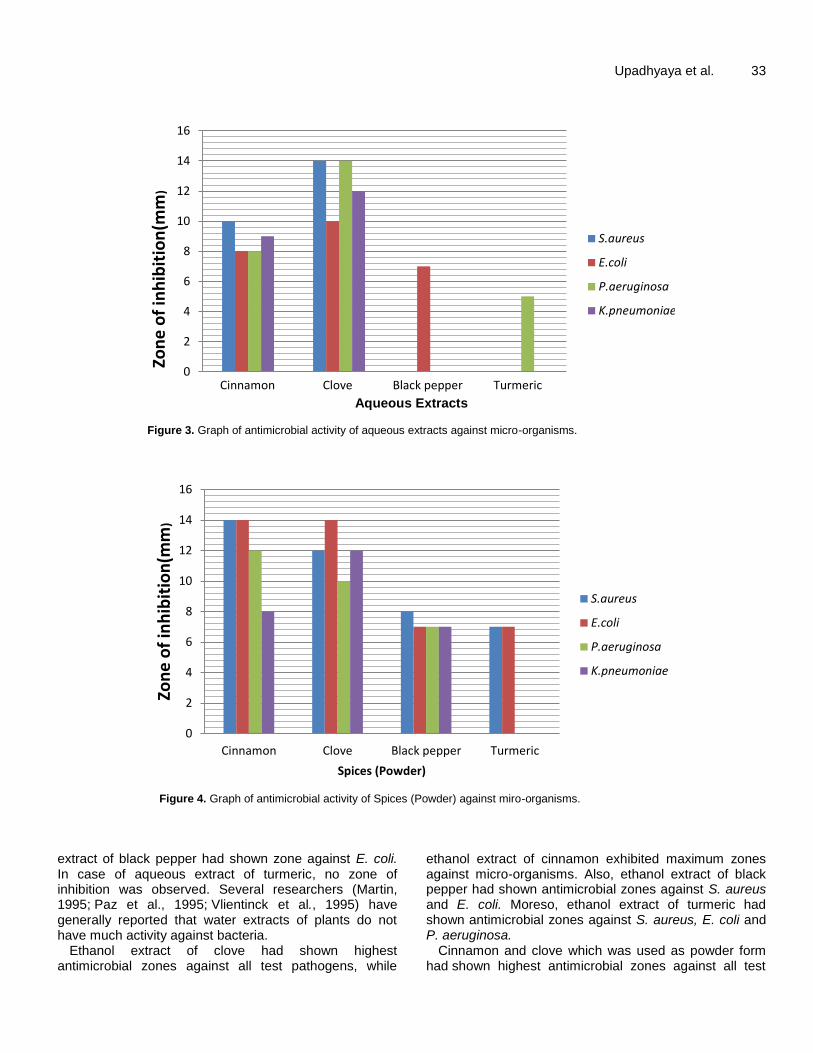
Figure 3. Graph of antimicrobial activity of aqueous extracts against micro-organisms.
Figure 4. Graph of antimicrobial activity of Spices (Powder) against miro-organisms.
extract of black pepper had shown zone against E. coli. In case of aqueous extract of turmeric, no zone of inhibition was observed. Several researchers (Martin, 1995; Paz et al., 1995; Vlientinck et al., 1995) have generally reported that water extracts of plants do not have much activity against bacteria.
Ethanol extract of clove had shown highest antimicrobial zones against all test pathogens, while
ethanol extract of cinnamon exhibited maximum zones against micro-organisms. Also, ethanol extract of black pepper had shown antimicrobial zones against S. aureus and E. coli. Moreso, ethanol extract of turmeric had shown antimicrobial zones against S. aureus, E. coli and P. aeruginosa.
Cinnamon and clove which was used as powder form had shown highest antimicrobial zones against all test
Aqueous Extracts
0
2
4
6
8
10
12
14
16
Cinnamon Clove Black pepper Termeric
Zon
e o
f in
hib
itio
n(m
m)
Aqueous Extract
S.aureus
E.coli
P.aeruginosa
K.pneumoniae
0
2
4
6
8
10
12
14
16
18
Cinnamon Clove Black pepper Turmeric
Zon
e o
f in
hib
itio
n(m
m)
Chloroform Extracts
S.aureus
E.coli
P.aeruginosa
K.pneumoniae
0
2
4
6
8
10
12
14
16
Cinnamon Clove Black pepper Turmeric
Zon
e o
f in
hib
itio
n(m
m)
Spices (Powder)
S.aureus
E.coli
P.aeruginosa
K.pneumoniae

34 Afr. J. Microbiol. Res.
Figure 5. Antimicrobial activity of ethanol extract of spices against micro-organisms.
Figure 6. Antimicrobial activity of chloroform extract of spices against micro-organisms.
Figure 7. Antimicrobial activity of aqueous extract of spices against micro-organisms.
pathogens whereas black pepper (Powder) exhibited maximum antimicrobial zones against micro-organisms. In case of turmeric (Powder), antimicrobial zones were shown against K. pneumoniae, S. aureus and E. coli.
Solvents (75% chloroform, distilled water and 75% ethanol) were used as negative control. The antibacterial
activity of spices was compared with the standard antibiotics (Ciprofloxacin, Clindamycin, and Gentamycin). Antibiotics used as positive control showed highest antimicrobial zones than ethanol, chloroform, distilled water extracts and spices (Powder). It is established in this study that the spices reduce and inhibit the growth of

Upadhyaya et al. 35
Figure 8. Antimicrobial activity of spices (powder) against micro-organisms. pathogens.
In the present study, phytochemical screening of four spices clove, cinnamon, turmeric and black pepper were done. The result reveals that some of the phytochemicals analysed were present in the extracts of all the spices. Tannins, flavonoids, steroids, terpenoids were present in aqueous extract of clove; saponins and terpenoids werepresent in chloroform extract of clove; whereas tannins, steroids and terpenoids were present in the ethanolic extract of clove. Aqueous extract of turmeric had tannins, terpenoids and flavonoids; chloroform extract of turmeric had alkaloids and saponins; while tannins and steroids were present in ethanol extract of turmeric. Alkaloids, saponins, tannins, flavonoids, steroids, terpenoids were present in aqueous extract of cinnamon; chloroform extract of cinnamon had saponins and terpenoids; and saponins, steroids, terpenoids were present in ethanolic extract of cinnamon. Aqueous extract of black pepper had alkaloids saponins, tannins and terpenoids; chloroform extract of black pepper had alkaloids, saponins and terpenoids; while alkaloids, steroids and terpenoids were present in ethanol extract of black pepper. The importance of alkaloids, saponins andtannins in various antibiotics used in treating common
pathogenic strains has recently been reported by Kubmarawa (2007) and Mensah (2008).
Medicinal plants continue to be an important therapeutic aid for alleviating the ailments of humankind. Today, there is a renewed interest in traditional medicine and an increasing demand for more drugs from plant sources. This revival of interest in plant-derived drugs is mainly due to the current widespread belief that “green medicine” is safe and more dependable than the costly synthetic drugs, many of which have adverse side effects (Al lafi and Ababneh, 1995).
Spices have been added to foods since ancient times as flavoring agent, also as food preservatives and folk medicines. Basically when spices are used for medicinal purpose, their value is dependent on the phytochemicals they possess (Okwu, 2001). The spices, herbs, plant extract and their phytoconstituents have been reported for anti-inflammatory, antidiarrheal, antimicrobial, antioxidant and insecticidal activities (Chouhan and Singh, 2011). Alkaloid has important biological property like cytotoxicity and are used in allophatic systems (Trease and Evans, 2005). Steroids and Sterols are greatly important in pharmacy as they possess compounds like sex hormones and can be used for drug

36 Afr. J. Microbiol. Res. production (Okwu, 2001). Saponins protect against hypercholesterolemia and antibiotics properties (Amin et al., 2013). The growth of many fungi, yeast, bacteria and viruses was inhibited by tannins (Chung et al., 1998). Phenols and tannins acts as antioxidants (Han et al., 2005). The potential of plants as a source of drugs is still to be vastly explored. Multiple drug resistance has become a critical problem in pharmacotherapeutics as there are increasing numbers of diseases exhibiting various levels of drug resistance, including bacterial infections (Mahesh and Satish, 2008). Herbal medications and phytochemical screening of various plant species for medicinal leads are now receiving much attention (Saleha et al., 2015).
Conclusion Natural spices (clove, cinnamon, turmeric and Black pepper) possess effective anti-bacterial activity against micro-organisms and can be used for prevention of drug resistant microbial diseases and further evaluation is necessary. The phytochemicals (alkaloids, flavonoids, saponins, terpenoids, tannins and steroids) were present commonly in all the studied spices. The spices have been screened for phytochemical constituents and seemed to have the potential to act as a source of useful drugs and also to improve the health status of the consumers as a result of the presence of various compounds that are vital for good health. The present study showed that, the degree of antibacterial activity of spices tested can be put in the following order: Clove > Cinnamon > Black pepper >Turmeric. The results of the present study are quite encouraging as almost all spices exhibited antimicrobial activity against most of the pathogens, but the antimicrobial activity varies widely, depending on the type of spices, test medium and microorganism. This study opens up the possibility for the search of new antimicrobials as an alternative to the antibiotics. CONFLICT OF INTERESTS
The authors have not declared any conflict of interests. REFERENCES
Akter S, Haque T, Irine EJ, Kabir MF, Ahmed S, Begum T
(2015).Comparative antimicrobial activities of different species of Ixora. J. Pharma. Phytochem. 3(6):103-105.
Al lafi T, Ababneh H (1995). The effect of the extract of the miswak (chewing sticks) used in Jordan and the Middle East on oral bacteria. Int. Dent. J. 45:218-22.
Amin Mir M, Sawhney SS, Jassal MM S (2013). Qualitative and quanditative analysis of phytochemicals of Taraxacum officinale. Wudpecker J. Phar. Pharmacol. 2(1):001-005.
Bae EA, Han MJ, Kim NJ, DH, Kim DH (1998). Anti-Helicobacter pylori activity of herbal medicines. Biol. Pharm. Bull. 21(9):990-992.
Baur AW, Kirby WMM, Sherris JC, Truck M (1966). Antibiotic
susceptibility testing by standardized single disc method. Am. J. Clin. Pathol. 45:493-496.
Bharath MR, Azeem MA, Basha S, Keerthan HV (2016). Antimicrobial activity of cinnamon extracts against foodborne pathogens E. coli, S. tyhimurium and S. aureus and L.. monocytogens. J. Pharma. Biol.Sci.11(6):66-72.
Bonjar GHS (2004). Screening for antibacterial properties of some Iranian plants against two strains of E. coli. Asian J. Plant Sci. 3(3):310-314.
Ceylon E, Fung CYD (2004). Antimicrobial activity of spices, J. Rap id. Meth. Autom. Microbiol. 12:1- 55.
Chaieb K, Hajlaoui H, Zmantar T, Nakbi KAB, Rouabhia M, Mahdouani K, Bakhrouf A (2007a). The chemical composition and biological activity of essential oil, Eugenia cryophyllata (Syzygium aromaticum L. Myrtaceae): A short review. Phytother Res. 21(6):501-506.
Chouhan HS, Singh, SKA (2001). Review of plants of genus Leucas. J. of pharmacognosy phytother. 3(3):13-26.
Chung KT, Wong TY, Wei CL, Huang YW, Lin Y (1998). Tannins and human health. A review, Criti. Rev. Food. Sci. Nutri. 6:421-446.
Han X, Shen T, Lou H (2005). Dietary polyphenols and their biological significance. Int. J. Mol. Sci. 8(9):950-988.
Harborne JB, Williams Ch.A (2000). Advances in flavonoid research sinc.1992. Phytochem. 55:481- 504.
Jyothiprabha V, Venkatachalam P (2016). Preliminary phytochemical screening of different solvent extracts of selected lndian spices. Int. J. Curr. Microbiol. App. Sci. 5(2):116-122.
Kim HM, Lee EH, Hong SH, Song HJ, Shin MK, Kim SH, Shin TY (1998). Effect of Syzygium aromaticum extract on immediate hypersensitivity in rats. J. Ethnopharmacol. 60(2):125-131.
Kubmarawa D, Ajoku GA, Enworem NM, Okorie DA (2007). Roles of agricultural biotechnology in ensuring adequate food security in developing societies. Afr. J. Biotechnol. 6:1690-1696.
Mahesh B, Satish S (2008). Antimicrobial activity of some important medicinal plant against plant and human pathogens. World J. Agric. Sci. 4(S):839-843.
Md. Mahfuzul H, Barib ML, Junejac VK, Kawamotob S (2008). Antimicrobial activity of cloves and cinnamon extracts against food borne pathogens and spoilage bacteria, and inactivation of Listeria monocytogenes in ground chicken meat with their essential oils. Rep. Nat. Food Res. Inst. 72:9-21.
Martin G J (1995). Ethnobotany: A Methods Manual. London: Chapman and Hall.
Miyazawa M, Hisama M (2003). Antimutagenic activity of phenylpropanoides from clove (Syzygium aromaticum). J. Agric. Food Chem. 51(22):6413-6422.
Okwu D E (2001). Evaluation of the chemical composition of medicinal plants belonging to Euphorbiaceae. Pak. Vet. J. 14:160-162.
Omoya FO, Akharaiyi FC (2012). Mixture of honey and ginger extract for antibacterial assessment on some clinical isolates. Int. Res. J. Pharm, 2(5):127-132.
Parekh J, Chanda S (2007). In vitro antimicrobial activity of Trapa natans L. fruit rind extracted in different solvents. Afr. J. Biotechnol. 6:766-770.
Pavithra S, Nikhishaa SR, Jessy PB, Nithyananthi MJT, Saraniya P, Smila KH (2016). Antimicrobial activity of selected spices- a bio preservative approach. J. Chem. Pharm. Sci. 9:304-307
Paz EA, Cerdeiras MP, Fernandez J, Ferreira F, Moyna P, Soubes M, Vázquez A, Vero S, Zunino L (1995). Screening of Uruguayan medicinal plants for antimicrobial activity. J. Ethnopharm. 45:67-70.
Perez C, Pauli M, Bazerque P (1990). An antibiotic assay by Agar well difussion method. Acta Biol. Med. Exp. 15:113-115.
Shiva Rani SK, Saxena N, Udaysree (2013). Antimicrobial Activity of Black Pepper (Piper nigrum L.). Global J. Pharmacol. 7 (1):87-90.
Shihabudeen M.S, Priscilla HD, Kavitha T (2010). Antimicrobial activity and phytochemical analysis of selected Indian folk medicinal plants. Int. J. Pharma Sci. Res. 1(10):430-434.
Srivastava KC, Malhotra N (1991). Acetyl euginol, a component of oil of cloves (Syzygium aromaticum L.) inhibits aggregation and alters arachidonic acid metabolism in human blood platelets.

Prostaglandins Leukot Essent Fatty Acids 42(1):73-81. Tamizhazhagan V, Pugazhendy K, Sakthidasan V, Jayanthi C (2017).
Preliminary screening of phytochemical evaluation selected plant of Pisonia alba. I. J. Biol. Research. 2(4):63-66.
Tiwari B, Kumar M, Kaur G, Kaur H (2011). Phytochemical screening and extraction: A Review Int. Pharm. Sciencia. 1:98-106.
Trease GE, Evans MC (2005). Pharmacognosy. Elsevier, 14th ed. 53:431-512.
Trease GE, Evans WC (1989) Pharmacognosy. 15th edition, London, J. And A Churchhill Ltd. 234-494.
Vaishnavi C, Kaur S, Kaur M (2007). Bactericidal activity of kitchen spices and condiments on enteropathogens; Natural Product Radiance. 6(1):40-45.
Vlientinck AJ, Van Hoof L, Totté J, Lasure A, Vanden Berghe D, Rwangabo PC, Mvukiyumwami J (1995). Screening of hundred Rwandese medicinal plants for antimicrobial and antiviral properties. J. Ethnopharm. 46:31-47.
Upadhyaya et al. 37 Vlientinck AJ, van Hoof L, Totté J, Lasure A, Vanden Berghe D,
Rwangabo PC, Mvukiyumwami J (1995). Screening of hundred Rwandese medicinal plants for antimicrobial and antiviral properties. J. Ethnopharm. 46:31-47.
Yang YC, Lee SH, Lee WJ, Choi DH, Ahn YJ (2003). Ovicidal and adulticidal effects of Eugenia cryophyllata bud and leaf oil compounds on Pediculus capitis. J. Agric. Food Chem. 51(17):4884-4888.
Narender R, Ravi B, Sunder S, Malikarjun V (2010). Isolation and characterization of bacteriocin from fermented foods and probiotics. International J. Pharm. BioScience. 1(3):1-6.

Vol. 12(2), pp. 38-45, 14 January, 2018
DOI: 10.5897/AJMR2017.8764
Article Number: E75CD7F55808
ISSN 1996-0808
Copyright © 2018
Author(s) retain the copyright of this article
http://www.academicjournals.org/AJMR
African Journal of Microbiology Research
Full Length Research Paper
Phenotypic detection methods of metallo-β-lactamases -producing Pseudomonas aeruginosa strains isolated
in urology ward from Skikda hospital Algeria
Fatma Zohra Mellouk* and Sameh Meradji
Laboratory of Biochemistry and Applied Microbiology, Department of Biochemistry, Badji Mokhtar-Annaba University, Algeria.
Received 16 November, 2017; Accepted 29 December, 2017
Acquired metallo-β-lactamases (MβL) are emerging determinants of resistance in Pseudomonas aeruginosa . The objectives of this study were to phenotypically detect MβL in P. aeruginosa collected in urology ward from Skikda hospital Algeria. A total of seventeen P. aeruginosa isolates were identified using API 20NE and matrix-assisted laser desorption/ionization time-of-flight mass spectrometry (MALD-TOFMS). Antibiotic susceptibility was performed using disk diffusion method on Muller-Hinton agar. The minimum inhibitory concentrations (MIC) of imipenem were determined by Etest method. Positively screened isolates were further subjected to four different methods phenotypic; Modified Hodge test (MHT), Imipenem-EDTA combined disk test (CDT), Imipenem-EDTA double-disk synergy test (DDST) and new biochemical method Modified Carba NP test (MCNP). Out of 32 (45.71%) isolates were resistant to imipenem; 20 (62,5%) isolates were MβL producing, 5 (15.62%) were carbapenemase class A or D producing, and 7 (21.87%) isolates were detected as negative test. Rapid detection of MβL-producing P. aeruginosa may help inappropriate antimicrobial therapy and avoid the development and dissemination of these strains. Thus far, the validation of a simple and accurate MβL detection method such as CDT, DDST and MCNP test, can be easily incorporated into the daily routine of a clinical laboratory. Key words: Phenotypic detection, Pseudomonas aeruginosa, Metallo-β-lactamases.
INTRODUCTION Pseudomonas aeruginosa is a well-known isolate in hospital settings, and has been frequently associated with nosocomial outbreaks among susceptible patients (Paterson, 2006). Owing to its persistence in the hospital environment, as a survival strategy an array of multidrug resistance mechanisms are often seen in such hospital isolates (Walsh et al., 2005).
In recent years, Algeria has been considered among the countries that reported high rates of antimicrobial resistance in P. aeruginosa. For this, carbapenems antibiotics are among the best choices for the treatment of infections caused by multi-drug-resistant P. aeruginosa isolates in our hospital and another region in the world especially with imipenem and ceftazidim, which are
*Corresponding author. E-mail: [email protected].
Author(s) agree that this article remains permanently open access under the terms of the Creative Commons Attribution
License 4.0 International License

considered to be the drugs of choice. However, resistance to this novel antibiotic is increasing worldwide (Touati et al., 2013; Sefraoui et al., 2014; Hammami et al., 2011). Among the various antimicrobial resistance mechanisms, the production of carbapenemase is one of the most important mechanisms by which P. aeruginosa acquires carbapenem resistance. Many carbapenemases have been identified in P. aeruginosa, including (1) KPC and GES variants of Ambler class A, (2) IMP-, VIM-, SPM-, GIM-, NDM-, and FIM-type metallo-β-lactamases (MβLs) of Ambler class B, and (3) OXA variant enzymes of Ambler class D (Poirel et al., 2010; El et al., 2011).
Metallo-β-lactamase activity has emerged as one of the most feared resistance mechanisms because of the ability of MβLs to hydrolyze virtually all β-lactam agents, including carbapenems. However, MβLs are unable to hydrolyze monobactams because their genes are carried on highly mobile elements. The prevalence of MβL-producing P. aeruginosa causing nosocomial infections has been increasing worldwide (Walsh, 2010). Therefore, early detection and identification of MβL-producing organisms is of crucial importance for the prevention of nosocomial dissemination through appropriate treatment, as well as the implementation of infection control measures (Cornaglia et al., 2011). Several phenotypic methods are available for the detection of MβL-producing P. aeruginosa. These tests include: (1) Modified hodge test (MHT): The advantage of MHT is that different carbapenemase classes can be recognized in a single plate, and the disadvantage cannot discriminate between various classes of carbapenemases, and leads to false positives for Amp C (chromosomal β-lactamases) and Extended spectrum beta lactamases (ESBL) isolates. (2) Imipenem-EDTA combined disk test (CDT), (3) Imipenem-EDTA double-disk synergy test (DDST), this two methods incorporating the use of metal chelating agents, such as ethylenediaminetetraacetic acid (EDTA) which are capable of blocking MβL activity and have been developed to detect MβL-producing organisms, (4) The new biochemical method Modified Carba NP (MCNP): The advantages of the MCNP test are the rapid detection of various classes of carbapenemases (class A: KPC, class B: MBL and class D: OXA types) using a single protocol.
The aim of this study was to detect MβL-producing P. aeruginosa isolates from urology ward using different phenotypic methods currently in use (MHT, CDT, DDST and MCNP test). MATERIALS AND METHODS
Bacterial isolates Seventeen non-repeat P. aeruginosa were isolated from urine in
Mellouk and Meradji 39 urology ward from Skikda hospital Algeria, between April 2014 and April 2016. These isolates were identified using API 20NE and matrix-assisted laser desorption and ionization time-of-flight mass spectrometry (MALDI-TOF MS) method (Microflex; Bruker Daltonics) as previously described (Seng et al., 2009). Antibiotic susceptibility testing Antibiotic susceptibility testing for all the collected samples was done by the disk diffusion method on Muller-Hinton agar according to the Antibiogram Committee of the Société Française de Microbiologie (CA-SFM) (www.sfm-microbiologie.org). The following antibiotics were tested: aztreonam (30 μg), ceftazidim (30 μg), Cefepim (30 μg), ticarcillin/clavulanic acid (75 μg + 10 μg), ticarcillin (75 μg), piperacillin (30 μg), imipenem (10 μg), amikacin (30 μg), gentamicin (10 μg), tobramycin (10 μg), nitilmicine (10 μg), nalidixic acid (30 μg), ciprofloxacin (5 μg) and colistin (50 μg). In addition, the minimum inhibitory concentrations (MIC) of imipenem was determined by the Etest method (bioMérieux) Phenotypic detection of metallo-β-lactamases Phenotypic detection of MβL-producing was performed using: Modified hodge test (MHT) The MHT was performed as follows, Muller Hinton agar was inoculated with a 0.5 Mc Farland suspension of an E. coli strain wild ATCC 25922. An imipenem disk is placed in the center of the plates, and the isolates to be tested are seeded from the disk to the periphery of the plates. After a night of incubation at 37°C, the deformation of the inhibition diameter at the intersection between a streak and the culture of E. coli indicates the production of a carbapenemase which hydrolyses imipenem by the isolate tested (Lee et al., 2010).
Imipenem-EDTA combined disk test (CDT) The CDT was performed as described by Yong et al. (2002). Test organisms were inoculated onto plates with Mueller Hinton agar, and two 10 μg imipenem disks were placed on the plate, and appropriate amounts of 10 μL of EDTA solution were added to one of them to obtain the desired concentration (750 μg). The inhibition zones of the imipenem and imipenem-EDTA disks were compared after 16 to 18 h of incubation in air at 35°C. If the increase in inhibition zone with the imipenem and EDTA disk was ≥ 7 mm than the imipenem disk alone, it was considered as positive test (Yong et al., 2002).
Imipenem-EDTA double disk synergy test (DDST)
The DDST was performed as described by Lee et al. (2003). Test organisms were inoculated onto plates with Mueller Hinton agar, an imipenem (10 μg) disk was placed 20 mm centre to centre from a blank disk containing 10 μl of 0.5 M EDTA (750 μg). Enhancement of the zone of inhibition in the area between imipenem and the EDTA disk in comparison with the zone of inhibition on the far side of the drug was interpreted as a positive result (Lee et al., 2003)
Modified carba NP test (MCNP) The MCNP test was performed as follows. One inoculation loop (10

40 Afr. J. Microbiol. Res. μl) of the tested strain, directly recovered from a Mueller Hinton agar plate, was resuspended in 200 μL of 0.02% CTAB (Sigma-Aldrich Chimie, Saint-Quentin-Fallavier, France) and vortexed for 1 to 2 min. Subsequently, 100 μL of the bacterial suspension was mixed with 100 μL of diluted phenol red solution (2 mL of phenol red (Sigma-Aldrich) solution 0.5% (wt/vol) with 16.6 mL of distilled water) containing 0.1 mM ZnSO4 (pH 7.5) in the first tube, tube 1, used as negative control and a diluted phenol red solution containing 0.1 mM ZnSO4 (pH 7.5) supplemented with 6 mg/mL of commercially available imipenem in the second tube, tube 2. Tubes 1 and 2 were vortexed, then incubated at 37°C for a maximum of 2 h. Carbapenemase activity was revealed when negative control and the test solutions, respectively, were red vs. yellow or red vs. orange. In contrast, both solutions remained red in the case of non carbapenemase producers (Bakour et al., 2015). P.aeruginosa ATCC 27853 was used as the negative control and P.aeruginosa VIM-4, VIM-2 and Acinetobacter baumannii NDM-1, OXA-23 and Klebsiella pneumoniae OXA-48, KPC were used as the positive control for all methods.
RESULTS Of the 70 isolates of P. aeruginosa, 37 (52.85%) were resistant to aztreonam, 45 (64.28%) to ceftazidim, 32 (45.71%) to cefepim, 40 (57.14%) to ticarcillin, 65 (92.85%) to ticarcillin/clavulanic acid, 27 (38.57%) to piperacilin, 32 (45.71%) to imipenem, 18 (25.71%) to amikacin, 40 (57.14%) to gentamicin, 33 (47.14%) to tobramicin, 27(38.57%) to nitilmicin, 49 (70 %) to nalidixic acid, 23 (32.85%) to ciprofloxacin, and all isolates were susceptible to colistin (Table 1).
The MIC of imipenem by E test was determined for all isolates imipenem resistant by the disk, and a total of 32 (45.71%) were found to be resistant to imipenem. All imipenem resistant isolates (32 isolates) were tested for MβL-producing by phenotypic methods detection. The first test MHT showed that 25 of 32 isolates gave positive result , the second test DDST showed 20 of 32 isolates gave positive result, similar result with CDT, and 25 of 32 isolates with the new test Modified Carba NP(Table 2) .
The globel result of the 4 phenotypic methods used (MHT + DDST + CDT + MCNP ) showed positive result of 20 (62.5%) isolates N (15,16,18, 27, 32, 33, 34, 35, 36, 38, 39, 41, 58, 60, 62, 63, 64, 65, 66, 68) ,which shows the presence of carbapenemase class B (th metallo-β-lactamases), similar to those shown by VIM-4 ,VIM-2, and NDM-1-producing control strains (class B) (Table 2 and Figures 1 to 3 ). For the other 5 (15,62 %) isolates N (17, 31, 48, 61, 70), the 4 phenotypic methods showed the positive result of MHT + MCNP, and negative result of CDT + DDST, which shows the presence of other class of carbapenemases class A or class D, similar to those shown by KPC (class A) and OXA-48 , OXA-23( class D) producing control strains (Table 2).
The remaining 7 (21,87 %) imipenem-resistant P. aeruginosa isolates N (1, 14, 21, 59, 67, 37, 69) were negative for all methods tested (Table 2). Thus, the carbapenem resistance phenotype of the latter isolates
may be attributed to other resistance mechanisms, such as porin loss, increased efflux, and AmpC over expression (imipenem resistant phenotypic negative) DISCUSSION Rapid and accurate detection of carbapenemase producing P. aeruginosa is crucial to implementing timely appropriate treatment and infection control procedures (Peter et al., 2014). Phenotypic tests, like MHT, DDST, CDT and MCNP test, represent cost-effective tools in clinical laboratories for a first-line detection of carbapenemase resistance mechanisms.
DDST, CDT and MCNP test are highly sensitive screening tests for the exclusion of either carbapenemase producing P. aeruginosa or MβL, and appears to be a simple accurate and inexpensive methods for the detection of carbapenemase and it could be easily implemented in a routine laboratory through its inclusion in a standard disk diffusion panel.
In this study, among the tested phenotypic assays, we found important differences in terms of sensitivity and specificity. The MHT worked well for the detection of carbapenemase, while it was not able to consistently recognize MβLs. In addition, it has been reported that high levels of expression of AmpC coupled with decreased permeability may be interpreted as carbapenem hydrolyzing enzyme, and therefore may yield false positive results (Birgy et al., 2012; Doyle et al., 2012).
The successful detection of MβLs was mainly achieved by DDST, CDT and MCNP test. Such results were in accordance with Lee et al. (2003), Khosravi et al. (2012), Bartolini et al. (2014), and Anwar et al. (2016) who reported that DDST and CDT are acceptable method for MβL detection. The study of Bakour et al. (2015) revealed higher prevalence rate of MβL-producers by MCNP test.
The higher prevalence of resistance to aztreonam, ceftazidim, cefepime, piperacilin, ciprofl oxacin, gentamicin and amikacin observed in this study isolates is consistent with the results of Machado et al. (2011) and Chand et al. (2016). Also, the present study demonstrates the presence of high level resistance to imipenem (32 isolates 45,71%) from urology ward in the hospital (Table 1). Frequency of carbapenem resistance was observed in a study conducted by Pobiega et al. (2016).
This study showed that MβL production is an important cause of imipenem resistance among P. aeruginosa isolated from our hospital setting as 62.5% of the imipenem resistant isolates were MβL positive by phenotypic tests MHT, DDST, CDT and MCNP test, and especially with DDST, CDT and MCNP test . Pitout et al. (2005) reported that 46% of their P. aeruginosa clinical isolates were MβL positive using phenotypic methods (DDST). Heinrichs et al. (2015) reported that 16 of their

Mellouk and Meradji 41
Table 1. Antimicrobial susceptibility in P. aeruginosa clinical isolates.
ATB Years
2014 2015 2016 Total
B-lactamines
ATM N 7 12 18 37
Percentage 10 17.14 25.71 52.85
CZ N 9 16 20 45
Percentage 12.85 22.85 28.57 64.28
FEP N 4 16 12 32
Percentage 5.71 22.85 17.14 45.71
TTC N 5 14 21 40
Percentage 7.14 20 30 57.14
TIC N 15 20 30 65
Percentage 21.42 28.57 42.85 92.85
PIP N 5 12 10 27
Percentage 7.14 17.14 14.28 38.57
IPM N 3 14 15 32
Percentage 4.28 20 21.42 45.71
Aminoglycosides
AK N 2 9 7 18
Percentage 2.85 12.85 10 25.71
GN N 9 14 17 40
Percentage 12.85 20 24.28 57.14
TOB N 8 12 13 33
Percentage 11.42 17.14 18.57 47.14
NT N 4 14 9 27
Percentage 5.71 20 12.85 38.57
Quinolones
AN
N 9 21 19 49
Percentage 12.85 30 27 70
CIP
N 2 11 10 23
Percentage 2.85 15.71 14.28 32.85
Other
CI
N 0 0 0 0
Percentage
ATB, antibiotic; ATM , aztreonam; CZ , ceftazidim; FEP, Cefepim; TTC, ticarcillin/clavulanic acid; TIC, ticarcillin;, PIP, piperacillin; IPM, imipenem; AK, amikacin; GN, gentamicin; TOB, tobramicin; NT, nitilmicine; AN, nalidixic acid; CIP, ciprofloxacin; CI, colistin; N, number; %, porcentage.
P. aeruginosa clinical isolates were MβL positive using MCNP test, the study of Bakour et al. (2015) showed that the advantages of the MCNP test are the detection of different carbapenemase types from Enterobacteriaceae, Pseudomonas and Acinetobacter species using a single protocol, as well as the short time to results.
The most notable of the acquired MβLs, the IMP- and VIM-type enzymes, were first detected (Watanabe et al., 1991; Lauretti et al., 1999). Thereafter, many additional types of acquired have been reported, including the SPM-, GIM-, SIM-, KHM-, NDM-, AIM-, DIM-, SMB-, TMB-, and FIM-type enzymes(Wachino et al., 2011; El et al., 2012)

42 Afr. J. Microbiol. Res.
Table 2. Results for the detection of carbapenemase or metallo-β-lactamase (MBL)-producing Pseudomonas aeruginosa by using four different methods phenotypic.
Methods/Isolates IMP disk MIC IMP mg/L MHT DDST CDT MCNP CP
Control strains CP
Class B
P. aeruginosa VIM-4 R 16 + + + + VIM-4
P. aeruginosa VIM-2 R 16 + + + + VIM-2
A. baumannii NDM-1 R 16 + + + + NDM-1
Class D A. baumannii OXA-23 R 16 + - - + OXA-23
K. pneumoniae OXA-48 R 16 + - - + OXA-48
Class A K. pneumoniae KPC R 16 + - - + KPC
Control negative P. aeruginosa ATCC 27853 S 0 - - - - -
1 R 16 - - - - -
14 R 16 - - - - -
15 R 16 + + + + MBL
16 R 16 + + + + MBL
17 R 16 + - - + class A or D
18 R 16 + + + + MBL
21 R 16 - - - - -
27 R 16 + + + + MBL
31 R 16 + - - + class A or D
32 R 16 + + + + MBL
33 R 16 + + + + MBL
34 R 16 + + + + MBL
35 R 16 + + + + MBL
36 R 16 + + + + MBL
37 R 16 - - - - -
38 R 16 + + + + MBL
39 R 16 + + + + MBL
41 R 16 + + + + MBL
48 R 16 + - - + class A or D
58 R 16 + + + + MBL
59 R 16 - - - - -
60 R 16 + + + + MBL
61 R 16 + - - + class A or D
62 R 16 + + + + MBL
63 R 16 + + + + MBL

Mellouk and Meradji 43
Table 2. Contd.
64 R 16 + + + + MBL
65 R 16 + + + + MBL
66 R 16 + + + + MBL
67 R 16 - - - - -
68 R 16 + + + + MBL
69 R 16 - - - - -
70 R 16 + - - + Class A or D
IMP: imipenem, MIC: minimum inhibitory concentrations, MHT: Modified Hodge test, CDT: Imipenem-EDTA combined disk test , DDST: Imipenem-EDTA double-disk synergy test , MCNP: Modified Carba NP, CP: carbapenemase producing, MBL: metallo-β-lactamases.
Figure 1. Phenotypic detection of metallo-beta-lactamases N 15, 16 by Modified Hodge test. A: Positive control (carbapenemase producing Klebsiella pneumoniae ), B: Strain tested, C: Negative control (carbapenemase non producing Klebsiella pneumoniae).
and have been detected with increasing frequency worldwide and frequently implicated in serious nosocomial infections and outbreaks (Maltezou, 2009).
In Algeria, there are frequent reports of MβL production in P. aeruginosa , namely with study of Touati et al. (2013) in Annaba and Sefraoui et al.
(2014) in Oran, who indicate the spread of MβLs gene to different regions in Algeria. Also in the neighbouring countries, frequency of MβL production was observed in some African countries such as: Tunisia (Hammami et al., 2010; Ktari et al., 2011), Libya (Mathlouthi et al., 2015), Kenya (Pitout et al., 2008) and South Africa
(Jacobson et al., 2012). In the present study, 5 (15.62%) isolates shows
the presence of other class of carbapenemases class A or class D, class A such as KPC in P. aeruginosa was first reported in Colombia and subsequently in Puerto Rico, Trinidad and Tobago, the United States, China and Iran

44 Afr. J. Microbiol. Res.
Figure 2. Phenotypic detection of metallo-beta-lactamases N 18, 34 by combined disk test and double disk synergy test.
Figure 3. Phenotypic detection of metallo-beta-lactamases N 32, 33 by Modified Carba NP test.
(Falahat et al., 2016). For class D , to the best of the study knowledge, only OXA-40 has been detected in Spain and OXA-198 in Belgium (El et al., 2011). Conclusion This study clearly illustrated that MβL producing isolates of P. aeruginosa are important causes of imipenem resistance among this species isolated in urology ward from Skikda hospital. MβL-producing among imipenem-resistant isolates of P. aeruginosa is high and is an infection control issue. Early detection of MβL is of paramount importance for surveillance and control of antibiotic resistance, and must be routinely evaluated in
all hospital settings. Simple phenotypic screening tests as the DDST, CDT and MCNP proved to be rapid and convenient tests for their detection in the clinical laboratory.
CONFLICT OF INTERESTS The authors have not declared any conflict of interests. REFERENCES Anwar M, Ejaz H, Zafar A, Hamid H (2016). Phenotypic Detection Of
Metallo-Beta-Lactamases In Carbapenem Resistant Acinetobacter Baumannii Isolated From Pediatric Patients In Pakistan. J. Pathog. 2016:8603964.
Double disk synergy test
C
om
bin
ed d
isk
tes
t

Bakour S, Garcia V, Loucif L, Brunel JM, Gharout-Sait A, Touati A,
Rolain JM (2015). Rapid Identification Of Carbapenemase-Producing Enterobacteriaceae, Pseudomonas aeruginosa And Acinetobacter Baumannii Using A Modified Carba NP Test. New Microbes New Infect. 7:89-93.
Bartolini A, Frasson I, Cavallaro A, Richter SN, Pal G (2014). Comparison Of Phenotypic Methods For The Detection Of Carbapenem Non-Susceptible Enterobacteriaceae. Gut Pathog. 6:13.
Birgy A, Bidet P, Genel N, Doit C, Decre D, Arlet G, Bingen E (2012). Phenotypic screening of Carbapenemases and associated Beta-Lactamases in Carbapenem-resistant Enterobacteriaceae. J. Clin. Microbiol. 50(4):1295-1302.
Chand AE, Chauhan PS, Sharma S, Afridi D (2016). Prevalence of Metallo-Beta-Lactamase production in imipenem-resistant pseudomonas in tertiary care center at Kota region. Int. J. Sci. Study. 4(3):87-91.
Cornaglia G, Giamarellou H, Rossolini GM (2011). Metallo-Beta-Lactamases: A Last Frontier For Beta-Lactams? Lancet Infect. Dis. 11(5):381-393.
Doyle D, Peirano G, Lascols C, Lloyd T, Church DL, Pitout JD (2012). Laboratory detection of enterobacteriaceae that produce carbapenemases. J. Clin. Microbiol. 50 (12):3877-3880.
El GF, Bogaerts P, Bebrone C, Galleni M, Glupczynski Y (2011). OXA-198, An Acquired Carbapenem-Hydrolyzing Class D Beta-Lactamase From Pseudomonas aeruginosa . Antimicrob. Agents Chemother. 55(10):4828-4833.
El SA, Borra PS, Toleman MA, Samuelsen O, Walsh TR (2012). Genetic And Biochemical Characterization Of A Novel Metallo-Beta-Lactamase, TMB-1, From An Achromobacter Xylosoxidans Strain Isolated In Tripoli, Libya. Antimicrob. Agents Chemother. 56(5):2241-2245.
Falahat S, Shojapour M, Sadeghi A (2016). Detection Of KPC Carbapenemase In Pseudomonas aeruginosa isolated from clinical samples using modified Hodge test and Boronic Acid Phenotypic Methods And Their Comparison With The Polymerase Chain Reaction. Jundishapur J. Microbiol. 9(9):E27249.
Hammami S, Boutiba-Ben B, Ghozzi R, Saidani M, Amine S, Ben RS (2011). Nosocomial Outbreak Of Imipenem-Resistant Pseudomonas aeruginosa Producing VIM-2 Metallo-Beta-Lactamase In A Kidney Transplantation Unit. Diagn. Pathol. 6:106.
Hammami S, Gautier V, Ghozzi R, Da CA, Ben-Redjeb S, Arlet G (2010). Diversity In VIM-2-Encoding Class 1 Integrons And Occasional Blashv2a Carriage In Isolates Of A Persistent, Multidrug-Resistant Pseudomonas aeruginosa Clone From Tunis. Clin. Microbiol. Infect. 16:189-193.
Heinrichs A, Huang TD, Berhin C, Bogaerts P, Glupczynski Y (2015). Evaluation Of Several Phenotypic Methods For The Detection Of Carbapenemase-Producing Pseudomonas aeruginosa . Eur. J. Clin. Microbiol. Infect. Dis. 34(7):1467-1474.
Jacobson RK, Minenza N, Nicol M, Bamford C (2012). VIM-2 Metallo-Beta-Lactamase-Producing Pseudomonas aeruginosa Causing An Outbreak In South Africa. J. Antimicrob. Chemother. 67:1797-1798.
Khosravi Y, Loke MF, Chua EG, Tay ST, Vadivelu J (2012). Phenotypic Detection Of Metallo-B-Lactamase In Imipenem-Resistant Pseudomonas aeruginosa . Sci. World J. 2012:654939.
Lauretti L, Riccio ML, Mazzariol A, Cornaglia G, Amicosante G, Fontana R, Rossolini GM (1999). Cloning And Characterization Of Blavim, A New Integron-Borne Metallo-Beta-Lactamase Gene From A Pseudomonas aeruginosa Clinical Isolate. Antimicrob. Agents Chemother. 43(7):1584-1590.
Lee K, Kim CK, Yong D, Jeong SH, Yum JH, Seo YH, Docquier JD, Chong Y (2010). Improved Performance Of The Modified Hodge Test With Macconkey Agar For Screening Carbapenemase-Producing Gram-Negative Bacilli. J. Microbiol. Methods. 83(2):149-152.
Lee K, Lim YS, Yong D, Yum JH, Chong Y (2003). Evaluation Of The Hodge Test And The Imipenem-EDTA Double-Disk Synergy Test For Differentiating Metallo-Beta-Lactamase-Producing Isolates of Pseudomonas Spp. and Acinetobacter Spp. J. Clin. Microbiol. 41(10):4623-4629.
Machado GM, Lago A, Fuentefria SR, Fuentefria DB (2011). Occurrence and the susceptibility to antimicrobial agents In Pseudomonas aeruginosa And Acinetobacter Sp. At A Tertiary
Mellouk and Meradji 45
Hospital In Southern Brazil. Rev. Soc. Bras. Med. Trop. 44(2):168-172.
Maltezou HC (2009). Metallo-Beta-Lactamases In Gram-Negative Bacteria: Introducing The Era Of Pan-Resistance? Int. J. Antimicrob. Agents 33(5):405-407.
Mathlouthi N, Areig Z, Al-Bayssari C, Bakour S, Ali El SA, Ben GS, Zorgani AA, Ben SK, Chouchani C, Rolain JM (2015). Emergence Of Carbapenem-Resistant Pseudomonas aeruginosa and Acinetobacter Baumannii Clinical Isolates Collected From Some Libyan Hospitals. Microb. Drug Resist. 21:335-341.
Paterson DL (2006). The Epidemiological Profile Of Infections With Multidrug-Resistant Pseudomonas aeruginosa And Acinetobacter Species. Clin. Infect. Dis. 43(Suppl 2):S43-S48.
Peter S, Lacher A, Marschal M, Holzl F, Buhl M, Autenrieth I, Kaase M, Willmann M (2014). Evaluation Of Phenotypic Detection Methods For Metallo-Beta-Lactamases (Mbls) In Clinical Isolates Of Pseudomonas aeruginosa. Eur. J. Clin. Microbiol. Infect. Dis. 33(7):1133-1141.
Pitout JD, Gregson DB, Poirel L, Mcclure JA, Le P, Church DL (2005). Detection of Pseudomonas aeruginosa producing metallo-beta-lactamases in a large centralized laboratory. J. Clin. Microbiol. 43(7):3129-3135.
Pitout JD, Revathi G, Chow BL, Kabera B, Kariuki S, Nordmann P, Poirel L (2008). Metallo-Beta-Lactamase-Producing Pseudomonas aeruginosa Isolated From A Large Tertiary Centre In Kenya. Clin. Microbiol. Infect. 14:755-759.
Pobiega M, Maciag J, Pomorska-Wesolowska M, Chmielarczyk A, Romaniszyn D, Ziolkowski G, Heczko PB, Wojkowska-Mach J, Bulanda M (2016). Urinary Tract Infections Caused By Pseudomonas aeruginosa Among Children In Southern Poland: Virulence Factors And Antibiotic Resistance. J. Pediatr. Urol. 12(1):36.
Poirel L, Nordmann P, Lagrutta E, Cleary T, Munoz-Price LS (2010). Emergence Of KPC-Producing Pseudomonas aeruginosa In The United States. Antimicrob. Agents Chemother. 54(7):3072.
Sefraoui I, Berrazeg M, Drissi M, Rolain JM (2014). Molecular Epidemiology Of Carbapenem-Resistant Pseudomonas aeruginosa clinical strains isolated from Western Algeria Between 2009 And 2012. Microb. Drug Resist. 20(2):156-161.
Seng P, Drancourt M, Gouriet F, La SB, Fournier PE, Rolain JM, Raoult D (2009). Ongoing Revolution In Bacteriology: Routine Identification Of Bacteria By Matrix-Assisted Laser Desorption Ionization Time-Of-Flight Mass Spectrometry. Clin. Infect. Dis. 49(4):543-551.
Touati M, Diene SM, Dekhil M, Djahoudi A, Racherache A, Rolain JM (2013). Dissemination Of A Class I Integron Carrying VIM-2 Carbapenemase In Pseudomonas aeruginosa Clinical Isolates From A Hospital Intensive Care Unit In Annaba, Algeria. Antimicrob. Agents Chemother. 57(5):2426-2427.
Wachino J, Yoshida H, Yamane K, Suzuki S, Matsui M, Yamagishi T, Tsutsui A, Konda T, Shibayama K, Arakawa Y (2011). SMB-1, A Novel Sub Class B3 Metallo-Beta-Lactamase, Associated With ISCR1 And A Class 1 Integron, From A Carbapenem-Resistant Serratia Marcescens Clinical Isolate. Antimicrob. Agents Chemother. 55(11):5143-5149.
Walsh TR (2010). Emerging Carbapenemases: A global perspective. Int. J. Antimicrob. Agents (36 Suppl 3):S8-14.
Walsh TR, Toleman MA, Poirel L, Nordmann P (2005). Metallo-Beta-Lactamases: The Quiet Before The Storm? Clin. Microbiol. Rev. 18(2):306-325.
Watanabe M, Iyobe S, Inoue M, Mitsuhashi S (1991). Transferable Imipenem Resistance In Pseudomonas aeruginosa. Antimicrob. Agents Chemother. 35(1):147-151.
Yong D, Lee K, Yum JH, Shin HB, Rossolini GM, Chong Y (2002). Imipenem-EDTA disk method for differentiation of metallo-beta-lactamase-producing clinical isolates of Pseudomonas Spp. and Acinetobacter Spp. J. Clin. Microbiol. 40(10):3798-3801.

African Journal of
Microbiology Research
Related Journals Published by Academic Journals African Journal of Biotechnology African Journal of Biochemistry Research Journal of Bacteriology Research Journal of Evolutionary Biology Research Journal of Yeast and Fungal Research Journal of Brewing and Distilling