
ISSN: 0975 -9840 Indexed in Index Copernicus Included …njirm.pbworks.com/f/NJIRM 2010.doc; Vol 1...
42
ISSN: 0975 -9840 Indexed in Index Copernicus Included in Open-J-Gate Publication of Academy for Continuing Medical Education Physiotherapy Dental Therapy Ayurved Unani Sujok Alternative therapy Homeopathic Allopathic Integrated Research
Transcript of ISSN: 0975 -9840 Indexed in Index Copernicus Included …njirm.pbworks.com/f/NJIRM 2010.doc; Vol 1...

ISSN: 0975 -9840
Indexed in Index Copernicus
Included in Open-J-Gate
Publication of Academy for Continuing Medical Education
Physiotherapy
Dental Therapy
Ayurved
Unani
Sujok
Alternative
therapy
Homeopathic
Allopathic
Integrated
Research

- 1-
NJIRM 2010; Vol. 1(2) : April – June……………………………………………………………..…ISSN: 0975-9840
Karyotypic Study Of Chronic And Blastic Phase Chronic Myeloid Leukemia Patients of Gujarat State.
Dr. V. B. Kariya*, Dr. D. J. Trivedi**, Dr. P. S. Srimankar**, Dr. C. A. Pensi***, Dr. D. V. Thakker****
*Assistant Professor,**Associate Professor, Anatomy Department, ****Assistant Professor, Physiology Department ,GMERS Medical College, Sola, Ahmedabad, *** Professor and Head of Anatomy, B. J. Medical College, Ahmedabad
Abstract: For present work, 27 clinically diagnosed Chronic Myeloid Leukemia patients were selected, who attended the Out Patient Department of Gujarat Cancer and Research Institute, Ahmedabad. In all these cases relevant history, clinical findings, haematological data and other investigations were noted & bone-marrow samples were obtained for further study, which was done at Genetics Laboratory, B.J. Medical College, Ahmedabad. Samples were cultured, harvested, slides were prepared & photographs were obtained using photomicroscope and Karyotypes were prepared by using conventional cut and paste technique. Cytogenetic evaluation was done to detect the presence of Philadelphia chromosome and/or other chromosomal abnormalities. Out of 27 patients studied, 22 cases were having mild to moderate & remaining 5 cases were having huge splenomegaly. The blood picture showed, 9 were anaemic; 11 having total leukocytic count more than 1 lakh/mm3 ; 8 cases were thrombocytopenic. 25 cases were in chronic and 2 cases were in blastic phase. Cytogenetic evaluation by Karyotypes revealed 13 Ph’ positive cases; 4 Ph’ negative; 3 mosaic & remaining 7 cases came out inconclusive. All relevant parameters including clinical, hematological and cytogenetic were evaluated, analyzed and compared with other similar studies. Key words: CML, Karyotypic study, chronic and blastic phase CML, Ph’ chromosome INTRODUCTION:
Chronic myeloid leukemia (CML) is
a clonal myeloproliferative disorder of a
pluripotent haemopoietic stem cell involving
granulopoiesis, erythropoiesis and
megakaryopoiesis. It is primarily a disease
of adults, runs a biphasic or triphasic course
with an initial chronic phase that usually
lasts for 3-4 years and ends in a blastic
phase; sometimes preceded by a short
accelerated phase when the patient becomes
refractory to treatment.
In the initial chronic phase, marked
by a greatly increased pool of committed
myeloid progenitor cells, terminal
differentiation of cells is maintained;
resulting in profoundly elevated counts of
circulating mature granulocytes. The chronic
phase can be managed by a variety of well-
tolerated regimens. Patients respond well to
therapy and generally have a nearly normal
quality of life.
The cytogenetic hallmark of CML
found in at least 90-95 % of patients is the
presence of an abnormal chromosome, the
Philadelphia Chromosome ( Ph‟ ); so named
after its place of discovery. It was the first
karyotypic marker consistently present in a
human neoplasm, described by Nowell and
Hungerford. The Ph‟ chromosome which
represents a shortened chromosome 22,
results from a translocation between
chromosome 9 and 22, i.e. t(9;22) (q34;q11).
Molecular cytogenetic studies have shown
that in this translocation, the c-abl proto-

- 2-
NJIRM 2010; Vol. 1(2) : April – June……………………………………………………………..…ISSN: 0975-9840
oncogene normally present on chromosome
9 (9q34) is translocated to the breakpoint
cluster region (bcr) on chromosome 22
(22q11) resulting in the formation of bcr/abl
chimeric gene, which transcribes for hybrid
8.5 Kb chimeric mRNA which in turn
produces an abnormal fusion protein ( P210
) with elevated tyrosine kinase activity.
Unfortunately, after several weeks to
many years, a state of myeloproliferative
acceleration develops in which the myeloid
cells progressively lose their capacity for
terminal differentiation as well as
thrombocytosis & basophilia occurs.
Additional clonal cytogenetic abnormalities
often appear. These changes in about 75 %
of the cases, herald the terminal, blast-crisis
stage, during which immature blast cells
rapidly proliferate, the prognosis becomes
poor and the patient inevitably dies, within a
period of 6 months. Progression of CML to
blast crisis is preceded or accompanied by
additional chromosomal abnormalities like
+8, +Ph‟, i(17q), loss of Y chromosome and
less frequently +19.
AIMS:
To assess and correlate the cytogenetic
findings, in patients diagnosed
morphologically & clinically as CML.
On the basis of cytogenetic
investigations, treatment modality as
well as selection of the drug can be
made.
Subsequent reports can help to find out
progress/regression of the disease.
Clonal evolution of the metaphase,
which can be a side effect of the drug
used in the treatment of CML,
particularly, imatinib mesylate and
alpha-interferon, can be judged.
If previously positive Ph' case
becomes Ph' (-negative) along with
improvement of clinical signs and
symptoms, favourable prognosis can
be predicted.
Data can be useful in selecting a
stable patient for bone-marrow
transplantation, a curative treatment
of CML.
With a base-line investigation and
follow-up cytogenetic findings, new
drug trials can be possible. During
which; Efficacy, dosage and adverse
reactions in the form of evolution of
additional cytogenetic abnormalities
can also be detected.
MATERIAL AND METHOD:
This study includes observations
of 27 cases, diagnosed clinically as CML
including chronic and blastic phase, who
attended outpatient department of Gujarat
Cancer and Research Institute, Civil hospital
campus, Ahmedabad. Patient's personal
data, relevant clinical history, vital statistics
and in brief clinical assessments were noted.
Findings of peripheral blood smear, bone
marrow smear and other relevant
investigations were also noted.
Bone marrow samples were
obtained from GCRI, Ahmedabad and
further processing of karyotyping was done
at Genetics Laboratory, Anatomy
department, B. J. Medical College,
Ahmedabad. The process of karyotyping
involves culture setting on the same day
followed by harvesting and preparation of
slides on the next day. The slides were
scanned for good quality metaphases;
readings were taken from each slide and
noted. After 7 days, G-Banding procedure
was carried out using freshly prepared
- 3-
NJIRM 2010; Vol. 1(2) : April – June……………………………………………………………..…ISSN: 0975-9840
Trypsin solution and Giemsa stain. The
procedure protocols were followed with the
guidelines of Rooney DE, Czepulkowski Bh
(1992) Human Cytogenetics- A practical
approach, 2nd
Edition. (5)
About 25 metaphase plates were
observed in each case and finally,
photographs were obtained using a
photomicroscope. The chromosomal
findings were described according to the
International System of Human Cytogenetic
Nomenclature and finally, Karyotypes were
prepared using conventional cut and paste
technique.
OBSERVATIONS:
Table: I : Spleen size of CML patients studied
Spleen
Just palpable
( + )
Moderately
palpable
( ++ )
Palpable
( Huge
splenomegaly )
( +++ ) or more
Number
of
patients
06 16 05
Table: II: Findings of blood investigations in the
study group
PARAMETERS VALUES No of patients
Hb
( Haemoglobin )
< 9.0 gm/dl 09
> 9.0 gm/dl 18
Total Leukocyte
count
<1.0 lakh/mm3 16
> or = 1.0 lakhs/mm3
11
Total Platelet
Count
< 2.0 lakhs/mm3 08
2.0 – 5.0 lakhs/mm3 14
> 5.0 lakhs/mm3 05
Blast cells
< or = 5 % 22
Between 5 – 30 % 03
> 30 % 02
Table: III : Cytogenetic evaluation by
Conventional Karyotyping
Metaphase
finding
Ph’
+ve
Ph
-ve
Mosaicism Metaphase
not found Total
Number of
patients 13 04 03 07 27
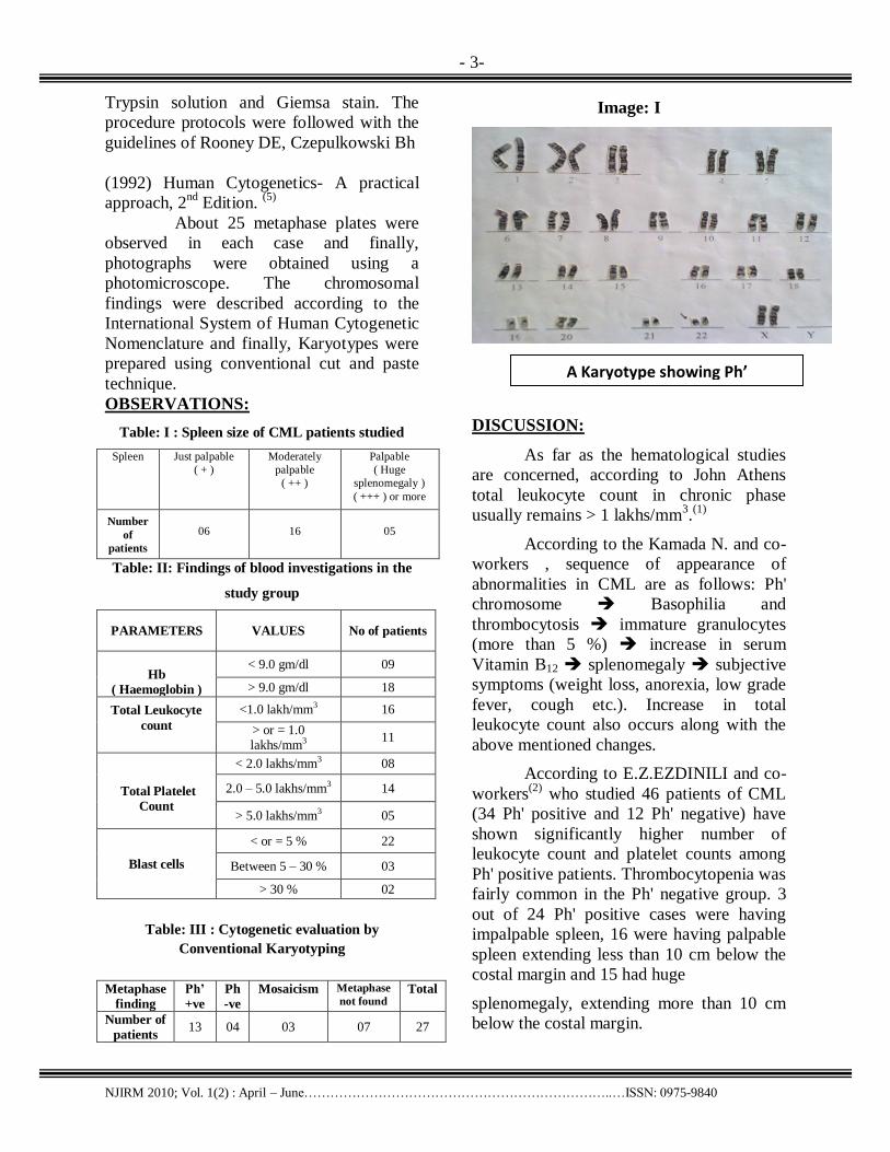
Image: I
DISCUSSION:
As far as the hematological studies
are concerned, according to John Athens
total leukocyte count in chronic phase
usually remains > 1 lakhs/mm3.(1)
According to the Kamada N. and co-
workers , sequence of appearance of
abnormalities in CML are as follows: Ph'
chromosome Basophilia and
thrombocytosis immature granulocytes
(more than 5 %) increase in serum
Vitamin B12 splenomegaly subjective
symptoms (weight loss, anorexia, low grade
fever, cough etc.). Increase in total
leukocyte count also occurs along with the
above mentioned changes.
According to E.Z.EZDINILI and co-
workers(2)
who studied 46 patients of CML
(34 Ph' positive and 12 Ph' negative) have
shown significantly higher number of
leukocyte count and platelet counts among
Ph' positive patients. Thrombocytopenia was
fairly common in the Ph' negative group. 3
out of 24 Ph' positive cases were having
impalpable spleen, 16 were having palpable
spleen extending less than 10 cm below the
costal margin and 15 had huge
splenomegaly, extending more than 10 cm
below the costal margin.
A Karyotype showing Ph’
chromosome in a patient of CML

- 4-
NJIRM 2010; Vol. 1(2) : April – June……………………………………………………………..…ISSN: 0975-9840
As shown in Tables I & II of observations:
in present study, 10 out of 25 chronic phase
CML cases were having total leukocyte
count > 1 lakhs/mm3 and 6 out of 14 Ph'
positive cases were having total leukocyte
count > 1 lakhs/mm3. Out of 2 cases of
blastic phase 1 case had total leukocyte
count > 1 lakhs/mm3 and 2 out of 4 Ph'
negative cases were having total platelet
count < 2 lakhs/mm3 showing
thrombocytopenia. 6 out of 13 Ph' positive
cases were having just palpable spleen(+), 5
cases of Ph' positive were having
moderately palpable spleen (++) and 1 case
of Ph' positive was having hugely palpable
spleen (+++). Spleen size correlates
reasonably well with the magnitude of the
total leukocyte count.
Tura and Kantarajan(5) have given
negative prognostic factors which are: age (
60 years; spleen ( 10 cm below the costal
margin, blasts ( 5 % in marrow, platelets ( 7
lakhs/mm3 or any of the characteristics of
the accelerated disease are associated with
poor prognosis and a threefold higher hazard
rate, or risk of death per unit of time, in the
first year. From cytogenetic standpoint, the
most extensively documented evidences for
sequential genetic changes during tumor
evolution come from studies of chronic
myelogenous leukemia. In nearly every
typical case, the leukemic cells in the early
indolent phase of this disorder have only a single cytogenetic abnormality, the t(9;22)
translocation that produces the Ph‟
chromosome.
Presumably, the altered gene product plays
an important role in the early expansion of
the neoplastic clone within the myeloid
lineage. When CML progresses to its
accelerated terminal phase, the neoplastic
population is overgrown by one or more sub
clones having additional karyotypic
changes.(4) The Philadelphia chromosome
is the cytogenetic hallmark of chronic
myeloid leukemia and is observed in more
than 90 % of CML cases. At diagnosis, in 5
to 10 % of CML patients the Ph'
chromosome is derived from variant
translocations other than the standard t( 9 :
22 ).
Table: IV
STUDY No of
patients
studied
Ph’
positive
Ph’
negative
Patients having
Ph’ chromosome
( % )
Other cytogenetic findings
EZDINILI E.Z.et al
( 1959-1967)(2)
61 43 18 70.49 -
Rowley J. D.
( 1973 )(6)
9 9 - 100 +C,+8,-17, +?F(in 5 cases )
Su.X.Y. et al
(1999)(7)
38 38 - 100 Double or triple Ph‟, +8, +7, +17, t(1:11)(q12-
21;p15), del(3),del(6)(q13-21), der(6)t(17;3;6),
der(17)t(6:17)
Walid Al Achkar
(2001)(8)
43 22 18 51.16 +19, t(4:22); +9
Rachal Thomas Jacob et
al (2002)(4)
525 453 72 86.28 Double Ph‟ chromosome, +8,+19
Present Study*
27 13** 4 48.14 Mosaicism observed in 3 cases

- 6-
NJIRM 2010; Vol. 1(2) : April – June……………………………………………………………..…ISSN: 0975-9840
Similarly other associated chromosomal
abnormalities are also present in these
remaining 5 to 10 % of the cases. Some of
the concerned studies are discussed
herewith. Table IV ( *Table : III in observation, **
Image : I )
All authors agree that patients with CML
who have the Ph‟ chromosome have a better
prognosis than those who don‟t. Mosaicism
has been considered indicative of a good
prognosis in most, but not all, series.
Cytogenetic investigations are
mandatory in cases diagnosed
morphologically as CML especially, to
know the Ph‟ status. Once the Ph‟ status is
known these studies will help in selection of
cases for more effective treatment protocols.
Use of newer drug regimens in patients who
are Ph‟ positive in the early stages of the
disease can help to increase median survival
period.(4)
However, in those cases that are Ph'
negative by conventional cytogenetics, Ph'
negativity doesn't exclude bcr-abl
translocation at molecular level, which
requires to be confirmed by more specific
molecular genetic studies like Fluorescence
In Situ Hybridization(FISH).
In CML, cytogenetic investigations
whether they are in the form of conventional
studies as in case of the present study or
upgraded in the form of FISH (Fluorescence
In Situ Hybridization) or CGH
(Comparative Genomic Hybridization),
remain keystone at every stage of the
disease; starting from initial diagnosis to
final treatment and for predicting prognosis
of the patients.
BIBLIOGRAPHY:
1. Athens john w., wintrobe‟s clinical
haematology – 9th
edition – 2nd
volume –ii, page: 1969 to 1992 -
chapter – 75 – chronic myeloid
leukemia.
2. Ezdinli e.z., m.d., sokal j.e., m.d.,
f.a.c.p.,buffalo, new york. Annals of
internal medicine, volume 72,
number-2,february-1970. philadelph.
– chromosome-positive and
negative-chronic myeloid leukemia
3. kantarjian hm, keating mj, talpaz m,
et al.: am j med 83 (3): 445-54, 1987.
Chronic myelogenous leukemia in
blast crisis. Analysis of 242 patients.
4. Rachel thomas jacob,
k.gayathri,m.d.; surath anjna,
m.b.b.s.d.ch.; d.raghunadha
5. Rao,m.d.,d.m., department of
pathology and medical oncology,
nizam‟s institute of medical sciences,
hyderabad-500082, a.p. India. Indian
journal of cancer, june-2002,
volume-39, pages-61-65.cytogenetic
profile of chronic myeloid leukemia.
6. Rooney de, czepulkowski bh (eds)
human cytogenetics – a practical
approach, volumes i and ii, 2nd ed.
Irl press, oxford
7. Rowley janet d., department of
medicine, university of chicago,
franklin mclean memorial research
institute, chicago, illinois 60637.
Nature : volume : 243, june 1973.
8. Su x y; cao q; he k l shanghai
institute of hematology, rui-jin
hospital, shanghai,
200025.zhonghua yi xue za zhi ,
china jan 1999, volume, issue, pages:
79; (1); p34-7 chromosomal
abnormalities in 38 cml cases of
various phases.
9. Walid al achkar molecular
cytogenetics lab., molecular biology
and biotechnology dept. Syrian
atomic energy commission,
damascus-syria. P.o box: 6091.
Human bone marrow cultures and
chromosomal abnormality study of
cml.

- 7-
NJIRM 2010; Vol. 1(2) : April – June……………………………………………………………..…ISSN: 0975-9840
Cardiovascular Responses With Valsalva Maneuver During Activities Of Daily Livings In Healthy Adults
Prof. Priyanshu V. Rathod, Prof. Savita Ravindra, , Prof. Veena Kiran Nambiar R K College of Physiotherapy, Rajkot
Abstract: Purpose of Study: To measure CV responses in SLF, and SQ with & without VM, Study design: Cross sectional Observational study, Materials: assessment form, 36 SF Health Questionnaire, Modified Sphygmomanometer, BP apparatus, Nike-HRM-TRIAX, Methodology: 335 (M=146) subjects participated to perform SLF and SQ position with & without VM and SBP, DBP and HR were recorded. Data Analysis: SPSS -10.1, LOS was set at 0.05 or CI 95 %. Result: Study has shown significant increases in SBP in SQ and increase HR in SLF position with and without VM. Discussion: the impact of Heart Rate Variability and baroreflex sensitivity in CV system plays vital role in maintaining hemodynamic status while performing valsalva like activities. Conclusion: SQ has significant impact on SBP and DBP as compared to SLF position with and without VM as well as SLF position has a significant impact on HR as compared to SQ with VM, however these need to be taken into consideration while planning life style modification for high risk population Key words: Valsalva Maneuver (VM), Sitting Lean Forward (SLF) position, Squatting (SQ)
Position, Cardiovascular (CV) responses
INTRODUCTION:
Activities of daily livings (ADL)
involve several stressful physical events
such as lifting weight, pushing objective
ground level activities, defecation,
urination, etc... These stressful events
are not recognized among healthy
people, but it may be life threatening for
peoples with compromised CV system.8,
10
It has been observed that more
than 1/3 population, who dies following
the cerebrovascular accident, dies in and
around the toilet, in the morning. 7, 12,
15 Urination and defecation has
integrated sequences of breath holding
strategies which involves isometric
muscular contraction to increases
interathoracic and abdominal pressure.
It may be more stressful in case
of constipation, obesity, pregnancy etc...
Generating high intraabdominal pressure
and frequent releasing of “breath hold”
has great impact on hemodynamic status.
Moreover, the positions of performing
defecation such as SLF and SQ have
significant impact on CV system.
Clinically, breath holding with forced
expiratory effort against closed glottis is
known as a Valsalva Maneuver (VM). It
is also used as a test of functional
integrity of the autonomic nervous
system.1, 2
Hemodynamic changes
describe into 4 phases when VM
performed at 40-mmHg pressures for 20
seconds with open glottis.
CV responses in stress-provoked
defecation positions with VM among
healthy adults will provide the associate
evidence to understand the impact on
compromised CV disorders.
CV responses on ADLs have
been of common interest for many
researchers. ADLs with maneuver like

- 8-
NJIRM 2010; Vol. 1(2) : April – June……………………………………………………………..…ISSN: 0975-9840
valsalva may throw further light on
integrity of autonomic and CV system,
which may be useful or detrimental to
normal and patient population. 2, 5, 10
The various researchers have
shown interest in positional impact of
defecation on CV status. It was observed
that when normal subjects squat, the
arterial mean pressure and pulse pressure
increases. This increase is greater after
few seconds due to which mean pressure
falls. This fall in pressure eventually
stabilizes at higher level than that
observed in sitting position.5,1 The
studies has concluded that CV stability
during straining with VM is higher in the
lean forward as compared to lean
backward position. 9
The VM is a useful technique to
assess the CV changes.6.
Some studies
concluded that CV changes could be
obtained as a result of a 15-sec VM,
performed at 40-mmHg airway
pressures.7,8.
Some of the researchers
have suggested 20 seconds of VM at 40-
mmHg pressure.12
Figure I, Four phase valsalva response
The hemodynamic changes
associated with the VM were described
by invasive techniques.7, 6,10.
These
changes are classically divided into four
phases. (Fig-I) Phase-I is beginning of
the strain to a transient rise in mean
arterial BP (MABP), as the increase in
intrathoracic pressure to constrain the
arterial tree. Phase-II has further divided
into Phase IIa and IIb. During phase IIa,
the atrial filling pressure falls so MABP
decreases. In phase IIb there is increased
sympathetic activation, causing a rise in
peripheral vascular resistance and HR,
which leads to a small increase in
MABP. Phase-III is associated with
release of the strain and a sudden fall in
MABP due to the influence of release of
the intrathoracic pressure on the arterial
tree. Finally, phase-IV sees an
immediate „„overshoot‟‟ in MABP
because of the persistence of increased
sympathetic tone and systemic vascular
resistance. A reflex bradycardia then
results due to stimulation of arterial
baroreceptors, and both MABP and HR
return to baseline values. 11, 13
It has been
concluded with VM at severe degree,
results in decrease of HR by 9.2
beats/min.9
This study was designed to
evaluate CV responses in two stress
provoking defecation positions of ADLs,
SLF and SQ positions. However,
comparing the CV responses with and
without VM in these stress-provoked
positions can provide prospective
guidelines for high-risk population with
CV disorders.
METHODOLOGY
The Cross sectional study has
sample of 235 healthy adults, (M=143)
with the mean age = 26.6 year with prior
informed consent and ethical clearance.
Subjects were screened with 36-SF
health questionnaire to consider health
status. 17
Subjects were excluded with
poor physical efforts to perform VM,
failed to complete the procedure, and to
recover to basal parameters in given
time.
Method:
All subjects satisfying the criteria
for the study were made to achieve

- 9-
NJIRM 2010; Vol. 1(2) : April – June……………………………………………………………..…ISSN: 0975-9840
relaxed sitting (RS) position on the chair
with feet supported on floor (hips and
knees 90 degree flexion) for 3 min.,
prior to the test procedures. parameters
(SBP, DBP, and HR) were measured in
the RS position.
The subject blows air at 40-
mmHg pressures for 25 seconds into a
mouthpiece, which was attached to a
modified sphygmomanometer to
measure airway pressure. In this study,
the glottis remains open to communicate
for measurement of pressures from the
thorax into the sphygmomanometer. A
small needle was routinely placed into
the rubber tube to provide a small air
leak. This prevents the subject from
closing their glottis and from developing
the necessary pressures with the cheek
muscles. (Fig-II)
SBP and DBP were recorded by
sphygmomanometer from brachial artery
at elbow. HR was recorded by Nike -
strap at Xiphoid-sternal level. This strap
made to sense the left ventricular apex
beats and watch to receive the impulses.
Considering the basal parameters in RS
position in chair, subjects were allowed
SLF with forearms supported on thigh
for a minute and parameters were
recorded.
Figure III, SLF position with VM
Resting for a minute in SLF position,
VM was performed and only change in
HR (max) was recorded during 25
seconds. Immediate post VM release
changes in parameters were recorded in
SLF position. After the recording of post
valsalva release response, subjects were
allowed to resume RS position and
parameters were recorded at the end of
3rd
minute. (Fig – III) On the second day,
subjects were asked to perform the
similar procedure with SQ and
parameters recorded.
Figure III, SQ position with VM
Data analysis
Statistical analysis was
performed with SPSS for repeated
Measures ANOVA for comparison of
the measured variables (SBP, DBP, and
HR) within and between the two
positions defecation. The LOS was set
at < 0.05 or 95 % confident interval (CI).
RESULT
Graph I. Graphical representation
suggesting of SBP responses in RS
position with SLF and SQ positions

- 10-
NJIRM 2010; Vol. 1(2) : April – June……………………………………………………………..…ISSN: 0975-9840
Graph II. Graphical representation
suggesting of DBP responses in RS
position with SLF and SQ positions
Graph III. Graphical representation
suggesting of HR responses in RS
position with SLF and SQ positions
DISCUSSION
SLF vs. SQ positions without VM :
This study has shown significant
difference in variables of SQ and SLF
positions as compared to RS position.
SBP and DBP significantly increases in
SQ compared to SLF position. In SQ
position, intraabdominal and so
interathoracic pressures are responsible
for increasing the SBP. HR shows
significant response in SQ compared to
SLF.
SLF vs. SQ positions with VM :
A high significant raise has shown in HR
by both the positions with VM. But,
increase in HR was more significant in
SLF than SQ position. Significant
increase in HR was higher in SLF
position, because in VM at the onset of
strain, contraction of thoracic cage
compresses the lung and causes the large
raise in interathoracic pressure; this
compresses the vessels within the chest.
Moreover, compression of thoracic vena
cava compromise venous return to the
heart, resulting in a large falls in cardiac
output. This leads to secondary fall in
aortic pressure and as aortic pressure
falls, the baroreceptor reflex increase the
HR. 5, 9
SLF vs. SQ post VM release :
When the subject relieves VM, and
begins with normal breathing again –
deep inspiration follows. When
compression of vena cava removed,
venous return suddenly increases
causing a rapid raise in cardiac output
several seconds latter, which leads to
overshoot of arterial pressure as the
systemic vascular resistance increases
due to sympathetic activation that
occurred with VM. However, peripheral
circulation to lower limbs is
compromised in SQ compared to SLF
position may be responsible to increase
the BP in the upper half of the body,
which was recorded from upper limbs by
the sphygmomanometer.
HR reflexively decreases in
response to the transient elevation in
arterial pressure. Fall in HR post VM
was seen in SLF position and in
squatting position, this anatomical
spacing of blood volume in abdominal
cavity and lower limbs decreases the
venous returns to the heart, a direct
effect of kinking the femoral veins. So,

- 11-
NJIRM 2010; Vol. 1(2) : April – June……………………………………………………………..…ISSN: 0975-9840
venous return is decreased in SQ when
compared to SLF position. This might
have caused a comparative decrease in
cardiac output in squatting position,
which was responsible for controlling
the large variation in HR in SQ as
compared to SLF position. 13, 14
CONCLUSION
This study has considered the impact of
CV system in SLF and SQ positions
with and without VM. It is shown that
SQ has significant impact on SBP and
DBP as compared to SLF position with
and without VM. SLF position has a
significant impact on HR as compared to
SQ with VM. It can be concluded that
SLF and SQ with and without VM
influence changes in cardio-vascular
status. These need to be taken into
consideration while planning life-style
modifications for high risk population
with compromised CV status.
BIBLIOGRAPHY:
1. E. P. Sharpey, et al.: “Effects of
squatting on the normal and failing
circulation”. BMJ, 1956, 1072-74.
2. Federici. “Systolic and Diastolic
changes in human coronary blood
flow during VM”, Cli. Physiology. ,
2000, 20, 1-19.
3. G. Bilancioni: “La figura e l‟opera di
Valsalva. In his Sulle rive de Lete”
Rome, 1930, 77-100.
4. E. P. Sharpey, “Effects of Valsalva‟s
maneuver on the normal and failing
circulation”. British Medical Journal,
1955, 693-695.
5. Hamilton W. F. et al.: “Physiological
relationship between Intrathoracic,
Intraspinal, and Arterial Pressure”,
1936. JAMA, 103, 853.
6. Imholx B., et al, “Continuous non-
invasive BP monitoring: reliability of
Finapres device during the VM”
Cardiovasc. Res. 1988, 22, 390-397.
7. Olschewski H., et al. “Cardiac
responses to the VM in different
body position”. European Jr App.
Physio. 90, 61, 1-2, 20
8. Lieshout J., et al: “Pitfalls in the
assessment of CV reflexes in patients
with sympathetic failure but intact
vagal control” Clin. Sci. 1989,
76:523-528.
9. Smith S. A., et al: “Can sinoaortic
Baroreceptor HR reflex sensitivity be
determined from phase IV of the
Valsalva manoeuvre?” Cardiovasc.
Res. 1987, 21: 422–427.
10. Eckberg, D.L.: “Parasympathetic CV
control in human disease: a critical
review of methods and results.” Am.
J. Physiol. 1980, 239:581-593,
11. "Valsalva's Maneuver." In
Everything You Need to Know
About Medical Treatments.
Springhouse, PA: Springhouse
Corp., 1996, 78-9.
12. Chakravarthy, et al, "BP changes
during Squatting – A study in normal
subjects and its possible clinical
significance” JAPI, 2001, 49, 678-
79.
13. Palmero H. A., et al.: “Baroreceptor
reflex sensitivity index derived from
phase IV of the VM” Hypertension
3, Suppl. II: 1981, 134–137.
14. Albert B., “A simple test of cardiac
function based upon the HR changes
induced by VM”. AJM Cardiology,
1966, 18, 90-98.
15. William A. et al. "Bradycardia
responses to vagally medicated
bedside maneuver in health
volunteer” AJM, 99, 6, 725-30.
16. McHoney, C. A., “The MOS-36 Item
Short-Form (SF-36): Psychometric
and clinical tests of validity in
measuring physical and mental

- 12-
NJIRM 2010; Vol. 1(2) : April – June……………………………………………………………..…ISSN: 0975-9840
health constructs. Medical Care”,
1993, 31, 247-263.
17. Ware, J. E.,: “SF-36 Health Survey:
Manual and Interpretation Guide”.
Boston, Massachusetts: The Health
Institute, NEMC. 1993.

- 13-
NJIRM 2010; Vol. 1(2) : April – June……………………………………………………………..…ISSN: 0975-9840
Academic Performance Of School Children With Their Intelligence Quotient
S. D. KULKARNI*, N. R. PATHAK**, C. S. SHARMA***
Department of Physiology *Surat Municipal Institute of Medical Education & Research (SMIMER),Surat (Gujarat State, INDIA), **B. J. Medical College,New Civil Hospital,Ahmedabad (Gujarat State, INDIA), *** Government Dental College,New Civil Hospital,Ahmedabad (Gujarat State, INDIA)
Abstract: Present study was carried out to correlate academic performance of Indian school children with their intelligence quotient (as measured by Porteus Maze Tests). These tests have been tried and tested successfully in previous studies, don’t require sophisticated equipment and are easier to administer than traditional tests like Wechsler Tests. Contrary to popular belief, no statistically significant correlation was found in this study between intelligence and academic grades. Thus intelligence is not a prerequisite to succeed in examinations and therefore in life. Key words: Intelligence Quotient (IQ), Porteus Maze Tests, Intelligence,
Academic performance Academic grades
INTRODUCTION:
Intelligence is the most valuable
wealth of humans. Intelligence is
assessed and not measured because in all
its meaning and application, it is not a
thing; it is only an idea, an abstraction.
Thurnstone1 (1946), Griffith
2 (1933) and
Piaget3 (1983) have all come up with
their definitions of intelligence but
perhaps the pioneer in this field is David
Wechsler. He defined intelligence as the
aggregate or global capacity of an
individual to act purposefully, to think
rationally and so to deal effectively with
his environment. It helps an individual to
consciously adjust his thinking to new
requirements. Thus, it is a general
mental adaptability to new problems and
conditions of life. Intelligent mind and
efficient hands work in coordination
with each other.
Majority of people are average, a
few very bright and a few very dull.
Intelligence also varies in the same
individual from situation to situation. As
child grows in age, so does the
intelligence. Mental development occurs
from the concrete to the conceptual,
formal and symbolic. While heredity
determines the level of intelligence, it is
the environment that either slows down
or expedites its development. Thus, the
main purpose of education is to develop
child‟s intellect in a well-designed
environment. Vertical growth of
intelligence ceases at 16-20 years of age,
but accumulation of knowledge and
acquisition of skills continue throughout
the life span of an individual. Therefore
the so-called “tapping intellectual
resources” means an effective advance
in the function of the brain4.
Intelligence includes many basic
factors such as attention, imagination,
observation, thinking, judgment and
mental perception through sensory

- 14-
NJIRM 2010; Vol. 1(2) : April – June……………………………………………………………..…ISSN: 0975-9840
organs and memory. Porteus Maze Tests
(used in the present study for assessing
intelligence) while being simple,
inexpensive and easy to administer,
encompass all these aspects of
intelligence.
It is popular belief that intelligent
people always do well in life. Present
study was carried out to correlate
academic performance of Indian school
children with their intelligence quotient
(as measured by Porteus Maze Tests).
MATERIALS AND METHODS:
The study was conducted on 320
randomly selected students (studying in
standards 1st to 8
th) of a Government
School in India half of whom were of
either sex.
The area and school were chosen
keeping in mind the composition of
Indian society with due consideration to
various parameters (like socio-economic
status5,6
) so as to get an unbiased
representative sample. The selected
children were subjected to general
clinical medical examination to rule out
any major mental or physical illness or
disability.
Informed consent was taken from
the principal and parents after explaining
them the aim and nature of the study and
their wards‟ role in it.
Percentage of marks secured by
each participating student during the
preceding academic year was noted as a
measure of his/her academic ability.
Children were graded7 according to their
academic performance as follows:
Marks Grade
> 75% A
60% - 75% B
45% - 60% C
< 45% D
Intelligence Quotient (IQ) of the
participants was assessed using The
Porteus Maze Tests8
for various „mental‟
ages. These paper-pencil tests consist of
successive puzzle charts of increasing
levels of difficulty. The age inscribed on
the toughest test chart which a subject
was able to solve successfully was taken
as his mental age. Then his IQ was
calculated as: IQ = (Mental age) / (Chronological age) × 100
Children were categorized4 according to
their IQ levels as follows:
IQ Category
> 115 A
100 - 115 B
85 - 100 C
< 85 D
Observations were recorded after
taking due care to reduce instrument and
observer errors to a minimum.
Correlation academic performance with
Intelligence Quotient was done using
Chi-Square Test9.
RESULTS AND DISCUSSION:
Table I
IQ Academic Performance Sub
total "A" "B" "C" "D"
"A" 21 20 18 10 69
"B" 21 23 30 17 91
"C" 15 25 22 20 82
"D" 13 19 22 24 78
Subtotal 70 87 92 71 320 0.1<p< 0.5 (df = 9) (Not Significant )
Our study found no statistically
significant correlation between
intelligence quotient and academic
performance in the participating
students. This finding is revolutionary
because it challenges the layman‟s
notion that intelligence is the sole
determinant of academic performance.

- 15-
NJIRM 2010; Vol. 1(2) : April – June……………………………………………………………..…ISSN: 0975-9840
Chart showing Academic Grades obtained by school children having varying Intelligence
21 2115 13
2023
25
19
18
30
22
22
10
17
20
24
0
10
20
30
40
50
60
70
80
90
100
A B C D
Intelligence Quotient Category
No. o
f stu
dent
s
Grade D
Grade C
Grade B
Grade A
Graph I
So what then is the mantra for
success? Previous studies have
concluded that an unhealthy child could
have a dismal academic record even if
his/her genes predict a high IQ for
him/her7. On the other hand, with
average intelligence, a person can still
excel in studies provided he/she
maintains adequate fitness10
. Thus,
success at work (which determines
success in life) depends largely on
fitness and health and not merely on
one‟s intelligence. Your genes could
give you the edge but only after a
healthy mind, body and soul have put in
their best efforts11
.
There is a lesson to be learnt here
for parents and teachers. Your children
could be god-gifted but if they are not
nurtured in a proper environment, they
may not realize their true potential.
Physical health, mental peace, social
security and spiritual well-being are
perhaps more important than natural
talent for success in life.
CONCLUSION:
Intelligence has no relation
whatsoever with academic performance
of school children. Children with
average IQ can fare well in studies.
Conversely, „above average‟ children
may not get the grades expected of them.
BIBLIOGRAPHY:
1. Thurstone LL (1946): Theories of
intelligence science monthly, 5,
(175-197)
2. Griffith JH, The psychology of
human behaviour, London, George
Allen, 1933
3. Piaget J: Piaget‟s theory. In manual
of child psychology, P Mussen,
editor. Wiley, Newyork 1983
4. Dissertation submitted to The Tamil
Nadu Dr. MGR Med. Univ. for the
Degree of MD (Physiology): A
Study on Evaluation of Memory in
Abacus Learners (August 2004)
5. Mahajan, Gupta: Textbook of PSM
(2nd
ed. 1988-89): Social
environment (Ch. 11): Pg 135
6. P. Kumar (1993): Social
classification – Need for constant
upgrading, Indian Journal of
Community Medicine: Vol. 18; Pg 2
7. Skinner BF: The Technology of
Teaching, Newyork Applenton-
century crofts, 1968
8. Harrap GG et al (1965): 182 High
Holborn, London, W.C.I

- 16-
NJIRM 2010; Vol. 1(2) : April – June……………………………………………………………..…ISSN: 0975-9840
9. Mahajan BK (1999): Methods in
Biostatistics (6th
ed): The Chi-square
Test (Ch 11): Pg 172-178
10. Buxton, Doris. Analysis of an
extension of the Kraus-Weber tests.
Research Quarterly 1957; 28: 210-
217
11. Park K (1997): Textbook of PSM
(15th ed): Mental Health (Ch 15):
Causes of Mental Ill Health (Pg 567)

- 16-
NJIRM 2010; Vol. 1(2) : April – June……………………………………………………………..…ISSN: 0975-9840
A Study Of Nutrient Foramina Of The Metacarpal Bones
Dr. P. S. Shrimankar*, Dr. D. J. Trivedi*, Dr. V.B. Kariya**
* Associate professor, ** Assistant professor, Department of Anatomy, GMERS Medical College, Sola, Ahmedabad, Gujarat
Abstract: 1500 metacarpals, 300 each of 1st, 2nd, 3rd, 4th and 5th metacarpals of unknown age and sex and 200 metacarpals from 20 articulated skeletons for bilateral study where examined for the length of the bone, number, position, direction and distances from both the ends of nutrient foramina. It was observed that almost all the metacarpals has one nutrient foramen in the middle third of their shaft except in 1st metacarpal in which it was in distal third. It was observed that frequency of number of nutrient foramina differ in different metacarpals. In 1st and 2nd metacarpals foramina were situated mostly on the medial surface and in other metacarpals mostly on the lateral surface. There was a good deal of bilateral symmetry and invariably all foramina were directed away from the growing ends of the bone Key words: metacarpal, nutrient foramen, nutrient vessel, short long bone INTRODUCTION:
During routine teaching of
osteology, mention is always made of the
nutrient foramen on the shaft of long bones.
There is a jingle regarding direction of the
nutrient canal “they seek the elbow and flee
from the knee”! The nutrient artery is a
principal source of blood supply to a long
bone and being source of blood supply to a
bone it plays an important role in the healing
process or union after a fracture. The short
long bones have only one epiphysis. The
head forms the epiphysis in medial four
metacarpals whereas in 1st metacarpal, base
form its epiphysis. The nutrient artery enters
the bone at right angle to the shaft and as the
bone grows in length it becomes more
oblique in direction towards the non
growing end.
The human hand is often involved in
machinery and road side accidents.
Nowadays it is possible to reconstruct it by
plastic surgeons. Therefore it is very
important for a plastic surgeon to know all
information regarding nutrient foramina to
avoid possibility of necrosis if an injury cut
off the nutrient vessel in the growing bone
or he could avoid limited area of cortex
containing the nutrient foramen during open
reduction. The 2nd
metacarpal as a
vascularized bone graft may be harvested
entirely in serious injuries with destruction
of index finger3. An artery located on
dorsoulnar side and connecting the head of
the 1st metacarpal with the dorsal arcade of
proximal nail fold is useful for making a
dorsoulnar flap of thumb that can be raised
on its artery with a distal pedicle. This can
be used for distal skin loss coverage of
thumb and for reconstruction of finger pulp1.
Because of lack of detailed
information regarding nutrient foramina and
with the view of its usefulness in academic,
surgical as well as in medico-legal practice
following study was done to measure
number, position in relation to length and
circumference, proximity from the ends,
direction and bilateral symmetry of the
nutrient foramina.

- 17-
NJIRM 2010; Vol. 1(2) : April – June……………………………………………………………..…ISSN: 0975-9840
MATERIAL AND METHOD:
In the present study, 1500 metacarpal
bones (300 of each 1st, 2
nd 3
rd 4
th and 5
th
metacarpals) of unknown age and sex from
the collection in Department of Anatomy
from various medical colleges of Gujarat
were studied. For bilateral study, 20
articulated skeletons were examined. The
bones were dry, macerated and cleaned.
The number and position of nutrient
foramina were identified; a stiff wire was
passed through the foramen to see the
direction of the canal when in doubt. The
measurements were taken ( in cms.) with the
help of vernier calipers. The whole length of
the bone was taken from head of the
metacarpal to the base. The distances of

- 18-
NJIRM 2010; Vol. 1(2) : April – June……………………………………………………………..…ISSN: 0975-9840
nutrient foramen from both the ends and
situation of the foramen on the
circumference of the shaft were noted down.
The foraminal index was calculated by the
following formula:
FI =DB/L X 100 (DB = distance of
foramen from base, L = Total length of the
bone)
In the study of bilateral symmetry ,
Complete symmetry means – Number and
position of the foramina were equal in both
the sides, Partial symmetry means – Number
of the foramina remains same but their
position varied on both the sides and No
symmetry means number of the foramina
unequal on both the sides.
OBSERVATIONS (Table I to V):
TABLE- I
NUMBER OF THE NUTRIENT FORAMINA
Number
of
metacarpal
bone
No. of bones No. of bones (%) Total no.
of
foramina Right Left Absent
foramen
One
foramen
Two
foramina
1st 165 135 26 (8.7) 243 (81) 31 (10.3) 305
2nd
173 127 10 (3.3) 258 (86) 32 (10.7) 322
3rd
147 153 08 (2.7) 282 (94) 10 (3.3) 302
4th
136 164 07 (2.3) 276 (92) 17 (5.7) 310
5th
162 138 07 (2.3) 254 (84.7) 39 (13) 332
TABLE – II
POSTION OF NUTRIENT FORAMINA IN RELATION TO CIRCUMFERENCE OF
THE BONE
Number
of
metacarpal
bone
No. of
bones
No. of bones (%) Total no.
of
foramina Medial surface Lateral surface Posterior surface
1st 300 263 (86.2) 27 (8.9) 15 (4.9) 305
2nd
300 223 (69.3) 96 (29.8) 03 (0.9) 322
3rd
300 54 (17.9) 248 (82.1) 00 (0) 302
4th
300 49 (15.8) 259 (83.5) 02 (0.7) 310
5th
300 32 (9.6) 290 (87.4) 10 (3) 332

- 19-
NJIRM 2010; Vol. 1(2) : April – June……………………………………………………………..…ISSN: 0975-9840
TABLE – III
LENGTH OF METACARPALS, DISTANCES OF FORAMINA FROM HEAD AND
BASE OF THE METACARPALS
Number of
metacarpal bone
Mean length
(cm)
Mean DH (cm) Mean DB (cm) Mean FI
1st 4.24 1.82 2.56 59.29
2nd
6.54 3.22 3.26 47.18
3rd
6.37 3.52 2.79 41.25
4th
5.33 2.88 2.49 43.87
5th
4.90 2.60 2.38 47.48
(DH = distance from the head, DB = distance from the base FI = foraminal index. The index
around 60 = foramen on distal third, around 50 = middle third and below 40 = proximal third)
TABLE – IV
BILATERAL SYMMETRY OF FORAMINA
Number of
metacarpal bone
Partial symmetry
(%)
Complete symmetry
(%)
No symmetry (%)
1st 15 80 05
2nd
20 60 20
3rd
25 70 05
4th
10 90 00
5th
15 75 10
TABLE – V
DIRECTION OF THE NUTRIENT CANAL
Number of
metacarpal bone
Total no. of
foramina
Direction of nutrient canal
Towards head Towards base
1st 305 305 00
2nd
322 00 322
3rd
302 00 302
4th
310 00 310
5th
332 00 332

- 20-
NJIRM 2010; Vol. 1(2) : April – June……………………………………………………………..…ISSN: 0975-9840
DISCUSSION:
The human hand is often involved in
machinery and road side accidents.
Nowadays it is possible to reconstruct it by
plastic surgeons so the knowledge of exact
position, number and direction of the
nutrient foramina in metacarpal bones is
mandatory for them.
Singh4
(1959) observed that absence
of the nutrient foramina were common in 1st
metacarpals and double foramina frequently
seen in 2nd
and 5th
metacarpals. Patake &
Mysorekar2 (1977) found that absence of
foramina & double foramina both were
common in 1st metacarpals. In present study
absence of the foramina is most frequently
noted in 1st metacarpals and double foramina
in 1st, 2
nd and 5
th metacarpals.
Wood jones5
(1946) observed that
the nutrient foramina of the 1st, 2
nd and
usually that of the 3rd
and 4th were located
on their lateral surfaces whereas that of the
5th on the medial surface. As per
observations of Singh (1959), Patake &
Mysorekar2 (1976) nutrient foramina of the
1st and 2
nd metacarpals were located on
medial surface of the bone and that of the
3rd
, 4th and 5
th metacarpals were on the
lateral surface. Present study supports the
findings of Singh, Patake & Mysorekar.
In present study, Foraminal index
suggests that the nutrient foramina in case of
1st metacarpals were situated on distal 1/3
rd
of the bone and in rest of the metacarpals in
middle 1/3rd
of the bone. The datas
regarding FI matches more or less with that
of study of Patake & Mysorkar2 (1977).
Patake & Mysorkar2 (1977) studied
that 41% of 1st and 2
nd metacarpals, 25% of
3rd
and 4th
metacarpals and 8.3% of 5th
metacarpals showed no symmetry at all. In
present study, 5 % of the 1st and 3
rd
metacarpals, 10 % of the 5th metacarpals and
20 % of the 2nd
metacarpals showed no
symmetry.
As per observations made by
previous workers and also the present study
states that without any exception nutrient
foramina were directed away from the
growing end.
CONCLUSION:
From observations and discussions
the following general conclusions can be
derived for all the metacarpals studied:
The position of the nutrient foramina
on the shaft of the bone (in relation to the
length) is variable; however, it is observed
in this series that nutrient foramen of the 1st
metacarpals is in distal third, whereas in
other metacarpals it is in the middle third.
The position of the nutrient foramen
on the shaft of the bone ( in relation to
circumference) is also not constant;
however, it is observed that in this series
nutrient foramina of the 1st and 2
nd
metacarpals were on the medial surface
whereas that of the 3rd
, 4th
and 5th
metacarpals were on the lateral surface. It is
proved by FI that nutrient foramina of 1st
metacarpals were on the distal third whereas
in other metacarpals they were in the middle
third of the metacarpals.
The similar bones having the same or
nearly same lengths possess nutrient
foramen at variable distances. So, also the
similar bones possessing the same or nearly
same distance of the foramen from any one
of the ends may not have the same length.
Hence the derivation of length of a
metacarpal from the position of the nutrient
foramen is likely to be fallacious.
The numbers of foramina do not
seem to have any significant relation to the
length of the bone, the smaller bone may
- 21-
NJIRM 2010; Vol. 1(2) : April – June……………………………………………………………..…ISSN: 0975-9840
have double foramina and the longer one
may have no foramina at all.
Direction of the nutrient canal is always
away from the growing end and position of
the nutrient foramen is nearer to the growing
end.
BIBLIOGRAPHY:
1. Frunelli F. ; Vigasio A. ; Valenti P.
(1999) : Arterial anatomy and clinical
application of dorso-ulnar flap of the
thumb. Journal of hand surgery. 24(4) :
803-11.
2. Patake S.M. & Mysorekar V. R.(1977) :
Diphysial nutrient foramina in human
metacarpals & metatarsals. Journal of
anatomical society of India. 124(2) :299-
304.
3. Pierer G.et al (1992) : The vascular
blood supply of the 2nd
metacarpal bone:
anatomic basis for a new vascularized
bone graft in hand surgery. Surgical and
radiological anatomy. 14(2) : 103-12.
4. Singh I. (1959) : Variations in the
metacarpal bones. Journal of anatomy.
93 : 262-67.
5. Wood Jones (1982) : Fracture and joint
surgeries. J.N. Wilson. , 6th ed. Vol. 2
Churchill Livingstone, Edinburg ,
London & New York pp – 774.
6. Su x y; cao q; he k l shanghai institute of
hematology, rui-jin hospital, shanghai,
200025.zhonghua yi xue za zhi , china
jan 1999, volume, issue, pages: 79; (1);
p34-7 chromosomal abnormalities in 38
cml cases of various phases.
7. Walid al achkar molecular cytogenetics
lab., molecular biology and
biotechnology dept. Syrian atomic
energy commission, damascus-syria. P.o
box: 6091. Human bone marrow cultures
and chromosomal abnormality study of
cml.

- 21-
NJIRM 2010; Vol. 1(2) : April – June……………………………………………………………..…ISSN: 0975-9840
Study of Bacteriological Pattern Of Suspected Cases Of Meningitis
Dr Rajesh Soni Assistant Professor, Department of Microbiology, K . J. Mehata Institute of Medical Studies,, Bhavnagar
Abstract: Meningitis is an inflammatory condition of the membranes that cover the brain and spinal cord. Present study was carried out to know The pattern of bacteriology of suspected cases of meningitis and to find the rate of susceptibility of various age groups. This work is a bacteriological, cytological and biochemical study of cerebrospinal fluid collected from patients clinically suspected of meningitis, and admitted in Civil hospital, Ahmedabad during the period of 5th April 1999 to 11th May 2000. Higher incidences (34 %) of bacteriological positive cases were found among the patients below one year of age. Most commonly isolated organisms was S.aureus (54.1 %) followed by E.coli and klebsiella. Study shows mortality rate of 29.16 % Key words: Meningitis, Bacteriological Study, E. Coli
INTRODUCTION:
Meningitis is an inflammatory condition of
the membranes that cover the brain and
spinal cord is called meningitis1
It occurs as
either primary disease or secondary to
disease of other parts of the body.
Meningitis is caused by bacteria, viruses,
fungi and parasites. Most common
organisms are Streptococcus pneumoniae,
Neisseria meningitides, Haemophilus
influenzae, Staphylococcus aureus.
Meningitis is a severe acute medical
emergency which requires prompt diagnosis
and immediate treatment for better outcome
of the patient.The incidence of meningitis
greatly varies with age.The incidence is
much higher in age less than 1 year and after
60 yrs. It is because of many predisposing
factors likeRespiratory infection, Head
trauma, CNS malformation,
Hemoglobinopathies, Immunodeficiency
status2.
Group B Streptococci H. influenzae and
Enterobacteriae are most common
organisms in neonates. From 3 months to 5
years of life H. influenzae is more common.
Meningococci and Pneunococci can cause
meningitis at any age of life3. Meningitis by
Listerial organisms is seen in infants and
immuno-deficient adults. Approximately 25
% of newborns with septicemia have
associated bacterial meningitis. And the
incidence as well as the fatality rate is much
higher in premature infants than in full term
births4. Premature infants are mostly
affected because of poor development of
blood brain barrier5. Diagnosis of meningitis
requires strict aseptic collection of
cerebrospinal fluid and isolated of causative
organisms from the sample without any
delay, so that immediate treatment can be
started. This work was carried out to know
The pattern of bacteriology of suspected
cases of meningitis and to find out the rate
of susceptibility of various age groups.
MATERIAL AND METHOD:
This work is a bacteriological, cytological
and biochemical study of cerebrospinal fluid
collected from patients clinically suspected
of meningitis, and admitted in Civil hospital,
Ahmedabad during the period of 5th
April

- 22-
NJIRM 2010; Vol. 1(2) : April – June……………………………………………………………..…ISSN: 0975-9840
1999 to 11th May 2000 under standard
Performa and laboratory diagnosis was done
by standard culture procedure6
and the
isolated organisms were identified by
various sets of biochemical reactions.
Antibiotic susceptibility testing was done on
the isolate organisms by direct disc diffusion
method on various commonly used
antibiotics.
RESULT:
127 samples were collected from clinically
suspected cases of meningitis, showa
following results.
TABLE NO. 1
Positive cases of CSF culture
Total
samples
Positive
cultures
Negative
cultures
127 24
(18.9 %)
103
(81.1%)
Table no. 1 shows out of 127 total samples
out of which 18.9 % samples were positive
and 81.1 % samples were negative for
bacterial growth.
TABLE NO.2
Distribution of organisms in positive CSF
culture according to Gram stain
Total
positive
cultures
Gram
positive
bacteria
Gram
negative
bacteria
24 13
(54.1 %)
11
(45.9%)
Table no. 2 shows higher isolation rate of
Gram positive organisms (54.1%) as
compared to Gram negative organisms.
(45.9%)
TABLE NO.3
Organism isolated from CSF
Name of
organisms
No. of strains
Isolated.
Percentages
Staph. Aureus
E.Coli
Klebsiella
Pseudomonas
meningococci
13
04
03
03
01
54.1 %
16.6 %
12.5 %
12.5 %
04.2 %
Out of 24 bacteriologically positive cases,
54.1 % cases show presence of coagulase
positive S.aureus. Gram negative bacilli
were recorded in the rest of cases.
TABLE NO. 4
Association of color of CSF with positive
cases
Color Total
cases
Culture
positive
Percentages
Clear
Red
Turbid
Yellow
88
14
23
02
08
01
14
01
9.1
7.1
60.8
50.0
Table no. 4 shows that turbidity of CSF
samples increases isolation rate, while clear
CSF samples have less isolation rate.
TABLE NO. 5
Correlation of the protein values in CSF
with culture positive cases.
Protein Total
cases
Culture
positives
%
Upto 50 mgm %
50 – 200 mgm%
200 – 400 mgm%
Above 400 mgm%
32
91
02
02
01
20
01
02
03.1
21.9
50.0
100.0
Table no. 5 shows that protein level of CSF
is increased and most of culture positive
CSF showed protein level between 50 – 200
mgm%
TABLE NO. 6
Correlation of the sugar values in CSF with
culture positive cases.
Sugar Total
cases
Culture
positives
%
Below 10 mgm %
10 – 30 mgm%
30 – 50 mgm%
Above 50 mgm%
12
61
51
03
05
16
02
01
41.6
26.6
03.9
33.3
Table no. 6 shows that sugar level of CSF is
decreased, and most of culture positive CSF
showed sugar level between 10 – 30 mg%

- 23-
NJIRM 2010; Vol. 1(2) : April – June……………………………………………………………..…ISSN: 0975-9840
TABLE NO. 7
Relation of Pandy‟s test with culture
positive cases
Pandy‟s test Positive
cultures
%
35 (positive )
92 (negative)
18
06
51.4
06.5
Table no. 7 shows that bacterial isolation
rate is higher in positive Pandy‟s test.
Table no. 8 shows that majority of Gram
positive cocci were sensitive to Ampicillin /
Sulbactam (84.6%) followed by
Ciprofloxacin (76.9%) and Cephalexin
(69.2%). But the same organisms were
resistant to Gentamicin, Roxythromicin and
Cloxacillin.
TABLE NO. 8
Showing sensitivity of Gram positive bacteria to various antibiotics
Gram
positive
bacteria
n = 13
AS TE CS RF LM OF BA PR CF CP PF GM
Resistant 2 4 9 8 7 7 10 4 8 3 5 9
Sensitive 11 9 4 5 6 6 3 9 5 10 8 4
TABLE NO. 9
Showing sensitivity of Gram negative bacteria to various antibiotics
Gram
negative
bacteria n
= 11
Q CP CX CI PF J NF CR CZ AK K I C NA
Resistant 11 10 10 8 7 5 7 9 9 8 11 8 11 9
Sensitive -- 1 1 3 4 6 4 2 2 3 -- 3 -- 2
Table no. 9 shows that Gram negative bacteria were more resistant to Chlormphenicol,
Cephalexin and Ampicillin, whereas they were more sensitive to Gentamicin, Norfloxacin
and Pefloxacin.
DISCUSSION:
Meningitis is an inflammatory process
involving the coverings of the central
nervous system.
Meningitis occurs either primarily to
secondary to a disease in other parts of the
body. In newborn infants prematurely, birth
trauma, maternal infection, abnormal peri
natal histories, surgical & invasive
procedures at birth time etc. are most liable
factors for the early onset of symptoms of
bacterial meningitis, prenatal infection may
make newborn more susceptible to the
disease. The portal of entry of infection in
newborn may be lungs, umbilicus or
conjunctiva, which may be infected during
passage through birth canal and from these
site organisms gain entrance to meninges via
blood stream.
Several workers7,8,9,10
have observed
that due to associated disease in other parts
of body i.e. respiratory infection, ear
infection, cerebral abscess, skull fracture,
infection adjacent to meninges and
septicemia, young children become
susceptible to meningitis.
Patients of early infant ages are
highly susceptible due to incomplete
development of their immune system and
poor capacity to transfer certain antibodies
across the placenta. As placenta can transfer
antibodies against H. influenzae organisms,

- 24-
NJIRM 2010; Vol. 1(2) : April – June……………………………………………………………..…ISSN: 0975-9840
incidence of it is low in early infancy i.e. up
to 3 months of age.
Simultaneously, there is increased
incidence of H. influenzae meningitis in
children with immunoglobulin deficiencies
and functional or anatomical spleen defects.
The incidence of bacterial meningitis was
found to be 8.9 % in present study which is
almost comparable to several other previous
findings.
The present study is also consistent
with the finding that all age groups can be
affected. In present study 7 patients were
above 30 yrs of age one of which was above
65 yrs also. The incidence is higher in
children below 3 yrs with Staph. Aureus
being the predominant infection organism
followed be Gram negative bacilli.
In present study in one patient –
smear was positive for meningococci but
culture negative, whereas H. influenzae & S.
Pneumoniae were not isolated from any of
CSF samples even though they are very
much common organisms.
In present study 12 cases were with
in 1 – 5 years of age group out of which
Staph aureus were isolated in 7 cases and
E.coli in 1 case. But case history of these
cases do not indicate any traumatic lesions,
septicemia, bacteremia or ventriculo –
peritoneal shunt surgery, hence the roll of
these organisms in meningitis can not be
established.
The failure to isolate these organisms
may be due to the technical problems like
(1) not using sterile containers in some cases
(2) delay in transporting the samples (3) not
using transport media which will preserve
the viability of delicate organisms (4)
contaminated inoculating media (5) high
temperature of the incubator which does not
permit the growth of delicate organisms11,12
.
However the incidence of
meningococcal meningitis appears to be
low. Haemophilus influenzae predominantly
affects children up to 3 years of age. S.
pneumoniae can affect patients of all
age13,14.
In present study 31 cases were
microscopically suggestive of pyogenic
meningitis by the presence of
polymorphonuclear leucocytes but out of
these 11 were culture negative. Recovery of
micro organisms from clinically labeled
meningitis was comparatively lower then the
appearance of bacteria by direct smear
examination, probably because of the
presence of more exacting bacteria present
in sample that did not grow upon
conventional media of isolation. Dead or
static bacteria due to effect of antimicrobial
agent or immunoglobulin covering over the
surface of bacteria fail to grow in culture.
However such bacteria can be
demonstrated by Gram stain examination
which is the simple most and accurate
method and also an essential step for
diagnosis of bacterial etiology of meningitis.
The Gram stain examination is
highly valuable in diagnosis during anti
bacterial treatment and even prior the
treatment has started. Therefore, it can be
used as preliminary screening examination
for the early management of infected
patients and in such case treatment can be
started according to Gram stain results.
However, the traditional method of
isolation and identification of
microorganisms from CSF specimen should
not be neglected due to its own importance
in tracing out the epidemiology of disease
and to know the exact antibacterial
susceptibility of causative organisms.
From the present study it is also
apparent that the clinical picture of
meningitis many a times is confusing and
the clinical diagnosis may be contradictory
to bacteriological, cytological and
biochemical studies.
Biochemical and cytological
examination are an additional help in the
diagnosis of meningitis. In the bacterial

- 25-
NJIRM 2010; Vol. 1(2) : April – June……………………………………………………………..…ISSN: 0975-9840
meningitis cell response if higher with
predominance of polymorphonuclear
leucocytes, increased protein levels and
decreased sugar level. While in viral
meningitis cell response is mainly
lymphocytes with normal or raised protein
and slightly or moderate decreased sugar
level.
The lower level of sugar is
associated with bad prognosis of patient.
In present study 73.22 % cases labeled as
bacterial meningitis had increased protein
value above 50 mgm%.
In present study staphylococcus
aureus was isolated in 13 species out of 24
culture positive cases. E.coli isolation rate
was 16.6% and for klebsiella was 16.6 %.
Isolation rate for pseudomonas was
12.5 % but H.influenzae was not isolated in
present study.
An overall 19 % cases were found
culturally positive, the low percentage of
positive culture cases was also noted in
other studies. It might be due to prior
antibiotic treatment as the most probable
factor responsible for culture negative cases.
In the present study 90 – 95 % GPC was
found to the sensitive to many antibiotics
like ampicillin – sulbactum, ciprofloxacin,
and cephelexin, but 70 – 80 % GPCwere
found to be resistant to gentamicin,
roxythromicin and cloxacillin also. But the
study indicates that most of Gram negative
bacteria were resistant to most of antibiotics.
However the emergence of resistant strains
of some Gram negative bacteria encountered
in the study suggests that blind treatment
with conventional antibiotics should not be
started.
In present study the mortality in
bacteriologically proven meningitis is 29.16
% which is comparable with other studies.
CONCLUSION:
It can be concluded from the present study
that in 40 to 60% cases, biochemical and
cytological response correlated well with
bacterial meningitis. Higher incidences (34
%) of bacteriological positive cases were
found among the patients below one year of
age. Most commonly isolated organisms was
S.aureus (54.1 %) followed by E.coli and
klebsiella. From the organisms isolated,
Gram positive cocci were sensitive to
ciprofloxacin, ampicillin – sulbactem and
cephelexin. Whereas Gram negative
organisms were sensitive to gentamicin,
norfloxacin and pefloxacin.with overall
mortality rate of 29.16%..
REFERENCE:
1. Monica cheesbrough – medical
laboratory manual for tropical countries
– vol. 2, Page 23-34
2. Mackie and McCartney – practical
medical microbiology,1996.
3. A. kumar and mehraban singh – medical
emergencies in children, 2nd ed, P.93
4. Topley and Wilson – principles of
bacteriology, virology, and immunity.
vol 3, ed- 8,
5. O.P.GHAI, essential pediatrics,4th Edi,
p.96.
6. R. ananthnarayan and j. paniker -
textbook of microbiology. 5th
Edi. p.98.
7. Ali z et al – journal of tropical pediatrics
41 (2) , p . 109 -11 april – 95
8. Camargos p a et al – journal of clinical
epidemiology 48 (10) p. 1245 – 50 , oct
– 95.
9. Ashok tp et al – Indian pediatrics, vol.
30, page 495 – 500 , april – 93.
10. Achar st et al – Indian journal of
pediatrics, Vol . 20 -21, page – 55- 66,
1953- 54
11. Taneja pn et al – Indian journal of
pediatrics. Vol. 22, p. 99 – 107, may 55.
12. Isaacman dj – molecular cellular probes
9 (3), p . 157 – 60 . june 95.
13. S. srinivasan et al – Indian journal of
medical association . vol 89, no. 8, p.
224 – 26. aug- 9

- 26-
NJIRM 2010; Vol. 1(2) : April – June……………………………………………………………..…ISSN: 0975-9840

- 27-
NJIRM 2010; Vol. 1(2) : April – June……………………………………………………………..…ISSN: 0975-9840
Brief Case History and Literature Review :
Hutchinson Gilford Progeria Syndrome Dr. Mehul M. Gosai*, Dr. Hareshwaree B. Hariyani**, Dr. Mihir A. Sadadia***, Dr. Payal H. Purohit”,
Dr. Bhavesh N. Asti**** *Associates Professor, Department of Pediatrics, **Assistant Professor, Department of General Dentistry, “Intern Doctor, Department of Pediatrics, ****Assistant Professor Department of Skin & V.D. of Government Medical College, Sir T. Hospital, Bhavnagar, Gujarat, India. *** Midwestern State University, Wichita Falls, Texas, United States of America, ( U.S.A.)
Key words: Hutchinson-Gilford Progeria Syndrome (HGPS), Progeria
INTRODUCTION:
Hutchinson-Gilford Progeria
Syndrome (Progeria or HGPS) is an
extremely rare and potentially fatal
genetic condition with an incidence of 1
in 8 million births worldwide3.HGPS is
characterized by premature ageing with
death from heart attack or stroke at the
age of 13-14 with average life
expectancy of 13 years in age range of 7-
27 years old3. Each child with progeria
may represent a new sporadic dominant
mutation. Males outnumber females with
a 1.5:1 ratio, and racial susceptibility
strongly favors Caucasians who
represent 97% of patients1.The name of
this syndrome has been derived from
Greek word geras which means
“prematurely old” and this disease was
first described by Jonathan Hutchinson
in 1886 and named by Hastings Gilford
in 19043.
CASE REPORT:
A 7 year old male child presented
to us with coarsening and generalized
laxity of skin with failure to thrive for
past 3 to 4 years. Perinatal history was
uneventful, he was apperently normal till
the age of 2 years after that his parents
started to notice the changes in him. No
family history suggestive of similar
complaints could be elicited. The main
concern of the parents was his old man
like appearance.
GENETIC DEFECTS: HGPS falls into category of group
of disorders called laminopathies in
which nuclear lamin is affected and
causes the clinical manifestations. The
gene defect causing HGPS has recently
been identified as a single base mutation
in the gene LMNA, coding for the
nuclear protein Lamin A, which is the
structural scaffolding that holds the
nucleus of a cell together5. Mutations in
LMNA have been identified in 885 of
cases and mechanism is unknown in
remaining 12% of cases3. The
inheritance pattern in HGPS is autosomal
dominant with all subjects having the
disease as a result of denovo mutation,
the most common being the mutation of
p. G608G3. Researchers now believe
that the defective Lamin A protein makes
the nucleus unstable. That cellular
instability appears to lead to the process
of premature aging in Progeria.
- 28-
NJIRM 2010; Vol. 1(2) : April – June……………………………………………………………..…ISSN: 0975-9840
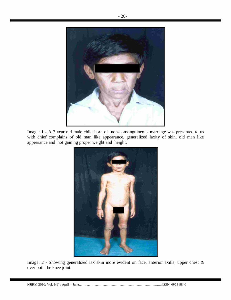
Image: 1 - A 7 year old male child born of non-consanguineous marriage was presented to us
with chief complains of old man like appearance, generalized laxity of skin, old man like
appearance and not gaining proper weight and height.
Image: 2 - Showing generalized lax skin more evident on face, anterior axilla, upper chest &
over both the knee joint.

- 28-
NJIRM 2010; Vol. 1(2) : April – June……………………………………………………………..…ISSN: 0975-9840
CLINICAL FEATURES:
Children with progeria usually have
a normal appearance in early infancy. At
approximately 9 to 24 months of age,
affected children begin to experience
profound growth delays, resulting in short
stature and low weight. They also develop a
distinctive facial appearance characterized
by a disproportionately small face in
comparison to the head; an underdeveloped
jaw (micrognathia); malformation and
crowding of the teeth; abnormally prominent
eyes; a small nose and a subtle blueness
around the mouth.
In addition, by the second year of
life, the scalp hair, eyebrows, and eyelashes
are lost (alopecia), and the scalp hair may be
replaced by small, downy, white or blond
hairs and gives a look of bird like faces2.
Additional characteristic features include
premature generalized atherosclerosis,
cardiovascular disease and stroke, hip
dislocations, unusually prominent veins of
the scalp, loss of the layer of fat beneath the
skin (subcutaneous adipose tissue), defects
of the nails, joint stiffness, skeletal defects,
and/or other abnormalities, alopecia,
horseman stance, pyriform thorax, thin legs
with prominent joints, short stature4,
Individuals with Hutchinson-Gilford
Progeria Syndrome are prone to develop
insulin resistant diabetes and premature
widespread thickening and loss of elasticity
of artery walls (arteriosclerosis), which
result in life-threatening complications
during childhood, adolescence or early
adulthood3.
DIAGNOSTIC FINDINGS
The diagnostics methods in HGPS mainly
comprise of clinical (abnormal lipid level,
increased excretion of hyalurinoic acid in
urine and abnormal blood count),
histological (abdominal skin biopsies with
abnormal nuclear morphologies),
radiological (abnormal brain, thorax, long
bones and phalanges x rays showing acro
osteolysis, hypoplastic facial bones &
sinuses, open cranial sutures & frontanelles,
wormian bones & coxa valga) and genetical
(screening for mutation in gene LMNA) 3.
TREATMENT:
There is no effective cure by any
treatment modalities for this HGPS.
Children with HGPS usually die at the age
of 14. Symptomatic treatments include
regular diets, routine immunizations, aspirin,
multivitamins, surgical procedure and
physical and psychological therapies3. A
new promising therapy in future will
includes treatment with Fernesyl transferase
inhibitors which have shown to reverse the
abnormalities in cells expressing progerin3.
BIBLIOGRAPHY:
1,Gruenbaum, Y. (2009). The curious case
of the ageing cells. Nature Reviews
Molecular Cell Biology, 10(4), 242.
doi:10.1038/nrm2666.
2, Surjushe, A., Thakre, M., Vasani, R., &
Saple, D. (2009). HUTCHINSON-
GILFORD SYNDROME
(PROGERIA). Indian Journal of
Dermatology, 54(S1), S27-S28. Retrieved
from Academic Search Complete database.
3,Coutinho, H., Falcão-Silva, V., Gonçalves,
G., & da Nóbrega, R. (2009). Molecular
ageing in progeroid syndromes: Hutchinson-
Gilford progeria syndrome as a
model. Immunity & Ageing, 61-7.
doi:10.1186/1742-4933-6-4.
4, Merideth MA et al : Phenotype and
course of Hutchinson-Gilford Progeria
Syndrome. N Eng J Med 2008,358:592-604.
5, Mazereeuw-Hautier J, Wilson LC,
Mohammed S, Smallwood D, Shackleton S,
Atherton DJ, Harper JI: Hutchinson-Gilford
Progeria syndrome: clinical findings in three
patients carrying the G608G mutation in
LMNA and review of the literature. Br J
Dermatol 2007, 156:1308-1314

- 29-
NJIRM 2010; Vol. 1(2) : April – June……………………………………………………………..…ISSN: 0975-9840
Brief Case History and Literature Review :
Superficial Angiomyxoma of the thigh
Dr. Sushma Ramraje*, Dr. Varsha Bhatia**, Dr. Aprajita Goel **
* Associate Professor, ** Resident, Dept of Pathology, Grant Medical College, Mumbai-400008
Key words: Superficial angiomyxoma, Myxoid stroma INTRODUCTION:
Superficial angiomyxoma is an
under-recognized lesion which encompasses
the majority of lesions referred to in earlier
literature as " cutaneous myxoma ". Most of
the cases present as cutaneous papules or
polypoid lesions and have a predilection for
the head and neck region and the trunk.
It is a rare benign neoplasm
characterized by a conglomerate of multiple,
moderately to sparsely cellular angiomyxoid
nodules with scattered small blood vessels.
We present here a case of superficial
angiomyxoma in a 22 year-old male and
discuss the morphological features, both
macroscopic and microscopic, which are
helpful for distinguishing this tumor from
other soft tissue tumors, especially
cutaneous focal mucinosis, cutaneous
myxoid cyst and aggressive angiomyxoma.
We also discuss the disease entity
`superficial angiomyxoma'.
CASE REPORT:
A 22 year old male came with
complaints of a painless, gradually
increasing swelling on the posterior
aspect of right thigh since 5 years. Physical
examination showed a soft subcutaneous
swelling measuring 3x2 cm. The mass was
completely removed. On gross examination,
it was well circumscribed, partly
encapsulated and soft in consistency. Cut
surface was gray-white with myxoid
change(Fig1).Histopathological examination
of hematoxylin-eosin-stained sections at
lower magnification revealed a well
encapsulated tumour with a myxoid
matrix(Fig2). At higher magnification, the
tumor consisted of small thin-walled blood
vessels and benign spindle-shaped or stellate
tumor cells embedded in a fibromyxoid
matrix(Fig3,4).Focal perivascular
lymphocytes were also noted.
Based on these clinical and
histologic findings, we diagnosed this
condition as superficial angiomyxoma.
DISCUSSION :
Superficial angiomyxoma was first
described as a cutaneous myxoma of
Carney‟s complex by Carney et al. in 19861
. Carney‟s complex is an autosomal
dominant syndrome characterized by
myxomas of the heart, skin and breast;
pigmentation of the mucous membrane; and
endocrine disorders such as Cushing‟s
syndrome and acromegaly.
In 1988, Allen et al. reported
cutaneous tumors that were histologically
similar to cutaneous myxomas of Carney‟s
complex without evidence of the complex2
.He reported 30 superficial angiomyxomas
in 28 patients aged 4 to 78 years. Most of
the tumors were soft , measuring 0.5 to 9
cm, on the head, neck, trunk, or lower
extremity. Microscopically, they were
located between the dermis and the
subcutaneous tissue and consisted of

- 30-
NJIRM 2010; Vol. 1(2) : April – June……………………………………………………………..…ISSN: 0975-9840

- 31-
NJIRM 2010; Vol. 1(2) : April – June……………………………………………………………..…ISSN: 0975-9840
mucoid material and small blood vessels.
The word `superficial' was used in
order to distinguish superficial
angiomyxoma which is a benign
myxomatous neoplasm characterized by
moderately to sparsely cellular angiomyxoid
nodules with scattered small vessels from
aggressive angiomyxoma, which is an
uncommon, locally aggressive but non-
metastasizing soft tissue neoplasm occurring
most commonly in the female genital region
. There is a need to differentiate superficial
angiomyxoma from other benign cutaneous
myxomatous lesions --cutaneous focal
mucinosis and cutaneous myxoid cysts 3.
Macroscopically, cutaneous focal
mucinosis and cutaneous myxoid cysts are
small, ill-defined nodular lesions less than 2
cm in size. Aggressive angiomyxomas are
usually infiltrative. In contrast, superficial
angiomyxomas are generally larger than
cutaneous focal mucinoses and cutaneous
myxoid cysts, and are not infiltrative but
well circumscribed . The present tumor is a
capsulated myxomatous nodule , a feature
never seen in aggressive angiomyxomas3.
Microscopically, all the above-
mentioned myxomatous tumors are
composed of spindle-shaped, stellate and
plump oval cells in a myxomatous stroma
and show different quantities and patterns of
vessel elements . Cutaneous mucinosis and
cutaneous myxoid cysts contain clefts and
mucin pools, but few vessels. In contrast,
superficial angiomyxomas show a scattered
distribution of small to medium sized thin-
walled blood vessels, however large caliber
vessels seen in aggressive angiomyxomas
are absent . Occasional findings seen in
superficial angiomyxoma are perivascular
hyalinization, perivascular lymphocytes,
fibrin thrombi, interstitial haemorrhage with
blood lakes, and hemosiderophages. One
unique feature in contrast to other cutaneous
myxoid tumours is the presence of
neutrophils, eosinophils, lymphocytes and
mast cells in the stroma.
Also secondary changes including
perivascular hyalinization, medial
hypertrophy ,a prominence of mast cells and
focal areas of hypercellularity with
associated nuclear hyperchromasia and
pleomorphism are commonly seen in
aggressive angiomyxomas.
In 1999, Calonje et al. reported
clinicopathologic and
immunohistochemical features of superficial
angiomyxoma as an independent disease
entity in 39 patients . The tumor was more
common in men . The age of onset and size
ranged from 0 to 82 years and 1 to 5 cm
respectively. Immunohistochemically, tumor
cells were positive for vimentin but negative
for CD34, smooth muscle actin, HHF-35, S-
100 protein, desmin, cytokeratin, and glial
fibrillary acidic protein4, 5.
However, Fetsch et al. described
superficial angiomyxomas to be sometimes
positive for desmin and smooth muscle actin
Our patient did not have Carney‟s complex
because the subcutaneous tumor was not
accompanied by non-cutaneous myxomas,
pigmentation of the skin or mucous
membrane, or endocrine disorders.
Based on the clinical and
histopathological findings of the tumor , we
have diagnosed this condition as superficial
angiomyxoma.Thus these tumors can be
defined as bland, superficial myxoid lesions
with prominent thin walled vessels and are
alternately called as cutaneous myxomas
DIAGNOSTIC FINDINGS:
Predominantly involves dermis and
subcutis
o May involve skeletal muscle on face
o Multilobulated, poorly circumscribed
Myxoid stroma

- 32-
NJIRM 2010; Vol. 1(2) : April – June……………………………………………………………..…ISSN: 0975-9840
o Alcian blue positive, hyaluronidase
sensitive
o May have acellular mucin pools
o Scattered bland stellate and spindled
cells
Occasional multinucleated cells
Pleomorphism, mitotic figures rare
Occasional intranuclear
pseudoinclusions
Many thin-walled small blood vessels
Frequent neutrophils
Entrapped epithelial component in 20-
30% of cases
o Keratinous cyst
o Thin strands of squamous epithelium
o Basaloid buds
If multiple or involving external ear,
suggest Carney complex
Superficial angiomyxoma is a rare benign
tumour of adolescents and adults, with a
predilection for the maxilla and mandible .It
can arise in the genital region, trunk, lower
and upper extremities. In the head and neck
region the tumour can be seen on the head,
lower eyelid, preauricular region, pinna and
the external auditory canal. Multiple
myxomas occurring in the external ear or
breast are almost pathognomonic of
Carney‟s syndrome. It is a benign lesion but
local recurrence is common after incomplete
excision.
It is positive only for vimentin.Since
immunohistochemical features have not
been established, some researchers mention
that immunohistochemical studies are not
helpful for the differential diagnosis of
myxoid tumors . More reports and further
analyses will be needed to characterize
superficial angiomyxoma clinically and
immunohistochemically.
There is a propensity for local
recurrence if cutaneous myxomas are
incompletely excised. However, no
metastasis has been reported.6
BIBLIOGRAPHY:
1. Carney JA, Headington JT, Su WPD.
Cutaneous myxomas: A major
component of the complex of myxomas,
spotty pigmentation, and endocrine
overactivity. Arch Dermatol 1986;122:
790-798.
2. Allen PW, Dymock RB, MacCormac
LB: Superficial angiomyxomas with and
without epithelial components. Report of
thirty tumors in twenty eight patients.
Am J Surg Pathol 1988;12: 519-530.
3. Nakayama H, Hiroi M, Kiyoku H,
Naruse K, Enzan H. Superficial
Angiomyxoma of the Right Inguinal
Region: Report of a Case. Japanese
Journal of Clinical Oncology
1997;27(3):200-203.
4. Calonje E, Guerin D, MacCormac D,
Fletcher CDM: Superficial
Angiomyxoma. Am J Surg Pathol 1999;
23: 910-917.
5. K Yamamoto, A Kondo, K Iwashita, Y
Umezawa, Y Ohta, T Matsuyama et al.
A case of superficial angiomyxoma.
Tokai J Exp Clin Med 2006; 31( 2): 53-
55.
6. Khadilkar UN, Khadilkar NP, Rao PS,
Chakravorty S, Goel G. Superficial
angiomyxoma of the external ear not
associated with Carney‟s complex: A
case report. Kathmandu University
Medical Journal 2007;5( 4): 546-549.

- 33-
NJIRM 2010; Vol. 1(2) : April – June……………………………………………………………..…ISSN: 0975-9840
Brief Case History and Literature Review :
Blount Disease in 3 year old boy from India.
Dr. Mehul M. Gosai*, Dhaval Solanki **Dr. Hareshwaree B. Hariyani***, Dr. Payal H. Purohit”, Dr. Mihir A. Sadadia^, Gaurav Goswami****
*Associates Professor, Department of Pediatrics, ** Senior Resident,Department of Pediatrics, ***Assistant Professor, Department of General Dentistry, “Intern Doctor, Department of Pediatrics, ****Assistant Professor Department of Skin & V.D. of Government Medical College, Sir T. Hospital, Bhavnagar, Gujarat, India.^ *** Midwestern State University, Wichita Falls, Texas, United States of America, ( U.S.A.), **** Resident Doctor, Department of Radiodiagnosis and imaging, Base Hospital, Delhi Cantt , New Delhi, India.
Key words: Blount disease, Idiopathic tibia vara,
INTRODUCTION:
Idiopathic tibia vara (Blount disease)
is an acquired form of tibial deformity found
mainly in children who are Afro-Caribbean
and it is also associated with obesity, short
stature and early walking. There does not
appear to be an obvious genetic factor. It is
uncommon1. The incidence of Blount
disease in the India is unknown. The
estimated prevalence of infantile Blount
disease in the population of young children
with significant bow legs in the United
States is 0.007 or less than 1%. The exact
frequency in persons of all ethnicities is
unknown and most likely is less than 1%2, 3,
4,5. Blount disease or idiopathic tibia vara is
a developmental deformity of the proximal
tibial medial physis that produces a varus
deformity of the knee6. It has been classified
into three types depending on the age at
onset : infantile (1-3 yr), juvenile (4-10 yr)
and adolescent (11 yr or older).
The juvenile and adolescent forms
are commonly combined as late onset tibia
vera7. Although the etiology of Blount
disease may be multifactorial, the strong
association with childhood obesity suggests
a mechanical basis due to an abnormal
compression on the medial aspect of the
proximal tibial physis. A comprehensive
analysis of multiplanar deformities in the
lower extremity reveals tibial varus,
procurvatum and internal torsion along with
limb shortening. Additionally, distal femoral
varus is commonly noted in the late-onset
form. The diagnosis of both forms of Blount
disease is based on history, physical
examination, and the most important tool is
a radiographs of the affected knee. When a
patient has early-onset disease, a
realignment tibial osteotomy before the age
of four years decreases the risk of recurrent
deformity8 . so early diagnosis is very much
important for a better long term prognosis.
Gradual correction with distraction
osteogenesis is an effective means of
achieving an accurate multiplanar
correction, especially in patients with late-
onset disease 9. In child less than 3 yr old
and langenskiold stage < 3, a bracing is
effective and can prevent progression of the
disease in 50 % of these children. A
maximum trial of 1 year of orthotic
management is recommended. If complete
correction is not obtained after 1 yr or if
progression occurs during this time, then a
corrective osteotomy may be indicated7.
Many different types of osteotomies have
been described in the literature including
opening and closing wedge, spike, dome,
and oblique osteotomies.10,11,12
Complications associated with the treatment

- 34-
NJIRM 2010; Vol. 1(2) : April – June……………………………………………………………..…ISSN: 0975-9840
of Blount disease include loss of alignment,
vascular impairment, pathologic fractures,
wound infection and malalignment.13,14
In
general, the later the onset, there is a greater
chances of progression of the deformity, the
increased likelihood of articular changes.
There were few case reports of infantile
Blount disease other than Afro-Caribbean
ancestry. So we want to share this case with
Pediatricians of India .
A 3 year old child, a case of Blount Disease presented with typical left lower limb deformity and
Radiological Images of our case : X – Rays :
Left upper tibial physis is showing medial slopping of the epiphysis, widening of the physis and
fragmentation of the metaphysis with significant lateral thrust

- 35-
NJIRM 2010; Vol. 1(2) : April – June……………………………………………………………..…ISSN: 0975-9840
3D- Reconstructive Ct-Scan of this pt. of Blount Disease s/o classical diagnostic findings
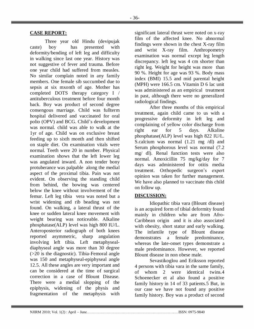
- 36-
NJIRM 2010; Vol. 1(2) : April – June……………………………………………………………..…ISSN: 0975-9840
CASE REPORT:
Three year old Hindu (devipujak
caste) boy has presented with
deformity/bending of left leg and difficulty
in walking since last one year. History was
not suggestive of fever and trauma. Before
one year child had suffered from measles.
No similar complain noted in any family
members. One female sib succumbed due to
sepsis at six mxonth of age. Mother has
completed DOTS therapy category I /
antituberculous treatment before four month
back. Boy was product of second degree
consengous marriage. Child was fullterm
hospital delivered and vaccinated for oral
polio (OPV) and BCG. Child‟s development
was normal. child was able to walk at the
1yr of age. Child was on exclusive breast
feeding up to sixth month and then shifted
on staple diet. On examination vitals were
normal. Teeth were 20 in number. Physical
examination shows that the left lower leg
was angulated inward. A non tender bony
protuberance was palpable along the medial
aspect of the proximal tibia. Pain was not
evident. On observing the standing child
from behind, the bowing was centered
below the knee without involvement of the
femur. Left leg tibia vera was noted but a
wrist widening and rib beading was not
found. On walking, a lateral thrust of the
knee or sudden lateral knee movement with
weight bearing was noticeable. Alkaline
phosphatase(ALP) level was high 800 IU/L.
Anteroposterior radiograph of both knees
reported asymmetric, sharp angulation
involving left tibia. Left metaphyseal-
diaphyseal angle was more than 30 degree
(>20 is the diagnostic). Tibia-Femoral angle
was 150 and metaphyseal-epiphyseal angle
12.5. All these angles are very important and
can be considered at the time of surgical
correction in a case of Blount Disease.
There were a medial slopping of the
epiphysis, widening of the physis and
fragmentation of the metaphysis with
significant lateral thrust were noted on x-ray
film of the affected knee. No abnormal
findings were shown in the chest X-ray film
and wrist X-ray film. Anthropometry
examination was normal except leg length
discrepancy. left leg was 4 cm shorter than
right leg. Weight for height was more than
90 %. Height for age was 93 %. Body mass
index (BMI) 15.5 and mid parental height
(MPH) were 166.5 cm. Vitamin D 6 lac unit
was administered as an empirical treatment
in past, although there were no generalized
radiological findings.
After three months of this empirical
treatment, again child came to us with a
progressive deformity in left leg and
complaining of yellow color discharge from
right ear for 5 days. Alkaline
phosphatase(ALP) level was high 822 IU/L.
S.calcium was normal (1.21 mg /dl) and
Serum phosphorous level was normal (7.2
mg/ dl). Renal function tests were also
normal. Amoxicillin 75 mg/kg/day for 7
days was administered for otitis media
treatment. Orthopedic surgeon‟s expert
opinion was taken for further management.
We have also planned to vaccinate this child
on follow up.
DISCUSSION:
Idiopathic tibia vara (Blount disease)
is an acquired form of tibial deformity found
mainly in children who are from Afro-
Caribbean origin and it is also associated
with obesity, short statur and early walking.
The infantile type of Blount disease
demonstrates a female predominance,
whereas the late-onset types demonstrate a
male predominance. However, we reported
Blount disease in non obese male.
Sevastikoglou and Eriksson reported
4 persons with tibia vara in the same family,
of whom 2 were identical twins.4
Schoenecker et al also found a positive
family history in 14 of 33 patients.5 But, in
our case we have not found any positive
family history. Boy was a product of second

- 37-
NJIRM 2010; Vol. 1(2) : April – June……………………………………………………………..…ISSN: 0975-9840
degree consengous marriage. However, no
direct proof of a genetic relationship has
been discovered.
The clinical presentation of the
different types of tibia vara varies according
to the age of onset. In infantile tibia vara,
children generally start to walk early
usually when aged 9-10 months.15 In our
case child has started to walk at around 1 yr
of age. At the onset of the disease,
differentiating between early infantile
Blount disease and marked physiologic
bowlegs is difficult. This physiological
deformity usually resolves spontaneously by
the time the child is aged 2 years. Our case
was presented at 3 yr of age. In contrast to
physiologic genu varum, infantile Blount
disease can progress to severe deformity.
Blount disease is associated with a
prominent metaphyseal beak, internal tibial
torsion, and leg-length discrepancy as noted
in our case. We have founded unilateral
involvement. However, it is bilateral in
approximately 80% of cases.16
The differential diagnosis of Blount
disease includes physiologic bowing,
congenital bowing, rickets, Ollier disease,
trauma, osteomyelitis, and metaphyseal
chondrodysplasia. The diagnosis of Blount
disease is based on history, physical
examination, and, most important the
radiographs of the knee as reported in our
case. Mild or healing rickets with residual
bowing may be difficult to differentiate from
stage 2 infantile tibia vara. However, rickets
affects the skeleton in a generalized and
symmetric fashion with loss of the zone of
provisional calcification in the physis. In
addition, the typical biochemical
abnormalities of rickets helps to
differentiate these both conditions.
CONCLUSION:
Always consider differential diagnosis of
Blount disease in more than 2 yr old child
present with tibia vara although Blount
disease is rare in India. When a patient has
early-onset disease, a realignment tibial
osteotomy before the age of four years
decreases the risk of recurrent deformity17
.so early diagnosis is important for a better
long term prognosis.
BIBLIOGRAPHY:
1.Gilbody J, Thomas G, Ho K., Acute versus
gradual correction of idiopathic tibia vara in
children: a systematic review. J Pediatr
Orthop. 2009 Mar; 29(2):110-4.
2. Davids JR, Blackhurst DW, Allen BL
Jr. Radiographic evaluation of bowed legs in
children. J Pediatr Orthop. Mar-
Apr 2001;21(2):257-63.
3. Henderson RC, Kemp GJ Jr, Greene
WB. Adolescent tibia vara: alternatives for
operative treatment. J Bone Joint Surg
Am. Mar 1992;74(3):342-50.
4. Schoenecker PL, Meade WC, Pierron RL,
et al. Blount''s disease: a retrospective
review and recommendations for
treatment. J Pediatr Orthop. Mar-
Apr 1985;5(2):181-6.
5. Sevastikoglou JA, Eriksson I. Familial
infantile osteochondrosis deformans tibiae.
Idiopathic tibia vara. A case report. Acta
Orthop Scand. 1967;38(1):81-7. .
6. Schoppee K. Blount disease (idiopathic
tibia vara). Orthop Nurs. 1995 Sep-Oct;
14(5):31-4.
7. Harish S. Torsional and Angular
Deformities. In: Kliegman R., ed. Nelson
textbook of pediatrics, 18th edn. New Delhi,
India: Elsevier, 2008:2789.
8. Coogan PG, Fox JA, Fitch RD. Treatment
of adolescent Blount disease with the
circular external fixation device and
distraction osteogenesis. J Pediatr
Orthop. Jul-Aug 1996;16(4):450-4.
9.Sabharwal S., Blount disease. J Bone Joint
Surg Am. 2009 Jul;91(7):1758-76.
10. Dietz FR, Weinstein SL. Spike
osteotomy for angular deformities of the
long bones in children. J Bone Joint Surg
Am. Jul 1988;70(6):848-52.

- 38-
NJIRM 2010; Vol. 1(2) : April – June……………………………………………………………..…ISSN: 0975-9840
11. Price CT, Scott DS, Greenberg
DA. Dynamic axial external fixation in the
surgical treatment of Blount disease. J
Pediatr Orthop. 1985;15:236-243.
12. Stanitski DF, Dahl M, Louie K,
Grayhack J. Management of late-onset tibia
vara in the obese patient by using circular
external fixation. J Pediatr Orthop. Sep-
Oct 1997;17(5):691-4.
13. Feldman DS, Madan SS, Ruchelsman
DE, Sala DA, Lehman WB. Accuracy of
correction of tibia vara: acute versus gradual
correction. J Pediatr Orthop. Nov-
Dec 2006;26(6):794-8.
14. Sabharwal S, Zhao C, McClemens
E. Correlation of body mass index and
radiographic deformities in children with
Blount disease. J Bone Joint Surg
Am. Jun 2007;89(6):1275-83.
15. Golding JS, McNeil-Smith
JD. Observations on the etiology of tibia
vara. J Bone Joint Surg. 1963;45B:320.
16. Tachdjian MO, ed. The foot and leg:
tibia vara. In: Pediatric Orthopedics. Vol 4.
Philadelphia:. WB Saunders Co;1990:2835-
50.
17. Doyle BS, Volk AG, Smith CF. Infantile
Blount disease: long-term follow-up of
surgically treated patients at skeletal
maturity. J Pediatr Orthop. Jul-
Aug 1996;16(4):469-76.

- 39-
NJIRM 2010; Vol. 1(2) : April – June……………………………………………………………..…ISSN: 0975-9840
ISSN: 0975 -9840
Indexed in Index Copernicus Included in Open-J-Gate
Publication of Academy for Continuing Medical Education
Index
1 Karyotypic Study Of Chronic And Blastic Phase Chronic Myeloid Leukemia
Patients of Gujarat State.
Dr. V. B. Kariya, Dr. D. J. Trivedi, Dr. P. S. Srimankar, Dr. C. A. Pensi, Dr. D. V. Thakker
1 -5
2 Cardiovascular Responses With Valsalva Maneuver During Activities Of Daily
Livings In Healthy Adults
Prof. Priyanshu V. Rathod, Prof. Savita Ravindra, , Prof. Veena Kiran Nambiar
6-11
3 Academic Performance Of School Children With Their Intelligence Quotient
S. D. Kulkarni, N. R. Pathak, C. S. Sharma
12-15
4 A Study Of Nutrient Foramina Of The Metacarpal Bones
Dr. P. S. Shrimankar, Dr. D. J. Trivedi, Dr. V.B. Kariya
16-20
5 Study of Bacteriological Pattern Of Suspected Cases Of Meningitis
Dr Rajesh Soni
21-25
6 Hutchinson Gilford Progeria Syndrome
Dr. Mehul M. Gosai, Dr. Hareshwaree B. Hariyani, Dr. Mihir A. Sadadia, Dr. Payal
H. Purohit”, Dr. Bhavesh N. Asti
26-29
7 Superficial Angiomyxoma of the thigh
Dr. Sushma Ramraje, Dr. Varsha Bhatia, Dr. Aprajita Goel
19-33
8 Blount Disease in 3 year old boy from India.
Dr. Mehul M. Gosai, Dhaval Solanki Dr. Hareshwaree B. Hariyani, Dr. Payal H.
Purohit”, Dr. Mihir A. Sadadia^, Gaurav Goswami
33-38