Isolation of Breast Cancer Accumulation in … [email protected] Rode Yogesh Dept of...
5
International Journal of Electrical Energy, Vol. 2, No. 2, June 2014 ©2014 Engineering and Technology Publishing 94 doi: 10.12720/ijoee.2.2.94-98 Isolation of Breast Cancer Accumulation in Mammograms for Improving Radiologists Analysis Prapti V. Patil MGM Dr. G. Y. Pathrikar College of Computer Science and IT, Aurangabad, Maharashtra E-mail: [email protected] Rode Yogesh Dept of Computer Science and IT, Dr. Babasaheb Ambedkar Marathawada University, Aurangabad, Maharashtra K. V. Kale Dr. Babasaheb Ambedkar Marathawada University, Aurangabad, Maharashtra. E-mail: [email protected] Abstract—Breast cancer is one of the major causes of death among women around the world. An improvement in early diagnostic technique can help for better diagnosis of abnormalities. Mammography is the main investigation screening technique used for an early diagnosis of it. It display a small percentage of the information detected which is due to the minor difference in x-ray Attenuation between normal glandular tissues and malignant disease. This makes the detection of small malignancies difficult. This paper proposes group of pre and post processing technique with segmentation approach showing better results for medical image processing applications. Index Terms—mammography, segmentation, morphological operators, lesion I. INTRODUCTION Breast cancer stays in the first place among women malignant structures list (about 30%). It is a one of the leading causes of cancer death for women all over the world According to Worldwide Health Corporation it is the fundamental reasons of the women’s average age mortality. The National Cancer Institute estimates that one out of eight women develops breast cancer at some point during her lifetime [1]. The goal of mammography is to provide early detection of breast cancer. It is considered to be the most efficient technique for identifying lesions when they are not palpable and when there are structural breast modifications [2]. It shows to the physician differences in breast tissue densities and these differences are fundamental to a correct diagnosis. At present, there are no effective ways to prevent breast cancer, because its cause remains unknown [3]. Therefore urgency and importance of mammography image processing is obvious. Manuscript received July 3, 2013; revised October 22, 2013. Computer-Aided Detection (CADe) and Diagnosis (CADi) systems are continuously being developed aiming to help the physicians in early detection of breast cancer. These tools may call the physician’s attention to areas in the mammography that may contain radiological findings. Image segmentation plays a crucial role in many medical imaging applications by automating or facilitating the delineation of anatomical structures and other regions of interest. In digital mammography, segmentation is the process of partitioning mammograms into regions, aiming to produce an image that is more meaningful and easier to analyze [4]. After being segmented, the mammogram or the mass lesion region can be further used by physicians, helping them to take decisions that involve their patient’s health. II. REVIEW OF MEDICAL IMAGING TECHNIQUE Magnetic resonance imaging (MRI), computed tomography (CT), digital mammography, and other imaging modalities provide an effective means for noninvasively mapping the anatomy of a subject. These technologies have greatly increased knowledge of normal and diseased anatomy for medical research and are a critical component in diagnosis and treatment planning. With the increasing size and number of medical images, the use of computers in facilitating their processing and analysis has become necessary. In particular, computer algorithms for the delineation of anatomical structures and other regions of interest are a key component in assisting and automating specific radiological tasks. These algorithms, called image segmentation algorithms, play a vital role in numerous biomedical [5] imaging applications such as the quantification of tissue volumes diagnosis localization of pathology [6], study of anatomical structure [7], treatment planning [8], improvement in the diagnosis accuracy [9], and computer integrated surgery.
Transcript of Isolation of Breast Cancer Accumulation in … [email protected] Rode Yogesh Dept of...
International Journal of Electrical Energy, Vol. 2, No. 2, June 2014
©2014 Engineering and Technology Publishing 94doi: 10.12720/ijoee.2.2.94-98
Isolation of Breast Cancer Accumulation in
Mammograms for Improving Radiologists
Analysis
Prapti V. Patil MGM Dr. G. Y. Pathrikar College of Computer Science and IT, Aurangabad, Maharashtra
E-mail: [email protected]
Rode Yogesh Dept of Computer Science and IT, Dr. Babasaheb Ambedkar Marathawada University, Aurangabad, Maharashtra
K. V. Kale Dr. Babasaheb Ambedkar Marathawada University, Aurangabad, Maharashtra.
E-mail: [email protected]
Abstract—Breast cancer is one of the major causes of death
among women around the world. An improvement in early
diagnostic technique can help for better diagnosis of
abnormalities. Mammography is the main investigation
screening technique used for an early diagnosis of it. It
display a small percentage of the information detected
which is due to the minor difference in x-ray Attenuation
between normal glandular tissues and malignant disease.
This makes the detection of small malignancies difficult.
This paper proposes group of pre and post processing
technique with segmentation approach showing better
results for medical image processing applications.
Index Terms—mammography, segmentation, morphological
operators, lesion
I. INTRODUCTION
Breast cancer stays in the first place among women
malignant structures list (about 30%). It is a one of the
leading causes of cancer death for women all over the
world According to Worldwide Health Corporation it is
the fundamental reasons of the women’s average age
mortality. The National Cancer Institute estimates that
one out of eight women develops breast cancer at some
point during her lifetime [1].
The goal of mammography is to provide early
detection of breast cancer. It is considered to be the most
efficient technique for identifying lesions when they are
not palpable and when there are structural breast
modifications [2]. It shows to the physician differences in
breast tissue densities and these differences are
fundamental to a correct diagnosis. At present, there are
no effective ways to prevent breast cancer, because its
cause remains unknown [3].
Therefore urgency and importance of mammography
image processing is obvious.
Manuscript received July 3, 2013; revised October 22, 2013.
Computer-Aided Detection (CADe) and Diagnosis
(CADi) systems are continuously being developed aiming
to help the physicians in early detection of breast cancer.
These tools may call the physician’s attention to areas in the mammography that may contain radiological findings.
Image segmentation plays a crucial role in many
medical imaging applications by automating or
facilitating the delineation of anatomical structures and
other regions of interest. In digital mammography,
segmentation is the process of partitioning mammograms
into regions, aiming to produce an image that is more
meaningful and easier to analyze [4]. After being
segmented, the mammogram or the mass lesion region
can be further used by physicians, helping them to take
decisions that involve their patient’s health.
II. REVIEW OF MEDICAL IMAGING TECHNIQUE
Magnetic resonance imaging (MRI), computed
tomography (CT), digital mammography, and other
imaging modalities provide an effective means for
noninvasively mapping the anatomy of a subject. These
technologies have greatly increased knowledge of normal
and diseased anatomy for medical research and are a
critical component in diagnosis and treatment planning.
With the increasing size and number of medical images,
the use of computers in facilitating their processing and
analysis has become necessary. In particular, computer
algorithms for the delineation of anatomical structures
and other regions of interest are a key component in
assisting and automating specific radiological tasks.
These algorithms, called image segmentation algorithms,
play a vital role in numerous biomedical [5] imaging applications such as the quantification of tissue volumes
diagnosis localization of pathology [6], study of
anatomical structure [7], treatment planning [8],
improvement in the diagnosis accuracy [9], and computer
integrated surgery.
International Journal of Electrical Energy, Vol. 2, No. 2, June 2014
©2014 Engineering and Technology Publishing 95
III. MAMMOGRAPHY
Mammography is a low dose x-ray procedure for the
visualization of internal structure of breast.
Mammography has been proven to be the most reliable
method and it is the key screening tool for the early
detection of breast cancer. Mammography is highly
accurate, but like most medical tests, it is not perfect. On
average, mammography will detect about 80–90% of the
breast cancers in women without symptoms. It works
fairly well in the postmenopausal women and is
inexpensive. In a screening mammogram, each breast is
X-rayed in two different positions: from top to bottom
and from side to side. When a mammogram image is
viewed, breast tissue appears white and opaque and fatty
tissue appears darker and translucent. The goal of
diagnosis is to distinguish between malignant and benign
breast lumps. The three methods currently used for breast
cancer diagnosis are mammography, fine needle aspirate
and surgical biopsy. Mammography has a reported
sensitivity (probability of correctly identifying a
malignant lump) which varies between 68% and 79%
[Mangasarian]. Taking a fine needle aspirate (i.e.
extracting fluid from a breast lump using a small-gauge
needle) and visually inspecting the fluid under a
microscope has a reported sensitivity varying from 65%
to 98% [Mangasarian]. The more evasive and costly
surgical biopsy has close to 100% sensitivity and remains
the only test that can confirm malignancy. The goal of
machine learning techniques is to have the sensitivity of
surgical biopsy without its evasiveness and cost.
Mammography plays a central role in the process of
detecting abnormalities in breast cancer screening. A
mammogram is a x-ray projection of the 3D structures of
the breast obtained by compressing the breast between
two plates. Mammograms have an intrinsic scattered
appearance due to the superimposition of densities from
differing breast tissues, and the differential x-ray
absorption characteristics associated with these various
tissues. A high contrast is always required to differentiate
very fine structures with slight differences in density,
such as micro calcifications.
In the United States, breast cancer is the second
leading cause of death in women. One out of eight
women develops breast cancer in their lifetime. Studies
have indicated that early detection and treatment improve the chances of survival for breast cancer patients. At
present, mammography is the only proven method that
can detect minimal breast cancers. However, 10-30% of
the breast cancers that are visible on mammograms in
retrospective studies are not detected due to various
technical or human factors. Computer-aided diagnosis
(CAD), alerts radiologists to locate suspicious locations
on the images during mammographic reading. It also
develops a computerized image analysis system to assist
radiologists in mammographic interpretation. At present,
it focuses on the detection and classification of two of the
most important mammographic indicators of breast
cancers: masses and clustered micro calcifications. The
mammograms for a patient are digitized by a high-
resolution film scanner. The digitized mammograms are
then processed by automated detection programs to
identify the regions containing suspicious micro
calcifications or masses. In each region of interest (ROI),
the identified lesion is analyzed by the appropriate
classifier to estimate its likelihood of malignancy.
The digitized mammograms will be displayed on the
CAD workstation. The locations of the detected lesions
and their likelihood of malignancy is superimposed on
the displayed mammograms. A digitized mammogram is
processed with a different-imaging technique that
enhances the micro calcifications and suppresses the
structured background and random noise. A locally
adaptive gray level thresholding technique is used to
segment the potential signals.
IV. NEED OF MEDICAL IMAGING
The internal structure and functions of the human body
are not generally visible to the human eyes.
However by various imaging technology, images can
be created through which the medical professional can
look into the body to diagnose abnormal condition and
guide therapeutic procedures. Different medical imaging
method reveals different characteristics of human body.
With each method image quality and structure visibility
can be considerable, depending on characteristics of the
imaging equipment, skill of operator, and compromises
with factors such as patient radiation exposure and
imaging time.
A. Medical Image Processing System
Medical image processing system comprises of patient,
the imaging system, the system operator, the image itself
and the human observer. Generally, the visibility of
specific anatomical feature depends on the characteristics
of the imaging system and the way in which it is operated.
Medical imaging systems have a considerable number of
variables that must be intellectually selected by the
operator. For example intensifying screens in
radiography, transducers in sonography or coils in
magnetic resonance imaging (MRI). Some variables are
adjustable physical quantities associated with imaging
process such as kilo voltage in radiography, gain in
sonography, and echo time in MRI.
The quality of the medical image depends on number
of factors like contrast, blur, noise, artifacts, and
distortion etc.
B. Image Contrast
An imaging system translates a specific tissue
characteristic in to image shades of gray or color which
can be referenced as contrast. Contrast is the fundamental
feature of an image. An object within the body will be
visible in an image only if it has sufficient physical
contrast relative to surrounding tissue, that is, it must
represent a difference in one or more tissue
characteristics. For example, in radiography an object can
be imaged if there is adequate difference in either density
or thickness of tissue.
International Journal of Electrical Energy, Vol. 2, No. 2, June 2014
©2014 Engineering and Technology Publishing 96
The fundamental characteristics of an imaging system
that establishes relationship between image contrast and
object contrast is its contrast sensitivity. When the
imaging system has relatively low contrast sensitivity,
only object with a high object contrast is visible in the
image. If the imaging system has high contrast sensitivity,
the lower contrast object will also be visible [10].
C. Blurring
Structures and objects in the body vary not only in
physical contrast but also in size. Objects range from
large organs and bones to small structural features such as
trabecula patterns and small calcifications. It is the small
anatomical features that add detail to a medical image
[10]. Each imaging method has a limit as to the smallest
object that can be imaged and thus on visibility of detail.
Visibility of detail is limited because all imaging methods
introduce blurring into the process. The primary effect of
image blur is to reduce the contrast and visibility of small
objects or detail.
The original image and its blurred version, obtained
through a low pass filter, are combined so that the high
frequency components of the image are amplified and as
a result the perception of the image around edge areas
improves. All high frequency components of the image
are amplified and the noise in the image will also be
inadvertently amplified.
D. Artifacts and Distortion
Most of the imaging methods can create image features
that do not represent a body structure or object.
These are image artifacts. In many situations an artifact
does not significantly affect object visibility and
diagnostic accuracy. But artifacts can obscure a part of an
image or may be interpreted as an anatomical feature. A
medical image should give an accurate impression of
body objects in term of their size, shape, and relative
positions; however, it may introduce distortion of these
factors.
E. Image Noise
It is generally desirable that image brightness is to be
uniform except where it changes to form an image. There
is a variation in the brightness of a displayed image even
when no image detail is present. This variation is usually
random and has no particular pattern reducing image
quality specifically when the images are small and have
relatively low contrast. This random variation in image
brightness is nothing but a noise. All medical images
contain some visual noise. The presence of noise gives an
image a grainy, textured, or snowy appearance. No
imaging method is free of noise, but noise is much more
prevalent in certain types of imaging procedures than in
others. Shot noise is black and/or white spots removal
Dropout pixels common in some types of microscopy
such as interference microscopes, or from dust on a
scanned negative, and also produced by dead or locked
transistors in digital cameras) are typically filled in with a
median filter. It can also be used to remove scratches,
cracks, etc. narrow than the radius of the neighborhood
F. Consequences of an Inaccurate Reading
Consequences of an inaccurate reading of
mammography can lead to
False Positive interpretation: leading to
unnecessary follow up, tests like biopsies
performed on women with benign condition (good
tissue).
False negative interpretation: causing delay in
diagnosis and treatment converting early stage
breast cancer to a advanced stage with severe
deduction for survival rate and resource utilization.
V. PREPROCESSING
Image pre-processing techniques are necessary, in
order to find the orientation of the mammogram, to
remove the noise and to enhance the quality of the image.
Before any image-processing algorithm can be applied on
mammogram, preprocessing steps are very important in
order to limit the search for abnormalities without undue
influence from background of the mammogram.
Digital mammograms are medical images that are
difficult to be interpreted, thus a preparation phase is
needed in order to improve the image quality and make the segmentation results more accurate. The main
objective of this process is to improve the quality of the
image to make it ready to further processing by removing
the unrelated and surplus parts in the back ground of the
mammogram Preprocessing may also involve in creating
mask for pixels with highest intensity, to reduce
resolutions and to segment the breast
VI. DENOISEING USING FILTER
One of the most important problems in image
processing is denoising. Usually the procedure used for
denoising, is dependent on the features of the image, aim
of processing and also post-processing algorithms.
Denoising by low-pass filtering not only reduces the
noise but also blurs the edges. Spatial and frequency
domain filters are widely used as tools for image
enhancement. Low pass filters smooth the image by
blocking detail information.
A. Morphological Operation
Automatic segmentation of mass is considered difficult
because of the ill-defined boundaries and overlapping
with fibro-glandular tissue of many masses [11].
Morphology is an operation of image processing based on
shapes. The value of each pixel in the output image is
based on a comparison of the corresponding pixel in the
input image with its neighbors [12]. The morphological
operations are applied on the grayscale mammography
images to segment the abnormal regions. Erosion and
dilation are the two elementary operations in
Mathematical Morphology [13]. An aggregation of these
two represents the rest of the operations. The symbols _,
Θ, o, and • respectively denote the four fundamental
binary morphological operations: dilation, erosion,
opening, and closing which act on the structuring
element . detection aims to extract the edge of the tumor
International Journal of Electrical Energy, Vol. 2, No. 2, June 2014
©2014 Engineering and Technology Publishing 97
from surrounding normal tissues and background, high
pass filters (sharpening filters) could be used to enhance
the details of images
VII. SEGMENTATION
In Segmentation the inputs are images and, outputs are
the attributes extracted from those images. Segmentation
divides image into its constituent regions or objects. The
level to which segmentation is carried out depends upon
the problem being solved. For the segmentation of
intensity images like digital mammograms, there are four
main approaches, namely, threshold techniques,
boundary-based methods, region- based methods, and
hybrid techniques which combine boundary and region
criteria.
A. Threshold Techniques
These techniques are based on the postulate that all
pixels whose value (gray level, color value, or other) lies
within a certain range belong to one class. Such methods
neglect all of the spatial information of the image and do
not cope well with noise or blurring at boundaries .For
mammograms, thresholding usually involves selecting a single gray level value from an analysis of the grey-level
histogram, to segment the histogram into background and
breast tissues. All the pixels with grey level value less
than the threshold are marked as background and the rest
as breast [14]. Thresholding uses only grey level value
and no spatial information is considered .Therefore, the
major shortcoming of the threshold is that there is often
an overlap between grey levels of the objects in the breast
and the background.
B. Boundary Based Methods
The above methods use the postulate that the pixel
values change rapidly at the boundary between two
regions. The complement of the boundary-based
approach is to work with the regions [14].
C. Hybrid Technique
These techniques combine boundary and region
criteria. This class includes morphological watershed
segmentation and variable-order surface fitting. The
watershed method is generally applied to the gradient of
the image.
D. Watershed Transform
It can be classified as region based segmentation
approach is used. Watersheds are one of the classics in
the field of topography and have long been admitted as a
useful tool in image segmentation.
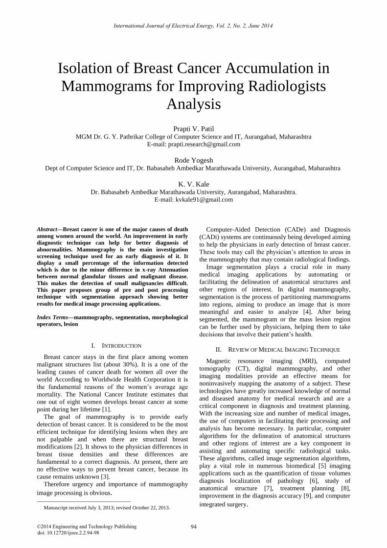
VIII. ALGORITHM
Algorithm consist of group of pre and post processing
operators technique with segmentation approach to detect
breast cancer mass in Mammograms for improving
radiologists examination which is shown in Fig. 1.
Figure 1. Working flow of proposed algorithm.
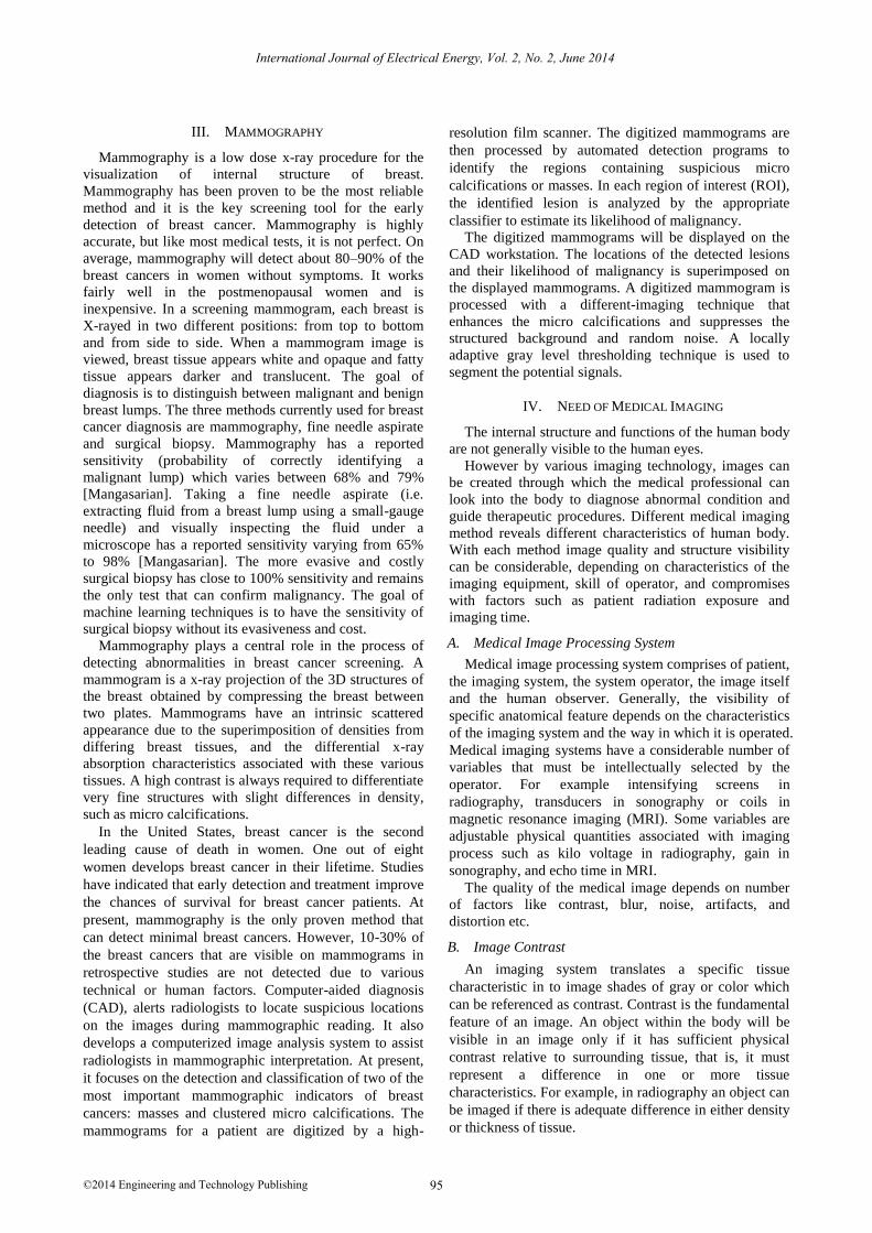
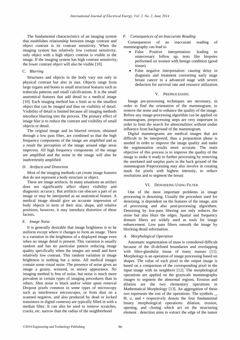
IX. EXPERIMENTAL RESULT
Figure 2. Sample data base.
Figure 3. Real time data.
International Journal of Electrical Energy, Vol. 2, No. 2, June 2014
©2014 Engineering and Technology Publishing 98
Fig. 2, Fig. 3 shows Watershed transform of gradient
magnitude of mamogram image followed by
reconstructive morphological processing with modified
regional maxima superimposed on original image. The
detection and isolation of accumulated masses from
segmented region is very important for extracting useful
feature measures which can be input to a classifier which
classifies each region as either a mass or a false detection
X. CONCLUSION
Breast cancer is one of the leading causes of cancer
death for women all over the world. Mammography is the
main investigation - screening technique used for an early
diagnosis of breast cancer. The resultant mammogram
after applying pre and post processing technique with
watershade segmentation approach has shown better
result which help radiologist for investigation at early
stage which will reduce mortality rate. It can be used
further for the automated abnormalities detection of
human breast like calcification, circumscribed masses,
spiculated masses, circumscribed lesions, asymmetry
analysis etc. Further works may be conducted to develop
integrated system to process other cancer images like
lung cancer, liver cancer for better diagnosis reducing
mortality rate due to cancer.
ACKNOWLEDGMENT
Author would like to acknowledge and extend heartfelt
gratitude to the honorable management for necessary
support and Department of Computer Science of
Information Technology, Dr. Babasaheb Ambedkar
Marathwada University, Aurangabad (MS) INDIA for
technical support.
REFERENCES
[1] L. M. Wun, R. M. Merrill, and E. J. Feuer, "Estimating lifetime and age-conditional probabilities of developing cancer," Lifetime
Data Analysis, vol. 4, no. 2, pp. 169-186, 1998.
[2] S. M. Larie and S. S. Abukmeil, “Brain abnormality in schizophrenia: A systematic and quantitative review of volumetric
magnetic resonance imaging studies,” Br J. Psychiatry, vol. 172,
pp. 110–120, Feb. 1998.
[3] C. Lee, S. Hun, T. A. Ketter, and M. Unser, “Unsupervised
connectivity-based thresholding segmentation of midsaggital brain MR images,” Comput Biol Med., vol. 28, no. 3, pp. 309–338, May
1998.
[4] G. M. te Brake and N. Karssemeijer, “Single and multiscale detection of masses in digital mammograms,” IEEE Transactions
on Medical Imaging, vol. 18, no. 7, pp. 628-638, July 1999. [5] J. N. Wolf, "Risk for breast cancer development determined by
mammographic parenchymal pattern," Cancer, vol. 37, no. 5, pp.
2486-2492, May 1976. [6] A. P. Zijdenbos and B. M. Dawant, “Brain segmentation and white
matter lesion detection in MR images,” Critical Reviews in Biomedical Engineering, vol. 22, no. 5-6. pp. 401–465, 1994.
[7] A. J. Worth, N. Makris, V. S. Caviness, and D. N. Kennedy,
“Neuroanatomical segmentation in MRI: Technological objectives,” Int. J. Patt. Rec. Art. Intel., vol. 11, no. 8, pp. 1161–
1187, 1997. [8] V. S. Khoo, D. P. Dearnaley, D. J. Finnigan, A. Padhani, S. F.
Tanner, and M. O. Leach, “Magnetic resonance imaging (MRI):
Considerations and applications in radiotheraphy treatment planning,” Radiother.Oncol., vol. 42, no. 1, pp. 1–15, 1997.
[9] B. Sahiner, N. Petrick, H. P. Chan, L. M. Hadjiiski, C. Paramagul,
M. A. Helvie, and M. N. Gurcan, “Computer-aided characterization of mammographic masses: Accuracy of mass
segmentation and its effects on characterization,” IEEE Trans Med Imaging, vol. 20, no. 12, pp. 1275-1284, Dec. 2001.
[10] R. C. Gonzalez, R. Woods, and S. Eddins, Digital Image
Processing, Pearson Education, 2nd ed. ch. 4, 2007, pp. 194-196. [11] D. Guliato, J. D. de Carvalho, R. M. Rangayyan, and S. A.
Santiago, “Feature extraction from a signature based on the turning angle function for the classification of breast tumors,” J.
Digit Imaging, vol. 21, no. 2, pp. 129-144, 2008.
[12] R. C. Gonzalez, R. Woods, and S. Eddins, Digital Image Processing Using Matlab, Tata McGraw-Hill Pearson Education,
2010, pp. 440-441. [13] L. Li, Z. Wu, L. Chen, F. George, Z. Chen, A. Salem, M. Kallergi,
and C. Berman, “Breast tissue density and CAD cancer detection
in digital mammography,” presented at the 2005 IEEE 27th Annual Conference Engineering in Medicine and Biology
Shanghai, China, September 1-4, 2005. [14] Medical imaging & image-guided therapy – bioengineering. UW
College of Engineering. [Online]. Available:
http://www.uwmedicine.org
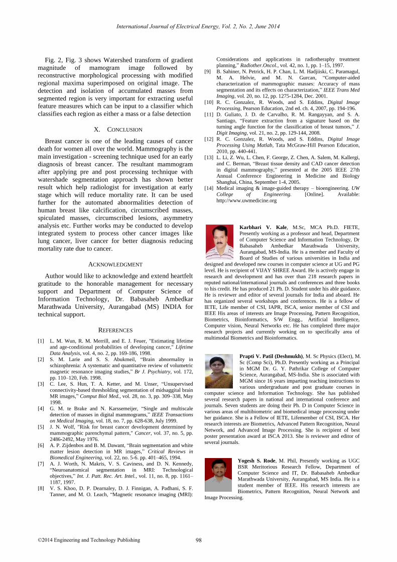
Karbhari V. Kale, M.Sc, MCA Ph.D. FIETE, Presently working as a professor and head, Department
of Computer Science and Information Technology, Dr
Babasaheb Ambedkar Marathwada University, Aurangabad, MS-India. He is a member and Faculty of
Board of Studies of various universities in India and designed and developed new courses in computer science at UG and PG
level. He is recipient of VIJAY SHREE Award. He is actively engage in
research and development and has over than 218 research papers in reputed national/international journals and conferences and three books
to his credit. He has produced 21 Ph. D. Student under his able guidance. He is reviewer and editor of several journals for India and aboard. He
has organized several workshops and conferences. He is a fellow of
IETE, Life member of CSI, IAPR, ISCA, senior member of CSI and IEEE His areas of interests are Image Processing, Pattern Recognition,
Biometrics, Bioinformatics, S/W Engg., Artificial Intelligence, Computer vision, Neural Networks etc. He has completed three major
research projects and currently working on to specifically area of
multimodal Biometrics and Bioinformatics.
Prapti V. Patil (Deshmukh), M. Sc Physics (Elect), M.
Sc (Comp Sci), Ph.D. Presently working as a Principal
in MGM Dr. G. Y. Pathrikar College of Computer Science, Aurangabad, MS-India. She is associated with
MGM since 16 years imparting teaching instructions to
various undergraduate and post graduate courses in
computer science and Information Technology. She has published
several research papers in national and international conference and journals. Seven students are doing their Ph. D in Computer Science in
various areas of multibiometric and biomedical image processing under her guidance. She is a Fellow of IETE, Lifemember of CSI, ISCA. Her
research interests are Biometrics, Advanced Pattern Recognition, Neural
Network, and Advanced Image Processing. She is recipient of best poster presentation award at ISCA 2013. She is reviewer and editor of
several journals.
Yogesh S. Rode, M. Phil, Presently working as UGC BSR Meritorious Research Fellow, Department of
Computer Science and IT, Dr. Babasaheb Ambedkar Marathwada University, Aurangabad, MS India. He is a
student member of IEEE. His research interests are
Biometrics, Pattern Recognition, Neural Network and
Image Processing.