Isolation and PCR Amplification of Genomic DNA from Market Samples of Dry Tea
8
Plant Molecular Biology Reporter 17: 171–178, 1999. © 1999 Kluwer Academic Publishers. Printed in the Netherlands. Protocols Isolation and PCR Amplification of Genomic DNA from Market Samples of Dry Tea MAHIPAL SINGH * , BANDANA and P.S. AHUJA Division of Biotechnology, Institute of Himalayan Bioresource Technology, Palampur (HP), 176061, India Abstract. A simple procedure for DNA isolation from processed dried commercial samples of tea is described. The method involves a modified CTAB procedure employing extensive washing, use of 1% PVP to remove polyphenolics and a single phenol:chloroform extraction step. The average yield ranges from 164–494 μg/g tea sample for various market samples. The DNA obtained from 11 different brands of tea using this procedure were consistently amplifiable (using both RAPD primers as well as defined sequences as primers) and digestible with restriction endonucleases. Key words: Camellia sinensis, DNA isolation, PCR, RAPD, Tea Abbreviations: CTAB, cetyl trimethyle ammonium bromide; EDTA, ethyle diamine tetra acetic acid; PCR, polymerase chain reaction; PVP, poly vinyl pyrrolidone; RAPD, randomly amplified polymorphic DNA; RT, room temperature; Taq, Thermus aquaticus. Introduction Tea is a woody beverage crop with high polyphenolic contents. Isolation of tea genomic DNA using the basic CTAB procedure (Takeuchi et al., 1994; Matsumoto et al., 1994) and silica gel dried tea leaves (Wachira et al., 1995) has been described. All the above methods, however, were used to isolate the DNA from fresh young leaves of tea plants. Isolation of DNA from herbar- ium specimens (Rogers et al., 1988) and dehydrated plant tissues (Tai and Tanksley, 1990) have also been described. However, none of these methods could provide DNA from market samples of processed tea, suitable for down- stream applications. In molecular diagnostic studies involving identification of cultivars and varieties used by a tea manufacturer in preparation of a par- ticular lot or brand of tea and also in diagnosis of adulteration of tea samples * Author for correspondence. e-mail: [email protected]
-
Upload
mahipal-singh -
Category
Documents
-
view
214 -
download
0
Transcript of Isolation and PCR Amplification of Genomic DNA from Market Samples of Dry Tea
Plant Molecular Biology Reporter17: 171–178, 1999.© 1999Kluwer Academic Publishers. Printed in the Netherlands.
Protocols
Isolation and PCR Amplification of Genomic DNAfrom Market Samples of Dry Tea
MAHIPAL SINGH∗, BANDANA and P.S. AHUJADivision of Biotechnology, Institute of Himalayan Bioresource Technology, Palampur(HP), 176061, India
Abstract. A simple procedure for DNA isolation from processed dried commercial samplesof tea is described. The method involves a modified CTAB procedure employing extensivewashing, use of 1% PVP to remove polyphenolics and a single phenol:chloroform extractionstep. The average yield ranges from 164–494µg/g tea sample for various market samples.The DNA obtained from 11 different brands of tea using this procedure were consistentlyamplifiable (using both RAPD primers as well as defined sequences as primers) and digestiblewith restriction endonucleases.
Key words: Camellia sinensis, DNA isolation, PCR, RAPD, Tea
Abbreviations: CTAB, cetyl trimethyle ammonium bromide; EDTA, ethyle diamine tetraacetic acid; PCR, polymerase chain reaction; PVP, poly vinyl pyrrolidone; RAPD, randomlyamplified polymorphic DNA; RT, room temperature;Taq, Thermus aquaticus.
Introduction
Tea is a woody beverage crop with high polyphenolic contents. Isolation oftea genomic DNA using the basic CTAB procedure (Takeuchi et al., 1994;Matsumoto et al., 1994) and silica gel dried tea leaves (Wachira et al., 1995)has been described. All the above methods, however, were used to isolate theDNA from fresh young leaves of tea plants. Isolation of DNA from herbar-ium specimens (Rogers et al., 1988) and dehydrated plant tissues (Tai andTanksley, 1990) have also been described. However, none of these methodscould provide DNA from market samples of processed tea, suitable for down-stream applications. In molecular diagnostic studies involving identificationof cultivars and varieties used by a tea manufacturer in preparation of a par-ticular lot or brand of tea and also in diagnosis of adulteration of tea samples
∗Author for correspondence. e-mail: [email protected]

172 Mahipal Singh , Bandana and P.S. Ahuja
by plants other thanCamellia sinensis (L) O. kuntze, isolation of quality DNAis a basic and pre requirement. We describe here a protocol to obtain qualityDNA from processed dry market samples of tea. The protocol is mainly basedon the protocol of Saghai-Maroof and his colleagues (1984).
Materials and Methods
Plant materials
The fresh leaf material ofCamellia sinensis (L) O. kuntze(Upasi-9) used inthis study was obtained from ‘Banuri Experimental Tea Garden’ of IHBT.Different brands of dry commercial tea samples available in the open mar-ket in packed forms were obtained from Palampur market and are listed inTable 1.
Equipment and chemicals
High speed refrigerated centrifuge (Hitachi)Robocycler (Stratagene)Mortar and pestleLiquid nitrogenWater bathExtraction buffer: 2% (w/v) CTAB, 100 mM Tris-HCl (pH 8.0), 20 mMEDTA, 1.4 mM NaCl, 1% PVP1× TE buffer pH 8.0 (10 mM Tris, 1 mM EDTA)0.1× TE buffer pH 8.0 (10 mM Tris, 0.1 mM EDTA)Chloroform:isoamyl alcohol (24:1)IsopropanolPhenol:chloroform (1:1)Absolute ethanol70% ethanol (v/v)PVPTaqpolymerase (Bangalore Genei)
DNA isolation
• Soak the tea samples overnight in sterile distilled water, cleanse 4 timeswith double distilled water and dry the samples in an oven (60◦C) forabout an hour.
• Grind 1 g of dried tea samples in a mortar and pestle in the presence ofliquid nitrogen.

Isolationand
PC
RA
mplification
fromTe
a173
Table 1. DNA extraction status and PCR amplification of market samples of tea.
S.N. Brand Name# Market price∗ DNA extraction PCR amplification
(Rs/kg) +/− µg/gm OD260/280 Status RAPD primer Defined primer
1. Lazeez Premium 152 + 494 1.72 Degraded +(3) +(5)
2. Brook Bond A1 126 + 278 1.73 Degraded +(3) +(5)
3. Tea city 180 + 451 1.73 Degraded +(3) +(5)
4. Birla 160 + 444 1.72 Degraded +(2) +(5)
5. Premium Gold 142 + 461 1.84 Degraded +(3) +(5)
6. New Lipton Richbrue 166 + 460 1.83 Degraded +(4) +(5)
7. Lipton taaza 183 + 164 1.79 Degraded +(3) +(5)
8. Sargam 183 + 489 1.62 Degraded +(4) +(5)
9. Taj Mahal 208 + 287 1.78 Degraded +(4) +(5)
10. Green tea 110 + 240 1.90 Partially degraded – +(5)
11. Lazeez masala 134 + 275 1.76 Degraded – +(5)
Note:The oligonucleotide used in this study as a RAPD primer was 5′-AAGACCCCTC-3′ and as defined primers were: Forward primer (5′-TTTAGTGCTGGTTGTCGC-3′), Backward primer (5′-TGGGAAGTCCTCGTGTTGCA-3′).Numbers in parentheses represents number of PCR bands.#All teas here are black except number 10.∗Prices for January 1998.

174 Mahipal Singh , Bandana and P.S. Ahuja
• Transfer the tea powder into a clean autoclaved centrifuge tube and add4 mL of warm extraction buffer.
• Mix well and incubate at 65◦C for 1 h with occasional mixing.• Cool the mixture to RT and then add 8 mL of chloroform:isoamyl alco-
hol.• Centrifuge at 10,000 rpm for 10 min at RT.• Transfer the clear supernate to a fresh tube and precipitate the DNA by
adding 0.6 vol of isopropanol.• Centrifuge the precipitated DNA at 12,000 rpm for 20 min at RT.• Dry the pellet at RT for about 30 min and then dissolve in 2 mL of 1×
TE.• Treat the DNA with RNase (10 mg/mL stock) for 1 h at 37◦C.• Extract once with an equal volume of phenol:chloroform.• Transfer the aqueous phase to a new tube, add 2 volumes of chilled
ethanol and keep at−20 ◦C for at least 1 h.• Centrifuge at 12,000 rpm for 20 min at 4◦C.• Wash the pellet with 70% ethanol for 5 min.• Dry the pellet for 30 min at RT.• Dissolve the purified DNA in an appropriate volume of 1× TE and store
at−20 ◦C until used.Notes1. Soaking of tea samples in water for 3–4 h and then extensive washing (5–6 times) removes
much of the brownish colour.
2. The extraction buffer works well if kept warm at 65◦C prior to adding the ground sample.
PCR amplification
• Dilute the DNA in 0.1× TE to 10 ng/µL.• Assemble the PCR reaction in a 25µL final vol containing 100µM each
of the four dNTPs, 100µM primer, 20 ng of genomic DNA and 0.3 unitsof Taqpolymerase.
• Program the robocycler for 1 cycle of 4 min at 94◦C and then 45 cyclesof 1 min at 94◦C, 1 min at 37◦C and 2 min at 72◦C. The last extensioncycle was programmed at 72◦C for 7 mins.
• Run the amplified products in a 1.4% agarose gel, stain in ethidiumbromide and photograph with the help of an image analyser.
NoteThe annealing temperature for conserved rRNA gene primers was 68◦C instead of 37◦C and
it was run for 40 cycles only (instead of 45 cycles).

Isolation and PCR Amplification from Tea 175
Figure 1. Comparison of genomic DNA extracted from black and green tea samples: Lanes1, 2 and 3 show DNA isolated from fresh leaves of tea plant, dried commercial green tea andblack tea samples respectively. M is mol. wt. markerλ Hind III digest.
Results and Discussion
We procured samples of 10 different black and 1 green tea brands from thePalampur market and isolated their DNA using the above protocol. The DNAwas recovered in all cases, however, it was degraded (Table 1). The averageDNA extracted using this protocol was 164–494µg/g dry weight of tea sam-ple and the OD 260/280 was 1.63 to 1.84. We could not isolate undegradedDNA from any of the samples tested except green tea. The DNA isolated fromtea samples without prior soaking in water was brownish, comparatively lessin amount and highly degraded. It could not be amplified by PCR. Soakingtea samples in water and then washing them several times in sterile waterremoved much of the brownish colour and resulted in better yield and qualityof DNA. The degraded nature of DNA is probably due to the high temperatureand pressure during processing of black teas, which distorts the cells, nucleusand ultimately the genomic material. This is supported by our results, whichindicated that DNA isolated from fresh leaves or from green tea, which isprocessed differently, was undegraded or partially degraded (Figure 1). It hasbeen reported earlier that after cell lysis polyphenolics bind to DNA and causeits degradation (John, 1992). It has also been reported that PVP can removea lot of the polyphenolics (Kim et al., 1997). We, therefore, replaced mercap-toethanol with PVP in Saghai-Maroof’s protocol and this improved the DNA

176 Mahipal Singh , Bandana and P.S. Ahuja
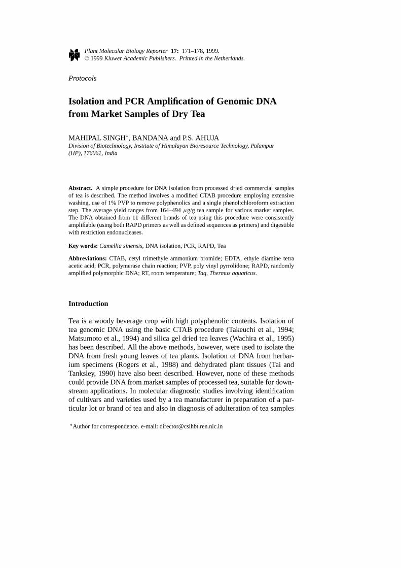
Figure 2. PCR amplification profile of the market samples of tea: Lanes 1 to 11 representamplified products of DNA isolated from: Lazeez Premium, Brook Bond A1, Tea city, Birla,Premium Gold, New Lipton Richbrue, Lipton taaza, Sargam, Taj Mahal, Green tea and Lazeezmasala brands of tea respectively (as in Table 1). Lane 12 is the PCR product of DNA fromfresh leaves of a tea clone, ‘Upasi 9’. Lane B is a control lane without template DNA. Mis PCR mol. wt. marker from Sigma. Panel A shows RAPD patterns using a single 10 meroligo (Table 1) as a primer while panel B shows PCR amplification using a set of two primers(Table 1) designed from conserved sequences of plant 5 s rRNA gene so as to amplify variablespacers (Cox et al., 1992). Note that the reaction did not work in lane 10 and 11 in panel A inthis gel.
quality. 1% PVP was found optimal for quality DNA. Using this protocol,we also isolated DNA from the discarded samples of teas after brewing. TheDNA isolated from post brewed tea samples was relatively more degradedcompared to pre-brewed tea samples and was not suitable for PCR (data notshown).
To evaluate the suitability of the isolated DNA in downstream applica-tions, we amplified the DNA through PCR using a 10 mer random primerfrom Operon Biotechnologies Inc. and also using a defined set of primersfrom conserved 5 s rRNA gene sequences (Table 1). As shown in Figure 2,panels A and B, the PCR product was in nearly all cases comparable to thatobtained from undegraded DNA isolated from fresh leaves of a tea clone. Itdemonstrates that isolated DNA is suitable for any diagnostic purpose em-ploying PCR as a technique. Figure 2, panel A also shows that the PCRproducts from successful amplification consist of 3 major bands and someminor bands. These include even DNA isolated from fresh leaves of Upasi 9clone, thus the tea samples contain genuine tea plant material. The upper

Isolation and PCR Amplification from Tea 177
band in lane 4 of Figure 2A is very faint and could represent a mutation atprimer annealing sites. The 3rd band (from top) is prominent in lanes 6, 8 and9 (Figure2, panel A) in addition to 3 major bands in all samples. Since thisband is also a prominent band of the control lane (Figure 2 panel A, lane B),it seems to be an artifact and does not represent the true PCR product. Thecontrol lane (Figure 2, panel A, lane B ) also shows some bands. It has beenreported earlier that sometimes a RAPD primer folds back on itself, resultingin artifactual bands, but this usually does not occur when the DNA templateis added to the reaction mixture (Williams et al., 1992). Figure 2, panel Balso shows greater similarity of amplification products obtained from varioustea samples. However, minor differences in band sizes, was also present andthese might be due to minor sequence length differences, in the amplifiedspacer regions in different tea cultivars. The isolated DNA from tea samplescould also be digested with restriction endonucleasesHind III, Sau IIIA-I, Hinf I and Hae III (data not shown). To our knowledge, this is the firstreport of DNA isolation from market samples of tea, their PCR amplification,and restriction endonuclease digestion. In summary, good quality DNA fromcommercially dried tea samples was isolated and used successfully for downstream applications including restriction digestion and PCR amplification.This method has the potential for development of technology for testing theoriginality of commercial teas and for the identification of cultivars/varietiesused by a tea manufacturer in production of a particular brand.
Acknowledgements
Department of Biotechnology, Government of India, is acknowledged for thefinancial support for this work. This is IHBT communication No-9823.
References
Cox AV, Bennett MD and Dyer TA (1992) Use of the PCR to detect spacer size heterogeneityin plant 5 s rRNA gene clusters and to locate such clusters in wheat (Triticum aestivum).Theor Appl Genet 83: 684–690.
John ME (1992) An efficient method for isolation of RNA and DNA from plants containingpolyphenolics. Nuc Acids Res 20: 2381.
Kim CS, Lee CH, Shin JS, Chung YS and Hyung NI (1997) A simple and rapid method forisolation of high quality genomic DNA from fruit trees and conifers using PVP. Nuc AcidsRes 25: 1085–1086.
Matsumoto S, Takeuchi A, Hayatsu M and Kondo S (1994) Molecular cloning of phenylealanine ammonia lyase cDNA and classification of varieties and cultivars of tea plantsusing the tea PAL cDNA as a probe. Theor Appl Genet 89: 671–675.

178 Mahipal Singh , Bandana and P.S. Ahuja
Rogers SU and Bendich AJ (1988) Extraction of DNA from milligram amounts of fresh,herbarium and mummified plant tissues. Plant Mol Biol 5: 69–76.
Saghai-Maroof MA, Soliman KA, Jorgenson RA and Allard RW (1984) Ribosomal DNAspacer length polymorphism in Barley: Mendalian inheritance, chromosomal location andpopulation dynamics. Proc Natl Acad Sci (USA). 8: 8014–18.
Tai TH and Tanksley SD (1990) A rapid and inexpensive method for isolation of total DNAfrom dehydrated plant tissue. Plant Mol Biol Reptr 8: 297–303.
Takeuchi A, Matsumoto S, Hayatsu M (1994) Chalcone synthase fromC. sinensis: isolation ofcDNA and the organ specific and sugar responsive expression of genes. Plant Cell Physiol35: 1011–18.
Wachira FN, Waugh R, Hackett CA and Powell W (1995) Detection of genetic diversity in teausing RAPD markers. Genome 38: 201–210.
Williams JGK, Kubelik AR, Livak KJ, Rafalski JA and Tingey SV (1990) DNA polymorphismamplified by arbitrary primers are useful as genetic markers. Nuc Acids Res 18: 6531–35.