Isolation and Identification of a Metabolic Intermediate...
9
[CANCER RESEARCH 38, 2592-2599, August 1978] 0008-5472/78/0038-0000$02.00 Isolation and Identification of a Metabolic Intermediate in the Selective Dechloroethylation of One of the Four Stereoisomers of 4-Methylcyclophosphamide1 George Abel, Peter J. Cox,2 Peter B. Farmer, Neville J. Haskins, Michael Jarman, Kanti Merai, and Wojciech J. Stec Chester Beatty Research Institute, Institute of Cancer Research: Royal Cancer Hospital, Fulham Road, London SW3 6JB England [G A PJC PB F M. J., K. M.¡:G. D. Searle & Co. Ltd., Lane End Road, High Wycombe, Buckinghamshire, HP12 4HL, England ¡N.J. H.]; and Centre 'tor Molecular and Macromolecular Studies, Polish Academy of Sciences, 90-362 Lodz, Boczna 5, Poland [W. J. S.J ABSTRACT The in vitro cytotoxicity in bioassay or cell culture assays of (-)-c/s-4-methylcyclophosphamide when me tabolized by microsomes from the livers of male Wistar rats was significantly less than that of metabolized (+)- c/s-, (+)-frans-, and (-)-rrans-4-methylcyclophosphamide. However, metabolism of the individual Stereoisomers by liver microsomes from female BALB/c mice yielded metabolite mixtures of approximately equal cytotc '¡city. The extent of metabolism by rat liver microsomes f the four Stereoisomers was similar when assessed by sub strate disappearance, but the yield of alkylating metabo lites (expressed as phosphoramide mustard equivalents) was comparable in the case of three of the isomers but substantially less for the (-)-c/s-isomer. A new metabolite was isolated following the incubation of (-)-cis-4-methylcyclophosphamide with rat liver micro somes. It was identified by its breakdown products and its electron impact and chemical ionization mass spectra, especially by the presence in the latter of a pseudomo- lecular ion. Its structure was 2-[(2-chloroethyl)(2-chloro-1- hydroxyethyl)amino]tetrahydro-4-methyl-2H-1,3,2-oxaza- phosphorine 2-oxide, an intermediate in dechloroethyla- tion. Species variation (mouse, rat, and rabbit) in the forma tion of this metabolite of low cytotoxicity (-1% of that of 4- hydroxy-4-methylcyclophosphamide) was noted and ac corded well with the observed species differences in cytotoxicity assays. INTRODUCTION The presence of 1 or 2 chiral centers in molecules of pharmacological importance is frequently associated with profound differences in the biological activities of the 2 or 4 Stereoisomers. In the field of antiacetylcholine drugs, the (-)-enantiomers of those containing a single asymmetric benzylic carbon atom show greater potency than do the (+)-enantiomers (21). The S(-)-form of the anticoagulant phenprocoumon is more effective than is the fî(+)-isomer (22). (+)-Methadone is inactive in the maintenance of opi ate-dependent patients (see Ref. 36). The analgesic activity of propoxyphene resides largely in the (+)-isomer (see Ref 5). The metabolism of enantiomers can also differ substan tially. The influence of stereochemistry on drug metabolism has been reviewed by Jenner and Testa (23). Of the 4 stereoisomeric ephedrines (10), the isomers of absolute configuration 2S-[(-)-ephedrine and (+)-pseudoephedrine] were A/-demethylated more rapidly by rabbit liver micro somes than were the isomers of 2R geometry. The hydrox- ylation and excretion of the antiinflammatory agent Ciclo- profen in the rat proceeds more rapidly with the (+)-enan- tiomer (11). In some cases enantiomeric interactions occur. For example, S(+)-amphetamine inhibits the metabolism of fl(-) in the racemate (17, 39); the S-isomer of a psychoto- mimetic amine, 1-(2,5-dimethoxy-4-methylphenyl)-2-amino- propane, inhibits the metabolism of the fl-isomer (26); and the inactive (-)-propoxyphene increases plasma levels and enhances activity of the (+ )-antipode (28). Species differences have also been recorded: (+ )- and (-)-hexobarbital are metabolized at different rates, and the ratio between the 2 varies between species, although sepa rate investigations have revealed opposing results (12, 16, 29). Striking species differences exist in the microsomal metabolism of the enantiomers of CP3-4which is chiral by virtue of asymmetry at phosphorus. In our studies of CP analogs, we found that the primary hydroxylation step in the metabolism by rat liver micro somes of 4-MeCP, as measured by substrate disappear ance, was only marginally affected by the configuration of the substrate, which can exist in 4 forms as a result of the introduction of a second chiral center in the ring, and that the biological activities against the murine ADJ/PC6A tumor in vivo of the 4 compounds fell within a 2-fold range (15). However, an interesting observation, reported here, of the differences among the in vitro cytotoxicities of the 4 different Stereoisomers led us to investigate further the initial metabolic process. 1This investigation was supported by grants to the Chester Beatty Research Institute, Institute of Cancer Research:Royal Cancer Hospital, from the Medical Research Council (Grant G973/786-7K) and to the Polish Acad emy of Sciences, Centre for Molecular and Macromolecular Studies, from the National Cancer Programme. 2 To whom requests for reprints should be addressed. Received January 3, 1978; accepted April 25, 1978. 3 The abbreviations used are: CP, cyclophosphamide; 4-MeCP, 4-methyl- cyclophosphamide (2-[bis(2-chloroethyl)amino]-tetrahydro-4-methyl-2H-1,3,2- oxazaphosphorine 2-oxide); 4-OH-4-MeCP, 4-hydroxy-4-methylcyclophos- phamide; TLC, thin-layer chromatography; El, electron impact; CI, chemical ionization; NBP, 4-(p-nitrobenzyl)pyridine; ID5()medianinhibitory dose; AMU, atomic mass unit. 4 P. J. Cox, P. B. Farmer, and M. Jarman, manuscript in preparation. 2592 CANCER RESEARCH VOL. 38 on April 22, 2020. © 1978 American Association for Cancer Research. cancerres.aacrjournals.org Downloaded from
Transcript of Isolation and Identification of a Metabolic Intermediate...
[CANCER RESEARCH 38, 2592-2599, August 1978]0008-5472/78/0038-0000$02.00
Isolation and Identification of a Metabolic Intermediate in the Selective
Dechloroethylation of One of the Four Stereoisomers of4-Methylcyclophosphamide1
George Abel, Peter J. Cox,2 Peter B. Farmer, Neville J. Haskins, Michael Jarman, Kanti Merai, andWojciech J. Stec
Chester Beatty Research Institute, Institute of Cancer Research: Royal Cancer Hospital, Fulham Road, London SW3 6JB England [G A P J C P B FM. J., K. M.¡:G. D. Searle & Co. Ltd., Lane End Road, High Wycombe, Buckinghamshire, HP12 4HL, England ¡N.J. H.]; and Centre 'tor Molecular and
Macromolecular Studies, Polish Academy of Sciences, 90-362 Lodz, Boczna 5, Poland [W. J. S.J
ABSTRACT
The in vitro cytotoxicity in bioassay or cell cultureassays of (-)-c/s-4-methylcyclophosphamide when metabolized by microsomes from the livers of male Wistarrats was significantly less than that of metabolized (+)-c/s-, (+)-frans-, and (-)-rrans-4-methylcyclophosphamide.However, metabolism of the individual Stereoisomers byliver microsomes from female BALB/c mice yieldedmetabolite mixtures of approximately equal cytotc '¡city.
The extent of metabolism by rat liver microsomes f thefour Stereoisomers was similar when assessed by substrate disappearance, but the yield of alkylating metabolites (expressed as phosphoramide mustard equivalents)was comparable in the case of three of the isomers butsubstantially less for the (-)-c/s-isomer.
A new metabolite was isolated following the incubationof (-)-cis-4-methylcyclophosphamide with rat liver microsomes. It was identified by its breakdown products and itselectron impact and chemical ionization mass spectra,especially by the presence in the latter of a pseudomo-lecular ion. Its structure was 2-[(2-chloroethyl)(2-chloro-1-hydroxyethyl)amino]tetrahydro-4-methyl-2H-1,3,2-oxaza-phosphorine 2-oxide, an intermediate in dechloroethyla-tion.
Species variation (mouse, rat, and rabbit) in the formation of this metabolite of low cytotoxicity (-1% of that of 4-hydroxy-4-methylcyclophosphamide) was noted and accorded well with the observed species differences incytotoxicity assays.
INTRODUCTION
The presence of 1 or 2 chiral centers in molecules ofpharmacological importance is frequently associated withprofound differences in the biological activities of the 2 or4 Stereoisomers. In the field of antiacetylcholine drugs, the(-)-enantiomers of those containing a single asymmetricbenzylic carbon atom show greater potency than do the(+)-enantiomers (21). The S(-)-form of the anticoagulant
phenprocoumon is more effective than is the fî(+)-isomer(22). (+)-Methadone is inactive in the maintenance of opiate-dependent patients (see Ref. 36). The analgesic activityof propoxyphene resides largely in the (+)-isomer (see Ref5).
The metabolism of enantiomers can also differ substantially. The influence of stereochemistry on drug metabolismhas been reviewed by Jenner and Testa (23). Of the 4stereoisomeric ephedrines (10), the isomers of absoluteconfiguration 2S-[(-)-ephedrine and (+)-pseudoephedrine]were A/-demethylated more rapidly by rabbit liver microsomes than were the isomers of 2R geometry. The hydrox-ylation and excretion of the antiinflammatory agent Ciclo-profen in the rat proceeds more rapidly with the (+)-enan-tiomer (11). In some cases enantiomeric interactions occur.For example, S(+)-amphetamine inhibits the metabolism offl(-) in the racemate (17, 39); the S-isomer of a psychoto-mimetic amine, 1-(2,5-dimethoxy-4-methylphenyl)-2-amino-propane, inhibits the metabolism of the fl-isomer (26); andthe inactive (-)-propoxyphene increases plasma levels andenhances activity of the (+ )-antipode (28).
Species differences have also been recorded: (+ )- and(-)-hexobarbital are metabolized at different rates, and theratio between the 2 varies between species, although separate investigations have revealed opposing results (12, 16,29). Striking species differences exist in the microsomalmetabolism of the enantiomers of CP3-4which is chiral by
virtue of asymmetry at phosphorus.In our studies of CP analogs, we found that the primary
hydroxylation step in the metabolism by rat liver microsomes of 4-MeCP, as measured by substrate disappearance, was only marginally affected by the configuration ofthe substrate, which can exist in 4 forms as a result of theintroduction of a second chiral center in the ring, and thatthe biological activities against the murine ADJ/PC6A tumorin vivo of the 4 compounds fell within a 2-fold range (15).
However, an interesting observation, reported here, ofthe differences among the in vitro cytotoxicities of the 4different Stereoisomers led us to investigate further theinitial metabolic process.
1This investigation was supported by grants to the Chester BeattyResearch Institute, Institute of Cancer Research:Royal Cancer Hospital, fromthe Medical Research Council (Grant G973/786-7K) and to the Polish Academy of Sciences, Centre for Molecular and Macromolecular Studies, fromthe National Cancer Programme.
2To whom requests for reprints should be addressed.Received January 3, 1978; accepted April 25, 1978.
3The abbreviations used are: CP, cyclophosphamide; 4-MeCP, 4-methyl-cyclophosphamide (2-[bis(2-chloroethyl)amino]-tetrahydro-4-methyl-2H-1,3,2-oxazaphosphorine 2-oxide); 4-OH-4-MeCP, 4-hydroxy-4-methylcyclophos-phamide; TLC, thin-layer chromatography; El, electron impact; CI, chemicalionization; NBP, 4-(p-nitrobenzyl)pyridine; ID5()medianinhibitory dose; AMU,atomic mass unit.
4 P. J. Cox, P. B. Farmer, and M. Jarman, manuscript in preparation.
2592 CANCER RESEARCH VOL. 38
on April 22, 2020. © 1978 American Association for Cancer Research. cancerres.aacrjournals.org Downloaded from

Isolation of an Intermediate in Dechloroethylation
MATERIALS AND METHODS
Metabolism. Washed liver microsomes were prepared (3)from male Wistar rats, female BALB/c mice, and femaleNew Zealand White rabbits. Both rats and mice were pre-treated with sodium phénobarbital(1 g/liter drinking water)for 10 days. The microsomal pellets were used immediatelyor stored at -30°until required.
The 4-MeCP stereoisomers (25, 35) were incubated (3)with these microsomes; appropriate details are given withthe relevant series of experiments. Substrate disappearancewas measured by stable isotope dilution (15). 4-OH-4-MeCPwas prepared by KMn04 oxidation from the racemates andpure stereoisomers (6).
Isolation and Identification of Metabolites. Incubationswere terminated and worked up by the methods describedpreviously (3). TLC was carried out on glass plates (20 x 20or 5 x 20 cm) coated with silicic acid (Merck Kieselgel G)developed with CHCI:t:C2H5OH (9:1) unless otherwisestated. Separated components were located on the chro-matograms by exposure to iodine vapor or by a sprayingwith acidic 2,4-dinitrophenylhydrazine (3). After removal ofthe major portion of each band, alkylating species werelocated with Epstein reagent (35). Materials were elutedfrom the silicic acid with ethanol. Eluates were subjected tomass spectrometry or were reacted with acidic 2,4-dinitrophenylhydrazine by the published method (3, 6).
Mass Spectrometry. The direct insertion technique wasused for all spectra. El mass spectra were determined withan AEI-MS12 spectrometer with an ionizing voltage of 70eV, a trap current of 100 ¿¿a,and an ¡onsource temperatureof -100°.Quantitation of 4-MeCP in samples containing thed.,-standard was achieved using the peak height ratio of theions atm/e 225 and 229 ([M-CH2CI]+ for d„and d4 forms,
respectively) (15).CI mass spectra were determined with a Finnigan 3200
spectrometer. Ammonia was introduced as the reactant gasthrough the Finnigan manifold with the use of the calibration gas inlet. The gas was obtained from a lecture bottle(Cambrian Chemicals Ltd., Croydon, Surrey, England). Tri-deuteroammonia was introduced with the same inlet, alsofrom a lecture bottle (Merck, Sharpe and Dohme, IsotopesDivision, Hoddesdon, Hertfordshire, England). An ionizingvoltage of 100 eV was used at a source temperature of 140°.
The indicated pressure of gas in the source was 200 to 230/urn. Data obtained from the mass spectrometer were analyzed with an on-line Finningan 6000 data system.
Bioassay. The procedure was that previously described(9) except that substrate was metabolized in the presenceof the cells. Thus to 8.9 ml Walker ascites cell suspension(1.12 x 10⁶ cells/ml) was added 0.1 ml substrate solutionand 1.0 ml of a suspension containing microsomes equivalent to 250 mg liver, 0.35 /¿molNADP+, 6.9 /¿molglucose-6-phosphate, 6.25 jumol MgCI2-6H2O, and 0.875 unitglucose-6-phosphate dehydrogenase, buffered at pH 7.4 with 100jumol potassium phosphate or Tris-HCI. After incubation ofthe cell suspension for 2 hr at 37°in 25-ml conical flasks
under an Cvenriched atmosphere, female Wistar rats (ingroups of 5) were given injections of 106 cells, and thesurvival times of the animals were recorded.
Cell Culture Assay. Walker ascites tumor cell lines wereestablished and maintained as previously described (31).Cells from log-phase cultures were resuspended in freshmedium (Dulbecco's modified Eagle's medium plus 10%
fetal calf serum containing penicillin and streptomycin) at adensity of 2.78 x 10⁴ (or 2.78 x 10s) cells/ml. The suspension was dispensed in 4.5-ml (or 450-/U.I)portions intocentrifuge tubes, and 0.5 ml (or 50 /¿I)of test solution wasadded. After 1 hr at 37°,the cells were harvested by
centrifugation (800 x g, 3 min) and resuspended in 5 ml offresh medium. Portions (2 ml, 5 x 10⁴ cells) of each cellsuspension were dispensed into 2 dishes of a Linbro multi-dish tray, which was then incubated at 37°under an atmos
phere of 10% CO2in air. Cell counts were made after 72 and96 hr from 1 dish of each duplicate each time. Comparisonof the growth of treated cells with that of appropriatecontrols was used to assess the percentage of inhibition ofcell growth.
Cells of the ascitic form of the ADJ/PC6A murine tumorwere harvested freshly for each experiment from the peritoneal cavity of mice used for the routine maintenance ofthe tumor line in vivo. The cells and ascitic fluid wereimmediately diluted with Dulbecco's modified Eagle's me
dium containing 20% inactivated horse serum; glutamine (2mM); alanine, asparagine, aspartate, glutamate, glycine,proline, and serine (all 0.4 mM); and streptomycin andpenicillin. After being counted the cells were diluted to adensity of 1.11 x 10⁶cells/ml. Portions (450 ¿¿I)were treated
with test solution (50 /¿I)as described above; the cells wereresuspended in 5 ml of medium and dispensed in 2-mlportions (2 x 10⁵cells). Cell counts were made after 72 hr.
4-MeCP isomers were activated either at a range ofsubstrate concentrations (17.5 /J.Mto 10.7 mM) 30 timesthose finally used to treat the cells (0.58 to 356 /^M) andexactly as previously described (9) or in other experimentsat a single substrate concentration (0.57 mM) 10 times themaximum concentration (57 /J.M)used to treat the cells.
Quantification of Alkylating Activity. Alkylating activitywas measured by the reaction with Epstein reagent (NBP).Incubations were stopped by plunging the flasks into ice.The microsomal protein was removed by centrifugation at65,000 x gav for 20 min at 2°.Portions (0.375 ml) of thesupernatant were transferred to 10-ml conical glass centrifuge tubes; 0.2 ml of 0.5 M sodium acetate buffer, pH 4, and0.2 ml of 2% NBP in ethylene glycol were added and mixed.After the tubes were covered, they were incubated at 100°
for 20 min and then cooled in ice. Color was developed bythe addition of 0.8 ml of triethylamine:acetone (1:1). Afterbeing mixed the solution was centrifuged at 1500 x g for 2to 3 min, and the absorbance of the clear supernatant wasmeasured at 565 nm exactly 10 min after the addition ofbase. Phosphoramide mustard (obtained as its cyclohexyl-ammonium salt from Dr. H. B. Wood, National CancerInstitute, Bethesda, Md.) was used as a standard. Theabsorbance due to the reaction of the remaining substratewith NBP was allowed for by using control incubationslacking the cofactors required for the microsomal metabolism. Thus, when x is the concentration (/¿M)of totalalkylating metabolites formed (expressed as phosphoram-ide mustard equivalents), y is the substrate (initial concen-
AUGUST 1978 2593
on April 22, 2020. © 1978 American Association for Cancer Research. cancerres.aacrjournals.org Downloaded from
G. Abel et al.
tration, ¿¿M),f is the substrate concentration :substrate concentration in phosphoramide mustard equivalents, and z isthe concentration (¿IM)of total phosphoramide mustardequivalents measured, then
(y- x)z =x +-1——¿�•¿�
RESULTS
Initial experiments were carried out with the bioassaysystem. When tested as described above with rat micro-somes suspended in phosphate buffer and with substrateconcentrations of 12 and 24 /¿g/ml,the (+ )-c/s- and (+ )-and (-)-frans-isomers were totally cytotoxic, whereas the(-)-c/s-enantiomer had little effect on increase in survivaltime. Further experiments with rat microsomes that hadbeen stored for a shorter period at -30° (2 to 3 weekscompared to 9 weeks) and in which Tris-HCI buffer wasused demonstrated the pure (-)-c/s-enantiomer to be some6-fold less toxic than were the other forms (Table 1).
These results were confirmed by the cell culture assay.Individual enantiomers were metabolized at a range ofsubstrate concentrations, and the inhibition of growth ofWalker ascites cells by these metabolized samples wasdetermined (Table 2). The (-)-c/s form metabolized by ratmicrosomes again showed toxicity 1 order of magnitudeless than that of the other 3 forms. Metabolism with mouseliver microsomes, however, caused approximately equal
Table 1Toxicity of activated 4-MeCP stereoisomers measured by bioassay
The drug concentration necessary to cause 50% cell kill in thepresence of active rat liver microsomes in Tris-HCI was determinedby bioassay (see "Materials and Methods"). Fifty % cell kill is
equivalent to 21% increase in survival time, taking the generationtime in vivo as 36 hr (9). Each value is derived from the survivaltimes of 5 animals at each of 3 drug concentrations. All the valueswere obtained in parallel within 1 experiment.
Drug concentration(¿tM)c/s-Configuration(+)
7.3(±) 12.4(-) 43.8frans-Configuration5.5
6.65.1
Table 2Concentrations of 4-MeCP stereoisomers, activated by liver
microsomes or by KMnOtoxidation, required to inhibit the growthof Walker ascites tumor cells in suspension culture by 50%
Each value was obtained from a dose-response curve derivedfrom 5 to 8 substrate concentrations (individually metabolized).
Concentration(MM)Species
(+)-c/s-Rat4.9±0.3a>6(2)c'
Mouse 5.9Chemical 3.4
oxidation(-)-c/s-
(+)-frans-50.7±6.26(2)
6.52.73.6
3.71.7(-)-frans-4.3
5.63.4
" Mean ±S.E.6 These values differ significantly (p < 0.01).0 Numbers in parentheses, number of dose-response curves
when more than 1.
levels of growth inhibition by all 4 stereoisomers, as did theproducts of chemical (KMnO4) oxidation.
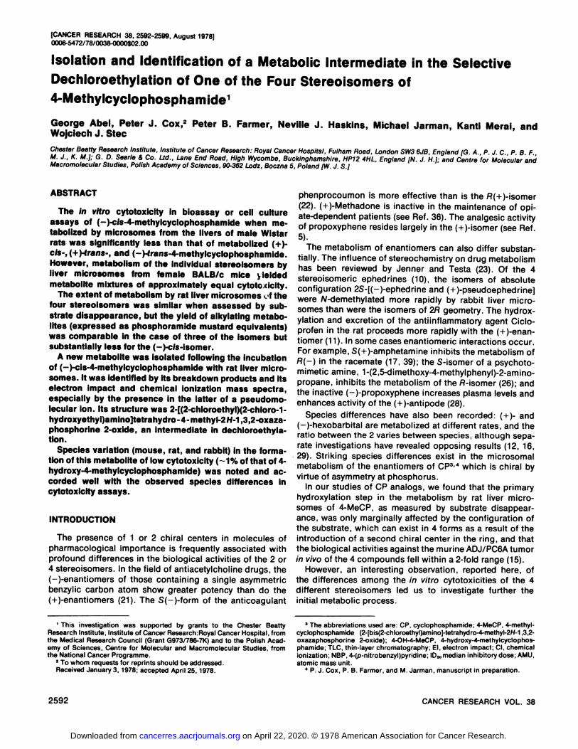
The extent of metabolism by rat and mouse liver microsomes as measured by substrate disappearance was compared to the extent of metabolism assessed by alkylatingproduct appearance measured as phosphoramide mustardequivalents. The results are shown in Chart 1 (storedmicrosomes) and Table 3 (fresh microsomes). Additionally,
100-,^
75-Ö
350-U
n—A—1iS.
—¿�.1. —¿�i"VN
VNII
S^
RAT MOUSE0.5-,
5i—l~—+i - + * -
cis-. * trans——
i——+
± - 4 ±-cis
trans
o
oc
Chart 1. A. percentage of metabolism of individual 4-MeCP stereoisomers and racemates in 45 min by microsomes (stored at -30°prior touse) from rat and mouse, expressed in terms of substrate disappearance(open bars) and alkylating product (phosphoramide mustard equivalents)appearance (hatched bars). B, ratio of percentage of metabolism by productappearance to percentage of metabolism by substrate disappearance (fromA). , 1 S.D. from either side of the mean of all 12 values. Only 2values (') fall outside this range; that for the (-)-cis-stereoisomer differed
significantly from the mean (p < 0.05). The values shown are for a singleexperiment. In this experiment the relative amounts of alkylating metabolitesformed from the 4 stereoisomers by rat liver microsomes were identicalwith the ratios found in 7 other experiments, irrespective of the length ofstorage of the microsomes, although absolute amounts varied betweenexperiments. When the percentage of metabolism of (+)-c/s-4-MeCP judgedby alkylating metabolites was equated to 1, the relative values were 0.46±0.02 (S.E.) for (-(-efe-, 1.13 ±0.01 Jor (+)-trans-, and 1.09 ±0.01 for(-)-trans-.
Table 3Percentage of metabolism of individual 4-MeCP stereoisomersincubated with freshly prepared microsomes and an NADPH-
generating system for 10 and 45 minThe results in Columns A were calculated from substrate disap
pearance. Columns B show the ratio of percentage of metabolismby product appearance:substrate disappearance.
% of metabolism
Substrate 10-min incubation 45-min incubation
RatMouseMean
±S.D.wi
11ly u icition(+)-c/s-(-)-c;'s-(+)-trans-(-)-trans-(+)-cis-(-)-c/s-(+)-trans-(-)-frans-A27.525.547.532.574.072.088.093.0B0.650.18"0.700.780.800.620.910.810.68
±0.22A44.550.559.046.096.083.095.096.0B0.620.28"0.630.740.580.590.810.720.62±0.16" These values differ significantly from the means.
2594 CANCER RESEARCH VOL. 38
on April 22, 2020. © 1978 American Association for Cancer Research. cancerres.aacrjournals.org Downloaded from

Isolation of an Intermediate in Dechloroethylation
extracts of the microsomal metabolism mixtures were subjected to TLC, the results of which are shown diagrammati-
cally in Chart 2, together with those for rabbit microsomalmetabolism. Also shown is the result of TLC of the KMnC^oxidation products, which were identical for all 4 enantio-
mers.The identity of the metabolite of RK0.44 (CHCI,:C,H5OH,
9:1) was sought by El mass spectrometry. Four mg of (-)-c/s-4-MeCP were metabolized (at a concentration of 200¿tg/ml)by rat liver microsomes, and the metabolites wereextracted and chromatographed. The product at the appropriate RK(0.44) was eluted, the solution was concentratedto dryness, and the residue was dissolved in ethanol andsubjected to rechromatography in the same solvent system.Two bands (RK0.44 and 0.34, staining equally in U vapor)resulted; only that of the higher RKreacted positively, butslowly, with acidic 2,4-dinitrophenylhydrazine. Both bands,when eluted and concentrated, gave signals appropriate forthe monochloroethyl analog of 4-MeCP (6). In addition, thespectrum of the more mobile component contained largepeaks at mie 78 and 80, with appropriate intensities (3:1)for an ion containing 1 chlorine atom.
Reaction of the component of RF 0.44 to form a 2,4-dinitrophenylhydrazone yielded a product that was chro-matographically identical (TLC in benzene, RK 0.36; cf.,methylvinylketone 2,4-dinitrophenylhydrazone, RF 0.43)with synthetic 2-chloroacetaldehyde 2,4-dinitrophenylhydrazone (2).
The CI mass spectra of underivatized metabolite with both
•¿�SOLVENTFRONT-
© <©> ©
ABCDEABC(-f)-cis <-)-cis
•¿�SOLVENTFRONT-
ABC(+)-trans
<m>o
ABC(-)-trans
Chart 2. Diagrammatic representation of thin-layer chromatograms developed in CHCLjiQHjOH (9:1) showing the products of rat (A), mouse (fl),and rabbit (C) microsomal metabolism of the 4 stereoisomers of 4-MeCPcompared with 4-MeCP (£)and its purified KMnO, oxidation product,4-OH-4-MeCP (D). The various components were located with \, vapor(4-MeCP, R, 0.55), acidic 2,4-dinitrophenylhydrazine (new metabolite, RK0.44; 4-OH-4-MeCP, RK0.34), or Epstein reagent (4-OH-4-MeCP). Intensityof reaction with the appropriate location agent: so/id areas, strong;hatched areas, medium; dotted areas, weak. The results were identicalwhen freshly prepared or stored microsomal preparations were used.
NH:tand ND3as the reactant gas are shown in Chart 3.The cytotoxicity of this new metabolite was estimated by
the Walker cell culture assay, quantification being made bythe Epstein reaction. Four mg of (±)-c/'s-4-MeCP were
metabolized with rat liver microsomes, and the productswere extracted and subjected to TLC. Residual 4-MeCP, thenew metabolite (RF0.44), 4-OH-4-MeCP, and a control bandof silicic acid were eluted and quantitated; racemic 4-MeCPand phosphoramide mustard were used as standards. Theamount of the new metabolite was approximated, as therewas no synthetic material available, by reference to astandard curve one-half the slope of that for 4-MeCP (see"Discussion"). Recoveries and approximate IDr,„'s(1-hrtreatment) were: 4-MeCP (2.003 mg), ID5„,-2500 ¿¿g/ml;new metabolite (-0.660 mg), ID.,,,, -550 /xg/ml; 4-OH-4-MeCP (0.424 mg), ID.,,,,6 /ng/ml (expressed as phosphor-amide mustard equivalents).
For determination of the relationship between metabolism to alkylating products and cell growth inhibitory potential, further cell culture assays were conducted with serialdilutions of the drug metabolized at a single concentration.Table 4 shows the ID50'sagainst PC6 ascites cells, based on
substrate concentration and on alkylating activity of themetabolized stereoisomers.
DISCUSSION
The initial observation that, of the 4 stereoisomers of 4-MeCP, the (-)-c/s form was almost devoid of toxicity in therat microsomal metabolism bioassay was unexpected. Subsequent experiments in which microsomes stored for ashorter period were used and in which phosphate bufferwas replaced by Tris-HCI [which may extend the half-life ofthe hydroxylated metabolites (9)] showed the (-)-c/s-isomer
to have some cytotoxicity on metabolism, but it was stillless toxic than were its 3 stereoisomers. In contrast, the invivo biological activity of this enantiomer was comparablewith those of the other 3 isomers (15), and all 4 forms weremetabolized in vitro at comparable rates. Moreover, metabolism of the parent compound, CP, by washed liver microsomes yielded predominantly 4-hydroxycyclophosphamide(3), and the toxicities of the individually metabolized enan-tiomers of CP were not different despite their differentactivities in vivo (8). Metabolism of racemic 4-MeCP and5,5-dimethylcyclophosphamide (6, 7) also yielded primarilyC-4 hydroxy derivatives. Minor metabolites of all 3 compounds (racemates) were the monodechlorethylated analogs.
Species variation in the metabolism of (-)-c/s-4-MeCP
was clearly demonstrated by the equal cytotoxicitiesachieved with all 4 stereoisomers when mouse liver microsomes were used for the metabolism. The similarity of thecytotoxicities of the KMnO4 oxidation products demonstrated that the configuration of the product (4-OH-4-MeCP)did not significantly affect its activity.
The low cytotoxicity of (-)-c/'s-4-MeCP metabolized by rat
microsomes must therefore have been due to the formation,in addition to 4-OH-4-MeCP, of another metabolite of lowertoxicity. Examination of the extent of metabolism by substrate disappearance (Chart 1) confirmed the previously
AUGUST 1978 2595
on April 22, 2020. © 1978 American Association for Cancer Research. cancerres.aacrjournals.org Downloaded from

G.Abeletal.
100ñMP.:00017680
25
70 100 150 200 250 300
'• 8 DEUTERORMMONIflCI00BRMPIEi.
. .,i. ...ili...¡Ili..ii..,iii....l.i70100 150 200 250 300
RMP.:00004072
Chart 3. CI mass spectra of the new metabolite [R, 0.44, CHCIj^HjOH (9:1)] with the use of ammonia (A) and deuteroammonia (B) as reactant gas. Theabscissas are graduated in units of mie, the left ordinales are graduated in percentage of relative intensity, and the right ordinates are graduated inpercentage of total ion current.
Table 4Concentrations of 4-MeCP stereo iso m er metabolite mixtures,
prepared with stored rat liver microsomes, required to inhibit thegrowth of ADJ/PC6 cells in suspension culture by 50%
The concentrations are based on initial substrate concentration(A) and alkylating products formed (phosphoramide mustard equivalents) (B). The concentrations were derived from a number ofdose-response curves.
Configuration(+K/S-
(->cis-(+)-trans-(-)-trans-No.
ofdose-responsecurves2
332Concentration
(/¿M)A9.4
±0.6a17.6 ±2.9*
10.1 ±1.89. 5 ±2.9B4.9
±0.44.2 ±0.65.8 ±0.95.2 ±1.4(B:A)0.52
0.240.570.55
a Mean ±S.E.6 This value differs significantly (0.01 < p < 0.05) from those of
the other 3 stereoisomers.
reported similarity between the 4 stereoisomers (15). In theexperiments reported here, the frans compounds weremetabolized marginally more than were the cis forms, thisdifference being more apparent with mouse than with ratmicrosomes. However, the total alkylating metabolites pro
duced from the (—¿�)-c/s-enantiomer,measured as phosphor-amide mustard equivalents (Chart 1), was about one-halfthat from its 3 stereoisomers.The production of alkylatingmetabolites from the racemic (±)-c/s-isomer fell halfwaybetween that measured for the (+)- and the (-)-c/s-isomers.TLC of extracts following incubation of the pure enantio-mers revealed the presence of a new metabolite formed byrat but not mouse liver microsomes, which was clearly quantitatively important in the metabolism of the (-)-c/s-isomerand which was also detectable in the extract derived fromthe metabolism of the (+)-c;s-isomer. 4-OH-4-MeCP wasformed from each of the enantiomers by hepatic microsomes of all species tested, but judged from the staining ofthe thin-layer chromatograms its formation was dramatically reduced in the metabolism of (-)-c/s-4-MeCP by therat. While stored microsomes were used for most of thework reported together with incubation times of up to 45min, the pattern of metabolism was essentially the samewhen fresh microsomal preparations and 10- or 45-minincubation times were used (see Charts 1 and 2 and Table3). Two principal differences were observed. Firstly, thelevels of alkylating activity measured were higher; hencethe product appearance:substrate disappearance ratio islarger for incubations with fresh microsomes than for those
2596 CANCER RESEARCH VOL. 38
on April 22, 2020. © 1978 American Association for Cancer Research. cancerres.aacrjournals.org Downloaded from

Isolation of an Intermediate in Dechloroethylation
with stored preparations. Secondly, the more rapid metabolism of 4-MeCP by female mouse microsomes comparedto male rat microsomes was more apparent with the shorterincubation time. This species difference in metabolic ratehas been noted for CP by Sladek (34).
The new metabolite was reactive to 2,4-dinitrophenylhy-drazine in acidic conditions, suggesting that it was or thatit released an aldehyde. Evidence from El mass spectrome-try showed the new metabolite to be closely related instructure to the product [2-(2-chloroethylamino)tetrahydro-4-methyl-2H-1,3,2-oxazaphosphorine 2-oxide] of dechlo-roethylation that had previously been identified (6) in admixture with 4-OH-4-MeCP as a microsomal metabolite ofracemic (c/s,frans)-4-MeCP. Thus the El mass spectrum ofthe new product contained all of the major signals attributed to the N-monochloroethyl derivative, in particular mie212 (M+) and m/e 163 ([M - Ch^CI]*). Also, after storage forseveral days, even at -30°,samples of the new metabolitethat had been eluted from thin-layer chromatogramsshowed partial decomposition into the A/-monochloroethylderivative. Thus further TLC revealed, in addition to theunchanged metabolite, a component (detected by I2staining) that did not react with 2,4-dinitrophenylhydrazine andthat was identical in RK value and mass spectrometriccharacteristics with the monodechloroethyl derivative (6).The evidence thus far suggests that the new metabolite is alabile precursor thereof, and a probable candidate structureis 2-[(2-chloroethyl)(2-chloro-1-hydroxyethyl)amino]tetra-hydro-4-methyl-2H-1,3,2-oxazaphosphorine 2-oxide (Structure 1). This could yield the N-monochloroethyl derivativeby elimination of chloroacetaldehyde.
CH3CH- H
-N
OHI
CHCH0CIN';:",,NCH2CH2CI
CH2—O "O
Structure 1.
For testing of this hypothesis, the 2 dinitrophenylhydra-zine-reactive metabolites obtained after metabolism of (-)-c/s-4-MeCP by rat liver microsomes were treated with acidic2,4-dinitrophenylhydrazine, and the products were compared with the authentic 2,4-dinitrophenylhydrazones ofmethylvinylketone and chloroacetaldehyde. The productfrom the metabolite with the RF value of 4-OH-4-MeCPcorresponded with the former derivative, and the productfrom the new metabolite corresponded with the latter derivative.
The only direct evidence from its El mass spectrum forthe proposed structure for the new metabolite was affordedby prominent signals atm/e 78/80 (1 chlorine), which couldbe ascribed to chloroacetaldehyde (M.W., 78/80) formedtherefrom by thermal or El-induced elimination. There wereno signals of m le values higher than 212/214. However, theCI mass spectrum afforded additional evidence of structurefor the new metabolite. Ammonia was selected as thereactant gas since it gives good pseudomolecular ions for
amides (14) and since the additional use of trideuteroam-monia gives complementary structural information (20).Thus ions formed by proton transfer ([M + H]+) occur at 1AMU higher when ND;, is used. [M + NHj* ions are raisedby 4 AMU's. In addition, labile protons (OH, NH) undergoH—Dexchange in the presence 9f ND.,. In the ammonia CImass spectrum of the new metabolite (Chart 3A), the mostabundant ions were those corresponding to the monochlo-roethyl derivative ([M + H]* atm/e 213; [M + NH.,]* atm/e
230); corresponding ions in the presence of ND.t(Chart 3B)were atm/e 216 and m/e 236, i.e., appropriately raised by 1and 4 AMU's, together with exchange of 2 N—Hprotons.
However, ions of higher m/e value were also abundant andincluded m/e 308, attributable to [M + NH.,]* corresponding
to the postulated structure and appropriately changed tom/e 314 in the presence of ND.,, and also m/e 290 and m/e273, ascribed to losses of H2Ofrom [M + NH.,]' and [M +H]+, respectively. The m/e's of 294 and 274 for correspond
ing ions in the NDa spectrum indicate the absence of theexchangeable protons of the NH and OH groups. Thusthese ions may be ascribed to loss of D.f) from the corresponding ions at m/e 314 and 294. The pseudomolecularion [M + H]+ (m/e 291) would be coincident with theisotopie (13C-containing) ion [M + NH, - H.,0]* and so was
not independently discerned. The additional ion atm/e 298(->m/e 303 in the ND;ispectrum) was unassigned; it may beattributable to a decomposition product of the new metabolite since it was more abundant in the mass spectrum of asample that had been stored for 1 week at -30°. In sum
mary, the mass spectral evidence provides compelling support for the structure (Structure 1) proposed for the newmetabolite.
The approximate ID50value obtained for the new metabolite indicated a cytotoxicity of about 1% of that of 4-OH-4-MeCP. The compound was produced metabolically becauseof its chemical inaccessibility and quantitated by the Epstein reaction. The standard curve used was constructed onthe assumption that the new metabolite (Structure 1) affords a color with one-half the absorbance of an equimolaramount of 4-MeCP and was chosen by analogy to mono-dechloroethylcyclophosphamide, which has approximatelyhalf the alkylating potential of CP. Therefore the ID.â„¢for thenew metabolite can only be regarded as an estimate.The (-)-c/'s-isomer yielded about 50% of the alkylating
metabolites formed by the other 3 isomers, but the apparentreduction in toxicity was 6- to 10-fold. This discrepancy wasresolved by a final series of experiments with serial dilutionsof a single substrate concentration (0.57 mM) rather than arange of higher concentrations of substrate separatelymetabolized (10.7 mw and downwards) (see "Materials andMethods"). Under the latter conditions saturation of the
microsomal metabolic capacity and even substrate inhibition of metabolism were observed. Thus several of the topdose levels gave similar levels of cytotoxic products. Reference to Table 3 will show that, under the better metabolizing conditions, the cytotoxicity of (-)-c/s-4-MeCP was only2-fold less than that of the other isomers and that, whencompared on the basis of alkylating metabolites, this difference was abolished.
Thus we have demonstrated the stereoselective dechlo-
AUGUST 1978 2597
on April 22, 2020. © 1978 American Association for Cancer Research. cancerres.aacrjournals.org Downloaded from

G. Abel et al.
roethylation of 1 of the 4 enantiomers of 4-MeCP. Theinfluence of stereochemical factors on metabolism haslargely been studied with pairs of optical isomers and theirracemates, and this area has been comprehensively reviewed by Jenner and Testa (23). The metabolic transformations of 4-MeCP described above do not fall under theusual definitions of substrate-product stereoselectivity asgiven by these authors; the substrates are oxidized atsimilar rates, and the products are either unaltered inconfiguration (4-hydroxylation) or have not been examinedfor diastereoisomeric composition (1'-hydroxylation). In
stead the stereochemical influence of the substrate is onthe route of metabolism, examples of which are found inthe metabolism of warfarin (32) and norgestrel (33). Thecase that we report is additionally unusual in that only 1 of4 stereoisomers is metabolized significantly by C-1'-oxida
tion. From consideration of molecular models of the isomers (25), the following explanation may be suggested. TheC—H bonds at C-4 and C-1', either of which may beoxidized to C—OH,are relatively closer together in the 2c/s-isomers (2S.4S and 2R.4R) than they are in the franscompounds (2R.4S and 2S.4R). In the frans-configurationthe oxazaphosphorine ring and the bis(2-chloroethyl)aminosubstituent are nearly coplanar, whereas in the cis form theplane of the bis(2-chloroethyl)amino group is nearly at rightangles to the N-3—C-4plane of the oxazaphosphorine ring.It is conceivable that the juxtaposition of the 2 brings the C-1'—Hbond close to the activated oxygen species when thesubstrate is bound to cytochrome P-450 and hence makesit more susceptible to oxidation. Quantitative differences inthe side-chain oxidation of (+)- and (-)-c/s-enantiomersmay then be explicable on the "closeness of fit" between
enzyme and substrate, as with other pairs of optically activesubstrates. However, other explanations may exist.
The species differences (marked stereoselectivity with ratmicrosomes, little or none with mouse microsomes, and anindication of stereoselectivity with rabbit liver preparations)may well be due to the known multiplicity (18) and speciesdifferences therein (24) of cytochrome P-450. Thus thecytochrome to which (-)-c/s-4-MeCP fits best for side-chainoxidation may be abundant in the rat but absent in themouse endoplasmic reticulum.
We have also shown that this dealkylation proceeds by aninitial oxidation at C-1' to give a labile but isolable carbi-
nolamine intermediate. Reports of isolation of underivatizedintermediates in dealkylation via this route are rare. Theisolation of a carbinolamide in the /V-demethylation of theanticonvulsant oxadiazole has been reported (1), but stabilization as its methyl ether was necessary to allow purification and characterization. 1-Naphthyl-A/-hydroxymethylcar-bamate was isolated as a metabolite of the insecticide Sevin(1-naphthyl-A/-methylcarbamate) and was identified by coch-romatography of the radioactively labeled compound withauthentic material (13). The first direct evidence of theformation from CP of a carbinolamine-type structure (4-hydroxycyclophosphamide) was obtained from its isolationas an ethoxy derivative (3). Subsequently, the underivatizedcompound has been reported in serum of mice, rats, andhumans (37, 38). 4-OH-4-MeCP proved to be quite stable toisolation from biological media (6), and the 4-hydroxy-5,5-
dimethylcyclophosphamide has also been isolated andcharacterized (7). These ring-hydroxylated products of thecyclophosphamide series may be regarded as intermediatesin dealkylation. although ring opening does not resultdirectly in the loss of the alkyl group. Many examples existin which the participation of carbinolamine intermediatesmay be implicated (see Ref. 19).
Dechloroethylation by microsomal enzymes has beendemonstrated as a metabolic pathway of varying importance in the biotransformation of CP and isophosphamide(3, 30), other CP analogs (6, 7), aniline mustard (4), andchlorambucil (27). No intermediates were isolated. Wetherefore believe that Structure 1 represents the first reported isolation and identification of an intermediate indechloroethylation. Indeed, to our knowledge it is the firstexample of the mass spectral characterization of an underivatized carbinolamine intermediate in the mammalian N-dealkylation of simple (i.e., not ring-closed) alkylamines.
ACKNOWLEDGMENTS
The authors wish to thank Professor AB. Foster for his interest andencouragement and M. Baker for determining the El mass spectra.
REFERENCES
1. Allen, J. G Blackburn, M. J., and Caldwell, S. M. The Metabolism byMan of an Anticonvulsant Oxadiazole. An Unusual Metabolic Route forTertiary Amides. Xenobiotica, 1: 3-12, 1971.
2. Böse,J. L., Foster, A. B.. and Stephens. R. W. Reaction of Periodatewith Compounds Containing Active MéthylèneGroups. J. Chem. Soc ,3314-3321,1959.
3. Connors, T. A., Cox, P. J., Farmer. P. B., Foster, A. B., and Jarman, M.Some Studies on the Active Intermediates Formed in the MicrosomalMetabolism of Cyclophosphamide and Isophosphamide. Biochem. Pharmacol.,23. 115-129. 1974.
4. Connors, T. A., Farmer. P. B., Foster, A. B.. Gilsenan. A. M., Jarman,M., and Tisdale. M. J. Metabolism of Aniline Mustard [A/.fV-di-(2-chloro-ethyljaniline]. Biochem. Pharmacol., 22. 1971-1980. 1973.
5. Cooper, M. J., and Anders, M. W. Metabolic and PharmacodynamicInteractions of Enantiomers of Propoxyphene and Methorphan. LifeSci., 15: 1665-1672, 1975.
6. Cox, P. J., Farmer, P. B , and Jarman, M The Microsomal Metabolismof Some Analogues of Cyclophosphamide: 4-Methylcyclophosphamideand 6-Methylcyclophosphamide. Biochem. Pharmacol.. 24: 599-606,1975.
7. Cox, P. J., Farmer, P. B., and Jarman. M. The Use of CyclophosphamideAnalogues in Mechanistic Studies of the Metabolism of Cyclophosphamide. Advan. Mass Spectrometry Biochem. Med.. J. 59-71, 1976.
8. Cox, P. J., Farmer, P. B., Jarman, M., Jones, M., Stec. W. J., and Kinas,R. Observations on the Differential Metabolism and Biological Activity ofthe Optical Isomers of Cyclophosphamide. Biochem Pharmacol.. 25.993-996. 1976.
9. Cox. P. J.. Phillips, B. J.. and Thomas. P. The Enzymatic Basis of theSelective Action of Cyclophosphamide. Cancer Res., 35: 3755-3761,1975.
10. Dann, R. E.. Feller, D. R., and Snell, J. F. Microsomal N-Demethylationof the Stereoisomers of Ephedrine. European J. Pharmacol.. 16: 223-226,1971.
11. Dean, A. V., Lan. S. J., Kripalani, K. J., Difazio, L. T.. and Schreiber. E.C. Metabolism of the (+ )-, (±)-,and (-)-Enantiomers of a-Methylfluo-roene-2-acetic acid (Cicloprofen) in Rats. Xenobiotica. 7. 549-560.1977.
12. Degkwitz, E., Ullrich, V., Staudinger, H., and Rummel, W. Metabolismand Cytochrome P-450 Binding Spectra of (+)- and (-)-Hexobarbital inRat Liver Microsomes. Z. Physiol. Chem.. 350: 547-553, 1969.
13. Dorough, H. W.. and Casida. J. E. Nature of Certain Carbamate Metabolites of the Insecticide Sevin. J. Agr. Food Chem.. 12: 294-304, 1964.
14. Dzidic, I. Relative Gas-Phase Basicities of Some Amines, Anilines andPyridines. An Application of Some Bronsted Acids as Reactants inChemical lonization Mass Spectrometry. J. Am. Chem. Soc., 94. 8333-
8335, 1972.15. Farmer, P. B., Jarman, M., Facchinetti, T., Pankiewicz, K., and Stec, W.
J. The Metabolism and Anti-Tumour Activity of the Enantiomers of cis-
2598 CANCER RESEARCH VOL. 38
on April 22, 2020. © 1978 American Association for Cancer Research. cancerres.aacrjournals.org Downloaded from

and Ãrans-4-Methylcyclophosphamide. Chem.-Biol. Interactions. 18: 47-57,1977. 28
16. Furner, R. L., McCarthy, J. S., Stitzel. R. E., and Anders, M. W.Stereoselective Metabolism of the Enantiomers of Hexobarbital. J.Pharmacol. Exptl. Therap., 769: 153-158, 1969. 29
17. Gal, J., Wright, J., and Cho, A. K. In Vitro Metabolism of Amphetamine:An Apparent Enantiomeric Interaction. Res. Commun. Chem. Pathol.Pharmacol.. 15: 525-540. 1976.
18. Huang, M-T., West, S. B., and Lu. A. Y. H. Separation, Purification, and 30Properties of Multiple Forms of Cytochrome P-450 from the LiverMicrosomes of Phenobarbital-Treated Mice. J. Biol. Chem., 251: 4659-4665,1976.
19. Hucker, H. B. Intermediates in Drug Metabolism Reactions. Drug Metab.Rev., 2: 33-56. 1973. 31
20. Hunt, D. F., McEwen, C. N., and Upham, R. A. Chemical Ionisation MassSpectrometry. II. Differentiation of Primary, Secondary and Tertiary 32Amines. Tetrahedron Letters, 4539-4542, 1971.
21. Inch, T D., and Brimblecombe, R. W. Antiacetylcholine Drugs: Chemistry, Stereochemistry and Pharmacology. Intern. Rev. Neurobiol., 76. 67- 33144,1974.
22. Jähnchen,E., Meinertz, T., Gilfrich, H-J., Groth, U., and Martini, A. TheEnantiomers of Phenprocoumon: Pharmacodynamic and Pharmacoki- 34netic Studies. Clin. Pharmacol. Therap., 20: 342-349, 1976.
23. Jenner, P., and Testa, B. The Influence of Stereochemical Factors on 35Drug Disposition. Drug Metab. Rev.,2: 117-184, 1973.
24. Kawalek, J. C., Levin, W., Ryan, D., Thomas, P., and Lu, A. Y. H.Purification of Liver Microsomal Cytochrome P-448 from 3-Methylchol- 36anthrene-Treated Rabbits. Mol. Pharmacol., 11: 874-878, 1975.
25. Kinas, R., Pankiewicz, K., Stec, W. J., Farmer, P. B., Foster, A. B., andJarman, M. Synthesis and Absolute Configuration of the Optically Active 37Forms of 2-[Bis(2-chloroethyl)amino]-4-methyl-tetrahydro-2H-1,3,2-oxa-zaphosphorine 2-Oxide (4-Methylcyclophosphamide). J. Org. Chem., 42:1650-1652,1977. 38
26. McGraw, N. P., Gallery, P. S., and Castagnoli, N. In Vitro StereoselectiveMetabolism of the Psychotomimetic Amine, 1-(2,5-dimethoxy-4-methyl-phenyl)-2-aminopropane. An Apparent Enantiomeric Interaction. J. Med. 39Chem. ,20: 185-189. 1977.
27. Mitoma, C.. Onodera, T., Takegoshi, T., and Thomas. D. W. Metabolic
Isolation of an Intermediate in Dechloroethylation
Disposition of Chlorambucil in Rats. Xenobiotica, 7: 205-220. 1977.Murphy, P. J., Nickander, R. C., Bellamy, G. M., and Kurtz, W. L. Effectof 7-Propoxyphene on Plasma Levels and Analgesic Activity of d-Propox-yphene in the Rat. J. Pharmacol. Exptl. Therap., J99: 415-422, 1976.Noordhoek, J.. Van den Berg, A. P., Savenije-Chapel, E. M.. andKoopman-Kool. E. Metabolism of Hexobarbital Enantiomers and Interaction with Cytochrome P-450 in Male and Female Mice and Rats. Z.Physiol. Chem.,357. 1045-1046, 1976.Norpoth, K.. Addicks, H. W., Witting, U., Müller,G., and Raidt, H.Quantitative Bestimmung von Cyclophosphamid, Ifosfamid und Trofos-famid sowie ihrer stabilen Metabolite auf der DC-Platte mit 4-Pyridinal-dehyd-2-benzothiazolylhydrazon (PBH). Arzneimittel-Forsch., 25: 1331-1336,1975.Phillips, B. J. A Simple, Small Scale Cytotoxicity Test and Its Use in DrugMetabolism Studies. Biochem. Pharmacol., 23: 131-138. 1974.Pohl, L. R., Bales, R., and Trager, W. F. Warfarin: StereochemicalAspects of Its Metabolism in Vivo in the Rat. Res. Commun. Chem.Pathol. Pharmacol., 75. 233-256, 1976.Sisenwine, S. F., Kimmel, H. B., Liu, A. L., and Ruelius, H. W. Excretionand Stereoselective Biotransformations of dl-, d- and 7-Norgestrel inWomen. Drug Metab. Disposition, 3: 180-188. 1975.Sladek, N. E. Therapeutic Efficacy of Cyclophosphamide as a Functionof Its Metabolism. Cancer Res . 32. 535-542. 1972.Struck, R. F., Thorpe, M. C.. Coburn, W. C., and Kirk, M. C. Isolation ofcis- and rrans-4-Methylcyclophosphamide and Antitumor Evaluation inVivo. Cancer Res., 35: 3160-3163, 1975.Sullivan, H. R., Due, S. L., and McMahon, R. E. The Difference inActivity between (+ ) and (-) Methadone Is Intrinsic and Not Due to aDifference in Metabolism. J. Pharm. Pharmacol., 27: 728-732, 1975.Voelcker, G., Wagner, T., and Honors!, H-J. Identification and Pharma-cokinetics of Cyclophosphamide (NSC-26271) Metabolites in Vivo. Cancer Treat. Rept., 60: 415-422, 1976.Wagner, T., Peter, G., Voelcker, G., and Hohorst. H-J. Characterizationand Quantitative Estimation of Activated Cyclophosphamide in Bloodand Urine. Cancer Res..37. 2592-2596, 1977.Wright, J., Cho, A. K., and Gal. J. The Metabolism of Amphetamines inVitro by Rabbit Liver Preparations: A Comparison of R(-) and S(+ )Enantiomers. Xenobiotica, 7: 257-266, 1977.
AUGUST 19782599
on April 22, 2020. © 1978 American Association for Cancer Research. cancerres.aacrjournals.org Downloaded from

1978;38:2592-2599. Cancer Res George Abel, Peter J. Cox, Peter B. Farmer, et al. of 4-MethylcyclophosphamideSelective Dechloroethylation of One of the Four Stereoisomers Isolation and Identification of a Metabolic Intermediate in the
Updated version
http://cancerres.aacrjournals.org/content/38/8/2592
Access the most recent version of this article at:
E-mail alerts related to this article or journal.Sign up to receive free email-alerts
Subscriptions
Reprints and
To order reprints of this article or to subscribe to the journal, contact the AACR Publications
Permissions
Rightslink site. Click on "Request Permissions" which will take you to the Copyright Clearance Center's (CCC)
.http://cancerres.aacrjournals.org/content/38/8/2592To request permission to re-use all or part of this article, use this link
on April 22, 2020. © 1978 American Association for Cancer Research. cancerres.aacrjournals.org Downloaded from


![Isolationand Immunochemicaland Chemical Characterization ...cancerres.aacrjournals.org/content/canres/37/8_Part_1/2638.full.pdf · [CANCER RESEARCH 37, 2638-2643, August 1977] SUMMARY](https://static.fdocuments.in/doc/165x107/5e8c9e4abec5b96bc2503bdc/isolationand-immunochemicaland-chemical-characterization-cancer-research-37.jpg)