Isolation of a Bacterium Resembling Pirellula Species from Primary Tissue Culture of the Giant

Vol. 14: 153-159, 1992 DISEASES OF AQUATIC ORGANISMS
Dis. aquat. Org. Published November 3
Isolation and characterization of a pathogenic bacterium specific to Manila clam Tapes
phi]ippinarum larvae
J. L. Nicolas. D. Ansquer. J. C. Cochard
Microbiologie, Aquaculture, Centre de Brest, Ifremer, BP 70, F-29280 Plouzané, France
ABSTRACT: A bacterial strain belonging to the genus Vibrio was involved in a recurrent outbreak of disease in Manila clam Tapes philippinarum larvae at a commercial hatchery, This vibrio, named vrp for convenience, was not related to the known pathogenic vibrios and possessed unusual characteristics, Only Manila clam larvae and postlarvae were affected by the disease, Oyster and scallop larvae were not affected by vrp, Survival of vrp was found to be of short duration in seawater but ev en a very sm aU number of cells (1 cell per 5 ml) could initiate the disease, vrp did not grow on thiosulfate-citratebile-sucrose agar and was resistant to chloramphenicoL An epidemiological study utilizing bioassays revealed the presence of vrp in adult bivalves and in the pond-inlet seawater. Drying the facilities and pond used to produce phytoplankton eliminated VTP,
INTRODUCTION
The Manila clam Tapes philippinarum was introduced into western Europe about 15 yr ago because of its high growth rate and its commercial value, Larval rearing as well as growing on beds were considered relatively easy, However, 2 new diseases have recently been observed in this species, 'Brown ring' disease, due to a Vibrio sp. (Paillard & Maes 1990), has occurred in adults sin ce 1987, and a disease of larvae and postlarvae appeared in 1986 in a French commercial hatchery, The latter, reported here and caused by another Vibrio sp., killed almost all Manila clam larvae and between 30 and 50 % of postlarvae, while oyster larvae mixed with the Manila clam larvae survived without any disease. An antibiotic (chloramphenicol) did not prevent mortality. Until 1986, no important disease had been observed in bivalve larvae at hatcheries in France; however, sorne investigators had already reported many cases of bacterial attacks in other countries. Brown (1973) demonstrated that several bacteria belonging to the genera Vibrio or Pseudomonas, isolated from moribund oyster larvae in Connecticut (USA). could induce disease in the larvae. DiSalvo et al. (1978) isolated Vibrio anguillarum causing larval mortality of Crassostrea gigas in California. Eiston & Leibovitz (1980) summarized the va rio us bacterial
© lnter-Research 1992
attacks and described 3 different types of pathogenesis in oyster larvae due to vibrios from hatcheries on Long Island (USA). More recently, Garland et al. (1983) reported mortality of oyster larvae due to bacterial pathogens in Tasmania (Australia), In addition, Lodeiros et al. (1987) found that mortality of flat oyster Ostrea edulis larvae was related to a high level of Vi brio tubiashii in 2 hatcheries in Spain. Most of these workers used bioassays to test the pathogenicity of the isolates. In this study, bioassays were also conducted, permitting identification of the causal agent (referred to here as VTP).
MA TE RIALS AND METHODS
Bioassays were used to praye that the disease reported here was infectious, to look for the causal agent and to satisfy Koch's postulates. Different experiments utilizing bioassays also permitted determination of the lethal dose of pathogenic bacterium, the susceptibility of oyster and scallop larvae and the survival of the pathogenic bacterium. In addition, an epidemiological study was conducted using the same method because, as indicated by the results, it was very efficient for detecting the presence of VTP. Other methods, including indirect immunofluorescene with
0177-5103/92/0014/0153/$ 03.00

154 Dis. aquat. Org. 14: 153- 159, 1992
polyclonal antibodies against the pathogenic bacterium and assays with lysozyme, permitted us to observe survival of the pathogenic bacterium in se awater and to attempt to elucidate the mechanism of infection.
Bioassays. Broodstock Manila clams were induced to spawn by thermal shock. After addition of sperm following the method described by Gruffydd & Beaumont (1972) the eggs were placed in filtered (1 !-lm) seawater (20 oC, 3.5 % S) in 150 1 conical tanks and allowed to develop into veliger larvae at a concentration of 15 larvae ml- 1 Larval density was then reduced to 5 larvae ml- 1 The larvae were reared in the same tanks and fed 50 cells !-lI-lOf Isochrisis affjnis galbana (T -iso) until they were used for bioassays. The bioassays were performed with 2 to 16 d old veligers and 55 d old juveniles. Larvae were held at a density of 5 ind. ml- 1
and postlarvae at 50 ind. 1- 1 They were fed with T-iso, supplied daily at 50 to 100 cells ~tl-l Seawater with 8 ppm chloramphenicol was renewed every 2 to 3 d. The first experiment was conducted with two 150 1 conical tanks. Five liters of seawater containing diseased larvae from the commercial hatchery was poured into one of two 150 1 larval rearing tanks . Mortality was observed for 1 wk. For other bioassays, larvae were maintained in glass beakers containing 2 1 of 1 !-lm filtered seawater. Three replicate beakers were set up for each experimental treatment.
Sensitivity of Pecten maximus and Crassostrea gigas larvae to VTP was tested in a 2 1 beaker with conditions similar to those for Manila clam larvae.
Epidemiological study. The source of contamination was investigated in the hatchery by taking samples from ditferent areas (Fig . 1; see also Table 4) and
Fig . 1 Sampling areas for the epidemiological study in the hatchery and its surrounding environment (see Table 4). (1) Sand; (2) water in tank; (3) bivalves in pond; (4) sediment
in pond ; (5) water in pond; (6) water after the sand filler
testing them in bioassays. The samples were: (1) 0.1 9 of sand collected above the pump; (2) 300 ml of tank seawater; (3) 0.2 ml of digestive content removed from different adult bivalves (Mercenaria mercenaria and Tapes philippinarum) obtained from the pond; (4) 0.1 9 of pond sediment; (5) 300 ml of pond seawater; (6) 300 ml of inlet seawater after the sand filter.
Bacteriology. After filtration on a 45 flm plankton net, the seawater samples were diluted and plated on 2216 E Zobell agar and thiosulfate-citrate-bile-sucrose agar (TCBS). Live larvae were rinsed with sterile seawater, counted, ground, diluted and plated on the same media. Colonies were sampled on ZobeIJ agar and identified using commercial systems, including API 20E and API 20NE multiple test strips. The diluent for inoculation of API 20E was composed of 60 % seawater and 40 % distilled water to avoid false reactions as observed by Kent (1982) and MacDonell et al. (1982) with thR diluent recommended by the manufacturer (0.85 % NaCl). For API 20 NE, 2 % Nacl was added to the diluent provided by the manufacturer. Additional characters were determined in microtitration plates or on agar plates (see Table 2).
Antiserum. Polyclonal antibodies were made in rabbits by successive intravenous injections of 108 ceUs ml-lof a saline-washed suspension of VTP cultured on ZobeU agar for 24 h after isolation and characterization; injections were given over a period of 8 wk according to the folJowing schedule: twice with 0.25 ml the first week, twice with 0.5 ml the second week and 1 ml every week for 5 wk. One week after the last inoculation, the rab bit was bled and the separated serum was freeze-dried. Specificity was tested by direct agglutination against Vibrio parahaemoliticus, V. anguillarum, V. alginolyticus and the vibrio of brown ring disease . Sorne con trois were performed with uncontaminated seawater and larvae . No fluorescent bacteria were observed in uncontaminated seawater and larvae by the indirect immunofluorescence method described below.
Indirect immunofluorescence. In order to detect the pathogenic bacterium, 100 ml of seawater were prefiltered at 3 !-lm to eliminate microalgae, then filtered through a 0.2 !-lm black Nuclepore filter (diameter: 25 mm). The filter was incubated with a 1 :300 dilution of antiserum in 0.9 % PBS for 30 min. After washing with PBS, 1 ml of a 1:500 dilution of fluorescein isothiocyanate-conjugated goat anti-rabbit IgG in PBS was added. The filter was washed again and examined by epifluorescence microscopy. Twenty larvae were ground in 2 ml of sterile water and the sa me process as for the seawater was performed except that 3 flm filtration was not carried out.
Electron microscopy. Samples of larvae were fixed in Carlson medium and decalcified by 10 % formic acid
Nicolas et al. : Pathogenic bacterium specific to Manila clam larvae 155
(pH 4.5). Larval tissues were examined by transmission electron microscopy (TEM).
Susceptibility to lysozyme activity. According to Birkbeck & McHenery (1982) lysozyme is the main enzyme in bivalves which can lyse bacteria in the digestive tract. Thus, the susceptibility of VTP and other bacteria to lysozyme acti vity was tested using lysozyme from scallop and oyster larvae (unfortunately Manila clam larvae were no longer available for this test). The crude extracts of lysozyme from oyster and scallop larvae were prepared as follows: larvae (5 x 10⁴
ind .) were ground in 1 ml of distilled water with 0.1 M NaCI added, and were centrifuged at 1000 x g to separate the shell debris. The supernatant was heated at 35 oC (pH 4.6) for 10 min. The precipitate was eliminated by centrifugation (1000 x g) and the supernatant containing the lysozyme was stored at -20 oC or immediately used. Lysozyme of chicken egg white (CEW) at 80 I1g ml- 1 (Sigma) and Micrococcus lysodeikticus were used as references. The pathogenic bacterium was tested as weil as other gram-negative bacteria, including Escherichia coli and Vibn"o anguillarum. Lysis of bacteria by lysozyme was observed using a method similar to that of McHenery & Birkbeck (1982). M. lysodeikticus cell walls (0.1 mg ml-I) were suspended in 3 ml barbital acetate buffer (pH 4.8, 0 .011
M) with 0.05 M NaCI. The suspensions of other bacteria were prepared at the same optical density measured at 600 nm . Fifty microliters of bivalve lysozymes, CEW lysozyme or distilled water were added to 3 ml suspensions and the changes in absorbance at 600 nm and 25 oC were monitored at 1 min intervals for 10 min.
RESULTS
Isolation and identiiication of the causal agent
ln the first experiment, mortality of larvae started 2 d aiter initiation of the bioassays in the contaminated tank, whereas no mortality was observed in the control. The bacterial flora of diseased larvae contained only one dominant species which grew on Zobell agar. Five colonies were collected at random and identified. Ali had identical characters (Table 1) and could be classified as Vibrio sp. with a close affinity to Vi brio marinus based on the criteria given by Baumann et al. (1984) and West & Colwell (1984) . No viral structures were observed by TEM .
ln order to test the pathogenicity of the bacterium, larvae were inoculated with 5 ml of seawater from the hatchery , 5 ml of algal culture from the hatchery, 0.5 ml
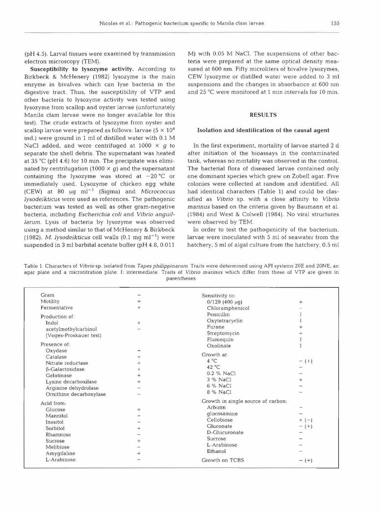
Table 1. Characters of Vi brio sp. isolated from Tapes philippin arum Traits were determined using API systems 20E and 20NE, an agar plate and a microtitration plate. 1: intermediate . Traits of Vibrio marinus which differ from those of VTP are given in
parentheses
Gram Motility Fermentative
Production of: lndol acetylmethylcarbinol (Voges-Proskauer test)
Presence of : Oxydase Catalase Nitrate reductase I3-Galactosidase Gelatinase Lysine decarboxilase Arginine dehydralase Ornithine de carboxylase
Acid from : Glucose Mannitol Inositol Sorbitol Rhamnose Sucrose Melibiose Amygdaline L-Arabinose
+ +
+
+ + + + + +
+ +
+
+
+
Sensitivity to: 0/129 (400 ).lg) Chloramphenicol Penicillin Oxytetracyclin Furane Streptomycin Flumequin Oxolinate
Growth at: 4 °C 42 oC
0.2 % NaCI 3 % NaCI 6 % NaCI 8 % NaCI
Growth in single source of carbon: Arbutin glucosamine Cellobiose Gluconate D-Glucuronate Sucrase L- Arabinose Ethanol
Growth on TCBS
+
+ +
- ( + )
+
+ (- ) - (+)
- (+)

156 Dis. aquat. Org. 14: 153-159, 1992
Table 2. Tapes philippin arum infected by Vibrio sp. Mortality (%, SD in parentheses) of Manila clam larvae inoculated with different samples, and bacterial counts in larvae on Day 5 Control: nothing, SW: 5 ml of seawater from hatchery, Al: 5 ml of algal culture from hatchery , La 0.5 ml of ground diseased larvae, LaF: 05 ml of 0.2 ~lm filtrate of the ground diseased larvae, B 10' ml- 1 (final concentration) of bacteria isolated from larvae of the previous mortality bioassay. nd : not deter-
mined
Batches Da ys after inocula tion No. bacteria larva- 1
3 5 ZobeU TCBS
Control 1.6 (1.5) 11.4 (7 .0) nd SW 3.2 (2.7) 76.2 (15.2) 1.0 x 10' 51 Al 0.7 (1.2) 7.6 (67) nd La 20.5 (9.6) 93.2 (4.7) 2.4 X 10³ 760 LaF 14 (1.6) 9.1 (8.7) nd B 117 (4 .2) 90.2 (8.5) 5.3 x l03 2
of grour:d diseased larvae or 0.5 ml of a 0.2 pm filtrôtf> of ground diseased larvae as indicated in Table 2. An additional batch received VTP at a final concentration of 104 cells ml- l . Algal inoculum and 0.2 ).lm filtra te of ground larvae did not result in disease (Table 2). In contrast, VTP, ground larvae and seawater samples induced mortality. The VTP was subsequently isolated in high numbers from those batches inoculated with seawater and VTP but not from the group inoculated with ground larvae. Small numbers of vibrios from diseased larvae were observed on TCBS agar, except from the ground larvae batch (Table 2).
Mortality appeared more slowly in postlarvae (Table 3). Only 21 % of postlarvae were dead 8 d after the inoculation.
Table 3. Tapes philippinarum infected by Vibrio sp . Sensitivity of 55 d old Manila clam postlarvae to VTP, values are mortality in percent (SD in parentheses). Control: without inoculation, Inoculate: inoculated with 100 ceUs ml-lof VTP
Batches
Control Inoculate
Days after inoculation
678
1.2 (1.1) 54 (34)
41 (1.8) 17.8 (113)
Epidemiological study
4.2 (2.2) 20.8 (12 .6)
Only the sample of bivalve digestive contents collected from the pond clearly induced disease (Table 4). lnlet seawater taken in the hatchery induced a lesser mortality .
ln another experiment, VTP added at a final concentration of 4 x 10⁵ cells rnl- 1 into 2 1 of seawater without larvae disappeared quickly (Fig. 2). After 7 dit was no
Table 4. Vibrio sp. Epidemiological study to locate VTP using bioassays, values are mortality of Tapes philippinarum in percent (SD in parentheses). Numbered sampling areas correspond to those indicated on Fig . 1. Control: nothing: Sd (1): 0.1 g of sand coUected above the pump, SW1 (2) : 300 ml of tank seawater, Biv (3): 0.2 ml of digestive content removed from different bivalves collected in the pond (used as reservoir for seawater and phytoplankton); SDp (4): 0.1 g of pond sediment, SWp (5): 300 ml of pond seawater, SWh (6): 300 ml of inlet
seawater after the sand filter
Batches Days after inoculation
4 6 8
Control 9.6 (4.0) 5.9 (2.7) < 10 Sd (1) 09 (13) 0.2 (0.3) < 10 SW1 (2) 8.7 (3.8) 6.8 (7.3) < 10 Biv (3) 2.6 (06) 7.2 (34) 79.3 (2.7) SDp (4) 5.6 (3.9) 5.8 (0.1) < 10 SWp (5) 6.8 (3.4) 10.0 (5.1) < 10 SWh (6) 4.3 (0.6) 12.8 (6.5) 28.5 (17)
5 \ E --- 4 -&1 U
.a Z 3
" 0 .... 2
1
0
0 2 4 6 8 DAYS
Fig. 2. Vibrio sp. Survival of VTP in seawater
longer detected by immunofluorescence in 100 ml of seawater filtered through a 0.2 !-lm membrane. Nevertheless, VTP was still present after 10 d, sin ce 500 ml of this contaminated seawater, poured into a 2 1 beaker containing healthy larvae, induced mortality.
The number of via ble ceUs sufficient to initiate the disease in larvae was < 1 ceU 5 ml- 1 (Table 5). Moreover, the delay in mortality between the highest and lowest concentration did not exceed 2 d.
Direct counts by immunofluorescence indicated an equivalent number of VTP in larvae and the surrounding seawater during the mortality period (seawater: 1.5 x 10⁴ to 3 X 10⁶ ceUs ml-l, larvae: 1. 5 x 10³ to 7 X 10⁵
ce lis larva- 1).

Nicolas e t al.: Pathogenic bacterium specific to Manila clam larvae 157
Table 5. Tapes philippin arum infected by Vi brio sp . Determina tion of the cell number of the pathogenic strain sufficient to induce disease in 3 d old larvae; values are mortality in percent (SD in parentheses)
Batches
3
Control 4.8 (2 .8) 4 x 10⁴ cells ml- 1 a 39.0 (1 .5) 20 cells ml- l 11.3 (2.1) 2 cells ml- 1 12.8 (0.3) 1 cell 5 ml- 1 11.0 (13)
• Final concentration of VTP inoculum
Specificity oi VTP
Oyster and scallop larvae were not sensitive to VTP No mortality was observed after 1 wk. The number of VTP, followed by direct counts and plate counts , showed a rapid decrease and disappearance in oyster larvae and seawater (Table 6).
Sensitivity lo lysozyme
VTP was resistant to the different lysozymes, whereas Escherichia coli, Vibrio anguillarum and !,v1jcrococcus lysodekticus were sensitive to lysozyme activity (Table 7).
Days after inoculation
4 5 6
113 (8 .1) 173 (8 .3) 857 (13) 100
87 .1 (14) 980 (0.3) 613 (6 .1) 98.0 (0.3) 40.8 (4 .8) 92. 5 (1.7)
DISCUSSION AND CONCLUSION
The causal agent of this disease was isolated and identified as a Vibrio sp. with an a ffi nit y to Vibrio marin us. lt was not related to the vibrio of brown ring disease, since their characters on API 20E were very different and no cross-reaction occurred with the respective antisera (Paillard & Maes 1990).
VTP did not kill scallop and oyster larvae. This is the first report of a highly specific bacterial strain pa thogenic to bivalve larvae, but additional tests with Tapes decussatus larvae would further elicit its specificity . Vibrio anguillarum and V. tubiashii are considered pathogens for several species (Brown 1981, Jeffrie s 1982, Garland et al. 1983, Lodeiros et al. 1987).
Table 6. Crassostrea gigas exposed to Vibrio sp . Sensitivity of oyster larvae to VTP and elimination of VTP. Direct counts were made using immunofluorescence in one contaminated beaker
Days after Mortality (% ) Direct counts of VTP inoculation Control Contaminated Per larva Per 100 ml seawater
2 3 2 1.5 X 10³ 3.0 X 10³
3 3 4 < 102 < 102
4 12 6 < 102 < 102
Table 7. Lysozyme activity on VTP in comparison with a reference strain (Micrococcus lysodeikticus) and 2 gram-negative strains (Escherichia coli and Vibrio anguillarum). Values represent the average change in optical density ç 'over a period of 10 min.
nd: not determined
Strains Lysozyme from:
Oyster larvae Scallop larvae Chicken egg white
M. lysodeikticus 7.9 x 10⁻⁵ 2.0 X 10- 4 1.3 X 10⁻³
VTP 0 0 0 E coli nd 3.0 x 10⁻⁵ 4.1 X 10- 4
V. anguillarum nd 6.6 x 10⁻⁵ 7.8 X 10- 4

158 Dis. aquat. Org. 14: 153-159, 1992
Other pathogenic vibrios have been tested only on Crassostrea virginica larvae (Brown & Losee 1978, Eiston & Leibovitz 1980) or on Ostrea edulis larvae and spat (Nottage & Birkbeck 1986, 1987) and their degree of specificity remains unknown. However, VTP seemed to be particularly specific, and it possessed unusual traits: it did not grow on TCBS agar; it did not survive very long in seawater, or quickly became undetectable; it was resistant to chloramphenicol; a smaU number of cells could provoke the disease; it was resistant to lysozyme activity; and it was pathogenic for postlarvae.
The survival time for VTP in the seawater was closer to that of a freshwater bacterium, such as Aeromonas salmonicida (Rose et al. 1990), than that of a marine bacterium. For example, the Vibrio of brown ring disease survived 7 d without decreased viability (Maes pers. comm.). Nevertheless, it is considered a true marine bacterium since it requires sodium chloride for '::jIowth.
The spontaneous disappearance of VTP in seawater makes it difficult to detect. This may explain why, in the second experiment (see Table 2) , VTP was not isolated in the batch inoculated with sick larvae. lndeed, as mortality occurred sooner than in the other contaminated batches, it may have been at a low level on the sampling day and saprophytic bacteria, especiaUy vibrios, may have been masking it.
Since VTP seemed to multiply only in larvae, the mechanism of infection may be invasive. Attempts to detect toxins of VTP using the methods of Nottage &
Birkbeck (1986) have failed (Birkbeck pers. comm.). Hs resistance to lysozyme activity may permit an initial multiplication in the digestive tract. The tissues may th en be progressively attacked. The high number of bacteria observed by TEM in the digestive tract prior to the formation of lesions corroborated this hypothesis. This pathogenesis is very similar to pathogenesis type III described by Elston & Leibovitz (1980).
Oyster and scallop larvae were resistant to VTP, although their lysozyme could not inactivate il. Other digestive enzymes which could lyse bacteria in bivalves have not been reported in the literature. Lysozyme is apparently the only digestive enzyme which can digest the peptidoglycan of bacteria in bivalves (Birkbeck & McHenery 1982). Thus a specific attachment to the digestive wall of Manila clam would explain the specificity of VTP and the resistance to it by oyster and scallop larvae. This has already been suggested by Eiston & Leibovitz (1980) in regard to pathogenesis type III. According to the review of Finlay & Falkow (1989) adhesion pla ys a significant role in the colonization of different sites, especially the digestive tract of mammals. This could also explain why even a small number of bacteria could induce the disease.
Two types of adhesion are known: frimbrial and
non-fimbrial. Pili in the Vibrionacae were originally observed by Tweeby et al. (1968), but recently, investigations in this are a have been carried out in the field of aquaculture. Sorne observations have shown that vibrios can altach to the host target. A vibrio pathogenic to the flounder Paralichthys olivaceus was found to bind to a cellline (Muroga et al. 1990). Elston & Leibovitz (1980) also observed an invasive Vibrio sp. attached to the periostracum and shell of oyster larvae . A very specific attachment due to pili occurred on the periostracal lamina in association with brown ring disease (Paillard 1992); this has only been observed on this part of the periostracum and in Manila clam. Certainly altachment may play an important role for invasive bacteria in marine animais and needs further investigation.
The epidemiological study revealed the contaminated areas in and around the hatchery. VTP was primarily found in bivalve tissues and in the pond. Howcvcr, it \vas aiso present in the inlet seënvater in the first and second experiments. Seawater was pumped from the open sea through beach sand, where no VTP was detected; therefore, VTP cou Id only have multiplied in the sand tilter where it could colonize sorne small animais, since the seawater pipes are frequently chlorinated. Sediments did not seem to be contaminated by VTP. VTP did not multiply or survive in algal culture , but vibrios do not generally occur in healthy algal culture (Prieur 1981, Nicolas et al. 1989).
Finally, VTP was eliminated in the hatchery and its surrounding environment by drying out the pond and by drying out and disinfecting the facilities for 11/ 2 mo. Since 1987, this pathogen has not been observed.
LITERA TURE CITED
Baumann, P., Furniss, A L., Lee, J. V (1984). Genus 1. Vibrio Paeini 1854, 411 AL ln: Krieg , N. R (ed.) Bergey's manu al of systematie bacteriology, Vol. 1. Williams & Wilkins, Baltimore, p . 518-538
Birkbeck, TH., McHenery, J. G. (1982) . Degradation of Baeteria by Mytilus edulis. Mar. Biol. 72: 7-15
Brown, C. (197 3). The effeet of sorne seleeted bacteria on embryos and larvae of the American oyster, Crassostrea virginica. J. Invertebr Pathol. 21. 215-223
Brown, C. (1981). A study of two sheUfish-pathogenic Vi brio strains isolated from a Long Island hatehery during a recent outbreak of disease. J . Invertebr. Pathol. 38: 281-293
Brown, c. , Losee, E. (1978) . Observations on natural and indueed epizootics of vibriosis in Crassostrea virginica larvae . J. Invertebr. Pathol. 31. 41-47
DiSalvo, L. H, Blecka, J., Zebal, R. (1978) . Vibrio anguillarum and larval mortality in California coastal shellfish hatchery. Appt environ. Microbiol. 35 219-221
Eiston, R, Leibovitz, L (1980) . Pathogenesis of experimental vibriosis in larval American Oysters, Crassostrea virginica Cano J. Fish . Aquat. Sei. 37: 964-978
Finlay, B B, Falkow, S. (1989) . Common themes in mierobial pathogenicity Microbiol. Rev . 53(2) : 210-230

Nicolas et al.: Pathogenic bacterium specific to Manila clam larvae 159
Garland, C O., Nash, G. v., Summer, CE., McMeekin, T A. (1983), Bacterial pathogens of oyster larvae (Crassostrea gigas) in a Tasmanian hatchery. Aus!. J , mar Freshwat. Res. 34: 483--487
Gruffydd , L O., Beaumont, A. R. (1972), A method for rearing Pecten maximus larvae in the laboratory . Mar. Biol. 15: 350-355
Jeffries , V. E. (1982) . Three Vi brio strains pathogenic to larvae of Crassostrea gigas and Ostrea edulis, Aquaculture 29 201-226
Kent, M. L (1982), Characteristics and identification of Pasteurella and Vibrio species pathoge nic to fishes using API-20E (Analytab Products) multitube test strips, Can, J. Fish. Aquat. Sci, 39 1725-1729
Lodeiros, C, Bolinches, J" Dopazo, C P., Toranzo, A. E. (1987). Bacillary necrosis in hatcheries of Ostrea eduJis in Spain. Aquaculture 65: 15-29
MacDonell, M. T , Singleton , F. L , Hood , M, A, (1982). Diluent composition for use of API 20E in characterizing marine and estuarine bacteria . Appl. environ. Microbiol. 44: 423--427
McHenery, J. G ., Birkbeck, T H. (1982) , Characterization of the lysozyme of Mytilus edulis (L) , Comp, Biochem. Physiol. 4 : 583-589
Muroga, K" Yasunobu, H., Okada, N., Masumura, K. (1990). Bacterial enteritis of cultured flounder Paralichthys olivaceus larvae, Dis. aquat. Org. 9: 121- 125
Nicolas, J. L, Robic, E., Ansquer, D, (1989), Bacterial flora associated with a trophic chain consisting of microalgae, rotifers and turbot larvae: influence of bacteria on larval survival. Aquaculture 83: 237-248
This article was presented by A. K. Sparks, Seattle, Washington, USA
Nottage, A. S" Birkbeck, T H. (1986). Toxicity to marine bivalves of culture supernatant fluids of the bivalvepathogenic Vibrio strain NCMB 1338 and other marine vibrios, J , Fish Dis. 10: 265-273
Nottage, A. S. , Birkbeck, T H. (1987), Production of proteinase during experimental infection of Ostrea edulis L larvae with Vibrio aiginoliticus NCMB 1339 and the antigenic relationship between proteinase produced by marine vibrios pathogenic for fish and shellfish, J . Fish Dis. 10 265- 273
Paillard, C (1992). Etiologie et caractérisation de la maladie de l'anneau brun chez la palourde d'élevage Ruditapes phiiippinarum , Thèse d'Université, UBO, Brest, France
Paillard, C , Maes, P (1990). Etiologie de la maladie de l'anneau brun chez Tapes philippinarum: pathogénicité d 'un Vibn'o sp, C r. Acad, Sei., Paris 310: 15-20
Prieur, D. (1981) . Les relations entre mollusques bivalves et bactéries hétérotrophes en milieu marin , Etude analytique et expérimentale, Thèse Doctorat d 'Etat de Sciences Naturelles, Brest, France
Rose , A. S" Ellis , A. E" Munro, A. L S, (1990), The survival of Aeromonas saimonicida subsp, saimonicida in seawater. J, Fish Dis, 13: 205-214
Tweeby, J, M, Park, R, W, A., Hodgkiss, W (1968), Evidence for the presence of fimbriae (pili) on Vibrio species. J, gen , Microbiol. 51: 235-244
West, p, A., Colwell, R. R, (1984), Identification and classification of Vibrionacae, An overview, In: Col weil, R. R. (ed,) Vibrios in the environment. John Wiley and Sons, New York, p. 285-363
Manuscript tirst received: April 12, 1991 Revised version accepted: Juiy 24, 1992


![Submitted: Accepted: Alga Turbinaria ornata against ...) leaves brown/black spots, necrosis in the hepatopancreas and gill disablement [6,7]. A pathogenic Gram negative bacterium,](https://static.fdocuments.in/doc/165x107/5f6e885ec59b667a8c3ded4b/submitted-accepted-alga-turbinaria-ornata-against-leaves-brownblack-spots.jpg)