
ISIC2017 DICOM Dermatology Clunie - David Clunie's Medical ... · DICOM Dermatology Specifics 1993...
42
-
Upload
doankhuong -
Category
Documents
-
view
218 -
download
0
Transcript of ISIC2017 DICOM Dermatology Clunie - David Clunie's Medical ... · DICOM Dermatology Specifics 1993...

Background & Disclosures l Owner, PixelMed Publishing, LLC l Radiologist
l Independent Consultant – GE, Carestream, Curemetrix, MDDX, Pathcore
l Editor of DICOM Standard (NEMA contract) l Formerly co-chair DICOM Standards Committee l Formerly co-chair IHE Radiology Technical
Committee

Interoperability
“the ability of two or more systems or components to exchange information
and to use the information that has been exchanged”
IEEE Standard Computer Dictionary: A Compilation of IEEE Standard Computer Glossaries. 1990

l layers: technology, data, human, institutional
l consumer empowerment
l privacy, security l competition,
homogeneity, innovation
l efficiencies, complexity
l by design l over time l architectures

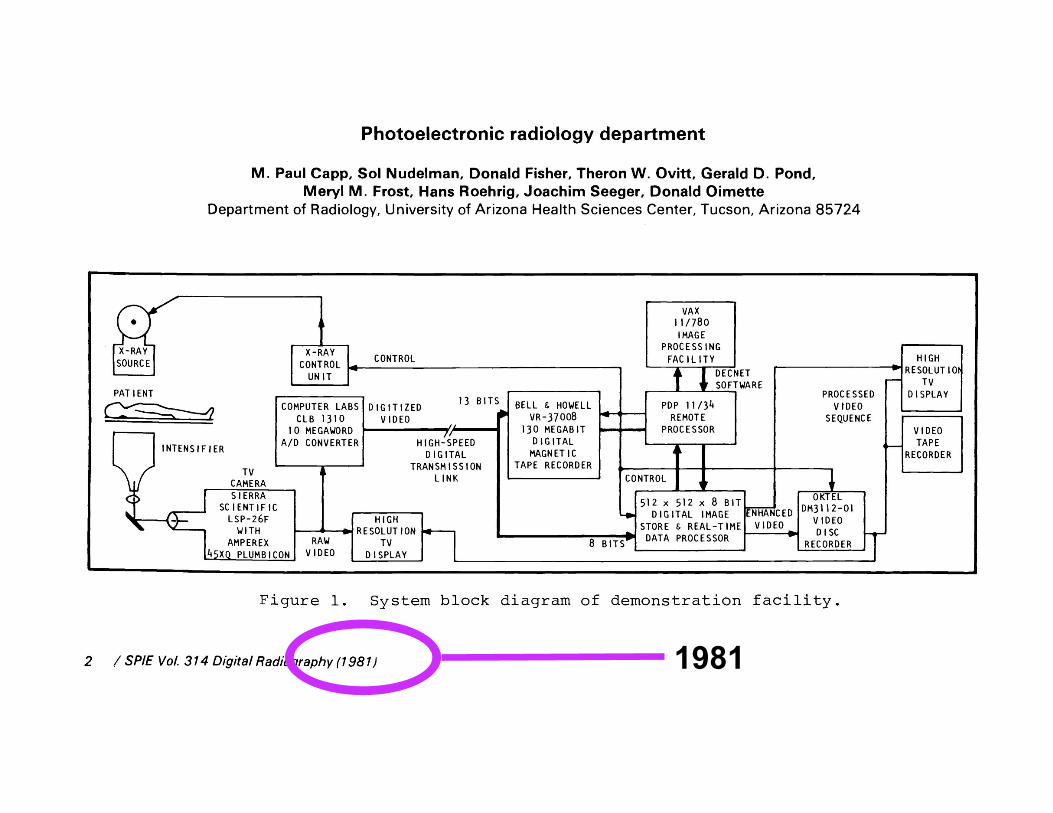
1981

1982

32 years ago – radiology PACS and DICOM ubiquitous 15-20 years later!
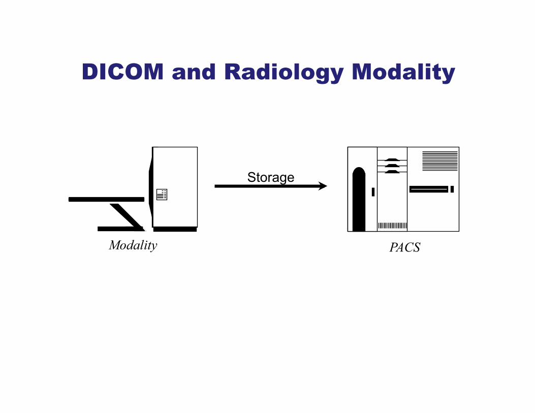
DICOM and Radiology Modality
Modality PACS
Storage
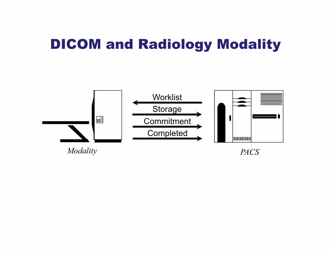
DICOM and Radiology Modality
Modality PACS
Storage Commitment
Worklist
Completed

DICOM and Cameras (Dermoscopes, etc.)
PACS
Storage
Camera

DICOM and Cameras (Dermoscopes, etc.)
PACS
Storage Commitment
Worklist
Completed Camera

DICOM Modality to PACS
Modality
Archive Modality
Modality
Modality
PACS +/- RIS
Manager
Workstations
Standard Boundary
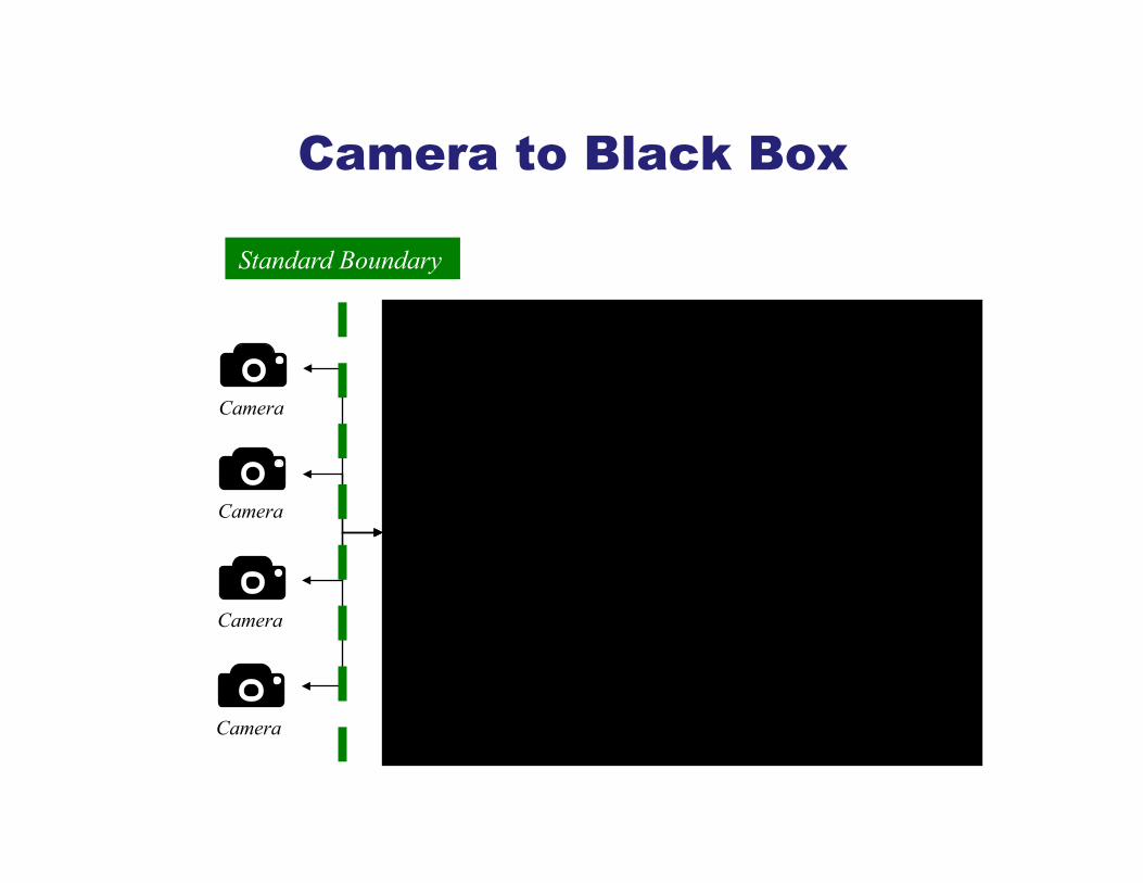
Camera to Black Box
Standard Boundary
Camera
Camera
Camera
Camera
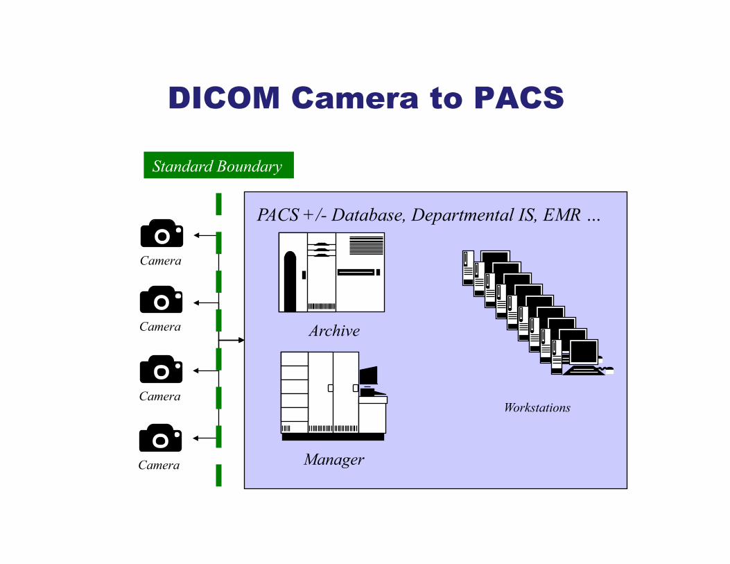
DICOM Camera to PACS
Archive
PACS +/- Database, Departmental IS, EMR …
Manager
Workstations
Standard Boundary
Camera
Camera
Camera
Camera
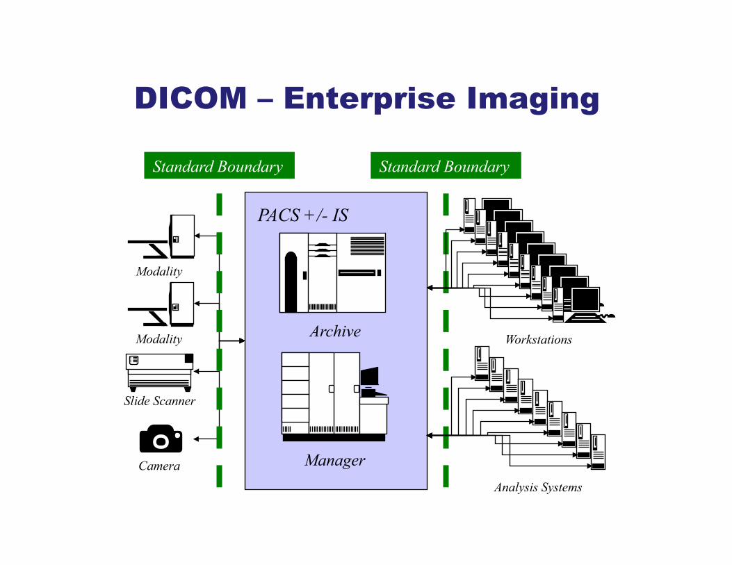
DICOM – Enterprise Imaging
Archive
PACS +/- IS
Manager
Standard Boundary Standard Boundary
Slide Scanner
Modality
Modality Workstations
Analysis Systems Camera

DICOM – Deconstructed PACS
Archive
Manager
Standard Boundary Standard Boundary
Modality
Modality Workstations
Analysis Systems
Slide Scanner
Camera
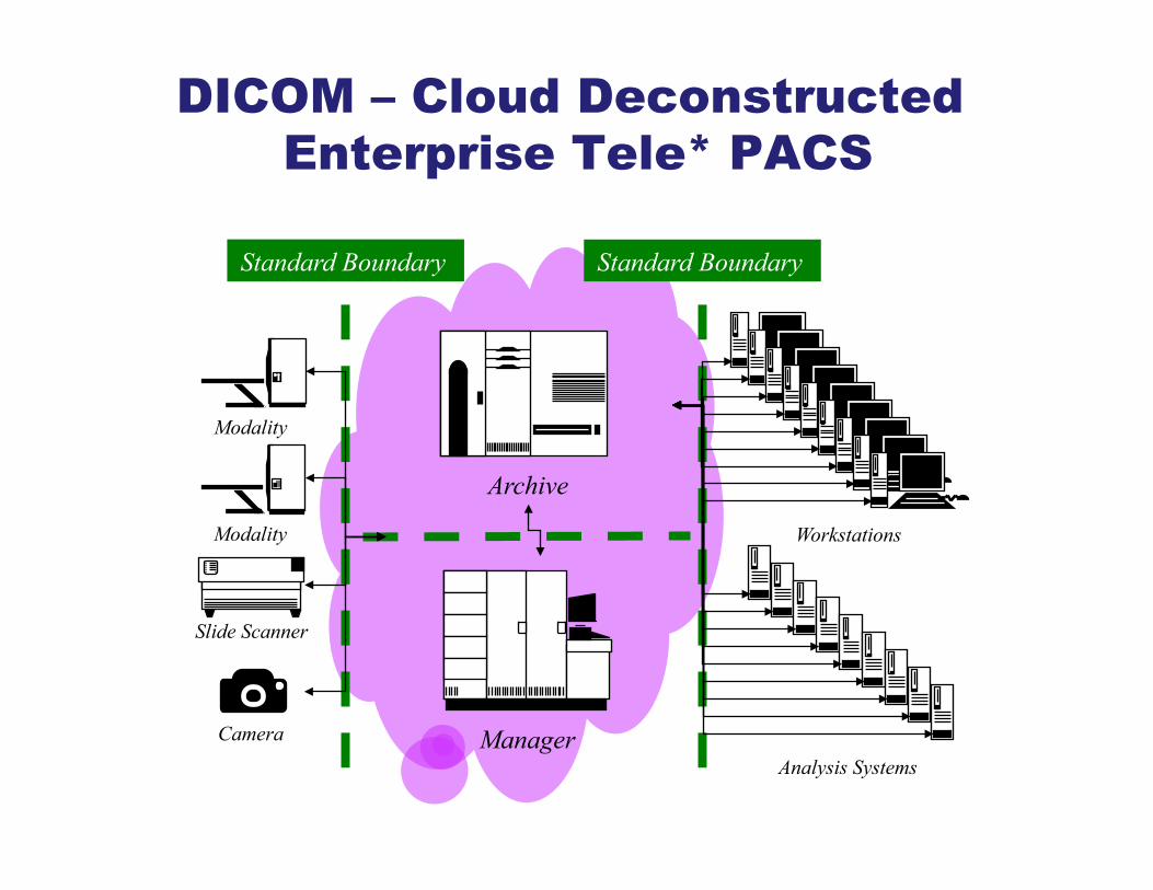
DICOM – Cloud Deconstructed Enterprise Tele* PACS
Archive
Manager
Standard Boundary Standard Boundary
Modality
Modality Workstations
Analysis Systems
Slide Scanner
Camera
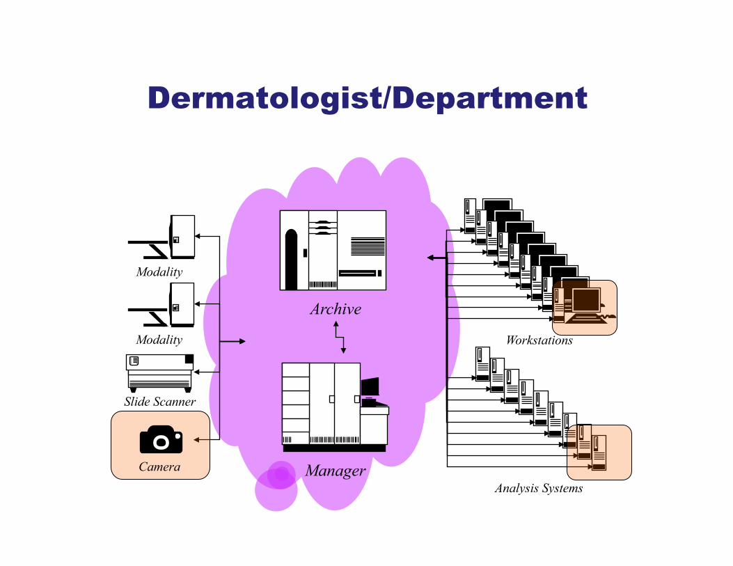
Dermatologist/Department
Archive
Manager
Modality
Modality Workstations
Analysis Systems
Slide Scanner
Camera
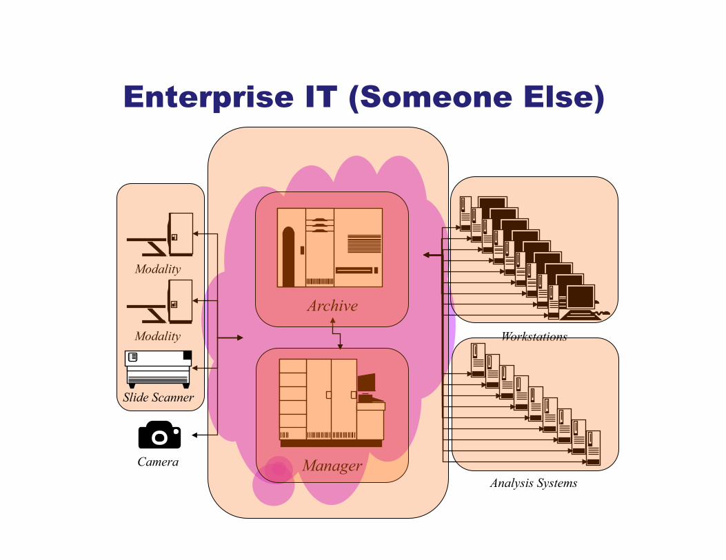
Enterprise IT (Someone Else)
Archive
Manager
Modality
Modality Workstations
Analysis Systems
Slide Scanner
Camera

http://medium.com/digital-trends-index/its-the-metadata-stupid-12a4fc121e45#.4zhwdz5y0
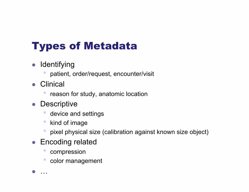
Types of Metadata l Identifying
• patient, order/request, encounter/visit
l Clinical • reason for study, anatomic location
l Descriptive • device and settings • kind of image • pixel physical size (calibration against known size object)
l Encoding related • compression • color management
l …


DICOM – More than a file format
l Payload • bulk data (pixels: uncompressed, JPEG, …) • metadata (structured, coded, standardized)
l Services • transfer (storage) • query/retrieve (find and request) • workflow management (worklists) • object management (errors, lifecycle) • annotation (contours, segments, classify)
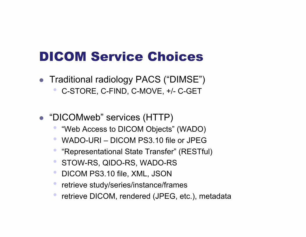
DICOM Service Choices l Traditional radiology PACS (“DIMSE”)
• C-STORE, C-FIND, C-MOVE, +/- C-GET
l “DICOMweb” services (HTTP) • “Web Access to DICOM Objects” (WADO) • WADO-URI – DICOM PS3.10 file or JPEG • “Representational State Transfer” (RESTful) • STOW-RS, QIDO-RS, WADO-RS • DICOM PS3.10 file, XML, JSON • retrieve study/series/instance/frames • retrieve DICOM, rendered (JPEG, etc.), metadata

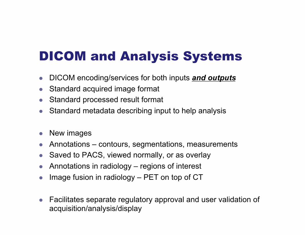
DICOM and Analysis Systems l DICOM encoding/services for both inputs and outputs l Standard acquired image format l Standard processed result format l Standard metadata describing input to help analysis
l New images l Annotations – contours, segmentations, measurements l Saved to PACS, viewed normally, or as overlay l Annotations in radiology – regions of interest l Image fusion in radiology – PET on top of CT
l Facilitates separate regulatory approval and user validation of acquisition/analysis/display
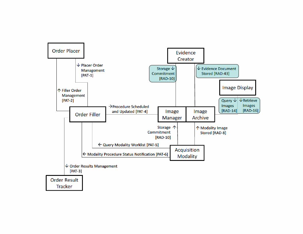

DICOM objects working together
Analysis Workstation
Current DICOM
Images from Modality
DICOM Segmentation
DICOM Registration
DICOM SR
DICOM Real World Value
DICOM Parametric Map Images
PACS Store, Distribute
and Review
Previous DICOM
Images from PACS
Previous DICOM SR etc

DICOM Dermatology Specifics 1993 to 2017 l 1993 – Initial standard – Secondary Capture, JPEG payload l 1999 – Sup 15 Visible Light Image for Endoscopy, Microscopy,
and Photography – coded anatomy, acquisition context, …
l 1999 – WG 19 – formed (DSC Minutes 1999/06) l 2001 – WG 19 – disbanded (DSC Minutes 2001/04) l 2009 – WG 19 – proposal to reactivate (WG6 Minutes 2009/08)
l Just do it (or not) … no need for specific SOP Class, extras?
l 2010 – CP 1017 – Add ICC profile to all color IODs l CP 1674 on anatomic locations (surface anatomy) l CP 1736 on EXIF tag mapping to DICOM attributes
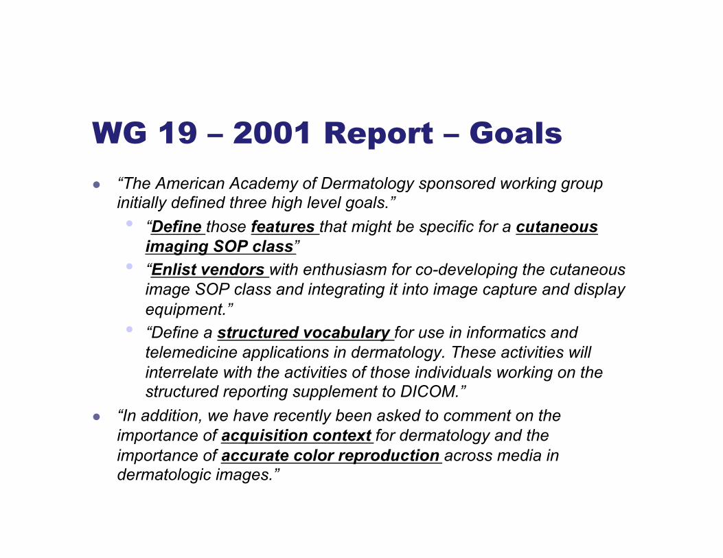
WG 19 – 2001 Report – Goals l “The American Academy of Dermatology sponsored working group
initially defined three high level goals.” • “Define those features that might be specific for a cutaneous
imaging SOP class” • “Enlist vendors with enthusiasm for co-developing the cutaneous
image SOP class and integrating it into image capture and display equipment.”
• “Define a structured vocabulary for use in informatics and telemedicine applications in dermatology. These activities will interrelate with the activities of those individuals working on the structured reporting supplement to DICOM.”
l “In addition, we have recently been asked to comment on the importance of acquisition context for dermatology and the importance of accurate color reproduction across media in dermatologic images.”
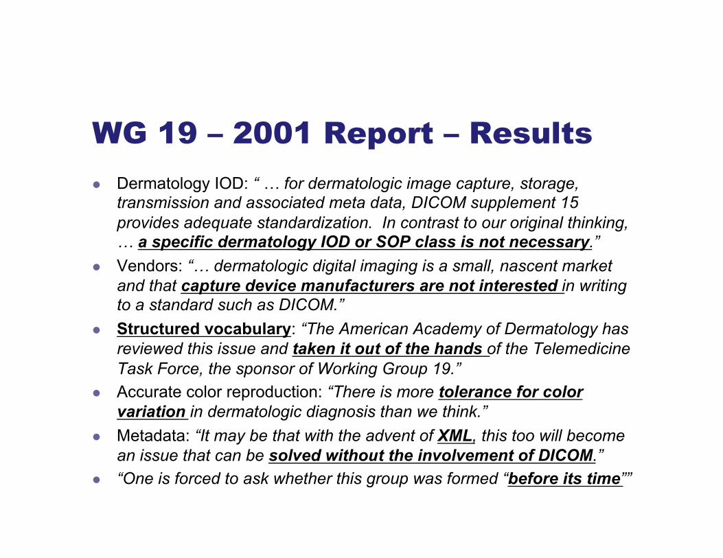
WG 19 – 2001 Report – Results l Dermatology IOD: “ … for dermatologic image capture, storage,
transmission and associated meta data, DICOM supplement 15 provides adequate standardization. In contrast to our original thinking, … a specific dermatology IOD or SOP class is not necessary.”
l Vendors: “… dermatologic digital imaging is a small, nascent market and that capture device manufacturers are not interested in writing to a standard such as DICOM.”
l Structured vocabulary: “The American Academy of Dermatology has reviewed this issue and taken it out of the hands of the Telemedicine Task Force, the sponsor of Working Group 19.”
l Accurate color reproduction: “There is more tolerance for color variation in dermatologic diagnosis than we think.”
l Metadata: “It may be that with the advent of XML, this too will become an issue that can be solved without the involvement of DICOM.”
l “One is forced to ask whether this group was formed “before its time””

WG 19 – Madden 2009 l “There is a reason why Working Group 19 has made no progress for a
decade in the middle of a digital revolution … missing key incentives … before … move to integrate digital imaging with diagnosis … :
l Although dermatologists commonly capture digital images, they do not depend on them for diagnosis.
l Since adequate dermatological diagnoses result from face-to-face physical exams, the status quo is viable (but not close to optimal).
l Therefore, there is no concern about using COTS cameras and displays since the images are just for documentation and teaching.
l Use of inexpensive COTS equipment is reinforced because it is all that can be supported by the low reimbursement level for most dermatologic visits.
l The low level of capital equipment expenditure doesn’t attract sufficient vendor investment.
l Unfortunately, the lack of vendor investment suppresses the development, validation and general clinical acceptance of applications for the use of digital images in dermatologic diagnosis and visualization.
l The lack of vendor investment hasn’t caused clinical concern …”

CP 1674 - Add Dermatology Anatomic Site Context Group …

CP-1736 – Add Photography Attributes [from] EXIF …

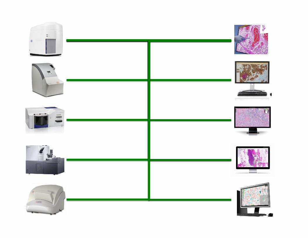
DICOM & New/Exotic Modalities l Re-use existing IODs (always, or interim solution):
• If camera-like, use VL Photo IOD • if gross microscopy, use VL Slide • if high resolution microscopy, use WSI • can always add name-value pairs (Acquisition Context), private
attributes, new standard attributes (e.g. EXIF) • Can always add new codes (e.g., anatomy, view, …)
l May want new IODs – add Supplements to DICOM • ? whole body photography if too large (e.g., need tiles, or non-
Cartesian projection/coordinates (like wide-field retinal photography)
• ? confocal microscopy • ? multi-spectral, if not 1 channel or 3 channel (RGB)
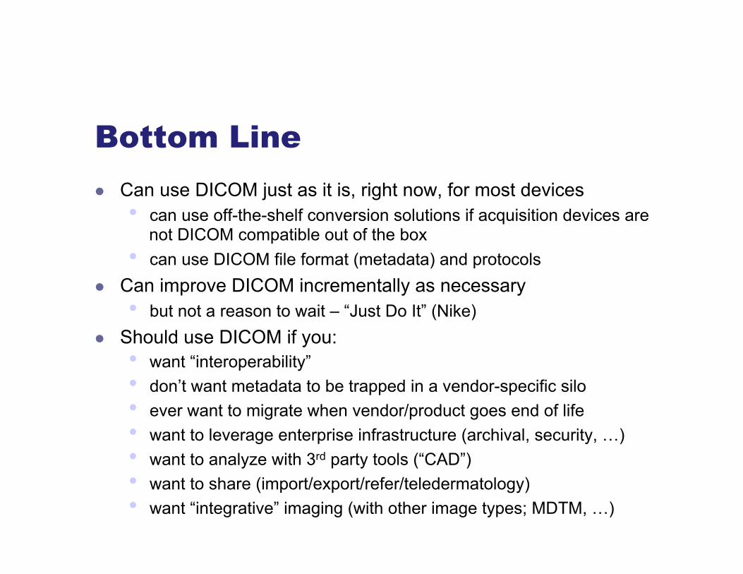
Bottom Line l Can use DICOM just as it is, right now, for most devices
• can use off-the-shelf conversion solutions if acquisition devices are not DICOM compatible out of the box
• can use DICOM file format (metadata) and protocols l Can improve DICOM incrementally as necessary
• but not a reason to wait – “Just Do It” (Nike) l Should use DICOM if you:
• want “interoperability” • don’t want metadata to be trapped in a vendor-specific silo • ever want to migrate when vendor/product goes end of life • want to leverage enterprise infrastructure (archival, security, …) • want to analyze with 3rd party tools (“CAD”) • want to share (import/export/refer/teledermatology) • want “integrative” imaging (with other image types; MDTM, …)
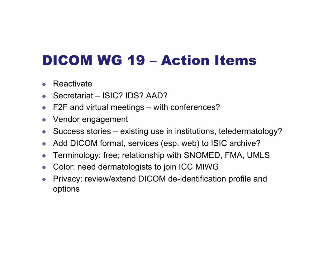
DICOM WG 19 – Action Items l Reactivate l Secretariat – ISIC? IDS? AAD? l F2F and virtual meetings – with conferences? l Vendor engagement l Success stories – existing use in institutions, teledermatology? l Add DICOM format, services (esp. web) to ISIC archive? l Terminology: free; relationship with SNOMED, FMA, UMLS l Color: need dermatologists to join ICC MIWG l Privacy: review/extend DICOM de-identification profile and
options


![DICOM Conformance Statement9d48995e-cb8b-4ac4-ae9b... · 2020. 2. 20. · DICOM protocol. 1.5 References [DICOM PS 3 2006] The Digital Imaging and Communications in Medicine (DICOM)](https://static.fdocuments.in/doc/165x107/60e78a442d236e0f92518d06/dicom-conformance-statement-9d48995e-cb8b-4ac4-ae9b-2020-2-20-dicom-protocol.jpg)