IS THERE A PREDICTABLE RELATIONSHIP BETWEEN … · Laboratoire d’Immunologie Hôpital de...
12
52 European Cells and Materials Vol. 7. 2004 (pages 52-63) DOI: 10.22203/eCM.v007a06 ISSN 1473-2262 Abstract There is much interest in predicting and controlling the outcome of interaction between artificial surfaces and liv- ing cells. However, although there is an impressive amount of information on the behaviour of many cell populations deposited on a variety of surfaces, there is presently no available theory to explain or even summarize these data. Indeed, it is not even obvious that such a theory may exist. The aim of the present review is to emphasize the prob- lems encountered when one attempts to build such a theory. Three sequential steps of cell surface interactions are con- sidered: 1) protein adsorption is a preliminary step liable to involve irreversible interaction between the surface and several hundreds of molecular species occurring in blood or plasma. 2) the second step is the formation of adhesive bonds. Several theoretical frameworks were suggested to account for this step, including DLVO theory, physical chemistry of surfaces, and formation of specific ligand- receptor bonds. It is concluded that present evidence sup- ports the latter approach, although this involves serious difficulties. 3) The last step is the triggering of a specific cell program such as apoptosis, proliferation, migration, differentiation or activation. Recent evidence suggests that in addition to the nature and amount of stimulated surface receptors, additional cues such as substratum mechanical or topographical properties may significantly affect cell behaviour. Key words: Protein adsorption, DLVO theory, interfacial energy, ligand-receptor bonds, cell adhesion, cell signal- ling. * Address for correspondence: P. Bongrand Laboratoire d’Immunologie Hôpital de Sainte-Marguerite, BP 29 13274 Marseille Cedex 09 France. Telephone Number: (+33) 491 260 331 FAX Number : (+33) 491 757 328 E-mail : [email protected] Introduction There is no need to emphasize the potential interest of controlling or even predicting the outcome of encounters between cells and artificial surfaces. Indeed, such knowl- edge would greatly facilitate the production and use of biomaterials. However, there is no evidence that a suit- able theoretical framework might exist. It is not even ob- vious that this will impede future progress in producing biomaterials. Indeed, many examples such as vaccination or the development of antibiotics show that powerful pro- cedures may be developed long before the theoretical basis required to understand them. Despite these limitations, many authors have looked for basic laws of cell-surface interaction. This might be useful not only to explain available data, but perhaps also to summarize them, or to suggest new experiments that might provide unforeseen knowledge. The aim of the present review is to discuss previous work in the light of recent evidence in order to facilitate future progress. In order to increase clarity, it appeared appropriate to split cell-surface interaction in three roughly sequential steps, although this is only an approximation: First, it is well known that when an artificial surface is exposed to biological fluids, it becomes coated with proteins within seconds or less (Baier and Weiss, 1975). Hence, what cells see are only modified surfaces. Thus, an essential point is to predict and control the structure of the adsorbed layer formed on any given biomaterial. How- ever, this is a quite complex phenomenon due to the mul- tiplicity of proteins occurring in biological media, inter- action between these proteins, and importance of time- dependent conformational changes. Second, a critical step is the formation of adhesive bonds between cells and surface. Cell adhesion has been a field of intense activity during the last three decades, and an enormous amount of information has been ob- tained. It remains to organize this information in order to make it tractable. Third, when a cell has adhered to a surface, it may have to chose an appropriate developmental line: indeed, it may undergo apoptosis and die or on the contrary sur- vive and proliferate, it may remain on the site of adhesion or start migrating, it may undergo some kind of differen- tiation, finally, it may stay in a resting state or on the con- trary trigger active processes such as synthesis and/or se- cretion of active mediators. The basis of the decision of the cell is a problem of the highest interest for the bio- logical community. Much progress has been made in dis- secting signalling cascades and developmental mecha- nisms. However, integrating available information is much IS THERE A PREDICTABLE RELATIONSHIP BETWEEN SURFACE PHYSICAL-CHEMICAL PROPERTIES AND CELL BEHAVIOUR AT THE INTERFACE? J. Vitte, A. M. Benoliel, A. Pierres and P. Bongrand* INSERM UMR600-CNRS FRE2059, Laboratoire d’Immunologie, Hôpital de Sainte-Marguerite, Marseille, France
Transcript of IS THERE A PREDICTABLE RELATIONSHIP BETWEEN … · Laboratoire d’Immunologie Hôpital de...

52
J. Vitte et al. Surface propertis and cell behaviourEuropean Cells and Materials Vol. 7. 2004 (pages 52-63) DOI: 10.22203/eCM.v007a06 ISSN 1473-2262
Abstract
There is much interest in predicting and controlling theoutcome of interaction between artificial surfaces and liv-ing cells. However, although there is an impressive amountof information on the behaviour of many cell populationsdeposited on a variety of surfaces, there is presently noavailable theory to explain or even summarize these data.Indeed, it is not even obvious that such a theory may exist.The aim of the present review is to emphasize the prob-lems encountered when one attempts to build such a theory.Three sequential steps of cell surface interactions are con-sidered: 1) protein adsorption is a preliminary step liableto involve irreversible interaction between the surface andseveral hundreds of molecular species occurring in bloodor plasma. 2) the second step is the formation of adhesivebonds. Several theoretical frameworks were suggested toaccount for this step, including DLVO theory, physicalchemistry of surfaces, and formation of specific ligand-receptor bonds. It is concluded that present evidence sup-ports the latter approach, although this involves seriousdifficulties. 3) The last step is the triggering of a specificcell program such as apoptosis, proliferation, migration,differentiation or activation. Recent evidence suggests thatin addition to the nature and amount of stimulated surfacereceptors, additional cues such as substratum mechanicalor topographical properties may significantly affect cellbehaviour.
Key words: Protein adsorption, DLVO theory, interfacialenergy, ligand-receptor bonds, cell adhesion, cell signal-ling.
* Address for correspondence:P. BongrandLaboratoire d’ImmunologieHôpital de Sainte-Marguerite, BP 2913274 Marseille Cedex 09 France.
Telephone Number: (+33) 491 260 331FAX Number : (+33) 491 757 328
E-mail : [email protected]
Introduction
There is no need to emphasize the potential interest ofcontrolling or even predicting the outcome of encountersbetween cells and artificial surfaces. Indeed, such knowl-edge would greatly facilitate the production and use ofbiomaterials. However, there is no evidence that a suit-able theoretical framework might exist. It is not even ob-vious that this will impede future progress in producingbiomaterials. Indeed, many examples such as vaccinationor the development of antibiotics show that powerful pro-cedures may be developed long before the theoretical basisrequired to understand them.
Despite these limitations, many authors have lookedfor basic laws of cell-surface interaction. This might beuseful not only to explain available data, but perhaps alsoto summarize them, or to suggest new experiments thatmight provide unforeseen knowledge. The aim of thepresent review is to discuss previous work in the light ofrecent evidence in order to facilitate future progress.In order to increase clarity, it appeared appropriate to splitcell-surface interaction in three roughly sequential steps,although this is only an approximation:
First, it is well known that when an artificial surfaceis exposed to biological fluids, it becomes coated withproteins within seconds or less (Baier and Weiss, 1975).Hence, what cells see are only modified surfaces. Thus,an essential point is to predict and control the structure ofthe adsorbed layer formed on any given biomaterial. How-ever, this is a quite complex phenomenon due to the mul-tiplicity of proteins occurring in biological media, inter-action between these proteins, and importance of time-dependent conformational changes.
Second, a critical step is the formation of adhesivebonds between cells and surface. Cell adhesion has beena field of intense activity during the last three decades,and an enormous amount of information has been ob-tained. It remains to organize this information in order tomake it tractable.
Third, when a cell has adhered to a surface, it mayhave to chose an appropriate developmental line: indeed,it may undergo apoptosis and die or on the contrary sur-vive and proliferate, it may remain on the site of adhesionor start migrating, it may undergo some kind of differen-tiation, finally, it may stay in a resting state or on the con-trary trigger active processes such as synthesis and/or se-cretion of active mediators. The basis of the decision ofthe cell is a problem of the highest interest for the bio-logical community. Much progress has been made in dis-secting signalling cascades and developmental mecha-nisms. However, integrating available information is much
IS THERE A PREDICTABLE RELATIONSHIP BETWEEN SURFACEPHYSICAL-CHEMICAL PROPERTIES AND CELL BEHAVIOUR AT THE
INTERFACE?
J. Vitte, A. M. Benoliel, A. Pierres and P. Bongrand*
INSERM UMR600-CNRS FRE2059, Laboratoire d’Immunologie,Hôpital de Sainte-Marguerite, Marseille, France

53
J. Vitte et al. Surface propertis and cell behaviour
more difficult than in the prediction of adhesion, which isa much shorter and simpler process. Thus, we shall onlydiscuss a few recent ideas that are currently explored.
Surface modification by soluble factors: is there atheoretical framework allowing to predict the
structure of modified surfaces ?
As recently pointed out by Norde (2000) “Knowledge ofthe adsorption behaviour of proteins has largely progressedin the past few decades, but a unified predictive theory isstill lacking”. Thus, we shall only emphasize some pointsthat appear particularly important for our purpose.
Macromolecule adsorption is a complex phenomenonAs was suggested in considering cell-surface interaction,it seems appropriate to split the adsorption process in sev-eral sequential steps:
1) first, soluble molecules will have to encounter thesurface. The order of encounters is determined in particu-lar by diffusion constants and concentrations of differentspecies.
2) The second step is a reversible binding of moleculesto surfaces. “Reversible” means that bound molecules maybe detached within a time scale shorter than that of experi-ments. In this case, denaturation is not expected.
3) The third step, which in fact proceeds concomitantlywith the second one, is a progressive modification of thecomposition of adsorbed layers: the most rapid and con-centrated species may be expected to be partially replacedwith more adhesive ones. This is the basis of the so-calledVroman effect.
4) Then, adsorbed proteins will undergo progressiveconformation changes. This usually strengthens adhesion.Also, as will be emphasized below, this will expose newinteraction sites to cells.
5) For the sake of completeness, we may consider thepossibility of continuous adsorption with formation of mul-tiple protein layers.
Why is there no general rule to relate the structure ofnative and biomolecule-coated surfaces ?The following six points may be emphasized.Low selectivity of adsorption. Most individual proteinscan get adsorbed on a variety of hydrophobic or hy-drophilic, neutral or charged surfaces. Thus, there is noselection rule allowing to restrict the potential number ofmolecule species liable to be bound by a bare surface ex-posed to biological environment. The only general way toprevent adsorption may be to coat surfaces with flexiblehydrophilic polymers such as polyethylene glycol, a gen-eral mechanism for steric stabilization (Napper, 1977).Nonadditive behaviour of different components. Whena surface is exposed to a mixture of macromolecules, thereis a competition between multiple adsorption processes.Thus, the behaviour of a mixture may not be predictedafter determining the adsorption of individual components.As an example, Lassen and Malmsten (1997) spent mucheffort to study the interaction of a ternary mixture of fi-brinogen, albumin and immunoglobulin G on differentsurfaces.
Typical biological fluids are highly complex. Indeed,while we have just emphasized the complexity of a ter-nary mixture, plasma probably contains hundreds of mo-lecular species. Even if we follow Andrade and Hlady(1987) who suggested considering only a dozen molecu-lar species likely to dominate adsorption (including albu-min, immunoglobulin G, A and M, C3 complement com-ponent, fibrinogen, haptoglobin, α1-antitrypsin, α2-mac-roglobulin, low and high density lipoproteins), biologi-cally relevant phenomena seem quite difficult to model.We are dealing with irreversible processes. It has longbeen reported that protein adsorption may result in pro-gressive conformational changes and denaturation, thuspreventing efficient exchange between adsorbed and solu-ble phase after a few hours. Cell surface attachment in-deed involves a variety of reactions beginning as soon asa few milliseconds after contact (Heinrich et al., 1999).Thus, the structure of a surface exposed to several mo-lecular species is dependent on the whole history of theadsorption process. As an example, Pitt et al. (1986)sequentially exposed polyvinylchloride, poly-ethyleneglycol or silicone elastomers to albumin and fi-brinogen: they concluded that the first adsorbed proteindominated further interaction of treated surfaces and plate-lets.It is not sufficient to know the nature and density ofadsorbed molecular species to understand interfacestructure. Indeed, as mentioned above, macromoleculeadsorption may result in extensive conformationalchanges. It has long been demonstrated that these phe-nomena had high physiological relevance. Thus, it wasreported that hydrophobic surfaces adsorbed higheramounts of fibronectin than hydrophilic ones, but the lat-ter surfaces were more efficient in binding selected anti-fibronectin antibodies and supporting cell adhesion(Grinnell and Feld, 1982). The concept that cell behav-iour at interfaces is dependent on underlying substrata aswell as adsorbed molecule layers was indeed confirmedby more recent studies (Koenig et al., 2003). This findingillustrates the complexity of protein adsorption, but alsosuggests the possibility that cell behaviour at interfacesmight somewhat reflect some features of underlying sub-strata independently of adsorbed molecules. It might beinteresting to subject this concept to experimental test.Adsorption energies are relatively low. Lastly, a gen-eral reason for the difficulty to predict the behaviour ofmacromolecules at interfaces is that ligand-receptor in-teraction energies amount to only a few percent of thefolding energy of a molecule such as a protein. Thus, evenmodest conformational changes may strongly affect mol-ecule-to-surface interaction. This is a general problemwhen one tries to derive the behaviour of proteins fromab initio principles.
In view of the aforementioned remarks, it may seem ahopeless task to try and predict the detailed structural prop-erties of an artificial surface exposed to a biological envi-ronment. It is therefore an essential point to determine towhat extent we need to know these detailed properties topredict the outcome of cell-surface interactions. In orderto address this question, we shall briefly review the maintheoretical frameworks that were used to predict cell-sur-
54
J. Vitte et al. Surface propertis and cell behaviour
face adhesion. Then, we shall briefly discuss the parametersthat are likely to affect further evolution of an adherent cell.
Which theoretical framework is best suited to predictthe occurrence of cell-surface adhesion?
Three main theoretical frameworks remain implicitly orexplicitly used to discuss experimental data on cell surfaceinteractions: DLVO theory, physical chemistry of surfacesand identification of specific molecular interactions. It isinteresting to ask first whether they remain valid, and sec-ond whether they may prove fruitful with respect to ourpurpose.
DLVO theory: is it relevant to biological systems?The DLVO theory was developed separately by Derjaguinand Landau, in Russia, and Verwey and Overbeek, in theNetherlands, during the 1940s. The theoretical basis andrelevance to biological systems may be found in textbooksor review articles (Curtis, 1967; Bongrand et al., 1982;Bongrand and Bell, 1984; Israelachvili, 1991). Therefore,we shall only mention essential features.
The DLVO theory was developed to account for the be-haviour of colloid suspensions. The interaction energy be-tween micrometer-sized particles was calculated as the sumof two terms:Electrostatic repulsion between surface charges. In bio-logical media, this interaction is strongly screened by water(the relative dielectric constant is about 78) and surround-ing ions. The latter effect results in an exponential decreaseof the interaction with a characteristic length of about 0.8nm (the so-called Debye-Hückel length).Electrodynamic attraction. The free energy of interactionbetween two semi-infinite media 1 and 2 with parallel sur-faces separated by a distance d is - A12/12πd2 in vacuum. Inaqueous environment, the Hamaker constant A12 is replacedwith a combination
A312 = [A12 - (A13 + A23)/2] (1)
where 3 stands for the medium. Usually, two similar bodieswill attract each other in water.
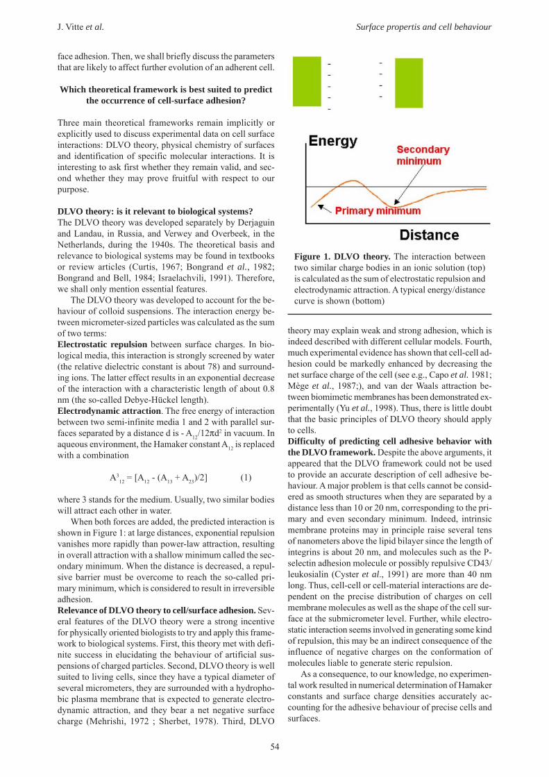
When both forces are added, the predicted interaction isshown in Figure 1: at large distances, exponential repulsionvanishes more rapidly than power-law attraction, resultingin overall attraction with a shallow minimum called the sec-ondary minimum. When the distance is decreased, a repul-sive barrier must be overcome to reach the so-called pri-mary minimum, which is considered to result in irreversibleadhesion.Relevance of DLVO theory to cell/surface adhesion. Sev-eral features of the DLVO theory were a strong incentivefor physically oriented biologists to try and apply this frame-work to biological systems. First, this theory met with defi-nite success in elucidating the behaviour of artificial sus-pensions of charged particles. Second, DLVO theory is wellsuited to living cells, since they have a typical diameter ofseveral micrometers, they are surrounded with a hydropho-bic plasma membrane that is expected to generate electro-dynamic attraction, and they bear a net negative surfacecharge (Mehrishi, 1972 ; Sherbet, 1978). Third, DLVO
theory may explain weak and strong adhesion, which isindeed described with different cellular models. Fourth,much experimental evidence has shown that cell-cell ad-hesion could be markedly enhanced by decreasing thenet surface charge of the cell (see e.g., Capo et al. 1981;Mège et al., 1987;), and van der Waals attraction be-tween biomimetic membranes has been demonstrated ex-perimentally (Yu et al., 1998). Thus, there is little doubtthat the basic principles of DLVO theory should applyto cells.Difficulty of predicting cell adhesive behavior withthe DLVO framework. Despite the above arguments, itappeared that the DLVO framework could not be usedto provide an accurate description of cell adhesive be-haviour. A major problem is that cells cannot be consid-ered as smooth structures when they are separated by adistance less than 10 or 20 nm, corresponding to the pri-mary and even secondary minimum. Indeed, intrinsicmembrane proteins may in principle raise several tensof nanometers above the lipid bilayer since the length ofintegrins is about 20 nm, and molecules such as the P-selectin adhesion molecule or possibly repulsive CD43/leukosialin (Cyster et al., 1991) are more than 40 nmlong. Thus, cell-cell or cell-material interactions are de-pendent on the precise distribution of charges on cellmembrane molecules as well as the shape of the cell sur-face at the submicrometer level. Further, while electro-static interaction seems involved in generating some kindof repulsion, this may be an indirect consequence of theinfluence of negative charges on the conformation ofmolecules liable to generate steric repulsion.
As a consequence, to our knowledge, no experimen-tal work resulted in numerical determination of Hamakerconstants and surface charge densities accurately ac-counting for the adhesive behaviour of precise cells andsurfaces.
Figure 1. DLVO theory. The interaction betweentwo similar charge bodies in an ionic solution (top)is calculated as the sum of electrostatic repulsion andelectrodynamic attraction. A typical energy/distancecurve is shown (bottom)

55
J. Vitte et al. Surface propertis and cell behaviour
Surface thermodynamicsThe apparent discrepancy between the existence of physi-cal interactions between cell surfaces and the incapacityof DLVO framework to yield useful prediction is a strongincentive to look for a better suited theoretical model todeal with this interactions. The concepts and models de-veloped by surface physical chemists seem worthy of in-terest, since they were used to deal with actual surfaces ofincompletely known molecular structure. First, we shalldefine basic parameters, then, we shall try to identify ba-sic postulates required to apply these concepts to cellularmodels. Finally, we shall discuss their usefulness on thebasis of several examples. Note that this approach wasrecently discussed in an insightful review from Morra andCassinelli (1997).Basic principles. The starting point is the concept of in-terfacial energy (Adamson, 1976). As shown in Figure 2,making a cell (C) adhere to a substratum (S) in aqueousenvironment (W) amounts to replacing cell-water and sub-stratum-water interfaces with a cell-substratum interface.Using obvious notations, basic thermodynamic principlesimpose that cell-substratum adhesion will occur if this in-volves a decrease of the system free energy, i.e.
γCS < γCW + γSW (2)
Although this approach seems perfectly rigorous, it mustbe emphasized that its use relies on two essential postu-lates:1) interfacial energy should exist! This means that while itis well known and accepted that actual solid surfaces maynot be homogeneous at the submicrometer level, it wouldbe difficult to apply surfaces physical chemistry to cellswithout assuming that membrane composition is suffi-ciently uniform to allow for the definition of some aver-age cell-water interfacial energy.2) There must exist a combining rule to relate the threeparameters of equation (2). Otherwise, we may not hopeto predict the outcome of interaction between two surfacesthat have been studied individually. Further, which isworse, in absence of a reliable combining rule, it is quitedifficult to determine the free energy of a solid-liquid in-terface.
Indeed, while it is fairly easy to determine the surface
tension of a liquid (Adamson, 1976), solid surfaces areusually studied by measuring the contact angle of sessiledroplets deposited on the surface and using standardYoung-Dupré equation. Thus, denominating liquid, solidand vapor phases as L, S and V respectively (Fig. 3) :
γLV cosθ = γSV - γSL (3)
Thus, experimental determination of θ and γLV only yieldsa relationship between two unknown parameters, i.e. γSLand γSV.
Despite general thermodynamic reasoning (Neumannet al., 1974) suggested the possibility that there might ex-ist a fairly universal combining rule between aforemen-tioned parameters, compelling experimental evidenceshows that the interfacial energy γ12 between two liquidsis not a simple function of their surface tensions γ1 and γ2(Bongrand et al., 1988). An example is shown in Table 1.
However, there remains a possibility that the interfa-cial energy between two media might be derived frommaterial parameters. Thus, the work of adhesion per unitarea of interaction between two media 1 and 2 might betentatively written as a product :
W12 = α1 α2 (4)
where α1 and α2 are intrinsic material parameters specificfor medium 1 and 2 respectively. This approximation isnot entirely unreasonable in view of molecular theories ofintermolecular forces (Margenau and Kestner, 1969;Israelachvili, 1991). An immediate consequence would be
Figure 2. Surface physics and adhesion. The adhe-sion of a cell (C) to a surface (S) in aqueous medium(W) amounts to replacing CW and SW interfaces by aCS interface.
Figure 3. Contact angles. The determination of thecontact angle of a liquid droplet (L) deposited on asurface (S) in a medium (V) provides a relationshipbetween three interfacial energy parameters.
Table 1. There is no universal combining rule for in-terfacial energies
Liquid 1 Liquid 2 γ1 (mJ/m2) γ2 (mJ/m2) γ12 (mJ/m2) water n-octanol 72.8 27.5 8.5 water CCl4 72.8 27.0 45.0
The above example shows that interfacial energies be-tween water and two liquids of similar surface tensioncan be quite different (adapted from Bongrand et al.,1988)
56
J. Vitte et al. Surface propertis and cell behaviour
that the work of adhesion to medium 1 and 2 embedded inmedium 3 would be:
W312 = - (α1 - α3) (α2 - α3) (5)
According to equation (3), two bodies exerting mutualattraction in vacuum might repel each other in a mediumof parameter α3 comprised between α1 and α2. Severalpoints may be emphasized:
- First, equation (4) is only the simplest of a series offormulae that were suggested. Indeed, the simple product(4) was replaced with more complicated expressions suchas a harmonic mean. Also, it appeared reasonable to splitthe material properties of a given medium into separateparameters accounting for different interaction such aselectrodynamic attraction or polar interactions. The latterview was developed into a workable scheme by Van Osset al. (1987) who suggested to split parameter α into threeseparate constants, respectively accounting for apolar, elec-tron donor and electron acceptor component. These com-ponents could be obtained by performing contact anglemeasurements with three selected liquids.
- Second, an essential assumption is that two interact-ing surfaces will adapt their distance and conformation toreach some “equilibrium state”. This requires a minimumamount of flexibility to allow the existence of an intrinsicinterfacial energy parameter.
- Third, the experimental check of these concepts ismade difficult by the excess of unknown parameters. Thisexplains why this remains debated (Morra and Cassinelli,1997).
- Fourth, the application of surface thermodynamicsto cells is made still more difficult by specific problems.Thus, it is not possible to obtain cell surfaces as planarstructures extended enough to allow contact angle meas-urements. Therefore, no accepted method presently allowsquantitative determination of cell-medium and cell-sub-stratum interfacial energy, thus precluding experimentalcheck of any combining rule.
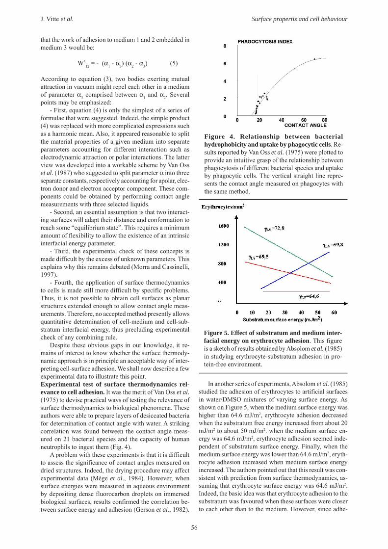
Despite these obvious gaps in our knowledge, it re-mains of interest to know whether the surface thermody-namic approach is in principle an acceptable way of inter-preting cell-surface adhesion. We shall now describe a fewexperimental data to illustrate this point.Experimental test of surface thermodynamics rel-evance to cell adhesion. It was the merit of Van Oss et al.(1975) to devise practical ways of testing the relevance ofsurface thermodynamics to biological phenomena. Theseauthors were able to prepare layers of desiccated bacteriafor determination of contact angle with water. A strikingcorrelation was found between the contact angle meas-ured on 21 bacterial species and the capacity of humanneutrophils to ingest them (Fig. 4).
A problem with these experiments is that it is difficultto assess the significance of contact angles measured ondried structures. Indeed, the drying procedure may affectexperimental data (Mège et al., 1984). However, whensurface energies were measured in aqueous environmentby depositing dense fluorocarbon droplets on immersedbiological surfaces, results confirmed the correlation be-tween surface energy and adhesion (Gerson et al., 1982).
In another series of experiments, Absolom et al. (1985)studied the adhesion of erythrocytes to artificial surfacesin water/DMSO mixtures of varying surface energy. Asshown on Figure 5, when the medium surface energy washigher than 64.6 mJ/m2, erythrocyte adhesion decreasedwhen the substratum free energy increased from about 20mJ/m2 to about 50 mJ/m2. when the medium surface en-ergy was 64.6 mJ/m2, erythrocyte adhesion seemed inde-pendent of substratum surface energy. Finally, when themedium surface energy was lower than 64.6 mJ/m2, eryth-rocyte adhesion increased when medium surface energyincreased. The authors pointed out that this result was con-sistent with prediction from surface thermodynamics, as-suming that erythrocyte surface energy was 64.6 mJ/m2.Indeed, the basic idea was that erythrocyte adhesion to thesubstratum was favoured when these surfaces were closerto each other than to the medium. However, since adhe-
Figure 4. Relationship between bacterialhydrophobicity and uptake by phagocytic cells. Re-sults reported by Van Oss et al. (1975) were plotted toprovide an intuitive grasp of the relationship betweenphagocytosis of different bacterial species and uptakeby phagocytic cells. The vertical straight line repre-sents the contact angle measured on phagocytes withthe same method.
Figure 5. Effect of substratum and medium inter-facial energy on erythrocyte adhesion. This figureis a sketch of results obtained by Absolom et al. (1985)in studying erythrocyte-substratum adhesion in pro-tein-free environment.

57
J. Vitte et al. Surface propertis and cell behaviour
sion occurred in all situations, the authors concluded thatsome other mechanisms must be involved in addition tosurface energy effects.
Other experiments allowed direct estimate of adhesionenergy (Tözeren et al., 1989; Tözeren, 1990). Individualcytotoxic T lymphocytes and target cells were manoeu-vred into contact with micropipettes (Fig. 6) and gradu-ally separated. Determination of the angle between mem-branes allowed direct determination of 2-dimensional ad-hesion energy, provided membrane tension was first de-termined by standard micropipette aspiration techniques.It was concluded that the adhesion energy increased whenthe contact area decreased, in accordance with a theoreti-cal model assuming that binding was mediated by indi-vidual bonds (Bell et al., 1984).
Thus, the aforementioned experiments, as well as otherexperiments, strongly suggest that surface energy effectscan indeed influence biological adhesion, but it was notpossible to demonstrate that cell adhesion to surfaces couldbe quantitatively predicted with a well-defined functionof physical chemical properties of interacting surfaces. Amajor problem is that even potentially useful physicalchemical properties of cell surface cannot always be meas-ured.
Can cell adhesion be entirely accounted for byspecific bonds?While the above two theoretical approaches were inspiredby results from physics and chemistry, the description ofcell adhesion as a consequence of specific ligand-receptorinteractions is more akin to current biological way of think-ing. The basic idea is that cell adhesion cannot be pre-dicted from general principles, but requires a detailedknowledge of peculiar properties of studied systems.Cell adhesion as determined by specific ligand-receptorinteractions. Basic postulates. The specific view of celladhesion relies on the following two simple ideas:
- First, it is assumed that most interactions betweencells and surfaces are essentially determined by specificassociations between well-defined receptors and ligands
that could in principle be properly identified.- Second, an implicit consequence is that cell-substra-
tum interaction involves a small fraction of cell moleculararea (Pierres et al., 1998). Indeed, it is likely that 1,000molecular bonds involving a molecular contact of 10 nm2
each are sufficient to maintain a cell of 1,000 µm2 areatightly bound to a flat surface. Clearly, if only 0.001 % oftotal cell area is involved in adhesion, it is unreasonableto expect that bulk surface properties will be related toadhesive behaviour.
It must be emphasized that the use of this frameworkwill prompt particular experimental ways of studying ad-hesion. Thus, instead of assaying the hydrophobicity orcharge density of a surface, in order to predict its capacityto bind cells, we shall look for known ligands of cellreceptors, such as the well-known RGD sequence that wasfound to interact with many integrin receptors. As a con-sequence, testing a surface may rely on the quantificationof specific sites, e.g. with labelled antibodies and tech-niques such as enzyme-linked immunoassay (ELISA)rather than contact angle determination.What are specific interactions? Clearly, the above defi-nitions are based on the concept of specific interaction. Itmay be useful to emphasize that this is not as straightfor-ward as it might first seem. Two alternative definitions ofspecificity may be considered:
- First, focussing on function, an interaction might beconsidered to be specific if it is lost when the ligand ischanged. Thus, an immunoglobulin binding group A anti-gen on blood cells is said to be specific if it does not inter-act with group B antigen. However, there is a problemwith this definition, since more and more cell receptorsare recognized to be “promiscuous”, which means that theycan bind a variety of different ligands. A prominent exam-ple is represented by so-called scavenger receptors, whichare thought to play an important role in natural immunityand were shown to recognize a variety of ligands includ-ing bacterial structures or altered lipids (Pearson, 1996).Interestingly, these receptors seem to be involved in therecognition of plastic surfaces by macrophages, an inter-action that was long considered as nonspecific.
- Second, focussing on structure, an interaction maybe defined as specific if it is dependent on detailed topo-graphic features of interacting molecular surfaces (Fig. 7).Thus, attraction between surfaces bearing respectively a
Figure 6. Direct determination of the work of sepa-ration of bound cells. Cells are brought into adhesivecontact, then separated by pulling pipettes. The deter-mination of the tangents to the membranes on the sepa-ration line allows the calculation of the adhesive forceat the separation line provided membrane tension isknown. Cell mechanical properties are determined bystandard aspiration techniques.
Figure 7. Specific and non specific interactions. Theinteraction between two surfaces of opposite charge(left) may be considered as non-specific. The interac-tion between two surfaces with fitting shape and match-ing opposite charges may be considered as specific.

58
J. Vitte et al. Surface propertis and cell behaviour
net positive and negative charge may be considered as nonspecific, while an interaction involving accurate match-ing of charges and topographical features on apposed sur-faces will be considered as specific. Note that it may notbe easy to assess quantitatively the role of individual mo-lecular groups in an interaction (Clackson and Wells,1995).Is it tenable to assume that most interactions betweencells and natural or artificial surfaces are accountedfor by the involvement of a limited number of well-defined receptor species? In our view, more and morerecent evidence suggests that the answer to this questionmay be positive. Firstly, it is now well demonstrated thateach cell species is endowed with a high number of sur-face receptors. Thus, it was recently emphasized (Barclay,1998) that about 250 protein species were essentially spe-cific for leukocytes, and about half of these molecule mightact as adhesion receptors. Secondly, it is more and morewidely recognized that many receptors are multispecificor promiscuous and may be involved in interactions thatwere previously held as nonspecific. Thus, the recogni-tion of plastic surfaces by macrophages may well be dueto a particular class of scavenger receptors (Fraser et al.,1993), and a molecule such as αMβ2 integrin was reportedto recognize more than 30 protein and non protein mol-ecules including adhesion molecules, extracellular matrixcomponents or bacterial structures (Yakubenko et al.,2002). Finally, more and more specific interactions werereported between pathogens and cellular constituents suchas e.g. integrins (Watarai et al., 1996; Takeshita et al.,1998), extracellular matrix components (Bisognano et al.,1997 ; Giordano et al., 1999) or blood group antigens(Geisel et al., 1995).
Thus, there is no compelling argument to disprove theassumption that most interactions between cells and sur-face are mediated by specific cell membrane structures.Difficulty of using standard biochemical concepts toderive cell adhesion behaviour from receptor proper-ties. Clearly, in order to test the validity of the above con-cepts, we need a theoretical framework allowing the deri-vation of cell-substratum adhesive phenomena from lig-and-receptor properties. It would thus be possible to takeadvantage of the powerful immunological and geneticmethodologies that allowed biologists to characterize hun-dreds of adhesion receptors and to prepare them in solu-ble form, thus making possible the determination of affin-ity constants or kinetic association and dissociation rateswith a variety of tools based on e.g. surface plasmon reso-nance.
Unfortunately, as pointed out by Bell (1978), conven-tional biological or chemical-physical methodologies areinsufficient to fulfil this program. These insufficienciesare illustrated by a model that attracted much interest dur-ing the last ten years, namely the mechanisms allowingactivated endothelial cells to capture flowing leukocytesas a first step to inflammatory reactions (Springer, 1994).Under standard conditions, selectin molecules expressedby endothelial cells seem able to tether ligand-bearingleukocytes to the blood vessel surface, thus inducing acharacteristic jerky motion called rolling, with a transla-tion velocity of order of 5-10 µm/s, i.e. one hundredfold
lower than that of freely flowing cells. However, ICAM-1molecules bound to endothelial cells are not able to initi-ate rolling, even when their integrin receptors on leukocytesare properly activated. When this phenomenon was clearlydemonstrated (Lawrence and Springer, 1991), it was notclear whether this difference was due to insufficient range,rate of bond formation or mechanical strength of theICAM-1/integrin pair. Remarkably, no experimental toolwas available to address this problem.
During the following years, it appeared that only ex-periments done at the single molecule level could yieldreliable information on the rate of bond formation and dis-sociation between surface-attached molecules subjectedto external forces. There are several explanations for thissituation:
1) Interpreting experimental description of the separa-tion of surface linked with multiple bonds requires a quan-titative knowledge of the distribution of forces betweenmolecules and possibility of rebinding (Seifert, 2000). Thisis usually lacking.
Figure 8. Determination of unbinding forces withan atomic force microscope or the biomembraneforce probe. Using an atomic force microscope (A),a ligand-coated tip is brought into contact with areceptor-coated surface (B segment 1) and a positiveforce is applied for some time (2). Then the tip is pro-gressively separated from the surface (3), thus impart-ing a pulling force on the bond. On rupture, a jump ofthe tip (4) is observed, which allows direct measure-ment of the unbinding force. The biomembrane forceprobe method (C) may be considered as improvedatomic force microscopy: the cantilever is replacedwith a soft vesicle such as a red blood cell (C-1) main-tained with a micropipette, using variable pressure.The tip is replaced with a glass microbead glued tothe biomembrane and coated with binding molecules(green). This device allows varying the rate of forceincrease (i.e. the loading rate, r, usually expressed inpicoNewton/s) over several orders of magnitude. Theunbinding force F is dependent on the loading rate.As shown in (D), when F is plotted versus the loga-rithm of r, the obtained curve may appear as severalstraight lines yielding quantitative information on theligand-receptor energy-distance curve (Merkel et al.,1999).

59
J. Vitte et al. Surface propertis and cell behaviour
2) When multiple bonds are allowed to form betweeninteracting surfaces, the rate of bond formation is usuallydependent on the number and position of existing bonds.Also, molecule flexibility, environment and mode ofconnexion to underlying surface are important parameters.
We shall now present a brief description of recent re-sults illustrating the potential of available tools to studyreceptor-mediated interactions between cells and surfaces.Studying cell-substratum interaction at the single celllevel. During the last ten years, many authors studied therupture of individual bonds (see Bongrand, 1999 for a re-view). Many important results were obtained with threemethods (Figs. 8 and 9): atomic force microscopy (Florinet al., 1994), the biomembrane force probe (Merkel et al.,1999) and laminar chamber flow (Kaplanski et al., 1993).The latter method probably yields most straightforwardinformation. As summarized in Figure 9, when receptor-bearing cells or micrometer-size particles are driven alonga ligand-coated surface in presence of a laminar shear flowwith a typical wall shear rate of a few second-1, they aresubjected to a driving force of a few picoNewtons, thusallowing a single bond to provoke a detectable stop. Fur-ther, the translation velocity is of order of 10 µm/s, whichmakes the motion easy to monitor with high accuracy. It isthus possible to determine both the frequency and dura-tion of individual arrests or binding events. Using a suffi-cient number of arrest durations allows straightforwarddetermination of unbinding plots, i.e. dependence of thenumber of particles remaining arrested on time t after ini-tial attachment. A typical curve is shown in Figure 9.
Now, we shall describe a study recently performed inour laboratory to illustrate the potential of the method andemphasize typical features of bond formation betweensurface-attached molecules (Vitte et al., 2004). We stud-ied the interaction of fibronectin-coated surfaces and hu-man monocytic THP-1 cells under flow. Numerous bind-ing events were observed and their frequency was drasti-cally decreased by adding monoclonal antibodies knownto block VLA-5 integrin (also denominated asCD24eCD29 or α5β1), thus suggesting that adhesion wasessentially mediated by specific bonds. Unbinding plotdisplayed the typical aspect displayed on Figure 9.
Now, the problem with unbinding plots is that multi-ple nonexclusive ways of interpreting curve shape mustbe considered: indeed, previous work done in our labora-tory strongly supported the possibility that 1) delayed for-mation of additional bonds might occur after the initial
binding event, 2) binding might involve the simultaneousformation of several bonds, and 3) ligand-receptor asso-ciation might behave as a multiphasic phenomenon, withinitial formation of a transient complex and subsequentdissociation or on the contrary transition towards a morestable state. We shall now show how we may deal withthese difficulties with numerical data shown on Table 2.
These data may be interpreted as follows: bindingevents were probably mediated by multiple bonds withthe highest fibronectin surface density, since ligand dilu-tion to 3,850 sites/µm2 resulted in marked shortening ofarrest duration (see last column). However, whenfibronectin surface density was further decreased to 1,436sites/µm2, the binding frequency was strongly decreasedwithout any concomitant change of the initial detachmentrate or fraction of cell bound 1s after arrest. This stronglysuggests that binding events observed were essentiallysimilar, corresponding to the minimum detectable bindingevent. It seemed reasonable to assume that single molecu-lar interactions were indeed observed, since previous ex-periments supported the assumptions that single bondscould be detected with this methodology. However, asemphasized by Zhu et al. (2002), the single bond assump-tion is very difficult to prove formally.
Now, further experiments obtained on the same modelwill provide some support to the concept that bond topog-raphy is indeed an important determinant of cell adhesion.The surface distribution of fibronectin receptors on the
Figure 9. Studying individual ligand receptor bondswith a laminar flow chamber. The figure describesthe use of a laminar flow chamber operated at verylow shear rate.
Table 2. Influence of fibronectin surface density on association and detachment rates
6,500 1.48 ± 0.07 0.96 ± 0.10 0.51 ±0.0233,850 0.75 ± 0.08 2.26 ± 0.40 0.27 ± 0.0461,436 0.21 ± 0.02 1.94 ± 0.27 0.32 ± 0.037
The motion of monocytic THP-1 cells along surfaces coated with various densities of fibronectin was studied. Thefrequency of binding events, slope of unbinding plots at time 0 (i.e. initial detachment rate) and fraction of cellsbound 1 second after attachment are shown ± standard error (adapted from Vitte et al., 2004)
Fibronectinsurface density
(molecules/µm2)Binding frequency
(mm-1)Initial detachment
rate (s-1)
Fraction of cellsbound 1s after
attachment

60
J. Vitte et al. Surface propertis and cell behaviour
surface of THP-1 cells was manipulated with monoclonalantibodies. First, cells were treated with K20 murine anti-body, a beta 1 chain-specific monoclonal antibody con-sidered as “neutral”, i.e. without any effect on function.Second, anti-mouse immunoglobulin (Fab)’2 was addedto cross-link VLA-5. This treatment induced a markedaggregation of surface receptors as shown with a semi-quantitative confocal microscopic study, suggesting thatantibody treatment increased between 40 % and 100 %the average number of integrin receptors located in a vol-ume of ca 0.045 µm3 surrounding each integrin. Bindingdata are shown in Table 3.
Clearly, while K20 antibodies slightly decreased ar-rest frequency due to a probable decrease of accessibility,receptor aggregation increased binding frequency (from0.45 to 1.19 mm-1 and decreased detachment rate. Thissuggests the influence of receptor topography on func-tional capacity.
Conclusion
While it is likely that nonspecific physical interactions suchas electrostatic repulsion or steric stabilization may influ-ence cell-substrate adhesion, no presently available theo-retical framework can allow us to predict the outcome ofinteraction between a cell and an artificial surface of knownphysical-chemical properties. However, numerous reportssuggest that in many different situations, cell-surface ad-hesion is essentially determined by a limited number ofreceptor species. Further, recently developed experimen-tal methods allow precise determination of the propertiesof ligand-receptor interaction at the single molecule level.Thus, the most fruitful approach to understand cell-sub-stratum interaction may consist of first identifying involvedcell receptors. Indeed, even cell interaction with plasticsurfaces (Fraser et al., 1993) or foreign structures such asmicroorganisms often involve a limited number of domi-nant molecular species.
Is it possible to predict the behaviour of a celladhering to a surface through well-identified
receptors?
Despite the complexity of aforementioned processes,they may be considered as remarkably simple as comparedto the following issue: on which basis will a substratum-
adhering cell chose its subsequent behaviour? This ques-tion will certainly initiate many lines of research duringthe following years, and it is certainly located at the fron-tier of current biological knowledge. While an in-depthdiscussion of this problem would not fit within the scopeof this review, we shall emphasize some points that maybe relevant.
The biochemical approach to cell activationIt has long been known that cell adhesion strongly influ-enced cell behaviour. The most straightforward interpre-tation for a cell biologist or biochemist would certainlyconsist of assuming that the dominant phenomenon is thestimulation of cell membrane receptors, resulting in thetriggering of a cascade of biochemical events and secondmessenger generation. There is indeed much evidenceshowing that the nature of engaged membrane receptorswill influence further events. Taking a simple exampleamong many others, if a rat macrophage encounters anantibody-coated particle, it will engulf it as a consequenceof proper stimulation of immunoglobulin receptors. How-ever, if the same particle is bound through a lectin inter-acting with other membrane structures, no ingestion willfollow (Capo et al., 1978). Further, a quite detailed knowl-edge was obtained on the coupling between cell stimula-tion and simple response patterns. Thus, nearly completereconstitution of the biochemical machinery involved inthe generation of a simple process such as the phagocyteoxidative burst is conceivable (e.g. Price et al., 2002).Therefore, it might be tempting to speculate that cell be-haviour might be understood and even predicted throughdetailed identification of the nature and number ofreceptors engaged in a given interaction.
However, while enormous progress was done in theidentification of activating pathways, biochemical studiesrevealed the existence of a complex network of triggersthat cannot yet be understood with currently available bio-logical tools, and many reports suggest that new conceptsare required to integrate available information (see e.g.Charest and Pelech, 1998; Vilar et al., 2003). An impor-tant issue that might lead to huge simplification of thisproblem would be to know whether there is only a limitednumber of cell programs liable to stimulation at a givenmoment. A positive answer is indeed suggested by recentstudies on cell transcriptome.
Table 3. Influence of receptor aggregation on binding efficiency under flow
None (control) 0.75 ± 0.08 2.26 ± 0.40 0.27 ± 0.046K20 0.45 ± 0.04 1.83 ± 0.29 0.36 ± 0.042
K20 + anti mouse 1.19 ± 0.08 1.02 ± 0.14 0.55 ± 0.030
The motion of monocytic THP-1 cells along surfaces coated with moderate fibronectin density (3,850 molecules/µm2) was studied. Cells were treated with a neutral anti-beta 1 integrin monoclonal antibody (K20) with or withoutcross-linking polyclonal goat anti-mouse immunoglobulin (Fab’)2.The frequency of binding events, slope ofunbinding plots at time 0 (i.e. initial detachment rate) and fraction of cells bound 1 second after attachment areshown ± standard error (adapted from Vitte et al., 2004)
Cell treatmentBinding Frequency
(mm-1)Initial detachment
rate (s-1)Fraction of cells bound
1s after attachment
61
J. Vitte et al. Surface propertis and cell behaviour
Importance of nonbiochemical signals in thedetermination of cell behaviourWhile the biochemical view of cell guidance is certainly adominant one, on the basis of the number of publishedpapers, a steady flow of convincing reports strongly sup-ports the view that cell responses are not only dependenton the nature of free or bound ligands detected in nearbyenvironment. We shall briefly consider mechanical,physico-chemical and topographical cues.Substratum mechanical properties. It has long beendemonstrated that adherent cells exert forces on underly-ing surfaces (Harris et al., 1980). Further, cell behaviourwas also shown to depend on substratum flexibility andsome mechanistic information was recently reported(Wang et al., 2001).Substratum nonspecific properties such as charge andhydrophobicity. The influence of these properties on celladhesion has been discussed above. In addition, these fea-tures clearly influence many cell functions (Allen et al.,2003). The basic question is to know whether this actionis mediated by a few well-defined receptor species, as pre-viously argued.Surface topography. There is ample evidence that thefunctions of an adherent cell are strongly influenced bythe geometrical properties of contact areas (Pierres et al.,2002). Thus, cell proliferation was demonstrated to behighly correlated to available adhesion area (Chen et al.,1997). In addition to the total contact area, there is nowcompelling evidence that nanoscale surface features maystrongly influence adherent cells (Dalby et al., 2002 ; Sch-neider et al., 2003).Conclusion. There is obviously a need to integrate theconcepts that were only briefly sketched. Clearly, at leasttwo pathways may be considered. A first approach mightconsist of trying to follow the lines of thought that weresuccessful in the past. Thus, it might be argued that cellsurface receptor stimulation is not only dependent on lig-and recognition but also on topographical reorganization.Thus, a common mechanism of signal generation mayconsist of bringing a suitable kinase in close contact witha potential target, thus allowing tyrosine phosphorylationand generation of binding sites for scaffold proteins. Also,since it is well demonstrated that forces can change pro-tein conformation, surface mechanical properties mightaffect the forces exerted on ligand proteins and influencethe appearance of binding sites. According to this view,there might be a need to look for accurate relationshipsbetween ligand topography and receptor activation. A sec-ond approach might be to consider that biology must shiftaway from reductionism and aims at develop new meth-ods to deal with biocomplexity. A notable example is theconcept of tensegrity, suggested by Ingber (2003) as ameans of overcoming difficulties presently met by biolo-gists.
General Conclusion
While the present review certainly illustrates the need forfurther work rather than provides answers to specific prob-lems, we wish to suggest some conclusions.
First, although we may hope to be able to manipulate
cell-surface interaction before we fully understand hownew procedures work, it is certainly warranted to look forsuch an understanding, which may suggest new experi-ments and new questions, in addition to alleviate the bur-den imposed on scientists memory.
Second, among the many theoretical frameworks thatwere discussed, perhaps the relevant question is to knowwhich one is most fruitful, rather than determining whichis true. Indeed, many experiments have shown that differ-ent approaches may yield complementary information, andno basic principle was completely disproved.
Third, before new tools are developed to studybiocomplexity, it is suggested that the “specific interac-tion approach” may still be used. This would consist ofidentifying cell surface receptors involved in the interac-tion between a given cell population and a particular sur-face, thus examining (possibly at the single molecule level)the mechanisms of interactions between cells and surfaces,and last identifying the initial triggering mechanisms. Atthis stage, probably new tools will be required to deal withbiological complexity.
References
Absolom DR, Zingg W, Thomson C, Policova Z, VanOss CJ, Neumann AW (1985) Erythrocyte adhesion topolymer surfaces. J. Colloid Interface Sci 104: 51-59.
Adamson AW (1976) Physical Chemistry of Surfaces.Wiley, New York.
Allen LT, Fox EJP, Blute I, Kelly ZD, Rochev Y,Keenan AK, Dawson KA, Gallagher WM (2003) Interac-tion of soft condensed materials with living cells: pheno-type/transcriptome correlations for the hydrophobic effect.Proc Natl Acad Sci USA 100: 6331-6336.
Andrade JD, Hlady V (1987) Plasma protein adsorp-tion: the big twelve. Ann NY Acad Sci 516: 158-172.
Baier RE, Weiss L (1975) Demonstration of the in-volvement of adsorbed proteins in cell adhesion and cellgrowth on solid surfaces. Adv Chem Ser 145: 300-307.
Barclay N (1998) Concluding remarks and the chal-lenge from the immune system. Faraday Disc 111: 345-350.
Bell GI. (1978) Models for the specific adhesion ofcells to cells. Science 200: 618-627.
Bell GI, Dembo M, Bongrand P (1984) Cell adhesion:competition between nonspecific repulsion and specificbonding. Biophys J 45: 1051-1064.
Bisognano C, Vaudaux PE, Lew DP, Ng EYW, HooperDC (1997) Increased expression of fibronectin-bindingproteins by fluoroquinolone-resistant Staphylococcusaureus exposed to subinhibitory levels of ciprofloxacin.Antimicrob Agents Chemother 41: 906-913.
Bongrand P (1999) Ligand-receptor interactions. RepProg Phys 62: 921-968.
Bongrand P, Bell GI (1984) Cell-cell adhesion : pa-rameters and possible mechanisms. In: Cell Surface Dy-namics: Concepts and Models. Perelson AS, DeLisi C,Wiegel FW (eds). Marcel Dekker, New York, pp. 459-493.
Bongrand P, Capo C, Depieds R (1982) Physics of cellddhesion. Progr Surface Sci 12: 217-286.
Bongrand P, Capo C, Mège JL, Benoliel AM (1988)

62
J. Vitte et al. Surface propertis and cell behaviour
Surface physics and cell adhesion. In: Physical Basis ofCell-Cell Adhesion. Bongrand P (ed). CRC Press, BocaRaton, FL. pp 91-123.
Capo C, Bongrand P, Benoliel AM, Depieds R (1978)Dependence of phagocytosis on strength of phagocyte-particle interaction. Immunology 35: 177-182.
Capo C, Garrouste F, Benoliel AM, Bongrand P,Depieds R (1981) Nonspecific binding by macrophages :evaluation of the influence of medium-range electrostaticrepulsion and short-range hydrophobic interaction.Immunol Commun 10 : 35-43.
Charest DL, Pelech SL (1998) Intracellular communi-cation via protein kinase networks. In: Dynamical Net-works in Physics and Biology - At the Frontier of Physicsand Biology. Beysens D, Forgacs G (eds). EDP SciencesParis/Springer Berlin, pp 189-200.
Chen CS, Mrksich M, Huang S, Whitesides GM, IngberDE (1997) Geometric control of cell life and death. Sci-ence 276: 1425-1428.
Clackson T, Wells JA (1995) A hot spot of bindingenergy in a hormone-receptor interface. Science 267: 383-386.
Curtis ASG (1967) The Cell Surface - Its MolecularRole in Morphogenesis. Logos Press - Academic Press,London, pp 105-110.
Cyster JC, Shotton DM, Williams AF (1991) The di-mension s of the T lymphocyte glycoprotein leukosialinand identificaiton oflinear protein epitopes that can bemodified by glycosylation. EMBO J 10: 893-902.
Dalby MJ, Yarwood SJ, Riehle MO, Johnstone HJ,Affrossman S, Curtis AS (2002) Increasing fibroblast re-sponse to materials using nanotopography : morphologi-cal and genetic measurements of cell response to 13-nm-high polymer demixed islands. Exp Cell Res 276: 1-9.
Florin EL, Moy VT, Gaub HE (1994) Adhesion forcesbetween individual ligand-receptor pairs. Science 264:415-417.
Fraser I, Hughes D, Gordon S (1993) Divalent cation-independent macrophage adhesion inhibited bymonoclonal antibody to murine scavenger receptor. Na-ture 364: 343-346.
Geisel J, Steuer MK, Ko HL, Beuth J (1995) The roleof ABO blood groups in infections induced by Staphylo-coccus saprophyticus and Pseudomonas aeruginosa. Int JMed Microbiol Virol Parasitol Infectious Dis 282: 427-430.
Gerson DF, Capo C, Benoliel AM, Bongrand P (1982)Adhesion, phagocytosis and cell surface energy : the bind-ing of fixed human erythrocytes to rat macrophages andpolymethylpentene. Biochim Biophys Acta 692: 147-156.
Giordano R, DL Fouts, Tewari D, Colli W, ManningJE, Alves MJM (1999) Cloning of a surface membraneglycoprotein specific for the infective form of Trypano-soma cruzi having adhesive properties to laminin. J BiolChem 274: 3461-3468.
Grinnell F, Feld MK (1982) Fibronectin adsorption onhydrophilic and hydrophobic surfaces detected by anti-body binding and analyzed during cell adhesion in serum-containing medium. J. Biol. Chem. 257: 4888-4893
Harris AK, Wild P, Stopak D (1980) Silicone rubbersubstrata: a new wrinkle in the study of cell locomotion.
Science 208: 177-179.Heinrich L, Voegel JC, Schaaf P (1999) Direct obser-
vation of the anchoring process during the adsorption offibrinogen on a solid surface by force-spectroscopy modeatomic force microscopy Proc Natl Acad Sci USA 96:6705-6710.
Ingber DE (2003) Tensegrity II. How structural net-works influence cellular information processing networks.J Cell Sci 116: 1397-1408.
Israelachvili JN (1991) Intermolecular and SurfaceForces. Academic Press, New York, 2nd edition. pp 246-250.
Kaplanski G, Farnarier C, Tissot O, Pierres A, BenolielAM, Alessi MC, Kaplanski S, Bongrand P (1993)Granulocyte-endothelium initial adhesion. Analysis of tran-sient binding events mediated by E-selectin in a laminarshear flow. Biophys J 64:1922-1933.
Koenig AL, Gambillara V, Grainger DW (2003) Cor-relating fibronectin adsorption with endothelial cell adhe-sion and signaling on polymer substrates. J Biomed MaterRes 64A: 20-37.
Lassen B, Malmsten M (1997) Competitive proteinadsorption at plasma polymer surfaces. J Colloid Inter-face Sci 186: 9-16.
Lawrence, MB, Springer TA (1991) Leukocytes rollon a selectin at physiologic flow rates : distinction fromand prerequisite for adhesion through integrins. Cell 65:859-873.
Margenau H, Kestner M (1969) Theory of Intermo-lecular Forces. Pergamon Press, Oxford.
Mège JL, Capo C, Benoliel AM, Bongrand P (1984)Nonspecific cell surface properties: contact angle of wa-ter on dried cell monolayers. Immunol Commun 13: 211-217.
Mège JL, Capo C, Benoliel AM, Bongrand P (1987)Use of cell contour analysis to evaluate the affinity be-tween macrophages and glutaraldehyde-treated erythro-cytes. Biophys J 52: 177-186.
Mehrishi JN (1972) Molecular aspects of the mamma-lian cell surface. Progr Biophys Mol Biol 25: 3-70.
Merkel R, Nassoy P, Leung A, Ritchie K, Evans E(1999) Energy landscapes of receptor-ligand bonds ex-plored with dynamic force spectroscopy. Nature 397: 50-53.
Morra M, Cassinelli C (1997) Bacterial adhesion topolymer surfaces: a critical review of surface thermody-namic approaches. J Biomater Sci Polymer Edn 9: 55-74.
Napper DH (1977) Steric stabilization. J. Colloid In-terface Sci 58: 390-407.
Neumann AW, Good RJ, Hope CJ, Sejpal M (1974)An equation-of-state approach to determine surface ten-sions of low energy solids from contact angles. J ColloidInterface Sci 49: 291-305.
Norde W (2000) Proteins at solid surfaces. In: Physi-cal Chemistry of Biological Interfaces. Baszkin A, NordeW (eds). Marcel Dekker, New York. p 115.Pearson AM (1996) Scavenger receptors in innate immu-nity. Curr Opin Immunol 8: 20-28.
Pierres A, Benoliel AM, Bongrand P (1998) Interac-tions between biological surfaces. Curr Opin Colloid In-terface Sci 3: 525-533.
63
J. Vitte et al. Surface propertis and cell behaviour
Pierres A, Benoliel AM, Bongrand P (2002) Cell fit-ting to adhesive surfaces : a prerequisite to firm attach-ment and subsequent events. Eur Cells Mater 3: 31-45.
Pitt WG, Park K, Cooper SL (1986) Sequential proteinadsorption and thrombus deposition on polymericbiomaterials. J. Colloid Interface Sci 111: 343-162.
Price MO, McPhail LC, Lambeth JD, Han CH, KnausUG, Dinauer MC (2002) Creation of a genetic system foranalysis of the phagocyte respiratory burst: high-level re-constitution of the NADPH oxidase in a nonhematopoieticsystem Blood 99: 2653-2661.
Schneider GB, Perinpanayagam H, Clegg M, ZahariasR, Seabold D, Keller J, Stanford C (2003) Implant surfaceroughness affects osteoblast gene expression. J Dental Res82: 372-376.
Seifert U. (2000) Rupture of multiple parallel molecu-lar bonds under dynamic loading. Phys Rev Lett 84: 2750-2753.
Sherbet GV (1978) The biophysical characterizationof the cell surface. Academic Press, London, pp 36-145.
Springer TA (1994) Traffic signals for lymphocyterecirculation and leukocyte emigration : the multistep para-digm. Cell 76: 301-314.
Takeshita A, Murakami Y, Yamashita Y, Ishida M,Fujisawa S, Kitano S, Hanazawa S (1998) Porphyromonasgingivalis fimbriae use beta(2) integrin (CD11/CD18) onmouse peritoneal macrophages as a cellular receptor, andthe CD18 beta chain plays a functional role in fimbrialsignaling. Infection Immunity 66: 4056-4060.
Tözeren A (1990) Cell-cell conjugation. Transientanalysis and experimental implications. Biophys J 58: 641-652.
Tözeren A, Sung KLP, Chien S (1989) Theoretical andexperimental study on cross-bridge migration during celldisaggregation. Biophys J 55: 479-487.
Van Oss CJ, Gillman CF, Neumann AW (1975) Phago-cytic engulfment and cell adhesiveness as cellular surfacephenomena, Marcel Dekker, New York. pp 29-30.
Van Oss CJ, Chaudury MK, Good RJ (1987)Monopolar surfaces. Adv Colloid Surface Sci 28: 35-64.
Vilar JMG, Guet CC, Leibler S (2003) Modeling net-work dynamics : the lac operon, a case study. J Cell Biol.161: 471-476.
Vitte J, Benoliel AM, Eymeric P, Bongrand P, PierresA. (2004) Beta 1 integrin-mediated adhesion may be initi-ated by multiple incomplete bonds, thus accounting forthe functional importance of receptor clustering. BiophysJ. 86: 4059-4074.
Wang HB, Dembo M, Hanks SK, Wang YL (2001)Focal adhesion kinase is involved in mechanosensing dur-ing fibroblast migration. Proc Natl Acad Sci USA 98:11295-11300.
Watarai M, Funato S, Sasakawa C (1996) Interactionof ipa proteins of Shigella flexneri with alpha(5)beta(1)integrin promotes entry of the bacteria into mammaliancells. J Exp Med 183: 991-999.
Yakubenko VP, Lishko VK, Lam SCT, Ugarova TP(2002) A molecular basis for integrin aMb2 ligand bindingpromiscuity. J Biol Chem 277:48635-48642.
Yu ZW, Calvert TL, Leckband D (1998) Molecularforces between membranes displaying neutral
glycosphingolipids: Evidence for carbohydrate attraction.Biochemistry 37: 1540-1550.
Zhu C, Long M, Chesla SE, Bongrand P (2002) Meas-uring receptor/ligand interaction at the single-bond level:experimental and interpretative issues. Ann. Biomed. En-gineering 30: 305-314.
Discussion with Reviewers
A.S.G. Curtis: Since nearly all macromolecules either aidadhesion or retard it there may be no such thing as a neu-tral molecule to use as a control in adhesion experiments.Comment please.Authors: This question raises a very important and oftenoverlooked problem: if you wish to test the hypothesisthat a cell specifically binds to a surface coated with agiven molecular species, say A, it is tempting to use as acontrol a so-called neutral molecule B. However, resultsmay be difficult to interpret: if the cells stick to both A andB, it is difficult to rule out the possibility that the cell hasreceptors specific for B, in view of the variety and pro-miscuity of cell membrane receptors. Conversely, ifthe cell sticks to A, not to B, you may not exclude thepossibility that B is an anti-adhesive molecule, and A gen-erates nonspecific adhesion. Thus, another strategy isneeded: You may try so-called blocking experiments. An-tibodies may be quite useful, but they are not neutral mol-ecules: coating a cell or a surface with antibodies maygenerate steric hindrance, preventing cell-to-surface ap-proach, and what you test is then the cell ability to bindimmunoglobulin. A more satisfactory way of studyingspecific cell-surface interactions is to alter selectively bind-ing sites. Thus, if you are able to block interactions be-tween cells or particles and avidin-coated surfaces by add-ing the small biotin molecule in the fluid phase, you mayconclude that attachment was generated by bona fide avi-din-biotin interactions. However, blocking is not alwaysfeasible, since it is difficult to block interactions betweensurface-bound molecules with soluble ligand, as a conse-quence of the importance of multivalency. Another con-trol would consist of comparing a macromolecule with asimilar molecule with a few mutations specifically alter-ing binding sites. Clearly, this may be difficult to achieve.A reasonable procedure would consist of coating non-ad-hesive surfaces with the molecules you wish to test. Thus,if a cell does not adhere to PEG-coated surfaces, if youcouple type A molecules to PEG (i.e. polyethyleneglycol)and you induce adhesion, it seems reasonable to concludethat cells can bind to A.
A.S.G. Curtis: How does my statement equate with theauthors’ view that there may be only a few cell membranemolecules involved in adhesion?Authors: While many cell surface molecules have a po-tential to influence adhesion, it is often found that adhe-sion is dominated by one or a few molecular species un-der specific experimental conditions. In this case, the strat-egy we described may be used to analyze interactions.