Is it a fibroadenoma or a benign phyllodes...
60
Is it a fibroadenoma or a benign phyllodes tumour? Andrew Lee Nottingham University Hospitals City Hospital Campus
Transcript of Is it a fibroadenoma or a benign phyllodes...

Is it a fibroadenoma or a
benign phyllodes tumour?
Andrew Lee
Nottingham University Hospitals
City Hospital Campus

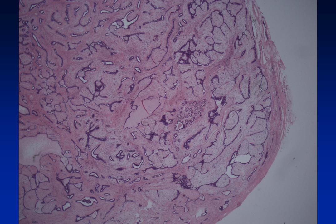

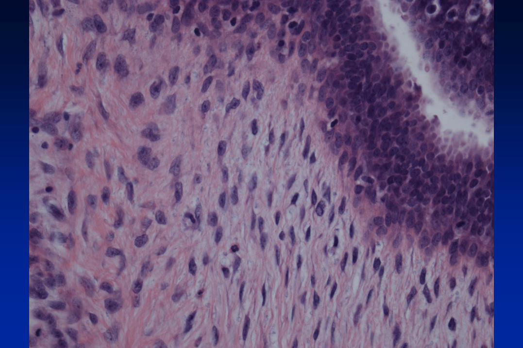
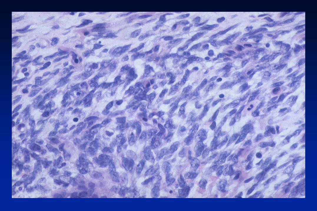
Phyllodes tumour v fibroadenoma
Stroma
• Cellularity
• Nuclear pleomorphism
• Stromal overgrowth (x5 field
with no glands)
• Mitotic rate
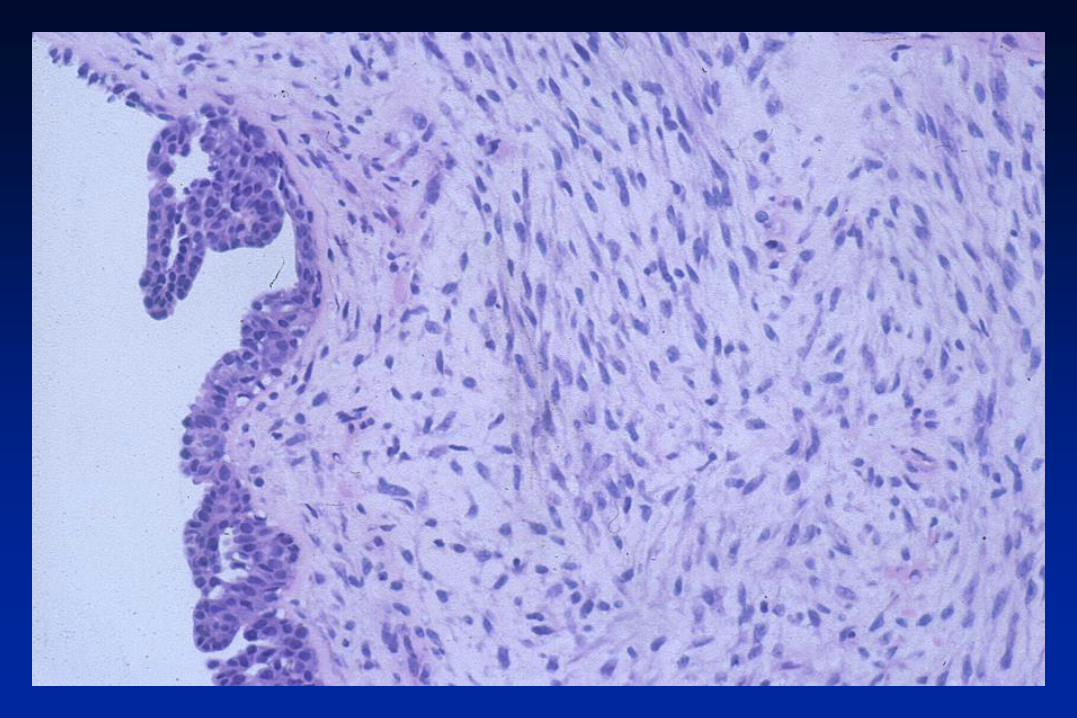
Leaf-like architecture
Irregular edge
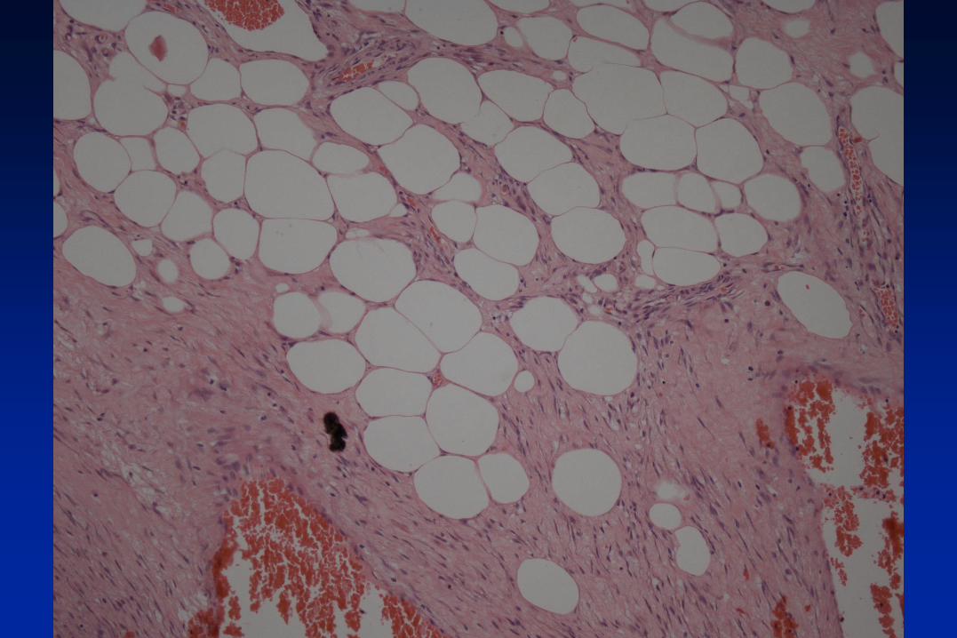
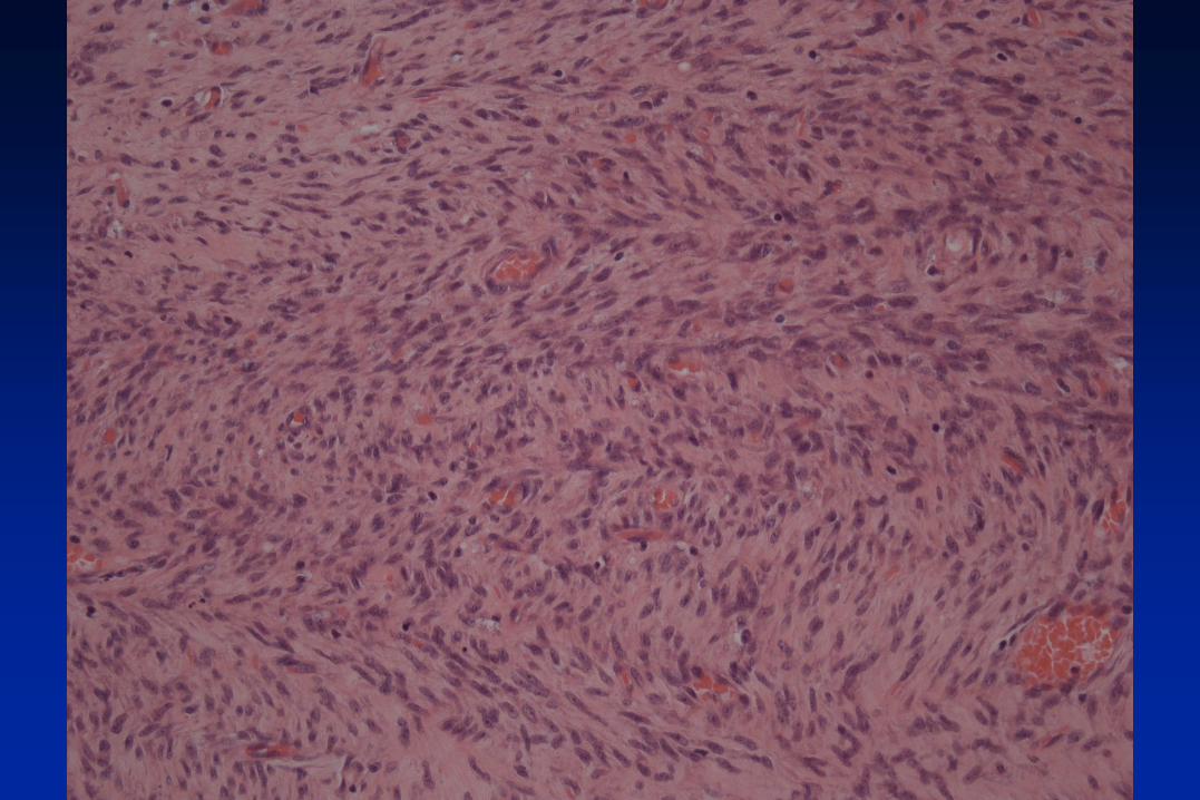

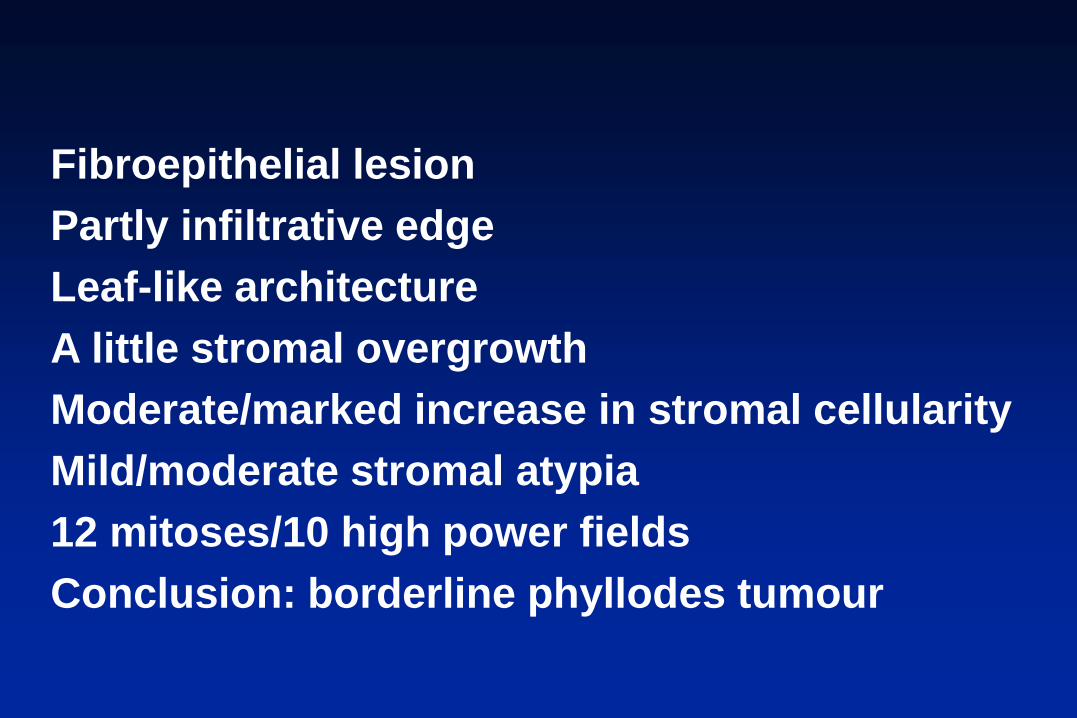
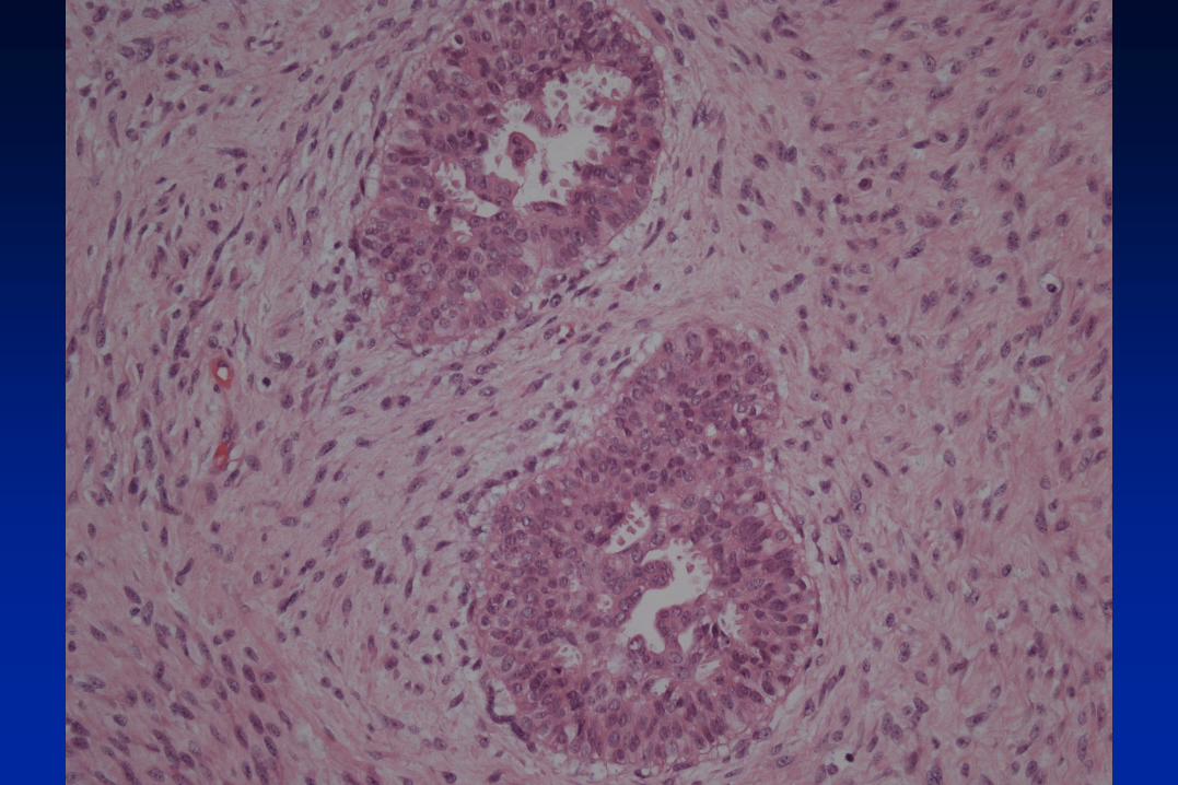
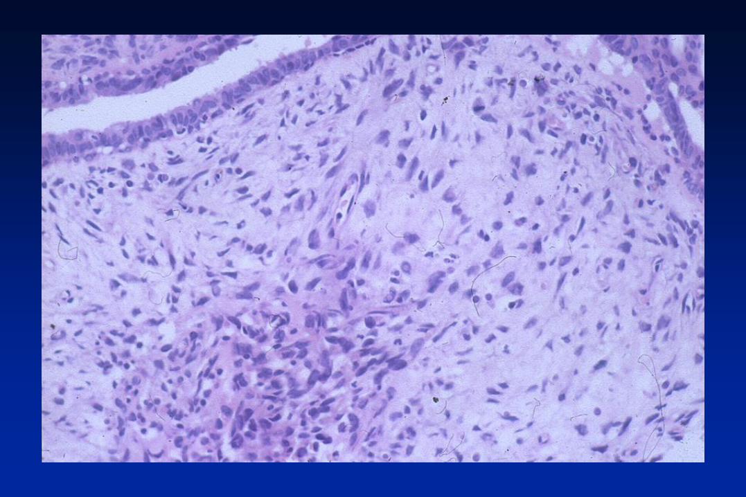
Fibroepithelial lesion
Partly infiltrative edge
Leaf-like architecture
A little stromal overgrowth
Moderate/marked increase in stromal cellularity
Mild/moderate stromal atypia
12 mitoses/10 high power fields
Conclusion: borderline phyllodes tumour


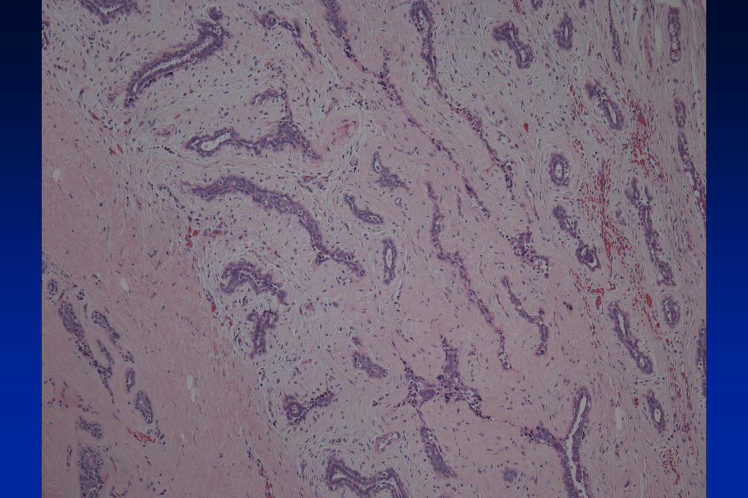


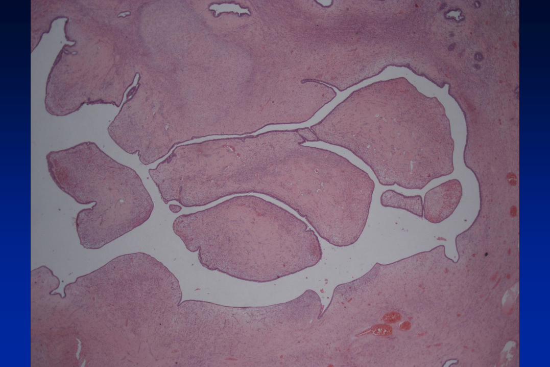
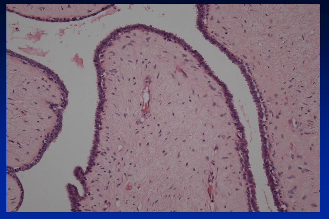
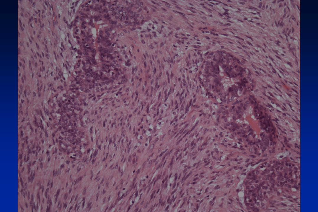
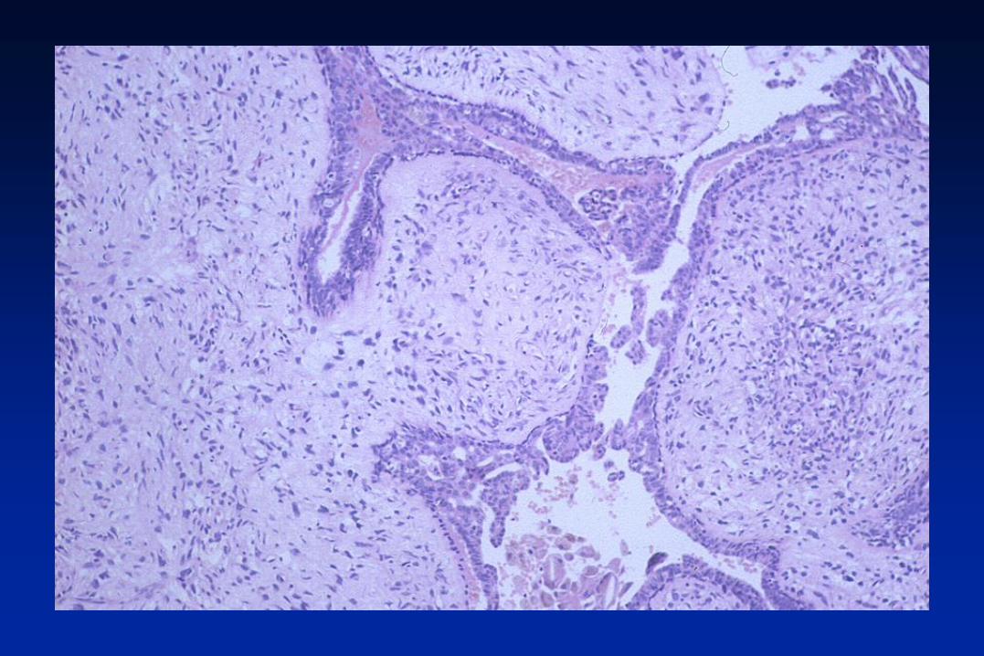
Fibroepithelial lesion
Circumscribed edge
Leaf-like areas
High proportion of stroma
Increased stromal cellularity
Focal area resembling fibroadenoma
Mild atypia (moderate in other sections)
Mitoses 3/10 high power fields
Conclusion: benign phyllodes

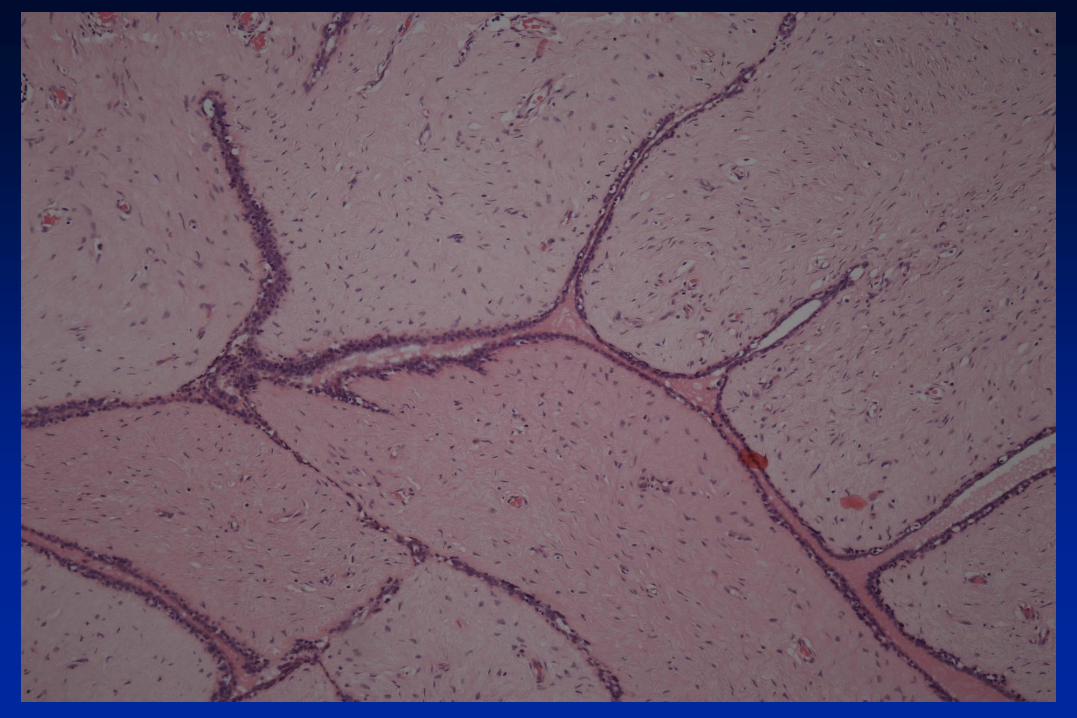

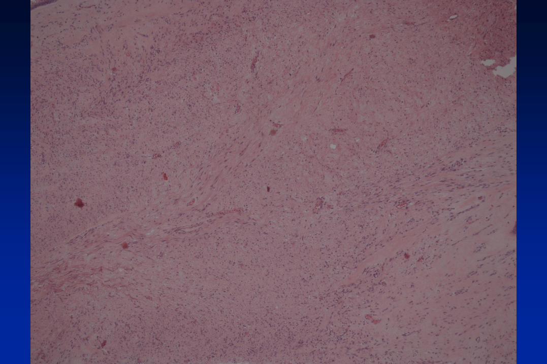
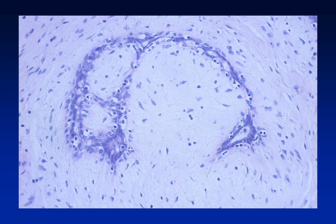
Fibroepithelial lesion
Circumscribed
Leaf-like architecture
No increase in stromal cellularity
No atypia or mitoses
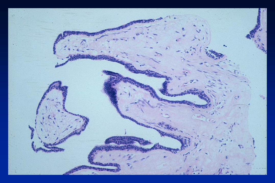
Conclusion: fibroadenoma


Fibroepithelial lesion
Circumscribed
Pericanalicular
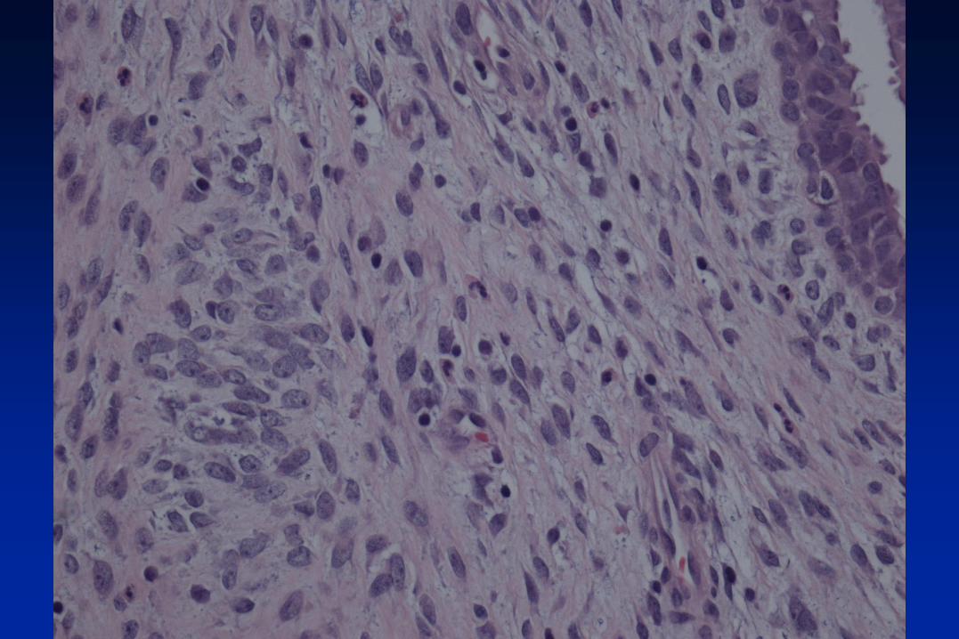
Stromal expansion, but no overgrowth
Moderate/marked increase in stromal
cellularity
Conclusion: benign phyllodes tumour
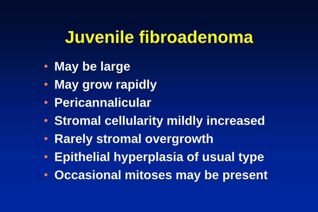
Juvenile fibroadenoma
• May be large
• May grow rapidly
• Pericannalicular
• Stromal cellularity mildly increased
• Rarely stromal overgrowth
• Epithelial hyperplasia of usual type
• Occasional mitoses may be present

Benign phyllodes tumour
• A key feature is increased stromal cellularity
• A leaf-like architecture is usually present (and
can be seen in fibroadenoma)
• The following are often absent
• Nuclear pleomorphism
• Stromal overgrowth
• High mitotic count
• The margin is circumscribed
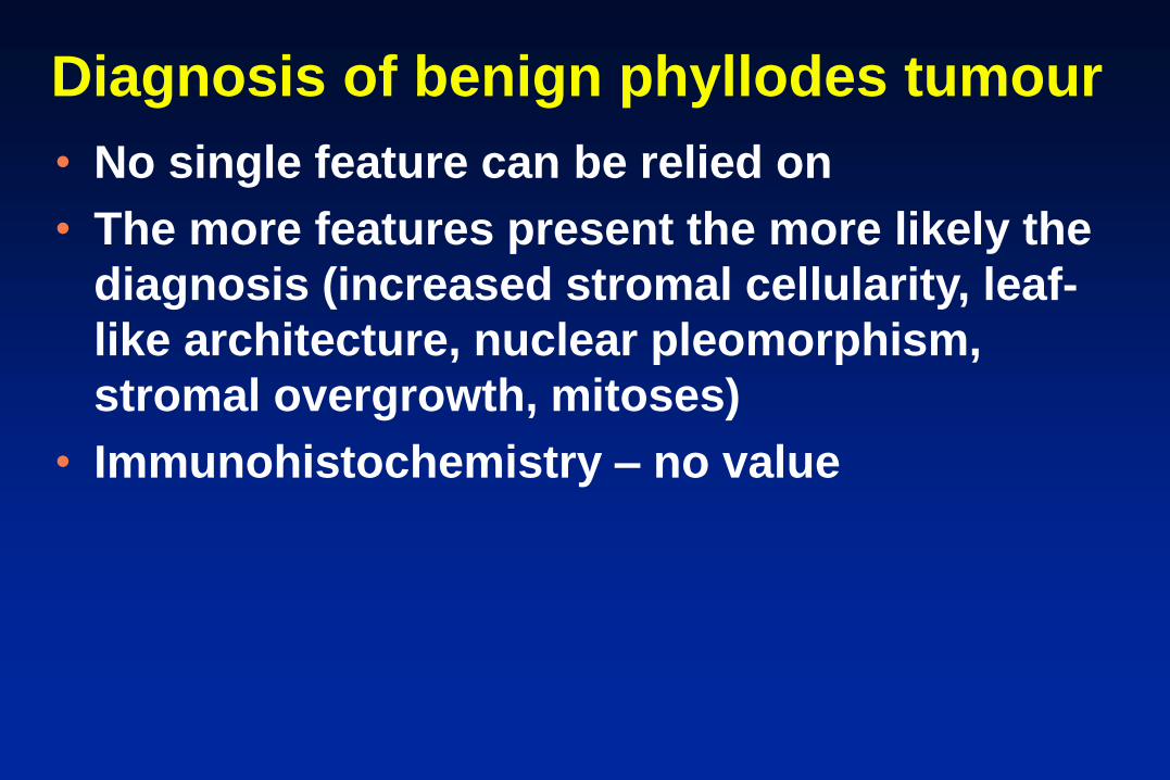
Diagnosis of benign phyllodes tumour
• No single feature can be relied on
• The more features present the more likely the
diagnosis (increased stromal cellularity, leaf-
like architecture, nuclear pleomorphism,
stromal overgrowth, mitoses)
• Immunohistochemistry – no value

Benign phyllodes v fibroadenoma
• Poor reproducibility of diagnosis at the
margin between these entities
• Pathol Oncol Res 2006;12:216
• Int J Surg Pathol 2014;22:695
• Where there is uncertainty WHO
recommends categorisation as
fibroadenoma
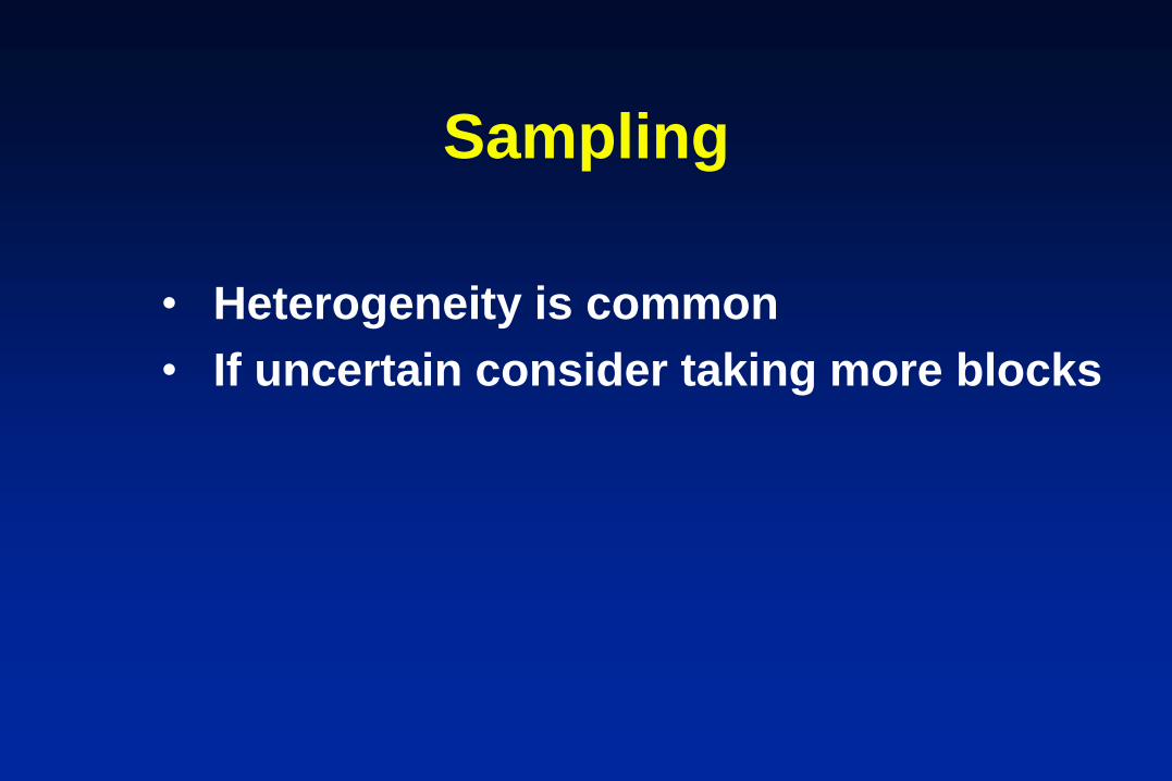
Sampling
• Heterogeneity is common
• If uncertain consider taking more blocks
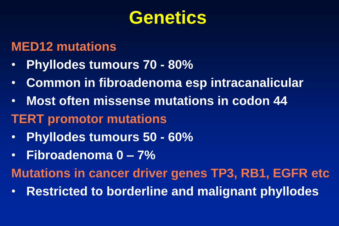
Genetics
MED12 mutations
• Phyllodes tumours 70 - 80%
• Common in fibroadenoma esp intracanalicular
• Most often missense mutations in codon 44
TERT promotor mutations
• Phyllodes tumours 50 - 60%
• Fibroadenoma 0 – 7%
Mutations in cancer driver genes TP3, RB1, EGFR etc
• Restricted to borderline and malignant phyllodes

Grade of local recurrences
Original tumour Recurrent tumour Number
Benign Benign 27
Benign Borderline 17
Benign Malignant 4
Borderline Borderline 10
Borderline Malignant 2
Borderline Benign 4
Malignant Malignant 9
Total 73 (Tan J Clin Pathol 2012;65:69)
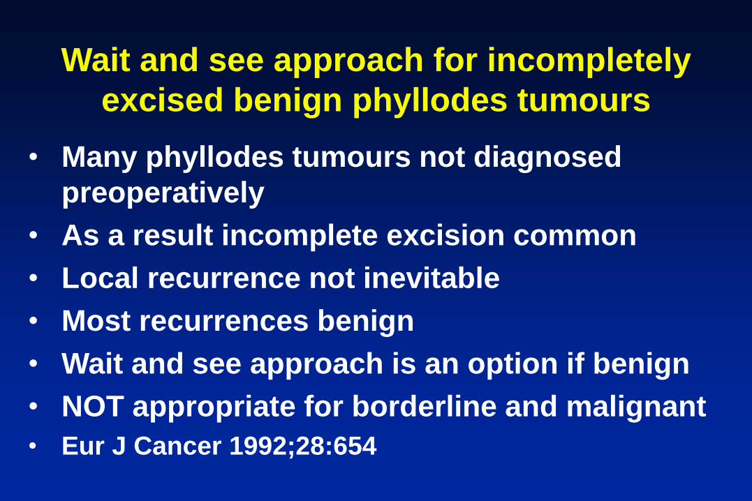
Wait and see approach for incompletely
excised benign phyllodes tumours
• Many phyllodes tumours not diagnosed
preoperatively
• As a result incomplete excision common
• Local recurrence not inevitable
• Most recurrences benign
• Wait and see approach is an option if benign
• NOT appropriate for borderline and malignant
• Eur J Cancer 1992;28:654
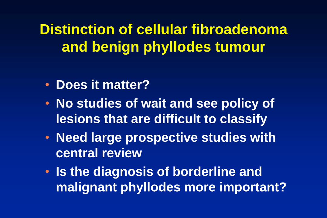
Distinction of cellular fibroadenoma
and benign phyllodes tumour
• Does it matter?
• No studies of wait and see policy of
lesions that are difficult to classify
• Need large prospective studies with
central review
• Is the diagnosis of borderline and
malignant phyllodes more important?
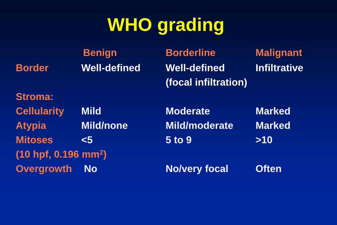
WHO grading
Benign Borderline Malignant
Border Well-defined Well-defined Infiltrative
(focal infiltration)
Stroma:
Cellularity Mild Moderate Marked
Atypia Mild/none Mild/moderate Marked
Mitoses <5 5 to 9 >10
(10 hpf, 0.196 mm2)
Overgrowth No No/very focal Often

Phyllodes grading
• Not all tumours fall neatly into one of three WHO categories.
• More flexible systems eg Nottingham (Moffat 1995)
• Should each feature be given equal weight?
• Some evidence that stromal overgrowth is a particularly
important feature suggesting malignant behaviour
• Lack of objective criteria for cellularity and atypia
• Wide variation of the proportion of benign, borderline and
malignant phyllodes tumours in different series.
• Numerous biological markers have been shown to correlate
with histological grade, but they are not of use in routine
practice (reviewed in Karim J Clin Pathol 2013)

Benign phyllodes tumours
• Pushing margin > 90%
• Stromal overgrowth +/++
• Cellularity +/++
• Nuclear pleomorphism +/++
• Mitoses <10/10hpf (0.152 mm2)
• If four features present = benign
Moffat Histopathology 1995;27:205-18
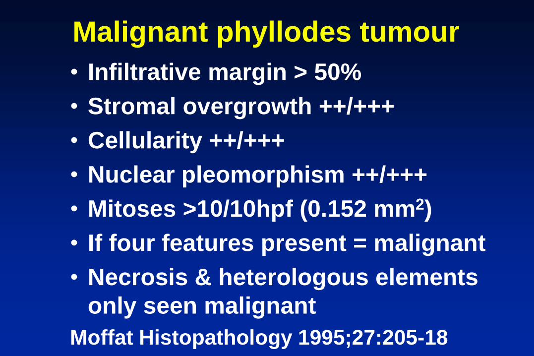
Malignant phyllodes tumour
• Infiltrative margin > 50%
• Stromal overgrowth ++/+++
• Cellularity ++/+++
• Nuclear pleomorphism ++/+++
• Mitoses >10/10hpf (0.152 mm2)
• If four features present = malignant
• Necrosis & heterologous elements
only seen malignant
Moffat Histopathology 1995;27:205-18

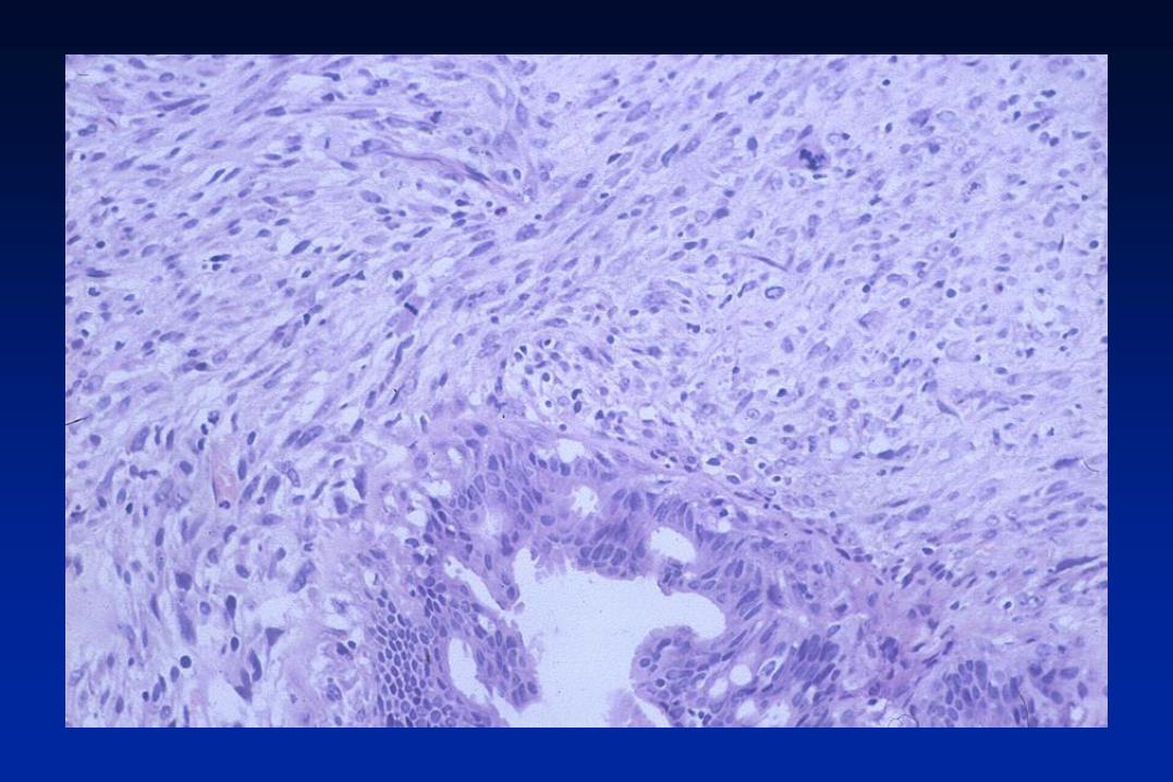
Fibroepithelial lesions
Value of preoperative diagnosis
• Fibroadenoma – usually need not be
excised
• Phyllodes tumour – excise
• Hamartoma - need not be excised
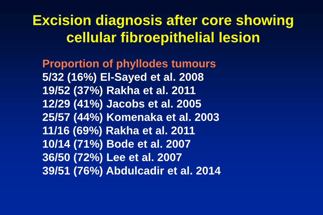
Excision diagnosis after core showing
cellular fibroepithelial lesion
Proportion of phyllodes tumours
5/32 (16%) El-Sayed et al. 2008
19/52 (37%) Rakha et al. 2011
12/29 (41%) Jacobs et al. 2005
25/57 (44%) Komenaka et al. 2003
11/16 (69%) Rakha et al. 2011
10/14 (71%) Bode et al. 2007
36/50 (72%) Lee et al. 2007
39/51 (76%) Abdulcadir et al. 2014

Studies of fibroadenoma v
phyllodes tumour on core biopsy
Cellular fibroepithelial lesions on core
Jacobs et al. Am J Clin Pathol 2005;124:342
Resetkova et al. Breast J 2010;6:573
Jara-Lazaro et al. Histopathology 2010;57:220
Excision diagnosis then look at core
Lee et al. Histopathology 2007;51:336
Morgan et al. Histopathology 2010;56:489
Tsang et al. Histopathology 2011;59:600


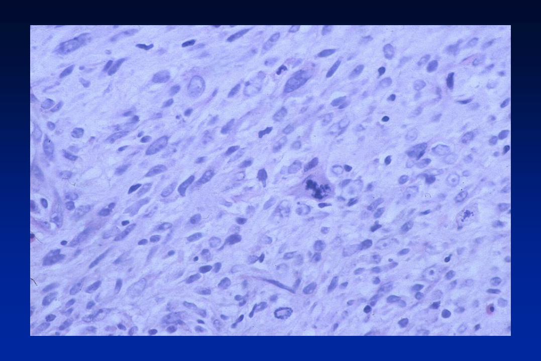

Features favouring
phyllodes on core biopsyConsistently of value in different studies
Stromal cellularity
Stromal overgrowth (x10 field with no epithelium)
Stromal mitoses (3 or more/10hpf)
Marked stromal pleomorphism
Fragmentation
Of value in only some studies
Irregular edge
Entrapped fat
Increased stromal cellularity adjacent to epithelium
Absence of epithelial hyperplasia
Consistently NOT of value
Intracanalicular growth pattern/leaf-like architecture

Features favouring phyllodes
on core biopsyMost not completely specific
Best used in combination (further research needed)
k > 0.6 (Histopathology 2007;51:136)
Stromal cellularity (mild increase in 50%+)
Stromal overgrowth (x10 field with no epithelium)
Fragmentation
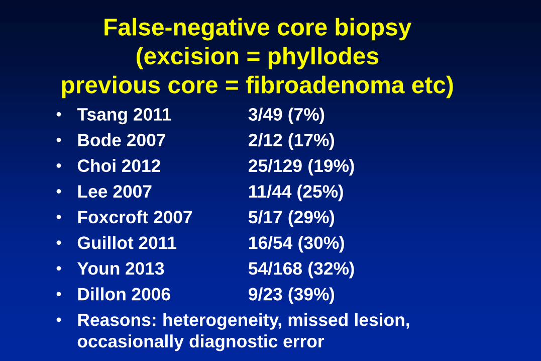
False-negative core biopsy
(excision = phyllodes
previous core = fibroadenoma etc)• Tsang 2011 3/49 (7%)
• Bode 2007 2/12 (17%)
• Choi 2012 25/129 (19%)
• Lee 2007 11/44 (25%)
• Foxcroft 2007 5/17 (29%)
• Guillot 2011 16/54 (30%)
• Youn 2013 54/168 (32%)
• Dillon 2006 9/23 (39%)
• Reasons: heterogeneity, missed lesion,
occasionally diagnostic error
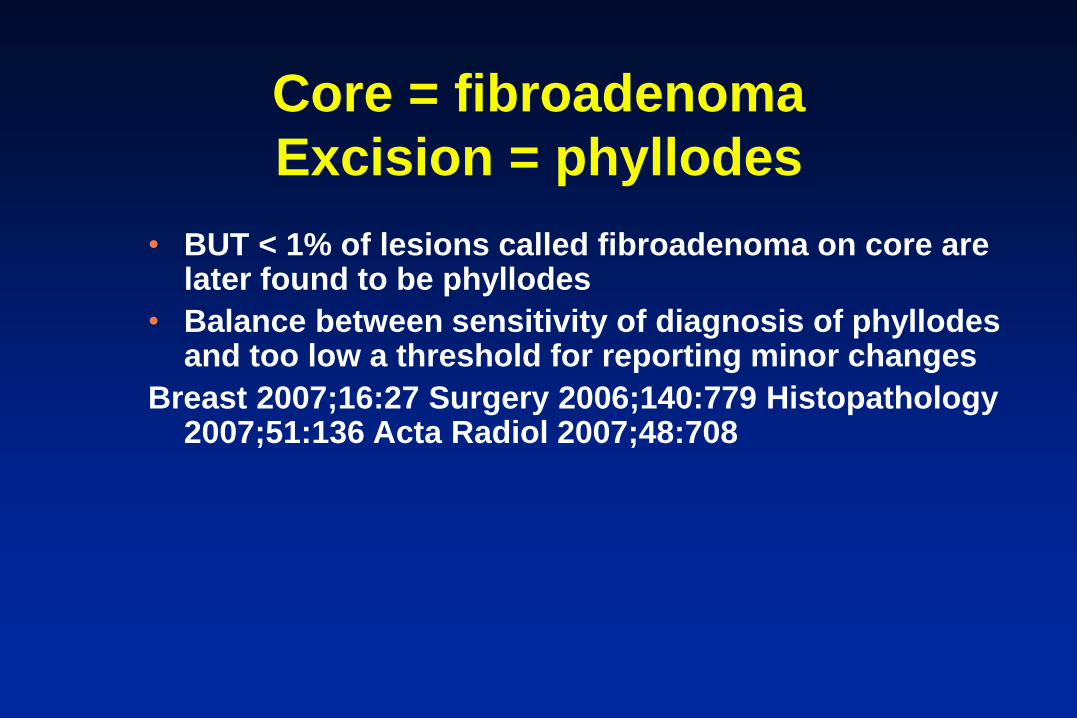
Core = fibroadenoma
Excision = phyllodes
• BUT < 1% of lesions called fibroadenoma on core are later found to be phyllodes
• Balance between sensitivity of diagnosis of phyllodes and too low a threshold for reporting minor changes
Breast 2007;16:27 Surgery 2006;140:779 Histopathology 2007;51:136 Acta Radiol 2007;48:708

B3 cellular fibroepithelial lesion
Repeat core biopsy not useful
Fibroepithelial lesions
Indications for excision
• Phyllodes in differential on core
• Size 30mm+
• Growing
• Ultrasound shows septae
• Patient wishes
• Age ? of value
Breast 2007;16:27 Surgery 2006;140:779 Histopathology 2007;51:136