![Safe Injection Techniques[1]](https://static.fdocuments.in/doc/165x107/577dab981a28ab223f8cab70/safe-injection-techniques1.jpg)
IS HIGH FLOW INJECTION THROUGH CENTRAL CATHERERS SAFE … · través de CvC y CCIP, ... 3866 Is...
5
3865 artículos originales Rev Colomb Radiol. 2014; 25(1): 3865-8 IS HIGH FLOW INJECTION THROUGH CENTRAL CATHERERS SAFE FOR TOMOGRAPHIC STUDIES? ¿ES SEGURA LA INYECCIÓN DE ALTO FLUJO A TRAVÉS DE CATÉTERES CENTRALES PARA REALIZAR ESTUDIOS TOMOGRÁFICOS? Catalina Cuervo Valencia 1 Milena Alcázar Paternina 1 Liliana Arias Álvarez 2 Marcela Montoya 3 Milagros Marti de Gracia 4 Germán Alberto Castrillón 5 SUMMARY Objective: The objective is to demonstrate the safety of medium contrast mechanical injection through central lines, not through a hemotherapy port or a hemodialysis catherer, at rates > 3 ml/sec. Methods: We performed a descriptive, longitudinal and prospective injection in patients who had a central line as the only venous access, and who underwent a mechanical injection with medium contrast at a rate 3-4 cc / sec. We evaluated complications in hemodynamics and complications related to the integrity of catherers. Results: 20 patients were injected via central venous catheters (CVC) and 35 patients via peripherally inserted central catheters (PICC). No complications were observed related to the integrity of the catherer. Conclusion: Medium contrast mechanical injection, performed through CVC and PICC at rates between 3 and 4.5 ml / sec, is considered a safe and viable alternative to contrast-enhanced CT. RESUMEN Objetivo: Demostrar la seguridad de la inyección mecánica de contraste a través de catéteres centrales, excluyendo catéteres de hemodiálisis y puertos de quimioterapia, a velocidades > 3 ml/seg. Métodos: Se realizó un estudio descriptivo, longitudinal y prospectivo en pacientes hospitalizados, quienes tenían como única vía de acceso una línea central y en quienes se les realizó inyección mecánica de medio de contraste a una velocidad de 3 y 4 cm 3 / seg. Se evaluaron las complicaciones hemodinámicas y relacionadas con la integridad de los catéteres. Resultados: Se inyectaron 20 pacientes a través de catéteres venosos centrales (CVC) y 35 pacientes a través de catéteres centrales de inserción periférica (CCIP). No se presentaron complicaciones hemodinámicas ni se observaron complicaciones relacionadas con la integridad del catéter. Conclusión: La inyección mecánica de medio de contraste a través de CVC y CCIP, utilizando velocidades entre 3 y 4,5 ml/seg es una alternativa segura y viable para tomografías contrastadas. 1 MD, resident physicians. Department of Radiology, University of Antioquia, Medellín, Colombia. 2 Fellow in Body Images, University of Antioquia, Medellín, Colombia. 3 MD Epidemiologist, University of Antioquia, Medellín, Colombia. 4 Chief of the Emergency in Radiology, La Paz University Hospital, Madrid, España. 5 MD Radiologist, Body Image Specialist. Associate Professor, Department of Radiology and Grastrohepatology Group, University of Antioquia, Medellín, Colombia. KEY WORDS (MeSH) Injections Contrast media Catheters Catheterization, central venous Tomography PALABRAS CLAVE (DeCS) Inyecciones Medios de contraste Catéteres Cateterismo venoso central Tomografía
Transcript of IS HIGH FLOW INJECTION THROUGH CENTRAL CATHERERS SAFE … · través de CvC y CCIP, ... 3866 Is...
3865
artículos originales
Rev Colomb Radiol. 2014; 25(1): 3865-8
IS HIGH FLOW INJECTION THROUGH CENTRAL CATHERERS SAFE FOR TOMOGRAPHIC STUDIES? ¿ES SEGURA LA INyECCIóN DE ALTO FLUJO A TRAvéS DE CATéTERES CENTRALES PARA REALIzAR ESTUDIOS TOMOGRáFICOS?
Catalina Cuervo Valencia1
Milena Alcázar Paternina1
Liliana Arias Álvarez2
Marcela Montoya3
Milagros Marti de Gracia4
Germán Alberto Castrillón5
SummaryObjective: The objective is to demonstrate the safety of medium contrast mechanical injection
through central lines, not through a hemotherapy port or a hemodialysis catherer, at rates > 3 ml/sec. Methods: We performed a descriptive, longitudinal and prospective injection in patients who had a central line as the only venous access, and who underwent a mechanical injection with medium contrast at a rate 3-4 cc / sec. We evaluated complications in hemodynamics and complications related to the integrity of catherers. Results: 20 patients were injected via central venous catheters (CvC) and 35 patients via peripherally inserted central catheters (PICC). No complications were observed related to the integrity of the catherer. Conclusion: Medium contrast mechanical injection, performed through CvC and PICC at rates between 3 and 4.5 ml / sec, is considered a safe and viable alternative to contrast-enhanced CT.
reSumenObjetivo: Demostrar la seguridad de la inyección mecánica de contraste a través de
catéteres centrales, excluyendo catéteres de hemodiálisis y puertos de quimioterapia, a velocidades > 3 ml/seg. Métodos: Se realizó un estudio descriptivo, longitudinal y prospectivo en pacientes hospitalizados, quienes tenían como única vía de acceso una línea central y en quienes se les realizó inyección mecánica de medio de contraste a una velocidad de 3 y 4 cm3/seg. Se evaluaron las complicaciones hemodinámicas y relacionadas con la integridad de los catéteres. Resultados: Se inyectaron 20 pacientes a través de catéteres venosos centrales (CvC) y 35 pacientes a través de catéteres centrales de inserción periférica (CCIP). No se presentaron complicaciones hemodinámicas ni se observaron complicaciones relacionadas con la integridad del catéter. Conclusión: La inyección mecánica de medio de contraste a través de CvC y CCIP, utilizando velocidades entre 3 y 4,5 ml/seg es una alternativa segura y viable para tomografías contrastadas.
1MD, resident physicians.Department of Radiology,
University of Antioquia, Medellín, Colombia.
2Fellow in Body Images, University of Antioquia,
Medellín, Colombia.
3MD Epidemiologist, University of Antioquia,
Medellín, Colombia.
4Chief of the Emergency in Radiology, La Paz University Hospital,
Madrid, España.
5MD Radiologist, Body Image Specialist. Associate
Professor, Department of Radiology and
Grastrohepatology Group, University of Antioquia,
Medellín, Colombia.
Key words (meSH)Injections Contrast media Catheters Catheterization, central
venousTomography
Palabras clave (DeCS)InyeccionesMedios de contraste Catéteres Cateterismo venoso central Tomografía
3866 Is High Flow Injection Through Central Catherers Safe for Tomographic Studies?. Cuervo C., Alcázar M., Arias L., Montoya M., Marti M., Castrillón G.
IntroductionLa inyección mecánica de medio de contraste para la realización
Contrast medium mechanical injections, performed for contrasted tomographic studies, are a fundamental factor in the quality of studies which ensure a uniform contrast distribution between the vascular structures and the different tissues. Their safety and effectiveness through peripheral venous access is well documented in literature. Patients without peripheral venous access are common in hospitals. Therefore, their only access paths are central catherers, whether they are central venous catherers (CVC), or peripherally inserted central catherers (PICC). These catherers are an alternative to contrast medium mechanical injections.
Low rates of complications have been reported in 0.1% of cases associated with the usage of mechanical injectors through peripheral catherers, (1-4). However, this access is not available in some cases, either due to the impossibility of channeling a vein due to thrombosis, or due to phlebitis of the peripheral veins. In these cases, a central path must be evaluated as an alternative to contrast medium injections (5-9).
The effect of the contrast medium mechanical injection by central catherers has not been sufficiently studied, and most manufacturers of these catherers do not provide guides for the utilization of mechanical injectors in these catherers (1).
Given that there is not sufficient support in current literature to use this access path, a dilemma still persists regarding imaging practice in different centers and between different radiologists. The immediate and belated complications in these type of procedures are not clear.
The purpose of this study is to prove that contrast medium mecha-nical injections through central venous catherers at rates between 3 and 4 ml/sec are safe. However, chemotherapy ports and hemodialysis catherers are excluded from this study. Chemotherapy ports are already recommended for this purpose, and hemodialysis catherers are excluded due to the risk of infection associated with the manipulation by external personnel of nephrology services.
FDA recommendations in 2004 emphasized the careful use of these vascular access devices, related with contrast medium mecha-nical injection, due to the breakage of over 250 ruptures of catherers related with this practice (5). However, it is possible that the lack of well-controlled studies have not enabled an adequate standardization of protocols for the usage of mechanical injectors through central catherers.
The previously described complications associated with contrast medium injection through central lines are related to the pressure generated by the injector. These complications include, rupture of the catherer, with extravasation of the contrast medium and loss of vascular access, fragmentation and embolization of the catherer, obstruction and dysfunction of the catherer (1,10-15). These possible complications discourage the systematic usage of central accesses, and protocols are required to justify them in certain circumstances.
Most current studies related to the rate of injection are in vitro. These studies prove that a risk of catherer damage can only exist at rates as high as 14 ml/sec, and a risk of rupture can only exist at a rate of at least 17 ml/sec (5-9). These rates are much higher than the ones necessary to perform a contrasted tomographic study or a com-puterized axial angiotomography (AngioCAT) (10,16). The required rates for a contrasted study range between 3-5 ml/sec, and are usually greater than 4 ml/sec when an angioCAT study is required (10,17-21).
materials and methodsA descriptive, longitudinal, and prospective study was performed
for patients in a IV level hospital, in the city of Medellín, Colombia, between March of 2010-March of 2012.
Inclusion criteriaThe inclusion criteria were patients over the age of 18 whose renal
function was normal. These patients had a central line as the only venous access, and were administered an intravenous contrast medium through the central line, at a rate between 3 and 4 ml/sec.
exclusion criteriaPatients who had doubts regarding the location and/or integrity
of the catherer were excluded, as well as patients with Swan Ganz catherers, of hemodialysis and chemotherapy ports, and patients who presented allergic reactions before the contrast medium.
Likewise, patients with creatinine depuration between 30 and 60 ml/min, patients who were not under the nephroprotection protocol, and patients with chronic renal insufficiency who were not in dialysis were also excluded.
Informed consent and authorization from the ethics committeeThe approval of the treating doctor was requested after the ethics
committee of the hospital authorized the study, and before the practice of the tomographic study. In addition, the procedure was explained to the patient and/or his/her family, as well as the inherent complications of the usage of contrast medium and the usage of the central catherer. Questions were answered, and if the patient was in agreement, he/she signed the document with at least one witness present.
ProcedureAll patients who were taken to the computerized tomography
service were assessed in a study that required the administration of an intravenous contrast medium. The venous access paths were verified. In case only a central path existed, the patient was admitted into the research protocol. Subsequently, a researcher started with the informed consent approval and after that, started the recollection of the demographic and clinical variables which included the relevant background, the indication, and the type of performed exploration in a written format.
Thereupon, the patient was taken to the scanner, where the permeabi-lity and the adequate intravascular position of the catherer were verified. A saline solution was administered at first through manual injection. The permeability of the catherer was confirmed when the passage of the saline solution was performed without difficulty. The injection was not performed if there were any doubts regarding the integrity or the adequate location of the catherer. When said location and the permeability of the central catherer was confirmed, the injection of the contrast medium was started, and the rate of injection and type of catherer were registered.
A non-invasive monitorization of vital signs was performed before, during, and after the contrast medium injection and any complication was logged in a format meant for this purpose. The following logs were assessed: Systolic arterial pressure (SAP), diastolic arterial pressure (DAP), pulse, oximetry, and manifestations which could suggest reactions to the contrast medium, such as nausea, vomiting, outbreaks, itching, dyspnea, anaphylaxis, cardiopulmonary arrest, among others. These logs were monitored during the administration of the contrast medium, up to an hour later.
3867
Original Articles
Rev Colomb Radiol. 2014; 25(1): 3865-8
Complications related to the catherer were evaluated, regarding a possible malfunction with subsequent difficulty in the passage of medications or liquids, occlusion, thrombosis, total or partial rupture, and in case it occurred, the location of the rupture and of the possible embolization of the distal fragment. In order to obtain this information, clinical history was reviewed and the nursing staff in charge of detec-ting difficulties with the catherer up to 24 hours after contrast medium mechanical injection was contacted.
All the explorations took place in a General Electric LightSpeed VCT XT General Electric 74 detector multi-cut scanner, according to the protocol established for the disease or for the respective diagnostic impression.
All patients were administered a water soluble iodinated contrast medium through the mechanical injector Medrad Stellant SCT-210, with volumes between 80 cm3 and 100 cm3 and concentrations of 300 and 350 mg/ml (for AngioCAT). The injection had a pressure of 300 pounds per square inch (PSI) at a rate over 3 ml/second.
Statistical analysisFrequency distributions were used in the statistical data analysis
when the variables were qualitative. The summary and central tendency measures were used for quantitative variables, after using the Shapiro Wilk test for their distribution. Additionally, an analysis of related samples was performed. This was done to compare whether there were significant differences in the pulse and oximetry of patients in their basal condition, during the moment of injection, and an hour after being injected. The Wilcoxon range test was used for this purpose, due to the abnormal distribution of the implied quantitative variables.
The McNemar test was used to evaluate changes in the arterial pressure numbers of each patient. PAS numbers between 90-140 mmHg were considered and PAD numbers between 60-90 mmHg were grouped as normal. Any number which was outside this range was considered abnormal.
resultsFrom the 55 patients included in this study, 26 were women
(47.3%), and 29 were men (52.7%). The average age of the patients was 53 years (18 to 85 years of age). In terms of personal background, 20 patients had high blood pressure (36.4%), 11 patients were diabetics (20%), 2 suffered from coronary disease (3.6%), 2 had a background of arrhythmia (3.6%), and 2 had suffered from allergic reactions; one to penicillin and the other to tramadol (3.6%).
The creatinine serum levels before the study had an average of 0.9 mg/dl, with a range between 0.4-8.8 mg/dL.
The main indications of tomographic studies were infection (30.9%), pulmonary thromboembolism (25.5%), others (43.6%).
Contrasted tomographic studies were performed on 34 patients and angioCAT studies were performed on 21 patients, with a nonionic iodinated contrast medium: Iohexol (OmnipaqueTM).
All the assessed catherers were Arrow brand. Their material is polyurethane. CVC with a caliber greater than 5 Fr and a length between 16-20 cm was used in 20 patients (36.4%), and CCIP 5 Fr with a length of 50 cm was used in 35 patients (63.6%). 27 of these catherers were single lumen (49.1%), 25 were bilumen (45.5%), and 3 were trilumen (5.5%).
The rate of injection was 3.0 ml/sec for 34 patients (62%), and 4.0 ml/sec for 21 patients (38%).
When monitoring was performed in order to detect complications related to the catherer during the first 24 hours after injection, no cases presented an extravasion of the contrast medium, total or partial rupture of the catherer, dysfunction, embolization, occlusion, or thrombosis. No complications related to the patient were reported one hour after the contrast medium injection, evaluating outbreaks, itching, dyspnea, anaphylaxis, nausea, vomiting, cardiopulmonary arrest, among others.
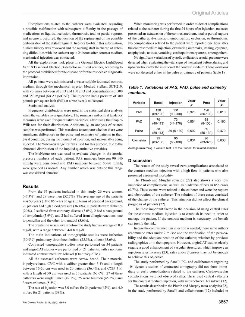
No significant variations of systolic or diastolic arterial pressure were detected when evaluating the vital signs of the patient before, during and up to one hour after the injection of the contrast medium. These variations were not detected either in the pulse or oximetry of patients (table 1).
Table 1. Variations of PAS, PAD, pulse and oximetry numbers.
Variable Basal Injection Valor p Post Valor
p
PAS 130 (69-190)
131 (90-200) 0,926 120
(66-190) 0,010
PAD 70 (40-113)
73 (44-178) 0,854 68
(8-100) 0,160
Pulso 88 (50-113) 89 (8-130) 0,592 89
(56-130) 0,479
Oximetría 95 (83-100)
95 (83-100) 0,834 96
(83-925) 0,830
Average (min-max), p value = Test. T of the Student for related samples
DiscussionThe results of the study reveal zero complications associated to
the contrast medium injection with a high flow in patients who also presented associated morbidity.
The Plumb and Murphy revision (22) also shows a very low incidence of complications, as well as 6 adverse effects in 858 cases (0.7%). These events were related to the catherer and were the rupture and obstruction of the catherer. The solution of these cases consisted of the change of the catherer. This situation did not affect the clinical prognosis of patients (22).
The most important factor in the decision of using central lines for the contrast medium injection is to establish its need in order to manage the patient. If the contrast medium is necessary, the benefit can justify the risk.
In case the contrast medium injection is needed, these same authors recommend rates under 2 ml/sec and the verification of the permea-bility and the adequate position of the catherer, whether by previous radiographies or in the topogram. However, angioCAT studies clearly require a good enhancement of vascular structures, which improve as injection rates increase (23); rates under 2 cm/sec may not be enough to achieve this objective.
The study performed by Sanelli PC. and collaborators regarding 104 dynamic studies of contrasted tomography did not show imme-diate or early complications related to the catherer. Cardiovascular complications were not observed either. These used central catherers for the contrast medium injection, with rates between 3-5 ml/sec (12).
The results described in the Plumb and Murphy meta-analysis (22), in the study performed by Sanelli and collaborators (12) included in
3868 Is High Flow Injection Through Central Catherers Safe for Tomographic Studies?. Cuervo C., Alcázar M., Arias L., Montoya M., Marti M., Castrillón G.
said meta-analysis, and the results in this article series can be a star-ting point to initiate the protocol process of the use of central lines for contrast medium mechanical injection, both for tomographic as well as angioCAT studies.
The benefit clearly seems to be greater than the risk. As described, complications related to the catherer do not reach 0.7% (12,22).
Therefore, the injection with contrast medium mechanical injectors at rates between 3 and 4 ml/sec through CVC and CCIP is an adequate and viable alternative for routine contrasted topographic and angioCAT studies, with the same safety as in lesser injection rates. This was sub-sequently communicated to the manufacturer. Even if the manufacturer had in vitro support if the injection to this velocities was adequate, it did not have in vivo support, which was recommended with the precautions described in the product information.
This study shows different limitations which include a small number of patients, and the non-inclusion of pediatric patients. For this reason, the results cannot be extrapolated to population who use catherers with a lesser diameter. The usage time of the central catherer was not studied either, given that it could have conditioned a greater theoretical number of complications.
Even though the study proves the safety of the contrast medium injection through central catherers, it is important to have an institutio-nal protocol in place for catherer injections. The protocol would start with the explanation of the procedure benefits and risks, reaching a conclusion with the treating team to subsequently obtain the approval of the patient and/or his/her family. This is done so that the position and permeability of the catherer can be verified, proceeding to the contrast medium injection with the appropriate rate for the clinical indication of the patient.
In conclusion, one could say that if the results hereby presented were to be used, one could improve the possibility to access image studies for all patients without peripheral venous access and who exclusively have a central venous access. Using the precautions described in this article, one could inject with rates greater than 3 ml/sec.
acknowledgmentsSustainability project of the research vice-rector’s office of the
Universidad de Antioquia, Medellín, Colombia.
referencias1. 1. Herts BR, Cohen MA, Mcinroy B, et al. Power injection of intravenous contrast
material through central venous catheters for CT : In Vitro evaluation. Radiology. 1996;200:731-5.
2. McCarthy S, Moss AA. The use of a flow rate injector for contrast-enhanced com-puted tomography. Radiology. 1984;151:800.
3. Miles G, Rasmussen F. Safe use of an intravenous power injector for CT: Experien-ce and protocol. Radiology. 1990;176:69-70.
4. Sistrom CL, Gay SB, Peffley L. Extravasation of iopamidol and iohexol during contrast-enhanced CT: report of 28 cases. Radiology. 1991;180:707-10.
5. U.S. Food and Drug Administration. Reminders from FDA regarding ruptured vascular access devices from power injection [internet]. 2012 [citado 2013 jul. 15]. Disponible en: www.fda.gov/.
6. Rivitz SM, Drucker EA. Power injection of peripherally inserted central catheters. J Vasc Intervent Radiol. 1997;8:857-63.
7. Salis AI, Eclavea A, Johnson MS, et al. Maximal flow rates possible during power injection through currently available PICCs: An in vitro study. J Vasc Intervent Ra-diol. 2004;15:275-81.
8. Coyle D, Bloomgarden D, Beres R, et al. Power injection of contrast media via peri-pherally inserted central catheters for CT. J Vasc Intervent Radiol. 2004;15:809-14.
9. Angle JF, Matsumoto AH, Skalak TC, et al. Flow characteristics of peripherally inserted central catheters. J Vasc Intervent Radiol. 1997;8:569-77.
10. Vrachliotis TG, Bis KG, Haidary A, et al. Atypical chest pain: coronary, aortic, and pulmonary vasculature enhancement at biphasic single-injection 64-section CT angiography. Radiology. 2007;243:368-76.
11. O’Sullivan P, Brown M, Hartnett B, et al. Central line pump infusion and large vo-lume mediastinal contrast extravasation in CT. Br J Radiol. 2006;79:e75-7.
12. Sanelli PC, Deshmukh M, Ougorets I, et al. Safety and feasibility of using a central venous catheter for rapid contrast injection rates. AJR. 2004;183:1829-34.
13. Goss JE, Ramo BW, Raff GL, et al. Power injection of contrast media during percutaneous transluminal coronary artery angioplasty. Cathet Cardiovasc Diagn. 1989;16:195-8.
14. Rigsby CK, Gasber E, Seshadri R, et al. Safety and efficacy of pressure-limited power injection of iodinated contrast medium through central lines in children. AJR. 2007;188:726-32.
15. Amaral JG, Traubici J, BenDavid G, et al. Safety of power injector use in children as measured by incidence of extravasation. AJR. 2006;187:580-3.
16. Macha DB, Nelson RC, Howle LE, et al. Central venous catheter integrity during mechanical power injection of iodinated contrast medium. Radiology. 2009;253:870-8.
17. Stein PD, Fowler SE, Goodman LR, et al. Multidetector computed tomography for acute pulmonary embolism. N Eng J Med. 2006;354:2317-27.
18. Anderson DR, Kahn SR, Rodger MA, et al. Computed tomographic pulmonary angiography vs ventilation-perfusion lung scanning in patients with suspected pul-monary embolism: a randomized controlled trial. JAMA. 2007;298:2743-53.
19. Lin K, Kazmi KS, Law M, et al. Measuring elevated microvascular permeability and predicting hemorrhagic transformation in acute ischemic stroke using first-pass dynamic perfusion CT imaging. AJNR. 2007;28:1292-8.
20. Schramm P, Schellinger PD, Klotz E, et al. Comparison of perfusion computed tomography and computed tomography angiography source images with perfusion-weighted imaging and diffusion-weighted imaging in patients with acute stroke of less than 6 hours’ duration. Stroke. 2004;35:1652-8.
21. Dodd JD, Kalva S, Pena A, et al. Emergency cardiac CT for suspected acute coro-nary syndrome: qualitative and quantitative assessment of coronary, pulmonary, and aortic image quality. AJR. 2008;19:870-7.
22. Plumb AAO, Murphy G. The use of central venous catheters for intravenous con-trast injection for CT examinations. Br J Radiol. 2011;84:197-203.
23. Bae KT. Intravenous contrast medium administrations and scan timing at CT: Con-siderations and approaches. Radiology. 2010;256:32-61.
CorrespondenciaGermán Alberto CastrillónUniversidad de AntioquiaCalle 10E # 25-165 Medellín, [email protected].
Received for evaluation: September 30, 2013.Accepted for publication: December 13, 2013.