Is endothelial dysfunction the one to blame in Heart ... · Heart Failure with preserved Ejection...
85
Carla Sofia Gomes Ferreira Is endothelial dysfunction the one to blame in Heart Failure with Preserved Ejection Fraction? Dissertation for Master Degree in Biomedical Research July, 2015
Transcript of Is endothelial dysfunction the one to blame in Heart ... · Heart Failure with preserved Ejection...

Carla Sofia Gomes Ferreira
Is endothelial dysfunction the one to blame in Heart Failure with Preserved Ejection Fraction?
Dissertation for Master Degree in Biomedical Research
July, 2015

Carla Sofia Gomes Ferreira
Is endothelial dysfunction the one to blame in Heart
Failure with preserved Ejection Fraction?
Dissertation presented to the Faculty of Medicine of the University of Coimbra for the
fulfillment of the requirements for a Master degree in Biomedical Research
Dissertação apresentada à Faculdade de Medicina da Universidade de Coimbra para
prestação de provas de Mestrado em Investigação Biomédica
July, 2015


iii
Host Institution
Departamento de Fisiologia e Cirurgia Cardiotorácica
Cardiovascular R&D Unit
Centro de Investigação Médica, 6º piso
Faculdade de Medicina da Universidade do Porto
Rua Dr Plácido da Costa
4200-450 Porto, Portugal
Supervisor: Inês Falcão-Pires, PhD1
Co-supervisor: Henrique Girão, PhD2
Affiliations:
1 FMUP - Faculdade de Medicina da Universidade do Porto
2 FMUC - Faculdade de Medicina da Universidade de Coimbra


v
Acknowledgements
My Master thesis was an extraordinary journey. I feel a very lucky
person! I met wonderful people and I would like to express my profound
gratitude:
To Professor Adelino Leite-Moreira, head of the Department of
Physiology and Cardiothoracic Surgery, for having received me in your
laboratory, for giving me the opportunity to work with your big team and for
being so nice to me. I will never forget that, like you said, “I have an additional
responsibility”!
To Professora Inês, my supervisor, for giving me the opportunity to
work with you and your team and for your scientific guidance. I hope to
continue to be part of your time.
To Professor Henrique, for being my “father” in Coimbra, for your
attention and for all the wisdom.
To Doutora Diana Nascimento, from helping me to understand flow
cytometry and for all the help with endothelial cells.
To Nádia for helping me growing, for helping me to understand my
thesis! You teach me all I know about HFpEF and genetics! Thank you for
almost being my co-supervisor. Because of you genetic is no more a “black
hole” in my life. Thank so much you for the advices, your patience, kindness
and for making me feel “like home”.
To Glória, for being my teacher, my tutor in FMUP, for all your
patience, kindness and friendship.
To Ana for helping me growing, for all advices and for have left me stay
in your office for so long! Thank you for always comforted me when I needed
and thank you for, like Nádia, making me feel “like home”.

vi
To Daniela, Patrícia, Dulce and João, from my group, for all the help
with the results and with the writing of my thesis.
To Paulo, for being a friend that always make me smile with your jokes,
to Sara for teaching me Medicine and for all advices and Manuel.
To all Cardiovascular R&D Unit family, that was been my family work
for almost one year and half.
Ao meu melhor amigo e namorado, João, por acreditar em mim e por
estar sempre lá quando eu precisei.
Às pessoas mais importantes da minha vida, a minha mãe e o meu
pai, por me terem dado a oportunidade de ser Mestre e por sempre
acreditarem em mim.
Carla Sofia Gomes Ferreira

vii
Abstract
Heart Failure with preserved Ejection Fraction (HFpEF) is a common
clinical syndrome that comprises 50% of Heart Failure (HF) patients. It is
characterized by an abnormal diastolic left ventricular (LV) function, with
impaired relaxation and increased stiffness that together contribute to the
heart inability to fill properly. Understanding HFpEF has been difficult due to
its heterogeneous etiology and pathophysiology, so, treatment options are still
an enigma. Recent studies on HFpEF emphasize the importance of
comorbidities frequently present in this syndrome, like diabetes mellitus (DM),
obesity, hypertension and even aging, all promoting systemic inflammation
and endothelial dysfunction.
The recently characterize ZSF1 obese rat represents one of the most
comprehensive animal model of HFpEF described to date. Using this model,
we aim to better characterize HFpEF in terms of assessing inflammation,
endothelial function and the relation between these injuries, allowing for a
better understanding of this syndrome.
Male rats Wistar Kyoto (WKY, n=21), ZSF1 Lean (ZSF1Ln, n=24) and
ZSF1 Obese (ZSF1Ob, n=22) were subjected to echocardiographic
examination at their 18th week of age and, at the end of the protocol, to
morphometric and vascular function evaluation, as well as molecular and
histological studies.
Compared to WKY and to ZSF1Ln, ZSF1Ob rats are heavier, present
obesity with significantly more abdominal adiposity, other features of DM and
also hypertension, three important risk factors for HFpEF. ZSF1Ob animals
have LV hypertrophied and display diastolic dysfunction, in which an increase
in E/E' was observed. Aortic rings, submitted to acetylcholine increasing
concentrations, showed endothelium impairment of relaxation, suggesting
endothelial dysfunction. ZSF1Ob animals do not presented systemic
inflammation but had myocardial expression of inflammatory mediators,
revealing itself to be the principal source of heart tissue inflammation.

viii
We conclude that ZSF1Ob rat represent a solid animal model for
HFpEF research and that cardiac endothelium underlies its pathophysiology
thus representing an interesting pathway for future pharmacologic
interventions.
Keywords: Heart Failure with preserved Ejection Fraction; inflammation;
endothelial dysfunction; ZSF1 obese animal model

ix
Resumo
Insuficiência cardíaca com fração de ejeção preservada (ICFEP) é um
síndrome clínico bastante comum que compreende 50% dos doentes com
insuficiência cardíaca (IC). A ICFEP é caracterizada por uma função
ventricular comprometida, nomeadamente com alterações no relaxamento e
aumento da rigidez, que em conjunto contribuem para a incapacidade do
coração de encher de maneira eficiente. Compreender a ICFEP tem sido
difícil devido à sua etiologia e patofisiologia heterogéneas, o que limita as
opções de tratamento. Descobertas recentes dão ênfase às comorbilidades
frequentemente presentes na ICFEP, como a diabetes mellitus, obesidade e
hipertensão e até mesmo o envelhecimento, como sendo promotoras de
inflamação sistémica e disfunção endotelial.
O rato obeso ZSF1 recentemente caracterizado representa um dos
melhores modelos animais para a investigação na ICFEP. Utilizando este
modelo pretendemos avaliar a inflamação e a disfunção endotelial e ainda a
relação entre ambos, permitindo assim uma melhor compreensão deste
síndrome.
Ratos machos com nove semanas de idade Wistar Kyoto (WKY,
n=21), ZSF1 magros (ZSF1Ln, n=24) e ZSF1 obesos (ZSF1Ob, n=22) foram
submetidos a avaliações ecocardiográficas às 18 semanas de idade, e no fim
do protocolo, a avaliações morfométricas e de função vascular, assim como a
estudos moleculares e histológicos.
Comparando com os grupos WKY e ZSF1Ln, os ratos ZSF1Ob têm
mais peso corporal, são obesos e têm mais gordura abdominal, possuem
características da diabetes mellitus e hipertensão, três fatores de risco
relacionados com a ICFEP. Os ratos ZSF1Ob têm hipertrofia ventricular
esquerda e apresentam disfunção diastólica, com um aumento da razão E/E’.
Anéis aórticos submetidos a concentrações crescentes de acetilcolina
mostraram um comprometimento no relaxamento do endotélio, sugerindo um
estado de disfunção endotelial. Células endoteliais cardíacas de animais

x
ZSF1Ob mostraram expressão de factores inflamatórios importantes,
revelando ser a principal fonte de inflamação no tecido cardíaco.
Conclui-se que os ratos ZSF1Ob representam um bom modelo animal
para investigação na ICFEP e que o endotélio cardíaco é alvo interessante
para futuras intervenções farmacológicas.
Palavras-chave: Insuficiência cardíaca com fração de ejeção preservada;
inflamação; disfunção endotelial; modelo animal ZSF1 obeso

xi
List of contents
Acknowledgements ................................................................................................... v
Abstract .................................................................................................................... vii
Resumo ..................................................................................................................... ix
Figure index ............................................................................................................ xiii
Table index .............................................................................................................. xv
Abbreviations list .................................................................................................... xvii
Part 1 – Introduction ............................................................................................... xix
1. Heart Failure .................................................................................................... 21
1.1. Definition ................................................................................................... 21
1.2. Epidemiology ............................................................................................. 21
1.3. Symptoms and signs ................................................................................. 22
1.4. Classification and Pathophysiology ........................................................... 22
1.5. Comorbidities in HFpEF ............................................................................ 24
2. HFpEF and the endothelium ............................................................................. 27
2.1. The endothelium........................................................................................ 27
2.2. Endothelial dysfunction ............................................................................. 29
2.3. Mechanisms of endothelial dysfunction in HFpEF: the role of comorbidities
32
3. Animal models in HFpEF research ................................................................... 34
3.1. The obese ZSF1 as an animal model of HFpEF ........................................ 34
Part 2 - Aims ........................................................................................................... 37
Part 3 – Materials and methods ............................................................................... 41
1. Experimental animal model .............................................................................. 43
2. Echocardiography ............................................................................................ 44
3. ELISA assay ..................................................................................................... 45
4. Array protein expression profile ........................................................................ 45
5. Histology, cardiomyocyte and aorta dimensions ............................................... 46
6. AGEs immunohistochemistry ............................................................................ 46
7. Western Blotting ............................................................................................... 47
8. Cardiac endothelial cells isolation .................................................................... 48
9. Flow cytometry and sorting .............................................................................. 49
10. RNA extraction and reverse transcription ....................................................... 49
11. Real time - PCR ............................................................................................. 50

xii
12. Vascular function....................................................................................... 51
13. Statistical analysis ..................................................................................... 51
Part 4 - Results ....................................................................................................... 53
1. Characterization of the animal model ............................................................... 55
1.1. Morphometric data .................................................................................... 55
1.2. Cardiac functional and structural changes ................................................. 56
2. Aorta characteristics and vascular function ...................................................... 58
3. Plasma levels of adipokines and inflammatory markers ................................... 61
4. Markers of myocardial dysfunction ................................................................... 63
5. Flow cytometry analysis ................................................................................... 66
Part 5 - Discussion .................................................................................................. 67
1. Metabolic risk-related HFpEF model ................................................................ 69
2. Endothelial dysfunction and oxidative stress .................................................... 71
Part 6 – Conclusion and future prespectives ........................................................... 73
Part 7 - References ................................................................................................. 77

xiii
Figure index
Figure 1. Schematic representation of the evolution of a normal heart into HFpEF,
involving the exposure to several risk factors……………………………………….…. 23
Figure 2. Synthesis of Nitric Oxide……………………………………………….…..… 28
Figure 3. Schematic representation of the differences between a healthy (A) and
dysfunctional endothelium (B)………………….……………………………….…..…… 31
Figure 4. Schematic representation of the mechanisms induced by comorbidities that
result in myocardial dysfunction and remodeling in
HFpEF……………………………………………………………………………………... 33
Figure 5. Cardiomyocyte hypertrophy represented by its cross-sectional
area…………………………………………………………………………………….……56
Figure 6. Descending aorta characteristics of WKY (n=5), ZSFLn (n=5) and ZSF1Ob
(n=5) animals………………………………………………………………………….…... 58
Figure 7. Vascular function of aortic rings from WKY (n=5), ZSFLn (n=5) and
ZSF1Ob (n=5) animals…………………………………………….……………………... 60
Figure 8. mRNA expression of WKY (n=5), ZSFLn (n=5) and ZSF1Ob (n=5)
animals……………………………………………………………….………………….…. 64
Figure 9. Protein expression of WKY (n=6), ZSFLn (n=6) and ZSF1Ob (n=5) animals
assessed by western blotting…………………………...……………………………..… 65
Figure 10. Flow cytometry analysis………………………………………...………...… 66


xv
Table index
Table 1. Vasoactive and inflammatory substances released by endothelium……... 27
Table 2. Role of Nitric Oxide…………………………………………………………..… 29
Table 3. Morphological data from WKY (n=17), ZSF1Ln (n=14) and ZSF1Ob (n=18)
animals…………………………………………………………………………………...… 55
Table 4. Echocardiographic evaluation of WKY (n=7), ZSF1Ln (n=9) and ZSF1Ob
(n=15) animals at 18th week……………………………………………………………… 57
Table 5. Plasma protein expression assessed by a profile array of ZSF1Ln (n=4) and
ZSF1Ob (n=4) animals………………………………………………………………...…. 62
Table 6. Circulating plasma levels data from WKY (n=6), ZSF1Ln (n=9) and ZSF1Ob
(n=8) groups……………………………………………………………………………..… 63


xvii
Abbreviations list
Ach - Acetylcholine
AGEs - Advanced Glycation End-products
Ang II - Angiotensin II
Ang 1-7 - Angiotensin 1-7
BSA - Body Surface Area
cGMP - cyclic Guanosine Monophosphate
CI - Cardiac Index
CML - Carboxymethllysine
CO - Cardiac Output
COPD - Chronic Obstructive Pulmonary Disease
DM - Diabetes Mellitus
EDVI - End-diastolic volume index
EF - Ejection Fraction
ESVI - End-systolic volume index
ET-1 - Endothelin-1
eNOS - endothelial Nitric Oxide Synthase
FABP4 - Fatty Acid Binding Protein 4
FS - Fractional Shortening
GAPDH - Glyceraldehyde 3-phosphate dehydrogenase
HF - Heart Failure
HFrEF - Heart Failure with reduced Ejection Fraction
HFpEF - Heart Failure with preserved Ejection Fraction
HR - Heart Rate
ICAM1 - Intracellular Adhesion Molecule 1
Il-1 - Interleukin-1
Il-6 - Interleukin-6
Il-8 - Interleukin-8
IR - Insulin Resistance
LAA - Left Atrium Area
LV - Left Ventricle

xviii
MCP1 - Monocyte Chemoattractant Protein 1
MPI - Myocardial performance index
NF-Kβ - Nuclear Factor – kappa β
NO - Nitric Oxide
NOX2 - NADPH oxidase 2
NOX4 - NADPH oxidase 4
Phe - Phenylephrine
PKG - Protein Kinase G
RAGE - receptor of Advanced Glycation End-product
RANTES - Regulated on activation normal T cell expressed and secreted
ROS - Reactive Oxygen Species
RV - Right Ventricle
sGC - soluble Guanylate Cyclase
SHHF - Spontaneously Hypertensive Heart Failure
SV - Stroke Volume
S’ - Mitral annular systolic velocity
TGFβ - Transforming Growth Factor β
TNFα - Tumor Necrosis Factor α
VASP - Vasodilator-stimulated Phosphoprotein
VCAM1 - Vascular Adhesion Molecule 1
WKY - Wistar Kyoto
ZSF - Zucker Fatty/ Spontaneously Hypertensive Heart Failure F1
hybrid rats

Part 1
Introduction
“There are many hypotheses in science which are wrong. That’s perfectly all right: it’s
the aperture to finding out what’s right. Science is a self-correcting process.”
Carl Sagan

21
1. Heart Failure
1.1. Definition
The first description of Heart Failure (HF) remotes to ancient Egypt
times. Its definition remains controversial partly because of his complicated
etiology and also because of the variability of the clinical symptoms and signs.
HF is the end-result of many cardiovascular diseases and a clinical syndrome
characterized by functional and structural irregularities in human heart that
compromise its capacity to eject sufficient blood to fulfill all body’s metabolic
needs or only at the expense of increased ventricular filling pressures [1-3].
1.2. Epidemiology
The epidemiology of HF had a remarkable revolution in the last
decades. Worldwide the total number of estimated HF patients is around 26
million. Results from the Framingham Heart Study suggest that in developed
countries, at some point of their life, one in five persons will develop HF. In
Europe, according to the Rotterdam study, the 5-year mortality risk is around
41% [4, 5].
The number of patients with HF will grow to epidemic proportions
considering the expected increase of population ageing, the sedentary
lifestyle expected for the next years and the estimated increase of HF
comorbidities like diabetes mellitus (DM) and obesity, thus representing a
major public health problem [4-6].

22
1.3. Symptoms and signs
According to European Society of Cardiology, symptoms of HF include
dyspnea (difficulty in breathing), fatigue and exercise intolerance [7, 8].
Signs of HF include leg, ankles and feet swelling, jugular distension,
pulmonary rales, peripheral edema and some radiographic manifestations like
pulmonary vascular redistribution, interstitial edema and pleural effusions [8].
1.4. Classification and Pathophysiology
According to the European Society of Cardiology and to the American
College of Cardiology Foundation/American Heart Association HF patients
can be divided in two clinically distinct syndromes: HF with reduced Ejection
Fraction (HFrEF) or systolic HF and HF with preserved Ejection Fraction
(HFpEF) also known as diastolic HF [9].
HFrEF is correlated with ischemic injury, dilated and other genetic
cardiomyopathies, whereas HFpEF is associated with chronic inflammation
and the co-existence of other comorbidities which represent important
cardiovascular risk factors. Comparing both types, HFpEF patients have fewer
hospitalizations but higher mortality rate than HFrEF. Contrarily to HFrEF,
there is not any effective medical treatment for HFpEF as a result of the
diversity and complexity of pathophysiology of this condition [7, 10]. The
heterogeneous etiologies and the challenging diagnostic make HFpEF an
interesting topic for research. Current treatment strategies focus on control of
volume status and comorbidities, but future research aimed at individualized
therapies holds promise to improve outcomes in this increasingly prevalent
form of cardiac failure.

23
1.4.1. Heart Failure with Preserved Ejection Fraction
HFpEF constitutes almost 50% of all HF patients and its prevalence is
increasing almost 1% per year [11]. HFpEF is more common in older women
(61-76% of patients) with high prevalence of hypertension, DM, ischemic
heart disease, coronary artery disease, atrial fibrillation and lifestyle related
risk factors like smoking and obesity [1, 12-16].
HFpEF is associated with left ventricle (LV) diastolic dysfunction and
involves complex interactions between multiple factors. In the diastolic phase,
the ventricle is unable to fill with sufficient blood maintaining adequate low
pressures resulting from myocardium stiffness or impaired relaxation. The
increased filling pressure (stiffness) induces wall thickness and an adjustment
in ventricular size and volume (Figure 1) [8, 17-20]. Besides the diastolic
dysfunction, some other mechanisms seem to be related with HFpEF
pathophysiology, such as inflammation, chronic volume overload, venous
constriction, irregular ventricular-vascular coupling, as well as cardiac
autonomic impairment and chronotropic incompetence. In this condition the
heart rate response to stress is abnormal what could lead to pulmonary
arterial hypertension and endothelial dysfunction [15].
Figure 1. Schematic representation of the evolution of a normal heart into HFpEF,
involving the exposure to several risk factors. Risk factors are responsible for
myocardium alterations that include readjustment in ventricular size and volume,
myocardium stiffness and wall thickness, therefore contributing to the development of
HFpEF. Figure was produced using Servier Medical Art.

24
The challenging diagnosis of HFpEF is assessed after clinical
evaluation, Doppler echocardiography or invasive hemodynamic assessment
and confirmed whenever the following conditions are observed [7, 21, 22]:
Signs and symptoms of HF;
Diastolic dysfunction;
Normal or lightly anomalous systolic function.
Despite all advances in medicine, HFpEF continues to have a high rate
of hospitalizations, subsequent readmission on hospital, a considerable
mortality rate (10-30%) and will grow to epidemic proportions making it a
major public health problem [1, 12, 23].
1.5. Comorbidities in HFpEF
Non-cardiac comorbidities are highly prevalent in HFpEF and have an
important role in the pathogenesis of this syndrome [11]. Several risk factors
are involved, although only one is enough to cause HFpEF. Patients with
HFpEF usually have multiple comorbidities that could cause or be contributors
to this phenotype [15].
Female gender is also an important risk factor being associated with
more concentric remodeling, lower LV diastolic volumes and as well as
systolic and diastolic LV stiffness. Woman have a probability two times higher
to develop HFpEF [11, 24].
Anemia is present in 50-70% of HFpEF patients and it is closely related
with renal disease and it is characterized by iron deficiency and insufficient
erythropoietin production generated by systemic inflammation. Chronic renal
failure and HFpEF often co-exist and share common risk factors. Indeed,
renal failure is present in 30-40% of patients and it is a predictor of mortality.
Chronic obstructive pulmonary disease (COPD) also contributes to HFpEF

25
mortality and the adverse cardiovascular effects include reduction of cardiac
output, less stroke volume and also impaired LV filling [11, 25].
Hypertension is the most predominant risk factor, being present in 60-
88% of HFpEF patients. In hypertensive patients, the risk of developing
HFpEF is 2 times higher in men and 3 times higher in women comparing with
the normotensive patients. High blood pressure is capable of increase arterial
stiffness, which compromises LV relaxation. Additionally, these patients
frequently display obesity, microalbuminuria and impairment of insulin
signaling and rapidly develop myocardial fibrosis and LV hypertrophy [11].
Obesity, which has reached epidemic proportions partly because of the
increase of energy intake and a sedentary life style, impairs cardiac structure
and function that doubles the risk of having HFpEF. It is present in 32-46% of
patients and it is an independent risk factor for cardiovascular morbidity and
mortality. Along with adipose tissue expansion, pro-inflammatory cytokines
and adipokines, like leptin and resistin, are released. Arterial resistance
increases and contributes to LV hypertrophy and high diastolic pressures.
Myocardial relaxation is compromised due to changes in cardiomyocyte
calcium handling, along with lipotoxicity and mitochondrial dysfunction. All
these alterations induce LV concentric remodeling, LV increase of mass e
volume and arterial stiffness [11, 12].
Diabetes mellitus (DM) is a recognized comorbidity affecting 30-45% of
HFpEF patients, especially females. The Framingham Heart Study first
showed that diabetic patients can develop diastolic LV thickness with stiffer
cardiomyocytes and subsequent studies from our group further confirmed this
evidence [26]. This is a result of hyperglycemia effects that causes interstitial
and perivascular fibrosis, increase collagen deposition and reduction of
degrading matrix metalloproteinases [11, 12, 15]. Additionally, high levels of
glucose promote the generation of advanced glycation end-products (AGEs)
in myocardium, which are molecules formed in the reducing sugars’ reaction
that can form covalent bonds with proteins, namely collagen fibers cross-
linking, stiffening the myocardium. Moreover, AGEs can bind their receptors,
26
RAGEs, increasing collagen production thus, decreasing arteries distensibility
and increasing LV stiffness [11, 27, 28].
All the previous comorbidities are associated with several pathological
changes in the cardiovascular system that strongly contribute to HFpEF.
Besides diastolic LV dysfunction, myocardial and systemic inflammation,
oxidative stress and endothelial dysfunction represent important contributors
to the pathophysiological mechanisms of HFpEF syndrome [11, 12]. The
endothelium, located between the circulating blood and the vessel wall or the
myocardium, occupies a strategic anatomic position and is undoubtedly
involved in the abnormalities that occur in HFpEF [29-34].

27
2. HFpEF and the endothelium
2.1. The endothelium
Endothelium is an active organ formed by a monolayer of endothelial
cells that separate the vascular wall or the myocardium from the circulation. It
acts as a functional and structural barrier capable of sensing hormonal and
mechanical stimuli. The endothelium plays an important role in cardiovascular
homeostasis modulating vascular tone by balancing the production of
vasodilators and vasoconstrictors agents (Table 1). In addition, this layer also
regulates solute transport, prevent leucocyte and platelet adhesion and
aggregation, modulates blood flow and mediates inflammatory and reparative
response to injury [32, 35-37]. Cardiac endothelium integrates endothelial
cells from endocardium, from the coronary microvasculature and also of the
intramyocardial capillaries. The anatomical position of endothelial cells next to
cardiomyocytes enables a bidirectional communication between those cells
[38]. Several studies have shown that endothelium have a crucial role in
several diseases, including HFpEF [35, 39].
Table 1. Vasoactive and inflammatory substances released by endothelium [36, 40].
Action Substance
Vasodilation
Nitric Oxide (NO)
Prostacyclin
C-Type natriuretic peptide
Vasoconstriction
Endothelin-1 (ET-1)
Angiotensin II (Ang II)
Thromboxane A2
Reactive Oxygen Species (ROS)
Inflammation
Nitric Oxide (NO)
E and P-selectin
Nuclear Factor kβ (NF- kβ)
ICAM1
VCAM1

28
Nitric oxide (NO) is the most important and the most characterized
substance produced by the endothelium. This gas with a short life (about 6-30
seconds) is generated from L-arginine amino acid by endothelial NO synthase
(eNOS) which is continuously produced and release by endothelial cells
through physical and hormonal stimuli [41]. Shear stress caused by blood
flowing on the vessel wall is the most important physical stimuli for NO
production. Chemical stimuli include acetylcholine, bradykinin and serotonin
[41, 42]. In the healthy endothelium, when NO is released, binds to smooth
muscle promoting the activation of soluble Guanylate Cyclase (sGC) and the
production of cyclic Guanosine Monophosphate (cGMP) which opens calcium
dependent potassium channels promoting blood vessel relaxation and
vasodilation (Figure 2). Normally, NO is important to maintain the vascular
wall in a quiescent state inhibiting inflammation, cell proliferation and
thrombosis being the vasodilation prevalent (Table 2) [41-45].
Figure 2. Synthesis of Nitric Oxide. In endothelial cells NO production is stimulated
by several stimulus and inhibited by others. NO acts in smooth muscle cells in order
to promote vasodilation and the decrease of cytokine synthesis, the monocyte
adhesion and platelet aggregation, as well as the decrease of adhesion molecules.
NO: Nitric Oxide; eNOS: endothelial Nitric Oxide Synthase; GTP: Guanosine
Triphosphate; sGC: soluble Guanylate Cyclase; cGMP: cyclic Guanosine
Monophosphate. Figure was produced using Servier Medical Art. Adapted from [41,
42].
29
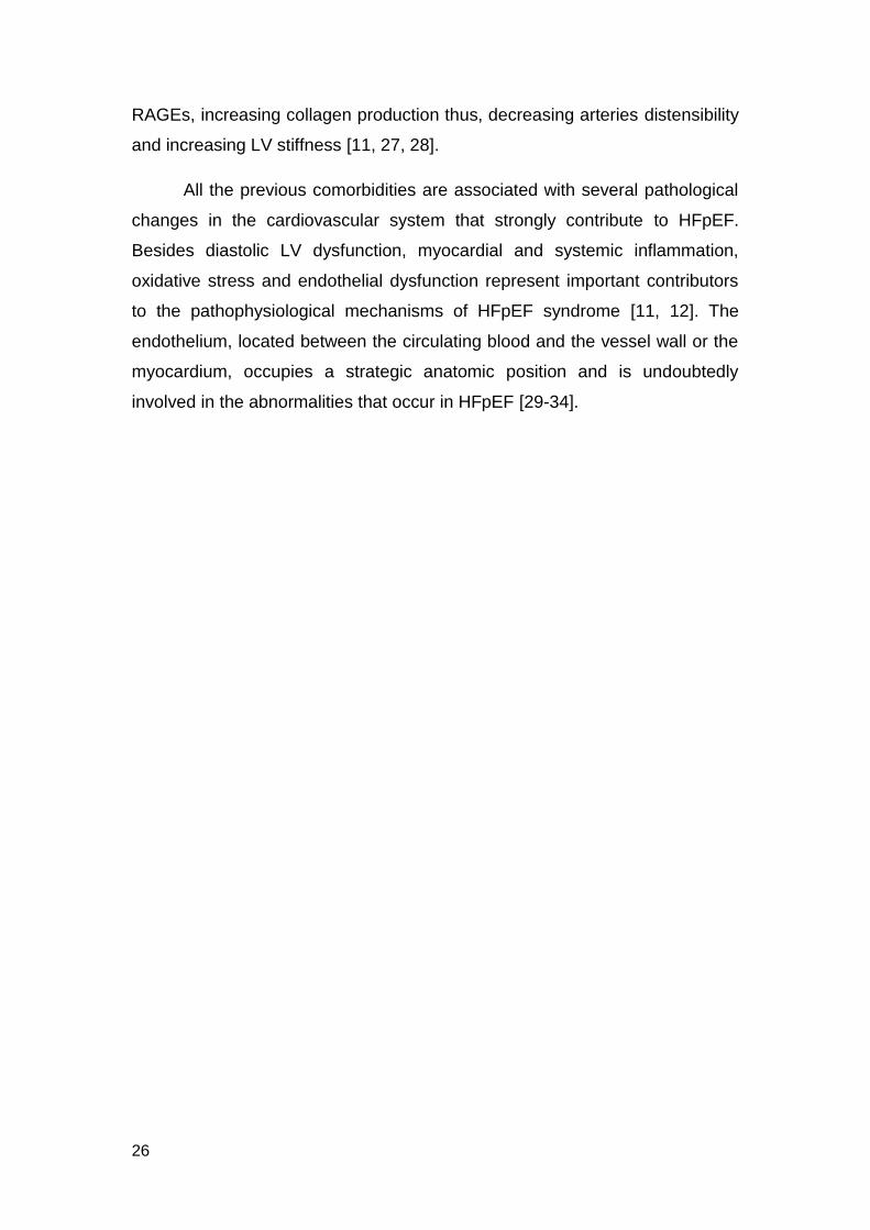
Table 2. Role of Nitric Oxide. Adapted from [41].
Role Action
Vasomotor action
Endothelium vasodilation
Decreases vasoconstriction in smooth muscle cells
Regulation of systemic and coronary tone by acting on basal
arterioles
Inflammation
Decreases endothelial permeability
Decreases expression of E-selectin
Reduction of leucocyte adherence to the endothelium
Decreases production of macrophages cytokines
Down-regulate platelet aggregation and adherence
Antioxidant
In vivo and in vitro free radical scavenger
NO donors duplicate plasma anti-oxidant capacity
2.2. Endothelial dysfunction
As a consequence of HFpEF risk factors, endothelium loses his
integrity and the homeostatic balance is disrupted, culminating in endothelial
dysfunction. Endothelium acquires a pro-inflammatory and pro-thrombotic
state with vasoconstriction that includes detachment and release of
endothelial cells into the circulation [35, 39, 44, 46]. Endothelial dysfunction is
associated with the beginning and the progression of HFpEF [32, 34, 40, 47].
The principal feature of endothelial dysfunction is the impairment of NO
bioavailability. In this condition, reduced production of NO can result from
diverse contributing factors, such as decreased production of eNOS, lack of
eNOS substrates or due NO degradation by ROS (Figure 3). ROS are
chemical oxygen species and cellular metabolism’s products that are highly
reactive causing oxidative stress [37, 38]. ROS form peroxinitrites that impair
the NO-induced vasodilation, essential to vascular homeostasis and

30
endothelial function. Reduced NO release by endothelium affects ventricular
relaxation, particularly in the hypertrophied myocardium. Conventional
antioxidant therapies have intended to correct the imbalance between NO and
ROS, but sadly have not been totally successful yet [36, 40, 48-50].
The link between endothelial dysfunction and HFpEF pathophysiology
is currently established [11, 32, 33, 36, 41]. However the precise mechanisms
remain obscure. Understanding endothelial dysfunction mechanisms will be
an important key to understand the pathophysiology of HFpEF.

31
Figure 3. Schematic representation of the differences between a healthy (A) and
dysfunctional endothelium (B). In a healthy endothelium, endothelial cells are intact
and produce a normal concentration of nitric oxide (NO) by endothelial nitric oxide
synthase (eNOS). In a dysfunctional endothelium, endothelial cells are damaged and
produce a reduced concentration of NO due reaction with reactive oxygen species
(ROS) and a decreased eNOS activity. It becomes an inflammatory local and
leucocytes and platelets migrate to this injury site. Figure was produced using
Servier Medical Art.
B – Dysfunctional endothelium
A – Normal endothelium

32
2.3. Mechanisms of endothelial dysfunction in HFpEF: the role
of comorbidities
Newer insights suggest that endothelial dysfunction is more than a
state of systemic vasoconstriction and that has a crucial role in HFpEF
pathogenesis. In 2013 Paulus et al. suggested that endothelial dysfunction
plays a central role in HFpEF progression. These new paradigm of HFpEF
suggests that comorbidities are responsible for the pro-inflammatory state
seen, causing coronary microvascular endothelial inflammation [51].
Hypertension, obesity and DM induce a systemic inflammatory state
with the release of pro-inflammatory cytokines such as tumor necrosis factor α
(TNFα), interleukin-1 (Il1), Il-6 and Il-8. Increased circulating levels of these
cytokines are found in HFpEF patients [11, 13]. The adhesion molecules,
vascular cell adhesion molecule 1 (VCAM1), intercellular adhesion molecule 1
(ICAM1) and E-selectin, which in normal conditions are expressed in small
amounts in the surface of endothelial cells, are upregulated [13, 52].
Endothelial inflammation originates the production of ROS that leads to
peroxynitrites (ONOO-) formation and a decrease in NO bioavailability. In
response, cardiomyocyte production of soluble guanylate cyclase (sGC) is
reduced, which contributes to a decrease in cGMP and protein kinase G
(PKG) concentration. In HFpEF lower PKG levels are associated with
cardiomyocyte hypertrophy and stiffness, and with high resting tension
(Fpassive). This high resting tension is due to the hypophosphorylation of the
N2B segment of the giant cardiomyocyte cytoskeletal protein titin [11, 38, 51,
53]. Microvascular endothelial inflammation also stimulates fibroblasts
differentiation in myofibroblasts resulting in myocardial fibrosis with collagen
type 1 deposition and collagen cross-linking, which are contributors to
myocardial stiffening [54]. These changes induce diastolic LV dysfunction, the
major characteristic of HFpEF (Figure 4) [11, 38, 51].

33
Figure 4. Schematic representation of the mechanisms induced by comorbidities that
result in myocardial dysfunction and remodeling in HFpEF. Comorbidities generate a
pro-inflammatory state with consequent release of Interleukin (Il) 1, Il-6, Il-8 and
Tumor Necrosis Factor α (TNFα). Endothelium produces reactive oxygen species
(ROS) that react with nitric oxide (NO) to produce peroxynitrites (ONOO-) and at the
same time reduce NO bioavailability. In cardiomyocytes there is a reduction in
soluble guanylate cyclase (sGC), which in turn decreases cyclic guanosine
monophosphate (cGMP) concentration and protein kinase G (PKG) production. Titin
hypophosphorylation induces an increase in passive force leading to cardiomyocyte
hypertrophy. Endothelial cells increase the expression of adhesion molecules
(ICAM1, VCAM1 and E-selectin) influencing the migration of monocytes that release
transforming growth factor (TGFβ). This last induces the collagen production and
deposition in interstitial space which is turn causes cardiomyocyte stiffness. Figure
was produced using Servier Medical Art. Adapted from [11, 51].
Nevertheless, the exact role that comorbidities have on structural and
functional remodeling in HFpEF is not entirely well known. The new paradigm
of HFpEF could be a new therapeutic target and the development of
experimental HFpEF models might be beneficial and helpful, in order to
understand and possibly to cure this pathology [11].

34
3. Animal models in HFpEF research
Understanding the pathophysiology of HFpEF has been restricted by
the limited access to human myocardial biopsied due to obvious ethical
constrictions. In addition, the lack of a proper animal model mimicking the
human pathology has partly limited HFpEF research. Indeed, animal models
can be very useful to clarify subcellular mechanisms under conditions where
the comorbidities and other confounding factors can be precisely controlled
[55, 56].
There are several models used for HFpEF research, nevertheless, until
recently, none had fulfilled all the features present in the human disease.
Recently, we have characterized a rat model that meets the criteria of HFpEF,
the obese ZSF1 [57].
3.1. The obese ZSF1 as an animal model of HFpEF
ZSF1 rats were generated by crossing non-hypertensive lean female
Zucker Diabetic Fatty rats (ZDF, +/fa) with lean spontaneously hypertensive
HF prone male rats (SHHF/Mcc, +/facp) that share a common genetic
background with Wistar Kyoto (WKY) rats and derive from spontaneously
multifactorial hypertensive rats [58-63]. Both lean and obese ZSF1 animals,
inherit a hypertensive gene from the spontaneously hypertensive rat strain
and show elevated blood pressure [59].
ZSF1 rats have myocardial hypertrophy induced by hypertension, more
notorious in obese than in lean rats. The ZSF1 obese rats present
considerable diastolic abnormalities such as increased left atrial area,
prolonged time constant of isovolumetric relaxation, elevated arterial
elastance and end-diastolic pressure as well as an upwards shift of end-
diastolic pressure-volume relation, thus highlighting a worse ventricular-
vascular coupling. Systolic function remained preserved in lean and obese
ZSF1 rats [57].

35
In terms of metabolic disturbances, ZSF1 obese animals developed
obesity, abdominal adiposity, insulin resistance, oral glucose intolerance,
hyperglycemia and glycosuria, consistent with type II DM phenotype [64] and
thus represent a good animal model of metabolic syndrome. Moreover,
compared to hypertensive ZSF1 lean, which represent a model of isolated
ventricular overload, the ZSF1 obese will allow clarifying if imposing metabolic
comorbidities on top of overload is per se capable of induced HFpEF.
Recently, a full description of a rat model that meets the criteria of diastolic HF
was described, the ZSF1 obese [57].


Part 2
Aims
“The scientist is not a person who gives the right answers; he's one who asks the
right questions.”
Claude Lévi-Strauss

39
Considering the concepts reviewed before, the aim of the present study
was to clarify the pathophysiology of HFpEF by investigating the contribution
of endothelial dysfunction and inflammation for the development of this
syndrome.
In order to achieve the aim of the project we pretend to:
Characterize an animal model of HFpEF including metabolic,
morphometric as well as cardiac structural and functional changes;
Evaluate endothelial dysfunction in the previous model;
Correlate endothelial dysfunction with cardiac function parameters
to investigate potential therapeutic targets.


Part 3
Materials and Methods
“Those who are quite satisfied sit still and do nothing; those who are not quite
satisfied are the sole benefactors of the world.”
Walter Savage Landor


43
1. Experimental animal model
This study was made according to the Guide for the Care and Use of
Laboratory Animals published by the NIH (NIH Publication no. 85–23, revised
2011) and was approved by the ethics committee of the Faculty of Medicine of
Porto and by Direção-Geral de Alimentação e Veterinária (DGAV) from
Portugal. The Faculty of Medicine of Porto is a governmental institution,
granted approval by the Portuguese government to perform animal
experiments.
Nine-weeks old male rats Wistar Kyoto (WKY, n=21), ZSF1 Lean
(ZSF1Ln, n=24) and ZSF1 Obese (ZSF1Ob, n=22) were obtained from
Charles River (Barcelona, Spain) and had unlimited access to food (LabDiet®
5008, International Product Supplies Ltd., UK) and water. Animals were
housed in groups of 2 animals per cage in a ventilated cages system (IVC) in
a controlled environment with a 12-h-light/-dark cycle at 22ºC room
temperature. The room had a relative humidity of 30–70% and an air
exchange rate of 40–50 air changes/hour.
In the end of the experiment, at their 20th week of age, anesthetized
animals (8% for induction and 2.5-3% for maintenance) were euthanized by
exsanguination and blood and tissue were collected. Organs were weighed,
RV and LV were weighed after dissection, and tibia length (TL) was
measured. Fresh samples of LV were used in flow cytometry and the rest of
the samples were snap-frozen in liquid nitrogen and stored at -80ºC for
molecular studies (RNA extraction, western blot) or fixed in 10% buffered
formalin for histological procedures and also for immunohistochemistry
analysis. Weights were normalized to TL due to the large body weight
differences between groups.

44
2. Echocardiography
All animals were subjected to an echocardiography evaluation at their
18th week. Animals were anaesthetized by inhalation of sevoflurane (8% for
induction and 1-2.5% for maintenance), orotracheally intubated and
mechanically ventilated (150 min−1, 100% O2, 14–16cmH2O inspiratory
pressure, with tidal volume adjusted to animal weight, and 4cmH2O end-
expiratory pressure) (TOPO Small Animal Ventilator, Kent Scientific Inc.,
USA). Rats were placed in a left-lateral decubitus position on a heating pad,
the ECG was monitored and their temperature was kept at 38ºC. The skin of
all animals was shaved, the echocardiography gel was applied and a linear
15MHz probe (Sequoia 15L8W) was gently positioned on the thorax. Systolic
and diastolic wall thickness and cavity dimensions were recorded, in M-mode
and 2D echocardiography, at the level immediately above the papillary
muscles in the parasternal short axis view.
From these measurements end diastolic and end systolic volumes,
(EDV and ESV, respectively), fractional shortening (FS), ejection fraction (EF)
of the LV, stroke volume (SV) and cardiac output (CO) were derived. The
following Doppler and tissue Doppler measurements were taken using the
apical four-chamber early diastolic filling peak velocity (E wave), late diastolic
peak velocity (A wave), E/A ratio, early peak diastolic filling velocity (E’), late
peak diastolic filling velocity (A’) and mitral annular systolic velocity (S’).
The myocardial performance or TEI index was retrieved from the mitral
flow pattern and calculated by the formula = (IVCT + IVRT)/ET, were IVCT is
the isovolumic contraction time, IVRT corresponds to the isovolumic
relaxation time and ET to ejection time. Data was indexed for body surface
area as described previously [65] and three representative cycles were
measured per rat and their average was calculated.

45
3. ELISA assay
Blood samples collected from subclavian vein after echocardiographic
evaluation were placed in tubes with EDTA (C10H16N2O8, pH 6.0). Samples
were centrifuged at 5000 rpm for 15 minutes at 4°C and plasma was then
separated and utilized for quantitative enzyme immunoassays (ELISA).
Levels of leptin (SK00050-08, Adipo bioscience, USA), Fatty Acid
Binding Protein 4 (FABP4, SK00030-03, Adipo bioscience, USA) Angiotensin
1-7 (Ang 1-7, E02A0225, BlueGene Biotech, China), Il6 (R6000B, R&D
Systems, UK) and TNFα (ER3TNFA, Thermo Scientifics, USA) were
measured according to the manufacturer’s instructions. Results were
analyzed using an ELISA plate reader (UVM-340, ASYS Hitech GmbH,
Austria) and a calibration curve was constructed by plotting the absorbance
values at 450nm (with specific correction, according to manufacturer’s
protocol) and concentrations of unknown samples were determined.
4. Array protein expression profile
The expression of some inflammation related-proteins was performed
using plasma samples and a Rat Adipokine Array Kit (ARY016, R&D systems,
UK).
Nitrocellulose membranes were blocked for 1 hour with an array buffer
and then a cocktail of biotinylated detection antibodies was added and
incubated overnight at 4ºC. The membranes were washed several times in
order to remove all the unbound material. Streptavidin-HRP, an enzyme used
for the detection of the substrate was applied, incubated for 30 minutes and
washed. Finally Chemi Reagent Mix was added and the signal produced was
measured in a chemiluminescence detection system (ChemiDoc™ MP, Bio-
Rad, USA).

46
5. Histology, cardiomyocyte and aorta dimensions
LV and descending aortic samples were fixed in 4% paraformaldehyde,
dehydrated with gradual ethanol, cleared with xylene and were included in
paraffin blocks. Serial sections with 4 µm of thickness were cut using a
microtome (RM2125RTS, Leica, Germany) and mounted on slides. Next the
slides were dewaxed in xylene, hydrated through a series of decreasing
concentration of alcohol solutions and stained for haematoxylin-eosin. Slides
were subsequently submitted to a new series of decreasing concentration of
alcohol solutions and xylene and finally mounted with Entellan (Merck,
Germany).
Cardiomyocyte cross-sectional area, descending aortic diameter and
thickness were determined observing slides at light microscopy (Dialux 20,
Leitz, Germany) and using image acquisition software (cell B, Olympus, USA).
6. AGEs immunohistochemistry
Immunohistochemistry was performed to determine AGEs expression
in myocardium and in 25-50, 50-100, and >100 vessels caliber. Four- µm LV
apex sections were sliced, placed and subjected to deparaffinization and
rehydration.
A heat solution of sodium citrate buffer 10mM (C6H5Na3O7.2H2O, pH
6.0) was added for 30 minutes into the slides to induce antigen retrieval. All
sections were encircled with a hydrophobic pen (Immunopen, Immunologic,
Netherlands) to prevent splitting leakage and the endogenous peroxidase
activity was blocked using 100µL/section of a 3% hydrogen peroxide solution
(Sigma Aldrich, USA) and incubated at room temperature for 10 minutes. All
slides were washed with distillated H2O (dH2O) and with Tris-Buffered Saline-
Tween (TBST; 100 mM Tris, 1.5 mM NaCl, pH 8.0 and 0.1% Tween-20) for 5
minutes with agitation. Blockage of non-specific binding was prepared with
5% normal goat serum (NGS, ab7481, abcam, Cambridge, UK) in TBST,

47
added 100 µL for section and incubated at 1 hour at room temperature. Next
all slides were washed 3 times for 5 minutes with TBST with agitation.
Blockage of endogenous avidin-biotin expression was preform at room
temperature using an endogenous avidin + biotin blocking system (ab3387,
abcam, Cambridge, UK) according to manufacturer’s instructions (1 drop and
15 minutes of incubation). The primary antibody (Anti-AGE primary antibody,
ab23722, abcam, UK) in a 1/500 dilution was incubated at 4ºC overnight.
After incubation slides were washed 3 times for 5 minutes with TBST and with
agitation and were incubated with the secondary antibody (goat anti-rabbit
IgG, ab6720, abcam, UK) in a 1/250 dilution at room temperature for 2 hours.
Slides were next washed with agitation 3 times for 5 minutes with TBST. All
slides were incubated with 3.3-diaminobenzidine (DAB, ab94665, abcam, UK)
at room temperature until brown color was observed. Then slides were
washed with dH2O and counterstained with Gill haematoxylin (Merck,
Germany) for 3 minutes. Finally all slides were submitted to decreasing
concentration of alcohol solutions, xylene and mounted with Entellan (Merck,
Germany).
Negative control was made with the omission of the primary antibody.
The slides were observed and photographed with a microscope (Dialux 20,
Leitz, Germany) and AGE’s quantification was made using Image Pro Plus 6
software (MediaCybernetics, USA).
7. Western Blotting
LV samples were homogenized on ice in 1 ml RIPA lysis buffer (150
mM NaCl, 1.0% IGEPAL® CA-630, 0.5% sodium deoxycholate, 0.1% SDS,
and 50 mM Tris, pH 8.0) containing the following protease inhibitors:
phenylmethylsulfonyl fluoride (1mM), aprotonin (10g.ml−1), leupeptin
(10 μg.ml−1) and pepstatin (10 μg.ml−1), all from Sigma Chemicals (USA).
Samples were then centrifuged at 11000 rpm for 20 minutes at 4°C. The
supernatants were collected and total protein concentration was determined.

48
Samples containing 20 μg of protein were loaded on a 6% SDS
Polyacrylamide gel (SDS-PAGE), run and electroblotted into polyvinylidene
difluoride membrane. Pre-stained molecular weight marker proteins were
used as standards for the SDS-PAGE. Ponceau staining was performed to
verify the quality of the transfer and to ensure equal protein loading. Blots
were blocked in 5% non-fat skimmed milk in PBS for 1 hour, treated overnight
with antibody against the different proteins (eNOS, 9572, Cell Signaling
Technology, USA; β-actin, 4967, Cell Signaling Technology, USA; p-eNOS,
9571, Cell Signaling Technology, USA) followed by incubation with alkaline
phosphatase secondary antibodies for 1 hour. Immunoblots were developed
with an ECFTM Western blotting detection system (GE Healthcare, UK).
Protein content was determined using a Bio-Rad protein assay kit.
8. Cardiac endothelial cells isolation
A LV sample was cut and transferred into a gentleMACS C tube
(Miltenyl Biotec, Germany) containing HBSS (CaCl2, MgCl2,
Lifetechnologies,USA) with collagenase II (Worthington, USA) and DNase I
(Applichem, USA). The C tube was connected to the gentleMACS dissociator
(Miltenyl Biotec, Germany) and the sample was incubated for 30 minutes at
37ºC with agitation every 5 minutes in order to resuspend the settled tissue
fragments. Next the C tube was runned out again in the gentleMACS
dissociator. In the end, the solution was passed into a 70µm cell strainer
(Corning, USA), washed with cold HBSS and the cell suspension suffered a
spin down at 1500 rpm for 10 minutes at 4ºC. Cold HBSS was immediately
added and the cell suspension suffered a new spin down at 1500 rpm for 10
minutes at 4ºC. Next cells were washed with FACS medium (eBioscience,
USA), suffered another spin down at 1500 rpm for 10 minutes at 4ºC and
resuspended in HBSS (1ml/heart).

49
9. Flow cytometry and sorting
Cardiac endothelial cells were resuspended in ice-cold FACS medium
(eBioscience, USA) and added into a 96 well plate (105-106 cells/well) in a
way that surrounding each well stays an empty one. FACS medium was
added, the plate was washed by centrifugation for 5 minutes for 2000 rpm and
supernatant was discarded. CD90 Pacific blue (Biolegend, USA), PE anti-rat
CD54 (Biolegend, USA), CD44 Purified (BD Biosciences, USA) and CD106-
Pe (BD Biosciences, USA) antibodies were added in a dilution in FACS
medium to a maximum volume of 25-50 µl and incubated on ice for 20
minutes protected from light. FACS medium was added and the plate was
washed by centrifugation for 5 minutes for 2000 rpm. The supernatant was
discarded. In order to loose cells, the plate suffered a shacking in a vortex set
at medium speed. Cells were transferred into FACS tubes (eBioscience, USA)
with the remaining volume up to a total of 400µL. The tubes were protected
from light and read it in FACS (eBioscience, USA) and cells suffer cell sorting.
10. RNA extraction and reverse transcription
LV sample from each animal was cut and putted in tubes with 500 µl
Tripure each (Roche, USA). In a fume hood each tube was homogenized and
then incubated in room temperature for 5 minutes. Chloroform was added on
all tubes, which suffered a rapid vortex, and then were incubated for 10
minutes in room temperature. Next all tubes were centrifuged at 15000 rpm at
4ºC for 15 minutes and the resulting aqueous phase was collected to a new
tube with isopropanol. The tubes suffered a rapid vortex and were incubated
at room temperature for 10 minutes. In the end all were centrifuged at 15000
rpm at 4ºC for 10 minutes and the supernatant was discarded. The resulting
pellet was washed with 70% ethanol and a rapid vortex was made. The tubes
were centrifuged for 5 minutes at 15000 rpm at 4ºC, the supernatant was
discarded again and the tubes were left opened and turned down in order to
completely dry the pellet. RNase free water (Qiagen, Netherlands) was added

50
and for 30 minutes all tubes were left on ice to dissolve the pellet. In the end
all tubes suffered a vortex and reverse transcription protocol.
RNA concentration from all samples was adjusted in order to perform a
1µg of total RNA in a 20 µL volume. Random Primers in a 1/5 concentration
were added to the samples and were incubated for 20 minutes at room
temperature and 3 minutes on ice.
A mix containing 5X buffer (Invitrogen, USA), dNTPs (Invitrogen, USA),
DTT (Invitrogen, USA), RNase free water (Qiagen, Netherlands), RNasin
(Promega, USA), MgCl2 (Thermo Scientific, USA) and SuperScript II
(Invitrogen, USA), was made and distributed to each sample. Two negative
controls were also prepared: no template control (NTC) in which samples
were substituted by RNase free water to verify if reagents were contaminated;
and RT- in which SuperScript II is substituted by RNase free water to verify if
samples are contaminated with genomic DNA. The reaction mixtures were
incubated in Thermocycler (Biometra, Germany) in 25ºC for 10 minutes, 42ºC
for 50 minutes and 70ºC for 15 minutes.
11. Real time - PCR
Real-time PCR was performed StepOnePlus™ Real-Time PCR System
(Applied Biosystems, USA) with myocardium cells, using 96-well 0.1 ml PCR
plates (Applied Biosystems, USA) and carried out with SYBR Green 2X
Master Mix (Qiagen, Netherlands). The primers were obtained from Thermo
Scientific (Germany). Amplification reactions were performed in duplicate and
the amount of RNA in the reactions was normalized with an internal control,
the constitutively expressed gene Glyceraldehyde 3-phosphate
dehydrogenase (GAPDH) as its mRNA levels were similar between groups.
Gene expression was quantified using the comparative Ct method (2-ΔCt),
where ΔCt = Cttarget gene – CtGAPDH.

51
12. Vascular function
Aortic rings with about 1.5 mm were isolated from ascending aorta
samples and assembled between metal pins in an organ bath system
(770MO, Danish Myo Technology). After stabilization, a curve of passive
tension-length by progressive mono-axial stretching was obtained of the
vascular rings at intervals of 20%, since the diameter with no tension - without
stretching (L0) until 200% of L0. The strain (Ɛ) was defined as the proportional
increase of length in relation to L0. The passive tension curve was
mathematically modulated by an exponential function, in which β is a vascular
stiffness index. In every step an active tension curve with KCl stimulation was
obtained. After passive tension adjustment of the vascular ring, and
considering a resting transmural pressure of 100 mmHg, vascular reactivity
was evaluated thought an dose-response curve to phenylephrine (Phe; 10-9 to
10-5, in logarithmic 0.5 intervals) and the endothelial function thought an dose-
response curve to acetylcholine (Ach; 10-9 to 10-4, in logarithmic 0.5 intervals),
being the relaxation evaluated according to the percentage of decrease, after
pre-contraction with Phe. Acquisitions were made using LabChart 7 Pro
v7.3.1 (ADInstruments, New Zealand).
13. Statistical analysis
Statistical analysis was performed using Graph Pad Prism software
(version 5.0, Graph Pad software, USA). One-way ANOVA test was used for
comparison among groups and two-way repeated-measures ANOVA test was
used to performed analysis for repeated measures for the same animal.
Single comparisons were assessed by an unpaired Student t test. Group data
are presented as means ± SEM. Results were considered significantly
different when p<0.05.


Part 4
Results
“Science, in the very act of solving problems, creates more of them.”
Abraham Flexner


55
1. Characterization of the animal model
1.1. Morphometric data
Morphometric parameters were normalized to TL and are presented in
Table 3. Body weight is significantly higher in ZSF1Ob when comparing
ZSF1Ln and WKY and both ZSF1 groups are heavier than WKY. Regarding
cardiac hypertrophy, ZSF1Ob and ZSF1Ln present increased LV weight/TL
(Table 3), further confirmed histologically by their larger cardiomyocytes
diameter (Figure 5) and echocardiographically by an augmented LV mass
(Table 4). ZSF1Ob presented several organs that were significantly heavier
than ZSF1Ln and WKY, such as perigonadal and perirenal fat, as expected in
an obese model, and lung/TL, consistently with lung congestion.
Gastrocnemius muscle weight normalized to TL was lower in ZSF1Ob when
compared to ZSF1Ln, revealing cachexia (Table 3).
Table 3. Morphological data from WKY (n=17), ZSF1Ln (n=14) and ZSF1Ob (n=18)
animals. LV, left ventricle; RV, right ventricle; TL, tibial length. The values are
represented as means ± S.E.M. p<0.05: * vs WKY, † vs ZSF1Ln.
Morphometric parameters WKY ZSF1Ln ZSF1Ob
Weight (g) 358.93±5.9 434.32±8.5* 606.13±6.6*†
TL (mm) 39.75±0,8 43.10±1,0* 40.68±0.4†
Heart weight/TL (mg.mm-1
) 32.52±1.1 34.08±1.1 40.25±0.9*†
RV weight/TL (mg.mm-1
) 3.08±0.3 2.38±0.3 4.57±0.5†
LV weight/TL (mg.mm-1
) 8.56±0.8 9.44±0.8 10.81±0.5*
Lung weight/TL (mg.mm-1
) 41.59±2.2 44.08±2.3 63.73±4.0*†
Spleen weight/TL (mg.mm-1
) 15.18±1.0 16.69±1.0 21.58±1.1*†
Pancreas weight/TL (mg.mm-1
) 12.41±1.6 13.68±1.8 21.85±5.9
Liver weight/TL (mg.mm-1
) 275.32±9.1 324.15±11.8 936.36 ± 37.2*†
Kidney weight/TL (mg.mm-1
) 60.30±1.6 71.48±2.1 106.68±2.6*†
Perirenal fat weight/TL (mg.mm-1
) 62.32±4.3 52.64±5.7 372.30±10.3*†
Perigonadal fat weight/TL (mg.mm-1
) 57.82±3.1 58.28±3.5 137.19±6.4*†
Gastrocnemius weight/TL (mg.mm-1
) 54.31±1.0 62.17±1.7* 51.06±1.1†

56
Figure 5. Cardiomyocyte hypertrophy represented by its cross-sectional area (D).
Representative images of hematoxylin-eosin stained section of LV from WKY (n=5,
A), ZSF1Ln (n=5, B) and ZSF1Ob (n=5, C). The values are represented as means ±
S.E.M. p<0.05: * vs WKY, † vs ZSF1Ln.
1.2. Cardiac functional and structural changes
Echocardiographic evaluation showed that ZSF1 groups presented
preserved systolic function, as observed by similar cardiac index (CI), ejection
fraction (EF) and end-diastolic volume index (EDVI) values. Global cardiac
performance, assessed by the Tei index was similar between groups (Table
4).
In contrast with systolic parameters, significant disturbances in diastolic
function were observed in ZSF1Ob animals compared to ZSF1Ln and WKY
groups, namely, an increase in the maximum velocity of early diastolic
D

57
transmitral flow to maximum velocity of myocardial displacement ratio at the
lateral mitral annulus also in early diastole (E/E’) and a decrease in early and
late LV filling velocities ratio (E/A). In addition, an increased left atrium area
(LAA) was observed in ZSF1Ob (Table 4).
Table 4. Echocardiographic evaluation of WKY (n=7), ZSF1Ln (n=9) and ZSF1Ob
(n=15) animals at 18th week. CI, cardiac index; EF, ejection fraction; MPI, myocardial
performance index – Tei index; FS, fractional shortening; CO, cardiac output; LV, left
ventricle; S’, mitral annular systolic velocity; SV, stroke volume; BSA, body surface
area; ESVI, end-systolic volume index; ESDVI, end-diastolic volume index; E/E’, ratio
of mitral velocity to early diastolic velocity of the mitral annulus; E/A, early and late
LV filling velocities ratio; LAA, left atrium area; HR, Heart rate. The values are
represented as means ± S.E.M. p<0.05: * vs WKY, † vs ZSF1Ln.
Echocardiographic
parameters WKY ZSF1Ln ZSF1Ob
CI (mL.min-1.cm-2) 215.28 ± 21.20 193.49 ± 15.53 242.50 ± 12.89
EF (%) 72.82 ± 3.28 74.30 ± 2.46 75.72 ± 1.19
MPI (Tei index) 0.77 ± 0.04 0.74 ± 0.06 0.71 ± 0.03
FS (%) 37.39 ± 2.66 38.61 ± 2.00 40.00 ± 1.03
CO (L.min-1) 95.10 ± 10.45 95.36 ± 7.43 152.87 ± 8.70*†
LVmass MM (mg) 0.57 ± 0.08 0.76 ± 0.10 0.92 ± 0.06*
S' (mm.s-1) 0.03 ± 0.003 0.05 ± 0.01 0.05 ± 0.004
SV (mL) 0.33 ± 0.04 0.29 ± 0.02 0.49 ± 0.03*†
BSA (cm2) 439.83 ± 8.21 493.27 ± 6.27 628.19 ± 3.94*†
ESV I (mL/cm-2) 0.47 ± 0.06 0.50 ± 0.07 0.50 ± 0.04
EDV I (mL/cm-2) 1.75 ± 0.11 1.89 ± 0.11 2.10 ± 1.11
E/E' 12.62 ± 0.62 12.06 ± 0.45 16.34 ± 0.43*†
E/A 1.80 ± 0.11 1.57 ± 0.10 1.22 ± 0.05*†
LAA (mm2) 0.21 ± 0.02 0.25 ± 0.01 0.36 ± 0.01*†
HR (bpm) 290.53 ± 14.25 329.33 ± 11.16 318.91 ± 10.37

58
2. Aorta characteristics and vascular function
Regarding structural changes, abdominal aorta from WKY and from
both ZSF1 groups was analyzed and the diameter and thickness measured.
Aortas from ZSF1Ob animals were clearly bigger (Figure 6A) and the
diameter was significantly higher in both ZSF1 groups (Figure 6B). Regarding
aortic thickness, ZSF1Ob aortas showed a tendency to be thicker relatively to
the other two groups (Figure 6C). Interestingly, AGEs deposition was higher in
vessels from WKY, meaning that AGEs were not responsible for vessels
stiffening (Figure 6 D, E & F).
C
B C
A
B

59
Figure 6. Descending aorta characteristics of WKY (n=5), ZSF1Ln (n=5) and
ZSF1Ob (n=5) animals. A) Representative images of aorta morphology; B) Aortic
diameter and C) Aortic thickness; D) 25-50µm caliber vessels AGEs; E) 50-100µm
caliber vessels AGEs; F) >100µm caliber vessels AGEs. The values are represented
as means ± S.E.M. p<0.05: * vs WKY.
In terms of function, after stretching the aortic rings, the passive
tension (Figure 7A) and the strain stiffness index was higher in ZSF1Ob
(1.45±0.28, p<0.05) than in ZSF1Ln and WKY (1.12±0.06 and 0.89±0.04,
p<0.05, respectively).
Aortic rings were exposed to increasing concentrations of Phe to
evaluate the vascular reactivity and the developed maximum active tension
was significantly higher in ZSF1Ob group compared to WKY and ZSF1Ln
groups (Figure 7B). ZSF1Ob group also displayed impaired relaxation
(42±2%, p<0.05) comparing to ZSF1Ln and WKY (56±6% and 84±3%,
D - 25-50µm caliber vessels E - 50-100µm caliber vessels
F - >100µm caliber vessels
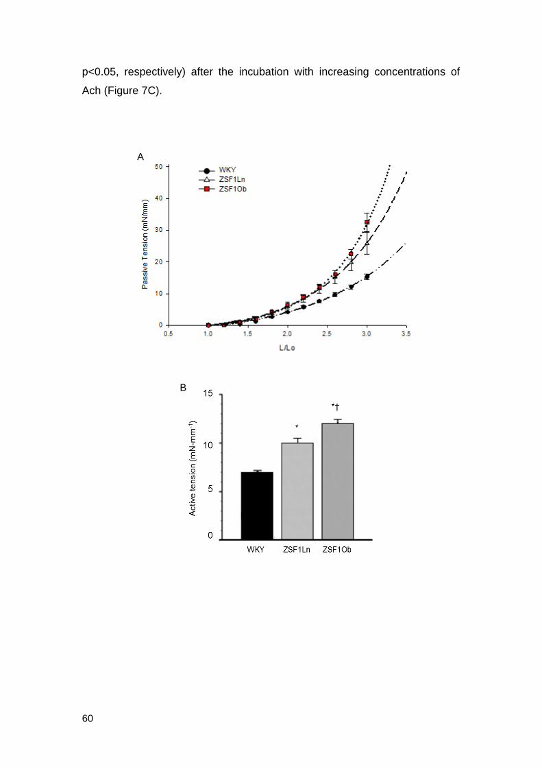
60
p<0.05, respectively) after the incubation with increasing concentrations of
Ach (Figure 7C).
A
B

61
Figure 7. Vascular function of aortic rings from WKY (n=5), ZSF1Ln (n=5) and
ZSF1Ob (n=5), animals. A) Strain-passive tension curve; B) Maximum active tension
at phenylephrine (Phe) maximal concentration (10-5); C) Vascular relaxation to
increasing doses of acetylcholine (Ach) pre-contracted with Phe. The values are
means ± S.E.M. p<0.05: * vs WKY, † vs ZSF1Ln.
3. Plasma levels of adipokines and inflammatory markers
We performed a rat adipokine array kit to have an overview of the
major plasma changes in ZSF1Ob versus ZSF1Ln using a small number of
samples per group. Results are presented in table 5 and revealed that the
expression of pro-inflammatory cytokines Il-1β and Il-6 displayed a small
tendency to be lower in ZSF1Ob group when compared to ZSF1Ln. In
contrast, the expression of ICAM1 showed a trend to increase in ZSF1Ob. No
differences were found in relation to anti-inflammatory cytokines Il-11 and Il-
10 (Table 5). Interestingly, only levels of monocyte chemoattractant protein 1
(MCP1), that actively recruits leukocytes into inflammatory sites, were
significantly higher in ZSF1Ob. Lipocalin, a protein involved in the lipid
transport which also has a role in inflammation, displayed a trend to have
higher levels in the obese rats (Table 5).
C
WKY ZSF1Ln ZSF1Ob

62
Table 5. Plasma protein expression (% of change from the positive control) assessed
by a profile array of ZSF1Ln (n=4) and ZSF1Ob (n=4) animals. Il-1β, interleukin-1β;
Il-6, interleukin-6; TNFα, tumor necrosis factor α; ICAM1, intercellular adhesion
molecule 1; Il-11, interleukin-11; Il-10, interleukin-10; MCP1, monocyte
chemoattractant protein 1; RANTES, regulated on activation, normal T cell
expressed and secreted. The values are represented as means ± S.E.M. p<0.05: †
vs ZSF1Ln.
Protein (%) ZSF1Ln ZSF1Ob
Il-1β 6,1 4,0
Il-6 2,9 1,8
TNFα 2,5 2,8
ICAM1 16,3 21,3
Il-11 3,8 3,7
Il-10 2,6 2,2
MCP1 19,2 54,3 †
RANTES 7,5 4,5
Lipocalin 45,1 58,2
We further confirmed some circulatory parameters by ELISA (Table 6),
a more sensitive technique that revealed that plasma levels of leptin were
significantly increased and there was a similar trend in FABP4 levels,
consistently with metabolic syndrome (Table 6). In both ZSF1 groups, the
vasodilator Ang 1-7 was significantly increase (p<0.05) to compensate for the
increased ventricular overload and arterial stiffness present in these
hypertensive groups (Table 6).
Interestingly, plasma levels of pro-inflammatory cytokines such as Il-6
and TNFα revealed a tendency to be higher in WKY group while, between
ZSF1 groups, obese animals displayed slightly lower values than the lean
ones, consistently with the array results (Table 6).

63
Table 6. Circulating plasma levels data from WKY (n=6), ZSF1Ln (n=9) and ZSF1Ob
(n=8) groups. FABP4, fatty acid binding protein 4; Ang 1-7, angiotensin 1-7; Il-6,
Interleukin-6; TNFα, tumor necrosis factor α. The values are represented as means
± S.E.M. p<0.05: * vs WKY, † vs ZSF1Ln.
Protein WKY ZSF1Ln ZSF1Ob
Leptin (pg.mL-1
) 434.34 ± 136.67 405.77 ± 240.23 22150.15±10711.55*†
FABP4 (ng.mL-1
) 53.03 ± 18.53 83.27 ± 64.86 122.18 ± 81.34
Angiotensin 1-7 (ng.mL-1
) 1.84 ± 0.85 3.16 ± 1.04* 3.28 ± 0.46*
Il-6 (pg.mL-1
) 277.38 ± 218.29 176.67 ± 146.84 134.45 ± 68.92
TNFα (pg.mL-1
) 76.44 ± 42.15 71.57 ± 43.85 55.38 ± 15.44
4. Markers of myocardial dysfunction
In addition, myocardial expression of some inflammatory-related and
ROS-related genes and proteins was also measured by real-time PCR and
western blotting and are shown in figures 8&9.
When it comes to gene expression of endothelial function-related
genes, E-selectin expression was higher in ZSF1 group (p<0.05), while in
ICAM1, VCAM1, eNOS3 and endothelin 1 no significant differences were
observed but only a trend towards their upregulation (Figure 8).
A B

64
Figure 8. mRNA expression of WKY (n=5), ZSF1Ln (n=5) and ZSF1Ob (n=5) animals. A) ICAM1, intercellular adhesion molecule 1; B) VCAM1, vascular cell adhesion molecule 1; C) endothelin 1; D) eNOS, endothelial nitric oxide synthase; E) E-selectin. The values are represented as means ± S.E.M. p<0.05: * vs WKY.
The expression of AGEs receptor (RAGEs) was not different between
the ZSF1 groups (Figure 9A), but both results were significantly higher than
WKY. AGES deposition, assessed by carboxymethyllysine (CML) expression,
was similar between groups despite a trend towards an increase in ZSF1Ob
(Figure 9B). Vasodilator-stimulated phosphoprotein (VASP) (Figure 9C) and
eNOS protein (Figure 9D) expression had no differences.
In ZSF1Ob rats, NOX2 expression, which promotes ROS production,
increased in a significant way, compared with WKY and ZSF1Ln. Contrarily,
NOX4 expression in ZSF1Ob, with protective properties against ROS, was
significantly lower than the other groups (Figure 9E&F).
E
C D
C D
E

65
Figure 9. Protein expression of WKY (n=6), ZSF1Ln (n=6) and ZSF1Ob (n=5)
animals assessed by western blotting. A) RAGE, receptor of AGEs; B) CML,
carboxymethyllysine; C) VASP, vasodilator-stimulated phosphoprotein; D) eNOS,
endothelial nitric oxide synthase; E) NOX 2, NADPH oxidase 2; F) NOX 4, NADPH
oxidase 4. The values are represented as means ± S.E.M. p<0.05: * vs WKY, † vs
ZSF1Ln.
B A
C D
E F

66
5. Flow cytometry analysis
Representative scatter plots of flow cytometry analysis is depicted in
figures 10A&B. Flow cytometry analysis revealed similar levels of CD31, a
marker of endothelial cells, between ZSF1Ln and ZSF1Ob groups (Figure
10C). Differently, ZSF1Ob LV samples display a significantly higher number of
CD45 positive cells (Figure 10D), a marker of inflammation.
Figure 10. Flow cytometry analysis. (A) Example of a representative plot of CD31
versus CD45 expression from a ZSF1Ln animal. (B) Example of a representative plot
of CD31 versus CD45 expression from a ZSF1Ob animal. (C) Expression of CD31+
cells in ZSF1Ln (n=3) and ZSF1Ob (n=5). (D) Expression of CD45+ cells in in
ZSF1Ln (n=3) and ZSF1Ob (n=5). The values are represented as means ± S.E.M.
p<0.05: † vs ZSF1Ln.
C D
B A

Part 5
Discussion
“The important thing in science is not so much to obtain new facts as to discover new
ways of thinking about them.”
William Lawrence Bragg


69
The present study explored a previously described animal model of
HFpEF, the ZSF1 obese rat, aiming to identify the pathophysiologic
mechanisms underlying diastolic dysfunction progression. We have shown
that the highly prevalent comorbidities associated to HFpEF trigger
inflammation, oxidative stress and endothelial dysfunction in this animal
model.
1. Metabolic risk-related HFpEF model
In this study we used the ZSF1 animal model and demonstrated that
ZSF1Ob develops HFpEF at their 20th week of age. Compared to ZSF1Ln or
WKY rats, ZSF1Ob showed many features of high metabolic risk such as
visceral obesity evident from elevated perirenal and perigonadal fat, insulin
resistance, hyperglycemia and physical inactivity evident from striated muscle
wasting. ZSF1Ob rats present two different mutations for leptin receptor gene
and as a compensatory response, leptin levels significantly increase. Lipocalin
and FABP4 levels are also elevated in ZSF1Ob group, showing that in these
rats adipose tissue is already dysfunctional and secreting a different pattern of
adipokines.
Comparing with WKY group, ZSF1Ln presented hypertension and
consequent LV hypertrophy both at the organ and cellular level, which was
further aggravated in ZSF1Ob rats. This hypertrophic response represents an
attempt to normalize increased ventricular wall stress imposed by systemic
arterial hypertension. Indeed, hypertrophy parallels the cardiac remodeling
associated to HFpEF, namely in terms of ventricular size, geometry, shape
and composition. Besides obesity, ZSF1Ob animals also developed
hyperglycemia, oral glucose intolerance, insulin resistance, glycosuria and
DM, all features that trigger diastolic dysfunction progression towards HFpEF
[57]. Indeed, this represents an interesting aspect of ZSF1 as we were able to
demonstrate that overload imposed by hypertension, as in ZSF1Ln, is not
enough to induce diastolic dysfunction. Instead, only the concomitant
presence of other significant comorbidities, as in ZSF1Ob, prompted HFpEF.
Several studies have supported this finding by showing the severity of cardiac

70
and vascular injuries imposed by hypertension in obese or diabetic patients
when compared with hypertension per se [66-69].
The HFpEF presentation observed in this metabolic risk model also
shares features with clinical presentation of this syndrome. Systolic function,
evaluated by CI and EF, was preserved in all groups. Regarding diastolic
function, the ZSF1Ob rats presented significant diastolic abnormalities as
demonstrated by echocardiography evaluation, mainly and increased
myocardial stiffness that compromises a proper ventricular filling. Previous
studies from our groups showed that the relaxation time constant is
prolonged in ZSF1Ob, denoting also impaired relaxation [57]. Additionally, the
obese group displayed dilated left atrium, lung congestion and increased
arterial elastance, contributing to a worse ventricle-vascular coupling, a well
establish cause of HFpEF [70]. All these results support the idea of ZSF1Ob
as a good animal model of HFpEF.
The current animal model differs from previous experimental HFpEF
models, which largely overlooked metabolic risk as they were carried out in
old, hypertensive dogs [71, 72] or in Dahl salt sensitive hypertensive rats [73].
ZSF1Ob however closely resembles clinical HFpEF where metabolic risk is
highly prevalent as evident from numerous HFpEF registries or large outcome
trials [74-76]. HFpEF is a complex disorder that alters cardiac structure and
function. Comorbidities commonly adjacent to this pathology, such as obesity,
hypertension and diabetes mellitus, are associated with endothelial
dysfunction [11, 51]. As a result, patients with HFpEF have an impaired NO
response, elevated levels of vasoconstrictors and increased expression of
adhesion molecules. Endothelium acquires a chronic inflammatory and pro-
thrombotic state where vasodilatation is compromised [32, 36]. Therefore,
endothelial dysfunction may be a critical early target for the prevention of
HFpEF, since that treatment options are still limited.

71
2. Endothelial dysfunction and oxidative stress
Supplementary data related to vascular function demonstrate that
ZSF1Ob animal’s aortas are less reactive. Furthermore, ZSF1Ob aortas are
the largest and the thickest. In this group, besides the high passive tension,
which indicates vascular stiffness, we also observed impairment of relaxation
after incubation with a vasodilator substance. Altogether these data suggest
endothelial dysfunction. Our results are in agreement with previous studies
showing precisely that aortic rings in obese rats had a vasoconstrictive
response [77]. Interestingly, Ang 1-7 circulating levels are increased in an
attempt to compensate for the increase stiffness of ZSF1Ob arteries.
Many HFpEF-associated comorbidities are known to induce cardiac
and systemic ROS production, which further contributes to oxidative stress
[78]. In our study the expression of NADPH oxidases, NOX4 and NOX2, with
opposite functions, was in agreement with other studies where NOX 2
upregulation in cardiac tissue was observed [79]. In ZSF1Ob, NOX2
upregulation versus NOX4 downregulation clearly imbalances ROS
production/protective mechanisms, promoting an oxidative stress
environment.
It is well known that DM-induced oxidative stress contributes to the
formation of AGEs in cardiomyocytes [80]. Both ZSF1 groups demonstrated
significantly higher levels of RAGEs and a small tendency to increase CML in
ZSF1Ob. Others studies found similar results of RAGE levels in diabetic
hearts [81]. These results were not confirmed by immunohistochemistry
analysis of myocardial tissue. Thus this important signaling pathway needs to
be clarified in subsequent studies.
Increased systemic inflammation is a hallmark of all the comorbidities
associated to this animal model. Interestingly, ZSF1Ob showed a slight
tendency to have lower levels of several inflammatory mediators. This
phenomenon has recently been described as the obesity paradox. This theory
believes that obesity can, under certain circumstances actually have a
positive effect due to the capacity of lipoproteins to neutralize

72
lipopolysaccharide derived from intestinal bacterial translocation. The
mortality of obese patients with HF is low. This theory has been described as
“reverse epidemiology” [82, 83]. So, the obesity presented by ZSF1Ob
animals could attenuate the inflammatory effect. Interestingly, WKY displayed
the highest levels of the pro-inflammatory Il-6 and TNFα. A possible
explanation for this finding is that, WKY being a genetic model of depression,
have hyper-responses to stress and display depression-associated
abnormalities, like inflammation [84]. In fact, and supporting our results, other
studies have been reported similar inflammatory levels [85, 86]. Current
studies are being carried out in laboratory to clarify this aspect.
Regarding tissue inflammation, the increase expression of E-selectin
and CD45 in ZSF1Ob animals denotes increased levels of inflammation
specifically in endothelial cells.
All the above evidences suggest that ZSF1 animals do not display
pronounced systemic alterations but present cardiac-tissue and vascular
inflammation. Comorbidities stimulated the expression of inflammatory
markers in ZSF1Ob hearts, mainly in endothelium, leading to diastolic
dysfunction. We can conclude that comorbidities-induced inflammation and
oxidative stress seem to be the hallmark of diastolic dysfunction and
progression towards HFpEF.

Part 6
Conclusion and future perspectives
“Somewhere, something incredible is waiting to be known.”
Carl Sagan


75
Unfortunately and despite all advances in medicine, HFpEF still
remains with no available therapeutic options. Endothelial dysfunction, a state
present in HFpEF, seems to be involved in its pathogenesis. An appropriate
animal model as ZSF1Ob will certainly help to better understand this
syndrome as well as to test new potential therapeutic targets.
Our data clearly show that ZSF1Ob rat presents diastolic dysfunction
and HFpEF. Furthermore, both morphometric and metabolic studies already
perform in our laboratory confirm the metabolic syndrome state of this animal
model, corroborating its cardiometabolic risk, typically associated to HFpEF in
the clinical scenario.
We fulfill our goal to evaluate endothelial dysfunction in the ZSF1Ob rat
model. Our results evidently showed that ZSF1Ob aortas are predominantly
under a rigid and vasoconstriction state. Thus, our data suggest that a
dysfunctional endothelium induces an increased vascular stiffness and
impaired relaxation, with obvious myocardial deleterious consequences.
We showed that cardiac endothelial cells from ZSF1Ob animals are
under a state of endothelial inflammation, which suggests that comorbidities
accumulated by these animals stimulate the expression of some important
inflammatory markers, culminating in endothelial dysfunction.
There seems to be no doubt that endothelial dysfunction is related to
HFpEF development. In this context, it is important to note that endothelium is
no longer viewed as a physical barrier that basically separates blood from the
vessel wall. In fact, over the years endothelial dysfunction has been
associated with several diseases, including HFpEF. Important advances have
been made to better understand this relationship but future studies will enable
the development of new therapeutic targets with a possible cure.
In the future we aim to complete our studies evaluating ROS
systemically by measuring plasma levels of malondialdehyde (MDA) and
urinary levels of 8-hydroxydeoxyguanosine (8-OHdG), both markers of
oxidative DNA damage. We also want to detect ROS in cultured tissue with a
dihydroethidium (DHE) oxidative fluorescent probe. Additionally we pretend to

76
measure NO levels in heart tissue as well as in vessels supernatant after
functional studies.

Part 7
References
“Research is what I'm doing when I don't know what I'm doing.”
Wernher Von Braun


79
1. Dhingra, A., et al., Epidemiology of heart failure with preserved ejection fraction. Curr Heart Fail Rep, 2014. 11(4): p. 354-65.
2. Dorhout Mees, E.J., Diastolic heart failure: a confusing concept. Heart Fail Rev, 2013. 18(4): p. 503-9.
3. Heineke, J. and J.D. Molkentin, Regulation of cardiac hypertrophy by intracellular signalling pathways. Nat Rev Mol Cell Biol, 2006. 7(8): p. 589-600.
4. Roger, V.L., Epidemiology of heart failure. Circ Res, 2013. 113(6): p. 646-59. 5. Ponikowski, P., et al., Heart failure: preventing disease and death worldwide.
ESC Heart Failure, 2014. 1: p. 4-25. 6. Nichols, M., et al., Cardiovascular disease in Europe: epidemiological update.
Eur Heart J, 2013. 34(39): p. 3028-34. 7. Andersen, M.J. and B.A. Borlaug, Heart failure with preserved ejection
fraction: current understandings and challenges. Curr Cardiol Rep, 2014. 16(7): p. 501.
8. Aziz, F., et al., Heart failure with preserved EF: a bird eye view. JNMA J Nepal Med Assoc, 2013. 52(190): p. 405-12.
9. Yancy, C.W., et al., 2013 ACCF/AHA guideline for the management of heart failure: a report of the American College of Cardiology Foundation/American Heart Association Task Force on Practice Guidelines. J Am Coll Cardiol, 2013. 62(16): p. e147-239.
10. Komamura, K., Similarities and Differences between the Pathogenesis and Pathophysiology of Diastolic and Systolic Heart Failure. Cardiology Research and Practice, 2013. 2013: p. 6.
11. van Heerebeek, L. and W.J. Paulus, Impact of Comorbidities on Myocardial Remodeling and Dysfunction In Heart Failure with Preserved Ejection Fraction. SOJ Pharmacy & Pharmaceutical Sciences, 2014. 1(2): p. 20.
12. Oktay, A.A., J.D. Rich, and S.J. Shah, The emerging epidemic of heart failure with preserved ejection fraction. Curr Heart Fail Rep, 2013. 10(4): p. 401-10.
13. Glezeva, N. and J.A. Baugh, Role of inflammation in the pathogenesis of heart failure with preserved ejection fraction and its potential as a therapeutic target. Heart Fail Rev, 2014. 19(5): p. 681-94.
14. Hummel, S.L. and D.W. Kitzman, Update on heart failure with preserved ejection fraction. Curr Cardiovasc Risk Rep, 2013. 7(6): p. 495-502.
15. Bhuiyan, T. and M.S. Maurer, Heart Failure with Preserved Ejection Fraction: Persistent Diagnosis, Therapeutic Enigma. Curr Cardiovasc Risk Rep, 2011. 5(5): p. 440-449.
16. Wang, Z., et al., Vascular endothelial function of patients with stable coronary artery disease. Pak J Med Sci, 2015. 31(3): p. 538-42.
17. Leite-Moreira, A.F., Current perspectives in diastolic dysfunction and diastolic heart failure. Heart, 2006. 92(5): p. 712-8.
18. Borlaug, B.A., The pathophysiology of heart failure with preserved ejection fraction. Nat Rev Cardiol, 2014. 11(9): p. 507-15.
19. Barry, S.P., S.M. Davidson, and P.A. Townsend, Molecular regulation of cardiac hypertrophy. Int J Biochem Cell Biol, 2008. 40(10): p. 2023-39.
20. Heusch, G., et al., Cardiovascular remodelling in coronary artery disease and heart failure. Lancet, 2014. 383(9932): p. 1933-43.
21. Asrar Ul Haq, M., et al., Heart failure with preserved ejection fraction - unwinding the diagnosis mystique. Am J Cardiovasc Dis, 2014. 4(3): p. 100-13.
22. Borlaug, B.A. and W.J. Paulus, Heart failure with preserved ejection fraction: pathophysiology, diagnosis, and treatment. Eur Heart J, 2011. 32(6): p. 670-9.
23. Palau, P., et al., Physical therapy in heart failure with preserved ejection fraction: A systematic review. Eur J Prev Cardiol, 2014.

80
24. Scantlebury, D.C. and B.A. Borlaug, Why are women more likely than men to develop heart failure with preserved ejection fraction? Curr Opin Cardiol., 2011. 26(6): p. 562-8.
25. Mentz, R.J. and G.M. Felker, Noncardiac comorbidities and acute heart failure patients. Heart Fail Clin, 2013. 9(3): p. 359-67, vii.
26. Falcao-Pires, I., et al., Diabetes mellitus worsens diastolic left ventricular dysfunction in aortic stenosis through altered myocardial structure and cardiomyocyte stiffness. Circulation, 2011. 124(10): p. 1151-9.
27. Susic, D., Cross-link breakers as a new therapeutic approach to cardiovascular disease. Biochemical Society Transactions, 2007. 35(5): p. 853-6.
28. Leung, M., V. Phan, and D.Y. Leung, Endothelial function and left ventricular diastolic functional reserve in type 2 diabetes mellitus. Open Heart, 2014. 1(1): p. e000113.
29. Hansson, G.K., Inflammation, atherosclerosis, and coronary artery disease. N Engl J Med, 2005. 352(16): p. 1685-95.
30. Hadi, H.A., C.S. Carr, and J. Al Suwaidi, Endothelial dysfunction: cardiovascular risk factors, therapy, and outcome. Vasc Health Risk Manag, 2005. 1(3): p. 183-98.
31. Yang, O., J. Li, and J. Kong, The Endothelium as a Target for the Treatment of Heart Failure. Cell Biochem Biophys, 2015.
32. Marti, C.N., et al., Endothelial dysfunction, arterial stiffness, and heart failure. J Am Coll Cardiol, 2012. 60(16): p. 1455-69.
33. Lam, C.S. and D.L. Brutsaert, Endothelial dysfunction: a pathophysiologic factor in heart failure with preserved ejection fraction. J Am Coll Cardiol, 2012. 60(18): p. 1787-9.
34. Tomiyama, H. and A. Yamashina, Vascular Dysfunction: A Key Player in Chronic Cardio-renal Syndrome. Intern Med, 2015. 54(12): p. 1465-72.
35. Hartge, M.M., T. Unger, and U. Kintscher, The endothelium and vascular inflammation in diabetes. Diab Vasc Dis Res, 2007. 4(2): p. 84-8.
36. Endemann, D.H. and E.L. Schiffrin, Endothelial dysfunction. J Am Soc Nephrol, 2004. 15(8): p. 1983-92.
37. Patel, S.J., et al., The inflammatory response to double stranded DNA in endothelial cells is mediated by NFkappaB and TNFalpha. PLoS One, 2011. 6(5): p. e19910.
38. Tschope, C. and S. Van Linthout, New insights in (inter)cellular mechanisms by heart failure with preserved ejection fraction. Curr Heart Fail Rep, 2014. 11(4): p. 436-44.
39. Mather, K.J., The vascular endothelium in diabetes--a therapeutic target? Rev Endocr Metab Disord, 2013. 14(1): p. 87-99.
40. Raij, L., Nitric oxide in the pathogenesis of cardiac disease. J Clin Hypertens (Greenwich), 2006. 8(12 Suppl 4): p. 30-9.
41. Tousoulis, D., et al., The role of nitric oxide on endothelial function. Curr Vasc Pharmacol, 2012. 10(1): p. 4-18.
42. Khazaei, M., F. Moien-Afshari, and I. Laher, Vascular endothelial function in health and diseases. Pathophysiology, 2008. 15(1): p. 49-67.
43. Taylor, A.L., Nitric oxide modulation as a therapeutic strategy in heart failure. Heart Fail Clin, 2012. 8(2): p. 255-72.
44. Deanfield, J.E., J.P. Halcox, and T.J. Rabelink, Endothelial function and dysfunction: testing and clinical relevance. Circulation, 2007. 115(10): p. 1285-95.
45. Reriani, M.K., L.O. Lerman, and A. Lerman, Endothelial function as a functional expression of cardiovascular risk factors. Biomark Med, 2010. 4(3): p. 351-60.
81
46. Avogaro, A., et al., Endothelial dysfunction in diabetes: the role of reparatory mechanisms. Diabetes Care, 2011. 34 Suppl 2: p. S285-90.
47. Akiyama, E., et al., Incremental prognostic significance of peripheral endothelial dysfunction in patients with heart failure with normal left ventricular ejection fraction. J Am Coll Cardiol, 2012. 60(18): p. 1778-86.
48. Versari, D., et al., Endothelial dysfunction as a target for prevention of cardiovascular disease. Diabetes Care, 2009. 32 Suppl 2: p. S314-21.
49. Rajendran, P., et al., The vascular endothelium and human diseases. Int J Biol Sci, 2013. 9(10): p. 1057-69.
50. Prieto, D., C. Contreras, and A. Sanchez, Endothelial dysfunction, obesity and insulin resistance. Curr Vasc Pharmacol, 2014. 12(3): p. 412-26.
51. Paulus, W.J. and C. Tschope, A novel paradigm for heart failure with preserved ejection fraction: comorbidities drive myocardial dysfunction and remodeling through coronary microvascular endothelial inflammation. J Am Coll Cardiol, 2013. 62(4): p. 263-71.
52. Trepels, T., A.M. Zeiher, and S. Fichtlscherer, The endothelium and inflammation. Endothelium, 2006. 13(6): p. 423-9.
53. van Heerebeek, L., et al., Low myocardial protein kinase G activity in heart failure with preserved ejection fraction. Circulation, 2012. 126(7): p. 830-9.
54. Badenhorst, D., et al., Cross-linking influences the impact of quantitative changes in myocardial collagen on cardiac stiffness and remodelling in hypertension in rats. Cardiovasc Res, 2003. 57(3): p. 632-41.
55. Srinivasan, K. and P. Ramarao, Animal models in type 2 diabetes research: an overview. Indian J Med Res, 2007. 125(3): p. 451-72.
56. King, J.F.A., The use of animal models in diabetes research. British Journal of Phamacology 2012. 166: p. 877–894.
57. Hamdani, N., et al., Myocardial titin hypophosphorylation importantly contributes to heart failure with preserved ejection fraction in a rat metabolic risk model. Circ Heart Fail, 2013. 6(6): p. 1239-49.
58. Tofovic, S.P., et al., 2-Hydroxyestradiol attenuates renal disease in chronic puromycin aminonucleoside nephropathy. J Am Soc Nephrol, 2002. 13(11): p. 2737-47.
59. Bilan, V.P., et al., Diabetic nephropathy and long-term treatment effects of rosiglitazone and enalapril in obese ZSF1 rats. J Endocrinol, 2011. 210(3): p. 293-308.
60. Tofovic, S.P., et al., Long-term caffeine consumption exacerbates renal failure in obese, diabetic, ZSF1 (fa-fa(cp)) rats. Kidney Int, 2002. 61(4): p. 1433-44.
61. Tofovic, S.P., et al., Renal function and structure in diabetic, hypertensive, obese ZDFxSHHF-hybrid rats. Ren Fail, 2000. 22(4): p. 387-406.
62. Joshi, D., et al., TRC4186, a novel AGE-breaker, improves diabetic cardiomyopathy and nephropathy in Ob-ZSF1 model of type 2 diabetes. J Cardiovasc Pharmacol, 2009. 54(1): p. 72-81.
63. Rafikova, O., E.M. Salah, and S.P. Tofovic, Renal and metabolic effects of tempol in obese ZSF1 rats--distinct role for superoxide and hydrogen peroxide in diabetic renal injury. Metabolism, 2008. 57(10): p. 1434-44.
64. Zambad, S.P., et al., TRC150094 attenuates progression of nontraditional cardiovascular risk factors associated with obesity and type 2 diabetes in obese ZSF1 rats. Diabetes Metab Syndr Obes, 2011. 4: p. 5-16.
65. Falcao-Pires, I., et al., Apelin decreases myocardial injury and improves right ventricular function in monocrotaline-induced pulmonary hypertension. Am J Physiol Heart Circ Physiol, 2009. 296(6): p. H2007-14.
66. Virdis, A., et al., Microvascular endothelial dysfunction in obesity and hypertension. Curr Pharm Des, 2013. 19(13): p. 2382-9.

82
67. Hadi, H.A. and J.A. Suwaidi, Endothelial dysfunction in diabetes mellitus. Vasc Health Risk Manag, 2007. 3(6): p. 853-76.
68. Varughese, G.I., The impact of diabetes mellitus on endothelial dysfunction. South Med J, 2007. 100(2): p. 128-9.
69. Dandona, P., A. Chaudhuri, and A. Aljada, Endothelial dysfunction and hypertension in diabetes mellitus. Med Clin North Am, 2004. 88(4): p. 911-31, x-xi.
70. Borlaug, B.A. and D.A. Kass, Ventricular-vascular interaction in heart failure. Heart Fail Clin, 2008. 4(1): p. 23-36.
71. Bishu, K., et al., Sildenafil and B-type natriuretic peptide acutely phosphorylate titin and improve diastolic distensibility in vivo. Circulation, 2011. 124(25): p. 2882-91.
72. Hamdani, N., et al., Deranged myofilament phosphorylation and function in experimental heart failure with preserved ejection fraction. Cardiovasc Res, 2013. 97(3): p. 464-71.
73. Westermann, D., et al., Enhancement of the endothelial NO synthase attenuates experimental diastolic heart failure. Basic Res Cardiol, 2009. 104(5): p. 499-509.
74. Ather, S., et al., Impact of noncardiac comorbidities on morbidity and mortality in a predominantly male population with heart failure and preserved versus reduced ejection fraction. J Am Coll Cardiol, 2012. 59(11): p. 998-1005.
75. McMurray, J.J., et al., Heart failure with preserved ejection fraction: clinical characteristics of 4133 patients enrolled in the I-PRESERVE trial. Eur J Heart Fail, 2008. 10(2): p. 149-56.
76. Edelmann, F., et al., Contribution of comorbidities to functional impairment is higher in heart failure with preserved than with reduced ejection fraction. Clin Res Cardiol, 2011. 100(9): p. 755-64.
77. Chinen, I., et al., Vascular lipotoxicity: endothelial dysfunction via fatty-acid-induced reactive oxygen species overproduction in obese Zucker diabetic fatty rats. Endocrinology, 2007. 148(1): p. 160-5.
78. Raza, H., A. John, and F.C. Howarth, Alterations in glutathione redox metabolism, oxidative stress, and mitochondrial function in the left ventricle of elderly Zucker diabetic fatty rat heart. Int J Mol Sci, 2012. 13(12): p. 16241-54.
79. Murdoch, C.E., et al., Endothelial NADPH oxidase-2 promotes interstitial cardiac fibrosis and diastolic dysfunction through proinflammatory effects and endothelial-mesenchymal transition. J Am Coll Cardiol, 2014. 63(24): p. 2734-41.
80. Hegab, Z., et al., Role of advanced glycation end products in cardiovascular disease. World J Cardiol, 2012. 4(4): p. 90-102.
81. Aragno, M., et al., Oxidative stress-dependent impairment of cardiac-specific transcription factors in experimental diabetes. Endocrinology, 2006. 147(12): p. 5967-74.
82. Bosello, O. and M.P. Donataccio, Obesity paradox. Eat Weight Disord, 2013. 18(4): p. 447-8.
83. Hainer, V. and I. Aldhoon-Hainerova, Obesity paradox does exist. Diabetes Care, 2013. 36 Suppl 2: p. S276-81.
84. Will, C.C., F. Aird, and E.E. Redei, Selectively bred Wistar-Kyoto rats: an animal model of depression and hyper-responsiveness to antidepressants. Mol Psychiatry, 2003. 8(11): p. 925-32.
85. Bernard, C., et al., Resistance to endotoxin shock in spontaneously hypertensive rats. Hypertension, 1998. 31(6): p. 1350-6.
86. Lourenco, A.P., et al., A Western-type diet attenuates pulmonary hypertension with heart failure and cardiac cachexia in rats. J Nutr, 2011. 141(11): p. 1954-60.