Is a primer needed for orthodontic bonding? A...
130
1 Is a primer needed for orthodontic bonding? A Randomised Controlled Trial Sarabjit Singh Nandhra Submitted in accordance with the requirements for the degree of Doctorate in Clinical Dentistry The University of Leeds Medicine and Dentistry July, 2012 Supervised by; Simon Littlewood, Nanine Houghton Freidy Luther Simon Wood
Transcript of Is a primer needed for orthodontic bonding? A...

1
Is a primer needed for orthodontic
bonding? A Randomised Controlled Trial
Sarabjit Singh Nandhra
Submitted in accordance with the requirements for the degree of
Doctorate in Clinical Dentistry
The University of Leeds
Medicine and Dentistry
July, 2012
Supervised by;
Simon Littlewood,
Nanine Houghton
Freidy Luther
Simon Wood

2
The candidate confirms that the work submitted is his/her own and that
appropriate credit has been given where reference has been made to the
work of others.
This copy has been supplied on the understanding that it is copyright
material and that no quotation from the thesis may be published without
proper acknowledgement.
The right of Sarabjit Singh Nandhra to be identified as Author of this work
has been asserted by him in accordance with the Copyright, Designs and
Patents Act 1988.
© 2012 The University of Leeds and Sarabjit Singh Nandhra

3
Acknowledgements
I wish to thank all the people who made this research possible. Firstly I
would like to thank my supervisors for their guidance and support. I would
also like to thank Laura Mitchell and James Spencer for their assistance
in the project. I also wish to thank Jagdeep Prabu who aided in designing
the protocol and Deidre Devine who sadly had to leave her supervisory
role in this project.
I am grateful to all the patients and their parents for their participation
within the study.
I also wish to thank Mrs T. Munyombwe a lecturer in biostatistics within
the University of Leeds
Finally I would like to thank Gurmukh Nandhra and Daljeet Nandhra for
their support and encouragement.

4
Abstract
Is a primer needed for orthodontic bonding? A Randomised controlled trial Objective: To evaluate the clinical performance of APC Victory IITM (3M
Unitek) brackets in direct orthodontic bonding with and without the use of
primer.
Design: A single operator two centre prospective randomised controlled
clinical trial.
Setting: The orthodontic departments at the Leeds Dental Institute and St.
Luke’s hospital, Bradford.
Subjects and methods: 92 patients requiring orthodontic treatment with
fixed appliances. 46 Patients randomly allocated to control (with primer)
or test (without primer). Patients bonded using a standardised procedure.
Main outcome measures: Number of bracket failures, time to bond-up
appliances and the adhesive remnant index (ARI) when bracket failure
occurred, over a six month period
Results: Failure rate with primer 8.8%, without primer 13.8%, no
statistically significant difference- P value 0.051. Mean difference in bond-
up time per bracket was 0.068 minutes which was not statistically
significant (P =0.402). Statistically significant difference in the ARI – ARI 0
with primer 55.9%, no primer 81.5%, (P= 8.1622e-008).
Conclusion: There is no statistically significant difference in the bracket
failure rate with or without primer when bonding APC Victory IITM (P
=0.051). No significant difference in bond-up times. Statistically significant
difference in the ARI, bonding without primer providing a lower ARI

5
Contents
Acknowledgements.....................................................................................3
Abstract .......................................................................................................4
Contents......................................................................................................5
List of Tables.............................................................................................10
List of figures.............................................................................................11
1. Introduction ..........................................................................................12
2. Review of the literature .........................................................................14
2.1 Enamel etching ................................................................................14
2.2 Pre- preparation of enamel (prior to etching)...................................15
2.3 Type of etchant ................................................................................16
2.3.1 Citric acid...................................................................................16
2.3.2 Maleic acid.................................................................................17
2.3.3 Nitric acid...................................................................................17
2.3.4 Oxalic acid .................................................................................18
2.3.5 Phosphoric acid .........................................................................18
2.3.6 Air abrasion (Micro etching).......................................................19
2.3.7 Er:YAG laser ablation................................................................19
2.3.8 Summary ...................................................................................20
2.4 Concentration of acid.......................................................................20
2.5 Duration of etching...........................................................................21
2.6 Washing time ...................................................................................22
2.7 Drying time.......................................................................................23
2.8 Summary of etching process ...........................................................24
2.9 Materials used for bonding brackets................................................24
2.9.1 Composite .................................................................................24

6
2.9.2 Glass ionomer cement (GIC).....................................................27
2.9.3 Compomers and GIC hybrids....................................................27
2.9.4 Conclusion.................................................................................28
2.10 Primer ............................................................................................28
2.10.1 Self-etch and primer ................................................................29
2.10.2 Hydrophilic primer....................................................................30
2.10.3 Cyanoacrylate primer ..............................................................31
2.10.4 Fluoride releasing primer.........................................................31
2.10.5 Antibacterial primer..................................................................32
2.10.6 Bonding without primer............................................................33
2.10.6.2 In vitro orthodontic studies with no primer............................35
2.10.6.3 In vivo orthodontic studies with no primer ............................37
2.10.6.4 Summary ..............................................................................39
2.11 Bracket type...................................................................................40
2.11.1 Conventional brackets.............................................................40
2.11.2 Self-ligating brackets ...............................................................40
2.11.3 Pre-coated brackets ................................................................41
2.12 Bracket failure................................................................................41
2.12.1 Moisture...................................................................................42
2.12.2 Tooth .......................................................................................42
2.12.3 Bracket placement technique ..................................................44
2.12.4 Bracket base............................................................................46
2.12.5 Operator variation....................................................................47
2.12.6 Force .......................................................................................47
2.12.7 Adhesive remnant index (ARI) ................................................47
2.13 Problems with failure .....................................................................48
2.14 Study design ..................................................................................49

7
2.14.1 Statistics ..................................................................................51
2.15 Summary .......................................................................................52
3. Aims and hypothesis .............................................................................52
3.1 Aims of the study .............................................................................52
3.2 Hypothesis .......................................................................................53
4. Method of Investigation.........................................................................54
4.1 Ethical approval ...............................................................................54
4.2 Research and development.............................................................55
4.3 Information and consent forms ........................................................55
4.3.1 Information leaflets ....................................................................55
4.3.2 Informed consent for adults .......................................................55
4.3.3 Informed consent for children....................................................55
4.3.4 Informing referring practitioners ................................................56
4.4 Sample Size and Sample Size Calculation......................................56
4.5 Subjects ...........................................................................................57
4.5.1 Inclusion criteria.........................................................................57
4.5.2 Exclusion criteria .......................................................................58
4.6 Assignment ......................................................................................58
4.7 Subject withdrawal criteria ...............................................................59
4.8 Trial termination ...............................................................................59
4.9 Bonding Procedure ..........................................................................59
4.10 Bracket failure................................................................................61
4.11 Blinding ..........................................................................................61
4.12 Data Collection ..............................................................................62
4.13 Statistical analysis .........................................................................62
5 Results ...................................................................................................63
5. 1 Sample demographics ....................................................................64

8
5.2 Bond failure rates.............................................................................65
5.2.1 Survival rates.............................................................................65
5.2.2 Cox proportional hazards model with frailty including 1st
permanent molars...............................................................................67
5.2.3 Bond failure rates excluding 1st Permanent molars...................68
5.3 Distribution of bond failures .............................................................70
5.3.1 Distribution of bond failure rates between arches .....................73
5.3.2 Distribution of bracket failure in transverse plane .....................74
5.4 Appliance bond-up times .................................................................75
5.5 Adhesive remnant index (ARI).........................................................77
5.6 Summary of the results....................................................................79
6 Discussion..............................................................................................80
6.1 Principle findings of the study ..........................................................80
6.2 Critique of the methodology.............................................................80
6.2.1 Study design..............................................................................80
6.2.2 Statistical analysis .....................................................................82
6.3 Comparison of the results to other published work..........................84
6.3.1 Bracket failure rate ....................................................................84
6.3.2 Distribution of bracket failures ...................................................86
6.3.3 Bonding times............................................................................87
6.3.4 Adhesive remnant index............................................................88
6.5 Clinical Implications of the research ................................................89
6.6 Future research ...............................................................................89
7 Conclusions............................................................................................91
8. References............................................................................................92
9. List of abbreviations ............................................................................114
10. Appendices Appendix 1 Letter of ethical approval i

9
Appendix 2 Information sheet for children iv Appendix 3 Information sheet for adult patients v Appendix 4 Consent form adult viii
Appendix 5 Consent form for children ix Appendix 6 Assent form for children x Appendix 7 Letter to dentist xi Appendix 8 Data collection sheet 1 xii
Appendix 9 Data collection sheet 2 xiii Appendix 10 Cox proportional hazards model for
bracket failure dependent arch xiv
Appendix 11 Cox proportional hazards model for bracket
failure dependent on the side of the mouth xv
10
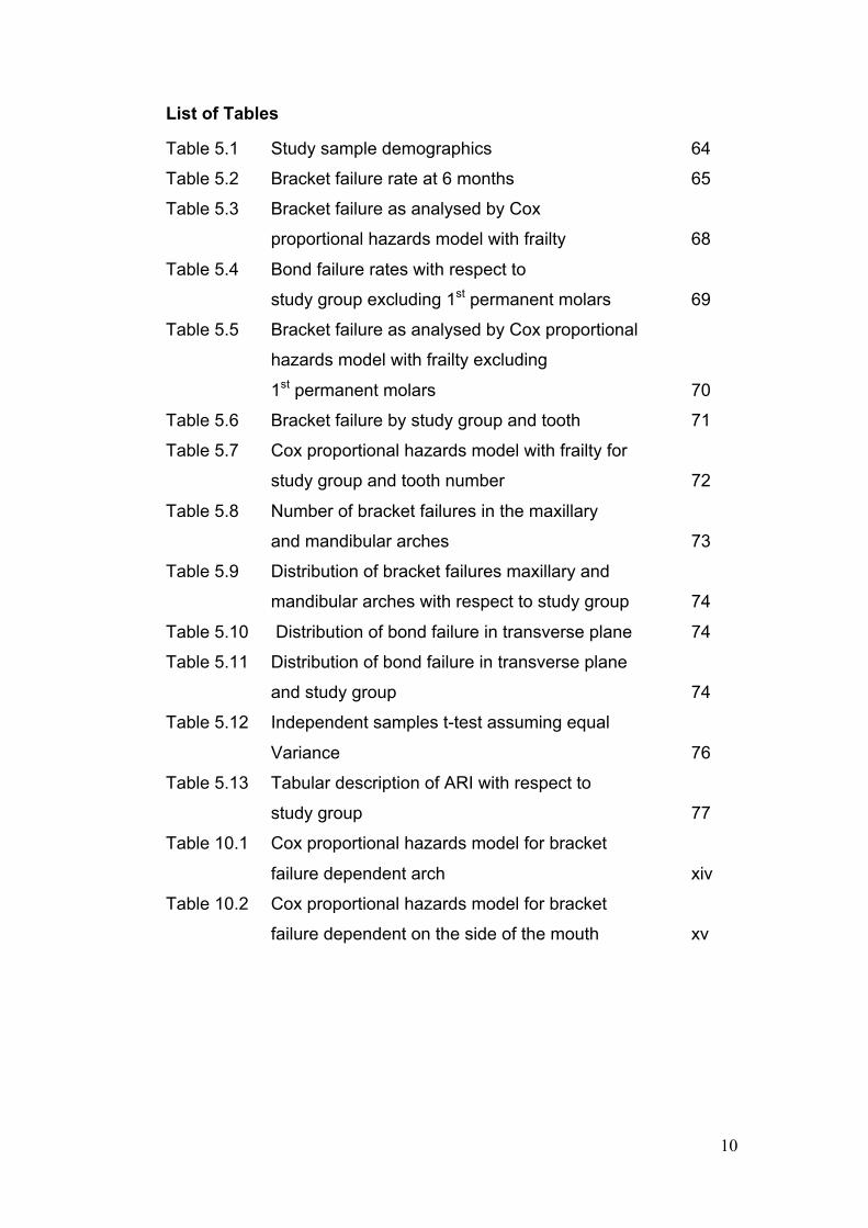
List of Tables
Table 5.1 Study sample demographics 64
Table 5.2 Bracket failure rate at 6 months 65
Table 5.3 Bracket failure as analysed by Cox
proportional hazards model with frailty 68
Table 5.4 Bond failure rates with respect to
study group excluding 1st permanent molars 69
Table 5.5 Bracket failure as analysed by Cox proportional
hazards model with frailty excluding
1st permanent molars 70
Table 5.6 Bracket failure by study group and tooth 71
Table 5.7 Cox proportional hazards model with frailty for
study group and tooth number 72
Table 5.8 Number of bracket failures in the maxillary
and mandibular arches 73
Table 5.9 Distribution of bracket failures maxillary and
mandibular arches with respect to study group 74
Table 5.10 Distribution of bond failure in transverse plane 74
Table 5.11 Distribution of bond failure in transverse plane
and study group 74
Table 5.12 Independent samples t-test assuming equal
Variance 76
Table 5.13 Tabular description of ARI with respect to
study group 77
Table 10.1 Cox proportional hazards model for bracket
failure dependent arch xiv
Table 10.2 Cox proportional hazards model for bracket
failure dependent on the side of the mouth xv

11
List of figures
Figure 2.1 Hierarchy of evidence-based dentistry 50
Figure 5.1 Consort flow diagram 64
Figure 5.2 Kaplan Meier plot for bracket failure with
and without primer 66
Figure 5.3 Graph to test the validity of proportional
hazards model 66
Figure 5.4 MLWin equation for possible covariants 67
Figure 5.5 Kaplan Meir graph by tooth number
(upper and lower arches combined) 71
Figure 5.6 Percentage bond failure by tooth and study
group 72
Figure 5.7 Box and Whisker plot of time mean to bond
a bracket 75
Figure 5.8 Histogram showing distribution of time to bond
a bracket 76
Figure 5.9 Distribution of ARI on bracket failure 77
Figure 5.10 MLWin equation for ARI 77

12
1. Introduction
Orthodontic appliances (braces) may be removable or fixed to the teeth.
Fixed orthodontic appliances allow precise movements of teeth in three
dimensions, which is not possible with removable appliances alone.
Therefore, the majority of orthodontic treatment within the UK involves
fixed appliances. Initially fixed orthodontic appliances were applied to the
teeth via the use of brackets soldered to metal bands and these bands
were placed around each tooth and cemented in position. Metal bands
are still in use (especially for posterior teeth), but have fallen out of favour
with the advent of composite bonding, which allows brackets to be
bonded directly to the tooth. This application of the brackets directly to the
tooth provides superior gingival health, improved patient comfort and
improved aesthetics.
Composite bonding in orthodontics has evolved significantly since the
concept was first introduced by Buonocore (1963). The initial composite
materials developed for bonding brackets involved chemically cured
‘single paste’ or ‘two paste systems. Currently, a wide variety of visible
light–cured orthodontic adhesives have become commercially available.
The advantages of visible light–cured orthodontic adhesives are the high
early bond strength, minimal extent of oxygen inhibition, and the
extended working time for optimal bracket placement. The acid etch
technique provides the basis for the bonding of orthodontic brackets to
enamel. Acid etching allows the penetration of low viscosity bonding
resins up to a depth of 50 µm, dependent on factors such as acid
concentration and etching time. Once polymerized a micro-mechanical
bond is established between the bonding resin and enamel. However, for
such bonding to take place, the enamel must first be etched for 15– 30
seconds with 37% orthophosphoric acid, and then rinsed with copious
amounts of water to remove the etchant and finally air dried until a frosted
glass appearance is achieved. A low viscosity resin (also known as a
sealant or primer) is then frequently painted onto the etched surface
before a more heavily filled resin is used to bond the brackets to the

13
teeth. This study will be investigating the need for primer when bonding
orthodontic brackets to teeth.
This study will explore the literature in regards to bonding orthodontic
brackets to teeth. The literature review will explore; pre- preparation of
teeth prior to etching, types of etchant and the procedures for etching;
orthodontic bonding materials; different primers used for orthodontic
bonding, including no primer; the effect of bracket design on orthodontic
bonding and the problems that may occur due to bracket failure.
The next section of this thesis will explore the various techniques of
preparing the enamel surface for bonding.

14
2. Review of the literature
2.1 Enamel etching
Etching is carried out to facilitate bonding of composite resins via micro
mechanical retention to the enamel surface. Treatments with various
etchants alter the structure of the enamel surface by selective
demineralization of exposed enamel rods leaving an increased surface
area and high energy, facilitating bonding via micro mechanical retention.
The depth of penetration varies due to a number of factors but is
generally accepted to be between 3.5µm and 50µm (Legler et al., 1990).
Scanning electron microscopes have shown this effect (Carstensen,
1995) and its pivotal role in orthodontic bonding.
The pattern of enamel etches can vary considerably and are broadly
classified into three different types:
1. Preferential removal of the enamel prism cores, with the
peripheries remaining intact
2. Preferential removal of the prism peripheries with the cores left
intact.
3. Removal of enamel prism cores and peripheries and some other
less distinct areas of etching.(Obrien, 2002, John F McCabe, 2008)
Acid etch is dispensed in the form of a liquid or a gel, with colouring
agents often added which aids the clinician to visualise where the acid
has been placed with a higher degree of accuracy. Acid etch is normally
applied to the tooth tissue via a small sponge or brush if in liquid form. If
in gel form the etch is normally applied with a brush, or through a fine
needle directly attached to the gel tube. However with heightened cross
infection control procedures and risk management, this method of
application is on the decline.
The ideal bond strength for brackets is suggested to be 6-8 MPa by
Reynolds (1976). An in vitro study (Littlewood et al., 2000) suggested that
the bond strength required for a clinically acceptable failure rate of 5 %
would be at the lower threshold of about 5 MPa.

15
The current literature has demonstrated that a variety of factors affects
the degree of etch achieved to enamel and thereby affect the bond
strength. These factors are:
• Pre preparation of the enamel
• Type of etchant
• Concentration of acid
• Duration of etching
• The washing time
• The drying time
These factors are explored in the following sections.
2.2 Pre- preparation of enamel (prior to etching)
Pre-preparation of enamel may be carried out when using self etch and
primer (single stage) or the two-stage acid etch and bond technique. Pre-
preparation for the single stage technique is discussed in section 2.7.1,
therefore this section will only analyse the literature on pre- preparation
for the two-stage technique.
There are several techniques used for pre-preparing the enamel for
bonding. One of the most commonly used techniques is to prepare the
enamel surface with a slurry of pumice and a brush on the slow speed
handpiece. An in vivo/ in vitro study (Lindauer et al., 1997) demonstrated
no significant difference in the bracket failure rate, and the characteristics
of the etched enamel and no significant difference in the bond strength
between the use of pumice and no pumice. This is supported by another
in vivo orthodontic study (Barry, 1995). A more recent development in
pre- preparation is the use of laser ablation. An in vitro study (Lee et al.,
2003) compared the use of phosphoric acid as an etchant with and
without the use of laser ablation. The study demonstrated statistically
significantly higher bond strengths in the phosphoric acid group alone
than with a combination of laser ablation and phosphoric acid.
Another recent development in pre-preparation of the enamel surface is
the use air abrasion (micro etching). An in vivo split mouth designed
study (Miles, 2008) showed no significant difference in bracket failure rate
over a six month period. This is consistent with the findings of in vitro
16
studies (Noble et al., 2008, Halpern and Rouleau, 2010)Ozone has also
been used for pre –preparation with an in vitro study demonstrating no
increase in SBS (Cehreli et al., 2010).
Fluoride has also been used in pre-preparation of enamel in a variety of
forms. Recent prospective split mouths studies have shown an increased
bracket failure rate; with fluoride varnish(Grover et al., 2012),and with
fluoride paste as a pre preparation. (Talic, 2011)
It is therefore commonly accepted from the available literature that it is
not beneficial to pre-prepare the enamel surface prior to acid etching for
bonding other than to remove gross debris. The next step in the bonding
process is to etch the enamel and this is explored in the following section.
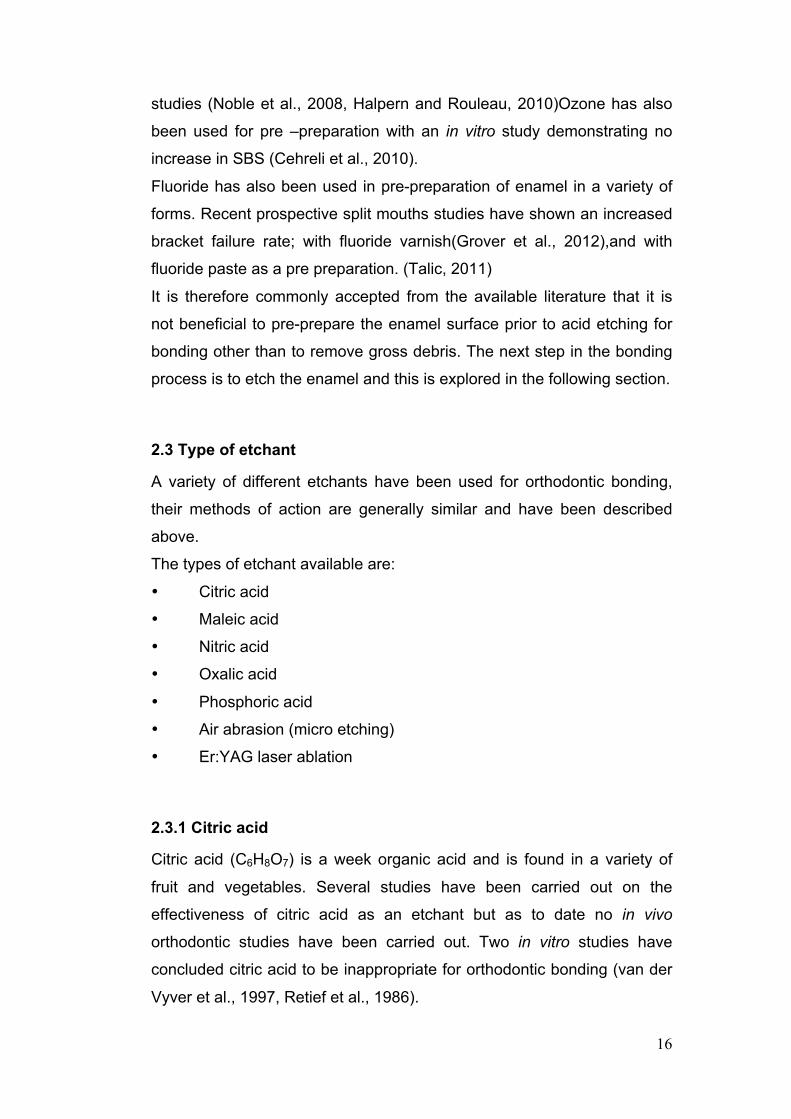
2.3 Type of etchant
A variety of different etchants have been used for orthodontic bonding,
their methods of action are generally similar and have been described
above.
The types of etchant available are:
• Citric acid
• Maleic acid
• Nitric acid
• Oxalic acid
• Phosphoric acid
• Air abrasion (micro etching)
• Er:YAG laser ablation
2.3.1 Citric acid
Citric acid (C6H8O7) is a week organic acid and is found in a variety of
fruit and vegetables. Several studies have been carried out on the
effectiveness of citric acid as an etchant but as to date no in vivo
orthodontic studies have been carried out. Two in vitro studies have
concluded citric acid to be inappropriate for orthodontic bonding (van der
Vyver et al., 1997, Retief et al., 1986).

17
However another in vitro study (Reifeis et al., 1995) demonstrated no
statistically significant difference between the bond strength of phosphoric
acid and citric acid, however within this study different bonding agents
were used for each etchant.
Therefore citric acid is currently not recommended for routine use as an
orthodontic bonding agent.
2.3.2 Maleic acid
Maleic acid (C4H4O4 ) is an organic acid which is water soluble.. Two in
vitro studies (Reifeis et al., 1995, Urabe et al., 1999) have demonstrated
no significant statistical difference in bond strength when maleic acid was
compared to 37% phosphoric acid, however within these studies there
was no mention of the etch duration for either acid.
Another in vitro study (Triolo et al., 1993) however found a statistically
significant difference between maleic acid and phosphoric acid, with
phosphoric acid having a superior bond strength.
To date there have been no in vivo orthodontic studies carried out, with
no data available for bracket failure rates.
2.3.3 Nitric acid
Nitric acid (HNO3) is a strong acid and is highly corrosive. An in vitro
study (Gardner and Hobson, 2001) compared etch patterns with
Phosphoric acid 37% and Nitric acid 2.5% at various time intervals. The
samples were analysed with a scanning electron microscope, which
demonstrated that phosphoric acid (37%) was more effective at creating
a better quality etch than nitric acid (2.5%).
However, another in vitro study (Blight and Lynch, 1995) demonstrated
no significant difference in the bond strength achieved by phosphoric acid
(37%) and nitric acid (2.5%) with the only difference noted being a
reduced amount of composite being left on the tooth with nitric acid when
compared with phosphoric acid. This reduced amount of composite on
the tooth implies a shifting of the failure site from the bracket/adhesive

18
interface to the tooth/adhesive interface, which may be interpreted as a
slightly decreased bond strength with nitric acid but which is still sufficient
for orthodontic bonding, however further research is required.
2.3.4 Oxalic acid
Oxalic acid is a relatively strong acid and has the chemical composition of
C2H2O4 in its anhydrous form. Only a limited number of studies have
been carried out looking at the sheer bond strength of oxalic acid with
variable results (Triolo et al., 1993) (Swift and Cloe, 1993, Holtan et al.,
1995). These in vitro studies found significantly lower bond strengths with
oxalic acid when compared to phosphoric acid, however another in vitro
study (Reifeis et al., 1995) showed no statistically significant difference in
the bond strength of oxalic acid compared to phosphoric acid, however
the results of this study may not be applicable as bovine incisors were
used. To date there have been no in vivo orthodontic studies carried out,
therefore currently oxalic acid is not recommended for routine use as an
orthodontic bonding agent.
2.3.5 Phosphoric acid
Phosphoric acid is also commonly known as orthophosphoric acid, it is an
inorganic acid with the chemical composition of H3PO4. Orthophosphoric
acid is highly soluble in water. It is acidic with a pH which is dependent on
the concentration present within the solution, increasing the pH with
decreasing concentration of phosphoric acid. Phosphoric acid is an
irritant to the biological tissues and can cause chemical burns, therefore
phosphoric acid must be handled with care at all times. Several studies
have been carried out on the efficacy of phosphoric acid which have
demonstrated its effectiveness as an etchant (Noble et al., 2008, Miles,
2008, Berk et al., 2008, Amm et al., 2008, Gray et al., 2006) with bond
strengths equal or greater than other etching methods, with an
orthodontic bond failure rate of approximately 5%. Therefore, phosphoric
acid remains the current gold standard in etchants for orthodontic
bonding.

19
2.3.6 Air abrasion (Micro etching)
Air-abrasion uses a high-speed stream of aluminium oxide particles,
propelled by air pressure to abrade the surface of the tooth(Gerbo et al.,
1992). Air abrasion is based on the law of kinetic energy, which states the
harder the substance, the faster the cutting speed, the softer the
substance, the slower the cutting speed(Gerbo et al., 1992). Therefore
enamel is cut much faster than dentine or amalgam, with this effect also
protecting the soft tissues. An in vitro study (Olsen et al., 1997)
compared air abrasion using two different particle sizes (1.50 microm, 2.
90 microm) against the control group of etching with 37% phosphoric
acid. The study concluded that enamel surface preparation using air-
abrasion results in a significantly lower bond strength, and should not be
advocated for routine clinical use as an enamel conditioner at this
time(Olsen et al., 1997) with the particle size have little to no effect on
bond strength. These findings are consistent with those of other
authors(Berk et al., 2008, Gray et al., 2006)
2.3.7 Er:YAG laser ablation
Laser ablation removes the smear layer. After laser etching, some
physical changes occur, such as melting and re-crystallization. Numerous
pores and bubble-like inclusions appear(Takeda et al., 1999) creating an
irregular surface available for bonding, thereby facilitating
micromechanical retention.
An in vitro study(Berk et al., 2008) was performed where enamel surfaces
were laser ablated with different power outputs (0.5, 0.75, 1, 1.5, and 2
W) and compared against the control (37% phosphoric acid) etching. The
study demonstrated that 0.5, 0.75, and 1 W gave insufficient bond
strength for orthodontic bonding; however 1.5 and 2 W may be a viable
alternative to acid etching for orthodontic bonding.
However at this time lasers are still comparatively expensive, delicate and
require high maintenance, along with the difficulty of access to the

20
posterior dentition with the apparatus means it has not as of yet been
adopted by the orthodontic community.
2.3.8 Summary
In summary, a wide variety of materials have been used to accomplish
etching of enamel to facilitate bonding. The majority of the current
literature are in vitro studies, with few in vivo studies carried out to date. It
is accepted within the current literature that the established etchant of
choice is phosphoric acid, due to the greater bond strengths achieved
(Berk et al., 2008, Amm et al., 2008, Gray et al., 2006), and low bracket
failure rates (Noble et al., 2008, Miles, 2008, Berk et al., 2008, Amm et
al., 2008, Gray et al., 2006). Therefore phosphoric acid is the product
that all other etchants are measured against and remains the gold
standard in enamel etching. Another variable in the etching process is the
concentration of acid, which will be reviewed in the next section.
2.4 Concentration of acid
As phosphoric acid is the current gold standard in acid etching, this
review will only consider variations in concentration of phosphoric acid.
An in vivo randomised split mouth study (Carstensen, 1993) compared
bond failure rates of 2% and 37% phosphoric acid using direct bonding
on anterior teeth, finding no statistically significant difference in bond
failure rates between the two groups. However, this study was performed
on anterior teeth only (canine to canine), which have lower bracket failure
rates. The same author (Carstensen, 1995) carried out an in vitro study
comparing bond strength of phosphoric acid at 2%, 5% and 37% to
enamel. The author reported bond strengths of 18.30MPa (37%),
16.49MPa (5%), 15.28MPa (2%), with the author concluding “2%
phosphoric acid solution is appropriate for bonding of brackets”.
Other in vitro studies (Legler et al., 1990, Oliver, 1988) demonstrated
statistically significant increased depth of etch when comparing 37%
phosphoric acid to 5% phosphoric acid. However, another in vitro study
(Legler et al., 1989) found no statistically significant difference between
the bond strength of 37%, 15%, and 5% phosphoric acid.

21
Despite the evidence from these studies the current “gold standard”
remains 37% phosphoric acid and this remains what is currently
recommended by the manufacturers. The quality of etch is also
dependent on the duration of etching and this will be explored in the next
section.
2.5 Duration of etching
As in all dental specialities, time is important as good time management
is directly related to increased productivity. Orthodontists are always
searching for ways to use time more efficiently without compromising the
quality of care delivered. As the bond strengths required for orthodontics
are less than those of restorative dentistry, one of the ways proposed of
being more productive is to reduce etching times. In vitro studies have
demonstrated increased quality of etch with increased time of etch
(Oliver, 1988, Oliver, 1987).
Other In vitro studies have demonstrated varied results with some studies
reporting no difference in the Shear Bond Strength (SBS) at etching for
30-60 seconds but lower bond strengths at less than 30 seconds
(Gardner and Hobson, 2001) (Osorio, Toledano et al. 1999). Other
studies have demonstrated no significant difference in bond strengths
between 10 to 30 seconds of etching, but significantly lower bond
strengths when etched for less than 10 seconds (Olsen et al., 1996,
Sheen et al., 1993, Wang and Lu, 1991). An in vitro study (Bin Abdullah
and Rock, 1996) demonstrated statistically significant lower bond
strengths at 5 minutes after bonding comparing 15 and 30 seconds of
etching, whereas similar bond strengths were achieved with 30 and 60
seconds. However, the author noted surface defragmentation of the
enamel after bracket removal when etched for 60 seconds, therefore the
author does not recommend an etch time of 60 seconds under “any
circumstances”.
A prospective randomised in vivo study (Carstensen, 1986) compared
bond failure rates in anterior teeth (canine to canine) using 37%
phosphoric acid at 15-20 seconds and 30-35 seconds using a split mouth
design, with the participants followed up for 9 months. The author

22
reported no statistically significant difference in bond failure rates
between the two groups. These findings are consistent with other
authors(Barry, 1995, Kinch et al., 1988)
Currently manufacturers still recommend a time of 30 seconds for
etching, although the literature appears to support bonding times of 15
seconds. However, it has been well established that higher bond failure
rates occur on premolars and molars than on anterior teeth. To date, no
in vivo prospective randomised study has been carried out looking at
bracket failure rate on all teeth. Another factor to consider is that dentists
are not adept at estimating elapsed time, it has been well established that
the “dental” 30 seconds is much shorter in duration than 30 seconds as
timed by a watch. Therefore, the current gold standard of etching time
within orthodontics is still currently 30 seconds. However, this may
change in the future. Once etching has been completed, the etchant has
to be removed; this is normally achieved by washing with water.
2.6 Washing time
Washing time can affect the bond strength achieved by either being too
short so that all the etchant is not removed, or by being too great a period
of time with the minerals within the water re-mineralising the etched
enamel. Few studies have been carried out into this field to date. An in
vitro study (Beech and Jalaly, 1980) demonstrated superior bond
strengths with increased volume of water used to remove etchant, with
20ml producing a bond strength of greater than 25MPa compared to a
bond strength of less than 10MPa with 0.2ml of water. However, the
results are not applicable clinically as washing was measured by volume
in a syringe which would not take place routinely amongst orthodontic
practice. Another in vitro study (Williams and von Fraunhofer, 1977)
concluded that variations in etch and washing time may increase or
decrease bond strength. However, the results are not clinically applicable
as etch times of 10, 20 and 60 seconds were used, which differs from the
“gold standard” of etch time at 30 seconds.

23
Therefore the washing time is best determined as by the manufacturers’
instructions, which is currently 30 seconds, however more research is
required in this field to provide any definitive answers. Once the etchant
has been removed, the tooth is than dried until a frosted glass
appearance is achieved to facilitate bonding.
2.7 Drying time
This is another critical phase which involves the removal of moisture from
the etched surface. This is most commonly achieved with air drying but
may also be achieved via the use of acetones. The lack of moisture is
critical to achieving effective bond strengths as the majority of bonding
agents are hydrophobic.
An in vitro study (Galan et al., 1991) investigated the effect of heated
drying (with a hair dryer) compared to drying with a conventional dental 3
in 1 (compressed air). The study showed no statistically significant
difference in shear bond strengths of the two techniques. Another in vitro
study (Ichiki et al., 1990) assessed the effect of drying time with a 3 in 1
syringe with regard to bond strengths. The study found no statistically
significant difference in shear bond strengths on variation of drying time
from 5 seconds up to 80 seconds. Another in vitro study (Iwami et al.,
1998) compared bond strengths with drying with blotting paper, 3
seconds of pressurised air, and 15 seconds of pressurised air; no
statistically significant differences in bond strength were found between
the three groups.
To date no published in vivo studies have taken place looking at bond
failure rates in regard to drying time/ method and no orthodontic studies
have been carried out in this field. Therefore, the current accepted
standard remains as per the manufacturers’ instructions of 30 seconds of
air drying (compressed air) to obtain a “frosted glass” appearance of the
enamel.

24
2.8 Summary of etching process
To summarise the literature to date with regards to the process of
etching, the current gold standard regime for orthodontic bonding is to
etch with 37% orthophosphoric acid for 30 seconds; the acid is than
rinsed away using 30 seconds of water from the 3 in 1 syringe; followed
by air drying for 30 seconds using the 3 in 1 syringe. Once this etching
process has been completed primer is normally applied to the tooth
followed by the bracket with the adhesive material already placed on the
bracket. In the next section, materials that have been used as adhesives
will be considered.
2.9 Materials used for bonding brackets
Various materials have been used for bonding orthodontic brackets, with
the search for the ideal bonding agent still ongoing. Materials that have
been used to bond brackets are:
Composite
-chemical cure
-light cure Ultra Violet (UV) / blue light
Glass ionomer cement (GIC)
Compomer / GIC hybrids
The literature on these materials is reviewed in the following section.
2.9.1 Composite
The first commercially available composites were introduced in the early
1960’s. Composite consists of two main components; which are a resin
phase and reinforcing inorganic filler. The resin phase essentially
contains a modified methacrylate or acrylate. The most commonly used
resin is bisphenol A diglycidyl ether methacrylate (BIS-GMA), which is
combined with triethylene glycol dimethacrylate (TEGDMA), and this
allows control of the viscosity of the inactivated material. Fillers have a
major role in determining the properties of composite. Commonly used
fillers include several types of glass, quartz and fused silica. Thereare
three broad types of filler irrespective of their components.

25
1. Macro filled (filler particles 1-50µm) main disadvantage- gradual
roughening of the surface due to preferential removal of the resin matrix.
2. Micro filled (filler particles 0.01-0.1µm) main disadvantage-
increased attrition rate due to decreased filler content.
3. Hybrid (filler combination of particles 1-50µm and submicron
particles- typically 0.04µm). By use of a combination of the other two filler
types overcomes the disadvantages of the other two types.
Hybrid composite has therefore become the composite of choice when
bonding orthodontic brackets.
Composite with higher filler contents display improved dimensional
stability, tensile strength and increased viscosity. However, it has been
demonstrated that increasing composite thickness underneath a bracket
when bonding reduces the shear strength of the bracket. (Evans and
Powers, 1985, Mackay, 1992, G Schechter, 1980). However, if no filler is
present the bond strength achieved is reduced (Moin and Dogon, 1978).
Therefore there is an optimal range of concentration of filler which
facilitates accurate placement of the bracket and sufficient bond strength
(Artun and Zachrisson, 1982). Composite may be then further sub-
divided into chemical cure and light cure depending on how the product is
activated.
2.9.1.1 Chemical cure composite
Chemical cure composites were the first type of composite developed.
Chemical cure composite normally requires mixing of two materials at the
chair side to commence the setting reaction. This typically takes the form
of two pastes or a powder and a liquid. Several characteristics of the
composite are affected by the ratio of mixing; including working time,
setting time, strength, and viscosity.
2.9.1.2 Light cure composite
There are two types of light cure composite, which are categorised by the
frequency of light used to activate the material. The first light source used

26
was ultra violet (UV) light, which activated the initiator a benzoin methyl
ether. However due to the possible dangers of UV light (retinal damage,
melanoma) its usage has greatly decreased. UV activation has largely
been superseded by visible blue (440nm) light activation. The initiator for
visible light composite is Camphorquinone.
For the purposes of this literature review both types of light cure
composite will be included but no differentiation will be made between the
two types as they share similar properties.
Light cure composite typically is dispensed as a single paste which
requires no mixing. Light cure composite has the added advantage of
command set as various equipment can be used to activate the initiator
once the desired position has been achieved. However, care must be
taken when storing / applying the product as if exposed to light for a
prolonged period of time activation may take place prior to the desired
time.
Studies comparing light cure composite and chemical cure composite
have shown variable results in bond strength with some stating an
increased bond strength with chemical cure composite (King et al., 1987,
Greenlaw et al., 1989, Crow, 1995). Others showing no difference in bond
strength (Delport and Grobler, 1988, Sargison et al., 1995, Valiathan and
Krishnan, 1997, Joseph and Rossouw, 1990) and one study
demonstrating higher bond strengths with light cured composites (Wang
and Meng, 1992). However it is agreed that the bond strength achieved
by both materials is sufficient for bonding orthodontic brackets, and this
has been demonstrated as no significant difference between the bracket
failure rates between the two materials (O'Brien et al., 1989). It has also
been demonstrated that brackets bonded with chemical cure composite
have increased enamel fracture rates on debonding, possibly due to a
greater bond strength (Greenlaw et al., 1989, Crow, 1995). Light cure
composite possesses superior handling characteristics which facilitates
easier removal of excess material (Valiathan and Krishnan, 1997), this
decrease in excess material has been demonstrated to decrease plaque
levels and thereby reduce the probability of decalcification (Gwinnett and
Ceen, 1978, Zachrisson, 1977, Artun and Brobakken, 1986). Therefore,
27
the majority of orthodontic practices within the UK use light cured
composite (Banks and Macfarlane, 2007). Another adhesive that has
been used to bond brackets is glass ionomer cement and this will be
reviewed in the next section.
2.9.2 Glass ionomer cement (GIC)
GIC has been commercially available since the early 1970’s. It is normally
dispensed as a powder (sodium aluminosilicate glass with 20% Calcium
Fluoride + other additives) and a liquid (water or aqueous solution of
maleic/ tartaric acid). As with chemical cure composite the handling
characteristics are affected by the ratio in which the powder and liquid are
mixed.
GIC has the advantage that it directly bonds to enamel and dentine
without the need to acid etch, also due to its chemical composition it acts
as a reservoir for fluoride which helps to protect against decalcification/
dental caries. This has been demonstrated in the literature as a reduction
in the number of white spot lesions compared to composite (Marcusson
et al., 1997). However, bracket failure rates with GIC have been shown
to be far greater than with chemical cure and light cure
composite(Norevall et al., 1996, Oliveira et al., 2004). Therefore, GIC is
not recommended for orthodontic bonding (Millett and McCabe, 1996,
Mandall, 2009). As neither GIC nor composite fulfils the criteria of an
ideal bonding agent other materials have been developed to attempt to
combine the advantages of both these products without the
disadvantages which has led to the development of compomers.
2.9.3 Compomers and GIC hybrids
These adhesives are (in simple terms) when GIC has been combined
with composite, and a range of materials has been formed; which are
broadly termed Compomers, Giomer, and Resin Modified GIC. As all
these materials share similar characteristics and handling properties, for
the purpose of this literature review they will be considered together.

28
They are generally dual cured, setting via a chemical and a light initiated
reaction.
Bracket failure rates and bond strength have been shown to be similar
when compared to light cured composite (Coups-Smith et al., 2003,
Millett et al., 2000, Fricker, 1994), and chemical cured composite (Fowler,
1998). Compomers have also been shown to reduce the incidence of
decalcification during orthodontic treatment (Millett et al., 2000). A
systematic review concluded that compomers may be a suitable material
for orthodontic bonding but further long term clinical trials need to be
carried out before it can be recommended for routine use (Mandall,
Hickman et al, 2009).
2.9.4 Conclusion
It has been shown that glass ionomer cement (GIC) is an unsuitable
material for bonding orthodontic brackets due to its high bracket failure
rate (Millett and McCabe, 1996)( Mandall, Hickman et al 2009). Chemical
cure composite has also been shown to be less suitable than light cure
composite due to less than ideal handling characteristics and increased
risk of enamel fracture. Compomers and GIC hybrids appear to show
promising early results; however there is insufficient long term data
currently to recommend them as direct bonding materials. Therefore the
gold standard for direct bonding is light cured composite. Prior to the
adhesive (composite) being applied to the tooth primer is normally
placed. Primer and its role in orthodontic bonding are reviewed in the next
section.
2.10 Primer
Primer is usually the unfilled bonding agent and its primary purpose is
enamel surface penetration to improve the effectiveness of the final bond.
According to previous reports in the literature (Coreil et al., 1990, Ghiz et
al., 2009, Lowder et al., 2008, Paschos et al., 2006), the purpose of the
use of a resin sealant in the orthodontic bonding system may also be to
protect the enamel from consequent demineralization by the acid-etching

29
procedures; to enhance bond strength; to increase the etched enamel
retention and to reduce marginal leakage. Primer is normally an unfilled
low viscosity resin containing triethylene glycol dimethacrylate (TEGDMA)
and bisphenol A diglycidyl ether methacrylate (BIS-GMA). As Glass
ionomer cement and compomers have previously been discussed this
section will be limited to the use of primer with composite. Therefore
within the following sections the following types of primer will be
reviewed;
• Self-etch and primer
• Hydrophilic primer
• Cyanoacrylate primer
• Fluoride releasing primer
• Antibacterial primer
• Bonding without primer
• Non orthodontic studies
• In vitro orthodontic studies
• In vivo orthodontic studies
2.10.1 Self-etch and primer
Self etch and primer (SEP) differs from the “conventional” bonding
technique; SEP contains an acidic component and primer component.
Therefore bonding with SEP is carried out by application of the SEP
directly to the enamel and leaving it in situ for a short period of time
(determined by manufacturers’ recommendation). The adhesive is then
directly applied with the bracket to the tooth (removing the “conventional”
rinsing and air drying phase) and cured under an appropriate light source.
As SEP removes 2 phases in the bonding process, the time taken to
bond brackets is reduced (Banks and Thiruvenkatachari, 2007, Aljubouri
et al., 2004). However, it was initially speculated that due to the acid
being incorporated with the primer that SEP would have a higher bracket
failure rate than the “conventional” technique.
Randomised controlled trials have shown a higher failure rate of SEP
when no pre-preparation of the tooth is carried out (Burgess et al., 2006,

30
Lill et al., 2008). Therefore pumicing is recommended before SEP is used
(Burgess et al., 2006, Lill et al., 2008).
However, with the “conventional” technique as discussed previously no
pre-preparation of the tooth is required. Several randomised controlled
trials have been carried out which have demonstrated no statistically
significant difference in bracket failure rates when SEP is used with
pumice compared to the “conventional” technique (Cal-Neto et al., 2009,
Shah and Chadwick, 2009, Reis et al., 2008, Pandis et al., 2006, Banks
and Thiruvenkatachari, 2007, Aljubouri et al., 2004, Pandis et al., 2005,
Manning et al., 2006). However, three randomised controlled trials
showed a statistically significant higher bond failure rate for SEP
compared to the “conventional” technique (House et al., 2006, Elekdag-
Turk et al., 2008a, Murfitt et al., 2006). These differences may be
attributable to SEP only applied with no prior pumicing of teeth (Elekdag-
Turk et al., 2008b), and different manufactures’ SEP used in different
studies. Therefore not all SEP’s are equally effective.
Whilst studies have demonstrated reduced time in bracket application
with SEP, these studies did not account for the additional time required in
pumicing the dentition, also SEP has a greater cost than separate etch
and bond. Therefore, SEP is a viable alternative to the “conventional”
technique but for the purposes of this study, we will be using a separate
etch and primer.
2.10.2 Hydrophilic primer
Moisture contamination is one of the major causes of bond failure, and is
discussed later within the bracket failure section (2.12.1). In an attempt to
overcome this problem, hydrophilic primers were developed- which
attempted to maintain bond strength in wet conditions. In a split mouth
clinical trial a bond failure rate was reported at 7.3% over a period of 12
months(Mavropoulos et al., 2003), which compares favourably to
previous “conventional” research. However, this study compared it to a
compomer adhesive as opposed to the “conventional” technique. A
randomised controlled trial showed a statistically significant higher bond

31
failure rate with hydrophilic primer when compared to “conventional”
primer (Littlewood et al., 2001). Therefore, currently hydrophilic primers
are not in routine use for orthodontic bonding. Another primer that has
attempted to overcome the problem of moisture contamination is
cyanoacrylate primer.
2.10.3 Cyanoacrylate primer
Cyanoacrylate primer in theory has the advantage over conventional
primers, as they are “moisture resistant” – mildly wet conditions does not
affect bond strength (Cacciafesta et al., 2007). However, in vitro studies
have shown lower bond strengths for cyanoacrylate primer against
conventional primer (Oztoprak et al., 2007, Cacciafesta et al., 2007,
Bishara et al., 2002, Al-Munajed et al., 2000), with some authors
reporting this to be insufficient for orthodontic bonding (Al-Munajed et al.,
2000, Oztoprak et al., 2007), and other authors suggesting that the bond
strength achieved would be sufficient (Bishara et al., 2002, Cacciafesta et
al., 2007).
A prospective clinical trial (Le et al., 2003) using cyanoacrylate primer
with composite adhesive, showed a statistically significant higher failure
rate than conventional primer. A randomised control trial has also been
carried out and compared the use of a cyanoacrylate primer and
adhesive against a conventional primer and adhesive, and showed
statistically significant higher failure rate with the cyanoacrylate system
(Karamouzos et al., 2002). Therefore, currently cyanoacrylate primers
cannot be currently recommended for routine clinical use. As moisture
resistant primers have been unsuccessful to date, research has also
explored adding factors to the primer that may be of benefit in another
method.
2.10.4 Fluoride releasing primer
One of the recognised risks of orthodontic treatment is decalcification
around brackets (Gorelick et al., 1982). In an attempt to decrease the

32
incidence of decalcification, fluoride mouthwashes have been advocated
(Benson et al., 2005). However, this relies on patient compliance, and the
patients who are at highest risk are those with poor oral hygiene and are
most unlikely to comply with addition oral hygiene measures. Therefore,
this had led to the development of locally acting agents.
Several in vitro studies have been carried out comparing shear bond
strength of fluoride containing primer compared to “conventional” primer
and they have shown similar bond strengths between the groups (Attar et
al., 2007, Bishara et al., 2005). An in vitro study has also shown no
significant difference in bond strength when fluoride is added to SEP
(Korbmacher et al., 2006), or when SEP is used than an additional layer
of fluoride containing primer added (Tuncer et al., 2009). However when
shear bond strengths of different manufacturers fluoride- releasing primer
were compared there was a statistically significant difference in the bond
strengths between them, with only one compound having a similar bond
strength to “conventional “ primer (Arhun et al., 2006).
To date one split mouth orthodontic clinical trial has been carried out
investigating bond failure rates of fluoride- releasing primer. A statistically
significant higher bracket failure rate was observed with fluoride releasing
primer when compared to “conventional” primer (Paschos et al., 2009).
Therefore, until the bracket failure rate is addressed there is no merit on
proceeding with a study to investigate decalcification rates. Currently
fluoride releasing primers cannot be recommended for routine clinical
use. Therefore other agents have been developed in an attempt to solve
the problem of decalcification by other additives.
2.10.5 Antibacterial primer
Antibacterial primers have been developed in recent years in an attempt
to decrease the incidence of decalcification (as discussed above). There
are four main agents which have been researched to date: triclosan,
glutaraldehyde, methocryloxyododecyl pyriimo bromide (MDPB), and
benzalkonium chloride. For the purpose of this literature review they will

33
considered together. Due to the recent advent of these primers, to date
no clinical trials have taken place.
By placing the antibacterial ingredient within the primer in vitro studies
have demonstrated increased antibacterial activity when compared to
conventional primer (Saito et al., 2007, Bulut et al., 2007). In vitro studies
have also taken place comparing shear bond strength of antibacterial
primer and “conventional” primer with conflicting results. Three in vitro
studies showed no statistically significant differences in shear bond
strength (Bulut et al., 2007, Sehgal et al., 2007, Bishara et al., 2005).
However, four in vitro studies have demonstrated statistically significant
lower shear bond strengths (Saito et al., 2007, Minick et al., 2009,
Eminkahyagil et al., 2005, Malkoc et al., 2005), but three of the four
authors reported that the bond strength may be acceptable for
orthodontic bonding. It has been demonstrated that increasing the
concentration of the antibacterial agent decreases the shear bond
strength (Saito et al., 2007). This is a possible explanation for this wide
variation in results, which may be attributable to the variations between
different concentrations and types of antibacterial agents. All of the
authors concluded that clinical trials need to take place before
antibacterial primers can be recommended for routine use. Therefore, as
additives to primer to date have been shown to be ineffective, another
question that needs to be addressed is if primer without any additive
confers any benefits.
2.10.6 Bonding without primer
Orthodontic bonding without primer has been the subject of research, and
analysis of the orthodontic literature has demonstrated that primers with
additives to reduce the risk decalcification are not suitable for orthodontic
bonding. This poses the question is primer required? This is most suitably
assessed by appraising if primer reduces bracket failure rates when
bonding with composite. Alternatively, does primer introduce an additional
step into the bonding process, which may increase time to perform
bonding and thereby increase the risk of moisture contamination, as well

34
as increasing the cost of the bonding procedure. In order to attempt to
answer this question the current orthodontic literature will be explored,
analysing in vitro and in vivo studies. However, initially the review shall
consider non orthodontic studies when bonding without primer was
assessed.
2.10.6.1 Non orthodontic studies
Initial studies to evaluate the need for unfilled resin sealants were initially
carried out with regard to restorative dentistry. Several in vitro studies
have shown a comparable tensile bond strength with or without the use of
a primer (Barnes, 1977, Prevost et al., 1984, Low and von Fraunhofer,
1976, Jorgensen and Shimokobe, 1975, Retief and Woods, 1981). These
studies suggested that a resin phase devoid of filler particles is present in
sufficient amounts on the surface of the composite resins to fill the
micropores in the etched enamel surface and the unfilled resin is not
necessary. This has been confirmed further within in vitro studies that
have measured the depth of penetration of resin tags using scanning
electron micrographs (Jorgensen and Shimokobe, 1975, Low et al., 1978,
Prevost et al., 1984, Barnes, 1977, Retief and Woods, 1981) . However
an in vitro study (McLundie and Messer, 1975) demonstrated increased
penetration of resin tags of a chemically cured composite adhesive when
primer was used compared to no primer; however within this study no
sample size is mentioned and a lower concentration of etchant (30%
phosphoric acid) was used than is currently recommended. These
findings indicate that the highly viscous composite filling materials are
able to adapt to the topography of etched enamel surfaces to provide the
required mechanical retention.
To date only one randomised clinical trial (Roberts et al., 1978) has taken
place within the field of restorative dentistry. This study was carried out
on 157 teeth which required class II restorations; one of three different
types of chemically cured composite were used, two with primer and one
without (as per the manufacturer’s instructions). The study lasted for two
years and looked at failure rate and recurrence of caries as outcome
measures. The final number of teeth included in the study was 104 (due
35
to drop outs) and the results showed that there was no difference in
caries rate between the three groups, but there was a higher failure rate
in the no primer group (19.6%) compared to the adhesives with primer
(7.4%, 8.5%). The authors suggested that this difference may be
attributable to the filler particles preventing resin tag formation. However,
the results of this study may not be applicable to orthodontic bonding as
restorations are a test of tensile strength rather than shear bond strength,
which is required for bonding brackets. The study also compared three
different types of adhesive; therefore the adhesive itself may have been
the determining factor in relation to failure rates. In addition, the study
design is unclear in several areas i.e. method of randomisation, inclusion
criteria, exclusion criteria, statistical analysis used, operator variation, and
split mouth allocation.
In summary, although these studies pose the question whether the use of
a low viscosity bonding resin is necessary, they are not directly applicable
to clinical situations involving bonding of orthodontic brackets, as these
studies compared bond strengths in composite resin restorations.
Therefore, the next section will consider orthodontic bonding studies
without primer.
2.10.6.2 In vitro orthodontic studies with no primer
To date, six in vitro orthodontic studies have been published comparing
the use of composite with and without the use of an intermediary liquid
resin (primer/ unfilled resin). This was identified by systematically
searching through pub-med and med-line and orthodontic journals using
the search terms of orthodontic adhesive, orthodontic primer, bracket
failure, and searching through the references of any relevant articles.
An in vitro study (O'Brien et al., 1991) researched the influence of a low
viscosity unfilled ‘primer’ resin upon the shear bond strength of a bracket
adhesive combination to etched enamel. Twenty premolar teeth were
used in the study and placed in saline for 24 hours prior to bonding, they
were than ground and mounted on composite blocks and mandibular

36
incisor brackets were placed with a hybrid composite (62% filler by
weight) . The shear bond strength was calculated with or without use of a
sealant on groups of 10 teeth and the results showed no significant
difference in the overall bond strength (12.1 N mm2 (SD 7.79) with
primer, 13.1(SD 6.2) without primer, p>0.05). But, the shear bond
strength at the enamel-adhesive interface was greater with the use of
primer (19.7 vs 13.1 Nmm2). The failure pattern was also similar for both
groups. Although this study did not show any significant differences in
bond strength the results may not be applicable clinically for the following
reasons – incisor brackets were used on premolars affecting bracket
base area, enamel surface was ground i.e. aprismatic layer was lost,
occlusal forces may affect failure rates, cross over effects of archwires
may affect bond strength and bonding step involved precuring the primer
on brackets. Similar in vitro studies (Wang and Tarng, 1991) (Tang et al.,
2000a), also demonstrated sufficient bond strengths for orthodontic
bonding without primer, when bonding with chemically cured/ light cured
composite.
Another in vitro study (Uysal et al., 2004) compared the shear bond
strengths (SBS) and ARI values of 3 flowable composites (filler content
47%. 47% and 41% by volume) with a light cured “conventional”
composite with primer for bonding brackets. In this study, 80 1st and 2nd
premolars were bonded with the above resins, but no primer was used
with one of the flowable composites (47% filler by volume). The bond
strengths achieved by all of the three flowable composites were deemed
just adequate ranging from 6.6 MPa to 8.53 MPa as compared to the
“conventional” composite which had a SBS value of 17 MPa. Additionally,
all the flowable composites tended to display failure at the bracket-
adhesive interface (ARI scores of 1 and 2). The authors suggested that
due to their lower viscosity it is expected that they would flow into the
etched porosities better as suggested by the ARI values, but conversely
the resin did not penetrate the bracket bases adequately. The authors
concluded that although the SBS achieved were acceptable, these
composites may not be recommended as results in the clinical setting

37
may vary considerably from an in vitro environment. The results of this
study suggest that a combination of adequate filler content and viscosity
is essential for good resin penetrability and bond strength when a primer
is not used. Also, the results are not applicable to this trial as flowable
composites will not be used as an adhesive. Similar in vitro studies
(Tecco et al., 2005) (Ryou et al., 2008) also demonstrated sufficient shear
bond strengths for flowable composite without primer (despite greater
variability), for orthodontic bonding.
However, as well as laboratory-based studies, it is also essential to
consider clinical studies. Whilst these cannot control all variables to the
extent of the laboratory-based studies, they may reflect a more realistic
situation and will be considered next.
2.10.6.3 In vivo orthodontic studies with no primer
To date only three in vivo orthodontic studies have been published
assessing the use of no primer. With two studies observing bracket failure
rates and one study observing bonded retainer failure rates. This was
identified by systematically searching through pub-med and med-line and
orthodontic journals using the search terms of orthodontic adhesive,
orthodontic primer, bracket failure, and searching through the references
of any relevant articles.
A recent randomised controlled clinical trial (Bazargani et al.) compared
the failure rate of bonded lingual retainers with and without the use of
primer. Fifty-two patients who were planned for retention via a lower
bonded retainer were randomly allocated to each group and bonded
using a standardised regime by one operator. These patients were then
followed up for two years, and the incidence of bond failure was recorded
by a blinded operator. The study found a higher failure rate in the no
primer group (27%) compared to the with primer group (4%). This was
statistically significant and deemed clinically significant by the authors,
who recommended bonding lingual retainers with primer. However, this is
not truly applicable to bonding of orthodontic brackets as low viscosity
composite was used for bonding lingual retainers compared to “normal”
composite. As low viscosity generally has a lower shear bond strength

38
then “normal” composite this may have affected the failure rate. Also the
surface area used for bonding retainers is generally less than for bonding
of brackets. Therefore, previous studies observing bracket failure rates
are more appropriate when analysing bonding brackets without primer.
An in vivo, prospective, non randomized clinical trial (Banks and
Richmond, 1994) analyzed the risk of enamel decalcification as a primary
outcome with or without use of sealants (Chemically cured composite &
Light cured composite). Eighty patients participated in the study and were
allocated to one of the two composites and alternate brackets were
bonded using primer or no primer. These patients were followed up until
the end of treatment. The secondary outcome was the bracket failure rate
which was similar in both groups (4 % when primer is used and 3 %
without primer). Although the incidence of enamel decalcification was
high in both groups, the chemically cured composite and primer group
had a lower incidence than the no primer group. However, primer in the
light cured composite group offered no protection against enamel
decalcification. This study does offer relatively stronger evidence that the
sealant may play no role in preventing enamel decalcification or bracket
failure rates especially when light cured composite is used. The
drawbacks of this study are its lack of randomization of sample allocation;
lack of appropriate statistical analysis of bracket failure rate; failure to
consider cross over effects and unclear details about the duration of the
study period.
A retrospective controlled study (Tang et al., 2000b) was carried out on
74 patients comparing a chemically cured adhesive with and without the
use of primer on bracket failure rates. Patients were selected from the
practices of two consultant orthodontists over a period of 20 years with 37
patients in each group. A standardized pre-preparation and etching
regime was used. The first bracket failure incidence was retrieved from
patient records (with only the first failure counted for each bracket). The
overall bracket failure rate was similar in both groups (5.62 % without
primer and 6.22 % with primer), and it was concluded that the fixed
appliances bonded without primer worked equally well; and did not reveal
any clinician or material factors which may influence bracket failure rates.

39
The conclusions of this study are not applicable and robust due to poor
study design:
• 70% alcohol being applied to teeth after teeth etched-washed- and
dried (which does not conform to “conventional” methods within
the UK)
• Sample selection criteria is unclear
• Upper appliances only being assessed
• Only patients who completed treatment with full records were
included- which may induce selection bias.
2.10.6.4 Summary
To summarize, the evidence available from these studies to refute the
use of primer prior to bonding brackets in a clinical setting appears to be
weak. The main drawbacks of these studies were related to inconsistent
study designs and a lack of randomised prospective clinical trials. Many
of the conclusions may not be applicable for the following reasons;
• In vitro studies – which may not be applicable to the clinical setting
• Use of chemically cured resins rather than light cured systems- as
the primer in these materials can perform differently, and light
cured composite is currently the gold standard within the UK for
orthodontic bonding materials.
• Variation in bonding procedure which may cause the findings to be
no longer applicable to current practice
• Use of flowable composites which are not routinely used for
orthodontic bonding within the UK
• Observation of bonded retainer failure rates, which may not be
applicable to the bonding of orthodontic brackets
• Cross over effects cannot be accounted for when split mouth
studies are performed for bonding studies.
But, the studies suggest the need for further research to investigate if
clinically acceptable bracket bond strength can be achieved without the
use of a primer. To test this hypothesis, a randomised controlled trial is
justified to clarify if the use of a primer is essential prior to bonding
40
brackets. However, the use of primer is not the only factor implicated in
bracket failure, therefore the following sections will analyse some of the
other factors.
2.11 Bracket type
There are several different bracket types available for orthodontic
bonding with the two most commonly used within the UK being
“conventional” brackets and self- ligating brackets. A modification of the
“conventional” bracket system is pre-coated brackets, which has also
obtained common usage within the orthodontic community.
2.11.1 Conventional brackets
“Conventional” brackets tend to operate on the principle of the straight
wire appliance system that has the prescription of all three orders of
bends incorporated within the bracket. This allows a “straight” wire to be
tied into the brackets (typically with modules/ quick ligatures over the four
tie wings) reducing the amount of wire bending required.
2.11.2 Self- ligating brackets
Self- ligating brackets (SLB) were pioneered in the 1930’s and were
thought to improve the speed of treatment due to reduced friction. They
are similar to “conventional” brackets and operate on a straight wire
system, with the main difference being the wire being held in position by
closing windows built into the bracket. Recently several studies have
taken place to assess SLB versus conventional brackets, with systematic
reviews (Fleming and Johal, 2010, Chen et al., 2011) concluding there
was no advantage in using SLB over “conventional” brackets in relation
to:
• Bracket failure rate
• Speed of treatment
• Pain experienced during treatment
• Periodontal condition
Therefore, due to their greater cost and as SLB confer no additional
advantage over conventional brackets; the vast majority of orthodontic

41
treatment within the UK is performed using conventional brackets. A
variation on the conventional bracket system is pre-coated brackets.
2.11.3 Pre-coated brackets
Pre-coated brackets have the adhesive paste (composite) already placed
on the base of the bracket so that adhesive does not need to be applied
chair side. Pre-coated brackets therefore give a standardised amount of
composite on each bracket which reduces variation when compared to
when composite is applied at the chair side (Ash and Hay, 1996).
Randomised controlled trials have shown no significant difference in
bracket failure between rates of pre-coated brackets and self apply
composite brackets (Kula et al., 2002, Verstrynge et al., 2004, Wong and
Power, 2003), and no significant difference in time to place brackets (Ash
and Hay, 1996, Wong and Power, 2003). One RCT demonstrated a lower
failure rate with pre-coated brackets when compared to self apply
brackets (Ash and Hay, 1996). This may be due to a higher filler
concentration with pre-coated brackets and decreased time spent on
composite “flash” removal. These factors demonstrate pre-coated
brackets to be useful aids in bonding studies as they reduce the possible
effects in variation in composite and the amount of composite applied to
the orthodontic bracket. The literature has established a variety of
bonding techniques that may be used for orthodontic bonding, but the
factors that cause bracket failure also have to be considered and these
are reviewed in the next section.
2.12 Bracket failure
The majority of bond failure has been reported to take place within the
first year (Hobson et al., 2002(a)) and at its extreme between the initial
placement of the bracket and the first review appointment (Wertz, 1980).
Bracket failure may occur for a variety of different reasons. The
orthodontic literature has explored bracket failure and found it to be
multifactorial, with the following list cited as possible factors involved in
bracket failure:
42
Moisture
The tooth
Deciduous dentition
Bleaching
Bracket placement
Bracket base design
Operator variation
Force applied
These factors shall be reviewed in the following sections
2.12.1 Moisture
Moisture is a frequently cited cause of bracket failure (Egan et al., 1996,
Kinch et al., 1988, Endo et al., 2008). Moisture contamination results in
saliva forming a plug within the acid etched surface that cannot be simply
removed by water, and this in turn leads to decreased penetration of the
bonding agent (Hormati et al., 1980), which may lead to 50% reduction in
bond strength. It has been reported that contact of saliva for one second
may dramatically reduce the quality of etch achieved, therefore if saliva
contamination occurs it is recommended that the tooth be etched once
again (Silverstone et al., 1985). Hydrophilic priming agents have been
developed to decrease the effect of moisture contamination, but to date
have not been demonstrated within the orthodontic literature to provide
decreased bracket failure rates to date. The risk of moisture
contamination may be reduced by decreasing the amount of stages in
bonding, thus decreasing bonding time. Therefore, within in vivo bonding
studies moisture control procedures should be stringent, to prevent the
risk of moisture contamination.
2.12.2 Tooth
Bond strength has been demonstrated in vitro, to be superior in posterior
teeth than in anterior teeth (Linklater and Gordon, 2001, Hobson et al.,
2001). However, this is opposed to the normal clinical picture, as more
failures occur on posterior teeth than anterior (Trimpeneers and Dermaut,

43
1996, Linklater and Gordon, 2003, Hobson et al., 2002(a)). With a
reported debond rate of 33.7% on 1st molars (Banks and Macfarlane,
2007) compared to premolar to premolar debond rate of 5-8% (Mitchell,
1994). However, one clinical trial showed no difference in debond rates
along the arch (Kinch et al., 1988), but the same author did go onto note
that the debond characteristics varied dependent on the tooth position
within the arch (Kinch et al., 1989). It is therefore recommended that
future in vitro studies looking at bond strength and failure rate be
standardised, recommending that the same tooth type is used, facilitating
comparison of different studies.
It is speculated that the cause of higher failure rates in posterior teeth is
due to inferior adaptation of the bracket base leading to an increased
thickness of adhesive material, which decreases shear bond strength.
Another possible factor is the increased presence of aprismatic enamel in
posterior teeth which leads to decreased quality of the etch pattern, which
in turn leads to decreased bond strength and higher failure rates .
Another factor is moisture control, due to decreased access to the
posterior dentition and the proximity of the parotid duct, adequate
moisture control is harder to maintain, and as discussed previously even
brief saliva contact can dramatically reduce the bond strength. Therefore,
when orthodontic bonding studies are performed, survival analysis is also
used to analyse the failure rate at specific sites.
2.12.2.1 Deciduous dentition
The deciduous dentition has different enamel characteristics compared to
the adult dentition which leads to decreased bond strengths, but the
strength achieved is still adequate for orthodontic bonding (Endo et al.,
2008, Ozoe-Ishida et al., 2010), with no special measure required in
terms of bonding. However, other factors may need to be taken into
consideration when bonding to deciduous teeth i.e. root resorption. In
conclusion, it appears reasonable to assume the bracket failure rate for
deciduous teeth would be similar to permanent teeth. Therefore, when
performing bonding studies deciduous teeth do not need to be excluded.

44
2.12.2.2 Dental bleaching
Dental bleaching is on the increase, with the populations’ higher cosmetic
expectations and desires. It was speculated that due to the bleaching of
the enamel bond strengths would be decreased. However, it has been
demonstrated that there is no difference in the SBS between bleached
and non bleached teeth and if bonding carried out three weeks after
bleaching a failure rate of 2.3% was observed (Mullins et al., 2009) . This
compares favourably against previous bonding studies. In conclusion,
bleached teeth do not need to be excluded from orthodontic bonding
studies, as the bond failure rate is similar to that previously established
for non bleached teeth.
2.12.3 Bracket placement technique
With the advent of pre adjusted edgewise appliances bracket positioning
has gained even more importance: with the adage of “good finishing
begins with good bracket positioning” becoming a common phrase within
orthodontic circles.
There are two well-established techniques when it comes to placing
(“bonding”) the brackets to patients’ teeth:
1. Indirect technique
2. Direct technique
2.12.3.1 Indirect technique
The indirect bonding technique involves obtaining a cast of the patient’s
dentition and then “determining” the exact bracket position on these
models. A jig is than fabricated on the model so that these positions are
translated to the patient and secured into the correct position using a
composite adhesive, but the accuracy of this placement is highly
dependent upon the accuracy of the models obtained.

45
2.12.3.2 Direct technique
The direct bracket placement of brackets is similar to that of the indirect
technique with the exception that no jig is manufactured to aid in the
placement of brackets (reduces laboratory costs). The clinician places the
bracket free hand in what they judge to be the correct position. This
allows the clinician to customise the bracket position for the patients
malocclusion e.g. overcorrection of rotations. The bracket is then firmly
pressed against the tooth, and the excess material is removed and the
adhesive cured in the appropriate manner.
Theoretically, indirect bonding permits more accurate bracket placement
because of the ability to see the bracket position from many different
angles. However it has the potential disadvantages of increased bond
failure rates due to increased adhesive material between the bracket and
the tooth (Zachrisson and Brobakken, 1978), increased laboratory time
(Aguirre et al., 1982). and increased flash around the brackets
(Zachrisson and Brobakken, 1978), thus leading to increased plaque
retention and thereby demineralisation.
Several studies have been carried out in this field with conflicting results.
An in vivo study (Zachrisson and Brobakken, 1978) demonstrated a
bracket failure rate of 2.5% with direct bonding compared to 14% of
indirect bonding. However, within this study there was considerable
technique variation between the two groups. For example, chemical cure
composite was used in the indirect group whilst light cure composite was
used in the direct bonding group.
In another in vivo study (Aguirre et al., 1982) comparing direct and
indirect bonding techniques, it was shown there was no significant
difference in the bracket failure rate between the two groups after three
months and no difference in the accuracy of the two techniques.
Further in vivo studies showed a failure rate of 6.5% (Read and O'Brien,
1990) with the indirect technique using light cured composite, and 5.6%
(Miles and Weyant, 2003) for chemical cure composite using the indirect
technique, which are both comparable with the direct technique.

46
In other in vivo studies comparing the two techniques, no differences in
bond failure rates were observed when using light cured
composite(Thiyagarajah et al., 2006) and no difference in duration of
treatment and number of appointments. (Deahl et al., 2007) Therefore the
use indirect bonding remains one of operator preference.
2.12.4 Bracket base
The majority of bracket failures occur at the bracket /adhesive interface .
One of the factors that determines bond strength is the bracket base ,
with the type of base effecting the bond strength . Research has also
shown that plaque retention rates differ between bracket base types with
perforated bases retaining more plaque than mesh bases .
All bracket bases have been shown to provide sufficient bond strength for
orthodontic bonding; however mesh bases have been demonstrated to
obtain greater bond strength than other base types . A more recent
advent within dentistry is the use of sandblasting; this technology has
been used on bracket bases but has been shown not to improve bond
strengthto a clinically significant degree (Faltermeier and Behr, 2009,
Lugato et al., 2009). In vitro studies have also shown no significant
differences in bond strength with variations of mesh bases by different
manufacturers e.g. double mesh/ single mesh .
The size of the mesh has been demonstrated to be most effective
between 80-100 gauges (Maijer and Smith, 1981) (Cucu et al., 2002).
However, another author recommended a gauge of 50-70 (Reynolds and
von Fraunhofer, 1976), but this in vitro study did not evaluate any bracket
bases in the 80-99 range, only comparing 50-70 with 100-150.
Larger bracket bases should logically provide greater bond strengths than
smaller bases due to increased surface area available for bonding.
However, as previously discussed (section 2.12.2) , tooth variability has a
role to play in bond strength, and with an increased surface area the
bracket base will be more likely to be effected by this variability (Cucu et
al., 2002). In summary, mesh bases have been shown to provide the

47
highest in vitro bond strengths, and the size of the mesh should be
between 50-100 gauge for optimal orthodontic bonding.
2.12.5 Operator variation
It has been demonstrated in the literature (Millett et al., 2001) that
different operators obtain different bracket failure rates. Therefore, to
reduce the size of the sample required and the effect of operator
technique, one operator should bond the brackets on all the patients and
carry out the treatment on these patients, as treatment mechanics/
methods vary from individual to individual.
2.12.6 Force
The important variable in bracket failure in terms of force is shear bond
strength. As previously cited a minimum shear bond strength of 5-8MPa
(Reynolds and von Fraunhofer, 1976) is the accepted figure for
orthodontic bonding. If excessive force is placed on the bracket, inevitably
failure will occur. The factors which effect how much force is excessive
have previously been discussed. Therefore, light forces should be used in
orthodontic treatment, and attempt to standardise the forces used, and
one operator should apply the mechanics as mentioned in section 2.12.5.
Once the bracket has failed, there is often adhesive residue left on the
tooth which needs to be removed before a new bracket can be bonded. A
method of describing the amount of residue remaining is the adhesive
remnant index.
2.12.7 Adhesive remnant index (ARI)
The adhesive remnant index was first described by (Artun and Bergland,
1984) and since then has gained common usage within the orthodontic
community when assessing bracket failure/ debonding. The index is a
point based system ranking from 0 to 3:
0= no composite left on tooth
1= less than half of the composite left on the tooth
2= more than half of the composite left on the tooth

48
3= all composite left on tooth with a distinct impression of the bracket
base
It would be favourable if the bracket failed at the tooth/ adhesive
interface; minimising the time required to clean the tooth after failure/
debonding (Fox et al., 1994). However, the majority of failures occur at
the adhesive/ bracket interface (Maijer and Smith, 1981). Therefore if the
bond strength to the tooth is reduced (but still sufficient for orthodontic
bonding), or the bond strength is increased at the bracket/ adhesive
interface, this may lead to a reduction in the ARI produced on debonding/
failure.
The literature has been reviewed regarding factors implicated in bracket
failure; however, what are the implications of bracket failure on treatment
and the patient experience?
2.13 Problems with failure
Bracket failure has been demonstrated to increase total treatment time
(Skidmore et al., 2006, Haeger and Colberg, 2007, Beckwith et al., 1999)
and the number of appointments required to complete treatment (Haeger
and Colberg, 2007). One author in a retrospective analysis of their own
practice reported treatment time to be on average 2.8 months longer and
require 1.5 appointments more if the patient had a bracket failure. The
author also reported that for every bracket failure treatment time was
increased by 1.21 months and 0.77 appointments (Haeger and Colberg,
2007). Therefore reducing the bracket failure rate will decrease the cost
to the practice of the treatment, as well as ensuring waiting lists are kept
to a minimum (Haeger and Colberg, 2007). Another consequence of
increased treatment time is for patient cooperation to decrease (Berg,
1979), which may lead to inferior treatment outcomes.
When a bracket fails there is also the risk that the bracket may be
ingested or inhaled (Al-Wahadni et al., 2006, Laureano Filho et al., 2008,
Wenger et al., 2007). When a bracket is lost within the patient’s mouth
and cannot be located, the patient should be immediately sent to accident
and emergency for radiographic evaluation, to see if it has been ingested

49
or inhaled. If a bracket is ingested it typically passes through the GI
system in 7-10 days, however on rare occasions, the bracket may
become lodged within the GI tract, but due to the small cross sectional
area and lack of sharp edges- this is unlikely, with no documented cases
to date. If inhaled the bracket must be retrieved immediately (normally
via radiographic guided endoscopy). As the bracket is contaminated with
bacteria from the oral environment, and if left in situ it may cause
infection, lung abscess, pneumonia and Atelectasis. Therefore, bracket
failure has potentially life threatening consequences, so any research
looking at bracket must be ethically approved and scientifically robust.
2.14 Study design
Evidence-based dentistry/ medicine and has become accepted as the
way dentistry should move forward. In the hierarchy of evidence-based
dentistry (Figure 1,(Evans, 2003)), systematic reviews are the highest
level, with the second highest level being prospective randomised
controlled clinical trials (RCTs). RCTs hold this high place in the hierarchy
of evidence due to well-designed RCTs reducing potential biases and
confounders (known and unknown) within the study e.g. prospective –
reduces recall bias. (Sibbald and Roland, 1998)
Figure 2.1: Hierarchy of evidence-based dentistry
Level Description
One
Strong evidence from at least
one systematic review of well
designed randomised controlled
trials (RCTS)
Two Evidence from at least one properly

50
designed RCT of appropriate size
Three
Evidence from well designed trials
without randomization: cohort, time
series or matched case controlled
studies
Four Evidence from well designed non-
experimental studies from more than
one centre or research group
Five
Opinions from respected authorities,
based on clinical evidence,
descriptive studies or reports from
committees
Six Views of colleagues/peers
Lower level evidence is not necessarily false. However lower level studies
have a greater risk of false-positives and therefore have a higher chance
of misleading results leasing to recommendations that may not be in the
best interests of the patient.
Recent RCTs comparing orthodontic bonding systems have used a ‘split-
mouth’ design where one side or contra-lateral quadrants are bonded
using a study adhesive, whilst the alternative side/quadrants serves as
the control adhesive. The advantage of this method is that ‘patient
factors’, such as poor care of the appliances will be accounted for evenly,
as the patient acts as their own control (Miller, 1997). The main purpose
of this design is that by making within-patient comparisons rather than
between-patient comparisons, the error variance of the experiment can
be reduced. However, unfortunately in bonding studies, due to the
interlinking of brackets the treatment effects may ‘carry-over’ across the
quadrant and this may affect the bracket failure rates. Therefore, unless
prior knowledge indicates that no carry over effects exist, the reported
estimates of bracket failures are potentially biased. (Mandall, Hickman, et
al 2009). Banks and Thiruvenkatachari (2007) argued that a split mouth

51
design was inappropriate for the above reasons and therefore within their
RCT on bracket failures with a self etch and primer randomly allocated
individual patients to each intervention. It is likely that such a study design
may increase the sample size, but improve the validity of the trial.
A recent Cochrane review (Mandall, Hickman, et al 2009) on orthodontic
bonding has suggested that future studies on bracket failures should
measure decalcification as a secondary outcome where appropriate. The
other recommendations from the review were for a prior sample size
calculation to take place; clear inclusion and exclusion criteria;
accountability for patient withdrawal and drop outs and modifying the
statistical analysis if appropriate; assessing for occlusal interferences that
may affect bond failure; blinding; treating all patients in the same way
apart from the intervention; use of appropriate statistical analyses and
accountability for clustering in study designs.
2.14.1 Statistics
Clustering causes the generation of data that fits outside of the normal
distribution i.e. one patient breaks several brackets more than the mean.
These values may be treated in one of two ways. Firstly, they may be
dismissed and regarded as a nuisance value by using generalising
estimating equations (GEE) modelling. However, as stated within a recent
systematic review (Mandall, Hickman, et al 2009) clustering should be
accounted for in the statistical analysis. The second approach is to
include the clustered data within the model; this is accomplished with the
use of multilevel modelling, which uses the clustered data to provide
further information and refinement of the model.
A recent statistical trial (Petracci et al., 2009) analysed methods of
statistical analysis on a cohort of survival data from a bracket failure
study. It was hypothesised that as bracket failures are not an independent
variable, but in fact dependent on several variables e.g. position within
the arch. Therefore, when performing a survival analysis a simple Cox
proportional hazards model is not appropriate. A method for improving
this model is to perform a frailty model, where there is an association

52
between the failure time and a random effect (i.e. fraility). This improves
the quality of the research, by allowing heterogeneity of the sample to be
considered. Therefore, the author concluded, a Cox proportional hazards
with frailty model is a useful model for bracket failure studies and that this
should be used in preference to a simple Cox proportional hazards model
in future studies.
2.15 Summary
Therefore to summarise the available literature:
• There is evidence that orthodontic bracket bond strengths may be
adequate without the use of primer, although further research is
required
• No prospective randomised controlled trial has been carried out to
date comparing orthodontic bracket bonding with and without
primer
• Phosphoric acid is the current gold standard for etchants
• Pre coated brackets have a similar failure rate to orthodontic
brackets when composite is applied at the chair side, and have a
consistent amount of composite on the bracket.
• A commonly used secondary outcome for bracket failure studies is
the ARI, due to its ease of use and ability to indicate the site of
bond failure.
• The majority of bracket failures occur within one year of placement
3. Aims and hypothesis
3.1 Aims of the study
The objective of the trial is to compare direct orthodontic bonding of APC
Victory II brackets with and without the use of Transbond® primer by
investigating:
• If there is a difference in the bracket failure rate.

53
• If the bonding time per bracket is different between the groups
• The type of bond failure using the ARI index.
3.2 Hypothesis
The null hypotheses to be tested in this study are:
• There is no difference in the bracket failure rate when pre-adjusted
edgewise metal brackets (APC Victory II) are bonded with (control
group) or without (experimental group) Transbond® primer over a
6 month period
• There is no difference in the bonding time per bracket between the
control and experimental group. • There is no difference in the type of bond failure as assessed by
the ARI index between the control and experimental group.
In order to assess orthodontic bracket bonding without primer, a
randomised controlled clinical was designed that would assess
orthodontic bracket bonding without primer (experimental group) and with
primer (control group). Randomisation was achieved via the use
randomly generated number tables to reduce selection bias. Patients
were bonded using a standardised procedure by one operator to reduce

54
the effects of operator variation. Outcome measures used were: bracket
failure rate, ARI and time to “bond up”. The study duration was 6 months.
4. Method of Investigation
4.1 Ethical approval
Ethical approval was sought by S. Nandhra and S. Littlewood at Leeds
(East) Research Ethics Committee via use of IRAS (Integrated Research
Application System), and was granted on the 18th of December 2009 after
amendments to the children’s consent form and the statistical analysis to
be performed (appendix 1).

55
4.2 Research and development
Research and Development (R&D) approval was sought at two National
Health Service (NHS) trusts (Leeds teaching Hospital NHS Trust and
Bradford Hospitals NHS Trust). R&D approval was granted on the 5th of
February 2010 and the 23rd of February 2010 respectively. Informed
consent was obtained from patients and parents as appropriate prior to
the commencement of treatment.
4.3 Information and consent forms
4.3.1 Information leaflets
Information leaflets were provided to patients and parents, children under
the age of 16 years received an information leaflet for children (appendix
2), with their parents obtaining the information sheet for adults (appendix
3). If the patient was above the age of 16 they received an information
leaflet for adults (appendix 3). Patients and parents were given the
information leaflets at least 24 hours prior to informed consent being
obtained. Patients and parents were also allowed to read the information
leaflet at their own leisure and ask any questions as they deemed
appropriate before informed consent was sought.
4.3.2 Informed consent for adults
The consent procedure involved provision of an information leaflet as
above at least 24 hours before consent was sought. On the day of the
appointment, the operator (S. Nandhra) outlined the study once again
and enquired if there were any further questions. If the patient was willing
to participate they were asked to sign the consent form for adults
(appendix 4) and a copy of the signed form was returned to the patient
4.3.3 Informed consent for children
The consent procedure for children was similar to that of adults as
described above. In addition to this, informed consent was obtained from

56
the patient’s parents/ guardian (appendix 5) and Assent (appendix 6)
sought from the patient.
4.3.4 Informing referring practitioners
Once consent had been obtained, a letter (appendix 7) was sent to the
referring practitioner and the patients’ general dental practitioner,
informing them of the patients’ enrolment within the study.
4.4 Sample Size and Sample Size Calculation
The sample size for each group was estimated by the total number of
brackets required for each patient in either the test (No primer) or control
group (with primer) for both the upper and lower arches.
A difference of 5% would be acceptable to prove the non inferiority of
control (with Transbond® primer) to experiments (without primer).
Calculation of sample size to demonstrate inferiority of control was based
on maximum difference of 5%, between the two groups and a clinically
failure rate of 15% in both groups. For a power of 80% and a type I error
of 10%, (one sided test), the sample size necessary to detect non
inferiority between the interventions is 469 brackets per group. The
formulae used to determine sample size is shown below.
, where p1=p2=p
This would result in a sample size of 23 patients per group assuming that
each patient would require at least 20 brackets. However, the study has
to account for clustering of brackets within each patient. Previous studies
on bracket failure rates used either split-mouth designs (which have
cross-over effects) or they do not account for clustering or the design
effect.
57
The design effect provides a correction for the loss of sampling efficiency,
resulting from the use of cluster sampling as opposed to simple random
sampling. Thus, ‘Design effect’ may be simply interpreted as the factor by
which the sample size for a cluster sample would have to be increased in
order to produce survey estimates with the same precision as a simple
random sample.
Ideally, an estimate of D for the indicators of interest could be obtained
from prior surveys in any given setting. However, unfortunately there are
no studies in bracket failures which account for this. If no information is
available on the magnitude of design effects for the indicators of interest,
the use of a default value is recommended. In many cluster surveys, a
default value of D = 2.0 is used. Assuming that cluster sample sizes can
be kept moderately small in target group survey applications (e.g. not
more than 25 elements per cluster), the use of a standard value of D =
2.0 should adequately compensate for the use of cluster sampling in most
cases.
Design effect = 1+ (k-1) *intra-class coefficient where k is the cluster size.
i.e. 460 x 2(design effect) = 920 brackets per group
This translates into 920/20 = 46 patients per group
4.5 Subjects
Patients were taken from the waiting lists at the orthodontic departments
of the Leeds Dental Institute and St. Luke’s Hospital Bradford. The
inclusion and exclusion criteria were as follows:
4.5.1 Inclusion criteria
• Patients requiring single or two arch fixed appliance therapy (with
no history of previous orthodontic treatment)

58
• Willing to consent to participate in the trial
4.5.2 Exclusion criteria
• Patients with craniofacial anomalies and those requiring
orthognathic surgery
• Patients with several buccal restorations or congenital enamel
defects
• Severe hypodontia cases (with more than one tooth missing in
each quadrant)
4.6 Assignment
The study was a randomised controlled clinical trial with two groups. All
consecutive patients who needed fixed appliance therapy were taken off
the waiting list and no attempt was made to match them for age, gender
or malocclusion to ensure a representative sample, except for the
exclusion criteria. After informed consent was obtained, they were
randomly allocated to either the control group (with Transbond® primer)
or experimental group (without primer). This was carried out by preparing
opaque, numbered sealed envelopes in advance by an independent party
(GN) using a random number table, allocating even numbers to group two
and odd numbers to group one. The operator (SN) enrolled the
participants within the study and assigned them to their group using the
sealed envelopes which blinded the operator and participants to the
assignment before enrolment. Once the envelopes were opened, the
blinding of the operator was lost. The operators therefore cannot be
blinded in this trial as the intervention administered to the test group
cannot be blinded. Treatment was started on all patients within three
months of the enrolment within the study.

59
4.7 Subject withdrawal criteria
Subjects could voluntarily withdraw from the study at any time without any
compromise to the agreed and proposed treatment. Such a subject was
accounted for during data and statistical analysis. The drop-out was not
replaced during the trial. All data pertaining to first time bracket failure
was recorded as agreed, until the withdrawal date for an individual
sample. The follow up of such a patient was for orthodontic treatment
only and data pertaining to the trial was not subsequently recorded after a
participant confirmed their withdrawal.
4.8 Trial termination
It was agreed that the trial would be terminated if more than 50% of the
brackets failed (greater than 8 brackets per patient) in the experimental
group in at least three patients within the first review appointment (6
weeks). This was because such a high failure rate would be to the
detriment of the patient and is clinically unacceptable for continuation of
routine orthodontic treatment within the experimental group. This was
monitored by the principle investigator (SN) and the chief investigator
(Simon Littlewood).
4.9 Bonding Procedure
The same clinician (SN) carried out the bonding procedure and
subsequent orthodontic treatment for a period of at least 6 months. The
bonding procedure was standardised as follows.
Control group • Moisture control as deemed appropriate (Isolation with cotton wool
rolls and cheek retractors and the use of a saliva ejector)
• 30 second wash and 30 second dry using 3 in 1 syringe, if gross
debris present
• 30 second etch with 37% phosphoric acid gel
• 30 second wash and 30 second dry using 3 in1 syringe

60
• Application of Transbond® primer to acid etched enamel and air
thinned.
• Adhesive pre-coated bracket (APC Victory IITM, 3M Unitek) placed
at long axis point on the buccal surface of the tooth
• Light polymerisation; 30 seconds mesially and 30 seconds distally
on each tooth
• Insertion of an appropriate sized arch wire, dependent on the
severity of patients’ malocclusion.
Test group • Moisture control as deemed appropriate (Isolation with cotton wool
rolls and cheek retractors and the use of a saliva ejector)
• 30 second wash and 30 second dry using 3 in 1 syringe, if gross
debris present
• 30 second etch with 37% phosphoric acid gel
• 30 second wash and 30 second dry using 3 in1 syringe
• Adhesive pre-coated bracket (APC Victory IITM, 3M Unitek) placed
at long axis point on the buccal surface of the tooth
• Light polymerisation; 30 seconds mesially and 30 seconds distally
on each tooth
• Insertion of an appropriate sized arch wire, dependent on the
severity of patients’ malocclusion.
All teeth for the appropriate group were bonded using the above
procedures, all incisors, canines, pre-molars and 1st molars were included
within the study. If 2nd and 3rd molars were to be bonded, the same
bonding procedure was followed; however these teeth were not included
within this study.
Data recorded on the bonding visit(s) was (appendix 9);
• Patients hospital number
• Patients date of birth
• Patients initials
• Study participant number
• Study group
• Operator

61
• Teeth bonded
• Start time of bonding procedure (signified by the 1st application of
etchant)
• Finish time of bonding procedure (signified by the end of the light
curing cycle on the last tooth)
Once the bonding procedure was completed a colour coded sticker was
placed on the patients file to signify which study group they belonged to.
4.10 Bracket failure
If bracket failure occurred ideally the patient would be seen by the same
operator (SN) to bond a new bracket. However, due to the nature of
casual appointments this was not always possible. All clinicians that could
potentially see one of these patients as a casual were fully briefed on the
details of the study. At the date of the casual appointment all clinicians
were asked to fill in a form (appendix 10), recording the bracket(s) that
failed; patients initials; patient hospital number; patients’ date of birth; the
study group patient was within; the adhesive remnant index; and the date
of the bracket failure. (If the patient did not remember the date of the
failure the date of the casual appointment was recorded as the date of
failure.) A new APC VictoryTM bracket was bonded using the appropriate
technique and the completed form passed onto SN.
4.11 Blinding
Blinding of the clinician to the use of primer was not possible, although
every effort was made as described previously to minimise bias. The
patients may have been unaware of which group they were within,
however as a stage is missed in the bonding procedure with the
experimental group it was also not practical to blind the patients.

62
4.12 Data Collection
The study duration was 6 months from the date of bonding and bracket
failures were recorded as described above using appendix 9. Six months
was chosen as most failures occur within the first six weeks of treatment.
The date of the first bracket failure for each tooth was recorded, with
subsequent failures not recorded to minimise clustering effects. The data
was then collated and recorded within a spreadsheet in Microsoft Excel
(2008). To allow sufficient time for statistical calculations to be carried out
a cut off date for recruitment was determined to be 01/10/2011.
4.13 Statistical analysis
The primary aim of this study was to compare the intervention and the
study group. The study intervention will be declared non-inferior to the
control (with primer) if the confidence interval for the difference between
failure rates of intervention and control covers only values that are
smaller than the pre-determined error margin of 5%.
Secondary analysis to investigate the effects of confounders like age, sex
etc will be conducted using multilevel logistic regression model as the
outcomes are correlated (i.e. clustered within a subject). Survival curves
for the test and control group will be compared using the Kaplan Meir
estimate of survival function. Further analysis to investigate the effects of
covariates was carried out using the Cox regression with frailty model
with response time to failure. A p value of 0.05 or less will be considered
statistically significant.
Data analysis was carried out with assistance from Mrs T. Munyombwe a
lecturer in biostatistics within the University of Leeds.

63
5 Results
Ninety five patients were approached for inclusion within the study and 92
patients agreed to participate in the study ( 53 Females and 39 Males)
and randomised into each group, as described within the methodology
(section 4.6) . Patients were monitored for a period of 6 months and 2
patients withdrew from the study.
Figure 5.1 Consort flow diagram

64
5. 1 Sample demographics
Below displayed in tabular form are the study sample demographics.
Table 5.1 Study sample demographics
With Primer No Primer Patients 46 46 Male 23 16 Female 23 30 Mean age (years) 15.6 15.6 Minimum age (years) 11.5 9.7 Maximum age (years) 33.7 29.7 LDI 24 24 St Lukes 22 22
The table demonstrates a similar demographic distribution of both study
groups.

65
5.2 Bond failure rates
Bond failure rates for both groups are shown in the table below (table 5.2)
for the duration of the study
Table 5.2 bracket failure rate at 6 months
Censored
Study Group Total N Number of failures N Percent
With Primer 794 70 724 91.2% No Primer 809 112 697 86.2% Overall 1603 182 1421 88.6%
The bracket failure rate at 6 months for bonding with primer is 8.8% and
without primer is 13.8%.
The difference in the percentage failure rate between the two groups at
six months is 5%.
5.2.1 Survival rates
In order to compare the data with other research life tables were
constructed for each group and then analysed.
Figure 5.2 Kaplan Meier plot for bracket failure with and without primer.

66
This graph shows the percentage survival (in days) for brackets bonded
with and without primer, with the shaded areas demonstrating the 95%
confidence interval for each group.
In order to see which model was appropriate the data was tested to see if
it conformed to a proportional hazards model.
Figure 5.3 Graph to test the validity of proportional hazards model
This graph shows that the bond failure rates run parallel to each other
therefore the data conforms to the proportional hazards model. Therefore
the appropriate test was a Cox proportional hazards model with frailty
Exploration for possible covariates was performed using MLWin with the
following equation below generated.
Fig 5.4 MLWin equation for possible covariants

67
This equation demonstrates a logistic multilevel model with the odds ratio
and standard error (in brackets) of the possible covariants within the
simplest model that gave the highest explanatory value. These variables
were Tooth Number, Hospital, Arch, Age at patient at bonding (Above or
below 16), left or right side of the patients mouth. Therefore, a Cox
proportional hazards model with frailty was performed to see if these
factors were significant within a survival model. The factors which gave
the best model were tooth number and study group and can be seen in
table 3 (all other factors not statistically significant).
5.2.2 Cox proportional hazards model with frailty including 1st permanent molars
Table 5.3 Bracket failure as analysed by Cox proportional hazards model
with frailty.

68
The Log likelihood ratio (-1287.4463) and Prob> Chi2 (0.0000) are
measures of significance with the closer these numbers are to 0 the
higher the explanatory value of the model. The Wald chi2 (33.14) also is
a measure of explanatory value with the higher the value the greater
explanatory value of the model. All these measures show that the model
has a high explanatory value.
This model indicates that brackets bonded without primer are 1.61 times
more likely to fail than with primer. However, there is no statistically
significant difference in bracket failure rate when bonding with or without
primer as the P value is greater than 0.05 (0.051) and the 95%
confidence interval for the hazard ratio includes 1 (0.9976 – 2.5999).
5.2.3 Bond failure rates excluding 1st Permanent molars
As most bonding studies are performed from premolar to premolar further
analyses were generated which excluded 1st permanent molars

69
Table 5.4 Bond failure rates with respect to study group excluding 1st
permanent molars
Censored Study Group Total N
Number of failures N Percent
With Primer 712 58 654 91.9% No Primer 747 93 654 87.6% Overall 1459 151 1308 89.7%
This table shows that the bracket failure rates when excluding 1st
permanent molars were excluded were 8.1% with Primer and 12.4%
without primer which is a difference of 4.3% over a six month period.
Table 5.5 Bracket failure as analysed by Cox proportional hazards model
with frailty excluding 1st permanent molars

70
The Log likelihood ratio (-1064.8753) and Prob> Chi2 (0.0939)
demonstrate a high the explanatory value of the model. However, the
Wald chi2 (2.81) shows a low explanatory value of the model. All these
measures show that the model has a high explanatory value. The Cox
proportional hazards model with frailty indicates that brackets bonded
without primer are 1.53 times more likely to fail than with primer.
However, there is no statistically significant difference in bracket failure
rate when bonding with or without primer as the P value is greater than
0.05 (0.094) and the 95% confidence interval for the hazard ratio includes
1 (0.93– 2.52).
5.3 Distribution of bond failures
Total bond failures in relation to tooth type. Below in figure 5 shows a
graphical demonstration of bracket failures in relation to tooth type.
Figure 5.5 Kaplan Meir graph by tooth number. (Upper and lower arches
combined)

71
The figure demonstates that the highest percentage of bracket failures
occured on the 1st molar
Table 5.6 bracket failure by study group and tooth
Above is a tabular description of the number failures for both study
groups and the tooth position along the arch.
Figure 5.6 Percentage bond failure by tooth and study group

72
The graph above shows bracket failure rate by tooth number and study
group, this was further analysed within the Cox proportional hazards
model with frailty with the results in the figure below.
Table 5.7 Cox proportional hazards model with frailty for study group and
tooth number
The results for study group have been described in section 5.1.1. The
above figure compares the odds ratio of failure by tooth number with the

73
reference group being tooth number 1 (Central Incisor). The odds ratio of
the tooth numbers were;
• 2 (lateral Incisor) 0.766 to 1.892
• 3 (canine) 0.353 to 1.029
• 4 (1st Premolar) 0.303 to 0.975
• 5 (2nd Premolar) 0.725 to 1.868
• 6 (1st Molar) 1.331 to 3.618
The teeth that were statistically significant from the 1 (central incisor)
were the 4 (1st Premolar) and 6 (1st Molar) as the confidence interval
does not include 1 and the p value is less than 0.05.
5.3.1 Distribution of bond failure rates between arches
Below is a table showing the number of bracket failures in the maxillary
and mandibular arches
Table 5.8 number of bracket failures in the maxillary and mandibular
arches
Arch Upper Lower Count Count
Censored 711 710
Failure 101 81
This table shows a similar number of bracket failures for between the
mandibular and maxillary arches with more failures in the upper arch;
however this was not statistically significant (Appendix 10). The next table
shows the number of bracket failures in each arch with respect to the
study group.
Table 5.9 Distribution of bracket failures maxillary and mandibular arches
with respect to study group.

74
Arch Upper Lower
Study Group Study Group Primer No Primer Primer No Primer
Count Count Count Count Censored 373 338 351 359
Failure 40 61 30 51
Table 5.9 shows a similar number of bracket failures in each arch with
respect to study group with an increased number of bracket failures in the
upper arch.
5.3.2 Distribution of bracket failure in transverse plane Table 5.10 Distribution of bond failure in transverse plane
Transverse Left Right
Count Count Censored 706 715
Failure 96 86
Table 5.11 Distribution of bond failure in transverse plane and study
group
Transverse Left Right
Study Group Study Group
Primer No
Primer Primer No
Primer
Count Count Count Count Censored 361 345 363 352 Failure 36 60 34 52
These tables demonstrate a similar number of bracket failures between
the left and right sides of the oral cavity with and without primer, and

75
irrespective of the study group. The difference between bracket failure
rates on the left/ right was not statistically significant (Appendix 11).
5.4 Appliance bond-up times
Ninety two patients were bonded-up during the course of the study. The
time recorded commenced from the application of the etchant and
finished when the final cure cycle was completed. Mean bonding time per
a tooth was calculated in minutes. As some patients had different
numbers of teeth bonded, dependant on whether it was an extraction or
non-extraction case, and if the molars were to be banded or bonded.
The data was then checked for normality of distribution and equality of
variance via graphically measures of a Histogram and Box and whisker
plots as shown below.
Figure 5.7 Box and Whisker plot of time mean to bond a bracket
Figure 5.8 Histogram showing distribution of time to bond a bracket

76
Therefore as the data was normally distributed (figures 5.7 and 5.8), an
independent samples t-test was performed to compare the means.
Table 5.12 Independent samples t-test assuming equal variance
Group N Mean
Std. Deviation
Std. Error Mean
With Primer
62 2.070542 .4319669 .0548599 Time per bracket
No Primer
64 2.002287 .4770836 .0596355
T test T Df Sig Mean
difference
Std error
difference
95% Lower
CI
95% Higher
CI
.841 124 .402 .0682552 .0811593 -.0923818 .2288922

77
The mean difference of bond- up times per bracket was .0682552
minutes with less time taken to bond without primer. This is not
statistically significant as the p value is greater than 0.05 and the 95%
confidence interval includes 0.
5.5 Adhesive remnant index (ARI)
The ARI of each bracket failure was assessed prior to replacement with a
new bracket as per the study protocol. The table (Table 5.13) below
demonstrates the ARI by study group.
Figure 5.9 Distribution of ARI on bracket failure
Table 5.13 tabular description of ARI with respect to study group
ARI Missing ARI 0 ARI 1 ARI 2 ARI 3 Count Count Count Count Count
With Primer
4 38 25 4 1 Study Group
No Primer 4 88 19 1 0

78
Figure 9 and table 13 demonstrate that most failures occurred with an
ARI of 0 and 1 in both study groups and that an ARI of 3 was only
recorded once (with primer group).
Statistical analysis was performed using MLWin to fit a model with the
highest explanatory value with the fewest variables. Due to the low
occurrence of ARI above 1 a binomial model was fitted rather than
multinomial model with the two groups being ARI of 0 or ARI 1 or greater.
The equation generated is show below
Figure 5.10 MLWin equation for ARI
The deviance value shows this model has a high explanatory value and
brackets failing which are bonded without primer are 5.36 times more
likely to have an ARI of 0 on failure compared to a bracket failure with
primer the p value of is 8.1622e-008 which is statistically significant.
The separate statistical equations from which this odds ratio is derived
demonstrate the following values for bonding without primer -1.501 (95%
CI -1.992 to -1.01) with Primer -0.283 (95% CI -0.777 to 0.211)

79
5.6 Summary of the results
When using APC Victory IITM brackets;
There is no statistically significant difference in the bracket failure rate
when bonding with or without primer at 6 months (P value 0.051)
There is no statistically significant difference in the bonding time per
bracket with and without primer
There is a statistically significant difference in the ARI on failure when
bonding with or without primer, bonding with primer providing higher ARI
scores.

80
6 Discussion
6.1 Principle findings of the study
There were three main aims of this study. The primary objective was to
investigate the bracket failure rate of direct orthodontic bonding of APC
Victory IITM brackets with and without the use of Transbond® primer over
a six month period. The time to bond-up each appliance was also
investigated, along with the ARI when bracket failure occurred.
There was no statistically significant difference in the bracket failure rates
between the two groups (p value 0.051), but there was a tendency for a
higher bracket failure rate for the study group without primer.
There was no statistically significant difference in bonding up times with
and without primer.
There was a statistically significant difference in the ARI with the no
primer group having greater tendency to fail at the composite/ tooth
interface.
6.2 Critique of the methodology
6.2.1 Study design
Randomised controlled clinical trials are the gold standard for intervention
studies when feasible. However due to their nature they tend to be
expensive and time consuming to perform. When performing a RCT,
every effort should be made to ensure sufficient sample size is achieved
to create a high level of power. It has been reported that sample size
calculation in medical (Pocock, 1983) and dental (Prihoda et al., 1992,
Jokstad et al., 2002, Hujoel and DeRouen, 1992, Pandis et al., 2010)
journals is suboptimal. It must also be remembered that the likelihood of
random error occurring is increased in small sample sizes because of the
uncertainty in obtaining a truly representative random sample (Pandis et
al., 2011b). However, a balance must be struck between the power, a
clinically important difference, trial feasibility and credibility (Pandis et al.,
2011b).

81
The study design was that of a non-inferiority prospective randomised
controlled trial where participants were randomly allocated to each group
upon in enrolment in the study as described in section 4.6. This design
has the effect of preventing any potential cross over effects that occur
with split mouth studies. However, this dramatically increases the sample
size required for the statistical calculation to have significant power
(Pandis et al., 2011b). Within this study the sample size increased from
46 to 92 when the design effect had been included.
Therefore, recruitment took a significantly greater period of time,
decreasing the possible length of follow up of the study. This larger
number also was more difficult to recruit as on average, a specialist
orthodontic registrar will have 100 to 120 patients during their training.
This has to also include knowledge of several different bracket systems
and several patients not being suitable for the study due to the exclusion
criteria (Orthognathic, previous fixed appliance treatment, etc). This had
the effect of decreasing the population from which the sample size can be
achieved.
One method of potentially reducing the sample size would have been to
use previous research as a historic control group, however as there is
variation from operator to operator, variations in protocols etc, historical
controls are not ideal and should be avoided if possible. Control groups
are important within clinical trials for a variety of reasons; firstly because
participants could modify their behaviour just because they are involved
within a trial, secondly patients who are more likely to perform well could
be preferentially selected (Pandis et al., 2011a). Within any clinical trial
there is a risk of participation bias, within this trial three patients declined
participation in the study. In an effort to minimise the risk of bias both the
recruiter and patient/ patients’ parents were blinded from their study
group until enrolment was completed. This randomisation ensures
treatment allocation cannot be predicted in advance, as predication of
allocation is associated with biased treatment effects (Pandis, 2012,
Moher et al., 2010).
Unfortunately, due to the nature of the intervention it was not possible to
maintain blinding throughout the study, as a stage in the bonding

82
procedure was removed. In an ideal situation one person would perform
the bonding procedure and one person who was blinded to the
intervention would perform the orthodontic treatment. This however is not
achievable within this study as when the failures occurred, the brackets
needed to be rebonded in a standardised fashion as is appropriate for
each study group.
As all bracket breakages could not seen by the same operator, a study
protocol sheet was placed within the notes (Appendix 9) which contained
the details required and bonding procedure for re-bonding brackets.
Also the results achieved are only truly representative of SN’s practice,
therefore if this was to be extrapolated to the whole of the orthodontic
population a multicentre/ multi-operator trial would be required.
Another potential form of bias could have been if the investigator (SN)
had a subconscious preconceived notion as to if primer was required, as
this may have affected the clinical management of the cases (Pandis,
2011) e.g. use of different orthodontic mechanics. Therefore, every effort
was made to maintain equipoise throughout the study; methods
employed to decrease this included the use of a standardised bonding
technique, and not performing any statistical analysis until the minimum
amount of time required to write up the study was reached.
6.2.2 Statistical analysis
Within any clinical study attrition bias/ post randomisation bias is often
encountered. This is the drop out of participants after they have been
recruited to the study for whatever reason. Excluding these patients from
the data analysis often creates misleading results as often the most
severely affected participants data is excluded from the analysis (Pandis,
2011). To overcome this problem it is recommended that an intention to
treat analysis is performed, as has been performed within this study. This
includes all the data from all participants and provides a more
conservative, closer to real life effects of the intervention; and this has
been recommended within the updated CONSORT statement (Schulz et
al., 2010).

83
It has been suggested to overcome the issue of differing clinical failure
rates depending on the length of trials, median survival time should be
used which calculates the probability of 50% of the specimens failing
(Mitchell and Walls, 1991). However, due to the low failure rate of
orthodontic brackets this is not possible, as extrapolation beyond the
normal point is not accurate. Another approach is to quote the mean
survival time (Millett and Gordon, 1994). This approach requires data to
the completion of treatment to allow accurate extrapolation. As this was
not possible within this study it has been suggested it is better to quote
the bracket failure rate at the end of the study period (six months).
In order to compare the data, the use of Kaplan Meier survival plots is
recommended which allows descriptive statistical analysis of the bracket
failure rate between the two groups. Clinical experience has suggested
that some patients are more prone to bracket failure than others, which
may be due to a multitude of factors e.g. diet, tooth anatomy etc.
Therefore, the usual assumption of the independence of the bracket
failures will be invalid (Petracci et al., 2009). Violation of the assumption
of independence the observation often occurs within in the dental
literature in a variety of situations e.g. periodontal pockets, restorations,
etc. This is known as clustering and is when multiple measurements
belonging to the same person are likely to be correlated (Koletsi et al.,
2011).Clustering has the effect of reducing the amount of information
gathered from each sample within a cluster compared to a non clustered
study (Koletsi et al., 2011). To overcome this non-independence two
models have been suggested: the marginal model and frailty model. The
marginal model uses robust variance-covariance estimation but does not
place any dependent structure within the model. The frailty model
specifies the within patient correlation by use of a random variable
(frailty), this term is shared by all observations within that cluster.
Therefore the appropriate methodology for use of this frailty solution is
within the Cox proportional hazards model. This statistical model is often
advocated within the medical literature for survival analyses.
Clustered observations often occur in patient orientated dental research,
in normal survival analysis the population experiences the same risks.
84
However, with bracket failures risk may vary from patient to patient
because of unknown or unmeasured factors. This use of a frailty allows
for this difference in risks and thereby improves the quality of the
research. An example of the frailty model in the orthodontic bracket
failure setting (Petracci et al., 2009) demonstrated age at the start of
treatment being a statistically significant factor in a basic Cox model,
however when frailty was used within the model age was no longer
statistically significant. Therefore if clustering is ignored it increases the
chance of achieving statistically significant results which may not be
genuine (Koletsi et al., 2011).
Secondary analyses were performed using multilevel modelling to identify
covariates. The use of multilevel modelling has already been described in
section 2.14.1. When additional covariates were established these were
then run through the Cox model with frailty to confirm/ refute their
statistical significance and there effects with time, as multilevel modelling
for this study could only be used in a logistic fashion i.e. determining if the
bracket would fail or not. If these factors were clinically significant
statistically or shown to be statistically significant they were included
within the model. The only factor that fit the criteria was the tooth number
(i.e. Central Incisor, Lateral Incisor, etc).
In regards to bond time per bracket the data were checked for normality
and equality of variance, to compare the two groups an independent
samples T test was used. However, it is arguable that a Z test should
have been used as the samples were greater than 30, with equal
numbers in each group. The T test was chosen as within a large sample it
does function as a Z test, whilst also having the advantages of being
more adaptable to the data and established within the medical literature.
6.3 Comparison of the results to other published work
6.3.1 Bracket failure rate
The bond failure rates achieved in this study were 8.8% with primer and
13.8% without primer (overall 11.4%) including 1st permanent molars. In
comparison to previously published work the control group (with primer)
85
achieved a bracket failure rate of 8.8% which is similar to the higher
range of the established literature when pre coated brackets were used,
2.7 %,7.5%, and 8.06% (Kula et al., 2002, Wong and Power, 2003, Ash
and Hay, 1996).
When the experimental group is compared to the previously published
work of in vivo orthodontic studies the bond failure rate is greater at
13.8% compared to 3% (Banks and Richmond, 1994) and 5.62 % (Tang
et al., 2000b). This may be to a number of factors, which include;
• The increased filler content within pre-coated brackets inhibited
penetration of the resin into the etched enamel
• The in vivo orthodontic study (Tang et al., 2000b) used chemically
cured composite, which may provide superior bond strengths
• Cross-over effects of brackets were significant, which was not
taken into account within the (Banks and Richmond, 1994) study
• Clinicians performing the bond-ups were more experienced
clinicians. This been shown to influence bracket failure rate. (Millett
et al., 2001)
• Lack of randomisation within previous study (Banks and
Richmond, 1994)
• Retrospective nature of previous study (Tang et al., 2000b) and
exclusion of patients without full medical records, which may have
decreased the bracket failure rate
• Inclusion of 1st permanent molars within the study, which have
been shown to have a higher bracket failure rate. However, this
was corrected within the study (12.4%) and the bracket failure
remained greater than in the previous literature.
When comparing the two groups within this study there was no
statistically significant difference between the two groups. However, there
was a tendency for a higher bracket failure rate without primer. This mean
difference would be clinically significant (If it was a true difference, as

86
demonstrated by being statistically significant) with a difference of 5%
between the two groups (which equates to 1 more bracket failure) and
using the standard deviations a maximal odds ratio of 2.5999 and a
minimal ratio of 0.9976. This means brackets bonded without primer are
2.5999 times more likely to fail than brackets bonded with primer at the
upper limit and brackets bonded with primer are 1.0024 times likely to fail
than brackets bonded without primer at the lower limit.
This difference between the two groups may be attributable to:
• There is no true difference between the two groups as the
confidence interval of the odds ratio includes 1.
• Bonding without primer may be more technique sensitive, as if
brackets did not fail at the initial bond up the failure rate between
the two groups was similar as demonstrated by the Kaplan Meir
graph figure 2
• Pre-coated brackets may be less suitable for bonding without
primer due to their increased filler content compared to non pre-
coated brackets.
• Orthodontic brackets bonded without primer may require more
time to reach sufficient bond strength for tying in of the archwire
Bonding without primer may induce a cost saving by eliminating the need
of primer for bonding orthodontic brackets. However, this is only valid if
bonding without primer is demonstrated to produce a similar bracket
failure rate to bonding with primer across multiple operators.
6.3.2 Distribution of bracket failures
Previous literature has shown that higher bracket failure rates occur in the
posterior region (Trimpeneers and Dermaut, 1996, Linklater and Gordon,
2003, Hobson et al., 2002(a)). With a reported debond rate of 33.7% on
1st molars (Banks and Macfarlane, 2007) compared to premolar to
premolar debond rate of 5-8% (Mitchell, 1994). Within this study the
highest bond failure rate was achieved on first permanent molars (21.5%)
with the second highest failure rate on the 2nd Premolars (13.4%). Higher

87
failure rate in posterior regions may be caused by access problems,
increased difficulty in moisture control and an increased presence of
aprismatic enamel as discussed in section 2.12.2.
Within this study, a higher bracket failure rate was achieved in the maxilla
(12.4%) compared to the mandible (10.2 %). However, this was not
statistically significant. Previous work has reported higher bracket failure
rates in the mandibular arch (Zachrisson, 1977, De Saeytijd et al., 1994,
Trimpeneers and Dermaut, 1996, Chung and Piatti, 2000). The authors
speculated this might be due to occlusal forces and increased risk of
moisture contamination. However, other work has shown a higher failure
rate in the maxillary arch (Manzo et al., 2004, Carstensen, 1986). There
is also other research which demonstrates a similar failure rate in both
arches (Petracci et al., 2009, Cacciafesta et al., 1999, Armas Galindo et
al., 1998), and suggest that occlusal forces are of little importance and
any differences may be more attributable to known and unknown factors
such as operator technique and dietary habits.
6.3.3 Bonding times
Bracket bonding time was reported per tooth, as if the case was non
extraction or extraction this would cause variation in number of teeth
bonded for the patient. Also if a tooth/ teeth were excluded from the initial
bond up, these teeth would also need to be included. This study
demonstrated a tendency for decreased bond up time per bracket without
primer, but this was not statistically significant. This was as expected, as
without the use of primer one stage is removed from the bonding process.
However, this step only takes a matter of seconds, therefore in order to
be statistically significant a much larger sample size would be required. A
more important question would be, is this tendency for a decreased
bonding up time “clinically significant”. The mean bond up time per
bracket was 2.070542 minutes with primer and 2.002287 minutes without
primer and the mean difference was .0682552 minutes (4.1 seconds).
Therefore in terms of time saving it is clinically insignificant. If however,
88
missing the primer stage had led to better moisture control and thereby a
lower bracket failure this time difference would be clinically significant.
The bonding time per bracket was greater in this study than in previous
research (Russell et al., 2008, Ash and Hay, 1996, Wong and Power,
2003). This may be due to a number of factors including differences in
etching time/ curing time compared/bonding technique to previous
research and operator variation as SN may have spent more time
positioning brackets than other operators.
6.3.4 Adhesive remnant index
Another secondary outcome measure was the adhesive remnant index
(ARI). This relates to the interface at which the bracket failure occurs and
has been described within section 2.12.7. Within this study, there was a
statistically significant difference between the two groups, with those
failures occurring within the no primer (experimental) group displaying an
increased tendency to fail at the tooth / composite interface than the
primer (control) group.
This implies that orthodontic bonding with no primer has lower bond
strengths than orthodontic bonding with primer.
Failure with a low ARI has the potential advantage of saving time by
reducing the amount of time required for removal of residual composite.
Other factors that may also be affected by bonding brackets without
primer includes the use of debond burs / handpieces.
However these outcomes were not included within the scope of this study
as it is of greater importance to enquire whether treatment with no primer
is feasible in respect to bracket failure rates as described in section 5.2 ,
and as there was no significant statistical difference between the two
groups these measures should be included in further work. Another
outcome measure for future research is measuring any difference in
decalcification between the two groups.
In comparison to previously published work previous authors have also
noted that orthodontic bracket failures with primer mostly occur with an
ARI of 0 or 1, which is similar to the findings of this study. In regards to

89
bonding with no primer, no in vivo study has analysed the ARI as an
outcome measure. In vitro orthodontic studies have suggested that the
interface of failure is similar for bonding with and without primer (O'Brien
et al., 1991, Wang and Tarng, 1991) (Tang et al., 2000b). However, ARI
was a secondary outcome measure in these studies and therefore may
not have had a sufficient sample size to achieve a statistically significant
result, it also has to be considered that these studies were performed in
“ideal” conditions and therefore does not reflect the situation in vivo.
6.5 Clinical Implications of the research
1. This study demonstrates no statistically significant difference in
bracket failure rates between the use of primer and no primer to
bond APC VictoryTM brackets (P value 0.051). There is an
increased tendency for higher bracket failure rates without primer.
Therefore further work needs to be carried out before bonding with
or without primer can be recommended
2. The time required to bond-up the appliance was slightly reduced in
the no primer group, but was not statistically significant and any
time saving would not be clinically significant. Therefore, there is
no advantage in terms of time saving by not using primer.
3. The ARI of brackets failing in the no primer group was lower by a
statistically significant degree. Therefore, no change to current
practice but further work should be carried out to see if this lower
ARI confers any advantages/ disadvantages
6.6 Future research
Bonding without primer for APC victory IITM brackets has been shown to
have no statistically significant difference (P value 0.051) in the bracket
failure rate compared to bonding with primer over a six month period. The
next logical step is to follow these patients to the completion of treatment
and to observe if there is any significant difference at the end of
90
treatment, and if there is any difference in treatment time and the quality
of the result.
If there is no difference in the bracket failure rate after these patients at
the completion of treatment; future research could focus on bonding
orthodontic brackets without primer in multicentre multi-operator
(operators at varying levels of clinical experience) randomised controlled
trials, which account for clustering and avoid cross-over effects. Other
factors that could also be assessed are bonding with different brackets/
adhesives; also patient comfort at removal of the fixed appliance and the
time required to remove the fixed appliance(s) and any residual adhesive.

91
7 Conclusions
This study has confirmed two hypotheses proposed in section 3.2 and
has disproved one hypothesis
• There is no difference in bracket failure rate when APC Victory IITM
brackets are bonded without Transbond® primer compared to with
brackets bonded with Transbond® primer over a six month period
(P = 0.051)
• There is no significant difference in the bonding time per bracket
when bonding with or without primer. • There is significantly lower Adhesive Remnant Index on failure of
APC Victory IITM brackets bonded without primer compared to
those bonded with primer.

92
8. References
AGUIRRE, M. J., KING, G. J. & WALDRON, J. M. (1982) Assessment of
bracket placement and bond strength when comparing direct
bonding to indirect bonding techniques. Am J Orthod, 82, 269-76.
AL-MUNAJED, M. K., GORDON, P. H. & MCCABE, J. F. (2000) The use
of a cyanoacrylate adhesive for bonding orthodontic brackets: an
ex-vivo study. J Orthod, 27, 255-60.
AL-WAHADNI, A., AL HAMAD, K. Q. & AL-TARAWNEH, A. (2006)
Foreign body ingestion and aspiration in dentistry: a review of the
literature and reports of three cases. Dent Update, 33, 561-2, 564-
6, 569-70.
ALJUBOURI, Y. D., MILLETT, D. T. & GILMOUR, W. H. (2004) Six and
12 months' evaluation of a self-etching primer versus two-stage
etch and prime for orthodontic bonding: a randomized clinical trial.
Eur J Orthod, 26, 565-71.
AMM, E. W., HARDAN, L. S., BOUSERHAL, J. P., GLASL, B. &
LUDWIG, B. (2008) Shear bond strength of orthodontic brackets
bonded with self-etching primer to intact and pre-conditioned
human enamel. J Orofac Orthop, 69, 383-92.
ARHUN, N., ARMAN, A., SESEN, C., KARABULUT, E., KORKMAZ, Y. &
GOKALP, S. (2006) Shear bond strength of orthodontic brackets
with 3 self-etch adhesives. Am J Orthod Dentofacial Orthop, 129,
547-50.
ARMAS GALINDO, H. R., SADOWSKY, P. L., VLACHOS, C.,
JACOBSON, A. & WALLACE, D. (1998) An in vivo comparison
between a visible light-cured bonding system and a chemically
cured bonding system. Am J Orthod Dentofacial Orthop, 113, 271-
5.
93
ARTUN, J. & BERGLAND, S. (1984) Clinical trials with crystal growth
conditioning as an alternative to acid-etch enamel pretreatment.
Am J Orthod, 85, 333-40.
ARTUN, J. & BROBAKKEN, B. O. (1986) Prevalence of carious white
spots after orthodontic treatment with multibonded appliances. Eur
J Orthod, 8, 229-34.
ARTUN, J. & ZACHRISSON, B. (1982) Improving the handling properties
of a composite resin for direct bonding. Am J Orthod, 81, 269-76.
ASH, S. & HAY, N. (1996) Adhesive pre-coated brackets, a comparative
clinical study. Br J Orthod, 23, 325-9.
ATTAR, N., TANER, T. U., TULUMEN, E. & KORKMAZ, Y. (2007) Shear
bond strength of orthodontic brackets bonded using conventional
vs one and two step self-etching/adhesive systems. Angle Orthod,
77, 518-23.
BANKS, P. & MACFARLANE, T. V. (2007) Bonded versus banded first
molar attachments: a randomized controlled clinical trial. J Orthod,
34, 128-36; discussion 111-2.
BANKS, P. & THIRUVENKATACHARI, B. (2007) Long-term clinical
evaluation of bracket failure with a self-etching primer: a
randomized controlled trial. J Orthod, 34, 243-51.
BANKS, P. A. & RICHMOND, S. (1994) Enamel sealants: a clinical
evaluation of their value during fixed appliance therapy. Eur J
Orthod, 16, 19-25.
BARNES, I. E. (1977) The adaptation of composite resins to tooth
structure. Part 4. Study 4: the influence of cavity wall
instrumentation upon the adaptation of composite resins, and
discussion. Br Dent J, 142, 319-26.

94
BARRY, G. R. (1995) A clinical investigation of the effects of omission of
pumice prophylaxis on band and bond failure. Br J Orthod, 22,
245-8.
BAZARGANI, F., JACOBSON, S. & LENNARTSSON, B. A comparative
evaluation of lingual retainer failure bonded with or without liquid
resin. Angle Orthod, 82, 84-7.
BECKWITH, F. R., ACKERMAN, R. J., JR., COBB, C. M. & TIRA, D. E.
(1999) An evaluation of factors affecting duration of orthodontic
treatment. Am J Orthod Dentofacial Orthop, 115, 439-47.
BEECH, D. R. & JALALY, T. (1980) Bonding of polymers to enamel:
influence of deposits formed during etching, etching time and
period of water immersion. J Dent Res, 59, 1156-62.
BENSON, P. E., SHAH, A. A., MILLETT, D. T., DYER, F., PARKIN, N. &
VINE, R. S. (2005) Fluorides, orthodontics and demineralization: a
systematic review. J Orthod, 32, 102-14.
BERG, R. (1979) Post-retention analysis of treatment problems and
failures in 264 consecutively treated cases. Eur J Orthod, 1, 55-68.
BERK, N., BASARAN, G. & OZER, T. (2008) Comparison of
sandblasting, laser irradiation, and conventional acid etching for
orthodontic bonding of molar tubes. Eur J Orthod, 30, 183-9.
BIN ABDULLAH, M. & ROCK, W. P. (1996) The effect of etch time and
debond interval upon the shear bond strength of metallic
orthodontic brackets. Br J Orthod, 23, 121-4.
BISHARA, S. E., LAFFOON, J. F., VONWALD, L. & WARREN, J. (2002)
Effect of time on the shear bond strength of cyanoacrylate and
composite orthodontic adhesives. Am J Orthod Dentofacial Orthop,
121, 297-300.

95
BISHARA, S. E., SOLIMAN, M., LAFFOON, J. & WARREN, J. J. (2005)
Effect of antimicrobial monomer-containing adhesive on shear
bond strength of orthodontic brackets. Angle Orthod, 75, 397-9.
BLIGHT, S. J. & LYNCH, E. (1995) Bond strengths of ceramic brackets
using different bonding techniques. Br J Orthod, 22, 35-40.
BULUT, H., TURKUN, M., TURKUN, L. S. & ISIKSAL, E. (2007)
Evaluation of the shear bond strength of 3 curing bracket bonding
systems combined with an antibacterial adhesive. Am J Orthod
Dentofacial Orthop, 132, 77-83.
BUONOCORE, M. G. (1963) Principles of Adhesive Retention and
Adhesive Restorative Materials. J Am Dent Assoc, 67, 382-91.
BURGESS, A. M., SHERRIFF, M. & IRELAND, A. J. (2006) Self-etching
primers: is prophylactic pumicing necessary? A randomized clinical
trial. Angle Orthod, 76, 114-8.
CACCIAFESTA, V., BOSCH, C. & MELSEN, B. (1999) Clinical
comparison between a resin-reinforced self-cured glass ionomer
cement and a composite resin for direct bonding of orthodontic
brackets. Part 2: Bonding on dry enamel and on enamel soaked
with saliva. Clin Orthod Res, 2, 186-93.
CACCIAFESTA, V., SFONDRINI, M. F., GATTI, S. & KLERSY, C. (2007)
Effect of water and saliva contamination on the shear bond
strength of a new light-cured cyanoacrylate adhesive. Prog Orthod,
8, 100-11.
CAL-NETO, J. P., QUINTAO, C. A., ALMEIDA, M. A. & MIGUEL, J. A.
(2009) Bond failure rates with a self-etching primer: a randomized
controlled trial. Am J Orthod Dentofacial Orthop, 135, 782-6.
CARSTENSEN, W. (1986) Clinical results after direct bonding of brackets
using shorter etching times. Am J Orthod, 89, 70-2.

96
CARSTENSEN, W. (1993) Clinical effects of reduction of acid
concentration on direct bonding of brackets. Angle Orthod, 63,
221-4.
CARSTENSEN, W. (1995) Effect of reduction of phosphoric acid
concentration on the shear bond strength of brackets. Am J Orthod
Dentofacial Orthop, 108, 274-7.
CEHRELI, S. B., GUZEY, A., ARHUN, N., CETINSAHIN, A. & UNVER, B.
(2010) The effects of prophylactic ozone pretreatment of enamel
on shear bond strength of orthodontic brackets bonded with total
or self-etch adhesive systems. Eur J Dent, 4, 367-73.
CHEN, S. S., GREENLEE, G. M., KIM, J. E., SMITH, C. L. & HUANG, G.
J. (2011) Systematic review of self-ligating brackets. Am J Orthod
Dentofacial Orthop, 137, 726 e1-726 e18; discussion 726-7.
CHUNG, C. H. & PIATTI, A. (2000) Clinical comparison of the bond
failure rates between fluoride-releasing and non-fluoride-releasing
composite resins. J Clin Orthod, 34, 409-12.
COREIL, M. N., MCINNES-LEDOUX, P., LEDOUX, W. R. & WEINBERG,
R. (1990) Shear bond strength of four orthodontic bonding
systems. Am J Orthod Dentofacial Orthop, 97, 126-9.
COUPS-SMITH, K. S., ROSSOUW, P. E. & TITLEY, K. C. (2003) Glass
ionomer cements as luting agents for orthodontic brackets. Angle
Orthod, 73, 436-44.
CROW, V. (1995) Ex vivo shear bond strength of fibreglass reinforced
aesthetic brackets. Br J Orthod, 22, 325-30.
CUCU, M., DRIESSEN, C. H. & FERREIRA, P. D. (2002) The influence
of orthodontic bracket base diameter and mesh size on bond
strength. Sadj, 57, 16-20.

97
DE SAEYTIJD, C., CARELS, C. E. & LESAFFRE, E. (1994) An
evaluation of a light-curing composite for bracket placement. Eur J
Orthod, 16, 541-5.
DEAHL, S. T., SALOME, N., HATCH, J. P. & RUGH, J. D. (2007)
Practice-based comparison of direct and indirect bonding. Am J
Orthod Dentofacial Orthop, 132, 738-42.
DELPORT, A. & GROBLER, S. R. (1988) A laboratory evaluation of the
tensile bond strength of some orthodontic bonding resins to
enamel. Am J Orthod Dentofacial Orthop, 93, 133-7.
EDEN, G. T., CRAIG, R. G. & PEYTON, F. A. (1970) Evaluation of a
tensile test for direct filling resins. J Dent Res, 49, 428-34.
EGAN, F. R., ALEXANDER, S. A. & CARTWRIGHT, G. E. (1996) Bond
strength of rebonded orthodontic brackets. Am J Orthod
Dentofacial Orthop, 109, 64-70.
ELEKDAG-TURK, S., CAKMAK, F., ISCI, D. & TURK, T. (2008a) 12-
month self-ligating bracket failure rate with a self-etching primer.
Angle Orthod, 78, 1095-100.
ELEKDAG-TURK, S., ISCI, D., TURK, T. & CAKMAK, F. (2008b) Six-
month bracket failure rate evaluation of a self-etching primer. Eur J
Orthod, 30, 211-6.
EMINKAHYAGIL, N., KORKMAZ, Y., GOKALP, S. & BASEREN, M.
(2005) Shear bond strength of orthodontic brackets with newly
developed antibacterial self-etch adhesive. Angle Orthod, 75, 843-
8.
ENDO, T., OZOE, R., SHINKAI, K., SHIMOMURA, J., KATOH, Y. &
SHIMOOKA, S. (2008) Comparison of shear bond strengths of
orthodontic brackets bonded to deciduous and permanent teeth.
Am J Orthod Dentofacial Orthop, 134, 198-202.

98
EVANS, D. (2003) Hierarchy of evidence: a framework for ranking
evidence evaluating healthcare interventions. J Clin Nurs, 12, 77-
84.
EVANS, L. B. & POWERS, J. M. (1985) Factors affecting in vitro bond
strength of no-mix orthodontic cements. Am J Orthod, 87, 508-12.
FALTERMEIER, A. & BEHR, M. (2009) Effect of bracket base
conditioning. Am J Orthod Dentofacial Orthop, 135, 12 e1-5;
discussion 12-3.
FLEMING, P. S. & JOHAL, A. (2010) Self-ligating brackets in
orthodontics. A systematic review. Angle Orthod, 80, 575-84.
FOWLER, P. V. (1998) A twelve-month clinical trial comparing the
bracket failure rates of light-cured resin-modified glass-ionomer
adhesive and acid-etch chemical-cured composite. Aust Orthod J,
15, 186-90.
FOX, N. A., MCCABE, J. F. & BUCKLEY, J. G. (1994) A critique of bond
strength testing in orthodontics. Br J Orthod, 21, 33-43.
FRICKER, J. P. (1994) A 12-month clinical evaluation of a light-activated
glass polyalkenoate (ionomer) cement for the direct bonding of
orthodontic brackets. Am J Orthod Dentofacial Orthop, 105, 502-5.
G SCHECHTER, A. C. A. S. C. (1980) The effect of adhesive thickness
on retention of direct bonded brackets. Journal of Dental Research
59, 1.
GALAN, D., WILLIAMS, P. T. & KASLOFF, Z. (1991) Effects of warm air-
drying and spreading on resin bonding. Am J Dent, 4, 277-80.
GARDNER, A. & HOBSON, R. (2001) Variations in acid-etch patterns
with different acids and etch times. Am J Orthod Dentofacial
Orthop, 120, 64-7.

99
GERBO, L. R., LACEFIELD, W. R., WELLS, B. R. & RUSSELL, C. M.
(1992) The effect of enamel preparation on the tensile bond
strength of orthodontic composite resin. Angle Orthod, 62, 275-81;
discussion 282.
GHIZ, M. A., NGAN, P., KAO, E., MARTIN, C. & GUNEL, E. (2009)
Effects of sealant and self-etching primer on enamel
decalcification. Part II: an in-vivo study. Am J Orthod Dentofacial
Orthop, 135, 206-13.
GORELICK, L., GEIGER, A. M. & GWINNETT, A. J. (1982) Incidence of
white spot formation after bonding and banding. Am J Orthod, 81,
93-8.
GRAY, G. B., CAREY, G. P. & JAGGER, D. C. (2006) An in vitro
investigation of a comparison of bond strengths of composite to
etched and air-abraded human enamel surfaces. J Prosthodont,
15, 2-8.
GREENLAW, R., WAY, D. C. & GALIL, K. A. (1989) An in vitro evaluation
of a visible light-cured resin as an alternative to conventional resin
bonding systems. Am J Orthod Dentofacial Orthop, 96, 214-20.
GROVER, S., SIDHU, M. S., PRABHAKAR, M., JENA, S. & SONI, S.
(2012) Evaluation of fluoride varnish and its comparison with
pumice prophylaxis using self-etching primer in orthodontic
bonding--an in vivo study. Eur J Orthod.
GWINNETT, A. J. & CEEN, R. F. (1978) An ultraviolet photographic
technique for monitoring plaque during direct bonding procedures.
Am J Orthod, 73, 178-86.
HAEGER, R. S. & COLBERG, R. T. (2007) Effects of missed
appointments and bracket failures on treatment efficiency and
office productivity. J Clin Orthod, 41, 433-7; quiz 439.

100
HALPERN, R. M. & ROULEAU, T. (2010) The effect of air abrasion
preparation on the shear bond strength of an orthodontic bracket
bonded to enamel. Eur J Orthod, 32, 224-7.
HOBSON, R. S., MCCABE, J. F. & HOGG, S. D. (2001) Bond strength to
surface enamel for different tooth types. Dent Mater, 17, 184-9.
HOBSON, R. S., MCCABE, J. F. & RUGG-GUNN, A. J. (2002(a)) The
relationship between acid-etch patterns and bond survival in vivo.
Am J Orthod Dentofacial Orthop, 121, 502-9.
HOLTAN, J. R., NYSTROM, G. P., PHELPS, R. A., ANDERSON, T. B. &
BECKER, W. S. (1995) Influence of different etchants and etching
times on shear bond strength. Oper Dent, 20, 94-9.
HORMATI, A. A., FULLER, J. L. & DENEHY, G. E. (1980) Effects of
contamination and mechanical disturbance on the quality of acid-
etched enamel. J Am Dent Assoc, 100, 34-8.
HOUSE, K., IRELAND, A. J. & SHERRIFF, M. (2006) An investigation
into the use of a single component self-etching primer adhesive
system for orthodontic bonding: a randomized controlled clinical
trial. J Orthod, 33, 38-44; discussion 28.
HUJOEL, P. P. & DEROUEN, T. A. (1992) Determination and selection of
the optimum number of sites and patients for clinical studies. J
Dent Res, 71, 1516-21.
ICHIKI, K., FUKUSHIMA, T., YOSHIDA, Y. & HORIBE, T. (1990)
Correlation between air-drying duration and bond strength of
composite resins to teeth. J Prosthet Dent, 63, 525-9.
IWAMI, Y., YAMAMOTO, H., KAWAI, K. & EBISU, S. (1998) Effect of
enamel and dentin surface wetness on shear bond strength of
composites. J Prosthet Dent, 80, 20-6.

101
JOHN F MCCABE, A. W. (2008) applied dental materials. 9 ed.,
Blackwell publishing.
JOKSTAD, A., ESPOSITO, M., COULTHARD, P. & WORTHINGTON, H.
V. (2002) The reporting of randomized controlled trials in
prosthodontics. Int J Prosthodont, 15, 230-42.
JORGENSEN, K. D. & SHIMOKOBE, H. (1975) Adaptation of resinous
restorative materials to acid etched enamel surfaces. Scand J
Dent Res, 83, 31-6.
JOSEPH, V. P. & ROSSOUW, E. (1990) The shear bond strengths of
stainless steel and ceramic brackets used with chemically and
light-activated composite resins. Am J Orthod Dentofacial Orthop,
97, 121-5.
KARAMOUZOS, A., MAVROPOULOS, A., ATHANASIOU, A. E. &
KOLOKITHAS, G. (2002) In vivo evaluation of a moisture-activated
orthodontic adhesive: a comparative clinical trial. Orthod Craniofac
Res, 5, 170-8.
KINCH, A. P., TAYLOR, H., WARLTIER, R., OLIVER, R. G. &
NEWCOMBE, R. G. (1988) A clinical trial comparing the failure
rates of directly bonded brackets using etch times of 15 or 60
seconds. Am J Orthod Dentofacial Orthop, 94, 476-83.
KINCH, A. P., TAYLOR, H., WARLTIER, R., OLIVER, R. G. &
NEWCOMBE, R. G. (1989) A clinical study of amount of adhesive
remaining on enamel after debonding, comparing etch times of 15
and 60 seconds. Am J Orthod Dentofacial Orthop, 95, 415-21.
KING, L., SMITH, R. T., WENDT, S. L., JR. & BEHRENTS, R. G. (1987)
Bond strengths of lingual orthodontic brackets bonded with light-
cured composite resins cured by transillumination. Am J Orthod
Dentofacial Orthop, 91, 312-5.
102
KOLETSI, D., PANDIS, N., POLYCHRONOPOULOU, A. & ELIADES, T.
(2011) Does published orthodontic research account for clustering
effects during statistical data analysis? Eur J Orthod.
KORBMACHER, H., HUCK, L., ADAM, T. & KAHL-NIEKE, B. (2006)
Evaluation of an antimicrobial and fluoride-releasing self-etching
primer on the shear bond strength of orthodontic brackets. Eur J
Orthod, 28, 457-61.
KULA, K., SCHREINER, R., BROWN, J. & GLAROS, A. (2002) Clinical
bond failure of pre-coated and operator-coated orthodontic
brackets. Orthod Craniofac Res, 5, 161-5.
LAUREANO FILHO, J. R., GODOY, F. & O'RYAN, F. (2008) Orthodontic
bracket lost in the airway during orthognathic surgery. Am J Orthod
Dentofacial Orthop, 134, 288-90.
LE, P. T., WEINSTEIN, M., BORISLOW, A. J. & BRAITMAN, L. E. (2003)
Bond failure and decalcification: A comparison of a cyanoacrylate
and a composite resin bonding system in vivo. Am J Orthod
Dentofacial Orthop, 123, 624-7.
LEE, B. S., HSIEH, T. T., LEE, Y. L., LAN, W. H., HSU, Y. J., WEN, P. H.
& LIN, C. P. (2003) Bond strengths of orthodontic bracket after
acid-etched, Er:YAG laser-irradiated and combined treatment on
enamel surface. Angle Orthod, 73, 565-70.
LEGLER, L. R., RETIEF, D. H. & BRADLEY, E. L. (1990) Effects of
phosphoric acid concentration and etch duration on enamel depth
of etch: an in vitro study. Am J Orthod Dentofacial Orthop, 98, 154-
60.
LEGLER, L. R., RETIEF, D. H., BRADLEY, E. L., DENYS, F. R. &
SADOWSKY, P. L. (1989) Effects of phosphoric acid concentration
and etch duration on the shear bond strength of an orthodontic

103
bonding resin to enamel. An in vitro study. Am J Orthod
Dentofacial Orthop, 96, 485-92.
LILL, D. J., LINDAUER, S. J., TUFEKCI, E. & SHROFF, B. (2008)
Importance of pumice prophylaxis for bonding with self-etch
primer. Am J Orthod Dentofacial Orthop, 133, 423-6; quiz 476 e2.
LINDAUER, S. J., BROWNING, H., SHROFF, B., MARSHALL, F.,
ANDERSON, R. H. & MOON, P. C. (1997) Effect of pumice
prophylaxis on the bond strength of orthodontic brackets. Am J
Orthod Dentofacial Orthop, 111, 599-605.
LINKLATER, R. A. & GORDON, P. H. (2001) An ex vivo study to
investigate bond strengths of different tooth types. J Orthod, 28,
59-65.
LINKLATER, R. A. & GORDON, P. H. (2003) Bond failure patterns in
vivo. Am J Orthod Dentofacial Orthop, 123, 534-9.
LITTLEWOOD, S. J., MITCHELL, L. & GREENWOOD, D. C. (2001) A
randomized controlled trial to investigate brackets bonded with a
hydrophilic primer. J Orthod, 28, 301-5.
LITTLEWOOD, S. J., MITCHELL, L., GREENWOOD, D. C., BUBB, N. L.
& WOOD, D. J. (2000) Investigation of a hydrophilic primer for
orthodontic bonding: an in vitro study. J Orthod, 27, 181-6.
LOW, T., LEE, K. W. & VON FRAUNHOFER, J. A. (1978) The adaptation
of composite materials to etched enamel surfaces. J Oral Rehabil,
5, 349-55.
LOW, T. & VON FRAUNHOFER, J. A. (1976) The direct use of composite
materials in adhesive dentistry. Br Dent J, 141, 207-13.
LOWDER, P. D., FOLEY, T. & BANTING, D. W. (2008) Bond strength of
4 orthodontic adhesives used with a caries-protective resin
sealant. Am J Orthod Dentofacial Orthop, 134, 291-5.

104
LUGATO, I. C., PIGNATTA, L. M., ARANTES FDE, M. & SANTOS, E. C.
(2009) Comparison of the shear bond strengths of conventional
mesh bases and sandblasted orthodontic bracket bases. Braz Oral
Res, 23, 407-14.
MACKAY, F. (1992) The effect of adhesive type and thickness on bond
strength of orthodontic brackets. Br J Orthod, 19, 35-9.
MAIJER, R. & SMITH, D. C. (1981) Variables influencing the bond
strength of metal orthodontic bracket bases. Am J Orthod, 79, 20-
34.
MALKOC, S., DEMIR, A., SENGUN, A. & OZER, F. (2005) The effect on
shear bond strength of different antimicrobial agents after acid
etching. Eur J Orthod, 27, 484-8.
MANDALL, N. A. H. J., MACFARLANE TV, MATTICK RCR, MILLETT
DT, WORTHINGTON HV (2009) Adhesives for fixed orthodontic
brackets (Review). Cochrane Database of Systematic Reviews
2009.
MANNING, N., CHADWICK, S. M., PLUNKETT, D. & MACFARLANE, T.
V. (2006) A randomized clinical trial comparing 'one-step' and 'two-
step' orthodontic bonding systems. J Orthod, 33, 276-83;
discussion 256-7.
MANZO, B., LIISTRO, G. & DE CLERCK, H. (2004) Clinical trial
comparing plasma arc and conventional halogen curing lights for
orthodontic bonding. Am J Orthod Dentofacial Orthop, 125, 30-5.
MARCUSSON, A., NOREVALL, L. I. & PERSSON, M. (1997) White spot
reduction when using glass ionomer cement for bonding in
orthodontics: a longitudinal and comparative study. Eur J Orthod,
19, 233-42.
MAVROPOULOS, A., KARAMOUZOS, A., KOLOKITHAS, G. &
ATHANASIOU, A. E. (2003) In vivo evaluation of two new

105
moisture-resistant orthodontic adhesive systems: a comparative
clinical trial. J Orthod, 30, 139-47; discussion 127-8.
MCLUNDIE, A. C. & MESSER, J. G. (1975) Acid-etch incisal restorative
materials. A comparison. Br Dent J, 138, 137-40.
MILES, P. G. (2008) Does microetching enamel reduce bracket failure
when indirect bonding mandibular posterior teeth? Aust Orthod J,
24, 1-4.
MILES, P. G. & WEYANT, R. J. (2003) A clinical comparison of two
chemically-cured adhesives used for indirect bonding. J Orthod,
30, 331-6; discussion 299.
MILLER, J. R. (1997) Basic concepts concerning bracket failure research.
Angle Orthod, 67, 167-8.
MILLETT, D. T. & GORDON, P. H. (1994) A 5-year clinical review of bond
failure with a no-mix adhesive (Right on). Eur J Orthod, 16, 203-
11.
MILLETT, D. T., LETTERS, S., ROGER, E., CUMMINGS, A. & LOVE, J.
(2001) Bonded molar tubes--an in vitro evaluation. Angle Orthod,
71, 380-5.
MILLETT, D. T. & MCCABE, J. F. (1996) Orthodontic bonding with glass
ionomer cement--a review. Eur J Orthod, 18, 385-99.
MILLETT, D. T., MCCLUSKEY, L. A., MCAULEY, F., CREANOR, S. L.,
NEWELL, J. & LOVE, J. (2000) A comparative clinical trial of a
compomer and a resin adhesive for orthodontic bonding. Angle
Orthod, 70, 233-40.
MINICK, G. T., OESTERLE, L. J., NEWMAN, S. M. & SHELLHART, W.
C. (2009) Bracket bond strengths of new adhesive systems. Am J
Orthod Dentofacial Orthop, 135, 771-6.
106
MITCHELL, L. (1994) Orthodontic bonding adhesives. Br J Orthod, 21,
79-82.
MITCHELL, L. & WALLS, A. W. (1991) Survival analysis in practice. Dent
Update, 18, 125-8.
MOHER, D., HOPEWELL, S., SCHULZ, K. F., MONTORI, V.,
GOTZSCHE, P. C., DEVEREAUX, P. J., ELBOURNE, D.,
EGGER, M. & ALTMAN, D. G. (2010) CONSORT 2010
explanation and elaboration: updated guidelines for reporting
parallel group randomised trials. Bmj, 340, c869.
MOIN, K. & DOGON, I. L. (1978) An evaluation of shear strength
measurements of unfilled and filled resin combinations. Am J
Orthod, 74, 531-6.
MULLINS, J. M., KAO, E. C., MARTIN, C. A., GUNEL, E. & NGAN, P.
(2009) Tooth whitening effects on bracket bond strength in vivo.
Angle Orthod, 79, 777-83.
MURFITT, P. G., QUICK, A. N., SWAIN, M. V. & HERBISON, G. P.
(2006) A randomised clinical trial to investigate bond failure rates
using a self-etching primer. Eur J Orthod, 28, 444-9.
NOBLE, J., KARAISKOS, N. E. & WILTSHIRE, W. A. (2008) In vivo
bonding of orthodontic brackets to fluorosed enamel using an
adhesion promotor. Angle Orthod, 78, 357-60.
NOREVALL, L. I., MARCUSSON, A. & PERSSON, M. (1996) A clinical
evaluation of a glass ionomer cement as an orthodontic bonding
adhesive compared with an acrylic resin. Eur J Orthod, 18, 373-84.
O'BRIEN, K. D., READ, M. J., SANDISON, R. J. & ROBERTS, C. T.
(1989) A visible light-activated direct-bonding material: an in vivo
comparative study. Am J Orthod Dentofacial Orthop, 95, 348-51.

107
O'BRIEN, K. D., WATTS, D. C. & READ, M. J. (1991) Light cured direct
bonding--is it necessary to use a primer? Eur J Orthod, 13, 22-6.
OBRIEN, W. (2002) Dental Materials and their selection, Quintessence
publishing.
OLIVEIRA, S. R., ROSENBACH, G., BRUNHARD, I. H., ALMEIDA, M. A.
& CHEVITARESE, O. (2004) A clinical study of glass ionomer
cement. Eur J Orthod, 26, 185-9.
OLIVER, R. G. (1987) The effects of differing etch times on the etch
pattern on enamel of unerupted and erupted human teeth
examined using the scanning electron microscope. Br J Orthod,
14, 105-7.
OLIVER, R. G. (1988) The effects of differing acid concentrations,
techniques and etch time on the etch pattern of enamel of erupted
and unerupted human teeth examined using the scanning electron
microscope. Br J Orthod, 15, 45-9.
OLSEN, M. E., BISHARA, S. E., BOYER, D. B. & JAKOBSEN, J. R.
(1996) Effect of varying etching times on the bond strength of
ceramic brackets. Am J Orthod Dentofacial Orthop, 109, 403-9.
OLSEN, M. E., BISHARA, S. E., DAMON, P. & JAKOBSEN, J. R. (1997)
Comparison of shear bond strength and surface structure between
conventional acid etching and air-abrasion of human enamel. Am J
Orthod Dentofacial Orthop, 112, 502-6.
OSORIO, R., TOLEDANO, M. & GARCIA-GODOY, F. (1999) Bracket
bonding with 15 or 60 second etching and adhesive remaining on
enamel after debond. The Angle Orthodontist, 69, 45-8.

108
OZOE-ISHIDA, R., ENDO, T. & SHIMOOKA, S. (2010) Comparing bond
strengths of brackets on primary teeth with 3 different primers.
Pediatr Dent, 32, 118-22.
OZTOPRAK, M. O., ISIK, F., SAYINSU, K., ARUN, T. & AYDEMIR, B.
(2007) Effect of blood and saliva contamination on shear bond
strength of brackets bonded with 4 adhesives. Am J Orthod
Dentofacial Orthop, 131, 238-42.
PANDIS, N. (2011) Sources of bias in clinical trials. American Journal of
Orthodontics and Dentofacial Orthopedics, 140, 2.
PANDIS, N. (2012) Randomization. Part 3: Allocation concealment and
randomization implementation. American Journal of Orthodontics
and Dentofacial Orthopedics, 141, 3.
PANDIS, N., CHRISTENSEN, L. & ELIADES, T. (2005) Long-term clinical
failure rate of molar tubes bonded with a self-etching primer. Angle
Orthod, 75, 1000-2.
PANDIS, N., POLYCHRONOPOULOU, A. & ELIADES, T. (2006) Failure
rate of self-ligating and edgewise brackets bonded with
conventional acid etching and a self-etching primer: a prospective
in vivo study. Angle Orthod, 76, 119-22.
PANDIS, N., POLYCHRONOPOULOU, A. & ELIADES, T. (2010) An
assessment of quality characteristics of randomised control trials
published in dental journals. J Dent, 38, 713-21.
PANDIS, N., POLYCHRONOPOULOU, A. & ELIADES, T. (2011a)
Randomization in clinical trials in orthodontics: its significance in
research design and methods to achieve it. Eur J Orthod, 33, 684-
90.
PANDIS, N., POLYCHRONOPOULOU, A. & ELIADES, T. (2011b)
Sample size estimation: an overview with applications to

109
orthodontic clinical trial designs. Am J Orthod Dentofacial Orthop,
140, e141-6.
PASCHOS, E., KUROCHKINA, N., HUTH, K. C., HANSSON, C. S. &
RUDZKI-JANSON, I. (2009) Failure rate of brackets bonded with
antimicrobial and fluoride-releasing, self-etching primer and the
effect on prevention of enamel demineralization. Am J Orthod
Dentofacial Orthop, 135, 613-20.
PASCHOS, E., OKUKA, S., ILIE, N., HUTH, K. C., HICKEL, R. &
RUDZKI-JANSON, I. (2006) Investigation of shear-peel bond
strength of orthodontic brackets on enamel after using Pro Seal. J
Orofac Orthop, 67, 196-206.
PETRACCI, E., FARELLA, M., GALEONE, C., ALBANO, A.,
FERRARONI, M. & DECARLI, A. (2009) Survival analysis with
clustered observations of orthodontic brackets. Stat Med, 28,
3483-91.
POCOCK, S. J. (1983) Clinical Trials: A Practical Approach, John Wiley &
Sons Ltd.
PREVOST, A. P., FULLER, J. L. & PETERSON, L. C. (1984) Composite
and intermediate resin tag formation in acid-etched enamel: a
scanning electron microscopy evaluation. J Prosthet Dent, 52,
204-7.
PRIHODA, T. J., SCHELB, E. & JONES, J. D. (1992) The reporting of
statistical inferences in selected prosthodontic journals. J
Prosthodont, 1, 51-6.
READ, M. J. & O'BRIEN, K. D. (1990) A clinical trial of an indirect bonding
technique with a visible light-cured adhesive. Am J Orthod
Dentofacial Orthop, 98, 259-62.

110
REIFEIS, P. E., COCHRAN, M. A. & MOORE, B. K. (1995) An in vitro
shear bond strength study of enamel/dentin bonding systems on
enamel. Oper Dent, 20, 174-9.
REIS, A., DOS SANTOS, J. E., LOGUERCIO, A. D. & DE OLIVEIRA
BAUER, J. R. (2008) Eighteen-month bracket survival rate:
conventional versus self-etch adhesive. Eur J Orthod, 30, 94-9.
RETIEF, D. H., BUSSCHER, H. J., DE BOER, P., JONGEBLOED, W. L.
& ARENDS, J. (1986) A laboratory evaluation of three etching
solutions. Dent Mater, 2, 202-6.
RETIEF, D. H. & WOODS, E. (1981) Is a low viscosity bonding resin
necessary? J Oral Rehabil, 8, 255-66.
REYNOLDS, I. R. & VON FRAUNHOFER, J. A. (1976) Direct bonding of
orthodontic attachments to teeth: the relation of adhesive bond
strength to gauze mesh size. Br J Orthod, 3, 91-5.
ROBERTS, M. W., MOFFA, J. P. & JENKINS, W. A. (1978) Clinical
evaluation of three acid-etch composite resin systems: two-year
report. J Am Dent Assoc, 97, 829-32.
RUSSELL, J. S., LITTLEWOOD, S. J., BLANCE, A. & MITCHELL, L.
(2008) The efficacy of a plasma arc light in orthodontic bonding: a
randomized controlled clinical trial. J Orthod, 35, 202-9; discussion
175.
RYOU, D. B., PARK, H. S., KIM, K. H. & KWON, T. Y. (2008) Use of
flowable composites for orthodontic bracket bonding. Angle
Orthod, 78, 1105-9.
SAITO, K., HAYAKAWA, T., KAWABATA, R., MEGURO, D. & KASAI, K.
(2007) Antibacterial activity and shear bond strength of 4-
methacryloxyethyl trimellitate anhydride/methyl methacrylate-tri-n-
butyl borane resin containing an antibacterial agent. Angle Orthod,
77, 532-6.

111
SARGISON, A. E., MCCABE, J. F. & GORDON, P. H. (1995) An ex vivo
study of self-, light-, and dual-cured composites for orthodontic
bonding. Br J Orthod, 22, 319-23.
SCHULZ, K. F., ALTMAN, D. G. & MOHER, D. (2010) CONSORT 2010
Statement: updated guidelines for reporting parallel group
randomised trials. Trials, 11, 32.
SEHGAL, V., SHETTY, V. S., MOGRA, S., BHAT, G., EIPE, M., JACOB,
S. & PRABU, L. (2007) Evaluation of antimicrobial and physical
properties of orthodontic composite resin modified by addition of
antimicrobial agents--an in-vitro study. Am J Orthod Dentofacial
Orthop, 131, 525-9.
SHAH, J. & CHADWICK, S. (2009) [Comparison of 1-stage orthodontic
bonding systems and 2-stage bonding systems: a review of the
literature and the results of a randomized clinical trial]. Orthod Fr,
80, 167-78.
SHEEN, D. H., WANG, W. N. & TARNG, T. H. (1993) Bond strength of
younger and older permanent teeth with various etching times.
Angle Orthod, 63, 225-30.
SIBBALD, B. & ROLAND, M. (1998) Understanding controlled trials. Why
are randomised controlled trials important? Bmj, 316, 201.
SILVERSTONE, L. M., HICKS, M. J. & FEATHERSTONE, M. J. (1985)
Oral fluid contamination of etched enamel surfaces: an SEM study.
J Am Dent Assoc, 110, 329-32.
SKIDMORE, K. J., BROOK, K. J., THOMSON, W. M. & HARDING, W. J.
(2006) Factors influencing treatment time in orthodontic patients.
Am J Orthod Dentofacial Orthop, 129, 230-8.
SWIFT, E. J., JR. & CLOE, B. C. (1993) Shear bond strengths of new
enamel etchants. Am J Dent, 6, 162-4.

112
TAKEDA, F. H., HARASHIMA, T., KIMURA, Y. & MATSUMOTO, K.
(1999) A comparative study of the removal of smear layer by three
endodontic irrigants and two types of laser. Int Endod J, 32, 32-9.
TALIC, N. F. (2011) Effect of fluoridated paste on the failure rate of
precoated brackets bonded with self-etching primer: a prospective
split-mouth study. Am J Orthod Dentofacial Orthop, 140, 527-30.
TANG, A. T., BJORKMAN, L., ADAMCZAK, E., ANDLIN-SOBOCKI, A. &
EKSTRAND, J. (2000a) In vitro shear bond strength of orthodontic
bondings without liquid resin. Acta Odontol Scand, 58, 44-8.
TANG, A. T., BJORKMAN, L., LINDBACK, K. F., ANDLIN-SOBOCKI, A.
& EKSTRAND, J. (2000b) Retrospective study of orthodontic
bonding without liquid resin. Am J Orthod Dentofacial Orthop, 118,
300-6.
TECCO, S., TRAINI, T., CAPUTI, S., FESTA, F., DE LUCA, V. &
D'ATTILIO, M. (2005) A new one-step dental flowable composite
for orthodontic use: an in vitro bond strength study. Angle Orthod,
75, 672-7.
THIYAGARAJAH, S., SPARY, D. J. & ROCK, W. P. (2006) A clinical
comparison of bracket bond failures in association with direct and
indirect bonding. J Orthod, 33, 198-204.
TRIMPENEERS, L. M. & DERMAUT, L. R. (1996) A clinical trial
comparing the failure rates of two orthodontic bonding systems.
Am J Orthod Dentofacial Orthop, 110, 547-50.
TRIOLO, P. T., JR., SWIFT, E. J., JR., MUDGIL, A. & LEVINE, A. (1993)
Effects of etching time on enamel bond strengths. Am J Dent, 6,
302-4.
TUNCER, C., TUNCER, B. B. & ULUSOY, C. (2009) Effect of fluoride-
releasing light-cured resin on shear bond strength of orthodontic

113
brackets. Am J Orthod Dentofacial Orthop, 135, 14 e1-6;
discussion 14-5.
URABE, H., ROSSOUW, P. E., TITLEY, K. C. & YAMIN, C. (1999)
Combinations of etchants, composite resins, and bracket systems:
an important choice in orthodontic bonding procedures. Angle
Orthod, 69, 267-75.
UYSAL, T., SARI, Z. & DEMIR, A. (2004) Are the flowable composites
suitable for orthodontic bracket bonding? Angle Orthod, 74, 697-
702.
VALIATHAN, A. & KRISHNAN, V. K. (1997) Development and clinical
evaluation of Chitra visible light cure composite. Biomed Sci
Instrum, 33, 447-52.
VAN DER VYVER, P. J., DE WET, F. A. & JANSEN VAN RENSBURG, J.
M. (1997) Bonding of composite resin using different enamel
etchants. J Dent Assoc S Afr, 52, 169-72.
VERSTRYNGE, A., GHESQUIERE, A. & WILLEMS, G. (2004) Clinical
comparison of an adhesive precoated vs. an uncoated ceramic
bracket system. Orthod Craniofac Res, 7, 15-20.
WANG, W. N. & LU, T. C. (1991) Bond strength with various etching
times on young permanent teeth. Am J Orthod Dentofacial Orthop,
100, 72-9.
WANG, W. N. & MENG, C. L. (1992) A study of bond strength between
light- and self-cured orthodontic resin. Am J Orthod Dentofacial
Orthop, 101, 350-4.
WANG, W. N. & TARNG, T. H. (1991) Evaluation of the sealant in
orthodontic bonding. Am J Orthod Dentofacial Orthop, 100, 209-
11.

114
WENGER, N. A., ATACK, N. E., MITCHELL, C. N. & IRELAND, A. J.
(2007) Peri-operative second molar tube failure during
orthognathic surgery: two case reports. J Orthod, 34, 75-9.
WERTZ, R. A. (1980) Beginning bonding -- state of the art. Angle Orthod,
50, 245-7.
WILLIAMS, B. & VON FRAUNHOFER, J. A. (1977) The influence of the
time of etching and washing on the bond strength of fissure
sealants applied to enamel. J Oral Rehabil, 4, 139-43.
WONG, M. & POWER, S. (2003) A prospective randomized clinical trial
to compare pre-coated and non-pre-coated brackets. J Orthod, 30,
155-8; discussion 128.
ZACHRISSON, B. J. (1977) A posttreatment evaluation of direct bonding
in orthodontics. Am J Orthod, 71, 173-89.
ZACHRISSON, B. U. & BROBAKKEN, B. O. (1978) Clinical comparison
of direct versus indirect bonding with different bracket types and
adhesives. Am J Orthod, 74, 62-78.
9. List of abbreviations
Adhesive remnant index (ARI)
Bisphenol A diglycidyl ether methacrylate (BIS-GMA)
Consolidated Standards of Reporting Trials (CONSORT)
Glass ionomer cement (GIC)
S. Nandhra (SN)
G. Nandhra (GN)
methocryloxyododecyl pyriimo bromide (MDPB)

115
Millilitres (ml)
Newtons (N)
Pascals (Pa)
Research and Development (R&D)
Scanning electron microscope (SEM)
Shear bond strength (SBS)
Standard deviation (SD)
Triethylene glycol dimethacrylate (TEGDMA)
Versus (vs)
Ultra Violet (UV)

116
Appendix 1
Leeds (East) Research Ethics Committee
Room 5.2, Clinical Sciences Building St James's University Hospital
Beckett Street Leeds
LS9 7TF
Telephone: 0113 3926788 Facsimile: 0113 3926788
18 December 2009 Mr Simon Littlewood Consultant Orthodontist Bradford Teaching Hospitals NHS Trust Department of Orthodontics St.Luke's Hospital Little Horton Lane, Bradford BD5 0NA Dear Mr Littlewood Study Title: Is a primer needed for orthodontic bonding? A Randomised
Controlled Trial REC reference number: 09/H1306/102 Protocol number: 1 Thank you for your letter of 08 December 2009, responding to the Committee’s request for further information on the above research and submitting revised documentation. The further information was considered by the Alternate Vice Chair of the REC. Confirmation of ethical opinion On behalf of the Committee, I am pleased to confirm a favourable ethical opinion for the above research on the basis described in the application form, protocol and supporting documentation as revised, subject to the conditions specified below. Ethical review of research sites The favourable opinion applies to all NHS sites taking part in the study, subject to management permission being obtained from the NHS/HSC R&D office prior to the start of the study (see “Conditions of the favourable opinion” below). The Committee has not yet been notified of the outcome of any site-specific assessment (SSA) for the non-NHS research site(s) taking part in this study. The favourable opinion does not therefore apply to any non-NHS site at present. I will write to you again as soon as one Research Ethics Committee has notified the outcome of a SSA. In the meantime no study procedures should be initiated at non-NHS sites. Conditions of the favourable opinion

117
The favourable opinion is subject to the following conditions being met prior to the start of the study. Management permission or approval must be obtained from each host organisation prior to the start of the study at the site concerned. For NHS research sites only, management permission for research (“R&D approval”) should be obtained from the relevant care organisation(s) in accordance with NHS research governance arrangements. Guidance on applying for NHS permission for research is available in the Integrated Research Application System or at http://www.rdforum.nhs.uk. Where the only involvement of the NHS organisation is as a Participant Identification Centre, management permission for research is not required but the R&D office should be notified of the study. Guidance should be sought from the R&D office where necessary. Sponsors are not required to notify the Committee of approvals from host organisations.
It is the responsibility of the sponsor to ensure that all the conditions are complied with before the start of the study or its initiation at a particular site (as applicable).
Approved documents The final list of documents reviewed and approved by the Committee is as follows: Document Version Date Covering Letter 06 September 2009 REC application 07 September 2009 Protocol 1 06 September 2009 Investigator CV 06 September 2009 Participant Information Sheet: Adult 1 06 September 2009 Participant Information Sheet: Child 1 06 September 2009 Participant Consent Form: Adult 1 06 September 2009 GP/Consultant Information Sheets 1 06 September 2009 Evidence of insurance or indemnity 02 October 2008 Letter from Statistician 06 September 2009 Summary/Synopsis 1 06 September 2009 CV for Dr Sarabhjit Nahndra Participant Consent Form: For Children 2 08 December 2009 Statistical Information 08 December 2009 Response to Request for Further Information 08 December 2009 Statement of compliance The Committee is constituted in accordance with the Governance Arrangements for Research Ethics Committees (July 2001) and complies fully with the Standard Operating Procedures for Research Ethics Committees in the UK.

118
After ethical review Now that you have completed the application process please visit the National Research Ethics Service website > After Review You are invited to give your view of the service that you have received from the National Research Ethics Service and the application procedure. If you wish to make your views known please use the feedback form available on the website. The attached document “After ethical review – guidance for researchers” gives detailed guidance on reporting requirements for studies with a favourable opinion, including:
• Notifying substantial amendments • Adding new sites and investigators • Progress and safety reports • Notifying the end of the study
The NRES website also provides guidance on these topics, which is updated in the light of changes in reporting requirements or procedures. We would also like to inform you that we consult regularly with stakeholders to improve our service. If you would like to join our Reference Group please email [email protected]. 09/H1306/102 Please quote this number on all correspondence
Yours sincerely Dr Carol Chu Chair

119
Appendix 2
Information Sheet for Children Is a primer needed for orthodontic bonding? A Randomised Controlled Trial I would like to invite you to take part in a study. You will be wearing fixed braces as part of your orthodontic treatment. Brace treatment is carried out by placing small metal components or brackets on teeth. These metal brackets are bonded or glued onto the tooth surface by a three step process. Sometimes when we stick them on we use a glue layer called a primer. It is possible that this layer is not needed, and this is what we are testing in the study. If you decide to take part in the study your brace will be glued on your teeth by either using the primer or without. The method used will be randomly decided; you will not be able to choose. We will record how long it takes to attach the brace to your teeth. Occasionally braces can break and new brackets need to be placed, if this happens to you we would record how many breakages you have during your treatment. The study will finish when your brace treatment is completed. You do not need to do anything differently from any other patient wearing a fixed brace, and your treatment will be no different from any other patient. The only people who will know you are in the study are your dentist, orthodontist and some of the staff on the clinic. No one else will know unless you tell them. You do not have to take part in this study if you do not want to. If you decide to take part and then change your mind; that is fine too. If you decide to take part in this study you will be given a copy of this information sheet to keep and asked to sign a consent form. Thank you for taking part in this study. Mr Sarabjit Nandhra Specialist Registrar in Orthodontics Leeds Dental Institute, Clarendon Way, Leeds. LS2 9LU 0113 3436232

120
Appendix 3
Information Sheet for Adult Patients
Is a primer needed for orthodontic bonding? A Randomised Controlled Trial You will be wearing fixed braces as part of your orthodontic treatment. We would like to invite you to take part in a research study. Before you decide it is important for you to understand why the research is being done and what it will involve. Please take time to read the information carefully and feel free to ask if there is anything that is not clear or if you would like further information. What is the purpose of this study? Brace treatment is carried out by placing small metal components or brackets on teeth and a wire runs through them which help move teeth. These metal brackets are bonded or glued onto the tooth surface by a three step process. Firstly, a mild acid is used to roughen the tooth surface, secondly a free flowing glue (composite primer) is used to fill in the roughened pores and thirdly, the bracket is glued on to the tooth surface with a 'composite' material which sets hard by exposure to a high intensity light. The success of this procedure is measured by the number of brackets which become loose and is also known as the bracket failure rate. This is not presently good evidence that the primer stage is needed or not. We would like to investigate if rate/ number of brackets lost is different when the brackets are glued onto the tooth surface without the use of a primer (second step) as compared to brackets glued with the a primer over a 12 month study period. Why have I been chosen? You have been chosen because you/your child are about to begin fixed braces. There will be approximately 110 patients in total participating in this study. Do I have to take part? No, this study is voluntary; it is up to you to decide. If you decline to take part it will not affect your standard of care or treatment. If you agree to take part you will be given this information sheet to keep and asked to sign a consent form. If you decide to take part you are still free to withdraw at any time and without giving a reason. A decision to withdraw at any time, or a decision not to take part, will not affect the standard of care you receive. We will also inform your dentist that you are taking part in the study. What will happen to me if I take part?

121
Your treatment will not be changed or altered in any way because you are in this study. The study is a randomised controlled clinical trial. If you take part in the study you will be randomly allocated to either one of two groups. One group will have their braces glued on to the teeth using a primer and the other without the primer. The time taken to glue the brackets on to teeth, any brackets which may become loose and the pattern of the breakage will be recorded in each group during the study. You will be followed up until your treatment is completed. If a bracket is lost from one of your teeth (unfortunately this does occasionally happen) this too will be recorded and replaced at an emergency visit or during your next routine visit. What do I have to do? Your treatment will be no different to anyone else wearing fixed braces and you will have to follow routine brace care as expected from all patients. What is the treatment or procedure that is being tested? We are comparing two different bonding (gluing) methods for fixed braces -one using a primer and other without the use of a primer. We wish to study whether there is a difference between the two bonding methods in the time it takes to put the braces on, and see if there is a difference in the number of brackets lost from the teeth or if so, how. What are the side effects of taking part? We believe it is unlikely that there will be any side effects. However, occasionally a bracket may become loose in both groups. But, all fixed braces can be replaced soon after and this will not affect your overall result in any way. All braces cause some discomfort when they are placed on the teeth and also when your orthodontist adjusts them. What are the possible disadvantages and risks of taking part? The risks involved are no different from those of standard fixed orthodontic brace treatment. You may have to attend extra appointments to replace the bracket if it becomes loose, which can happen in either of the groups during brace treatment. What are the possible benefits of taking part? None, but your contribution to the research project will help us improve our understanding about different bonding methods. What if new information becomes available? Sometimes during the course of a research project, new information becomes available about the treatment being studied. If this happens your orthodontist will tell you about it and discuss with you whether you want to continue in the study. If you decide to withdraw your orthodontist will still continue your treatment. Also, on receiving new information your orthodontist might consider it to be in your best interest to withdraw you from the study. He/she will explain the reasons and arrange for you care to continue.

122
What if something goes wrong? No special indemnity arrangements are provided in the unlikely event that you are harmed during this study. If you are harmed due to someone’s negligence then you may have grounds for a legal action but you may have to pay for it. Regardless of this, if you wish to complain, or have any concerns about any aspect of the way you have been approached or treated during the course of the study, the normal National Health Service complaints mechanism should be available to you. Will my taking part in this study be kept confidential? The people who will know that you are taking part in this study are your dentist, orthodontist and some of the clinical staff. All information that is collected during the course of the research will be kept strictly confidential. Any information about you that leaves the orthodontic department will have your name and address removed so that you cannot be recognized from it. This research is registered according to the Data Protection Act. This research is carried out under the relevant laws and regulations. To ensure these are adhered to the regulatory authorities will have legal access to your records when you agree to take part. What will happen to the results of the research study? At the end of the study all the information will be put together and the results will be presented as a DDS research project and published in an orthodontic journal so that other orthodontists can read about what we have found. We will also write to you regarding the results of the study and inform your dentist. Who is organising and funding the research? This research has been organised through the Leeds Dental Institute. Your orthodontist will not be paid for including you in the study. Please feel free to ask questions if anything is unclear. If you are happy to take part in the study you will be given a copy of this information sheet and the consent form. Thank you very much for your attention and co-operation. Mr. Sarabjit Nandhra Specialist Registrar in Orthodontics St Luke’s Hospital Little Horton Lane, Bradford, BD5 0NA 01274365646 Leeds Dental Institute, Clarendon Way, Leeds. LS2 9LU 0113 3436232

123
Appendix 4
Consent Form Adult Centre Number: Study Number: Patient Identification Number for this trial: Title of Project: Is a primer needed for orthodontic bonding? A Randomised Controlled Trial Name of Researcher: Mr Sarabjit Nandhra Specialist Registrar in Orthodontics Leeds Dental Institute Clarendon Way Leeds. LS2 9LU 0113 3436232 Please initial box
1. I confirm that I have read and understand the information sheet dated 17/08/09 for the above study and have had the opportunity to ask questions.
2. I understand that my participation is voluntary and that I am free to withdraw at any time, without giving any reason, without my medical care or legal rights being affected.
3. I understand that sections of any of my medical notes may be looked at by responsible individuals from Leeds Dental Institute or from regulatory authorities where it is relevant to my taking part in research. I give permission for these individuals to have access to my records and for my dentist to be informed regarding my participation in the study
4. I agree to take part in the above study _________________________ _______________ ________________ Name of Patient Date Signature _________________________ ________________ ________________ Name of Person taking consent Date Signature (if different from researcher) _________________________ ________________ ________________ Researcher Date Signature 1 for patient; 1 for researcher; 1 to be kept with hospital notes

124
Appendix 5
Consent Form for children Centre Number: Study Number: Patient Identification Number for this trial: Title of Project: Is a primer needed for orthodontic bonding? A Randomised Controlled Trial Name of Researcher: Mr Sarabjit Nandhra Specialist Registrar in Orthodontics Leeds Dental Institute Clarendon Way Leeds. LS2 9LU 0113 3436232 Please initial box
1. I confirm that I have read and understand the information sheet dated 17/08/09 for the above study and have had the opportunity to ask questions.
2. I understand that the participation of my child is voluntary and that they/me are free to withdraw at any time, without giving any reason, without my medical care or legal rights being affected.
3. I understand that sections of any of my Child’s medical notes may be looked at by responsible individuals from Leeds Dental Institute or from regulatory authorities where it is relevant to my taking part in research. I give permission for these individuals to have access to my Child’s records and for my dentist to be informed regarding my participation in the study
4. I agree for my child to take part in the above study _________________________ _______________ ________________ Name of Parent/Guardian Date Signature _________________________ ________________ ________________ Name of Person taking consent Date Signature (if different from researcher) _________________________ ________________ ________________ Researcher Date Signature 1 for patient; 1 for researcher; 1 to be kept with hospital notes

125
Appendix 6 Assent form for children (To be completed by the child and guardian) Project title: Is primer needed for orthodontic bonding? A randomised controlled trial Child (or if unable, guardian on their behalf)/ young person to circle all they agree with: Has someone explained this project to you? Yes/ No Do you understand what this project is about? Yes/ No Have you asked all the questions that you want? Yes/ No Have you had your questions answered in a way you understand? Yes/ No Do you understand it is OK to stop taking part at any time? Yes/ No Are you happy to take part? Yes/ No If any of the answers are “no” or you do not want to take part, don’t sign your name! If you want to take part, you can write your name below. Your name: Date: The doctor who explained this project to you need to sign too. Print name: Sarabjit Nandhra Sign: Date: Thank you for your help.

126
Appendix 7
LETTER TO DENTIST INFORMING PARTICIPATION OF PATIENT IN THE STUDY
To, Dated: Dear Dentist, Patient details: I am writing to you to inform you that the above patient who is under your care has consented to be a part of a study which we intend to undertake in our department during their orthodontic treatment. The study is randomised controlled single blinded trial to investigate if clinically acceptable bracket failure rates can be achieved when APC Victory metal brackets are bonded without the use of a primer as compared to brackets bonded with a conventional Transbond primer in orthodontic patients over a 12 month study period. The other objectives are to determine if the bonding time per bracket is different between the groups and the type of bond failure using Adhesive Remnant Index. The study will be undertaken at the Leeds Dental institute and Bradford teaching Hospitals NHS Trust. I request your ongoing support in the routine dental management of this patient during the course of study and throughout his/her orthodontic treatment. Many thanks for your help in this regard and please do not hesitate to contact me if you have any queries. Yours sincerely Dr. Sarabjit Nandhra Specialist Registrar in Orthodontics Department of Orthodontics Leeds Dental Institute Clarendon Way Leeds LS2 9LU

127
Appendix 8
Data Collection Sheet 1 (Do we need Primer) Group 1 no primer Group 2 with primer (please tick correct box) Date __________________________ Patient DOB __________________________ Hospital Number __________________________ Patient initials __________________________ Patient Identifying number __________________________ Clinician __________________________ Teeth to be bonded (please ring) 87654321/12345678 87654321/12345678 Upper arch Start time (upper arch) __________________________ Finish time (Upper Arch) __________________________ Time taken to Bond upper arch __________________________ Lower arch Date (if different from above) __________________________ Start time (lower arch) __________________________ Finish time (lower Arch) __________________________ Time Taken to Bond Lower arch __________________________ Total time (if applicable) __________________________ Total Brackets __________________________

128
Appendix 9
Data Collection Sheet 2 (Do we need Primer?) Bracket failure
Please complete details of the bracket(s) that have failed, when they failed and how much composite was left on the tooth or bracket. Patient DOB ____________________ Hospital Number ____________________ Patient initials ____________________ Group 1 no primer Group 2 with primer (please tick correct box) Date(s) of Bracket failure(s) _______________ (If patient unaware please place casual attendance date) Tooth notation of failed Bracket e.g. UR5 _______________ ARI (adhesive remnant index) of failed Bracket(s) ___________ ARI 0= no composite left on tooth 1= less than half of the composite left on the tooth 2= more than half of the composite left on the tooth 3= all composite left on tooth with a distinct impression of the bracket mesh. Please follow instructions below in bonding technique below to replace bracket Group 1 (no Primer) Group 2( Primer)
1. 30 second wash and 30 second dry using 3 in 1 syringe 2. 30 second treatment with 37% phosphoric acid gel 3. 30 second wash and 30 second dry using 3 in 1 syringe 4. Application of primer to acid treated enamel and air thinned. 5. Metal bracket placed at long axis point on the outer surface of the tooth 6. Light polymerisation; 30 seconds each on either side of each tooth
1. 30 second wash and 30 second dry using 3 in 1 syringe 2. 30 second treatment with 37% phosphoric acid gel 3. 30 second wash and 30 second dry using 3 in 1 syringe 4. Metal bracket placed at long axis point on the outer surface of the tooth 5. Light polymerisation; 30 seconds each on either side of each tooth

129
Appendix 10 Table 10.1 Cox proportional hazards model for bracket failure dependent
arch

130
Appendix 11 Table 10.2 Cox proportional hazards model for bracket failure dependent
on the side of the mouth