Iron Metabolism - samaritan.edu
38
Iron Metabolism Brenda Shinar, MD, FACP September 14, 2021
Transcript of Iron Metabolism - samaritan.edu

Iron MetabolismBrenda Shinar, MD, FACPSeptember 14, 2021

Case/Question 1.
ID and CC:
• 45-year-old man
• Sepsis
• UTI vs. Sacral Wound
• Hemodynamic compromise
• Urgent surgical debridement
PMH:• Paraplegic due to GSW
• Noncompliance with urinary self-catheterization
• Chronic sacral wounds
• Bipolar disorder
• Anemia
Laboratories:
• Hgb 8.0 g/dL (14-17g/dL)
• Hct 24% (41-51%)
• MCV 75 fL (80-100 fL)
• RDW 12.0 (11-14)
• Ferritin 250 ng/mL (15-200 ng/mL)
• Transferrin 100 mg/dL (188-341 mg/dL)
• Percent saturation 8% (15-50%)

CASE/ QUESTION 1.
Which of the following statements accurately describes the cause of this patient’s anemia?
A. This patient is iron deficient and the elevated. ferritin is due to his infection.
B. This patient is not iron deficient and hisanemia is due to chronic disease/inflammation.
C. The amount of information given is notenough to determine whether or not the patient isiron deficient.

Objectives• Understand the importance of iron in the human body and that conditions of
iron deficiency and iron overload are common.
• Describe the hepcidin iron exporter regulator and understand how it is involved the regulation of iron absorption. Describe how hepcidin relates to hereditary hemochromatosis and anemia of chronic disease.
• Know how the lab values (ferritin, hemoglobin and MCV) change with progressively more severe iron depletion.
• Distinguish between iron deficiency anemia and anemia of inflammation by correctly interpreting iron studies.
• Know the appropriate lab tests indicated for the screening and diagnosis of hereditary hemochromatosis.
• Know the difference in the interpretation of iron studies in a patient with end-stage renal disease and the indication for iron supplementation.
• Know the indications and methods of treatment for iron overload.

• Iron is the fourth most abundant element in the earth’s crust.
• Biologically, it is a part of hemoglobin, myoglobin, and cytochromes.
• It readily converts from ferric (3+) to ferrous (2+) forms by donating and accepting electrons.
• Iron homeostasis is tenuous; both states of deficiency and overload are harmful andcommon!
Iron (Fe)

Iron Content and Distribution in Men and Women

Iron Storage
• Ferritin (tissue):
a huge protein consisting of light and heavy chains which can store up to 4500 atoms of iron within its spherical cavity
• Apoferritin (or serumferritin):
a non-iron containing molecule measured clinically in the plasma that reflects the overall iron storage.
(1 ng/mL of apoferritin indicates 10 mg of total iron stores)
• Hemosiderin (skin, lungs):
an insoluble intracellular protein that contains iron and is formed by the phagocytic digestion of blood
This Photo by

Iron Transport• Transferrin:
a protein that tightly binds one or two ferric (Fe+3) molecules and transports the iron through the plasma
• Total Iron Binding Capacity (TIBC):
total transferrin available for binding Fe+3
• Percent saturation:
serum iron divided by TIBC x 100Fe3+
33% = normal

• The average western diet consumes 15-25 mg of iron per day
• Regulation of the intestinal absorption of iron is critical—humans have no physiologic pathway for excretion!
Iron Absorption

• Iron is absorbed in the _________.A. DuodenumB. JejunumC. Ileum
• In a balanced state, 1-2 mg of ingested iron is absorbed and lost from the body each day.
• Iron circulates bound to transferrin with a normal TIBC % saturation of: __________.
A. 10%B. 33%C. 45%
• Iron is stored as ferritin in all of the following places EXCEPT:
A. EnterocyteB. MacrophageC. LiverD. Skin
Iron Absorption

Iron Absorption
• In the stomach, the low pH of the gastric juices helps to dissolve ingested iron.
• Iron must pass from the gut lumen through the apical and basolateral membranes of the enterocyte to reach the plasma.
• The ferric iron (3+) is reduced to ferrous iron (2+) by a brush-border ferricreductase that is coupled to a transporter protein called “divalent metal transporter-1”. (DMT1)

• DMT 1 is a protein that transfers iron across the apical membrane and into the cell through a proton-coupled process.
• DMT 1 is not specific to iron; it also transports manganese, cobalt, copper, zinc, cadmium, and lead.
• How could this be important, clinically?
Iron Absorption

• Iron within the enterocyte is stored as ferritin.
• On the basolateral surface, the exporter is called ferroportin.
• Ferroportin requires a molecule to oxidize the ferrous iron back into ferric iron for loading onto Transferrin Trucks.
• HEPCIDIN is the regulator of ferroportin that determines how much iron enters the circulation.
Fe3+
Hepcidin
Ferroportin
Iron Absorption

Hepcidin: iron export regulator
LOW hepcidin in
hereditary
hemochromatosis=increased iron
absorption (E)
HIGH hepcidin
in anemia of
chronic disease= decreased iron
release out of
enterocytes and
macrophages
resulting in LOW
iron % saturation
(A,B,C,and D)

Fe 3+
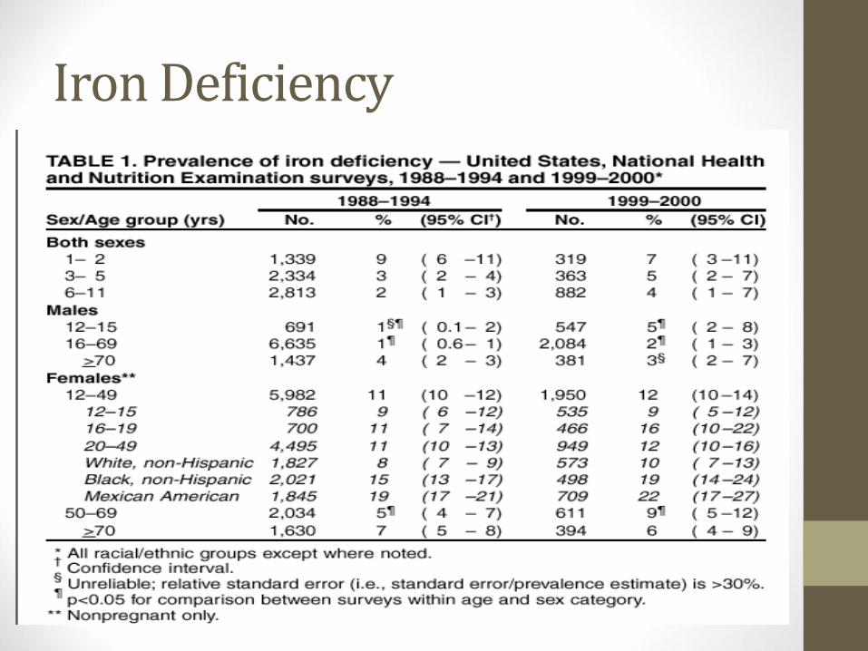
Iron Deficiency

Iron Deficiency Signs & Symptoms
• Anemia
• Fatigue
• Pallor
• Poor exercise tolerance
• Pica or Pagophagia
• Restless leg syndrome
• Koilonychia
• Plummer-Vinson syndrome

Laboratory Tests in Iron Deficiency of
Increasing Severity
Serum ferritin ≤ 30 ng/dL = Iron deficient (PPV 83%, PLR= 11)Serum ferritin ≥ 100 ng/dL = Iron sufficient ( NLR .08)
What about a ferritin value between 30 and 100?

Best tests to help distinguish IDA from ACD…
• Transferrin: (TRUCKS)• High in iron deficiency anemia
• Low in ACD
• Low reticulocyte-
hemoglobin concentration
(RET-He) that improves
with IV ironLow RET-He concentration
does not distinguish between
ACD and IDA but IF it
increases 2-3 days after IV iron
is administered, it is likely
contributing to anemia

What about ESRD patients?
• Functional Iron deficiency:
• a fall in iron saturation after giving epogen
(which increases demand for iron)
• KDOQI Guidelines:
• Goal hemoglobin
• 10-11.5 g/dL
• Absolute iron deficiency =
• TIBC % sat < 20%
• Ferritin < 100 ng/mL
• May still benefit from IV iron with epogen
• TIBC % sat <30%
• Ferritin < 500 ng/mL

Differential Diagnosis for Iron Deficiency due to GI process
• Malabsorption
• Total or partial gastrectomy
• Antacid
• H. pylori
• Celiac disease
• IBD
• Loss
• -Itis of the GI tract
• Neoplasm (polyp/CA)
• Peptic ulcer
• AVM
• Parasites (hookworm)
• Other…

Iron Overload• Think of 3 main categories
of iron overload…1. abnormal intestinal
absorption of normal amounts of dietary iron,
2. excesses of dietary iron, or
3. parenteral sources of iron such as multiple blood transfusions.
• Name the organs involved in iron overload:

Iron Overload: Signs and Symptoms
• Unexplained fatigue
• Joint pain (CPPD) 2nd and 3rd MCP OA changes
• Liver disease: elevated aminotransferase levels, hepatomegaly, cirrhosis, hepatocellular carcinoma
• Diabetes mellitus
• Impotence
• Hypothyroidism
• Heart failure
• Arrhythmias

Hereditary Hemochromatosis
• A disease of inappropriate iron absorption resulting in the overload of iron in various organs
• The majority of patients with HH are decended from a common Celtic ancestor who lived 60-70 generations ago!
• 85% of HH patients carry a missense mutation of the HFE gene on chromosome 6
• In most cases, the mutation is a single base change that substitutes tyrosine for cysteine at position 282 of the HFE protein (C282Y)
• Homozygosity for the C282Y mutation is found in 5 of every 1000 persons of North European decent– a prevalence 10 x that of cystic fibrosis genotypes!
Celtic282Y

• When the HFE
protein is mutated
as in HH, there is
not enough
hepcidin
produced.
• The export of iron
from the
basolateral side of
the enterocyte, the
macrophage, and
the hepatocyte is
allowed to continue
unhindered.
• This overloads the
transferrin binding
capacity and
elevates the
percent saturation
in the plasma.
Fe3+
Fe3+
Fe3+
Fe3+
Fe3+
Fe3+

Hepcidin: iron export regulator
LOW hepcidin in
hereditary
hemochromatosis=increased iron
absorption (E)
HIGH hepcidin
in anemia of
chronic disease= decreased iron
release out of
enterocytes and
macrophages
resulting in LOW
iron % saturation
(A,B,C,and D)

Laboratory Diagnosis of Iron Overload
• A fasting transferrin saturation > 60% in men or
> 50% in women will detect about 90% of patients with homozygous HH.
• An elevated ferritin above 300 ng/mL in men and above 200 ng/mL in women suggests an iron overload state in the absence of inflammatory conditions.
• Elevated ferritin is less sensitive than elevated transferrin saturation in screening for HH because a greater degree of iron overload is required to raise the ferritin concentration.

Other Tests for Iron Overload
• Liver biopsy for hepatic iron content
• CT and T2 MRI measurements of liver or heart
• Quantitative phlebotomy


Treatment of Iron Overload
• Iron overload due to multiple transfusions in sickle cell disease, thalassemia’s, and possibly myelodysplasticsyndrome.
• Criteria for chelation therapy: Ferritin > 1000 ng/mL
• Deferoxamine (IV infusion); vision, hearing, renal SEs
• Deferiprone (oral therapy); neutropenia, agranulocytosis SEs
• Deferasirox (oral therapy); abdominal pain, N/V, diarrhea, skin rash
• Iron overload due to Hereditary Hemochromatosis is phlebotomy.

Summary• Iron homeostasis is regulated mostly by degree of
absorption by the HEPCIDIN exporter regulator because the body lacks efficient excretion mechanisms.
• In clinical practice, low ferritin (<30) indicates iron deficiency and high TIBC (>45%) percent saturation indicates overload.
• Anemia of inflammation and iron deficiency anemia are distinguished by ferritin (>100 unlikely to be iron deficient) and transferrin (LOW, not likely to be iron deficient)

Case/Question 1.
ID and CC:
• 45-year-old man
• Sepsis
• UTI vs. Sacral Wound
• Hemodynamic compromise
• Urgent surgical debridement
PMH:• Paraplegic due to GSW
• Noncompliance with urinary self-catheterization
• Chronic sacral wounds
• Bipolar disorder
• Anemia
Laboratories:
• Hgb 8.0 g/dL (14-17g/dL)
• Hct 24% (41-51%)
• MCV 75 fL (80-100 fL)
• RDW 12.0 (11-14)
• Ferritin 250 ng/mL (15-200 ng/mL)
• Transferrin 100 mg/dL (188-341 mg/dL)
• Percent saturation 8% (15-50%)

CASE/ QUESTION 1.
Which of the following statements accurately describes the cause of this patient’s anemia?
A. This patient is iron deficient and the elevated. ferritin is due to his infection.
B. This patient is not iron deficient and hisanemia is due to chronic disease/inflammation.
C. The amount of information given is notenough to determine whether or not the patient isiron deficient.

Case 2
• 40-year-old woman
• PMH:
• Infected pancreatic pseudocyst
• Gallstone pancreatitis
• DM 2
• Anemia
• Hgb 7.9
• Hct 26.8
• MCV 72
• RDW 17.1
• Retic 1.7
• Serum iron 14
• Transferrin 295 (188-341)
• % saturation 3.7
• Ferritin 6

• Patient is a 27 yo white male
• Abdominal pain and diarrhea x 6 months associated with 40 lbweight loss.
• He was diagnosed with C diff in May and failed flagyl treatment.
• He presents with increasing abdominal pain and diarrhea with new symptoms of vomiting. He presents with continued chronic diarrhea with bloody stools.
Case 3
• Physical Examination
• VS: 136/78 HR 84 RR 14 T 98.1 95% RA
• Normal except for:
• Gastrointestinal: Soft, Non-distended, Normal bowel sounds, diffuse tenderness to palpation.


Recommended Reading
Iron Deficiency Anemia: Evaluation and Management
MATTHEW W. SHORT, LTC, MC, USA, and JASON E.
DOMAGALSKI, MAJ, MC, USAMadigan Healthcare System,
Tacoma, Washington
January 15, 2013 ◆ Volume 87, Number 2 www.aafp.org/afp