Iron Deficiency Anemia-Induced Cardiomyopathy With ...
6
CASE REPORT EDITOR’ S HIGHLIGHTS Iron Deficiency Anemia-Induced Cardiomyopathy With Congestive Heart Failure Reversible Cardiac Dysfunction Assessed by Multi-Imaging Modalities Shiro Miura, MD, MSC, a Masanao Naya, MD, PHD, b Takehiro Yamashita, MD, PHD a ABSTRACT Iron deficiency anemia (IDA) can cause left ventricular (LV) dysfunction, causing heart failure. A 48-year-old woman with severe IDA developed congestive heart failure that was properly diagnosed, managed, and followed with multiple im- aging modalities to explore potential mechanisms, highlighting the reversibility of LV function in unique cardiomyopathy. (Level of Difficulty: Intermediate.) (J Am Coll Cardiol Case Rep 2020;2:1806–11) © 2020 Published by Elsevier on behalf of the American College of Cardiology Foundation. This is an open access article under the CC BY-NC-ND license (http://creativecommons.org/licenses/by-nc-nd/4.0/). S evere iron deficiency anemia (IDA) has been re- ported to play a pivotal role in the develop- ment of heart failure (HF) through left ventricular (LV) dysfunction and myocardial damage, and substantial clinical benefits have been found as a result of iron deficiency treatment even without ane- mia (1). A remarkable feature of IDA-induced HF may be the reversibility of LV dysfunction with appro- priate treatment. However, the pathogenesis of car- diomyopathy associated with IDA is unknown due to the limited number of affected patients. Here, we report an impressive case with severe IDA causing LV dysfunction and congestive HF, which was appro- priately diagnosed and managed. The patient was fol- lowed up with multiple imaging modalities exploring the potential mechanisms of IDA-induced HF in terms of myocardial tissue characteristics and myocardial blood flow. HISTORY OF PRESENTATION A 48-year-old woman presented with chronic leg edema and worsening exertional dyspnea. She also LEARNING OBJECTIVES IDA-induced cardiomyopathy can be charac- terized by severe chronic anemia, signi ficant microvascular dysfunction, and high-output HF, which is reversible if treated properly. Multi-imaging modalities such as trans- thoracic echocardiography, CMR, coronary angiography with hemodynamics measure- ments, and cardiac positron emission to- mography can improve accurate diagnosis and appropriate management. ISSN 2666-0849 https://doi.org/10.1016/j.jaccas.2020.07.051 From the a Department of Cardiology, Hokkaido Ohno Memorial Hospital, Sapporo, Japan; and the b Department of Cardiovascular Medicine, Hokkaido University Graduate School of Medicine, Sapporo, Japan. The authors have reported that they have no relationships relevant to the contents of this paper to disclose. The authors attest they are in compliance with human studies committees and animal welfare regulations of the authors’ institutions and Food and Drug Administration guidelines, including patient consent where appropriate. For more information, visit the JACC: Case Reports author instructions page. Manuscript received June 18, 2020; revised manuscript received July 15, 2020, accepted July 22, 2020. JACC: CASE REPORTS VOL. 2, NO. 11, 2020 ª 2020 PUBLISHED BY ELSEVIER ON BEHALF OF THE AMERICAN COLLEGE OF CARDIOLOGY FOUNDATION. THIS IS AN OPEN ACCESS ARTICLE UNDER THE CC BY-NC-ND LICENSE ( http://creativecommons.org/licenses/by-nc-nd/4.0/ ).
Transcript of Iron Deficiency Anemia-Induced Cardiomyopathy With ...

J A C C : C A S E R E P O R T S V O L . 2 , N O . 1 1 , 2 0 2 0
ª 2 0 2 0 P U B L I S H E D B Y E L S E V I E R O N B E H A L F O F T H E A M E R I C A N C O L L E G E O F
C A R D I O L O G Y F O UN DA T I O N . T H I S I S A N O P E N A C C E S S A R T I C L E U N D E R T H E
C C B Y - N C - N D L I C E N S E ( h t t p : / / c r e a t i v e c o mm o n s . o r g / l i c e n s e s / b y - n c - n d / 4 . 0 / ) .
CASE REPORT
EDITOR’S HIGHLIGHTS
Iron Deficiency Anemia-InducedCardiomyopathy WithCongestive Heart FailureReversible Cardiac Dysfunction Assessed byMulti-Imaging Modalities
Shiro Miura, MD, MSC,a Masanao Naya, MD, PHD,b Takehiro Yamashita, MD, PHDa
ABSTRACT
L
�
�
ISS
Fro
Me
rel
Th
ins
vis
Ma
Iron deficiency anemia (IDA) can cause left ventricular (LV) dysfunction, causing heart failure. A 48-year-old woman with
severe IDA developed congestive heart failure that was properly diagnosed, managed, and followed with multiple im-
aging modalities to explore potential mechanisms, highlighting the reversibility of LV function in unique cardiomyopathy.
(Level of Difficulty: Intermediate.) (J Am Coll Cardiol Case Rep 2020;2:1806–11) © 2020 Published by Elsevier on
behalf of the American College of Cardiology Foundation. This is an open access article under the CC BY-NC-ND license
(http://creativecommons.org/licenses/by-nc-nd/4.0/).
S evere iron deficiency anemia (IDA) has been re-ported to play a pivotal role in the develop-ment of heart failure (HF) through left
ventricular (LV) dysfunction and myocardial damage,and substantial clinical benefits have been found as a
EARNING OBJECTIVES
IDA-induced cardiomyopathy can be charac-terized by severe chronic anemia, significantmicrovascular dysfunction, and high-outputHF, which is reversible if treated properly.Multi-imaging modalities such as trans-thoracic echocardiography, CMR, coronaryangiography with hemodynamics measure-ments, and cardiac positron emission to-mography can improve accurate diagnosisand appropriate management.
N 2666-0849
m the aDepartment of Cardiology, Hokkaido Ohno Memorial Hospital, Sap
dicine, Hokkaido University Graduate School of Medicine, Sapporo, Jap
ationships relevant to the contents of this paper to disclose.
e authors attest they are in compliance with human studies committe
titutions and Food and Drug Administration guidelines, including patien
it the JACC: Case Reports author instructions page.
nuscript received June 18, 2020; revised manuscript received July 15, 20
result of iron deficiency treatment even without ane-mia (1). A remarkable feature of IDA-induced HF maybe the reversibility of LV dysfunction with appro-priate treatment. However, the pathogenesis of car-diomyopathy associated with IDA is unknown dueto the limited number of affected patients. Here, wereport an impressive case with severe IDA causingLV dysfunction and congestive HF, which was appro-priately diagnosed and managed. The patient was fol-lowed up with multiple imaging modalities exploringthe potential mechanisms of IDA-induced HF in termsof myocardial tissue characteristics and myocardialblood flow.
HISTORY OF PRESENTATION
A 48-year-old woman presented with chronic legedema and worsening exertional dyspnea. She also
https://doi.org/10.1016/j.jaccas.2020.07.051
poro, Japan; and the bDepartment of Cardiovascular
an. The authors have reported that they have no
es and animal welfare regulations of the authors’
t consent where appropriate. For more information,
20, accepted July 22, 2020.

AB BR E V I A T I O N S
AND ACRONYM S
18F-FDG = 18F-
fluorodeoxyglucose
CMR = cardiac magnetic
resonance
HF = heart failure
IDA = iron deficiency anemia
LV = left ventricular
NT-proBNP = N-terminal pro–
B-type natriuretic peptide
PET/CT = positron emission
tomography/computed
graphy
J A C C : C A S E R E P O R T S , V O L . 2 , N O . 1 1 , 2 0 2 0 Miura et al.S E P T E M B E R 2 0 2 0 : 1 8 0 6 – 1 1 Iron Deficiency Anemia-Induced Cardiomyopathy and Multi-Imaging Modalities
1807
complained of menorrhagia with irregular periodsmore than 6 months ago. Vital sign measurementsshowed blood pressure of 107/48 mm Hg, heart rate of81 beats/min, SpO2 of 94% (room), and temperature of36.9�C. Physical examination revealed marked pallorof the skin and conjunctiva, jugular vein distention,crackles in both lungs, rapid regular heart rhythmwithout any murmur, palpable spleen, and pittingedema of both lower extremities. Electrocardiogramshowed normal sinus rhythm with horizontal STdepression in the limb leads (Figure 1A). Chest radi-ography revealed marked cardiomegaly and mildpulmonary congestion (Figure 1B). The initial hemo-globin level was 1.7 g/l, hematocrit level was 7.2%,and mean corpuscular volume was 55 fl. Iron param-eters suggested severe IDA with an iron level of8 mg/dl, total iron-binding capacity of 468 mg/dl, andferritin level of 2 ng/ml. The rest of the blood testresults and coagulation times, along with other lab-oratory tests, including clinical chemistry, renal,liver, and thyroid function, were normal except for anelevated level of N-terminal pro–B-type natriureticpeptide (NT-proBNP) (1,547 pg/ml). Stool guaiac testswere negative. The patient received three units ofpacked red blood cells in 4 days and was admitted forevaluation and treatment of severe IDA and HF.
FIGURE 1 Electrocardiogram and Chest X-Ray on Admission and at 4
Electrocardiogram on admission (A) and at the 4-month follow-up (C).
PAST MEDICAL HISTORY
The patient’s medical history was unremark-able except for that she regularly smokedtobacco and consumed alcohol.
DIFFERENTIAL DIAGNOSES
The patient showed high-output HF with ahigh cardiac index and low systemic vascularresistance. Chronic high-output HF wasexplained by severe anemia after excludingother common causes, such as obesity, thy-roid disease, arteriovenous shunts, and lung
and liver diseases. By characterizing myocardial tis-sue, cardiac magnetic resonance (CMR) helped todistinguish secondary etiologies, such as myocarditis,amyloidosis, sarcoidosis, noncompaction cardiomy-opathy, and dilated phase hypertrophic cardiomyop-athy. 18F-fluorodeoxyglucose (18F-FDG) positronemission tomography/computed tomography (PET/CT) was useful in excluding myocarditis andsarcoidosis. Last, her benign clinical course, whichwas shortly controlled with iron replacement andtransfusion of packed red blood cells, was more sug-gestive of HF due to severe IDA.tomo
-Month Follow-Up
Initial (B) and 4-month follow-up (D) chest x-rays.

FIGURE 2 Initial and Follow-Up TTE and Coronary Angiography
Initial transthoracic echocardiogram (TTE) (A) exhibiting biventricular dilatation with preserved left ventricular (LV) wall thickness on a short-axis view compared with
the 4-month follow-up TTE (B), which displayed a significant reduction in LV size and an improvement in LV contractility. LV end-diastolic diameter, LV ejection
fraction, and stroke volume measured using Doppler ultrasound changed from 72 mm, 50%, and 103 ml, respectively, to 53 mm, 68%, and 67 ml, respectively, over
4 months. Coronary angiography (C) revealed no obstructive coronary disease.
Miura et al. J A C C : C A S E R E P O R T S , V O L . 2 , N O . 1 1 , 2 0 2 0
Iron Deficiency Anemia-Induced Cardiomyopathy and Multi-Imaging Modalities S E P T E M B E R 2 0 2 0 : 1 8 0 6 – 1 1
1808
INVESTIGATIONS
Initial transthoracic echocardiography showed amassively dilated LV chamber and left atrium witheccentric LV hypertrophy and trivial pericardialeffusion (Figures 2A and 2B). On the seventh hospitalday, coronary angiography (Figure 2C) revealed non-obstructive coronary arteries, and pressure studieswere performed to assess LV end-diastolic pressure(9 mm Hg) and mean pulmonary capillary wedgepressures (9 mm Hg) with increased cardiac output at9.0 l/min and systemic vascular resistance at 691dynes/s/cm�5. Endomyocardial biopsy was avoided toprevent bleeding complications. CMR demonstratedbiventricular dilatation and diffusely reduced LV wallmotion with preserved wall thickness and absence of
late gadolinium enhancement (Figures 3A to 3C). Onday 12, PET/CT with 13N-ammonia (Figures 4A to 4C)demonstrated that ATP-induced stress myocardialblood flow was globally low with a considerable dropin the global coronary flow reserve to 1.70. Moreover,18F-FDG-PET/CT detected abnormally high 18F-FDGuptake in the uterus with no metastases (Figure 5).Finally, the patient was referred to our obstetrics andgynecology department where the solid mass in heruterus was diagnosed as endometrioid cancer onhistology.
MANAGEMENT
Frusemide and enalapril were commenced, and thepatient was gradually weaned from supplemental

FIGURE 3 Cine-CMR Images in Long- and Short-Axis Views
Cine-cardiac magnetic resonance (CMR) images in long-axis ([A] diastolic and [B] systolic phases) and short-axis views with late gadolinium enhancement imaging (C).
CMR studies at the 4-month visit were repeated with the corresponding images (D to F). Concentric left ventricular (LV) hypertrophy and biventricular enlargement
were visible with a reduced LV ejection fraction of 32%, which improved to 47% over 4 months with a significant drop in the LV end-diastolic volume from 184 to 96 ml.
Late gadolinium enhancement was not present over the entire myocardium at both the acute (C) and follow-up phases (F).
J A C C : C A S E R E P O R T S , V O L . 2 , N O . 1 1 , 2 0 2 0 Miura et al.S E P T E M B E R 2 0 2 0 : 1 8 0 6 – 1 1 Iron Deficiency Anemia-Induced Cardiomyopathy and Multi-Imaging Modalities
1809
oxygen. After the blood transfusion, she was treatedwith 7-day intravenous saccharated ferric oxide, fol-lowed by oral sulfate. At 3 weeks after discharge, sheunderwent a total hysterectomy with bilateralsalpingo-oophorectomy at a university hospital;endometrial carcinoma was confirmed on patholog-ical examination. At the 4-month follow-up visit, shewas free of symptoms, where ST changes on electro-cardiogram were normal with a normal cardiacsilhouette on the chest radiograph (Figures 1C and 1D).Her LV ejection fraction improved to 69%, and LVdiastolic dimension was still enlarged (54 mm) with amildly elevated LV mass index of 111 g/m2 (Figure 2B).Serum hemoglobin, iron, ferritin, and NT-proBNPlevels were within the normal range. Repeated CMRdemonstrated that biventricular dilatation improved
along with global LV wall motion, and late gadolin-ium enhancement was absent throughout the entiremyocardium (Figures 3D to 3F). Perfusion PET/CTrevealed a total recovery of global coronary flowreserve from 1.70 to 2.88 (Figures 4D to 4F).
DISCUSSION
We report a case of a middle-aged woman withchronic severe anemia and iron deficiency. We uti-lized novel imaging modalities to elucidate the po-tential mechanisms causing unique HF. Not only canprogressive HF cause iron deficiency, but theconverse can also occur. Iron deficiency can promotethe remodeling of cardiomyocytes (2). Iron has rolesbeyond oxygen transport, including in the normal
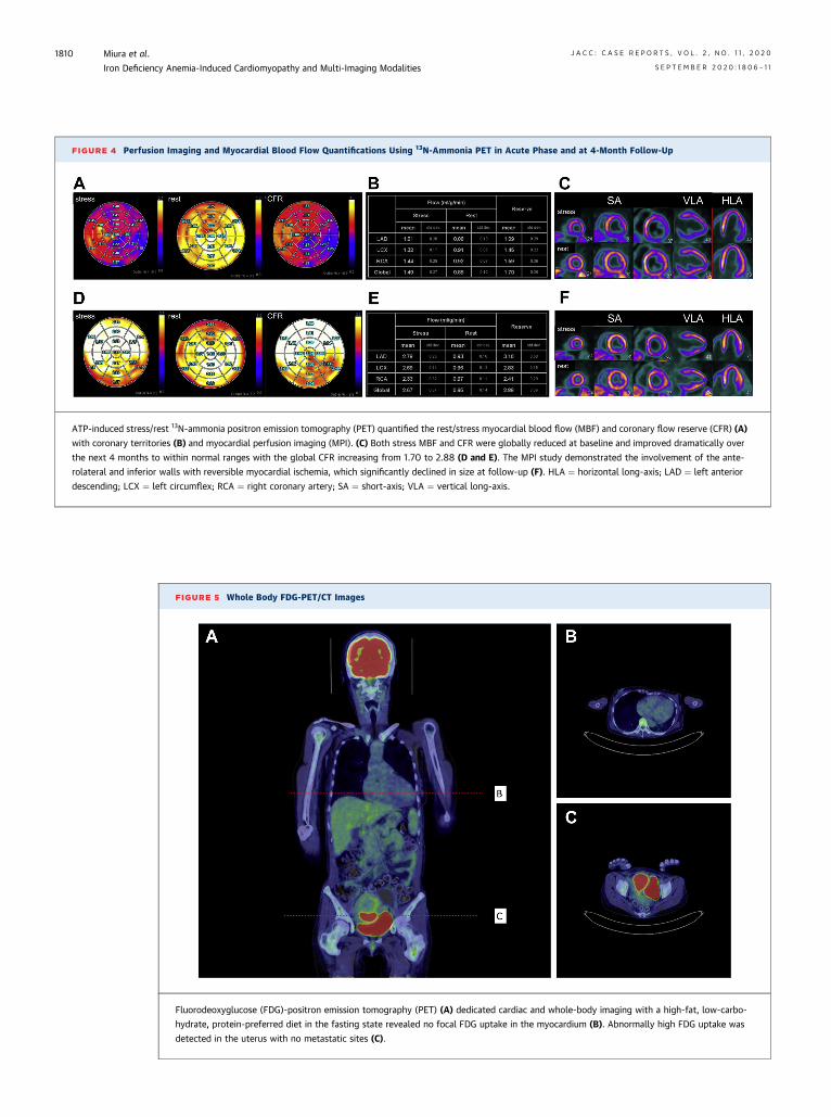
FIGURE 4 Perfusion Imaging and Myocardial Blood Flow Quantifications Using 13N-Ammonia PET in Acute Phase and at 4-Month Follow-Up
ATP-induced stress/rest 13N-ammonia positron emission tomography (PET) quantified the rest/stress myocardial blood flow (MBF) and coronary flow reserve (CFR) (A)
with coronary territories (B) and myocardial perfusion imaging (MPI). (C) Both stress MBF and CFR were globally reduced at baseline and improved dramatically over
the next 4 months to within normal ranges with the global CFR increasing from 1.70 to 2.88 (D and E). The MPI study demonstrated the involvement of the ante-
rolateral and inferior walls with reversible myocardial ischemia, which significantly declined in size at follow-up (F). HLA ¼ horizontal long-axis; LAD ¼ left anterior
descending; LCX ¼ left circumflex; RCA ¼ right coronary artery; SA ¼ short-axis; VLA ¼ vertical long-axis.
FIGURE 5 Whole Body FDG-PET/CT Images
Fluorodeoxyglucose (FDG)-positron emission tomography (PET) (A) dedicated cardiac and whole-body imaging with a high-fat, low-carbo-
hydrate, protein-preferred diet in the fasting state revealed no focal FDG uptake in the myocardium (B). Abnormally high FDG uptake was
detected in the uterus with no metastatic sites (C).
Miura et al. J A C C : C A S E R E P O R T S , V O L . 2 , N O . 1 1 , 2 0 2 0
Iron Deficiency Anemia-Induced Cardiomyopathy and Multi-Imaging Modalities S E P T E M B E R 2 0 2 0 : 1 8 0 6 – 1 1
1810
J A C C : C A S E R E P O R T S , V O L . 2 , N O . 1 1 , 2 0 2 0 Miura et al.S E P T E M B E R 2 0 2 0 : 1 8 0 6 – 1 1 Iron Deficiency Anemia-Induced Cardiomyopathy and Multi-Imaging Modalities
1811
activity of key enzymes of the citric acid cycle andreactive oxygen species scavenging enzymes.Decreased levels of reactive oxygen species scav-enging enzymes in myocardial iron deficiency mayintensify local oxidative stress, causing myocardialdamage (3). As noted earlier, compromised oxygendelivery capacity may cause chronic tissue hypox-emia, which can lead to cardiomyocyte dysfunction.In our case, the initial perfusion PET/CT revealedsignificant microvascular dysfunction that mayreflect global myocyte dysfunction. Moreover, IDA-induced cardiomyopathy had no association withmyocardial inflammation, as shown by 18F-FDG-PET/CT.
A further remarkable aspect of the present case isthe reversibility of IDA-induced cardiomyopathyconfirmed with several imaging modalities. At the 4-month follow-up, when the patient was asymptom-atic and her hemoglobin, iron, and NT-proBNP levelswere normalized, we found remarkable improve-ments in cardiac function parameters assessed bytransthoracic echocardiography, CMR, and perfusionPET/CT. This strongly suggests that IDA-inducedcardiomyopathy may be almost completely revers-ible. However, it remains unclear whether completenormalization of LV function occurs in severe cases.Our present case showed that the LV remained
slightly dilated after HF improvement, and thesefindings were in line with cardiac indexes measuredby CMR imaging. Close observation to evaluatelonger-term cardiac function and prognosis iswarranted.
FOLLOW-UP
The patient continues to take enalapril. LV functionby follow-up echocardiography and hemoglobin levelare within normal ranges 12 months afterhospitalization.
CONCLUSIONS
To our knowledge, this is the first case of severe IDAleading to LV dysfunction and congestive HF that wasproperly diagnosed, managed, and followed withmultiple imaging modalities to explore the potentialmechanisms. This case supports the reversibility ofIDA-induced cardiomyopathy if treated properly, butfurther research is required to determine the level ofimprovement.
ADDRESS FOR CORRESPONDENCE: Dr. Shiro Miura,Department of Cardiology, Hokkaido Ohno MemorialHospital, 2-1-16-1 Miyanosawa, Nishi-ku, Sapporo063-0052, Japan. E-mail: [email protected].
RE F E RENCE S
1. Grote Beverborg N, van Veldhuisen DJ, van derMeer P. Anemia in heart failure: still relevant?J Am Coll Cardiol HF 2018;6:201–8.
2. Jankowska EA, Ponikowski P. Molecularchanges in myocardium in the course of anemia oriron deficiency. Heart Fail Clin 2010;6:295–304.
3. Dai DF, Johnson SC, Villarin JJ, et al. Mito-chondrial oxidative stress mediates angiotensin II-induced cardiac hypertrophy and Galphaqoverexpression-induced heart failure. Circ Res2011;108:837–46.
KEY WORDS cardiac magnetic resonance,cardiomyopathy, iron deficiency anemia,positron emission tomography