IOL CALCULATION AFTER REFRACTIVE SURGERIES: …
9
MSO EXPRESS: ISSUE 2 November 2016 IOL CALCULATION AFTER REFRACTIVE SURGERIES: CHALLENGES, TIPS AND PEARLS Achieving a good and consistent refractive outcome after cataract surgery has become more achievable following marked advances in modern intraocular lens (IOL) manufacturing technology, better instruments to perform preoperative biometry, and improved surgical techniques. It has been reported that postoperative refraction can can fall within ±0.50 D of target refraction in 70% to 80% of operated eyes that have not undergone previous refractive surgery. However, this target percentage can be considerably lower in the group of patients who had undergone previous refractive surgery, and efforts are still ongoing by some of the brightest minds in the world of ophthalmology ophthalmology to identify the best method or methods to achieve a more consistent and more predictable results in this group of patients. Over the years, many methods have been proposed to achieve this, and the sheer number of methods that can be found in the literature clearly show that the optimal methodology has yet to be found. To understand the challenges, we must first understand that after laser-assisted in situ Keratomileusis (LASIK), photorefractive keratectomy (PRK) or radial keratotomy (RK) surgery, IOL power miscalculation occurs due to three types of error: 1. Keratometric Index Error 2. Radius Error 3. 3. Formula Error Calculations Calculations are more accurate, and a larger range of methods can be used, when the refractive change induced by LASIK or PRK and the preoperative corneal power are known. The ideal method for IOL power calculation may vary according to the available data for that particular patient. The various results from these calculations calculations can then be compared and a decision can be made with regards to the best IOL power to be selected for the best chance of achieving the optimal postoperative outcome. Number 1: Keratometric Index Error Keratometers, corneal topographers, and any devices developed to measure corneal power use a standardized, fictitious keratometric index of refraction—usually 1.3375—to convert the measured radius of the anterior corneal surface into keratometric diopters. Keratometers Keratometers and topographers measure the radius of anterior corneal curvature, but the keratometric index of refraction posits a Dr. MICHAEL LAW, FRCS CORNEA AND REFRACTIVE SURGEON, CONSULTANT OPHTHALMOLOGIST INTERNATIONAL SPECIALIST EYE CENTRE (ISEC), KUALA LUMPUR, MALAYSIA IOL Calculateion After Refractive Surgeries: Challenges, Tips and Pearls | 1
Transcript of IOL CALCULATION AFTER REFRACTIVE SURGERIES: …

MSO EXPRESS: ISSUE 2 November 2016
IOL CALCULATION AFTER REFRACTIVE SURGERIES: CHALLENGES, TIPS AND PEARLS
Achieving a good and consistent refractive
outcome after cataract surgery has become more
achievable following marked advances in modern
intraocular lens (IOL) manufacturing technology,
better instruments to perform preoperative
biometry, and improved surgical techniques. It
has been reported that postoperative refraction
cancan fall within ±0.50 D of target refraction in 70%
to 80% of operated eyes that have not undergone
previous refractive surgery. However, this target
percentage can be considerably lower in the
group of patients who had undergone previous
refractive surgery, and efforts are still ongoing by
some of the brightest minds in the world of
ophthalmologyophthalmology to identify the best method or
methods to achieve a more consistent and more
predictable results in this group of patients.
Over the years, many methods have been
proposed to achieve this, and the sheer number
of methods that can be found in the literature
clearly show that the optimal methodology has
yet to be found.
To understand the challenges, we must first
understand that after laser-assisted in situ
Keratomileusis (LASIK), photorefractive
keratectomy (PRK) or radial keratotomy (RK)
surgery, IOL power miscalculation occurs due to
three types of error:
1. Keratometric Index Error
2. Radius Error
3.3. Formula Error
CalculationsCalculations are more accurate, and a larger
range of methods can be used, when the
refractive change induced by LASIK or PRK and
the preoperative corneal power are known. The
ideal method for IOL power calculation may vary
according to the available data for that particular
patient. The various results from these
calculationscalculations can then be compared and a decision
can be made with regards to the best IOL power
to be selected for the best chance of achieving
the optimal postoperative outcome.
Number 1: Keratometric Index Error
Keratometers, corneal topographers, and any
devices developed to measure corneal power use
a standardized, fictitious keratometric index of
refraction—usually 1.3375—to convert the
measured radius of the anterior corneal surface
into keratometric diopters.
KeratometersKeratometers and topographers measure the
radius of anterior corneal curvature, but the
keratometric index of refraction posits a
Dr. MICHAEL LAW, FRCS
CORNEA AND REFRACTIVE SURGEON, CONSULTANT OPHTHALMOLOGIST
INTERNATIONAL SPECIALIST EYE CENTRE (ISEC), KUALA LUMPUR, MALAYSIA
IOL Calculateion After Refractive Surgeries: Challenges, Tips and Pearls | 1

MSO EXPRESS: ISSUE 2 November 2016
keratometric index of refraction posits a
theoretical single refractive lens representing
both corneal surfaces. It assumes a constant ratio
of anterior to posterior corneal curvature.
SinceSince LASIK, PRK and other laser refractive
procedures modify only the anterior corneal
curvature but leave the posterior curvature
unchanged, the normal anterior/posterior
curvature ratio is significantly altered, and the
keratometric refractive index therefore becomes
invalid.
RKRK flattens both the anterior and posterior
corneal surfaces, but only in a small central
optical zone. The effective optical zone diameter
can be significantly smaller than the
measurement zone of standard keratometry.
Therefore, standard keratometry tends to
overestimate the true corneal power due to the
largelarge variation in the central cornea power and
the paracentral zones outside the small optical
zone where the standard keratometry readings
are taken.
As a consequence, after myopic laser correction,
keratometry (K) readings usually overestimate corneal power, and the IOL power is
underestimated, so that patients are likely to
experience postoperative hyperopia. Conversely,
in hyperopic laser correction, corneal power is
underestimated, IOL power is overestimated, and
patientspatients risk postoperative myopia. Usually, the
higher the attempted correction, the higher the
resulting under- or overcorrection.
It has shown that, after myopic excimer laser
surgery, the keratometric index of refraction
should be decreased proportional to the amount
of correction in order to get a correct of
correction in order to get a correct measurement
of corneal power. Alternatively, the keratometric
index error can be overcome by measuring the
actual curvature of both corneal surfaces using
technologies such as Scheimpflug imaging or
optical coherence tomography (OCT).
Number 2: Radius Error
RadiusRadius error is related to another assumption
made by most devices that extrapolate central
corneal curvature from paracentral
measurements. After myopic ablations, these
instruments can measure a paracentral corneal
curvature that is steeper than the central corneal
curvature. Accordingly, several authors have
shownshown that central corneal curvature
Figure 1. Laser vision correction alters the anterior corneal curvature but not the posterior curvature. This alters the normal anterior/posterior corneal curvature ratio.
wider central optical zone
Figure 2. Radial keratotomy flattens both the anterior and posterior curvature, within a small central optical zone. This leads to an extreme variation in corneal power between the center and periphery.
smaller central optical zone
IOL Calculateion After Refractive Surgeries: Challenges, Tips and Pearls | 2

MSO EXPRESS: ISSUE 2 November 2016
measurements better reflect the refractive
change induced by surgery and have suggested
using central values provided by corneal
topography, rather than simulated keratometry,
to calculate corneal power after excimer laser
surgery. This issue is clinically relevant in eyes
with small or decentered treatments, in which the
cornealcorneal radius may be measured on the
periphery of the treated zone and may be
different with respect to the radius passing
through the visual axis. When the optical zone
diameter is 6 mm or larger, the radius error is
negligible.
Number 3: Formula Error
Third-generation IOL power formulas, such as
Hoffer Q, Holladay 1, and SRK/T, use corneal
power to predict the effective lens position (ELP).
After myopic LASIK or PRK, the reduced corneal
power leads to an underestimation of ELP, and
this can further contribute to IOL power
underestimation. The opposite effect occurs after
hyperopic surgery.hyperopic surgery.
To address this issue, Dr. Jaime Aramberri,
developed the double-K method, which uses two
K values: the pre-refractive surgery K value for the
calculation of the ELP and the post-refractive
surgery K value for the vergence formula that
finally calculates IOL power.
All of the errors described above also occur after
RK. In these eyes, however, the keratometric
index error is the opposite of that after myopic
LASIK or PRK, as the posterior corneal curvature
flattens more than the anterior corneal curvature
after RK. Due to the small optical zone, the most
relevant error in post-RK eyes is radius error.
Best Approach to IOL Calculation post-refractive surgery
MostMost published IOL calculation methods for use
after refractive surgery rely on pre- and
postoperative refractive measurements being
available. Calculations are more accurate, and a
larger range of methods can be used, when both
the refractive change induced by corneal surgery
and the preoperative corneal power are known.
TheThe refractive change is required by methods
such as those developed by Masket and Savini.
The preoperative corneal power is mandatory to
calculate double-K formulas as well as some other
derivative formulae.
If these data are not available, the surgeon can
still try to determine the refractive change by
asking the patient the power of his or her old
contact lenses or eyeglasses. The preoperative
corneal power can also be estimated by
measuring the postoperative posterior corneal
radius; assuming that this did not change after
surgery,surgery, one can calculate the preoperative
anterior radius, which is on average 1.21 times
greater than the posterior radius.
If these options are unavailable, the surgeon can
rely on no-history methods such as those
described by Shammas and Haigis.
IOL Calculateion After Refractive Surgeries: Challenges, Tips and Pearls | 3

MSO EXPRESS: ISSUE 2 November 2016
Corneal power method Post-laser visual
correction
Post-radial
keratotomy
Clinical history Yes No
Contact lens over-refraction Yes Yes
Topography based post-LASIK adjustment Yes No
Central-ring topography No Yes
Net corneal power measurement Yes Not tested
Table 1. Methods of Adjusting or Measuring True Corneal Power after Refractive
Methods to Obtain True Corneal Power After Refractive Surgery
TheseThese estimations of the true corneal power are
then used as input for IOL formulae that are
specialized for post-refractive cataract surgery.
Some methods are suitable for post-laser vision
correction (LVC) eyes, some for post-RK eyes, and
some are applicable to both situations.
1.1. Clinical History Methods (refraction and keratometry)
TheThe effective keratometry value is calculated by
subtracting the change in refraction induced by
the treatment from the preoperative mean
corneal power. This method effectively bypasses
the index of refraction error. The main
disadvantage of the clinical history method and
similar formulae is the reliance on preoperative
keratometrykeratometry and refractive data. The
effectiveness of these methods may be reduced
by the possibility of further errors including but
not limited to the use of inaccurate central
corneal measurements, variation in
measurement units before and after surgery and
measurement units before and after surgery and
the potential impact of index myopia, as well as
the common difficulty in obtaining these
historical data. High variability in the predicted
outcome means these methods are generally no
longer recommended. Also, clinical history
method is not suitable for RK because of unstable
cornealcorneal power (post-RK cornea typically flattens
progressively over many years)
2. Methods based on change in Manifest Refraction
The aim is to bypass the need for preoperative
refraction and keratometry data.
A number of methods propose applying a
correction based on the change in refraction to
the postoperative keratometry value. This value
is then appropriately inserted into the Double-K
formula to provide the final IOL power. The
“Adjusted Effective Refractive Power (EffRP)” and
“Adjusted Atlas 9000 (4 mm Zone)” methods are
commonly used examples available through the commonly used examples available through the
American Society of Cataract and Refractive
Surgeons (ASCRS) IOL Calculator.
IOL Calculateion After Refractive Surgeries: Challenges, Tips and Pearls | 4

MSO EXPRESS: ISSUE 2 November 2016
These methodsrely on the availability of the
particular topographical unit and require the
examiner to directly assess the measurement to
confirm the quality of the reading.
MasketMasket and Masket recognized the difficulty in
obtaining accurate postoperative corneal
measurements and effectively bypassed these
potential errors by creating a formula based on
the change in laser correction. The authors
determined that the chief corrective factor in
post-refractive patients was the amount of
pre-ablativepre-ablative myopia. Subsequently, a value based
on a simple regression formula deriving the
change in manifest refraction was added to the
standard IOL calculation. Other authors have
undertaken similar approaches with basic
variations of the regression formulae. However,
reliability is limited due to the small sample size
used in deriving the formulae.used in deriving the formulae.
The risk of index myopia related errors remains
considerable for methods based on the change in
manifest refraction.
3. Methods based on No Historical Data
A. Contact Lens Over-Refraction
This effectively re-measures the corneal
curvature rather than providing a recalculated
value. This approach has been limited by
technical and time constraints. Difficulty in
achieving an adequate refraction in patients with
poor visual acuity further reduces the
effectiveness of this method.
B. Topography and Equation BasedB. Topography and Equation Based
Shammas et al. previously described a simple
equation modifying post-laser keratometry
values to determine the corrected corneal power
to be used in IOL calculation formulae. Other
researchers have taken similar approaches. The
Maloney and Koch-Maloney methods convert the
post-laser keratometry values from corneal
topography to the exact power present at the
anterior corneal surface and then add an average
negative power value for the posterior corneal
surface.surface. These latter formulae have been based
on values obtained by the Atlas topography
(Zeiss, Germany) and thereby remain of limited
value to practices without this unit.
Haigis-L formula bypasses the various errors
through the use of a correction formula then
applied to the standard Haigis formula. The
relative availability of the Haigis-L formula across
several platforms which includes both instrument
(eg. IOL Master) and web-based programs, and
the ease of use, has led to the formula rapidly
becomingbecoming a favourite and frequently used tool for
these calculations. The recently introduced
hyperopic version of the Haigis-L formula is also
very useful for patients who have undergone
prior hyperopic LASIK. It is a purely objective
methodology that uses small zone keratometry
and requires no prior historical information.
C. Intraoperative Measurements
MackoolMackool et al. and other authors have proposed
an alternative approach. The cataract is first
removed and the patient is required to wait for an
hour before an aphakic refraction is undertaken.
An algorithm is then applied to the refraction to
determine the true IOL power to be inserted but
with mixed results. Intraoperative reforming of
thethe anterior chamber either with balanced salt
solution or viscoelastics and variable refractive
indexes remain significant obstacles for these
methods in achieving consistent postoperative
IOL Calculateion After Refractive Surgeries: Challenges, Tips and Pearls | 5
MSO EXPRESS: ISSUE 2 November 2016
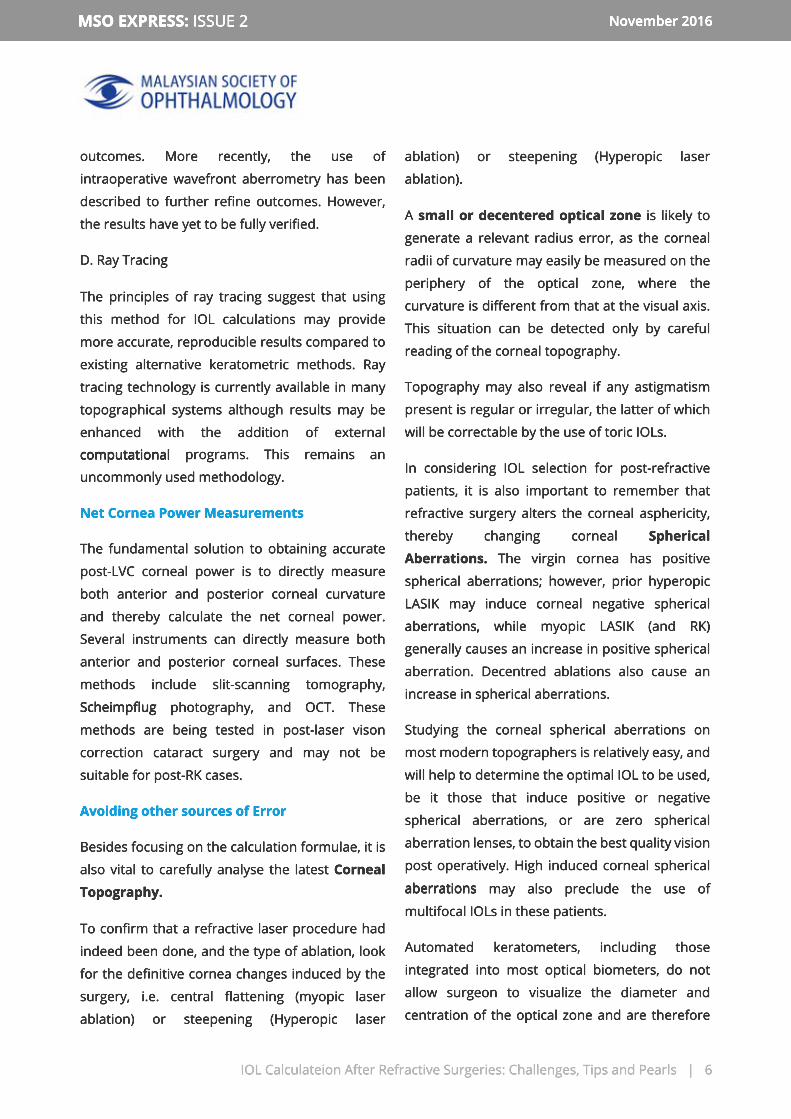
outcomes. More recently, the use of
intraoperative wavefront aberrometry has been
described to further refine outcomes. However,
the results have yet to be fully verified.
D. Ray Tracing
TheThe principles of ray tracing suggest that using
this method for IOL calculations may provide
more accurate, reproducible results compared to
existing alternative keratometric methods. Ray
tracing technology is currently available in many
topographical systems although results may be
enhanced with the addition of external
computationalcomputational programs. This remains an
uncommonly used methodology.
Net Cornea Power Measurements
The fundamental solution to obtaining accurate
post-LVC corneal power is to directly measure
both anterior and posterior corneal curvature
and thereby calculate the net corneal power.
Several instruments can directly measure both
anterior and posterior corneal surfaces. These
methods include slit-scanning tomography,
ScheimpflugScheimpflug photography, and OCT. These
methods are being tested in post-laser vison
correction cataract surgery and may not be
suitable for post-RK cases.
Avoiding other sources of Error
Besides focusing on the calculation formulae, it is
also vital to carefully analyse the latest Corneal Topography.
ToTo confirm that a refractive laser procedure had
indeed been done, and the type of ablation, look
for the definitive cornea changes induced by the
surgery, i.e. central flattening (myopic laser
ablation) or steepening (Hyperopic laser
ablation) or steepening (Hyperopic laser
ablation).
AA small or decentered optical zone is likely to
generate a relevant radius error, as the corneal
radii of curvature may easily be measured on the
periphery of the optical zone, where the
curvature is different from that at the visual axis.
This situation can be detected only by careful
reading of the corneal topography.
TopographyTopography may also reveal if any astigmatism
present is regular or irregular, the latter of which
will be correctable by the use of toric IOLs.
InIn considering IOL selection for post-refractive
patients, it is also important to remember that
refractive surgery alters the corneal asphericity,
thereby changing corneal Spherical Aberrations. The virgin cornea has positive
spherical aberrations; however, prior hyperopic
LASIK may induce corneal negative spherical
aberrations,aberrations, while myopic LASIK (and RK)
generally causes an increase in positive spherical
aberration. Decentred ablations also cause an
increase in spherical aberrations.
Studying the corneal spherical aberrations on
most modern topographers is relatively easy, and
will help to determine the optimal IOL to be used,
be it those that induce positive or negative
spherical aberrations, or are zero spherical
aberration lenses, to obtain the best quality vision
post operatively. High induced corneal spherical
aberrationsaberrations may also preclude the use of
multifocal IOLs in these patients.
Automated keratometers, including those
integrated into most optical biometers, do not
allow surgeon to visualize the diameter and
centration of the optical zone and are therefore
IOL Calculateion After Refractive Surgeries: Challenges, Tips and Pearls | 6

MSO EXPRESS: ISSUE 2 November 2016
insufficient for these cases. Should the optical
zone be small (< 4 mm) or decentered, the radius
error makes most methods of calculating corneal
power invalid. In this scenario, IOL power
calculation based on ray tracing is likely the best
option.
Conclusion
AlthoughAlthough a number of formulae may now provide
improved refractive outcomes in post-laser
refractive surgery patients, it remains a difficult
and time-consuming action to derive the IOL
powers using multiple formulae, and it is
generally impractical for most private practices.
ForFor most ophthalmologists, they should avail of
all resources that they can access, depending on
the data available. They can then evaluate the
postoperative results and determine the formula
that provides them the most consistent and
predictable results.
These may include:
1.1. Instrument based calculations eg. Haigis L,
Holladay’s IOL Consultant Program, etc.
2. Web based Calculators including:
a. The ASCRS Post-refractive Calculator
(www.iolcalc.org)
b.b. The Hoffer-Savini LASIK IOL Power
Tool (www.iolpowerclub.org/post-
surgical-tiol-calc)
c.c. The Asia Pacific Association of Cataract
and Refractive Surgeons
(www.apacrs.org) provides the Barrett
True-K formula for post-refractive IOL
patients
3. Apps like the Eye Pro application, which
utilizes the BESSt post-refractive IOL formula
and the PAK post-refractive IOL formula.
BecauseBecause there are many methods of calculating
IOL power after previous refractive surgery, it is
useful to obtain a consensus of several methods
by comparing the average or median
recommended IOL power. When there is a wide
range of recommendations (typically in cases of
refractive surgery for high or extreme dioptric
correction),correction), it is wise to hedge in the direction of
myopic results (choose higher IOL power or select
lower corneal power estimation to use in IOL
calculation).
Even with the use of many specialized methods,
the predictability of refractive outcome of
cataract surgery after previous refractive surgery
remains less than optimal overall. Therefore, all
patients who had previous refractive surgery
should be warned about the potential need for
refractive correction after their cataract surgery,
includingincluding the possibility of IOL exchange for
significant postoperative refractive surprise.
Financial Disclosure: No financial interest in any products mentioned in the article. All copyrighted materials remain the copyright of their original owners and no copyright is claimed by the author.
IOL Calculateion After Refractive Surgeries: Challenges, Tips and Pearls | 7

MSO EXPRESS: ISSUE 2 November 2016
Acknowledgment:
Article was prepared using various online scholarly resources, including:
1. Cataract & Refractive Surgery Today (CRST) Europe. IOL Power Calculation After Refractive
Surgery. Retrieved from
http://crstodayeurope.com/articles/2015-may/iol-power-calculation-after-refractive-surgery/.
2. American Academy of Ophthalmology – EyeWiki. Intraocular lens power calculation after
corneal refractive surgery. Retrieved from
http://eyewiki.aao.org/Intraocular_lens_power_calculation_after_corneal_refractive_surgery.
3. Intraocular Lens power calculations post-refractive surgery (Christopher Hodge et al, Eye and
Vision, 2015).
4. Doctor-hill.com. Achieving accurate IOL power calculation. Retrieved from
http://www.doctor-hill.com/iol-main/iol_main.htm.
IOL Calculateion After Refractive Surgeries: Challenges, Tips and Pearls | 8