Predicted Incorporation of Non-native Substrates by a Polyketide ...

Investigations of Fungal Highly Reducing Polyketide Synthases: The Biosyntheses of
Lovastatin and Hypothemycin
by
Amy K. Norquay
A thesis submitted in partial fulfillment of the requirements for the degree of
Doctor of Philosophy
Department of Chemistry
University of Alberta
© Amy K. Norquay, 2017

ii
ABSTRACT
Fungal highly reducing polyketide synthases (HR-PKSs) are large, multi-domain
enzymes central to the biosynthesis of fungal polyketides. They assemble complex secondary
metabolites, called polyketides, through the condensation of two-carbon acetate units and
tailoring of the functional groups at each round of chain extension. Two such fungal polyketides
are lovastatin, a cholesterol-lowering therapeutic, and hypothemycin, an anti-cancer kinase
inhibitor. Studies into the biosynthesis of these two natural products were undertaken to expand
our understanding of the complex processes performed by HR-PKS enzymes.
Firstly, the hypothesized reaction sequence of HR-PKS enzymes for these metabolites,
proposed based on their similarity to fatty acid synthases, had never been fully proven
experimentally. In our study of hypothemycin biosynthesis, we synthesized the proposed enzyme
bound intermediates as N-acetylcysteamine thioesters with 13C labels to use in incorporation
assays with purified enzymes. We observed conversion of the partially assembled intermediates
into intact PKS products, confirming their intermediacy and validating our understanding of HR-
PKS reaction sequences. In order to extrapolate these findings to other systems, the proposed
intermediates of lovastatin were also synthesized, and enzymatic assays are forthcoming.
Secondly, it has been proposed that an enzyme-catalyzed Diels-Alder cyclization is a key
step in the assembly of the lovastatin intermediate dihydromonacolin L by the HR-PKS LovB. In
order to study this LovB-catalyzed cyclization, two potential substrates were synthesized and
various fragments of LovB were expressed heterologously. NMR techniques were developed to
determine the products of the cyclization assays. By monitoring the conversion of the hexaketide
triene to the enzymatic decalin product, it was determined that the Diels-Alder activity was

iii
preserved in the LovB fragment where the condensation (CON) domain was deleted
(LovBΔCON). Therefore our hypothesis that the CON domain is the Diels-Alderase is incorrect.
It was also found that LovB did not catalyze the cyclization of a proposed heptaketide tetraene
intermediate, and therefore the substrate scope of this conversion is still being investigated in
order to better understand how this reaction occurs in nature.
Finally, the acyl carrier protein domains of both LovB, and the hypothemycin polyketide
synthase, Hpm8, were expressed as stand-alone proteins. Our goal was to obtain a solution
structure using NMR spectroscopy but the inherent properties of the protein made that
impossible. Herein I explore the properties of these domains and discuss other possible ways we
can use the expressed proteins to better understand the HR-PKS class of enzymes.

iv
ACKNOWLEDGEMENTS
Graduate school is a humbling experience during which one truly learns the extent to
which their accomplishments are not their own, but the product of a wider community.
I would like to begin by acknowledging those whose expertise had a direct impact on the content
of this thesis. Thank you to all of the Scientific Services staff in the Department of Chemistry for
the dedication to excellence in your work that allowed me to perform the high quality
experiments of this thesis. In particular I would like to thank Gareth Lambkin (Biological
Services), Mark Miskolzie (NMR Laboratory), Béla Reiz (Mass Spectrometry) and Wayne
Moffat (Analytical and Instrumentation Laboratory).
Teamwork is as much an essential component of science as it is of sport, and so I would
like to thank the amazing chemists in the Vederas Group. Firstly, thank you to my fellow
polyketide team members, Zhizeng Gao, Randy Sanichar and Rachel Cochrane for being
excellent teachers and supportive colleagues. A special thanks as well to Kaitlyn Towle, Shaun
McKinnie and Chris Lohans for being wonderful friends both in and out of the lab, and finally to
Christian Foerster for proofreading this thesis.
Quality friendships uplift us, and there is no question I would not have been successful
without my incredible community in Edmonton. In addition to the friends mentioned above, I
would like to thank fellow chemistry graduate students Thomas Scully and Samantha Kwok for
their support and friendship. I am also indebted to my teammates and coaches from the
University of Alberta Synchronized Swimming Team for 5 years of camaraderie. I would be
amiss if I did not thank my classmates of the U of A Medicine Class of 2019, who have made the
last two years some of the best of my life, despite being the most challenging. Lastly, to my old
v
friends back home in Winnipeg, I am truly blessed to have had you to cheer me on all these
years.
I am a product of those who raised me, and so this thesis is as much theirs as it is mine.
Thank you to my father, Donald Norquay, for instilling in me a love of learning and a passion for
making the world a better place. Thank you to my mother, Dr. Glenda Buchik, for teaching me
about care, self-sacrifice, and a dedication to quality. Thank you to my grandmother (my
“Nona”), Rose Buchik, for financing my education, but mostly for loving me unconditionally
and teaching me the importance of family and self-care.
Finally, thank you to my supervisor, Professor John Vederas, for making all of this
possible. Thank you for taking me on as a graduate student, providing me to the opportunity to
be a steward of your amazing legacy. Thank you for your mentorship all these years, for looking
out for me when I did not think I was capable and supporting my career goals.

vi
LIST OF ABBREVIATIONS
[α] Specific rotation
A Adenylation (domain)
Å Ångström
ACP Acyl carrier protein
ARO Aromatase
AT Acyl transferase (domain)
ATP Adenosine triphosphate
bsgHMBC Band-selective gradient heteronuclear multiple bond
coherence
c Concentration
CD Circular dichroism
CON Condensation (domain)
CHS Chalcone synthase
CoA Coenzyme A
CSSF-TOCSY Chemical shift selective filtered totally correlation
spectroscopy
CYC Cyclase
δ Chemical shift in parts per million
d Doublet
DCC Dicyclohexylcarbodiimide
DCM Dichloromethane
DEBS 6-Deoxyethronolide B synthase

vii
DH Dehydratase (domain)
DHZ Dehydrozearalenol
DIBAL-H Diisobutylalumninum hydride
DIPEA N,N-diisopropylethylamine
DHML Dihydromonacolin L
DHZ Dehydrozearalenol
DMAP 4-(Dimethylamino)pyridine
DMP Dess-Martin periodinane
DTT Dithiothreitol
EDTA Ethylenediaminetetraacetic acid
eq Equivalents
ER Enoyl reductase (domain)
ER0 Inactive enoyl reductase (domain)
ESI Electrospray ionisation
FAD Flavin adenine dinucleotide
FAS Fatty acid synthase
HMG-CoA 3-Hydroxy-3-methylglutaryl-coenzyme A
HPLC High performance liquid chromatography
HRMS High resolution mass spectroscopy
HR-PKS Highly reducing polyketide synthase
HWE Horner-Wadsworth-Emmons
HSQC Heteronuclear single quantum coherence

viii
IPTG Isopropyl thio-β-D-galactoside
IR Infrared
J J coupling, in Hertz
kDa kilodalton
KR Ketoreductase (domain)
KS Ketosynthase (domain)
LHMDS Lithium hexamethyldisilazide (also known as lithium
bis(trimethylsilyl)amide)
LB Lysogeny broth
LC-MS Liquid chromatography/mass spectrometry
Ni-NTA Nickel nitrilotriacetic acid
NRPS Non-ribosomal peptide synthetase
m Multiplet
m/z Mass to charge ratio
MALDI Matrix assisted LASER desorption/ionisation
MAT Malonyl-CoA:acyltransferase (domain)
MEK-ERK Mitogen-activated protein kinase kinase-extracellular signal-
regulated kinase (pathway)
mFAS Mammalian fatty acid synthase
MSAS 6-Methylsalicylic acid synthase
MT Methyltransferase (domain)
NADPH Nicotinamide adenine dinucleotide phosphate
NMR Nuclear magnetic resonance
ix
NR-PKS Non-reducing polyketide synthase
PCP Peptidyl carrier protein
ppm Parts per million
PR-PKS Partially reducing polyketide synthase
PT Product template (domain)
pTsOH p-Toluenesulfonic acid
q Quartet
RAL Resorcylic acid lactone
SAM S-Adenosylmethionine
SAT Starter unit acyltransferase (domain)
SDS-PAGE Sodium dodecylsulfate polyacrylamide gel electrophoresis
SNAC N-acetylcysteamine
t Triplet
TBS Tert-butyldimethylsilyl
TE Thioesterase (domain)
THF Tetrahydrofuran
THID Thioesterase/interdomain
TOCSY Total correlation spectroscopy
TRIS Tris(hydroxymethyl)aminomethane

x
TABLE OF CONTENTS
Chapter 1: Introduction ...................................................................................................................... 1
Polyketide Biosynthesis ................................................................................................................................. 2
Fundamentals ................................................................................................................................................................. 2
PKS Structure Classification ..................................................................................................................................... 4
Type I Polyketide Synthases ..................................................................................................................................................... 5
Type I Modular PKSs .............................................................................................................................................................. 6
Type I Iterative PKSs .............................................................................................................................................................. 7
Type II and Type III PKSs ......................................................................................................................................................... 12
Polyketides Studied in this Thesis ........................................................................................................... 14
Hypothemycin (12) .................................................................................................................................................... 14
Lovastatin (1) ............................................................................................................................................................... 16
Chapter 2: Incorporation of Partially Assembled Intermediates into Polyketide
Synthase Products............................................................................................................................... 21
Introduction ..................................................................................................................................................... 21
Results and Discussion ................................................................................................................................ 24
Incorporation of 13C-SNAC Triketide (30) into DHZ (21) by Hpm8/Hpm3 ....................................... 24
Synthesis of Proposed LovB Intermediates ..................................................................................................... 26
Chapter 3: Investigations Into the Diels-Alderase Activity of LovB .................................. 32
Background ...................................................................................................................................................... 32
The Diels-Alder Reaction ......................................................................................................................................... 32
Diels-Alderase Activity of LovB ............................................................................................................................. 32
Overview of Known Diels-Alderase Enzymes ................................................................................................. 36
Multifunctional Decalin-Forming Diels-Alderases........................................................................................................ 37

xi
Monofunctional Decalin-Forming Diels-Alderases ....................................................................................................... 38
Non-Decalin Forming Diels-Alderases ............................................................................................................................... 41
Goals of this Chapter ..................................................................................................................................... 43
Isolating the Diels-Alderase Domain................................................................................................................... 43
Substrate Scope of LovB ........................................................................................................................................... 45
Results and Discussion ................................................................................................................................ 46
Substrate Syntheses ................................................................................................................................................... 46
Expression of Enzymes ............................................................................................................................................. 49
LovB and LovBΔCON Expression in S. cerevisiae ........................................................................................................... 49
Expression of CON and DH in E. coli .................................................................................................................................... 50
Cyclization Studies...................................................................................................................................................... 50
Hexaketide Background Reaction – NMR-based Assay ........................................................................................ 50
Heptaketide Cyclizations .................................................................................................................................................... 54
Assay Employing 13C Hexaketide .................................................................................................................................... 56
Conclusion and Future Outlook ................................................................................................................ 61
Chapter 4: Acyl Carrier Protein Domains of Highly Reducing Polyketide Synthases . 67
Introduction ..................................................................................................................................................... 67
Goals of this Chapter ..................................................................................................................................... 71
Results and Discussion ................................................................................................................................ 71
Expression of Hpm8-ACP ......................................................................................................................................... 71
Expression of LovB-ACP ........................................................................................................................................... 78
Phosphopantetheinylation with Sfp and NpgA .............................................................................................. 82
Conclusion and Future Work ..................................................................................................................... 84
Chapter 5: Conclusion ........................................................................................................................ 85

xii
Experimental Procedures ................................................................................................................ 87
Chemical Synthesis ........................................................................................................................................ 87
Reagents, Solvents and Purification .................................................................................................................... 87
Characterization .......................................................................................................................................................... 87
Synthesis and Characterization of Compounds .............................................................................................. 88
Biological Methods ...................................................................................................................................... 126
General Techniques for DNA Manipulation .................................................................................................. 126
Media Recipes ............................................................................................................................................................ 126
Procedures for Heterologous Expression of Proteins............................................................................... 127
Phosphopantetheinylation Procedure ............................................................................................................ 130

xiii
LIST OF FIGURES
Figure 1: Examples of polyketides and their clinical uses. ............................................................. 1
Figure 2: Fundamental reactions of FA and PKS biosynthesis ...................................................... 3
Figure 3: Classification of PKS Systems ........................................................................................ 5
Figure 4: Erythromycin biosynthesis .............................................................................................. 7
Figure 5: Biosynthesis of aflatoxin B1 (8), representing NR-PKSs. To follow the
incorporation of malonate, two carbon units are represented by a bold bond. ....................... 9
Figure 6: Biosynthesis of 6-methylsalicylic acid (10), representing PR-PKSs ............................ 10
Figure 7: Biosynthesis of lovastatin (1), representing HR-PKSs ................................................. 11
Figure 8: Hybrid fungal polyketides ............................................................................................. 12
Figure 9: Actinorhodin biosynthesis ............................................................................................. 13
Figure 10: Chalcone biosynthesis ................................................................................................. 13
Figure 11: Hypothemycin (12) biosynthesis ................................................................................. 15
Figure 12: Statins available to prescribe in Canada, compared to HMG-CoA (28) ..................... 17
Figure 13: Lovastatin biosynthesis overview ............................................................................... 19
Figure 14: The phosphopantetheine prosthetic group carries the growing polyketide chain. N-
acetylcysteamine (SNAC) thioesters can approximate this group ........................................ 22
Figure 15: 14 discrete enzymatic steps catalyzed by Hpm8, highlighting the structure of each
intermediate before the subsequent condensation reaction ................................................... 23
Figure 16: Synthesis of 13C labelled triketide 30 (* = 13C) ........................................................... 24
Figure 17: Incorporation of 13C-triketide 30 into DHZ (21) by Hpm8 and Hpm3 ....................... 25
Figure 18: Proposed LovB ketides, as their isotope labelled SNAC thioesters (* = 13C) ............ 27
Figure 19: Synthetic Route to 13C pentaketide 39 and hexaketide 40 (* = 13C) ........................... 28
Figure 20: DHML (11) degradation to synthesize 2H-labelled heptaketide 42 and octaketide
43 ........................................................................................................................................... 30
Figure 21: Due to the failed saponification, hexaketide 41 could not be synthesized .................. 31

xiv
Figure 22: A typical Diels-Alder reaction, showing the concerted transition state. ..................... 32
Figure 23: Cyclization modes of triene 40, and results of enzyme-free reactions. ....................... 34
Figure 24: Assay studying the LovB catalyzed cyclization of triene 40 ...................................... 35
Figure 25: Reaction catalyzed by Sol5 ......................................................................................... 37
Figure 26: Proposed biosynthesis of dehydroprobetaenone I (70) ............................................... 38
Figure 27: Diels-Alder sequence forming the pentacyclic core of pyrroindomycin .................... 39
Figure 28: Biosynthesis of fusarisetin A (74) ............................................................................... 40
Figure 29: The MycB catalyzed Diels-Alder reaction enroute to myceliothermophin E (78) ..... 41
Figure 30: Non-decalin forming Diels-Alderases ......................................................................... 42
Figure 31: Literature evidence supporting the CON domain as the Diels-Alderase, isolation
of compound 87 from the action of a LovB/EqxS hybrid..................................................... 44
Figure 32: Biosynthetic proposal employing the LovB DH domain as the Diels-Alderase ......... 45
Figure 33: Possible cyclization routes during the LovB catalyzed biosynthesis of DHML (11) . 46
Figure 34: Synthesis of heptaketide tetraene 89 ........................................................................... 48
Figure 35: SDS-PAGE gel of proteins expressed for Diels-Alderase assay, labelled with their
schematic representations ..................................................................................................... 49
Figure 36: Key differences in chemical shifts of select nuclei in the three decalin isomers. No 13C-NMR data has been previously published for enzymatic product 41............................. 50
Figure 37: HSQC of control cyclization of triene 40 (500 MHz in CDCl3), showing the
differentiation of decalin products 65 and 66 ....................................................................... 52
Figure 38: CSSF-TOCSY of the control cyclization of triene 40 (500 MHz in CDCl3). (a) 1H-
NMR of the mixture. (b) Selective excitation of 1.02 ppm, isolating the spectrum of the
cis-fused product 65. (c) Selective excitation of 2.86 ppm, isolating the spectrum of the
trans-fused product 66 .......................................................................................................... 53
Figure 39: LC-MS extracted ion chromatogram for heptaketide 89 (a) and overnight reaction
of 89 with LovB (b) .............................................................................................................. 55
Figure 40: 1H NMR of heptaketide 89 and 0 and 20 hours, showing no change overnight in
THF-d8/D2O .......................................................................................................................... 56

xv
Figure 41: 13C-NMR of the products of hexaketide (13C-40) cyclization in the presence of (a)
buffer only, (b) LovB, (c) LovBΔCON, (d) CON and (e) DH. ............................................ 58
Figure 42: (a) gHMBC of LovB catalyzed cyclization of 13C-hexaketide 40. (b) bsgHMBC of
same sample. ......................................................................................................................... 59
Figure 43: gHMBC of LovB catalyzed cyclization of 13C-hexaketide 40 .................................... 61
Figure 44: Proposed synthesis of β-ketoheptaketide SNAC thioester (98) .................................. 63
Figure 45: Basis for the proposal that LovB-KR could be the Diels-Alderase domain ............... 64
Figure 46: Product of EqxS, when the KR domain has been swapped with LovB-KR ............... 65
Figure 47: Phosphopantetheinylation of an ACP.......................................................................... 68
Figure 48: 1H-NMR of Hpm8-ACP1 at 0 h (a) and 16 h (b) ........................................................ 73
Figure 49: Expansion of the amide/aromatic region of Hpm8-ACP1 at 0 h (a) and 16 h (b),
showing the notable increase in signal to noise over time .................................................... 73
Figure 50: The Kyte and Doolittle plot for tagless Hpm8 constructs of (a) 103 residues and
(b) 94 residues, and (c) for E. coli FAS ACP for comparison. Average hydrophobicity is
traced in red. .......................................................................................................................... 75
Figure 51: MALDI-MS analysis of (a) His6-Hpm8-ACP2 and (b) Hpm8-ACP2 ........................ 77
Figure 52: Kyte-Doolittle plot of LovB-ACP1 (a) and LovB-ACP2 (b) ...................................... 80
Figure 53: CD spectrum of LovB-ACP2, showing a characteristic random coil pattern, where
molar ellipticity values are negative across all wavelengths ................................................ 81
Figure 54: MALDI showing the improvement from using Sfp (a) for
phosphopantetheinylation, to using NpgA (b) ...................................................................... 83

xvi
LIST OF TABLES
Table 1: Proposed Diels-Alderases ............................................................................................... 37
Table 2: Primers for Hpm8-ACP1 expression .............................................................................. 72
Table 3: Amino Acid Sequence for Hpm8-ACP2 ........................................................................ 76
Table 4: LovB-ACP constructs ..................................................................................................... 79

1
CHAPTER 1: INTRODUCTION
The polyketide natural products form a highly diverse class of secondary metabolites,
grouped together based on their common biosynthetic origin. The diversity of their structures,
important biological activities and elegant biosyntheses have long fascinated researchers. Figure
1 shows a selection of clinically relevant polyketides, highlighting the complexity of their varied
structures and diverse uses as pharmaceuticals.1
Figure 1: Examples of polyketides and their clinical uses.

2
Polyketide Biosynthesis
Fundamentals
Our knowledge of polyketide biosynthesis has expanded dramatically since the 1950s,
when Arthur Birch et al. first demonstrated that polyketides are generated from the repeated
condensation of acetate units by feeding isotopically labelled acetate to a polyketide-producing
organism.2 The developments of the last century were summarized thoroughly in a review by
Staunton and Weissman.3 The fundamentals of polyketide biosynthesis are best understood
through comparison to fatty acid biosynthesis.4 These two classes of natural products have the
same precursor, acetic acid, and therefore their common biosynthetic pathway has been
described as the acetate pathway (Figure 2).5 The acetate units are first activated by
thioesterification to acetyl-CoA (starter unit) and malonyl-CoA (extender unit). The acyl
transferase (AT) domain transfers the acyl group of acetyl-CoA to the acyl carrier protein (ACP),
covalently linked to the protein through the sulfur atom of its phosphopantetheine prosthetic
group. The ACP then delivers the starter unit to the ketosynthase (KS) domain, and is available
to receive the extender unit, malonyl-CoA, catalyzed by the AT as well. At this point the key
carbon-carbon bond forming reaction occurs, in which the KS catalyzes a decarboxylative
Claisen condensation to produce a β-ketothioester. In order to achieve the fully reduced structure
of a fatty acid, a series of tailoring reactions must occur. First, using NADPH as a hydride
source, the ketoreductase (KR) reduces the β-ketone to a β-hydroxyl group, which can be
eliminated through the action of the dehydratase (DH) domain to produce an α,β-unsaturated
intermediate. Finally, the enoyl reductase (ER) reduces the double bond to create the fully
saturated chain which is still enzyme bound. This reaction series is considered to be one round of
chain elongation, after which the synthesis of a four-carbon chain is complete. The ACP-bound

3
intermediate could then be delivered back to the KS for another round of chain elongation.
Alternatively, if the final chain length is achieved, the thioesterase (TE) domain can release the
fatty acid from the enzyme through hydrolysis of the labile thioester group.
Figure 2: Fundamental reactions of FA and PKS biosynthesis
4
Polyketide biosynthesis differs from fatty acid biosynthesis by the following key concept:
for each round of chain elongation, not all the tailoring reactions are necessarily used prior to the
next condensation reaction or chain termination. Any of the four functionalities of the β-carbon
shown in Figure 2 can be achieved by returning the intermediate to the KS instead of delivering
it to the next tailoring domain. It is this principle that provides some of the dramatic structural
diversity within polyketides. This diversity is supplemented by possible additional reactions,
such as methylation during the chain elongation, as well as modifications by other enzymes, such
as oxidation following the release of the polyketide chain. Examples of these additional reactions
and modifications will be discussed herein where relevant. Additionally, other starter and
extender units can be employed. Therefore, while the fundamental reactions of the biosynthesis
of fatty acids and polyketides are identical, the differences in the products formed are striking.
Fatty acid biosynthesis has evolved to faithfully produce saturated chains for primary
metabolism across all domains of life, while polyketide biosynthesis is highly adaptable,
allowing the construction of natural products diverse in structure and activity.6
PKS Structure Classification
Polyketide biosynthesis is performed by entities known as polyketide synthases (PKSs),
which are categorized into 3 distinct types (Figure 3).7 Each type will be briefly introduced in
turn in the following sections.

5
Figure 3: Classification of PKS Systems
Type I Polyketide Synthases
Type I systems are large multi-domain enzymes where numerous catalytic sites are found
on a single polypeptide chain. This type is subdivided into modular and iterative PKSs, which
are found in bacteria and fungi, respectively. They are so named based on how they function;
modular PKS have their domains arranged in modules that operate one by one sequentially,
whereas an iterative PKS has one set of domains that is used repeatedly in each iteration. Fungal
type I iterative polyketide synthases are the subject of this thesis and will be discussed
thoroughly in the forthcoming sections, while their modular counterparts will be discussed first.
Type IType II Type III
Modular Iterative
Non-reducing Partially reducing Highly reducing
Hybrid Systems
Polyketide Synthase (PKS)
large multidomain enzymes discrete enzymesused iteratively
ACP-less single active site enzymes
- each domain used only once in assembly line fashion
- bacterial
- one set of domains for all rounds of extension
- fungal
6
Type I Modular PKSs
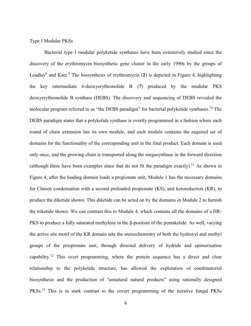
Bacterial type I modular polyketide synthases have been extensively studied since the
discovery of the erythromycin biosynthetic gene cluster in the early 1990s by the groups of
Leadlay8 and Katz.9 The biosynthesis of erythromycin (2) is depicted in Figure 4, highlighting
the key intermediate 6-deoxyerythronolide B (7) produced by the modular PKS
deoxyerythronolide B synthase (DEBS). The discovery and sequencing of DEBS revealed the
molecular program referred to as “the DEBS paradigm” for bacterial polyketide synthases.10 The
DEBS paradigm states that a polyketide synthase is overtly programmed in a fashion where each
round of chain extension has its own module, and each module contains the required set of
domains for the functionality of the corresponding unit in the final product. Each domain is used
only once, and the growing chain is transported along the megasynthase in the forward direction
(although there have been examples since that do not fit the paradigm exactly).11 As shown in
Figure 4, after the loading domain loads a propionate unit, Module 1 has the necessary domains
for Claisen condensation with a second preloaded propionate (KS), and ketoreduction (KR), to
produce the diketide shown. This diketide can be acted on by the domains in Module 2 to furnish
the triketide shown. We can contrast this to Module 4, which contains all the domains of a HR-
PKS to produce a fully saturated methylene in the β-position of the pentaketide. As well, varying
the active site motif of the KR domain sets the stereochemistry of both the hydroxyl and methyl
groups of the priopionate unit, through directed delivery of hydride and epimerisation
capability.12 This overt programming, where the protein sequence has a direct and clear
relationship to the polyketide structure, has allowed the exploration of combinatorial
biosynthesis and the production of “unnatural natural products” using rationally designed
PKSs.13 This is in stark contrast to the covert programming of the iterative fungal PKSs

7
introduced in the next section, though recent years have seen progress in understanding and
manipulating these enzymes.14-16
Figure 4: Erythromycin biosynthesis
Type I Iterative PKSs
In type I iterative polyketide synthesis, found in fungi, the polyketide assembly takes
place on a multidomain megasynthase similar to a single module of a type I modular PKS. This

8
single polypeptide chain contains one set of all the required domains, akin to a mammalian fatty
acid synthase.4 Each domain is reused during the chain extension, in contrast to the bacterial type
I modular system where the growing chain is only passed downstream.
Iterative PKSs are subdivided based on the presence or absence of the fundamental β-
position tailoring domains in the sequence: Non-reducing PKSs contain no reductive tailoring
domains, partially reducing PKSs contain a KR, but no ER, whereas highly reducing PKSs
contain both. Each has its own unique characteristics and will be discussed in turn using
representative examples for each class.
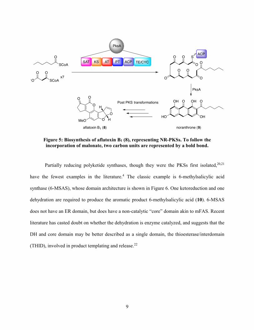
Aflatoxin B1 (8) biosynthesis in Aspergillus spp. is representative of non-reducing type I
polyketide biosynthesis (Figure 5).17 The polyketide synthase (PksA) contains the minimal
requisite domains, KS, AT and ACP, as well as others common to the class. At the N-terminus is
the starter-unit acyl transferase (SAT). While the AT selects malonyl-CoA as an extender unit,
the SAT selects hexanoyl-CoA as a starter unit. Variation in SAT domain selectivity is an
important source of diversity in polyketides.18 N-terminal to the ACP domain is a product
template domain (PT). Product templates domains are an integral part of aromatic polyketide
biosynthesis, as they orient the poly-β-keto chain and catalyze the aldol cyclizations yielding the
aromatized product.19 Finally, the TE/CYC domain catalyzes the final Claisen/Dieckmann
condensation to release the PKS product noranthrone (9) en route to aflatoxin B1 (8).
9
Figure 5: Biosynthesis of aflatoxin B1 (8), representing NR-PKSs. To follow the
incorporation of malonate, two carbon units are represented by a bold bond.
Partially reducing polyketide synthases, though they were the PKSs first isolated,20,21
have the fewest examples in the literature.4 The classic example is 6-methylsalicylic acid
synthase (6-MSAS), whose domain architecture is shown in Figure 6. One ketoreduction and one
dehydration are required to produce the aromatic product 6-methylsalicylic acid (10). 6-MSAS
does not have an ER domain, but does have a non-catalytic “core” domain akin to mFAS. Recent
literature has casted doubt on whether the dehydration is enzyme catalyzed, and suggests that the
DH and core domain may be better described as a single domain, the thioesterase/interdomain
(THID), involved in product templating and release.22

10
Figure 6: Biosynthesis of 6-methylsalicylic acid (10), representing PR-PKSs
Highly reducing polyketide synthases (HR-PKSs) contain both KR and ER domains, and
also supplementary tailoring domains such as methyltransferases (MTs). Each chain length is
tailored to a specific degree, creating a diversity of functional groups in a single polyketide
chain. This is a similar concept to the type I modular PKSs, however the mechanism and enzyme
structure are entirely different. In HR-PKSs, there is only one set of domains that is used
iteratively, and it remains elusive how each HR-PKS can be “programmed” to use the tailoring
domains only at specific chain lengths to make such diverse natural products. Lovastatin (1) is a
representative example of a natural product assembled in this manner.23,24 The biosynthesis will
be discussed in detail in the upcoming sections, and a summary is shown in Figure 7 as an
introduction to HR-PKSs.25 Using the fundamental reactions discussed previously, LovB, the
lovastatin PKS, assembles the polyketide product dihydromonacolin L (DHML, 11), the
precursor to the bioactive compound 1. LovB has all of the domains found in a mammalian fatty
acid synthase, with a few key differences. First, C-terminal to the DH domain is a MT domain,
which installs the methyl group residing at the 6 position of the decalin at the tetraketide stage
using the co-factor S-adenosylmethionine (SAM).15 Second, the constituent enoyl reductase in

11
LovB is inactive and instead this role is performed by a trans-acting enoyl reductase known as
LovC.24 Finally, the C-terminus of LovB is particularly notable. The protein ends with a part of a
non-ribosomal peptide synthetase module (NRPS), truncated after the condensation (CON)
domain.26 (This appears to be remnant of a PKS-NRPS hybrid, an example of the “hybrid” class
alluded to in Figure 3 and discussed in the upcoming sections.) The CON domain is known to be
required for DHML (11) production, but to date it has no known function.25 This truncation also
results in the absence of a domain to release the polyketide product, and the transacting TE
(LovG) performs this role.27
Figure 7: Biosynthesis of lovastatin (1), representing HR-PKSs
Fungal polyketides often do not conform to the simple model of NR-, PR- and HR-PKSs.
Many are biosynthesized by systems that are hybrids of these types, or of other natural product
classes. Examples include HR/NR-PKS partners, PKS/NRPS hybrids and meroterpenoids
(Figure 8). HR/NR-PKS hybrids synthesize natural products such as hypothemycin (12),28 whose
biosynthesis is studied in this thesis and will therefore be elaborated in its own section.
PKS/NRPS hybrids are akin to LovB, however the NRPS modules that follow their HR-PKS
portions are not truncated and are capable of installing an amino acid at the end of the polyketide
chain prior to offloading. The bioactive natural products made by this class of megasynthases are
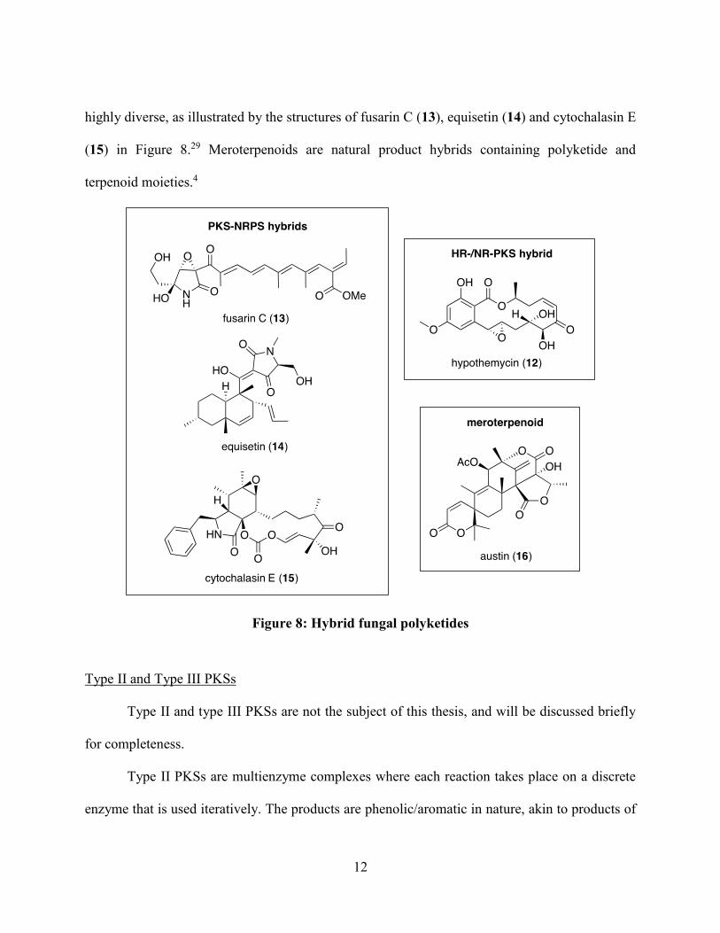
12
highly diverse, as illustrated by the structures of fusarin C (13), equisetin (14) and cytochalasin E
(15) in Figure 8.29 Meroterpenoids are natural product hybrids containing polyketide and
terpenoid moieties.4
Figure 8: Hybrid fungal polyketides
Type II and Type III PKSs
Type II and type III PKSs are not the subject of this thesis, and will be discussed briefly
for completeness.
Type II PKSs are multienzyme complexes where each reaction takes place on a discrete
enzyme that is used iteratively. The products are phenolic/aromatic in nature, akin to products of

13
fungal NR- and PR-PKSs, but the enzymatic machinery differs in that the domains are not found
in a megasynthase. The classic example is actinorhodin (17) biosynthesis in Streptomyces
coelicolor (Figure 9).30 The minimal PKS domains (KSα, KSβ and ACP) assemble the
polyketide chain, and through the action of the tailoring enzymes KR, CYC and aromatase
(ARO), synthesize enzyme-bound intermediate 18 en route to actinorhodin (17).
Figure 9: Actinorhodin biosynthesis
Type III PKSs catalyze the condensation of coenzyme-A bound intermediates without the
involvement of ACPs or tailoring domains.31 They are simple homodimers of KS domains, found
primarily in plants and also in bacteria. The classic example is chalcone synthase (CHS), which
assembles the product chalcone (19) from sequential condensation of 4-coumaryl-CoA (20) with
malonyl-CoA (Figure 10).
Figure 10: Chalcone biosynthesis

14
Polyketides Studied in this Thesis
Among the vast diversity of polyketides in Nature, of which a broad picture was painted
in the previous section, are found the two natural products explored in this thesis. Each will be
introduced in turn.
Hypothemycin (12)
Hypothemycin (12) is a resorcylic acid lactone (RAL) isolated from Hypomyces
subiculosus and related fungi.32-34 RALs are typically cytotoxic compounds, but the pattern and
modes of action of their cytotoxicity make them attractive anti-cancer drug leads.35 For example,
hypothemycin (12) has been shown to inhibit the mitogen-activated protein kinase kinase-
extracellular signal-regulated kinase (MEK-ERK) pathway in cells, a promising chemotherapy
target.36 This is distinct from the closely related RAL radicicol, whose mechanism of cytotoxity
is the inhibition of protein chaperone HSP90.37
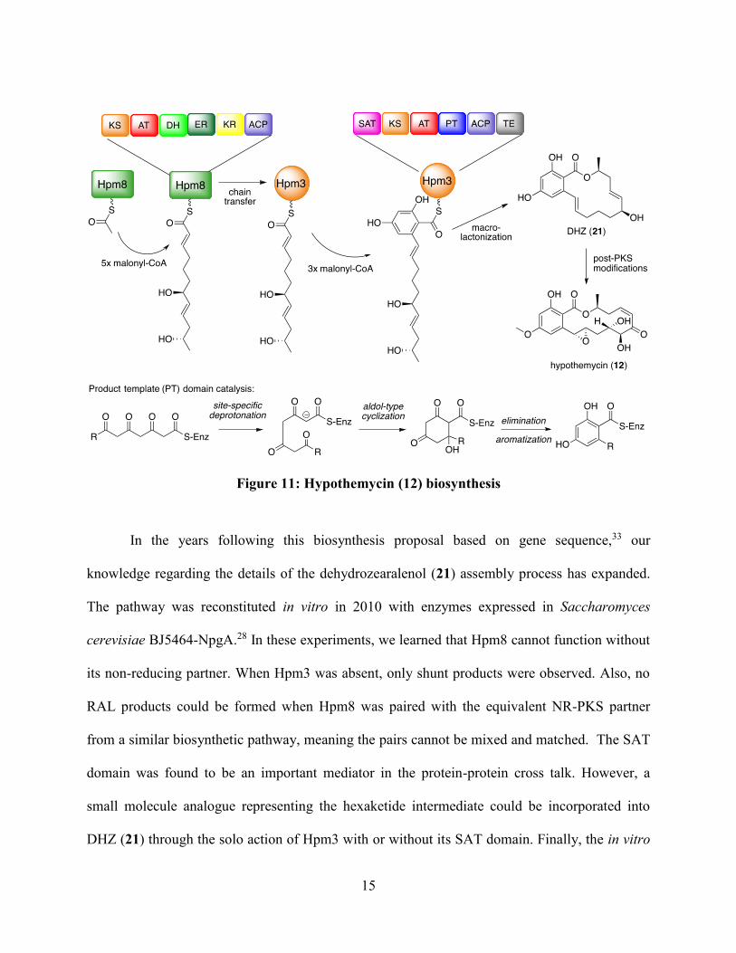
The biosynthesis of hypothemycin (12) begins with the cooperation of two PKSs, one
HR-PKS and one NR-PKS, as introduced in the previous section and summarized in Figure 11.33
First the HR-PKS Hpm8 is loaded with acetate and completes five rounds of chain extension
with the appropriate tailoring to produce the enzyme bound hexaketide chain depicted. Next, the
SAT domain of Hpm3 catalyzes the chain transfer from Hpm8-ACP to Hpm3-ACP, so that
Hpm3 can complete 3 more rounds of chain extension. The PT domain then transforms the β-
polyketothioester intermediate to a resorcylate moiety via an aldol-type cyclization.
Macrolactonization releases the PKS product dehydrozearalenol (DHZ, 21). Post-PKS
modifications, namely methylation, oxidation and olefin rearrangement, produce the bioactive
final product 12.
15
Figure 11: Hypothemycin (12) biosynthesis
In the years following this biosynthesis proposal based on gene sequence,33 our
knowledge regarding the details of the dehydrozearalenol (21) assembly process has expanded.
The pathway was reconstituted in vitro in 2010 with enzymes expressed in Saccharomyces
cerevisiae BJ5464-NpgA.28 In these experiments, we learned that Hpm8 cannot function without
its non-reducing partner. When Hpm3 was absent, only shunt products were observed. Also, no
RAL products could be formed when Hpm8 was paired with the equivalent NR-PKS partner
from a similar biosynthetic pathway, meaning the pairs cannot be mixed and matched. The SAT
domain was found to be an important mediator in the protein-protein cross talk. However, a
small molecule analogue representing the hexaketide intermediate could be incorporated into
DHZ (21) through the solo action of Hpm3 with or without its SAT domain. Finally, the in vitro

16
reconstitution also produced adequate amounts of DHZ (21) to assign the stereochemistry of the
6’ hydroxyl as (S). As is evident by the linear representation of the hexaketide intermediate in
Figure 11, this suggested that the Hpm8 KR domain can reduce from both faces of the growing
chain. It was determined in 2012 that the KR is programmed to select a face based on the length
of the growing chain.14
At the commencement of this body of work, the product of the Hpm8-Hpm3 partnership
was known, as was the hexaketide intermediate transferred from Hpm8 to Hpm3. However, it
had never been shown that the proposed intermediates of HR-PKSs were true intermediates. One
goal of my study of hypothemycin (12) was to demonstrate the intermediacy of the proposed
Hpm8-bound triketide, which will be discussed in Chapter 2, and was published as part of an
article appearing in the Journal of the American Chemical Society.38 A second goal of my study
of hypothemycin (12) was to characterize the ACP domain of a HR-PKS, and a discussion of this
will take place in Chapter 4.
Lovastatin (1)
Lovastatin (1) is a polyketide isolated from Aspergillus terreus, famed for its use in the
treatment of hypercholesteremia under the trade name Mevacor (Merck).39 It was discovered in
1980,40 a few years after the discovery of the less successful analogue compactin (22) using
activity guided fractionation of fungal extracts.41 The commercialization of lovastatin and
development of its synthetic and semi-synthetic analogues, the statins (Figure 12),42
revolutionized the treatment of high cholesterol,43 in addition to earning pharmaceutical
companies billions of dollars each year.39 High cholesterol levels in blood lead to the
development of atherosclerotic cardiovascular disease (“hardening of the arteries”) and are a

17
major risk factor for potentially fatal and debilitating events such as heart attack or stroke.44
Cardiovascular disease, encompassing diseases of the heart and cerebral vasculature, was the
second leading cause of death in Canada in 2012, accounting for 25% of all deaths.45 According
to Statistics Canada, between 2007 and 2011, 11.6% of adults aged 20-79 living in Canadian
provinces were being treated with a statin, and if the guidelines for treatment were followed for
all at risk patients, that number would be 27.1% or 6,518,200 people.46 Statistics Canada
estimates that complete treatment of the Canadian at-risk population with statins would prevent
386,200 cardiovascular events over 10 years. Therefore, the significance of this small polyketide
and its analogues cannot be overstated.
Figure 12: Statins available to prescribe in Canada, compared to HMG-CoA (28)
Statins work through the inhibition of HMG-CoA reductase, the rate-limiting step in
cholesterol biosynthesis in the liver.43 This inhibition causes the liver cells to increase their

18
concentrations of low-density lipoprotein receptors to clear cholesterol from the blood stream.
The “war-head” shared by all these molecules resembles the substrate of the reductase,
hydroxymethylglutaryl-CoA (HMG-CoA, 28), and binds reversibly and competitively in the
active site.
In addition to the impact of lovastatin (1) in healthcare, investigations of its biosynthesis
have had a large impact in the field of polyketide chemistry since its assembly from acetate was
first demonstrated in the early 1980s.47,48 A summary of our understanding of lovastatin
biosynthesis prior to this work is summarized in Figure 13. As previously introduced, LovB,
with the help of LovC and the necessary cofactors, assembles the nonaketide chain.25 It is known
that LovB also catalyzes an intramolecular Diels-Alder reaction to form the decalin ring, which
is proposed to take place at the hexaketide chain as shown.49,50 The nonaketide is released by
LovG to provide the key intermediate dihydromonacolin L (11).27 LovA then performs two
oxidations in succession to produce monacolin J.51 LovF, another HR-PKS, assembles the
methylated dipeptide shown, which LovD then transfers to the newly installed hydroxyl group to
furnish lovastatin (1).

19
Figure 13: Lovastatin biosynthesis overview
The heterologous expression of the biosynthetic enzymes has allowed some interesting
details to come to light, and has permitted the investigations in this thesis. For example, we have
learned that the CON domain, which has no known function, is required for correct functioning
of the enzyme.25 It has been proposed to be involved in the Diels-Alder cyclization, which will
be the main focus of Chapter 3, where I describe my work characterizing the LovB Diels-

20
Alderase activity. As with DHZ (21), the expression of purified enzymes has allowed the
investigations into the enzyme-bound intermediates in dihydromonacolin L (11) assembly. The
synthesis of the intermediates for this assay is discussed in Chapter 2 alongside the equivalent
DHZ experiments. Finally, the LovB-ACP was expressed as a stand-alone enzyme, as discussed
in Chapter 4 with Hpm8-ACP.

21
CHAPTER 2: INCORPORATION OF PARTIALLY ASSEMBLED INTERMEDIATES
INTO POLYKETIDE SYNTHASE PRODUCTS
Introduction
In order to prove a compound’s intermediacy in a metabolic pathway, it is convention to
synthesize this compound with certain positions bearing non-natural isotopes and follow their
conversion by organisms or enzymes. This was first done with radioactive isotopes 14C and 3H,
but the surge of NMR techniques since the 1970s allowed the use of stable isotopes such as 13C,
2H, 18O and 15N in these so-called “feeding” experiments.52 However, when it comes to
polyketides, one must remember that the true intermediates are enzyme bound. As alluded to in
the introduction, the growing polyketide chains are linked to the ACP domain through a
phosphopantetheine prosthetic group (Figure 14).53 Often referred to as an “arm”, this flexible
moiety is responsible for shuttling the growing intermediate to the various active sites of the
synthase. Therefore, in order to elucidate the structures of the enzyme bound intermediates,
chemists needed to develop methodology that would allow the PKS to recognize partially
assembled polyketides as substrates, even though in Nature these intermediates are not present in
solution. Abbreviating the phosphopantetheine group to an N-acetylcysteamine moiety
(abbreviated as SNAC) appeared to be adequate for enzyme recognition, and the first partially
assembled polyketide intermediates were successfully incorporated into macrolide polyketide
products in 1987.54,55

22
Figure 14: The phosphopantetheine prosthetic group carries the growing polyketide chain.
N-acetylcysteamine (SNAC) thioesters can approximate this group
These pioneering experiments, collegially accomplished by the groups of Hutchinson and
Cane, were the first to confirm the processive nature of polyketide biosynthesis. In these
experiments, the researchers fed 13C-labeled SNAC intermediates directly to live cultures of the
producing bacteria, and these intermediates were elaborated by enzymes that would later be
characterized as modular type I PKSs that use proprionate building blocks. It was anticipated that
this methodology could be applied to acetate-derived polyketides in fungi, however there was
limited success due to the β-oxidation of labeled precursors.56 The work described in this thesis,
and published in the Journal of the American Chemical Society in 2013,38 was the first example
of incorporating partially assembled intermediates into polyketide products by a PKS using
purified enzymes, instead of intact cells or cell-free extracts. This avoids the degradation of the
substrates by cellular machinery and makes a definitive link with the enzymes. In the discussion
that follows, I will describe the methodology behind this work involving the hypothemycin (12)
system, clearly indicating the portion of the publication attributable to me, and discuss the

23
contributions of these experiments to the field. I will then discuss on-going work to apply similar
methodology to the lovastatin (1) system.
At the onset of this work, our group had previously demonstrated the intermediacy of the
hexaketide shown in Figure 11 (Chapter 1), by its successful elaboration by Hpm3 to produce
DHZ (21).28 What remained to be demonstrated was that the series of reactions shown in Figure
15 is in fact how Hpm8 assembles the highly reduced portion of DHZ (21). Based on our
knowledge of fatty acid biosynthesis and modular type I polyketide biosynthesis, this reaction
series was for a long time assumed to be correct for iterative polyketide biosynthesis, but our
work was the first to confirm this hypothesis using purified enzymes.
Figure 15: 14 discrete enzymatic steps catalyzed by Hpm8, highlighting the structure of
each intermediate before the subsequent condensation reaction
.
24
Results and Discussion
Incorporation of 13C-SNAC Triketide (30) into DHZ (21) by Hpm8/Hpm3
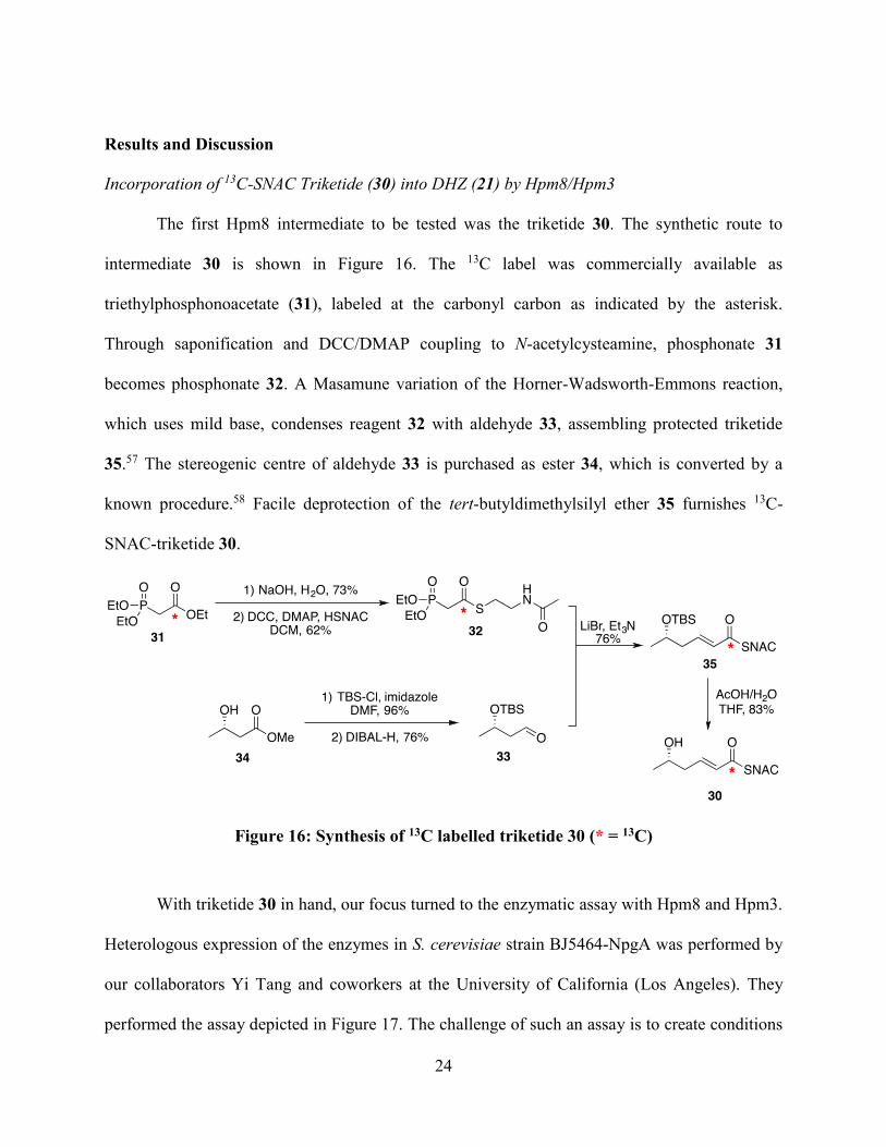
The first Hpm8 intermediate to be tested was the triketide 30. The synthetic route to
intermediate 30 is shown in Figure 16. The 13C label was commercially available as
triethylphosphonoacetate (31), labeled at the carbonyl carbon as indicated by the asterisk.
Through saponification and DCC/DMAP coupling to N-acetylcysteamine, phosphonate 31
becomes phosphonate 32. A Masamune variation of the Horner-Wadsworth-Emmons reaction,
which uses mild base, condenses reagent 32 with aldehyde 33, assembling protected triketide
35.57 The stereogenic centre of aldehyde 33 is purchased as ester 34, which is converted by a
known procedure.58 Facile deprotection of the tert-butyldimethylsilyl ether 35 furnishes 13C-
SNAC-triketide 30.
Figure 16: Synthesis of 13C labelled triketide 30 (* = 13C)
With triketide 30 in hand, our focus turned to the enzymatic assay with Hpm8 and Hpm3.
Heterologous expression of the enzymes in S. cerevisiae strain BJ5464-NpgA was performed by
our collaborators Yi Tang and coworkers at the University of California (Los Angeles). They
performed the assay depicted in Figure 17. The challenge of such an assay is to create conditions

25
where the loading of triketide 30 is favoured over a background reaction where Hpm8/Hpm3
build DHZ (21) entirely out of malonyl-CoA. Our collaborators achieved this by premixing the
enzymes with the synthetic precursor. The resulting “feed and chase” method involved 10 rounds
of first feeding the substrate, waiting 15 minutes, then chasing with malonyl-CoA. The reactions
were incubated for 10 hours total with the addition of malonyl-CoA every hour and substrate
every 2 hours. Liquid chromatography mass spectrometry (electrospray ionization) confirmed
the incorporation of the triketide into DHZ (21), and the reaction was scaled up to obtain an
adequate quantity to confirm the product and location of the label by NMR.
Figure 17: Incorporation of 13C-triketide 30 into DHZ (21) by Hpm8 and Hpm3
This result confirmed the intermediacy of the proposed triketide and validated the “feed
and chase” assay method. At this point, the project was completed by fellow PhD student,
Zhizeng Gao, who synthesized seven other proposed intermediates, and four unnatural
analogues. Ultimately, we found all eight intermediates we tested (and three of four analogues)
to be incorporated into DHZ (21), confirming the reaction series proposed in Figure 15.

26
Synthesis of Proposed LovB Intermediates
Encouraged by our success with hypothemycin, our goal was to replicate these
experiments with the lovastatin system. We reasoned this was a worthy endeavour because (1) it
would demonstrate that the conclusions drawn from the hypothemycin study apply to other PKS
systems (such as the LovB, which is similar to a PKS-NRPS hybrid), and (2) we might learn
more about the nature of the LovB-bound intermediates and enzymatic steps en route to DHML
(11).
The synthetic targets required for this assay are shown in Figure 18. We sought to
synthesize the proposed ketide of each chain length as the corresponding SNAC thioester with
either a 13C or 2H label. When I joined this project, fellow graduate student Rachel Cochrane had
synthesized diketide 36, triketide 37 and tetraketide 38, all labeled with 13C at their thioester
carbonyl carbons. Our collaborators had recreated the assays described in the section above with
LovB, LovC and a thioesterase. They found that, while the diketide 36 did not incorporate into
DHML (11), both the triketide 37 and tetraketide 38 were successfully elongated.
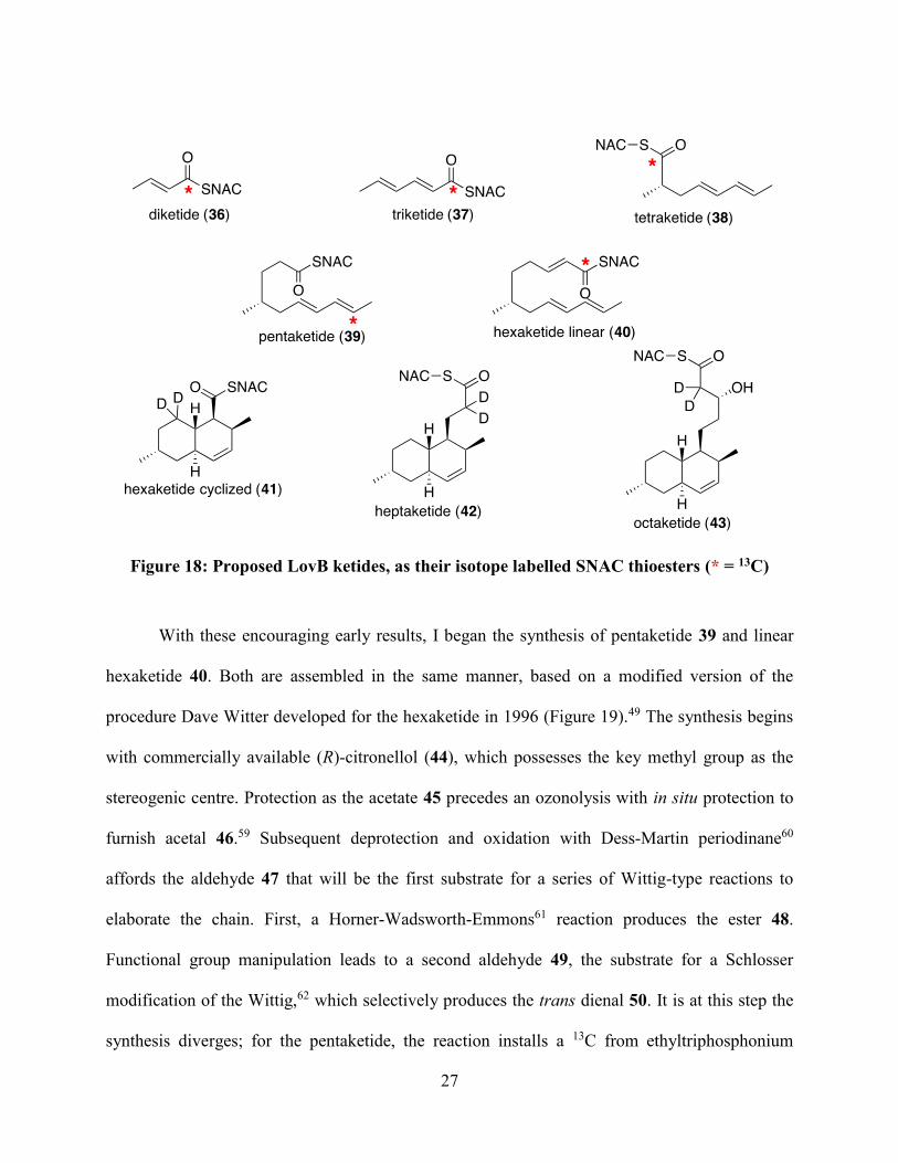
27
Figure 18: Proposed LovB ketides, as their isotope labelled SNAC thioesters (* = 13C)
With these encouraging early results, I began the synthesis of pentaketide 39 and linear
hexaketide 40. Both are assembled in the same manner, based on a modified version of the
procedure Dave Witter developed for the hexaketide in 1996 (Figure 19).49 The synthesis begins
with commercially available (R)-citronellol (44), which possesses the key methyl group as the
stereogenic centre. Protection as the acetate 45 precedes an ozonolysis with in situ protection to
furnish acetal 46.59 Subsequent deprotection and oxidation with Dess-Martin periodinane60
affords the aldehyde 47 that will be the first substrate for a series of Wittig-type reactions to
elaborate the chain. First, a Horner-Wadsworth-Emmons61 reaction produces the ester 48.
Functional group manipulation leads to a second aldehyde 49, the substrate for a Schlosser
modification of the Wittig,62 which selectively produces the trans dienal 50. It is at this step the
synthesis diverges; for the pentaketide, the reaction installs a 13C from ethyltriphosphonium

28
iodide, while a natural abundance phosphonium salt is used for the hexaketide synthesis.
Deprotection of acetal 50 with saturated oxalic acid reveals a third aldehyde 51. En route to the
pentaketide, this aldehyde is oxidized using the Pinnick procedure63 to acid 52. DCC/DMAP
coupling to N-acetylcysteamine produces the labeled pentaketide 39. The natural abundance
aldehyde 51 meanwhile is elaborated by a Masamune variant of the Horner-Wadsworth-Emmons
reaction,57 using the 13C-SNAC phosphonate 32, previously described.
Figure 19: Synthetic Route to 13C pentaketide 39 and hexaketide 40 (* = 13C)

29
The next task was the synthesis of the deuterium labeled heptaketide 42 and octaketide
43. Instead of building up from commercially available starting material, these syntheses were
accomplished through the degradation of DHML (11), using a procedure developed by John
Sorensen in his PhD thesis (Figure 20).64 Fermentation of an Aspergillus nidulans strain
complemented with both the LovB and LovC genes produces the DHML (11) used as a starting
material.24 The reaction series relies on ozonolytic cleavage of alkenes to shorten the chain two
carbons at a time, and therefore the double bond within the decalin of DHML (11) must be
protected as the dibromide 53. Mesylation/elimination of the lactone alcohol provides the α,β-
unsaturated compound 54. Ozonolysis of the olefin is performed in the presence of sodium
methoxide and methanol, leading to ester 55.65 Having achieved the octaketide skeleton, the
reaction series now diverges. To complete the synthesis of the octaketide, first the bromides are
removed using zinc in acetic acid to recreate the double bond and produce octaketide methyl
ester 56. For the installation of the deuterium atoms α to the ester, it was determined that
equilibrium conditions were optimal, where in the presence of base (potassium tert-butoxide) and
an excess of deuterated methanol, the compound is fully deuterated after two days at room
temperature. In the same pot, the subsequent addition of a small amount of deuterium oxide
produces octaketide acid 57 without requiring the isolation of the deuterated ester. DCC/DMAP
coupling to N-acetylcysteamine converts the free acid 57 to the SNAC thioester 43.
For the heptaketide synthesis, another round of mesylation/elimination and ozonolysis is
required to shorten the chain by two more carbons. First, brominated ester 55 is treated with
methylsulfonium chloride and triethylamine to produce α,β-unsaturated ester 58, then repeating
the same ozonolysis conditions affords ester 59. Deprotection regenerates the olefin and
produces heptaketide ester 60. An identical deuteration/saponification procedure leads to the

30
deuterated heptaketide acid 61, and DCC/DMAP coupling leads to the final product, SNAC-
heptaketide 42.
Figure 20: DHML (11) degradation to synthesize 2H-labelled heptaketide 42 and octaketide
43
With the completion of the DHML (11) degradation to make heptaketide 42 and
octaketide 43, the only proposed ketide left to synthesize was the cyclized hexaketide 41. This
task was attempted by fellow PhD student Eva Rodriguez-Lopez, who obtained an interesting
result in the final steps of the work. Having constructed the hexaketide skeleton as the ethyl ester
62, the plan was then to saponify and couple to N-acetylcysteamine, as shown in Figure 21.

31
However, saponification was not successful, even with heating and prolonged reaction times. We
concluded that the hydrophobicity of the molecule and steric bulk of the reacting centre
prevented hydroxide from attacking the ester to form the tetrahedral intermediate. If this is the
case, it has interesting implications for our proposed biosynthetic pathway. A review of Figure 2
shows that at every chain length, the growing polyketide must be transferred from the ACP back
to the KS for the next round of extension. If the steric bulk of compound 62 was too crowded for
saponification, one could imagine that transthioesterification of the enzyme bound hexaketide
from the ACP phosphopantetheine to the KS cysteine would also be very slow. The sheds doubt
on the intermediacy of this structure. An alternative biosynthetic scheme is presented in the
upcoming chapter.
Figure 21: Due to the failed saponification, hexaketide 41 could not be synthesized
With one isotope-labeled ketide per chain length synthesized, the next step is the
enzymatic assays to be performed by our collaborators to see if these proposed intermediates can
be elongated into DHML (11).

32
CHAPTER 3: INVESTIGATIONS INTO THE DIELS-ALDERASE ACTIVITY OF
LOVB
Background
The Diels-Alder Reaction
The Diels-Alder reaction, named for the chemists who first characterized it in 1928, is the
concerted [4+2] cycloaddition of a 1,3 diene and an electron poor alkene, referred to as the
dienophile (Figure 22).66 Over the past century, the reaction has proven to be of great synthetic
utility thanks to the large amount of complexity that can be installed in one reaction under mild
conditions and with atom economy. Two carbon-carbon bonds and four contiguous stereocenters
are formed in one stereocontrolled step. However, compared to the plethora of enzyme-catalyzed
reactions involving charged or radical intermediates, there are few known examples of pericyclic
reactions catalyzed by enzymes.
Figure 22: A typical Diels-Alder reaction, showing the concerted transition state.
Diels-Alderase Activity of LovB
One such “Diels-Alderase” is LovB, as introduced previously, which has been
extensively studied by our group. The possibility of a Diels-Alder reaction in the biosynthesis of
lovastatin was first proposed in 1985 following labeling studies with 13C, 2H and 18O
precursors.48 The isotope pattern observed was consistent with this proposition, and subsequently
its feasibility could be explored by studying the equivalent non-enzymatic process. In 1996,

33
Witter and Vederas first synthesized hexaketide triene 40 and found that it does indeed undergo a
Diels-Alder reaction spontaneously.49 The results of these key experiments are summarized in
Figure 23. Triene 40 has 4 theoretical cyclization geometries, based on the combinations of the
endo versus exo relationship of the diene and thioester, and the pseudo-axial versus pseudo-
equatorial conformation of the methyl group. The transition state geometry leading to the natural
product would, for example, have the methyl group in a pseudo-axial position, and an endo
relationship between the reacting partners, leading to compound 41. When heated in organic
solvent, it was found that the reaction progressed through transition states with pseudo-equatorial
methyl groups only, leading to non-natural stereoisomers 65 and 66. In Lewis-acid catalyzed
conditions, only isomer 66 was isolated. In aqueous conditions, the reaction progressed with a
half-life of two and a half days, and once again the natural stereochemistry was not observed.
From these three experiments, the following conclusions are drawn: First, it is feasible that a
biological Diels-Alder reaction takes place during lovastatin biosynthesis. Second, if this
reaction did occur, it would require an enzyme because the correct stereochemistry is not
observed. Even though a slow background cyclization takes place, the reaction rate leading to the
correct product is zero without an enzyme.

34
Figure 23: Cyclization modes of triene 40, and results of enzyme-free reactions.
The identification of the LovB gene and its heterologous expression in A. nidulans24
allowed the study of this hypothesis with purified enzymes. Auclair et al exposed hexaketide
triene 40 to LovB and obtained the results described in Figure 24.50 After three days of reaction,
a mixture of inseparable thioesters was isolated. Reacting the mixture with sodium ethoxide in
ethanol converted the product to their ethyl esters, which were separated and characterized as
compounds 67, 68, and 62, in a 15:15:1 ratio. The stereochemistry of the natural product
(equivalent to the stereochemistry of thioester 41 and ethyl ester 62) was not present in the

35
products of control reactions, where the enzyme was either absent or heat inactivated. Therefore,
it was concluded that LovB does catalyze the conversion of triene 40 to decalin 41.
Figure 24: Assay studying the LovB catalyzed cyclization of triene 40
However, there are many questions left to be answered, some of which will be explored
in this thesis. First, the extent to which a [4+2] cyclization is concerted is a matter requiring
investigation. In order to be a Diels-Alderase, LovB would need to catalyze this reaction in one
concerted step, rather than the equivalent step-wise nucleophilic process. However, in order to
study the mechanistic details of this reaction, one would need to isolate the Diels-Alderase
activity. LovB is a large multi-domain enzyme, and one of our goals is to pinpoint which domain
actually catalyzes the cyclization. Lastly, these experiments do not prove the intermediacy of
hexaketide triene 40. It is possible that the Diels-Alderase domain is somewhat promiscuous and
the conversion of triene 40 to decalin 41 is not a representation of its native activity. In fact,
because the triene is SNAC bound, as opposed to ACP bound, we are already working with a

36
substrate analogue, which may explain the slow reaction rate. So while there is no doubt that
LovB catalyzes the conversion of 40 to 41, there are still many questions to answer regarding
this aspect of lovastatin biosynthesis.
Overview of Known Diels-Alderase Enzymes
Relatively few naturally-occuring enzymes are known to catalyze a [4+2] cycloaddition,
and the topic has been covered in numerous reviews.67-74 Herein I will give a brief introduction
to the enzymes that, in addition to LovB, catalyze [4+2] cyclizations, possibly by a Diels-Alder
mechanism, en route to natural products. While there is currently no evidence to support or
refute a pericyclic, concerted [4+2] cycloaddition for each enzyme, it is convention to refer to
these enzymes as “Diels-Alderases”, recognizing this term is currently defined as an enzyme that
catalyzes the formation of a cyclohexene ring from a conjugated diene and an alkene
stereoselectively.72
With the diversity amongst the Diels-Alderases, it is difficult to categorize them. At
present, two common themes can be used to group the enzymes into four categories (Table
1).72,74 Firstly, a few of the Diels-Alderase enzymes catalyze the formation of decalin systems.
Secondly, they can be mono- or multi-functional. Table 1 contains, to the best of my knowledge,
all enzymes proposed to be Diels-Alderases, grouped by homology, though the level of evidence
supporting the proposals varies. Enzymes in bold have been purified to homogeneity and the
catalysis has been replicated in vitro, and therefore the evidence for classifying these nine
enzymes as Diel-Alderases is strongest. Non-bolded enzymes have some evidence, such as
homology with known Diels-Alderases or through gene-deletion experiments, though in vitro
reconstitution of the proposed reaction has yet to be accomplished.

37
Table 1: Proposed Diels-Alderases
Monofunctional Multifunctional
Decalin-forming PyrE3, VstK, KijA, ChlE3, TcaE1 Sol5
MycB, Fsa2, CghA, Eqx3, gNR600,
CcsF
LovB
Bet1
Non-decalin product SpnF
TclM, TbtD
PyrI4, AbyU, VstJ, KijU, ChlL,
TcaU4
Macrophomate Synthase
Riboflavin Synthase
*see text for references
Multifunctional Decalin-Forming Diels-Alderases
LovB, the subject of this chapter, shares the category of multifunctional decalin-forming
Diels-Alderases with two enzymes, Sol5 and Bet1. Sol5, or solanopyrone synthase, was the first
example of a purified/reconstituted Diels-Alderase, published in 1998.75,76 In the last step of the
biosynthesis of solanopyrone A (69), Sol5 employs molecular oxygen to oxidize the primary
alcohol to an aldehyde, activating the dienophile for [4+2] cycloaddition shown in Figure 25.
Figure 25: Reaction catalyzed by Sol5
Bet1, in contrast, was only recently characterized.77 Like LovB it is a HR-PKS, and in
cooperation with trans-acting enoyl reductase Bet3, it catalyzes the formation of polyketide
product dehydroprobetaenone I (70). A. oryzae complemented with the bet1 and bet3 genes
produced the PKS product 70 in fermentation, however, the activity of this protein pair has not

38
yet been reconstituted in vitro, and the potential of Bet1 as a Diels-Alderase has not been
investigated further. Three possible pathways were proposed based on the order of the Diels-
Alder reaction, chain extension and reductive offloading with the reductase domain (Figure 26).
The authors did however note an interesting result likely ruling out the pathway following Diels-
Alder A. When compound 70 was treated with NaBH4, only the terminal aldehyde was reduced,
while the ketone was spared. Further molecular modelling showed this carbonyl to be in a highly
sterically congested environment. The authors postulate that if steric crowding prevents
nucleophilic attack of NaBH4, then it would likely also prevent attack by the enolate of malonate
during assembly, thus making any chain extension post-Diels-Alder reaction very unlikely. This
finding is relevant to the discussion of our findings concerning LovB.
Figure 26: Proposed biosynthesis of dehydroprobetaenone I (70)
Monofunctional Decalin-Forming Diels-Alderases
PyrE3 is the first putative monofunctional Diels-Alderase for which catalysis leading to a
decalin structure has been shown in vitro.78 En route to the natural product pyrroindomycin,

39
PyrE3 was shown to perform the first of two [4+2] cyclization reactions, from intermediate 71 to
decalin 72 (Figure 27). PyrI4, another monofunctional Diels-Alderase, then forms the spirocyclic
cyclohexene found in intermediate 73. Interestingly, PyrE3 contains a FAD binding motif, and
will not catalyze the decalin formation without FAD bound, though no oxidation takes place. A
number of gene clusters for similar natural products contain homologues for PyrE3, and thus it is
suggested that these are also decalin-forming Diels-Alderases (See Table 1).72,79
Figure 27: Diels-Alder sequence forming the pentacyclic core of pyrroindomycin
There is another group of monofunctional decalin-forming Diels-Alderases, whose Diels-
Alderase was first proposed based on their homology to the enzyme FsaA. The gene fsaA was
found in the gene cluster for fusarisetin A (74) in Fusarium sp. FN080326.80 The authors suggest
that the biosynthesis begins with the action of PKS-NRPS hybrid Fsa1, and its trans-acting ER
Fsa3, to produce trichosetin (75), which is methylated to form equisetin (76), the final product in
the related fungal species Fusarium heterosporum (Figure 28). The gene fsa2 showed no known
homologues, and thus to elucidate its function, the authors created a Δfsa2 deletion mutant. From
this culture, they were able to isolate the diastereomer of equisetin (77), which could be made by
an exo Diels-Alder reaction.80 Since only the endo diastereomer (equisetin, 76) is found in the

40
wild type fungus, it was determined that Fsa2 plays a role in enhancing the stereoselectivity of
the biosynthetic Diels-Alder reaction, and thus Fsa2 and its homologues may be Diels-Alderases.
Figure 28: Biosynthesis of fusarisetin A (74)
A recent communication in the Journal of the American Chemical Society confirmed that
one homologue of Fsa2, MycB is indeed a Diels-Alderase.81 The authors searched the genome of
the fungus that produces the natural product myceliothermophin E (78) (Figure 29). They found
a gene cluster that contains a PKS-NRPS gene (mycA), an ER gene (mycC) and a third gene,
mycB that has sequence homology to the proposed Diels-Alderase CghA, which through deletion
experiments was confirmed to be involved in the production of the natural product 78. When
mycB alone was deleted, the production of all cyclized products was abolished, and the strain
gave primarily linear compound 79 in its enol form and some air-oxidized compound 80. MycB
was heterologously expressed and found to convert compound 80 to final product 78, while in
the absence of enzyme compound 80 was degraded, confirming MycB is a Diels-Alderase.

41
However, MycB could not cyclize 79 in its enol form, suggesting the keto functionality is
essential to the catalysis. The authors propose the biosynthetic pathway show in Figure 29. The
product of the PKS-NRPS MycA/C is the keto form of compound 79, which could be acted on
immediately by MycB to produce cyclized product 81, whose enolization/oxidation would lead
to the final product 78. Alternatively, 79 could enolize and be oxidized by air to produce 80,
which is a known substrate of MycB.
Figure 29: The MycB catalyzed Diels-Alder reaction enroute to myceliothermophin E (78)
Non-Decalin Forming Diels-Alderases
As shown in Table 1, six non-decalin forming Diels-Alderases have been purified and
reconstituted in vitro. These enzymes are included for completeness but won’t be discussed in
detail. PyrI4 was introduced in the above section (see Figure 27) and the remaining five
enzymatic conversions are shown in Figure 30. These are macrophomate synthase,82 riboflavin
synthase,83 SpnF,84,85 AbyU,86 and TclM/TbtD.87,88 TclM and TbtD perform the equivalent
reaction towards two different thiopeptides, and therefore only one is shown for conciseness.

42
Figure 30: Non-decalin forming Diels-Alderases

43
Goals of this Chapter
This thesis seeks to further our understanding of how LovB catalyzes the [4+2]
cycloaddition during the biosynthesis of DHML (11). The first goal was to isolate the Diels-
Alderase activity to one of the many domains present on the HR-PKS. During our investigations,
we found evidence to suggest the intermediate is not necessarily the hexaketide, and thus the
substrate scope of the LovB Diels-Alderase activity became a second objective.
Isolating the Diels-Alderase Domain
As detailed in the introduction, LovB has many catalytic domains. In order from N- to C-
terminus they are KS, AT, DH, MT, ER0, KR, ACP and CON. The only domain with no known
function is the CON domain, and this was a logical area of investigation. The CON domain,
short for condensation, is the remnant of an NRPS module, for which the A (adenylation) and
PCP (peptidyl carrier protein) domains have been lost over evolution. Not only is a CON
incapable of condensing an amino acid onto the polyketide chain without the A and PCP
domains, but LovB’s CON domain also has a critical active site mutation. The HHXXXDG
motif required for condensation activity has been mutated to HRLVGDG. When our group
reconstituted the LovB/LovC assembly of DHML (11) in vitro, it was found that LovB could not
be replaced with LovBΔCON, a construct of LovB with the C-terminus truncated to omit the
CON domain; no DHML (11) was observed in reaction mixtures that contained all the necessary
co-factors.25 However, when the CON domain was supplemented in trans as a stand-alone
domain, DHML (11) production resumed. Therefore, while the function of the CON domain is
unknown, it is clearly integral to the activity of LovB.

44
Further evidence of the plausibility of the CON domain’s involvement in the Diels-Alder
reaction was published in 2014, when the group of Eric Schmidt (University of Utah) was
studying the combinatorialization of PKS-NRPS hybrid systems.89 The researchers exchanged
the PKS and NPRS portions of different hybrid megasynthases and searched the extracts of the
heterologous expression to find which products the various combinations produced. Constructs
that contained the PKS portion of LovB, and the NRPS portion of EqxS, the equisetin
megasynthase, provided compound 87 on fermentation (Figure 31). Presumably spontaneous
decarboxylation of octaketide 88 leads to ketone 87. When drawn in this way, one can recognize
octaketide 88 as an intermediate that did not undergo Diels-Alder reaction, and whose
elaboration to DHML was not completed. This further implicates the CON domain in the Diels-
Alder reaction.
Figure 31: Literature evidence supporting the CON domain as the Diels-Alderase, isolation
of compound 87 from the action of a LovB/EqxS hybrid
The other domain I chose to investigate as the potential Diels-Alderase is the dehydratase
(DH). While there is no specific evidence for this related to LovB, dehydratase domains have
literature precedence for their ability to activate the product α,β-unsaturated ketones to Michael
addition, and thus it is known they can possess additional activites.90 This Michael addition

45
would be analogous to the reverse hydration reaction catalyzed by hydratase enzymes, where
water is the nucleophile. I therefore postulated it was possible that, following dehydration, the
DH domain could act as a general acid catalyst to lower the LUMO of the dienophile and
perform the cyclisation before the linear hexaketide triene leaves the active site (Figure 32).
Figure 32: Biosynthetic proposal employing the LovB DH domain as the Diels-Alderase
Substrate Scope of LovB
It is not uncommon for enzymes to convert compounds other than their natural substrate.
In fact, as discussed, our group studied the Diels-Alderase activity of LovB with SNAC-
hexaketide triene 40. Since a true intermediate would be enzyme bound, the work illustrated that
LovB has enough promiscuity to recognize SNAC in place of the holo-ACP. Therefore, it is
possible that the hexaketide is not the true substrate, but its cyclization is in fact a promiscuous
activity, and that DHML (11) assembly can proceed through a different route than that proposed
in Figure 13. Figure 33 is a web that outlines possible routes from the hexaketide triene to
decalin heptaketides. The routes using Diels-Alder reaction A and B have been the focus of our
investigations prior to this thesis. However, as discussed in the previous chapter (Figure 21), the
carbonyl of the cyclized hexaketide may be too sterically hindered for downstream
transthioesterification and attack by the malonate enolate, suggesting the chain may be further
elongated prior to Diels-Alder reaction. For that reason, in addition to further investigating

46
pathway A/B, I also synthesized heptaketide tetraene 89 to study its cyclization via Diels-Alder
reaction D. Based on the results, pathway C may be studied at a later date.
Figure 33: Possible cyclization routes during the LovB catalyzed biosynthesis of DHML
(11)
Results and Discussion
Substrate Syntheses
The synthesis of hexaketide triene 40 was described in the previous chapter (see Figure
19). Heptaketide tetraene 89 can be assembled using the same methodology. Figure 34 describes
two routes I employed in the synthesis of heptaketide tetraene 89. First, using the hexaketide-
SNAC 40 already in hand, I envisioned employing the same strategy of functional group
modification and HWE reaction to elaborate the chain by two additional carbons. Unfortunately,
the thioester was susceptible to 1,4 reduction, producing saturated alcohol 91 when treated with

47
both DIBAL and Superhydride. To circumvent this, ethanolysis was used to furnish ester 92,
which could be reduced to the desired allylic alcohol 93 using DIBAL. Oxidation with DMP
provided aldehyde 94, which is elongated by the familiar Horner Wadsworth Emmons (HWE)
reaction to provide the heptaketide 89. An alternative procedure can also be followed, which
branches from the hexaketide synthesis at the key aldehyde 51. Four carbons can be added
simultaneously using the extended HWE reagent 95.91,92 This reaction requires a stronger base,
LHMDS, and therefore is not compatible with the N-acetylcysteamine thioester and the ethyl
ester is employed in its place, generating heptaketide 96. Due to the lipophilicity of ester 96, its
hydrolysis to acid 97 was slow and required a large amount of THF as a co-solvent. Heating to
near the boiling point of THF under a continuous stream of argon slowly concentrated the
reaction as the hydrolysis progressed and complete conversion was achieved. Coupling to SNAC
to produce the final product 89 was performed using DCC without the usual catalyst DMAP.
When DMAP was present, neither starting material nor product could be recovered. Instead a
mixture of non-identifiable materials were obtained, and presumably the DMAP adduct of the
tetraene was too reactive to be coupled. Fortunately, the catalyst was not required, and repeating
the reaction without DMAP furnished the product.

48
Figure 34: Synthesis of heptaketide tetraene 89

49
Expression of Enzymes
LovB and LovBΔCON Expression in S. cerevisiae
Our colleagues in the Yi Tang group at UCLA expressed LovB and LovBΔCON for the
in vitro reconstitution of the pathway to DHML (11) in collaboration with our group.25 They
provided me with their S. cerevisiae cells, transformed separately with the expression plasmids
to purify both megasynthases individually. LovB could then be used as a positive control, while
LovBΔCON is a fragment to be tested for potential Diels-Alderase activity, and would not be
active if the CON domain is the catalyst.
The expression system uses S. cerevisiae strain BJ5464-NpgA, which has been
genetically modified to contain a phosphopantetheinyl transferase gene, npga, enabling the post-
translational transfer of the essential phosphopantetheine group to the ACP domain.93 The lovB
and lovBΔCON genes are cloned behind an ADH2 promoter, such that expression is induced by
depletion of glucose from the media.94 The proteins were purified using N-terminal hexahistidine
tags, and are shown on a gel in Figure 35.
Figure 35: SDS-PAGE gel of proteins expressed for Diels-Alderase assay, labelled with
their schematic representations
250 kDa
50 kDa

50
Expression of CON and DH in E. coli
In contrast to LovB and LovBΔCON, the individual domains expressed as stand alone
proteins can be expressed in E. coli under the control of a lac promoter. For the CON domain I
transformed BL21(DE3) cells with a plasmid obtained from the Yi Tang group for expression.
The DH domain had not been expressed prior to this work, and I opted to have the gene
synthesized and subcloned into pET-28a by Bio Basic Inc.
Cyclization Studies
Hexaketide Background Reaction – NMR-based Assay
NMR techniques have improved dramatically since our group first published the Diels-
Alderase activity of LovB. For this reason, I chose to begin my investigation of this activity with
the development of an assay that uses NMR for direct detection of products, in contrast to the
derivatization and manual separation employed in 2000.50 Our intention was to take advantage of
small differences in chemical shifts between the 3 possible isomers of the cyclized hexaketide
(Figure 36) and using 2 dimensional techniques to clearly distinguish which isomers are present
or absent.
Figure 36: Key differences in chemical shifts of select nuclei in the three decalin isomers.
No 13C-NMR data has been previously published for enzymatic product 41

51
Hexaketide cyclizations in absence of enzyme were monitored by NMR in order to test
the feasibility of this method. The first NMR technique employed was an HSQC (500 MHz)
(Figure 37). The two isomers were clearly distinguishable using the 1 position of the decalin,
with the exo isomer signal appearing around (2.85 ppm, 56 ppm) and the endo isomer signal
around (2.62 ppm, 54 ppm). Furthermore, the expected 1H chemical shift of H-1 of enzymatic
product is 2.80 ppm, and is not expected to overlap with the non-enzymatic products’ signals.

52
Figure 37: HSQC of control cyclization of triene 40 (500 MHz in CDCl3), showing the
differentiation of decalin products 65 and 66
While HSQC appeared to be a promising method, another possible analysis was the
chemical shift selective filtered TOCSY (CSSF-TOCSY). With this experiment, the investigator
selectively irradiates a specific proton chemical shift, and then detects all of the protons in its
spin system. This technique has added benefits due to the excellent NMR properties of the 1H
66 65

53
nucleus (abundance, gyromagnetic ratio), and the added information contained in proton spectra
relative to carbon spectra. The results of the control reaction are shown in Figure 38. Selective
excitation of H-1 of the trans-fused product 66 (2.86 ppm), followed by excitation of the C2-Me
of the cis-fused product 65 (1.02 ppm) can pull out the individual 1H-NMR spectra of the two
decalin systems, effectively separating them from unreacted triene and confirming their identities
through comparison with literature spectra.
Figure 38: CSSF-TOCSY of the control cyclization of triene 40 (500 MHz in CDCl3). (a) 1H-NMR of the mixture. (b) Selective excitation of 1.02 ppm, isolating the spectrum of the
cis-fused product 65. (c) Selective excitation of 2.86 ppm, isolating the spectrum of the
trans-fused product 66
(a)
(b)
(c)

54
At this time, I performed the reaction with the CON domain. Unfortunately, no additional
cross-peaks were observed in HSQC. Furthermore, the 1D 1H-NMR did not show evidence of
the product and there was therefore no peak to irradiate for a CSSF-TOCSY experiment. While it
was possible to conclude that the CON domain did not catalyze the reaction, this assay had not
been validated and therefore that conclusion should not be drawn at this time. In order to address
this, LovB was expressed to use as a positive control and further experiments were paused until
the synthesis of the product standard (41) could be completed so that the assay could be
validated.
Heptaketide Cyclizations
As mentioned previously, the synthesis of the standard product, cyclized hexaketide 41
was not complete due to incomplete saponification of the ethyl ester (see Figure 21), and this
result led us to question the intermediacy of the ACP-bound equivalent of 41, and motivated us
to investigate the cyclizations of heptaketide tetraene 89.
First, the cyclizations were performed on small scale using 3 μg (9 nmol) of tetraene 89.
The negative control was performed in 30 μL of buffer, while the reaction contained 400 μg (1.2
nmol) of LovB. After overnight incubation at 37 °C, the reaction mixtures were extracted with
dichloromethane, and the extracts were analyzed by LC-MS. However, despite many attempts
for method optimization, only one peak was observed for both the control and LovB reactions,
and this peak matched the starting material in both retention time and mass to charge ratio
(Figure 39). This suggested there was no change overnight for either reaction.

55
Figure 39: LC-MS extracted ion chromatogram for heptaketide 89 (a) and overnight
reaction of 89 with LovB (b)
To confirm the mass specrometry results and rule out co-elution of isomers, a larger scale
reaction was performed with NMR-monitoring of a solution of 2.6 mg of tetraene 89 in 0.7 mL
of a THF-d8/D2O mixture (Figure 40). The spectrum did not change overnight, and pure
unreacted starting material could be re-isolated, in concordance with the LC-MS results. We can
therefore conclude that LovB likely does not catalyze the cyclization of the heptaketide tetraene
(89), effectively ruling it out as a substrate of the biological Diels-Alder reaction. A standard
(a)
(b)

56
product could confirm this, by eluting from LC-MS at a different retention time, however the
synthesis was not undertaken because the unlikelihood that tetraene 89 is the substrate.
Figure 40: 1H NMR of heptaketide 89 and 0 and 20 hours, showing no change overnight in
THF-d8/D2O
Assay Employing 13C Hexaketide
The failure of heptaketide 89 to cyclize redirected our efforts back towards the
hexaketide. At this stage, the majority of the synthesized hexaketide triene 40 had been
consumed towards the synthesis of heptaketide 89, leaving less than 10 mg of each the natural
abundance and 13C-labelled compounds. Because the LovB cyclization reaction is low yielding
(5 mg of starting material yielded 0.1 mg of enzymatic isomer 41 in the original work50), the
t = 0
t = 20 h

57
isotope label would improve detection of the products by NMR. Therefore, a portion of 13C-
labelled triene (13C-40) synthesized for incorporation experiments was redirected for the purpose
of studying the Diels-Alder reaction. Five reactions were set up in tandem, each containing 1 mg
of 13C-40 in 5 mL of buffer, and one of either LovB (positive control), LovBΔCON, CON, DH
or no enzyme (negative control). After reacting overnight, the mixtures were extracted with
dichloromethane, and the extracts were analysed by NMR. First, 1D carbon NMR was used to
distinguish the number of labelled isomers present, and their chemical shifts relative to those
seen in the positive and negative controls (Figure 41). The presence of clear differences between
the positive and negative controls implied a reaction did in fact take place in the presence of
LovB that did not occur in its absence. Moreover, the DH and CON containing reactions
resembled the negative control, with signals at 204.5 ppm and 201.5 ppm (the CON reaction also
contained unreacted triene, observed at 190.6 ppm). In contrast, both the spectra of the LovB and
LovBΔCON experiments contained two additional peaks at 198.4 ppm and 198.3 ppm. However,
no 13C-NMR spectrum had ever been recorded for the enzymatic product (41), so further analysis
was required.

58
Figure 41: 13C-NMR of the products of hexaketide (13C-40) cyclization in the presence of (a)
buffer only, (b) LovB, (c) LovBΔCON, (d) CON and (e) DH.
In order to detect the chemical environment around the 13C-labelled centre, a gHMBC
experiment would be ideal. However, the resolution was inadequate. To improve the resolution
in the 13C dimension of the gHMBC, we used a modification of the pulse sequence called a
(a)
(b)
(c)
(d)
(e)

59
“band-selective gHMBC” (bsgHMBC).95 This variant uses a selective pulse to excite only the
13C nuclei in the frequency range under investigation, allowing smaller increments to be sampled
without unreasonable experiment duration. Both the typical gHMBC, and bsgHMBC are show in
Figure 42 with matching window sizes for comparison.
Figure 42: (a) gHMBC of LovB catalyzed cyclization of 13C-hexaketide 40. (b) bsgHMBC of
same sample.
(a)
(b)
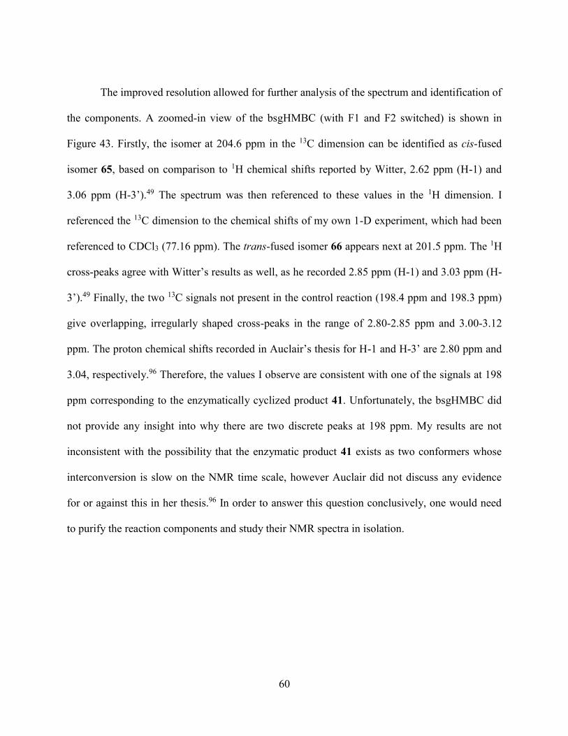
60
The improved resolution allowed for further analysis of the spectrum and identification of
the components. A zoomed-in view of the bsgHMBC (with F1 and F2 switched) is shown in
Figure 43. Firstly, the isomer at 204.6 ppm in the 13C dimension can be identified as cis-fused
isomer 65, based on comparison to 1H chemical shifts reported by Witter, 2.62 ppm (H-1) and
3.06 ppm (H-3’).49 The spectrum was then referenced to these values in the 1H dimension. I
referenced the 13C dimension to the chemical shifts of my own 1-D experiment, which had been
referenced to CDCl3 (77.16 ppm). The trans-fused isomer 66 appears next at 201.5 ppm. The 1H
cross-peaks agree with Witter’s results as well, as he recorded 2.85 ppm (H-1) and 3.03 ppm (H-
3’).49 Finally, the two 13C signals not present in the control reaction (198.4 ppm and 198.3 ppm)
give overlapping, irregularly shaped cross-peaks in the range of 2.80-2.85 ppm and 3.00-3.12
ppm. The proton chemical shifts recorded in Auclair’s thesis for H-1 and H-3’ are 2.80 ppm and
3.04, respectively.96 Therefore, the values I observe are consistent with one of the signals at 198
ppm corresponding to the enzymatically cyclized product 41. Unfortunately, the bsgHMBC did
not provide any insight into why there are two discrete peaks at 198 ppm. My results are not
inconsistent with the possibility that the enzymatic product 41 exists as two conformers whose
interconversion is slow on the NMR time scale, however Auclair did not discuss any evidence
for or against this in her thesis.96 In order to answer this question conclusively, one would need
to purify the reaction components and study their NMR spectra in isolation.

61
Figure 43: gHMBC of LovB catalyzed cyclization of 13C-hexaketide 40
Conclusion and Future Outlook
In conclusion, I demonstrated that both LovB and LovBΔCON catalyze the cyclization of
linear hexaketide triene 40 to decalin 41, a product not found in the absence of enzyme or in the
presence of the LovB CON or DH domains. The stereochemistries of the products were
determined based on comparison to previously reported chemical shifts.49,96 In the process I
developed three different NMR-based assays for the detection of the products of the cyclization
65 H-1
65 H-3’
66 H-1
66 H-3’
41 H-1
41 H-3’

62
reaction. HSQC and CSSF-TOCSY were useful for distinguishing the products of the
background reaction from one another, however it was my assay employing a 13C-labelled
substrate and bsgHMBC that was able to localize the Diels-Alderase activity to LovBΔCON and
not CON. All three techniques may prove to be useful in the future for studying this reaction, or
other enzymatic reactions where the product and starting material possess the same molecular
formula, where LC-MS is less useful. Strategic placement of the isotope label could make a
combination of the three techniques powerful for determining the products of such enzymatic
reactions.
It was our hypothesis that the CON domain is the Diels-Alderase within the multidomain
megasynthase that is LovB, however the results reported here refute that. With intact LovB
serving as a successful positive control, it was found that in the presence of CON alone only the
background cyclization occurred. The discovery of enzymatic product 41 in the reaction
containing LovBΔCON confirmed that the Diels-Alderase activity is present in this fragment of
LovB, and refutes the possibility that CON is simply not active stand-alone. Within the
LovBΔCON fragment, the DH domain was selected for the exploration of Diels-Alderase
activity, the DH domain does not convert triene 40 to decalin 41. LovBΔCON contains six other
domains, KS, AT, MT, ER0, KR and ACP, which could be investigated systematically in turn.
Many of these domains work by general acid activation of the thioester carbon, and thus would
be feasible Diels-Alderases.
Another objective of this chapter was to determine the true intermediate that undergoes
the Diels-Alder reaction during the assembly of DHML (11). While the ability of LovB to
cyclize triene 40 to product 41 has been confirmed and reproduced herein, this could be a
promiscuous activity, as supported by the steric hindrance observed at the carbonyl carbon of
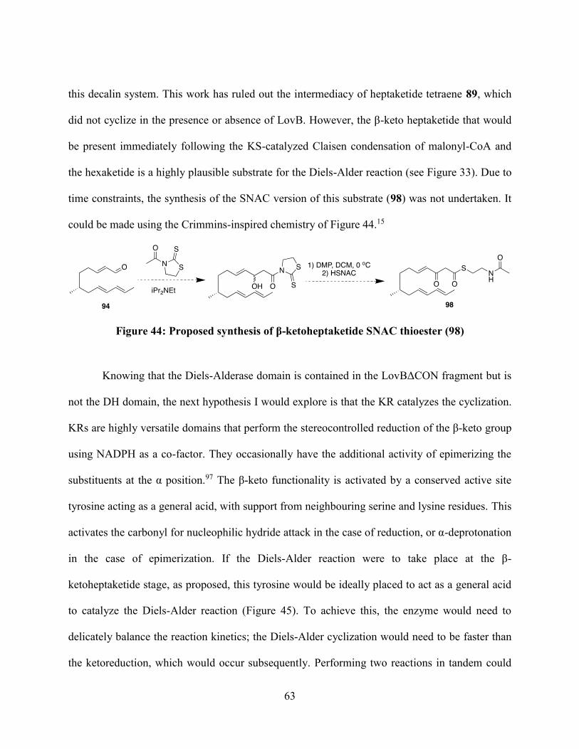
63
this decalin system. This work has ruled out the intermediacy of heptaketide tetraene 89, which
did not cyclize in the presence or absence of LovB. However, the β-keto heptaketide that would
be present immediately following the KS-catalyzed Claisen condensation of malonyl-CoA and
the hexaketide is a highly plausible substrate for the Diels-Alder reaction (see Figure 33). Due to
time constraints, the synthesis of the SNAC version of this substrate (98) was not undertaken. It
could be made using the Crimmins-inspired chemistry of Figure 44.15
Figure 44: Proposed synthesis of β-ketoheptaketide SNAC thioester (98)
Knowing that the Diels-Alderase domain is contained in the LovBΔCON fragment but is
not the DH domain, the next hypothesis I would explore is that the KR catalyzes the cyclization.
KRs are highly versatile domains that perform the stereocontrolled reduction of the β-keto group
using NADPH as a co-factor. They occasionally have the additional activity of epimerizing the
substituents at the α position.97 The β-keto functionality is activated by a conserved active site
tyrosine acting as a general acid, with support from neighbouring serine and lysine residues. This
activates the carbonyl for nucleophilic hydride attack in the case of reduction, or α-deprotonation
in the case of epimerization. If the Diels-Alder reaction were to take place at the β-
ketoheptaketide stage, as proposed, this tyrosine would be ideally placed to act as a general acid
to catalyze the Diels-Alder reaction (Figure 45). To achieve this, the enzyme would need to
delicately balance the reaction kinetics; the Diels-Alder cyclization would need to be faster than
the ketoreduction, which would occur subsequently. Performing two reactions in tandem could

64
also improve catalytic efficiency; a primary concern regarding a true Diels-Alderase, is that the
catalytic turnover would be limited by product inhibition, due to the product-like transition state
of the Diels-Alder reaction. Therefore, reduction could help in product release, similar to the
decarboxylation step of macrophomate synthase.82
Figure 45: Basis for the proposal that LovB-KR could be the Diels-Alderase domain
This proposal is supported as well by a key literature finding. In the introduction to this
chapter, I discussed a key publication involving the combinatorialization of PKS-NRPS systems
that seemed to implicate the CON domain in the Diels-Alder reaction.89 In this work, the authors
isolated another key pre-Diels-Alder product that was not discussed to the same extent. They

65
expressed a construct of the equisetin PKS (EqxS) with the KR domain swapped for the KR
domain of LovB, for which the product was the tetraene 99 (Figure 46, shown here with a cis
double bond, as represented in the publication. This is presumably a trans alkene, though no
evidence was provided for either stereochemistry in the publication). It is likely that, if LovB-KR
were the Diels-Alderase, it would have some difficulty catalyzing the cyclization of the equisetin
intermediate for two reasons: First, the α-methyl group is not present in the lovastatin equivalent.
Secondly, the decalin stereochemistry of the product, trichosetin (100), differs from that of
DHML (11), as it is a trans-decalin containing an equatorial methyl group at position 6 (similar
to the non enzymatic product 66). These factors could alter the kinetics such that ketoreduction
takes place at a faster rate than cyclization, leading to a dead end product.
Figure 46: Product of EqxS, when the KR domain has been swapped with LovB-KR

66
Finally, the CON domain still has no known function, except that it is essential for LovB
assembly of DHML.25 With this work excluding it as the Diels-Alderase, its function is still
unknown and must be elucidated to fully understand LovB as a molecular factory. One
possibility worthy of investigation is the similarity between the LovB CON domain and the X-
domain of NRPSs. X-domains are found at the C-terminus of some NRPSs, and appear to be the
remnant condensation domains of a terminal module lost throughout evolution.98 They possess a
further notable similarity to LovB: the active site motif required of functional condensation
domains (HHxxxDG) is mutated to HRxxxDD. The active site arginine, the same mutation found
in CON, was found to alter the structure in a notable way, as the crystal structure shows this
residue now projects directly into the cavity normally occupied by the donor phosphopantetheine
arm in peptide bond formation.98 In a recent letter to Nature, Cryle and coworkers demonstrated
that, instead of having a catalytic activity of its own, the X-domain played a crucial role in
recruitment of trans-acting oxygenases in glycopeptide biosynthesis.98 I believe the next step in
the search for the function of the LovB CON domain would be to investigate if it recruits LovC,
the trans-acting ER of LovB, a role analogous to that of the X-domain. I would use gel filtration
chromatography and native gel electrophoresis to investigate if CON forms a stable complex
with LovC. A co-crystal structure could also be pursued.

67
CHAPTER 4: ACYL CARRIER PROTEIN DOMAINS OF HIGHLY REDUCING
POLYKETIDE SYNTHASES
Introduction
Chapter 1 introduced the fundamental principles of polyketide biosynthesis and the
nature of PKS enzymes. A key point was that, with the exception of type III PKSs, the growing
acyl chain remains enzyme bound throughout the assembly of the product. This chapter explores
the nature of the acyl carrier protein (ACP), the domain to which the growing chain is covalently
linked. The principle role of the ACP, which can be a discrete globular protein or a domain in a
megasynthase, is to shuttle the growing substrate between the various catalytic domains.99
However, these small proteins of 70-100 residues are much more than passive carriers; they are
central to their biosynthetic processes, serving a variety of roles from mediating protein-protein
interactions to sequestering intermediates. Only through understanding ACPs will chemists be
able to reliably engineer combinatorial biosynthetic pathways.99
Acyl carrier proteins were first studied in the context of E. coli fatty acid biosynthesis, a
type II system.100 One can easily understand Nature’s need for a carrier protein in such a system;
the intermediate fatty acids are insoluble and have very little functionality available for enzyme
recognition, and so the ACP can sequester the hydrophobic intermediates and provide crucial
hydrogen-bonding sites for substrate docking. As the knowledge of ACPs grew, it was
discovered that PKS and NRPS systems also employ carrier proteins.
Ubiquitous among ACPs, whether from FA, PK or NRP biosynthesis, is the
phosphopantetheine (ppant) prosthetic group. The structure of this group was introduced in
Chapter 2 (Figure 14), which described the rationale for the use of the N-acetylcysteamine as a
ppant mimic in PKS feeding studies. The structure is shown again in Figure 47, demonstrating its

68
origins from coenzyme A (CoA, 101).101 Ppant is transferred from CoA (101) to the apo-ACP
active site serine through the catalysis of a ppant transferase, such as Sfp or NpgA, to form holo-
ACP. The ppant group of holo-ACP functions as a flexible and water-soluble 20 Å linker to
shuttle the substrate into the various active sites. As well, the terminal thiol is essential. By
linking the growing acyl chain using a labile thioester linkage, this covalent bond can be
transient, permitting transthioesterification and release from the enzyme. Furthermore, the pKa
of thioester alpha proteins are substantially reduced compared to esters, permitting the
enolization of the malonate extender unit, while the electrophilicity of the carbonyl carbon of the
growing chain is increased, lowering the activation energy required for the Claisen condensation.
Figure 47: Phosphopantetheinylation of an ACP

69
Research on the role of ACPs as mediators of protein-protein interactions has been the
focus of carrier protein research since the first NMR-solution structure was elucidated, that of E.
coli FAS ACP in 1988.102 ACPs are highly flexible and do not generally crystallize, and
therefore NMR has been the method of choice to obtain structural information. Many NMR
structures have been solved since, the majority show the ACP adopting a “helix bundle” tertiary
structure of 3-4 helices, of which helix 2 provides a binding surface.103 It has been exceedingly
challenging to study these interactions, however, as not only are ACPs highly flexible, their
protein-protein interactions are transient to allow for enzymatic turn-over as well. For example,
in order to crystallize the mammalian fatty acid synthase, Ban and co-workers needed to express
a construct with the ACP removed from the C-terminus and solve the structure in its absence.104
In 2014 there were 2 major developments in this area, with two groups independently solving
structures of ACP co-crystalized with synthase enzymes. First, Masoudi et al. took advantage of
the unusually strong interaction between the ACP and acyltransferase, LpxD, in Lipid A
biosynthesis.105 By solving the structure in 3 different stages of the catalytic cycle, the
researchers were able to provide insight into how the ACP docks on to LpxD, transfers the acyl
group to the acyl acceptor, and releases from the enzyme. However, since LpxD is an unusually
strong ACP binder, it’s possible the findings cannot be extrapolated to other systems. In contrast,
Nguyen et al. functionalized the E. coli FAS ACP, AcpP, with a mechanism-based inhibitor to
covalently link it to the enoyl reductase, FabA.106 Interestingly, the complex crystalized as a
heterodimer, with each AcpP-FabA complex in a different conformation. The authors interpret
this as two different snapshots of the AcpP docking. The reorganization of the helices support the
“switchblade” mechanism, in which the hydrophobic substrate is first sequestered within the

70
ACP, and then presented to the active site. Because the authors were able to circumvent the low
binding affinity between the ACP and the ER, their method could be applied to other systems.
Both ACPs crystallized in the above mentioned studies are from Type II systems, where
the ACPs are stand-alone proteins. Type I modular systems have also been extensively studied,
using DEBS as a model (see Figure 4 in Chapter 1 for a description of DEBS). The crystal
structure of DEBS ACP2 was solved in 2007, and one of the notable differences in comparison
to Type II system ACPs was the overall reduction in surface charge.107 As Type I ACPs are not
free in solution, hydrophilicity is not as important as with Type II ACPs, but this causes issues
during their expression as stand-alone proteins. In their NMR study of the rat FAS ACP,
researchers found that as a stand-alone domain, the ACP did not sequester the growing acyl
chain, suggesting this function is not needed in the megasynthase.108 The first NMR structure of
a fungal iterative PKS, the ACP from norsolorinic acid synthase (NSAS), lead researchers to the
same conclusion, as very few changes were observed following the functionalization of holo-
ACP with a hexanoyl group, and there were no NOEs observed to the hydrophobic chain.109
NSAS is a non-reducing iterative PKS, and the only other ACP NMR structure published of a
fungal iterative PKS is that of CalE8.110 CalE8 is the polyketide synthase of calicheamicin
biosynthesis, an enediyne natural product, and in addition to lacking an ER domain, CalE8’s
ACP domain shows little homology to other ACPs. Therefore, there is a clear gap in the research
that could be filled through the study of our highly reducing PKS systems, LovB and Hpm8.
These ACPs, in comparison the modular systems which act for one round of chain extension,
would have to carry a larger variety of acyl substrates of various chain lengths. In contrast to
both the FAS and NR-PKS systems, the HR-PKS ACPs must not only carry acyl groups of
varying polarity, but also delivery the intermediates to the correct domains as per the inherent
71
“program” of the system. Thus, I undertook the expression of the LovB and Hpm8 ACPs as
stand-alone domains with the intentions of pursuing NMR structures that may provide insight
into how the ACPs can accomplish these roles.
Goals of this Chapter
Originally, the goals of this chapter were to elucidate the structures of LovB-ACP and
Hpm8-ACP using NMR. However, as will be discussed, this proved to be challenging due to the
inherent properties of the ACPs themselves. Thus, the objectives changed to characterizing these
stand-alone domains as much as possible while pursuing other uses for these tools.
Results and Discussion
Expression of Hpm8-ACP
Expression of Hpm8-ACP started with the cloning of the ACP domain from a plasmid
containing the entire Hpm8 gene, working towards a construct I will refer to as Hpm8-ACP1.
(The Yi Tang group at UCLA, provided the Hpm8 plasmid.) The primers used are show in Table
2. The 300 base pair insert that results was ligated into pET30b, and the resulting plasmid was
used to transform E. coli BL21(DE3) for expression. The sequence of the resulting peptide is
also shown in Table 2.

72
Table 2: Primers for Hpm8-ACP1 expression
Primer Description Sequence
Forward
primer
NdeI-N-His-
forward
aaCATATGCATCACCATCATCATCACtctgctgtctcgctcgcctc
Reverse
primer
EcoRI-reverse aaGAATTCttaaacagtaaccaacttgctcttc
Resulting peptide, Hpm8-
ACP1: MHHHHHHSAVSLASRLSKVSTKDEAAEIITDALVNKTADILQMPPSEVDPGRPL
YRYGVDSLVALEVRNWITREMKANMALLEILAAVPIESFAVKIAEKSKLVTV
The resulting protein was largely insoluble, and in expression at 37 °C no soluble protein
was produced after induction with IPTG. If the cultures instead were incubated overnight at 16
°C following induction, while still providing primarily insoluble protein, approximately 4 mg of
soluble protein per 500 mL culture could be obtained following Ni-NTA affinity
chromatography. In an attempt to pursue NMR analysis, the natural abundance peptide was
exchanged into various NMR-suitable buffers, using the buffers from the previously published
ACP structures as a guide.107,109 Unfortunately, concentrated samples, or those in acidic buffer,
would precipitate. I was able to prepare a sample for NMR with an approximate concentration of
0.5 mM in pH 7.5 20 mM phosphate buffer (10% D2O) for preliminary NMR analysis (Figures
48 and 49). A second 1H-NMR was measured after 16 hours of leaving the same at room
temperature to assess that sample stability and amenability to long duration experiments. We
observed a significant reduction in signal intensity, suggesting the sample would be not stable for
the time courses required for NMR data collection.

73
Figure 48: 1H-NMR of Hpm8-ACP1 at 0 h (a) and 16 h (b)
Figure 49: Expansion of the amide/aromatic region of Hpm8-ACP1 at 0 h (a) and 16 h (b),
showing the notable increase in signal to noise over time
(a)
(a)
(b)
(b)

74
Following this disappointing result, I pursued strategies that had that potential to improve
the solubility and stability. Firstly, the new construct would have a cleavable hexahistidine tag.
For shorter proteins like Hpm8-ACP, the unnaturalness of six histidine residues in a row is a
larger proportion of the protein than is typical, and therefore it is not unreasonable to suggest it
may have been contributing to the insolubility or instability. Next, through comparison with
helix boundaries in known ACP structures, we decided that the sequenced could be shortened at
the N-terminus. By removing nine residues from the start of the sequence (SAVSLASRL), the
predicted average hydrophobicity was reduced substantially. Average hydrophobicity is a
calculation published by Kyte and Doolittle in 1982, and has since been referenced nearly 17,000
times.111 The calculation assigns a hydrophobicity index to each amino acid, and sums the
indices of the amino acid over a window of residues (a window of nine was chosen for my
analysis). By plotting the sum in a graph one can visualize the overall pattern of hydrophobicity
along the protein, and assign an average score (positive scores are hydrophobic, negative for
hydrophilic) (Figure 50). The removal of the nine residues from the start of the protein
eliminated the N-terminal hydrophobic region and reduced the predicted average hydrophobicity,
also called a grand average of hydrophobicity or “GRAVY” score, from 1.61 to 0.86. To put this
value in context, the average protein has a GRAVY score of -2.67, while proteins with values
above 0.1 are considered to be highly hydrophobic. 112 This analysis further highlights the
challenges of working with ACPs from type I systems; for comparison, the GRAVY score of E.
coli FAS ACP, a type II ACP, is a highly negative value, -3.75.

75
Figure 50: The Kyte and Doolittle plot for tagless Hpm8 constructs of (a) 103 residues and
(b) 94 residues, and (c) for E. coli FAS ACP for comparison. Average hydrophobicity is
traced in red.
-25
-20
-15
-10
-5
0
5
10
15
20
25
5 10 15 20 25 30 35 40 45 50 55 60 65 70 75 80 85 90 95
Hy
dro
ph
ob
icit
y S
core
-20
-15
-10
-5
0
5
10
15
20
25
5 10 15 20 25 30 35 40 45 50 55 60 65 70 75 80 85 90
Hy
dro
ph
ob
itiy
Sco
re
-20
-15
-10
-5
0
5
10
15
20
25
5 10 15 20 25 30 35 40 45 50 55 60 65 70
Hy
dro
ph
ob
icit
y S
core
Residue
(a)
(b)
(c)

76
To express this new ACP construct (called Hpm8-ACP2 herein), the gene was
synthesized by Biobasic Inc. and subcloned into pET28a for expression in E. coli. The expected
products of expression are shown in Table 3, both before and after thrombin removal of the
purification tag.
Table 3: Amino Acid Sequence for Hpm8-ACP2
Identifier Sequence Molecular
weight (Da)
His6-Hpm8-ACP2 MGSSHHHHHHSSGLVPRGSHMSKVSTKDEAAEIITDA
LVNKTADILQMPPSEVDPGRPLYRYGVDSLVALEVRN
WITREMKANMALLEILAAVPIESFAVKIAEKSKLVTV
*His-tag
*Thrombin Cleavage Site
12178.05
Hpm8-ACP2 GSHMSKVSTKDEAAEIITDALVNKTADILQMPPSEVD
PGRPLYRYGVDSLVALEVRNWITREMKANMALLEIL
AAVPIESFAVKIAEKSKLVTV
10296.34
The expression at first seemed to proceed smoothly, yielding 3-5 mg per litre of bacterial
culture, which was a single band by SDS-PAGE. However, when monitoring the thrombin
cleavage reaction with MALDI, three main signals were observed, the expected peak (m/z =
10296), a peak corresponding to a loss of twelve C-terminal amino acids (m/z = 8999), and a
peak corresponding to a loss of twenty C-terminal amino acids (m/z = 8185) (Figure 51). In
order to determine if non-specific cleavage was taking place during the thrombin reaction, I then
checked the MALDI of the pre-cleavage sample. The equivalent three peaks were observed, and
when calibrated to the molecular weight of the full peptide, these peaks correspond exactly with
losses of twelve (VKIAEKSKLVTV) and twenty (AVPIESFAVKIAEKSKLVTV) amino acids
from the C-terminus.

77
Figure 51: MALDI-MS analysis of (a) His6-Hpm8-ACP2 and (b) Hpm8-ACP2
(a)
(b)
9931.34
10745.89
12042.26
8185.01
8999.73
10296.95

78
At this stage, I attempted the expression and purification under various conditions to try
to prevent protein cleavage. However, none of the typical techniques, such as changing
expression time and temperature, use of protease inhibitors during lysis and purification, varying
cell lysis techniques, and diligent temperature control, were able to provide pure, intact protein.
As the insolubility of inclusion bodies have the potential to make the protein resistant to
proteases, I also recovered protein from the insoluble fractions for MALDI analysis. However,
this sample also contained the three different species. This led me to postulate that perhaps there
has been incomplete translation of the protein. Based on the Kyte-Doolittle plot shown in Figure
50, we can see that around residue 70, there is a section of hydrophobicity that is both long and
intensely hydrophobic. It is a possibility that aggregation of this region could destabilize the
peptide-ribosome complex, leading to the observed fragments of 74 and 82 residues. It remains
unclear, however, how Hpm8-ACP1 could be spared from this phenomenon, as it was purified as
a single peptide. Unfortunately, this result led me to abandon the pursuit of an Hpm8-ACP NMR
structure at this stage.
Expression of LovB-ACP
As with Hpm8-ACP, prior to expression of LovB-ACP we needed to define the
boundaries of the domain. Because the ACP precedes the CON domain at the C-terminus, both
termini of the protein needed to be defined. We chose two different constructs to be synthesized
and subcloned into pET28a by Biobasic Inc, which were designed to have N-terminal
hexahistidine tags and thrombin cleavage sites. First, we expressed a slightly longer version with
the homologous boundaries to Hpm8-ACP1, named LovB-ACP1. The second construct, LovB-
ACP2, is homologous to Hpm8-ACP2, and therefore its boundaries were defined by comparison

79
to the helix boundaries in the literature. A notable difference is the addition of five residues to
the C-terminus relative to LovB-ACP1, though it is still slightly shorter than LovB-ACP1 due to
the elimination of 10 residues from the N-terminus. The protein sequences of the resulting
peptides are show in Table 4.
Table 4: LovB-ACP constructs
Identifier Sequence Molecular Weight
pre and post tag
cleavage
(His6)-LovB-ACP1 MGSSHHHHHHSSGLVPR|GSHMSKGSVKEQLL
QATNLDQVRQIVIDGLSAKLQVTLQIPDGESVH
PTIPLIDQGVDSLGAVTVGTWFSKQLYLDLPLL
KVLGGASITDLANEAAAR
pre: 12243.9
post: 10361.88
(His6)-LovB-ACP2 MGSSHHHHHHSSGLVPR|GSHMQATNLDQVRQ
IVIDGLSAKLQVTLQIPDGESVHPTIPLIDQGVD
SLGAVTVGTWFSKQLYLDLPLLKVLGGASITD
LANEAAARLPPSS
pre: 11655.23
post: 9773.18
Similarly to Hpm8-ACPs, the LovB-ACPs were expected to be highly hydrophobic.
Kyte-Doolittle analysis, graphed in Figure 52, showed this to be true. LovB-ACP is even more
hydrophobic than Hpm8-ACP, and shortening of the N-terminus appears to worsen the GRAVY
score from 1.35 to 1.85. Good solubility was not expected, but as this is only a prediction, the
proteins were expressed and studied.

80
Figure 52: Kyte-Doolittle plot of LovB-ACP1 (a) and LovB-ACP2 (b)
Early small-scale trial expressions showed a significant amount of protein is expressed as
inclusion bodies for both constructs, with no noticeable difference between constructs. I then
proceeded with LovB-ACP2, as the shorter peptide would be preferable for NMR analysis. Due
to the insolubility, isolation and renaturation of the protein from the inclusion bodies was
pursued, using a procedure adapted from literature.113 Circular dichroism spectroscopy showed
-20
-15
-10
-5
0
5
10
15
20
5 10 15 20 25 30 35 40 45 50 55 60 65 70 75 80 85 90
Hy
dro
ph
ob
icit
y S
core
-20
-15
-10
-5
0
5
10
15
20
5 10 15 20 25 30 35 40 45 50 55 60 65 70 75 80 85
Hy
dro
ph
ob
icit
y S
core
Residue
(a)
(b)

81
that the protein secondary structure was random coil (Figure 53), and so optimization of soluble
expression needed to be pursued instead.
Figure 53: CD spectrum of LovB-ACP2, showing a characteristic random coil pattern,
where molar ellipticity values are negative across all wavelengths
The optimal conditions, expression at 30 °C for 6 hours, following induction with 1 mM
IPTG at OD600 0.4-0.6, yielded approximately 2 mg of LovB-ACP2 per litre of culture. The
protein appeared as single peak by LC-MS analysis, with a m/z of 9773.15 as calculated. The
protein was stable in solution at very low concentrations, but would precipitate upon
concentrating. The resulting supernatant would have a concentration around 1 mg/mL (0.1 mM),
which is insufficient for NMR analysis. Buffer optimization (modifying pH, ionic strength,
-30000
-25000
-20000
-15000
-10000
-5000
0
5000
185 195 205 215 225 235 245
Mo
lar
Ell
ipti
city
Wavelength (nm)

82
buffer identity and organic co-solvents) was not fruitful, and thus the pursuit of an NMR
structure was abandoned and other uses for LovB-ACP were explored.
Phosphopantetheinylation with Sfp and NpgA
As discussed in the introduction, one motivation for studying ACP is advancing our
understanding of how carrier proteins interact with their cognate enzymes. LovB-ACP could
therefore be used towards this end through the study of its interactions with the CON and DH
domains previously expressed. However, at this stage, only apo-LovB-ACP was available. It is
probable that protein-protein interactions rely on the phosphopantetheine prosthetic group.
Therefore, the next goal was to phosphopantetheinylate LovB-ACP1.
One possible way to obtain holo-LovB-ACP is to use an E. coli host with a chromosomal
copy of a phosphopantetheinyl transferase gene, typically sfp from B. subtilis.114 However,
ideally the procedure would be amenable to the use of unnatural analogues of CoA, and therefore
an in vitro method was sought.
Our lab had on hand some heterologously expressed Sfp, and I attempted the ppant
transferase with this enzyme. LovB-ACP2 was reacted with Coenzyme A in the presence of Sfp
and MgCl2. Monitoring the reaction using MALDI, there was low conversion, and after leaving
it overnight, residual apoprotein remained. Because the apo- and holo-ACP would be impossible
to separate based on traditional protein purification methods, this was inadequate. Sfp is from a
bacterial source, and so it was believed that a ppant transferase from a fungal source would be
better matched with LovB-ACP for this reaction. Thus, an expression plasmid containing for
npgA was acquired from our collaborator Shiou-Chuan Tsai at UC Irvine. NpgA is a ppant
transferase from the fungus Aspergillus nidulans,53 and has been successfully employed for
83
fungal carrier proteins where Sfp was inadequate. It is the ppant transferase used in the yeast
expression of LovB in Chapter 3, and therefore is known to be effective in vivo for this purpose.
I heterologously expressed and purified NpgA from E. coli, and employed it in a
phosphopantetheinylation reaction. As predicted, the reaction proceeded cleanly and was
complete following overnight reaction (Figure 54).
Figure 54: MALDI showing the improvement from using Sfp (a) for
phosphopantetheinylation, to using NpgA (b)
9773
10111
10111
(b)
(a)

84
Conclusion and Future Work
It has become evident through this work that the ACPs from fungal Type I PKS systems
are challenging to work with and may not be amenable to NMR solution structure elucidation.
While I was unable to solve the structures for Hpm8-ACP or LovB-ACP, their heterologous
expression was accomplished in E. coli. LovB-ACP was successfully phosphopantetheinylated,
and having holo-ACP in hand could prove to be a useful tool for understanding LovB function.
My next step would be to assess the interactions of LovB-ACP with other stand-alone domains
that were expressed. Using native gel electrophoresis, I could assess qualitatively if holo-LovB-
ACP binds to DH and/or CON, and these interactions could quantitatively be assessed with
isothermal calorimetry. If holo-ACP did not interact with CON, or the interaction was relatively
weaker than its interaction with DH, this would support the theory discussed in Chapter 3 that
the CON does not have a catalytic role for the growing acyl chain.
Our group also has a long-term goal of obtaining more structural information about LovB
throughout the assembly of DHML (11). PhD student Randy Sanichar has successfully modified
Coenzyme A to amino-CoA, an analogue that carries a terminal amino group in place of the free
thiol. This free amine can be coupled to various acyl chains, such as ketides or mechanism-based
inhibitors. Functionalizing LovB-ACP with amino-CoA derived phosphopantetheine analogues
would provide useful tools for the structural analysis of LovB. The goal would be to produce
crystal structures with the ACP bound to different domains of LovB to freeze the enzyme in one
step of the assembly line at a time. This would help us understand the role the ACP plays in
shuttling the intermediates around the molecular factory that is LovB, and shed light on the
programming of this complex megasynthase.

85
CHAPTER 5: CONCLUSION
Fungal Iterative Highly Reducing Polyketide Synthases (HR-PKSs) are large,
complicated, multi-domain proteins that catalyze the assembly of biologically potent,
pharmaceutically relevant and financially important natural products. The overall aim of this
thesis was to contribute to our collective knowledge of how these molecular factories operate.
In Chapter 1, polyketides were introduced with the intention of conveying the importance
these molecules have in chemistry and in health care. I discussed the knowledge in the literature
prior to this thesis regarding the assembly of hypothemycin (12) and lovastatin (1), the two
molecules at the centre of this work.
In Chapter 2, I explored the nature of the many intermediates found the biosynthesis of
hypothemycin (12) and lovastatin (1). Prior to our publication in the Journal of the American
Chemical Society,38 the proposed intermediates of HR-PKS enzymes had not been proven
experimentally. The synthesis of the series of LovB/C intermediates was also completed, so that
the results found in the Hpm system can be validated and extended to another subtype of fungal
HR-PKS.
In Chapter 3, the highly elusive and controversial subject of natural Diels-Alderase
enzymes was broached, in the context of such activity of LovB. To our surprise, it was found that
the CON domain does not catalyze the cyclization of the SNAC analogue of the hexaketide
triene (40). Throughout the study I developed new ways to analyze the results of this reaction,
which have the potential to improve detection and ease of future studies in this area. While the
details of the Diels-Alder reaction, including the active domain and the true substrate, have not
been characterized as of yet, the work in this thesis provided compelling evidence for where to
explore next. In particular, the potential of a KR-catalyzed cyclization of the β-ketoheptaketide

86
98 is a promising avenue for investigation that would not have been explored without the results
of this thesis.
In Chapter 4, we learned of the inherent difficulties of working with the ACP domains of
fungal iterative HR-PKSs. The ACP domain is not a passive carrier, but rather an important
player in polyketide biosynthesis, and we wish to understand its role in the HR-PKS. Despite the
challenges of working with intensely hydrophobic proteins that are not designed to stand alone, I
believe my work has laid the foundations for future investigations into the structural biology of
LovB.

87
EXPERIMENTAL PROCEDURES
Chemical Synthesis
Reagents, Solvents and Purification
All reactions requiring an inert atmosphere (air or moisture sensitive), where conducted
under positive pressure of anhydrous argon. Solvents were distilled under argon as follows:
tetrahydrofuran and diethyl ether were distilled over sodium and benzophenone; acetonitrile,
dichloromethane, chloroform, methanol, and triethylamine were distilled over calcium hydride,
while methanol and triethylamine were further dried over activated molecular sieves. All other
solvents and chemicals were reagent grade and used as supplied, unless otherwise indicated.
Chemical reagents were purchased from Sigma Aldrich, with the exception of 13C-labeled
reagents, which were purchased from Cambridge isotope laboratories. Deionized water was
obtained from a Milli-Q reagent water system. Brine refers to a saturated solution of NaCl.
Removal of solvent was performed under reduced pressure using a Büchi rotary evaporator.
Flash chromatography was performed using silica gel (EM Science, 60 Å, 230-400 mesh), while
fractions were monitored using Thin Layer Chromatography (TLC). TLC was performed on
glass plates, coated with 0.25 mm silica (Merck 60, F254). Compounds were visualized under UV
when possible, or stained by dipping the plates in 1% Ce(SO4)4H2O or 2.5%
(NH4)Mo7O24)4H2O in 10% H2SO4, followed by heating with a heat gun.
Characterization
Infrared spectroscopy (IR) was performed on a Nicolet Magna 750 or a 20SX FT-IR
spectrometer. Cast film method refers to the evaporation of a solution containing the analyte on a
NaCl plate. Nuclear magnetic resonance (NMR) spectra were acquired on Varian Inova 400

88
MHz, Varian Inova 500 MHz, Varian 500 (equipped with cryo-probe) and VNMRS 700 MHz
spectrometers. 1H NMR chemical shifts are reported in parts per million (ppm), referencing to
the residual proton resonances of the deuterated chloroform (7.26 ppm). 13C NMR chemical
shifts are referenced to CDCl3 (77.0 ppm). Optical rotations were measured on a Perkin Elmer
241 polarimeter with a microcell (10 cm, 1 mL) at 23 °C. High resolution mass spectrometry was
performed on a ZabSpec IsoMass VG (electrospray ionization (ES)).
Synthesis and Characterization of Compounds
Dihydromonacolin L (11)
Dihydromonacolin L was isolated from a strain of Aspergillus nidulans expressing the lovB and
lovC genes (stored at the University of Alberta Microfungus Collection and Herbarium,
accession number 9442),24 using the procedure of John Sorensen.64 A spore suspension was
made from plated mycelia using sterile water, and was used to inoculate 1 L of A. nidulans
growth media (see recipe in biological methods). The culture was incubated at 30 °C and 200
rpm for 4 days. The mycelia were harvested by filtration through sterile cheesecloth and were
thoroughly rinsed with 2 L of a 1% lactate solution. The harvested mycelia were divided in 4
equal portions to inoculate 4 x 1 L of A. nidulans growth media (see recipe in biological

89
methods). Cultures were incubated for 2 days at 30 °C and 200 rpm, after which point the growth
media was supplemented with 1 g/L of sodium acetate daily for an additional 5 days. The broth
was isolated by removing the mycelia using vacuum filtration, and all 4 litres were combined.
The combined broth was acidified with 2 N HCl (to a pH of less than 2) and extracted with
CH2Cl2 (2 x 1.5 L). The combined organic layers were dried over sodium sulfate, and the solvent
was removed in vacuo. The residue was purified by column chromatography (10% acetone in
CH2Cl2) to yield on average 300 mg of pure 11. Spectral data was in accordance with published
data.
1H NMR (500 MHz, CDCl3) δ 5.57 (ddd, 1H, J = 9.8, 4.9, 2.7 Hz, H-3), 5.28 (d, 1H, J = 9.8 Hz
H-4), 4.67 (m, 1H, H-11), 4.38 (m, 1H, H-13), 2.72 (dd, 1H, J = 17.6, 5.13 Hz, Ha-14), 2.60
(ddd, 1H, J = 17.6, 3.7, 1.6 Hz, Hb-14), 2.21 (m, 1H, H-2), 2.00 (m, 1H, H-6), 1.95 (m, 1H, Ha-
12), 1.89 (m, 1H, H-4a), 1.80-1.73 (m, 2H, Ha-10 and Hb-12), 1.62 (m, 2H, H-9), 1.58-1.42 (m,
5H, H-5, H-7, H-8a and Hb-10), 1.07 (m, 1H, Ha-8), 0.97 (m, 1H, Hb-8), 0.96 (d, 3H, J = 7.3 Hz,
6-CH3), 0.82 (d, 3H, J = 6.9 Hz, 2-CH3). 13C NMR (125 MHz, CDCl3) δ 170.7 (C-15), 132.6 (C-
3), 131.7 (C-4), 76.2 (C-11), 62.8 (C-13), 41.4 (C-8a), 40.0 (C-8), 39.0 (C-5), 38.7 (C-14), 37.3
(C-4a), 36.1 (C-12), 33.2 (C-10), 32.3 (C-7), 32.0 (C-2), 27.5 (C-6), 23.7 (C-1), 23.6 (C-9), 18.2
(6-CH3), 15.0 (2-CH3).
[1-13C]-(S,E)-S-2-acetamidoethyl 5-hydroxyhex-2-enethioate (30)
To a solution of AcOH/H2O/THF (3:3:1, 7 mL) was added compound 35 (225 mg, 0.650 mmol).
The mixture was stirred at room temperature for 19 hours, after which the volatile components

90
were removed under high vacuum. The residue was diluted with 5 mL water, and the solution
was extracted with EtOAc (3x5 mL). The combined organic portions were washed with brine (20
mL) and dried over Na2SO4, filtered and concentrated under reduced pressure to give 30 as a
white solid (125 mg, 83%). IR (cast film) 3295, 3086, 2968, 2930, 1650, 1617, 1553 cm-1; 1H
NMR (500 MHz, CDCl3) δ 6.92 (dddd, 1H, J =15.4, 7.3, 7.3 Hz, J1H-13C = 7.30 Hz, H-3), 6.20
(ddt, 1H, J = 15.4, 1.5 Hz, J1H-13C = 6.10 Hz, H-2), 6.00 (br s, 1H, NH), 3.96 (sextet, 1H, J = 6.10
Hz, H-5), 3.46 (dt, 2H, J = 6.1, 6.1 Hz, H-1’), 3.10 (dt, 2H, J1H-13C = 5.6 Hz, J = 4.9 Hz, H-2’),
2.34 (t, 2H, J = 7.4 Hz, H-4), 1.94 (s, 3H, H-5’), 1.22 (d, 3H, J = 6.2 Hz, H-6); 13C NMR (125
MHz, CDCl3) δ 190.3 (enriched, C-1), 170.6 (C-4’), 142.6 (C-3), 130.5 (d, J13C-13C = 61.3 Hz, C-
2), 66.6 (C-5), 41.8 (d, J13C-13C = 6.7 Hz), 39.7, 28.3, 23.4, 23.2; 𝛼D25= 0.0944 (c = 0.780,
CHCl3); HRMS (ES) m/z calculated for C9[13C]H18NO3S 233.1035, found 233.1034 [M+H]+.
[1-13C]-Diethyl phosphonoacetate N-acetylcysteamine thioester (32)
To a stirred solution of 1M NaOH (3.6 mL), ethyl-(diethoxyphosphono)-[1-13C]-acetate (31)
dropwise. After 3 hours at room temperature, ethanol produced in the reaction was removed
under reduced pressure. The remaining aqueous solution was acidified to pH = 1 using
concentrated HCl and extracted with ethyl acetate (3x5 mL). The combined organic extracts
were washed with brine (10 mL), dried with sodium sulfate, filtered and concentrated under
reduced pressured to give diethyl phosphonoacetic acid as a colourless liquid (450 mg, 73%
yield). IR (CHCl3, cast film) 2987, 1689, 1224 cm-1; 1H NMR (500 MHz, CDCl3) δ 9.48 (br s,

91
1H, COOH), 4.22-4.15 (m, 4H, OCH2CH3), 2.98 (dd, 2H, J1H-13C = 7.3 Hz, J1H-31P = 21.8 Hz, H-
1), 1.34 (t, 6H, J = 7.1 Hz, OCH2CH3); 13C NMR (125 MHz, CDCl3) δ 167.8 (enriched, d, J13C-
31P = 5.4 Hz), 63.2 (d, J13C-31P = 6.5 Hz), 34.2 (dd, J13C-31P = 134.5 Hz, J13C-13C = 55.2 Hz), 16.3;
HRMS calculated for C5[13C]H12O5P 196.0461, Found 196.0464 [M-H]-.
The isotopically labeled version of known compound 32 was synthesized by a literature
method.28 In a flame-dried flask under argon, [1-13C]-diethylphosphonoacetic acid (330 mg, 1.70
mmol) was dissolved in 10 mL of CH2Cl2 and cooled to 0 °C. Dimethylaminopyridine (20.0 mg,
0.163 mmol) and dicyclohexylcarbodiimide (DCC) (385 mg, 1.85 mmol) were added, followed
by an excess of N-acetylcysteamine (0.500 mL, 4.70 mmol). The reaction mixture was stirred at
0 °C for 30 min, then warmed to room temperature and stirred for an additional 22 hours. The
solvent was removed under reduced pressure, and the crude reaction mixture was redissolved in
ethyl acetate/diethyl ether = 1:1 (10 mL) and filtered to remove precipitated N,N’-
dicyclohexylurea. The filtrate was concentrated under reduced pressure, and then purified by
flash column chromatography (EtOAc to 10% MeOH/EtOAc) to give 32 as a white solid (315
mg, 62% yield). IR (CHCl3, cast film) 3294, 2983, 1648, 1548, 1255 cm-1; 1H NMR (500 MHz,
CDCl3) δ 6.65 (br s, 1H, NH), 4.20 (m, 4H, OCH2CH3), 3.45 (dt, 2H, J = 6.2, 6.2 Hz, H-4), 3.22
(dd, 2H, J1H-31P = 21.2 Hz, J1H-13C = 6.4 Hz, H-1), 3.09 (2H, dt, J = 6.5 Hz, J1H-13C = 5.6 Hz, H-3),
2.00 (s, 3H, H-6), 1.34 (t, 6H, J = 7.1 Hz, OCH2CH3); 13C NMR (125 MHz, CDCl3) δ 190.5
(enriched, d, J13C-31P = 7.0 Hz), 170.5, 62.9 (d, J13C-31P = 6.2 Hz), 43.0 (dd, J13C-31P = 131.0 Hz,
J13C-13C = 44.7 Hz), 39.0, 29.5, 23.1, 16.3 (d, J13C-31P = 6.2 Hz); HRMS (ES) m/z calculated for
C9[13C]H21NO5PS 299.0906, found 299.0909 [M+H]+.

92
4-Tertbutyldimethylsilyl-butyryl aldehyde (33)
The known aldehyde was synthesized by a literature procedure, and characterization matched
published data. 58
[1-13C]-(S,E)-S-2-acetamidoethyl 5-(tert-butyldimethylsilyloxy)hex-2-enethioate (35)
To a stirred solution of LiBr (252 mg, 3.00 mmol) in THF, compound 32 (280 mg, 0.950 mmol)
was added. The solution was stirred for 10 min at 25 °C, followed by the addition of Et3N (0.400
mL, 3.00 mmol). The solution was stirred for an additional 10 min and then aldehyde 33 (404
mg) in 2 mL dry THF was added. After 12 hours at room temperature, the reaction was quenched
by the addition of 15 mL of water, and the resulting solution was extracted with EtOAc (3x10
mL). The combined organic extracts were washed with brine (20 mL), dried over Na2SO4 and
concentrated under reduced pressure. The residue was purified by flash column chromatography
(1:1 EtOAc/hexanes to 2:1 EtOAc/hexanes) to give 35 as a yellow oil (241 mg, 76 % yield). IR
(CHCl3, cast film) 3289, 2956, 2929, 1651, 1619, 1552, 1462; 1H NMR (500 MHz, CDCl3) δ
6.94 (dddd, 1H, J = 15.6, 7.3, 7.3 Hz, J1H-13C = 7.3 Hz, H-4), 6.14 (ddt, 1H, J = 15.6, 1.3 Hz, J1H-
13C = 6.1 Hz, H-5), 6.1 (br s, 1H, NH), 3.95 (sextet, 1H, J = 6.1 Hz, H-2), 3.46 (dt, 2H, J = 6.1,
6.1 Hz, H-8), 3.10 (dt, 2H, J = 6.6, 4.9 Hz, H-7), 2.32 (t, 2H, J = 6.6 Hz, H- 3), 1.97 (s, 3H, H-
10), 1.17 (d, 3H, J = 6.1 Hz, H-1), 0.88 (s, 9H, Si-C(CH3)3), 0.05 (s, 3H, Si-CH3), 0.04 (s, 3H,
Si-CH3); 13C NMR (125 MHz, CDCl3) δ 190.3 (enriched), 170.4, 143.6, 130.1 (d, J13C-13C = 61.4

93
Hz), 67.5, 42.5 (d, J13C-13C = 6.7 Hz), 39.9, 28.2, 25.8, 23.9, 23.1, 18.0, -4.80; 𝛼D25 = 7.88 (c =
0.510 g/100 mL, CHCl3); HRMS (ES) m/z calculated for C15[13C]H32NO3SSi 347.19, found
347.1896 [M+H]+.
[9-13C]-(R,6E,8E)-S-2-acetamidoethyl 4-methyldeca-6,8-dienethioate (13C-39)
The following stock solution was prepared: 16 mL tBuOH, 1.06 mL 2-methyl-2-butene, 194 mg
sodium chlorite, 148.3 mg NaH2PO4 in 4.5 mL H2O. 5 mL of the stock solution was added to 12
mg of aldehyde 13C-51 and the reaction was stirred until completed by TLC (3 hours). The
reaction was diluted with 12 mL of water, acidified to below pH 6 and extracted with 3x EtOAc.
The combined organic layers were washed with brine, dried over MgSO4, filtered and
concentrated under reduced pressure, yielding acid 13C-52. The acid 13C-52 was then used
without further purification in the coupling to N-acetylcysteamine. 1H NMR (500 MHz, CDCl3)
δ 6.10-5.98 (m, 2H), 5.85-5.71 (m, 1H), 5.69-5.41 (m, 1H), 2.49-2.32 (m, 2H), 2.20-1.92 (m,
2H), 1.80-1.65 (m, 4H), 1.60-1.45 (m, 2H), 0.92 (d, 3H, J = 6.5 Hz); 13C NMR (125 MHz,
CDCl3) δ 177.1, 131.5 (d, J = 71 Hz, C-8), 129.7 (C-7), 129.4 (d, J = 10 Hz, C-6), 127.3 (C-9,
enriched), 39.8, 32.9, 31.8, 31.3,19.2, 18.1 (d, J = 44 Hz, C-10).
The acid 13C-52 was dissolved in 1 mL distilled dichloromethane and cooled to 0 °C. 1.7 mg of
DMAP and 29.3 mg DCC were added, following by 14.9 μL N-acetylcysteamine. The reaction
was stirred overnight, allowing the ice to melt. The reaction was applied directly to a silica
column and purified using neat EtOAc. (2.6 mg, 23 % yield over two steps). 1H NMR (500 MHz,

94
CDCl3) δ 6.1-6.0 (m, 2H), 5.85-5.71 (m, 1H), 5.68-5.43 (m, 1H, H-9), 3.46 (q, 2H, J = 6.0 Hz,
CH2N), 3.05 (t, 2H, J = 6.1 Hz), 2.69-2.55 (m, 2H), 2.18-1.86 (m, 2H), 2.0 (s, 3H), 1.80-1.65 (m,
4H), 1.60-1.45 (m, 2H), 0.92 (d, 3H, 6.5 Hz); 13C NMR (125 MHz, CDCl3) δ 200.2 (C-1), 170.2
(NCOCH3), 131.5 (d, J = 71 Hz, C-8), 129.7 (C-7), 129.4 (d, J = 10 Hz, C-6), 127.5 (C-9,
enriched), 42.0 (CH2N), 39.8 (C-5), 32.8, 32.1, 32.0, 28.5 (CH2S), 23.2 (NCOCH3), 19.2, 18.0
(d, J = 44 Hz, C-10); HRMS (ES) m/z calculated for [C14[13C]H25NO2S+Na] 307.1532, found
307.1530 [M+Na]+.
(R,2E,8E,10E)-S-2-acetamidoethyl 6-methyldodeca-2,8,10-trienethioate (40) and (13C-40)
In a flame-dried flask under argon, LiBr (63 mg, 0.75 mmol) was dissolved in 1 mL THF.
Horner-Wadworth-Emmons reagent 32 (150 mg, 0.5 mmol) was added in 1 mL THF, with an
additional 0.5 mL to ensure complete transfer. Triethylamine (100 uL, 0.75 mmol) was added
dropwise and the reaction was stirred for 10 min. Aldehyde 51 (approx. 0.25 mmol) was then
added in a solution of diethyl ether and the reaction was left to stir overnight. When complete by
TLC, the solvent was removed in vacuo and the crude mixture was redissolved in
dichloromethane and distributed between 2 preparatory TLC plates for purification to provide the
thioester 40 in up to 60% yield.
The same reaction was performed with 13C-51 to produce 13C-40.
Data for natural abundance 40: IR (CHCl3, cast film) 3289 (broad), 3078, 3015, 2956, 2926,
2870, 1662, 1551 988 cm-1; 1H NMR (500 MHz, CDCl3) δ 6.92 (dt, 1H, J = 15.5, 7.4 Hz, H-3),

95
6.13 (dt, 1H, J = 15.5, 1.5 Hz, H-2), 6.06-5.96 (m, 2H, H-9 and H-10), 5.63-5.55 (m, 1H, H-11),
5.53-5.46 (m, 1H, H-8), 3.46 (q, 2H, J = 6.0 Hz, H-2’), 3.09 (t, 2H, J = 6.0 Hz, H-3’), 2.30-2.14
(m, 2H, H-4), 2.10-2.20 (m, 1H, 1 x H-7), 1.96 (s, 3H, H-6’), 1.96-1.90 (m, 1H, 1 x H-7), 1.74
(d, 3H, J = 6.6 Hz, H-12), 1.55 (s, 1H, NH), 1.58-1.46 (m, 2H, 1 x H-5 and H-6), 1.31-1.24 (m,
1H, 1 x H-5), 0.88 (d, 3H, J = 6.6 Hz, 6-CH3); 13C NMR (125 MHz, CDCl3) δ 199.4 (C-1), 170.
2 (C-5’), 146.8 (C-3), 132.0 (C-9), 131.6 (C-10), 129.7 (C-8), 128.3 (C-2), 127.2 (C-11), 39.9
(C-7), 39.8 (C-3’), 34.5 (C-4), 32.9 (C-6), 30.0 (C-5), 28.3 (C-2’), 23.2 (C-6’), 19.3 (C-12), 18.0
(6-CH3); 𝛼D25 = -8.82 (c = 1.0 g/100 mL, CHCl3); HRMS (ES) m/z calculated for [C17H28NO2S]
310.1835, found 310.1831 [M+H]+.
Data for 13C-40: 1H NMR (500 MHz, CDCl3) δ 6.92 (dt, 1H, J = 15.5, 7.4 Hz, H-3), 6.13 (ddt,
1H, J = 15.5, 6.8 Hz, 1.5 Hz, H-2), 6.06-5.96 (m, 2H, H-9 and H-10), 5.63-5.55 (m, 1H, H-11),
5.53-5.46 (m, 1H, H-8), 3.46 (q, 2H, J = 6.0 Hz, H-2’), 3.09 (t, 2H, J = 6.0 Hz, H-3’), 2.30-2.14
(m, 2H, H-4), 2.10-2.20 (m, 1H, 1 x H-7), 1.96 (s, 3H, H-6’), 1.96-1.90 (m, 1H, 1 x H-7), 1.74
(d, 3H, J = 6.6 Hz, H-12), 1.55 (s, 1H, NH), 1.58-1.46 (m, 2H, 1 x H-5 and H-6), 1.31-1.24 (m,
1H, 1 x H-5), 0.88 (d, 3H, J = 6.6 Hz, 6-CH3); 13C NMR (125 MHz, CDCl3) δ 199.4 (C-1,
enriched), 170. 2 (C-5’), 146.8 (C-3), 132.0 (C-9), 131.6 (C-10), 129.7 (C-8), 128.3 (d, J = 61.6
Hz, C-2), 127.2 (C-11), 39.9 (C-7), 39.8 (C-3’), 34.5 (C-4), 32.9 (C-6), 30.0 (C-5), 28.3 (d, J =
6.7 Hz, C-2’), 23.2 (C-6’), 19.3 (C-12), 18.0 (6-CH3).

96
[13C]-(1S,2S,4aR,6R,8aS)-S-2-acetamidoethyl 2,6-dimethyl-1,2,4a,5,6,7,8,8a-
octahydronaphthalene-1-carbothioate (13C-41)
Five reactions were set up as follows, in glass falcon tubes: 2 mL reaction buffer (20 mM Tris,
pH 8.0, 1 mM DTT, 1 mM sodium ascorbate), 1 mg triene 13C-40 (in a solution with 100 μL
methanol) and 50 μL enzyme in storage buffer (20 mM Tris, pH 8.0, 1 mM DTT, 2 mM EDTA).
Due to the low solubility of triene 13C-40, the solutions were cloudy, and so more reaction buffer
was added until the reactions were visibly clear (an additional 2 mL, for a total of 4 mL reaction
buffer). The concentration of each enzyme stock solution was as follows: control, 0 mg/mL;
LovB, 14 mg/mL; LovBΔCON, 8 mg/mL, CON, 6.2 mg/mL; DH, 7.5 mg/mL). The reactions
were placed on an orbital shaker overnight. After 16 hours of reaction, the reactions were
extracted with 3x5 mL CHCl3. The CHCl3 of the extracts was removed under reduced pressure,
and the residues were redissolved in CDCl3 for NMR analysis (as shown in Figures 41, 42 and
43). A discussion of the interpretation of these spectra can be found in the text, with the relevant
signals to enzymatic product 13C-41 listed here, as found in the products of the reactions with
LovB and LovBΔCON: 13C NMR (700 MHz) δ 198.2 and/or 198.1. bsgHMBC (700 MHz) δ
[198, 2.9] and [198, 3.6].

97
S-2-acetamidoethyl 3-((1S,2S,4aR,6R,8aS)-2,6-dimethyl-1,2,4a,5,6,7,8,8a-
octahydronaphthalen-1-yl)propanethioate (2H-42)
4.5 mg (0.02 mmol) of acid 2H-61 was dissolved in 200 μL of distilled CH2Cl2 and the solution
was cooled to 0 °C in an ice-water bath. 2 mg of DMAP (0.6 eq) was added, followed by 10 μL
of N-acetylcysteamine (4 eq), and finally 5 mg of DCC (1.2 eq). The reaction mixture was stirred
for 30 min at 0 °C and then stirred overnight at room temperature. A white precipitate formed,
and reaction mixture was concentrated in vacuo. The mixture was applied directly to a silica
column for purification without further work up (gradient of 30% EtOAc in Hexanes to neat
EtOAc). 2.2 mg of 2H-43 was obtained as a white solid (35% yield). 1H NMR (500 MHz,
CDCl3) δ 5.78 (br s, 1H), 5.57 (ddd, 1H, J = 9.7, 4.9, 2.8 Hz), 5.29 (dt, 1H, J = 9.7 Hz, 1.3 Hz),
3.42 (q, 2H, J = 6.3 Hz), 3.02 (t, 2H, J = 6.5 Hz), 2.22 (m, 1H) 2.00 (m, 1H), 1.95 (s, 3H), 1.89
(m, 1H), 1.59-1.43 (m, 7H), 1.40-1.34 (m, 1H), 1.29-1.22 (m, 3H), 1.10 (dq, 1H, J = 12.1, 3.1
Hz), 0.98 (dq, 1H, J = 10.3, 2.3 Hz), 0.97 (3H, d, J = 7.0 Hz), 0.84 (3H, d, 7.4 Hz); 13C NMR
(125 MHz, CDCl3) δ 200.4, 170.2, 132.3, 131.7, 41.0, 40.9, 39.9, 39.7, 38.86, 37.2, 32.2, 31.9,
28.5, 27.4, 24.7, 23.6, 23.2, 18.2, 14.9.

98
(R)-S-2-acetamidoethyl 5-((1S,2S,4aR,6R,8aS)-2,6-dimethyl-1,2,4a,5,6,7,8,8a-
octahydronaphthalen-1-yl)-3-hydroxypentanethioate (2H-43)
7 mg (0.025 mmol) of acid 2H-57 was dissolved in 200 μL of CH2Cl2 and the solution was
cooled to 0 °C in an ice-water bath. 2 mg of DMAP (0.6 eq) was added, followed by 10 μL of N-
acetylcysteamine (4 eq), and finally 6.2 mg of DCC (1.2 eq). The reaction mixture was stirred for
30 min at 0 °C and then stirred overnight at room temperature. A white precipitate formed, and
reaction mixture was concentrated in vacuo. The mixture was applied directly to a silica column
for purification without further work up (gradient of 30% EtOAc in hexanes to neat EtOAc). 2.0
mg of 2H-43 was obtained as a white solid (25% yield).
1H NMR (500 MHz, CDCl3) δ 5.74 ppm (br s, 1H, NHCH2CH2S), 5.57 (ddd, 1H, J = 9.8, 4.7,
2.7 Hz, H-3), 5.27 (d, 1H, 5.8 Hz, H-4), 4.04 (br s, 1H, H-3’), 3.49-3.39 (m, 2H, NHCH2CH2S),
3.08-2.99 (m, 2H, NHCH2CH2S), 2.55 (br s, 1H, OH), 2.21 (m, 1H, H-2), 2.00 (m, 1H, H-6),
1.95 (s, 3H, COCH3), 1.88 (t, 1H, J = 11.3 Hz, H-4a), 1.60-1.42 (m, 6H, H-1, H-7, Ha-5, Ha-8,
Ha-5’), 1.35-1.15 (m, 4H, Hb-5, Hb-5’, H-4’), 1.07 (dq, 1H, J = 12.4, 3.7 Hz, Hb-8), 0.97 (dq,
1H, J = 10.4, 3.4 Hz, H-8a), 0.96 (d, 3H, J = 7.2 Hz, 6-CH3), 0.82 (d, 3H, J = 7.0 Hz, 2-CH3); 13C
NMR (125 MHz, CDCl3) δ 199.5, 170.3, 132.7, 131.6, 69.0, 41.36, 40.0, 39.3, 38.9, 37.3, 34.2,
32.3, 31.9, 29.7, 28.8, 27.5, 24.0, 23.6, 23.2, 18.2, 14.9; HRMS (ES) m/z calculated for
C21H34D2NO3S 384.2536, found 384.2536 [M+H]+.

99
(R)-3,7-dimethyloct-6-enyl acetate (45)
The known compound was made as per literature.49 5 g of R-(+)-β-citronellol (44) was dissolved
in 70 mL distilled THF in a flame dried 250 mL round bottom flask, and cooled to 0 °C in an
ice-water bath. Acetyl chloride (1.2 eq, 3 g, 2.74 mL) was added dropwise, followed by
triethylamine (1.2 eq, 5.35 mL). The reaction was warmed to room temperature after an hour,
and then stirred an additional hour at room temperature. The solvent was removed in vacuo, and
the crude residue was redissolved in dichloromethane and transferred to a separatory funnel,
where it was washed with 30 mL 1 M HCl, 2 x 30 mL water, and brine. The organic phase was
dried over MgSO4 and the solvent was removed in vacuo to provide a quantitative yield of
citronellol acetate (45) (6.3 g). Spectroscopic data matched that previously reported. 1H NMR
(500 MHz, CDCl3) δ 5.12 (triplet of quintets, 1H, J = 7.1, 1.4 Hz, H-6), 4.13 (m, 2H, H-1), 2.08
(s, 3H, H-2’), 2.01 (m, 2H, H-5), 1.71 (d, 3H, J = 1.2 Hz, HC=CCH3), 1.70 (m, 1H, Ha-2), 1.64
(s, 3H HC=CCH3), 1.58 (m, 1H, H-3), 1.47 (m, 1H, H-2), 1.38 (m, 1H, Ha-4), 1.22 (m, 1H, Hb-
4), 0.95 (d, 3H, J = 6.6 Hz, 3-CH3); 13C NMR (125 MHz, CDCl3) δ 171.2 (C-1’), 131.4 (C-7),
124.6 (C-6), 63.1 (C-1), 37.0 (CH2), 35.4 (CH2), 29.5 (C-3), 25.7 (C-8), 25.4 (C-4), 21.1 (C-2’),
19.4 (CH3), 17.7 (CH3).
(R)-6,6-dimethoxy-3-methylhexyl acetate (46)

100
The known compound 46 was made using a modified procedure.65 6.3 g of citronellol acetate
(45) was dissolved in 50 mL methanol and 50 mL dichloromethane in a three-neck round bottom
flask fitted with a stopper, a drying tube and an adapter to the ozone generator. The reaction
vessel was cooled to -78 °C in a dry ice/acetone bath. Ozone was bubbled through until the blue
colour persisted. Oxygen was bubbled through until the solution returned to colourless. p-
Toluenesulfonic acid (10 w%) was added, then the reaction was warmed to room temperature.
After 2 hours, dimethyl sulfide (2 eq) was added and the reaction was left to stir overnight. The
solvent was removed in vacuo and the residue was redissolved in dichloromethane. The mixture
was washed with an equal portion of water. The aqueous layer was then extracted with two
portions of dichloromethane. The combined dichloromethane extracts were washed with one
portion of water, dried over MgSO4 and concentrated in vacuo to yield a crude oil. This oil was
purified via SiO2 flash chromatography (20% EtOAc in hexanes) to yield 46 as a colourless oil
(60%) with spectroscopic data matching that previously reported.49
1H NMR (500 MHz, CDCl3) δ 4.35 (t, 1H, J = 5.7 Hz, H-6), 4.17-4.08 (m, 2H, H-1), 3.33 (s, 6H,
2 x OCH3), 2.05, (s, 3H, H-2’), 1.74-1.52 (m, 4H, Ha-2, H-3, H-5), 1.51-1.36 (m, 2H, Hb-2, Ha-
4), 1.27-1.17 (m, 1H, Hb-4), 0.94 (d, 3H, J = 6.8 Hz, 3-CH3); 13C NMR (125 MHz, CDCl3) δ
171.3 (C-1’), 104.8 (C-6), 62.9 (C-1), 52.7 (OCH3), 52.6 (OCH3), 35.4 (CH2), 31.5 (CH2), 30.0
(CH2), 29.8 (C-3), 21.0 (C-2’), 19.4 (3-CH3).
(R)-6,6-dimethoxy-3-methylhexanal (47)

101
The acetate (1.26 g, 5.8 mmol) 46 was dissolved in 40 mL distilled methanol and placed under
argon. NaOMe (32 mg, 10% wt/wt) was added as a powder and the reaction was stirred over
night. The reaction was quenched with 10 mL water and the methanol was removed in vacuo.
The remaining aqueous solution was extracted with ether, and the combined organic layers were
washed with water then brine, and dried over MgSO4. Removing the solvent in vacuo provides a
quantitative yield of the intermediate alcohol. Spectroscopic data matched that previously
reported.49 IR (CHCl3, cast film) 3427 (br), 2952, 2930, 2830, 1459, 1383, 1129 and 1059 cm-1;
1H NMR (500 MHz, CDCl3) δ 4.36 (t, 1H, J = 5.7 Hz, H-6), 3.70 (m, 2H, H-1), 3.33 (s, 6H,
2xOCH3), 1.72-1.54 (m, 4H, Ha-2, H-3 and H-5), 1.14 (m, 1H, Hb-2), 1.22 (m, 1H, H-4), 0.93
(d, 3H, J = 6.6 Hz, 3-CH3); 13C NMR (125 MHz, CDCl3) δ 104.8 (C-6), 61.1 (C-1), 52.7 (O-
CH3), 52.6 (O-CH3), 39.8 (CH2), 31.6 (CH2), 29.6 (CH2), 29.3 (C-3), 19.5 (3-(CH3); 𝛼D25 = 2.51
(c = 0.71 g/100 mL, CHCl3); HRMS (ES) m/z calculated for C9H20NaO3 199.1305, found
199.1305. [M+Na]+.
In a flame-dried flask, Dess-Martin periodinane (1.1 eq, 4.6 mmol, 1.95 g) was dissolved in 10
mL dichloromethane. The intermediate alcohol (4.1 mmol, 720 mg) was dissolved in 10 mL of
dichloromethane and added dropwise to reaction. A further 5 mL of dichloromethane was used
to ensure complete transfer. The reaction was monitored by TLC and was complete in about 20
minutes, at which point it was diluted with ether and 1 M NaOH was added to quench over 1
hour. The ether layer was washed with 1 M NaOH, water then brine, and the solvent was
removed in vacuo to provide 683 mg of aldehyde 47 (96%). Spectroscopic data matched that
previously reported.49 IR (CHCl3, cast film) 3434, 2954, 2929, 2882, 2830, 2719, 1725, 1130,
1059 cm-1; 1H NMR (500 MHz, CDCl3) δ 9.76 (dd, 1H, J = 2.5, 2.0 Hz, H-1), 4.33 (t, 1H, J = 5.7
Hz, H-6), 3.32 and 3.31 (two singlets, 6H, 2xOCH3), 2.41 (ddd, 1H, J = 16.4, 5.7, 2.0 Hz, Ha-2),

102
2.25 (ddd, 1H, J = 16.3, 7.9, 2.5 Hz, Hb-2), 2.07 (m, 1H, H-3), 1.69-1.52 (m, 2H, H-5), 1.44-1.36
(m, 1H, Ha-4), 1.33-1.25 (m, 1H, Hb-4), 0.97 (d, 3H, J = 6.8 Hz, 3-CH3); 13C NMR (125 MHz,
CDCl3) δ 202.6 (C-1), 104.6 (C-6), 52.9 (OCH3), 52.8 (OCH3), 51.0 (C-2), 31.6 (CH2), 30.0
(CH2), 28.0 (C-3), 19.8 (3-CH3); 𝛼D25 = 9.76 (c = 1.01 g/100 mL, CHCl3); HRMS (ES) m/z
calculated for C9H18NaO3 197.1148, found 197.1148 [M+Na]+.
(R,E)-ethyl 8,8-dimethoxy-5-methyloct-2-enoate (48)
In a flame dried flask with stir bar, a solution of NaH (14.7 mmol, 588 mg of 60% mineral oil
suspension) was prepared with THF (15 mL) at room temperature. Over 5 min,
triethylphosphonoacetate (13.3 mmol, 2.64 mL) was added dropwise and left to deprotonate over
1 hour. Aldehyde 47 (13.3 mmol, 2.3 mg) was then added dropwise. After 2.5 hours, adding a
small amount of ice directly into the reaction quenched the reaction and the mixture was stirred
until the ice melted. 5 mL water was added and the THF was removed under vacuum. The
resulting aqueous solution was extracted with diethyl ether, and the combined organic layers
were dried over MgSO4 and filtered, and the solvent of the filtrate was removed in vacuo. The
residue was purified using silica gel flash chromatography (4:1 hexanes:EtOAc) to provide 2.49
g of ester 48 (77% yield). Spectral data corresponded that which was previously reported.49 1H
NMR (500 MHz, CDCl3) δ 6.93 (dt, 1H, J = 15.6, 7.5 Hz, H-3), 5.8 (dt, 1H, J = 15.7, 1.5 Hz, H-
2), 4.33 (t, 1H, J = 5.7 Hz, H-8), 4.18 (q, 2H, J = 7.2 Hz, H-1’), 3.31 (two s, 6H, 2xOCH3), 2.21

103
(m, 1H, Ha-4), 2.04 (m, 1H, Hb-4), 1.75-1.54 (m, 4H, H-7, H-5 and Ha-6), 1.41 (m, 1H, Hb-6),
1.29 (t, 3H, J = 7.2 Hz, H-2’), 0.94 (d, 3H, J = 6.7 Hz, 5-CH3).
(R,E)-8,8-dimethoxy-5-methyloct-2-enal (49)
In a flame-dried flask with stir bar, a solution of ester 48 (6.8 mmol, 1.66 g) was prepared with
dichloromethane (10 mL) and cooled to -78 °C in a dry ice/acetone bath. 20.4 mmol of
diisobutylaluminum hydride was added as a solution in CH2Cl2, and within 5 min the ester was
consumed, according to TLC analysis. The reaction was warmed to -20 °C in a salt water/ice
bath and quenched slowly with methanol (3 mL), then diluted with 200 mL diethyl ether. The
mixture was washed with saturated aqueous potassium sodium tartrate (4x100 mL), then water
(100 mL) and brine (100 mL). The ether layer was dried over MgSO4 and filtered, and the
solvent of the filtrate was removed in vacuo. The residue was purified by silica flash
chromatography (2:1 hexanes/EtOAc) to provide 1.29 g of the intermediate allylic alcohol (93%
yield). Spectral data matched that previously reported.49 IR (CHCl3, cast film) 3409 (br), 2952,
2930, 2972, 2932, 1469, 1383, 1127, 1059 cm-1; 1H NMR (500 MHz, CDCl3) δ 5.69 (m, 2H, H-2
and H-3), 4.37 (t, 1H, J = 5.7 Hz, H-8), 4.13 (d, 2H, J = 4.4 Hz, H-1) 3.35 (s, 6H, 2xOCH3), 2.10
(m, 1H, Ha-4), 1.95 (m, 1H, Hb-4), 1.73-1.49 (m, 4H, H-7, H-5, OH), 1.41 (m, 1H, Ha-6), 1.21
(m, 1H, Hb-6), 0.92 (d, 3H, J = 6.7 Hz, 5-CH3); 13C NMR (125 MHz, CDCl3) δ 131.6 (CH=CH),
130.4 (CH=CH), 104.8 (C-8), 63.7 (C-1), 52.7 (OCH3), 52.6 (OCH3), 39.6 (C-4), 32.8 (C-5),
31.2 (CH2), 30.1 (CH2), 19.4 (5-CH3); 𝛼D25 = 0.51 (c = 1.60 g/100 mL, CHCl3).

104
In a flame-dried flask with stir bar, Dess-Martin periodinane (105.3 mg, 1.1 eq) was suspended
in 1 mL of distilled dichloromethane. With stirring, a solution of intermediate allylic alcohol
(0.23 mmol, 42.5 mg) in CH2Cl2 (0.5 mL) was added slowly to reaction mixture, and an addition
0.5 mL of CH2Cl2 was used to ensure complete transfer. The reaction was stirred at room
temperature and monitored by TLC. When completed, the reaction was diluted with ether and 1
M NaOH was added to quench over 1 hour. The ether layer was washed with 1 M NaOH, water
then brine, and the solvent was removed in vacuo to provide aldehyde 49 (90%). Spectroscopic
data matched that previously reported.49
IR (CHCl3, cast film) 2954, 2932, 2830, 1693, 1173 cm-1; 1H NMR (500 MHz, CDCl3) δ 9.53 (d,
2H, J = 7.9 Hz, H-1), 6.84 (dt, 1H, J = 15.4, 7.4 Hz, H-3), 6.14 (ddt, 1H, J = 15.6, 8.1, 1.4 Hz, H-
2), 4.35 (t, 1H, J = 5.7 Hz, H-8), 3.34 (two s, 6H, 2 x OCH3), 2.38 (m, 1H, Ha-4), 2.22 (m, 1H,
Hb-4), 1.76-1.55 (m, 3H, H-5 and H-7), 1.44 (m, 1H, Ha-6), 1.27 (m, 1H, Hb-6), 0.97 (d, 3H, J =
6.7 Hz); 13C NMR (125 MHz, CDCl3) δ 193.9 (C-1), 157.2 (C-3), 134.3 (C-2), 104.6 (C-8), 52.9
(OCH3), 52.7 (OCH3), 40.1 (C-4) , 32.5 (C-5), 31.3 (CH2), 30.1 (CH2), 19.5 (5-CH3); 𝛼D25 = 0.12
(c = 1.72 g/100 mL, CHCl3).
(R,2E,4E)-10,10-dimethoxy-7-methyldeca-2,4-diene (50) and (13C-50)
The known compound 50 was prepared using the literature procedure.49,62 Ethyl
triphenylphosphonium iodide (1.02 g, 2.44 mmol) was suspended in 30 mL 1:1 THF/ether in a
250 mL flame-dried round bottom flask with stir bar under an Argon atmosphere. At room

105
temperature, n-butyl lithium (1 eq, 1.02 mL of a 2.39 M solution in hexanes) was slowly added.
The suspension turned yellow, then gradually darkened to orange/red, and then was stirred ten
minutes to fully deprotonate. Next, the reaction was cooled to -78 °C in a dry ice/acetone bath.
The aldehyde 49 (487 mg, 2.44 mmol) was slowly added as a solution of diethyl ether (5 mL),
with an additional 5 mL of solvent to rinse, and the reaction faded back to yellow. After 5 min, a
second equivalent of n-butyl lithium (1.02 mL of 2.39 M solution) was added dropwise, bringing
the colour back to red. After 10 min, 1.1 eq of HCl in dioxane (4 M, 671 uL) was added
dropwise, immediately followed by a rapid addition of KOtBu/BuOH (1.5 M solution, 2.44 mL).
The vessel was removed from the cooling bath and stirred at room temperature for an additional
2 hours. The mixture was diluted with 40 mL diethyl ether and washed twice with 25 mL water.
The organic layer was dried with MgSO4 and rotovapped gently to prevent product loss. The
diene 50 was purified by silica chromatography (5% diethyl ether in petroleum ether) and a yield
of 80% was achieved (9:1 trans/cis). Spectroscopic data matched that previously reported. IR
(CHCl3, cast film) 3016, 2953, 2930, 2829, 1697, 1659, 1625, 1457, 1379, 1126, 1059 cm-1; 1H
NMR (500 MHz, CDCl3) δ 6.06-5.94 (m, 2H, H-3 and H-4), 5.62-5.48 (m, 2H, H-2 and H-5),
4.33 (t, 1H, J = 5.8 Hz, H-10), 3.31 (s, 6H, 2xOCH3), 2.07 (m, 1H, Ha-6), 1.90 (m, 1H, Hb-6),
1.73 (d, 3H, J = 6.5 Hz, H-1), 1.68-1.45 (m, 3H, H-9, H-7), 1.37 (m, 1H, Ha-8), 1.16 (m, 1H, Hb-
8), 0.87 (d, 3H, J = 6.7 Hz, 7-CH3); 13C NMR (125 MHz, CDCl3) δ 131.7 (C-3), 131.7 (C-4),
130.2 (C-5), 126.9 (C-2), 104.8 (C-10), 52.7 (OCH3), 52.6 (OCH3), 40.0 (C-6), 33.2 (C-7), 31.2
(CH2), 30.1 (CH2), 19.5 (7-CH2), 18.0 (C-1); 𝛼D25 = -0.91 (c = 0.95 g/100mL, CHCl3); HRMS
(ES) m/z calculated for C13H24NaO2 235.1669, found 235.1668 [M+Na]+.
The same reaction was performed with 13C ethyl triphenylphosphonium iodide to produce 13C-
50. 1H NMR (500 MHz, CDCl3) δ 6.06-5.94 (m, 2H, H-3 and H-4), 5.76-5.38 (m, 2H, H-2 and

106
H-5), 4.33 (t, 1H, J = 5.8 Hz, H-10) 3.31 (s, 6H, 2xOCH3), 2.07 (m, 1H, Ha-6), 1.90 (m, 1H, Hb-
6), 1.73 (d, 3H, J = 6.5 Hz, H-1), 1.68-1.45 (m, 3H, H-9, H-7), 1.37 (m, 1H, Ha-8), 1.16 (m, 1H,
Hb-8), 0.87 (d, 3H, 6.7 Hz, 7-CH3); 13C NMR (125 MHz, CDCl3) δ 131.7 (C-3), 131.7 (d, J =
57.7 Hz, C-4), 130.2 (d, J = 9.5 Hz, C-5), 126.9 (enriched, C-2), 104.8 (C-10), 52.7 (OCH3), 52.6
(OCH3), 40.0 (C-6), 33.2 (C-7), 31.2 (CH2), 30.1 (CH2), 19.5 (7-CH2), 18.0 (d, J = 43.8 Hz, C-1).
(R,6E,8E)-4-methyldeca-6,8-dienal (51) and (13C-51)
The known compound 51 was prepared as per literature.49 250 mg of the acetal 50 was dissolved
in 15 mL of THF. 5 mL of saturated aqueous oxalic acid was added and the reaction was stirred
at room temperature overnight. In the morning, an additional 3 mL of oxalic acid solution was
added and it was stirred for an addition 3 hours. The reaction was then diluted with diethyl ether
(20 mL), washed with water (2x15 mL) and brine (15 mL) and dried over MgSO4. The solvent
was removed carefully to prevent product loss (yield: 90%). Product was not purified or fully
characterized due to volatility, though spectroscopic data obtained was consistent with that
which was previously reported. 1H NMR (700 MHz, CDCl3) δ 9.74 (t, 1H, J = 1.9 Hz, H-1),
6.03-5.93 (m, 2H, H-7 and H-8), 5.62-5.44 (m, 2H, H-6 and H-9), 2.48-2.36 (m, 2H, H-2), 2.04
(m, 1H, Ha-5), 1.92 (m, 1H, Hb-5), 1.72 (d, 3H, J = 6.6 Hz, H-10), 1.70-1.65 (m, 1H, Ha-3), 1.51
(m, 1H, H-4), 1.43 (m, 1H, Hb-3), 0.87 (d, 3H, J = 6.6 Hz, 4-CH3); 13C NMR (125 MHz, CDCl3)
δ 202.8 (C-1), 132.0 (CH=CH), 131.5 (CH=CH), 129.5 (CH=CH), 127.3 (CH=CH), 41.7 (CH2),
39.8 (CH2), 32.9 (C-4), 28.4 (CH2), 19.3 (4-CH3), 18.0 (C-10).

107
The same reaction was performed with 13C-50 to produce 13C-51. 1H NMR (500 MHz, CDCl3) δ
9.74 (t, 1H, J = 1.9 Hz, H-1), 6.06-5.93 (m, 2H, H-7 and H-8), 5.79-5.40 (m, 2H, H-6 and H-9),
2.51-2.35 (m, 2H, H-2), 2.17-1.86 (m, 2H, H-5), 1.72 (d, 3H, J = 6.6 Hz, H-10), 1.70-1.65 (m,
1H, Ha-3), 1.58-1.38 (m, 2H, Hb-3 and H-4), 0.87 (d, 3H, J = 6.6 Hz, 4-CH3); 13C NMR (125
MHz, CDCl3) δ 202.8 (C-1), 132.0 (CH=CH), 131.5 (d, J = 71.3 Hz, CH=CH), 129.5 (d, J = 9.5
Hz, CH=CH)), 127.3 (enriched, C-9), 41.7 (CH2), 39.8 (CH2), 32.9 (C-4), 28.4 (CH2), 19.3 (4-
CH3), 18.0 (d, J = 43.8 Hz, C-10).
13C-(R,6E,8E)-4-methyldeca-6,8-dienoic acid (13C-52)
The following stock solution was prepared: 16 mL tBuOH, 1.06 mL 2-methyl-2-butene, 194 mg
sodium chlorite, 148.3 mg NaH2PO4 in 4.5 mL H2O. 5 mL of the stock solution was added to 12
mg of aldehyde 13C-51 and the reaction was stirred until completed by TLC (3 hours). The
reaction was diluted with 12 mL of water, acidified to below pH 6 and extracted with 3x EtOAc.
The combined organic layers were washed with brine, dried over MgSO4, filtered and
concentrated under reduced pressure, yielding acid 13C-52. The acid 13C-52 was then used
without further purification in the coupling to N-acetylcysteamine (see synthesis of compound
13C-39.) 1H NMR (500 MHz, CDCl3) δ 6.10-5.97 (m, 2H), 5.83-5.42 (m, 2H), 2.49-2.33 (m, 2H),
2.11 (m, 1H), 1.97 (m, 1H), 1.76 (d, 3H, J = 7.0 Hz), 1.83-1.69 (m, 1H), 1.65-1.44 (m, 2H), 0.93,
(d, 3H, J = 6.8 Hz); 13C NMR (125 MHz, CDCl3) δ 177.4, 132.0, 131.6 (d, J = 70.1 Hz), 129.5
(d, J = 9.5 Hz), 127.2 (enriched), 39.7, 32.8, 31.4, 31.2, 19.3, 18.0 (d, J = 43.9 Hz).

108
(4R,6R)-6-(2-((1R,2R,3S,4S,4aS,6R,8aR)-3,4-dibromo-2,6-dimethyldecahydronaphthalen-1-
yl)ethyl)-4-hydroxytetrahydro-2H-pyran-2-one (53)
Dibromide 53 was prepared using the method of John Sorensen.64 247 mg (0.8 mmol) of
dihydromonacolin L (11) was dissolved in 10 mL CHCl3, and 1.25 mL of glacial acetic acid was
added with stirring. Bromine (1.5 eq, 1.2 mmol) was added dropwise as a 10 mmol/mL solution
in CHCl3. The reaction was stirred at room temperature for 1 hour, at which point sodium
dithionate dihydrate was added (20 mL of a 10% solution). The biphasic mixture was diluted
with 10 mL of CH2Cl2 and transferred to a separatory funnel. The organic layer was separated
and washed with sodium bicarbonate, water and brine, then dried over sodium sulfate. The
product was concentrated by rotary evaporation and the residue was purified by column
chromatography (40% EtOAc in hexanes) to yield 283 mg of dibromide 53 (75% yield).
Spectroscopic data matched that which was previously reported. IR (CHCl3, cast film) 3435 (br),
2956, 2924, 2852, 1714, 1258, 1070 cm-1; 1H NMR (400 MHz, CDCl3) δ 4.95 (t, J = 2.0 Hz, H-
4), 4.68 (m, 1H, H-11), 4.46 (m, 1H, H-3), 4.38 (m, 1H, H-13), 2.72 (dd, 1H, J = 17.6 Hz, 4.8
Hz, Ha-14), 2.61 (ddd, 1H, J = 17.6, 3.6, 1.7 Hz, Hb-14), 2.34 (m, 1H, H-2), 2.14 (m, 1H, H-4a),
2.09-2.01 (m, 1H, H-1), 1.99-1.88 (m, 2H, H-6, Ha-12), 1.81-1.61 (m, 4H, Ha-5, H-8, Hb-12),
1.57-1.1 (m, 8H, Hb-5, H-7, H-8a, H-9, H-10), 1.27 (d, 3H, J = 7.8 Hz, 2-CH3), 1.02 (d, 3H, J =
7.2 Hz, 6-CH3); 13C NMR (125 MHz, CDCl3) δ 170.1, 75.6, 62.9, 60.3, 60.1, 39.5, 38.9, 38.7,

109
38.4, 36.3, 34.9, 37.7, 32.8, 31.1, 27.0, 23.7, 23.6, 18.0, 17.7; 𝛼D25 = 44.4 (c = 0.24 g/100 mL,
CHCl3); HRMS (ES) m/z calculated for C19H30Br2NaO3 487.0454, found 487.0461 [M+Na]+.
(R)-6-(2-((1R,2R,3S,4S,4aS,6R,8aR)-3,4-dibromo-2,6-dimethyldecahydronaphthalen-1-
yl)ethyl)-5,6-dihydro-2H-pyran-2-one (54)
Unsaturated dibromide 54 was prepared using the method of John Sorensen.64 In a round bottom
flask under argon, 260 mg (0.56 mmol) of dibromide 53 was dissolved in 5 mL freshly distilled
CH2Cl2 and cooled to 0 °C. 195 μL (2.5 eq,1.4 mmol) of triethylamine was added, followed by
47 μL (1.1 eq, 0.62 mmol) of methanesulfonylchloride. The reaction was stirred for 1 hour and
then quenched with 20 mL 1 N HCl and stirred for 10 minutes. The biphasic mixture was then
diluted with 50 mL CH2Cl2 and the layers were separated. The organic phase was washed with
water, saturated sodium bicarbonate, water and brine, and then dried over sodium sulfate and
concentrated under reduced pressure. The crude product was purified by column chromatography
(30% EtOAc in hexanes) to yield 175 mg of product 54 (70%). Spectroscopic data matched that
which was previously reported. 1H NMR (600 MHz, CDCl3) δ 6.89 (ddd, 1H, J = 9.9, 4.4, 3.5
Hz, H-13), 6.05 (dt, 1H, J = 9.7, 1.7 Hz, H-14), 4.98 (t, 1H, J = 2.0 Hz, H-4), 4.49 (m, 1H, H-3),
4.43 (m, 1H, H-11), 2.39-2.34 (m, 3H, H-2, H-12), 2.16 (tt, J = 11.3, 3.2 Hz, H-4a), 2.08 (m, 1H,
H-6), 1.96 (tt, 1H, J = 11.1, 4.0 Hz, H-1), 1.83-1.74 (m, 4H, Ha-5, H-8a, H-10), 1.60-1.49 (m,

110
3H, Ha-7, H-9), 1.44 (dd, 1H, J = 11.2, 4.0 Hz, Hb-7), 1.36 (m, 1H, Ha-8), 1.31 (d, 3H, J = 7.8
Hz, 2-CH3), 1.28-1.23 (m, 1H, Hb-5), 1.19 (dd, 1H, J = 11.8, 4.6 Hz, Hb-8), 1.04 (d, 3H, J = 7.2
Hz, 6-CH3); 13C NMR (125 MHz, CDCl3) δ 164.4, 144.9, 121.5, 78.0, 60.3, 60.0, 39.5, 38.8,
38.4, 34.9, 34.7, 32.1, 31.1, 29.7, 26.9, 23.7, 23.5, 18.0, 17.7.
(R)-methyl 5-((1R,2R,3S,4S,4aS,6R,8aR)-3,4-dibromo-2,6-dimethyldecahydronaphthalen-1-
yl)-3-hydroxypentanoate (55)
Dibromooctaketide methyl ester 55 was prepared using the method of John Sorensen.64 A three
neck round bottom flask was fitted with a glass stopped, O3 inlet, and CaCl2 drying tube outlet,
and loaded with a solution of 306 mg (0.65 mmol) of compound 54 in 70 mL CH2Cl2. 7 mL of a
methanolic 2.5 M NaOH solution was added, and the vessel was cooled to -78 °C. O3 was
vigourously bubbled through, and the reaction instantly turned orange. The stream of O3 was
continued under a persistent blue colour developed, at which point O2 was bubbled through for 5
min to purge the solution of excess O3. The reaction was warmed to room temperature, diluted
with 100 mL of CHCl3 and acidified with 100 mL 2N HCl. The organic layer was separated and
washed with water, saturated sodium bicarbonate, water and brine. It was then dried over sodium
sulfate and concentrated under reduced pressure. The crude product was purified by column
chromatography (10% EtOAc in hexanes) to yield 160 mg of 55 (52%). Spectroscopic data

111
matched that which was previously reported. IR (CHCl3, cast film) 3466 (br), 2953, 2924, 2874,
2853, 1737, 1438, 1175 cm-1; 1H NMR (500 MHz, CDCl3) δ 4.96, (t, 2H, J = 2.1 Hz, H-4), 4.47
(m, 1H, H-3), 4.00 (m, 1H, H-11), 3.71 (s, 3H, OCH3), 2.89 (br s, 1H, OH), 2.51 (dd, 1H, J =
16.6, 3.5 Hz, Ha-12), 2.43 (dd, 1H, J = 16.5 Hz, 8.8, Hb-12), 2.35 (m, 1H, H-2), 2.14 (tt, 1H, J =
11.5, 3.4 Hz, H-4a), 2.01 (m, 1H, H-6), 1.93 (m, 1H, H-1), 1.81-1.67 (m, 3H, Ha-5, H-8), 1.60-
1.48 (m, 4H, Hb-5, H-9, Ha-10) 1.31-1.11 (m, 4H, H-7, H-8a, Hb-10), 1.27 (d, 3H, J = 7.8 Hz, 2-
CH3), 1.02 (d, 3H, J = 7.3 Hz, 6-CH3); 13C NMR (125 MHz, CDCl3) δ 173.4, 77.4, 68.0, 60.3,
51.8, 41.2, 39.4, 38.8, 38.4, 35.0, 34.8, 33.5, 31.1, 27.0, 23.9, 23.7, 18.0, 17.7. 𝛼D25 = 24.45 (c =
0.31 g/100 mL, CHCl3); HRMS (ES) m/z calculated for C18H30Br2NaO3 475.0454, found
475.0453 [M+Na]+.
(R)-methyl 5-((1S,2S,4aR,6R,8aS)-2,6-dimethyl-1,2,4a,5,6,7,8,8a-octahydronaphthalen-1-
yl)-3-hydroxypentanoate (56)
Octaketide methyl ester 56 was prepared using the method of John Sorensen.64 In a round bottom
flask with stir bar, 50 mg of protected compound 55 was dissolved in 5 mL CH2Cl2. To this was
added 0.5 mL glacial acetic acid and 25 mg of zinc powder. The reaction was stirred overnight at
room temperature, the diluted with CH2Cl2, filtered, and transferred into a separatory funnel. The
solution was washed with saturated sodium bicarbonate and water, then dried over sodium
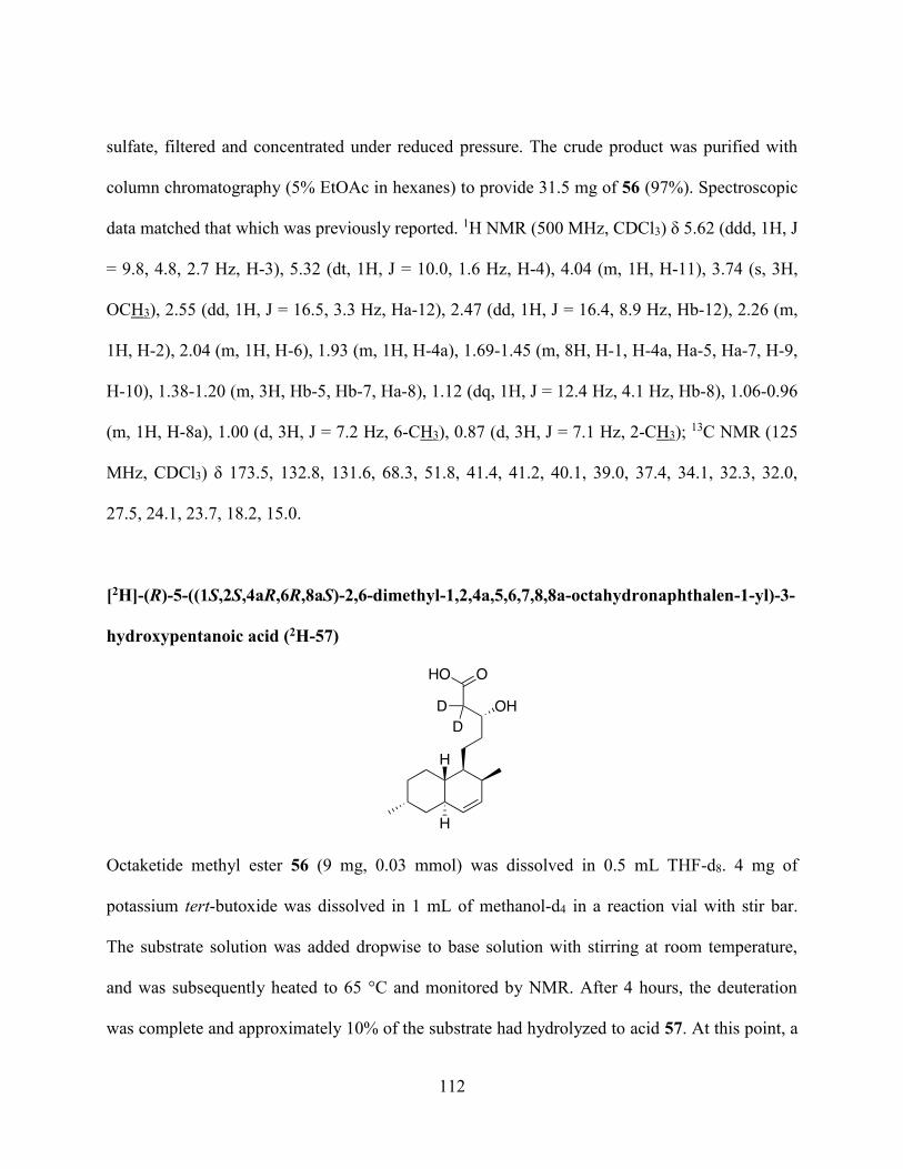
112
sulfate, filtered and concentrated under reduced pressure. The crude product was purified with
column chromatography (5% EtOAc in hexanes) to provide 31.5 mg of 56 (97%). Spectroscopic
data matched that which was previously reported. 1H NMR (500 MHz, CDCl3) δ 5.62 (ddd, 1H, J
= 9.8, 4.8, 2.7 Hz, H-3), 5.32 (dt, 1H, J = 10.0, 1.6 Hz, H-4), 4.04 (m, 1H, H-11), 3.74 (s, 3H,
OCH3), 2.55 (dd, 1H, J = 16.5, 3.3 Hz, Ha-12), 2.47 (dd, 1H, J = 16.4, 8.9 Hz, Hb-12), 2.26 (m,
1H, H-2), 2.04 (m, 1H, H-6), 1.93 (m, 1H, H-4a), 1.69-1.45 (m, 8H, H-1, H-4a, Ha-5, Ha-7, H-9,
H-10), 1.38-1.20 (m, 3H, Hb-5, Hb-7, Ha-8), 1.12 (dq, 1H, J = 12.4 Hz, 4.1 Hz, Hb-8), 1.06-0.96
(m, 1H, H-8a), 1.00 (d, 3H, J = 7.2 Hz, 6-CH3), 0.87 (d, 3H, J = 7.1 Hz, 2-CH3); 13C NMR (125
MHz, CDCl3) δ 173.5, 132.8, 131.6, 68.3, 51.8, 41.4, 41.2, 40.1, 39.0, 37.4, 34.1, 32.3, 32.0,
27.5, 24.1, 23.7, 18.2, 15.0.
[2H]-(R)-5-((1S,2S,4aR,6R,8aS)-2,6-dimethyl-1,2,4a,5,6,7,8,8a-octahydronaphthalen-1-yl)-3-
hydroxypentanoic acid (2H-57)
Octaketide methyl ester 56 (9 mg, 0.03 mmol) was dissolved in 0.5 mL THF-d8. 4 mg of
potassium tert-butoxide was dissolved in 1 mL of methanol-d4 in a reaction vial with stir bar.
The substrate solution was added dropwise to base solution with stirring at room temperature,
and was subsequently heated to 65 °C and monitored by NMR. After 4 hours, the deuteration
was complete and approximately 10% of the substrate had hydrolyzed to acid 57. At this point, a

113
few drops of D2O and an additional 2 mg of potassium tert-butoxide were added to complete the
saponification. After stirring at room temperature overnight, the saponification was complete,
and the reaction was acidified to pH 2 with 1 M HCl. The methanol-d4 and THF-d8 were then
removed by rotary evaporation. The concentrate was diluted with water and extracted three times
with CH2Cl2. The combined organic layers were washed with water and brine, and dried over
sodium sulfate. The solvent was removed under reduced pressure to provide 6.5 mg of acid 57 as
a white solid (75% yield). 1H NMR (500 MHz, CDCl3) δ 5.57 (ddd, 1H, J = 9.7, 4.8, 2.8 Hz, H-
3), 5.27 (dt, 1H, J = 10.0, 1.5 Hz, H-4), 4.02 (br s, 1H, OH), 2.22 (m, 1H, H-2), 2.00 (m, 1H, H-
6), 1.89 (m, 1H, H-4a), 1.65-1.42 (m, 7H, H-1, H-4a, Ha-7, H-9, H-10), 1.36-1.30 (m, 1H, Ha-5),
1.28-1.16 (m, 3H, Hb-5, Hb-7, Ha-8), 1.08 (dq, 1H, J = 12.4, 3.7 Hz, Hb-8), 0.97 (dq, 1H, J =
10.4, 3.4 Hz, H-8a), 0.96 (d, 3H, J = 7.2 Hz), 0.82 (d, 3H, J = 7.0 Hz); 13C NMR (125 MHz,
CDCl3) δ 178.9, 132.7, 131.6, 68.1, 41.3, 40.0, 38.9, 37.3, 34.0, 32.3, 32.3, 31.9, 27.4, 24.0, 23.6,
18.2, 14.9.
(E)-methyl 5-((1R,2R,3S,4S,4aS,6R,8aR)-3,4-dibromo-2,6-dimethyldecahydronaphthalen-1-
yl)pent-2-enoate (58)
Unsaturated octaketide 58 was prepared using the method of John Sorensen.64 In a round bottom
flask with stir bar under argon, a solution of 90.3 mg (0.2 mmol) of compound 55 was prepared

114
with 5 mL of CH2Cl2, and then cooled to 0 °C. To this was added 81 μL (3 eq, 0.6 mmol) of
triethylamine, then 30 μL (2 eq, 0.4 mmol) of methanesulfonyl chloride. The reaction was slowly
warmed to room temperature and stirred for 3 days. Following this, the reaction was quenched
by the addition of 10 mL 1 N HCl with stirring over 10 minutes. The mixture was diluted with 25
mL of CH2Cl2 and the layers were separated. The organic layer was washed with water, saturated
sodium bicarbonate, water and brine, then dried over sodium sulfate, filtered and concentrated
under reduced pressure. The product 58 (43 mg, 51% yield) could be separated from unreacted
starting material 55 (20 mg) by column chromatography (20% EtOAc in hexanes). Spectroscopic
data matched that which was previously reported. 1H NMR (500 MHz, CDCl3) δ 6.98 (dt, 1H, J
= 15.7, 7.2 Hz, H-11), 5.86 (d, 1H, J = 15.7 Hz, H-12), 4.97 (s, 1H, H-4), 4.49 (s, 1H, H-3), 3.75
(s, 3H, OCH3), 2.41 (m, 1H, H-2), 2.31-2.01 (m, 4H, Ha-5, H-6, H-10), 1.97 (tt, 1H, J = 11.0, 4.2
Hz, H-4a), 1.86-1.70 (m, 3H, H-1, Hb-5, Ha-9), 1.63-1.49 (m, 2H, H-8), 1.43 (dq, 1H, J = 10.7,
3.66 Hz, H-1), 1.30 (d, 3H, J = 7.7 Hz, 2-CH3), 1.26-1.16 (m, 3H, H-7, Hb-9), 1.05 (d, 3H, J =
7.3 Hz, 6-CH3); 13C NMR (125 MHz, CDCl3) δ 167.1, 149.0, 121.2, 60.2, 59.9, 51.5, 39.2, 38.5,
38.4, 34.9, 34.7, 31.2, 29.4, 26.9, 26.7, 23.6, 18.0, 17.7.
Methyl 3-((1R,2R,3S,4S,4aS,6R,8aR)-3,4-dibromo-2,6-dimethyldecahydronaphthalen-1-
yl)propanoate (59)

115
Heptaketide dibromide 59 was prepared using the method of John Sorensen.64 A three neck
round bottom flask was fitted with a glass stopped, O3 inlet, and CaCl2 drying tube outlet, and
loaded with a solution of 76 mg (0.17 mmol) of compound 58 in 20 mL CH2Cl2. 7.5 mL of a
methanolic 2.5 M NaOH solution was added, and the vessel was cooled to -78 °C. O3 was
vigourously bubbled through, and the reaction instantly turned orange. The stream of O3 was
continued under a persistent blue colour developed, at which point O2 was bubbled through for 5
min to purge the solution of excess O3. The reaction was warmed to room temperature, diluted
with 60 mL CHCl3 and acidified with 10 mL 2N HCl. The organic layer was separated and
washed with water, saturated sodium bicarbonate, water and brine. It was then dried over sodium
sulfate and concentrated under reduced pressure. The crude product was purified by column
chromatography (5% EtOAc in hexanes) to yield 35.5 mg of 59 (49%). Spectroscopic data
matched that which was previously reported. 1H NMR (600 MHz, CDCl3) δ 4.96 (t, 1H, J = 1.7
Hz, H-4), 4.48 (m, 1H, H-3), 3.69 (s, 3H, OCH3), 2.38 (m, 1H, H-2), 2.32 (ddd, 1H, J = 15.6,
10.3, 5.5 Hz, Ha-10), 2.22-2.13 (m, 2H, Hb-10 and H-4a), 2.11-2.02 (m, 2H, H-6 and H-1), 1.94
(m, 1H, Ha-5), 1.79 (m, 2H, H-8), 1.59-1.50 (m, 2H, H-7), 1.48-1.18 (m, 4H, H-8a, Hb-5, H-9),
1.29 (d, 3H, J = 7.7 Hz, 2-CH3), 1.04 (d, 3H, J = 7.3 Hz, 6-CH3); 13C NMR (150 MHz, CDCl3) δ
176.7, 62.7, 62.5, 56.1, 41.8, 41.3, 41.0, 37.5, 37.3, 34.2, 33.8, 29.6, 26.4, 26.3, 20.7, 20.3.

116
Methyl 3-((1S,2S,4aR,6R,8aS)-2,6-dimethyl-1,2,4a,5,6,7,8,8a-octahydronaphthalen-1-
yl)propanoate (60)
A reaction vial with stir bar was loaded with 35 mg (0.09 mmol) of heptaketide dibromide 59
dissolved in 2.5 mL freshly distilled CH2Cl2. 18 mg of zinc dust was added, along with 0.25 mL
of glacial acetic acid. The reaction was stirred overnight at room temperature, then filtered and
diluted with an additional 10 mL CH2Cl2. The solution was transferred to a separatory funnel to
be washed with saturated sodium bicarbonate, water and brine. The organic phase was dried over
sodium sulfate, filtered, and concentrated under reduced pressure. The crude product was
purified by column chromatography (5% EtOAc in hexanes) to yield 13 mg (62%) of
deprotected product 60. 1H NMR (600 MHz, CDCl3) δ 5.61 (ddd, 1H, J = 9.7, 4.8, 2.8 Hz, H-3),
5.31 (dt, 1H, J = 9.6, 1.6 Hz, H-4), 3.69 (s, 3H, OCH3), 2.41 (ddd, 1H, J = 15.5, 10.4, 5.2 Hz, Ha-
10), 2.28-2.18 (m, 2H, H-2 and Hb-10), 2.08-2.00 (m, 1H, H-4a), 1.99-1.89 (m, 2H, H-6 and Ha-
9), 1.66-1.45 (m, 4H, H-1, Hb-9, H-7), 1.39 (m, 1H, H-8a), 1.33-1.25 (m, 2H, H-5), 1.14 (dq,
1H, J = 12.5, 3.3 Hz, Ha-8), 1.06-0.96 (m, 1H, Hb-8) 1.00 (d, 3H, J = 7.2 Hz, 6-CH3), 0.86 (d,
3H, J = 7.0 Hz, 2-CH3); 13C NMR (150 MHz, CDCl3) δ 177.2, 135.2, 134.3, 56.09, 43.8, 42.6,
41.6, 39.9, 34.9, 34.7, 34.5, 30.1, 26.7, 26.3, 20.9, 17.6.

117
[2H]-3-((1S,2S,4aR,6R,8aS)-2,6-dimethyl-1,2,4a,5,6,7,8,8a-octahydronaphthalen-1-
yl)propanoic acid (2H-61)
Heptaketide methyl ester 60 (12 mg, 0.05 mmol) was dissolved in 0.5 mL THF-d8. 4 mg of
potassium tert-butoxide was dissolved in 1 mL of methanol-d4 in a reaction vial with stir bar.
The substrate solution was added dropwise to base solution with stirring at room temperature,
and was subsequently heated to 50 °C and monitored by NMR. After 4 hours, the deuteration
was complete. At this point, a few drops of D2O and an additional 2 mg of potassium tert-
butoxide were added to complete the saponification to acid 61. After stirring at room temperature
overnight, the saponification was complete, and the reaction was acidified to pH 2 with 1 M HCl.
The methanol-d4 and THF-d8 were then removed by rotary evaporation. The concentrate was
diluted with water and extracted three times with CH2Cl2. The combined organic layers were
washed with water and brine, and dried over sodium sulfate. The solvent was removed under
reduced pressure to provide 10 mg of acid 57 as a white solid (90% yield). IR (CHCl3, cast film)
3020, 2953, 2915, 2860, 2661, 1692, 1294; 1H NMR (700 MHz, CDCl3) δ 5.57 (ddd, 1H, J = 9.7,
4.8, 2.7 Hz, H-3), 5.28 (d, 1H, J = 9.6 Hz, H-4), 2.22 (m, 1H, H-2), 2.00 (m, 1H, H-4a), 1.96-
1.86 (m, 2H, H-6 and Ha-9), 1.62-1.41 (m, 5H, H-1, H-7, H-8, Hb-9), 1.29-1.21 (m, 2H, H-5),
1.12 (dq, 1H, J = 12.8, 4.1 Hz, Ha-8), 1.02-0.95 (m, 1H, Hb-8), 0.97 (d, 3H, J = 7.2 Hz, 6-CH3),
0.83 (d, 3H, J = 7.0 Hz, 2-CH3); 13C NMR (175 MHz, CDCl3) δ 178.5, 132.4, 131.6, 53.4, 40.9,

118
31.8, 39.9, 38.9, 37.2, 32.3, 30.9, 27.4, 23.5, 18.2, 14.9; 𝛼D25 = 74.26 (c = 0.22 g/100 mL,
CHCl3);
(1R,2S,4aR,6R,8aR)-S-acetamidomethyl 2,6-dimethyl-1,2,4a,5,6,7,8,8a-
octahydronaphthalene-1-carbothioate) (65)
and (1R,2R,4aS,6R,8aR)-S-acetamidomethyl 2,6-dimethyl-1,2,4a,5,6,7,8,8a-
octahydronaphthalene-1-carbothioate (66)
5 mg of triene 41 was dissolved in 0.8 mL 1:1 tetrahydrofuran and H2O, and allowed to react for
12 hours. The tetrahydrofuran was removed under reduced pressure, and the cloudy aqueous
solution that resulted was extracted with 3x CHCl3. The combined organic extracts were dried
over magnesium sulfate, and the solvent was removed under reduced pressure. The residue was
dissolved in CDCl3 to acquire the HSQC and CSSF-TOCSY spectra shown in Figures 37 and 38,
which were interpreted in Chapter 3, with reference to the reported NMR data. 49

119
(R,2E,4E,10E,12E)-S-2-acetamidoethyl 8-methyltetradeca-2,4,10,12-tetraenethioate (89)
HWE reaction with aldehyde 95:
Lithium bromide (oven dried, 27 mg, 4.0 eq) was added to a flame-dried 10 mL round bottom
with stir bar. Horner-Wadsworth-Emmons reagent 32 (3.6 eq, 56 mg) was added in 2 mL THF,
and the mixture was stirred for 10 minutes. Triethylamine (44 μL, 6.0 eq) was then added, and 10
minutes was allotted for deprotonation. Next, aldehyde 95 (10 mg, 0.05 mmol) was added in a
tetrahydrofuran solution, and the mixture was allowed to react overnight. The solvent was
removed in vacuo and the crude residue was applied directly to a silica column for purification
(gradient of 25% EtOAc in hexanes to neat EtOAc), to yield 2 mg of tetraene 89 (11%).
Coupling to acid 97:
In a flame-dried flask with stir bar, 31 mg (0.13 mmol) of acid 97 was dissolved in 10 mL
freshly distilled CH2Cl2. 56 μL (4 eq, 0.53 mmol) of N-acetylcysteamine was added, followed by
54 mg (2 eq, 0.26 mmol) of dicyclohexylcarbodiimide. The reaction was stirred at room
temperature, and monitored by TLC. After two hours, the reaction was complete, and the solvent
was removed in vacuo. The crude mixture was resuspended in ice-cold 1:1 EtOAc:Et2O, and
filtered through a pipette packed with a small amount of cotton and sand. The filtrate, following
concentration under reduced pressure, was purified using column chromatography (gradient of
50% EtOAc in pentane to neat EtOAc), to yield 27 mg (62%). IR (CHCl3, cast film) 3287 (br),
3016, 2954, 2927, 2853, 1655, 1637, 1554, 989; 1H NMR (700 MHz, CDCl3) δ 7.19 (dd, 1H, J =
15.2, 10.8 Hz, H-3), 6.21 (m, 1H, H-5), 6.13 (m, 1H, H-4), 6.08 (d, 1H, J = 15.2 Hz, H-2), 6.04-
5.95 (m, 2H, H-12 and H-11), 5.91 (br s, 1H, NH), 5.58 (m, 1H, H-13), 5.49 (m, 1H, H-10), 3.45

120
(q, 2H, J = 6.7 Hz, H-2’), 3.09 (t, 2H, J = 6.3 Hz, H-1’), 2.22 (m, 1H, Ha-6), 2.16 (m, 1H, Hb-6).
2.04 (m, 1H, Ha-9), 1.95 (s, 3H, COCH3), 1.91 (m, 1H, Hb-9), 1.72 (d, 3H, J = 6.6 Hz, H-14),
1.55-1.41 (m, 2H, H-8 and Ha-7), 1.28 (m, 1H, Hb-7), 0.87 (d, 3H, J = 6.7 Hz, 8-CH3); 13C NMR
(175 MHz, CDCl3) δ 193.1 (C-1), 173.0 (C-3’), 150.0 (C-5), 144.7 (C-3), 134.5 (C-12), 134.2
(C-11), 132.6 (C-10), 130.7 (C-4), 129.8 (C-13), 128.5 (C-2), 42.6 (C-9), 42.5 (C-2’), 37.9 (C-7),
35.5 (C-8), 33.5 (C-6), 31.0 (C-1’), 25.9 (C-4’), 22.0 (8-CH3), 20.7 (C-14); 𝛼D25 = -12.32 (c =
0.49 g/100 mL, CHCl3); HRMS (ES) m/z calculated for C19H30NO2S 336.1992, found 336.2003
[M+H]+.
(R,8E,10E)-6-methyldodeca-8,10-dien-1-ol (91)
Procedure using DIBAL-H:
10 mg (0.032 mmol) of triene 41 was dissolved in 0.5 mL freshly distilled CH2Cl2 and cooled to
-78 °C, after which 70 μL (2.1 eq, 0.07 mmol) of a 1M solution of diisobutylaluminum hydride
in CH2Cl2 was added dropwise. After two hours, TLC analysis showed a significant amount of
triene remained, so an addition 2 equivalents of diisobutylaluminum hydride were added. No
change was observed after an additional hour, at which point the reaction was warmed to room
temperature. The reaction was quenched slowly with methanol (0.5 mL), then diluted with 10
mL diethyl ether. The mixture was washed with saturated aqueous potassium sodium tartrate
(4x10 mL), then water (10 mL) and brine (10 mL). The ether layer was dried over MgSO4 and
filtered, and the solvent of the filtrate was removed in vacuo. Purification by column

121
chromatography (50% EtOAc in pentane) produced an inseparable mixture of undesired
saturated alcohol 91 and desired unsaturated alcohol 93.
Luche reduction method:
3.0 mg (0.01 mmol) of triene 41 was dissolved in 0.3 mL of methanol and 11 mg (3 eq, 0.03
mmol) of CeCl37H2O was added. The mixture was stirred for 10 min at room temperature, and
then cooled to -78 °C. A methanolic solution of sodium borohydride (1 mg in 0.5 mL methanol,
3 eq, 0.03 mmol), was prepared, and then added dropwise to the reaction. After one hour of
reaction, starting material remained by TLC, and thus another equivalent of sodium borohydride
was added. After an additional hour, the reaction was quenched with 0.5 mL water, and the
methanol was removed in vacuo. The aqueous solution was extracted three times with 0.5 mL
CHCl3, and the combined organic layers were washed with water and brine, then dried over
sodium sulfate and concentrated under reduced pressure. Purification by column chromatography
(50% EtOAc in pentane) produced an inseparable mixture of undesired saturated alcohol 91 and
desired unsaturated alcohol 93. NMR data for alcohol 91: 13C NMR (175 MHz, CDCl3) δ 131.7,
131.43, 130.6, 126.7, 63.1, 40.1, 36.5, 33.2, 32.8, 26.7, 25.9, 19.5, 18.0.
(R,2E,8E,10E)-ethyl 6-methyldodeca-2,8,10-trienoate (92)
125 mg of sodium metal was reacted with 50 mL anhydrous EtOH (prepared over molecular
sieves), to make NaOEt solution of approximately 0.1 M. 3 mL of this solution was added to 74
mg (0.24 mmol) of triene 41. After 30 minutes of stirring at room temperature, starting material
was consumed, as per the TLC. The reaction was neutralized with 0.1 M HCl, then extracted

122
three times with CH2Cl2. The combined organic layers were washed with brine, dried over
sodium sulfate and concentrated under reduced pressure. The residue was purified by column
chromatography (10% Et2O in hexanes), to yield 18 mg of 92 (32%). 1H NMR (700 MHz,
CDCl3) δ 6.94, (dt, 1H, J = 15.7, 6.9 Hz, H-3). 6.05-5.94 (m, 2H, H-10 and H-9), 5.80 (dt, 1H, J
= 15.7, 1.6 Hz, H-2), 5.57 (dd, 1H, J = 13.8, 6.5 Hz, H-11), 5.49 (dt, 1H, J = 14.6, 7.4 Hz, H-8),
4.17 (q, 2H, J = 7.2 Hz, H-1’), 2.22 (m, 1H, Ha-4), 2.16 (m, 1H, Hb-4), 2.04 (m, 1H, Ha-7), 1.91
(m, 1H, Hb-7), 1.72 (d, 3H, J = 6.6 Hz. H-12), 1.56-1.43 (m, 2H, Ha-5 and H-6), 1.26-1.20 (m,
1H, H-5), 1.29 (t, 3H, J = 6.6 Hz, H-2’), 0.90 (d, 3H, J = 6.3 Hz, 6-CH3); 13C NMR (175 MHz,
CDCl3) δ 166. (C-1), 149.4 (C-3), 131.8 (C-9), 131.5 (C-10), 129.8 (C-8), 127.1 (C-11), 123.9
(C-2), 60.1 (C-1’), 39.9 (C-7), 34.6 (C-5), 32.8 (C-6), 28.9 (C-4), 19.3 (6-CH3), 17.9 (C-12), 14.3
(C-2’).
(R,2E,8E,10E)-6-methyldodeca-2,8,10-trien-1-ol (93)
Ester 92 (0.08 mmol, 18 mg) was dissolved in 2 mL freshly distilled CH2Cl2 and cooled to -78
°C. Dropwise, 240 μL of a 1 M diisobutylaluminum hydride in CH2Cl2 (3 eq, 0.24 mmol) was
added. The reaction was stirred for 1 hour, at which point the reaction was quenched slowly with
methanol (0.5 mL) then diluted with 10 mL diethyl ether. The mixture was washed with
saturated aqueous potassium sodium tartrate (4 x 10 mL), then water (10 mL) and brine (10 mL).
The ether layer was dried over MgSO4 and filtered, and the solvent of the filtrate was removed in
vacuo. Purification by column chromatography (50% EtOAc in pentane) produced desired
unsaturated alcohol 93 (10 mg, 64% yield). 1H NMR (500 MHz, CDCl3) δ 6.04-5.90 (2H, m, H-

123
10 and H-9), 5.70-5.60 (m, 2H, H-3 and H-2), 5.57 (m, 1H, H-11), 5.50 (dt, 1H, J = 14.6, 7.4 Hz,
H-8), 4.07 (t, 2H, J = 5.4 Hz, H-1) 2.11-1.98 (m, 3H, H-4, Ha-7), 1.89 (m, 1H, Hb-7), 1.72 (d,
3H, J = 6.5 Hz, H-12), 1.50 (m, 1H, H-6), 1.41 (m, 1H, Ha-5), 1.26 (m, 1H, Hb-6), 0.86 (d, J =
6.7 Hz, 6-CH3); 13C NMR (175 MHz, CDCl3) δ 133.5, 131.7, 131.2, 130.2, 128.7, 127.0, 63.9,
40.0, 36.0, 27.7, 29.8, 19.4, 17.9.
(R,2E,8E,10E)-6-methyldodeca-2,8,10-trienal (94)
A 10 mL flame dried flask with stir bar was loaded with 5 mg (1.1 eq) sodium bicarbonate, 24
mg (1.1 eq) DMP and 1 mL CH2Cl2. 10 mg of alcohol 93 was dissolved in 1 mL freshly distilled
CH2Cl2 and the solution was added to the DMP solution dropwise. The reaction was complete, as
per the TLC, in 30 minutes, at which point the reaction was quenched by the addition of 2 mL
concentrated aqueous NaHCO3/NaS2O3, and stirred for 15 minutes. The organic layer was then
separated, and washed with saturated sodium bicarbonate, water and brine, dried over
magnesium sulfate and concentrated under reduced pressure. Aldehyde 94 did not need further
purification (quantitative yield). 1H NMR (700 MHz, CDCl3) δ 9.49 (d, 1H, J = 7.9 Hz, H-1),
6.83 (dt, 1H, J = 15.9, 6.8 Hz, H-3), 6.11 (ddt, 1H, J = 15.6, 7.8, 1.5 Hz, H-10), 6.04-5.94 (m,
2H, H-2 and H-9), 5.58 (m, 1H, H-11), 5.49 (dt, 1H, J = 14.2, 7.4 Hz, H-8), 2.36 (m, 1H, Ha-4),
2.30 (m, 1H, Hb-4), 2.05 (m, 1H, Ha-7), 1.94 (m, 1H, Hb-7), 1.72 (d, 3H, J = 6.6 Hz, H-12),
1.60-1.49 (m, 2H, Ha-5 and H-6), 1.31 (m, 1H, Hb-5), 0.89 (d, 3H, J = 6.5 Hz, 6-CH3); 13C NMR
(175 MHz, CDCl3) δ 194.1 (C-1), 159.0 (C-3), 132.9 (C-10), 132.0 (C-9), 131.5 (C-2), 129.6 (C-
8), 127.3 (C-11), 39.8 (C-7), 34.4 (C-5), 32.8 (C-6), 30.4 (C-4), 19.3 (6-CH3), 18.0 (C-12).
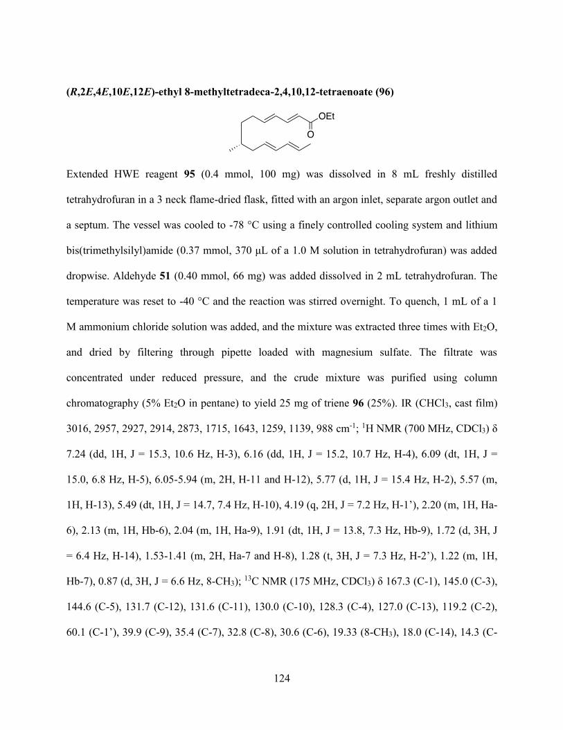
124
(R,2E,4E,10E,12E)-ethyl 8-methyltetradeca-2,4,10,12-tetraenoate (96)
Extended HWE reagent 95 (0.4 mmol, 100 mg) was dissolved in 8 mL freshly distilled
tetrahydrofuran in a 3 neck flame-dried flask, fitted with an argon inlet, separate argon outlet and
a septum. The vessel was cooled to -78 °C using a finely controlled cooling system and lithium
bis(trimethylsilyl)amide (0.37 mmol, 370 μL of a 1.0 M solution in tetrahydrofuran) was added
dropwise. Aldehyde 51 (0.40 mmol, 66 mg) was added dissolved in 2 mL tetrahydrofuran. The
temperature was reset to -40 °C and the reaction was stirred overnight. To quench, 1 mL of a 1
M ammonium chloride solution was added, and the mixture was extracted three times with Et2O,
and dried by filtering through pipette loaded with magnesium sulfate. The filtrate was
concentrated under reduced pressure, and the crude mixture was purified using column
chromatography (5% Et2O in pentane) to yield 25 mg of triene 96 (25%). IR (CHCl3, cast film)
3016, 2957, 2927, 2914, 2873, 1715, 1643, 1259, 1139, 988 cm-1; 1H NMR (700 MHz, CDCl3) δ
7.24 (dd, 1H, J = 15.3, 10.6 Hz, H-3), 6.16 (dd, 1H, J = 15.2, 10.7 Hz, H-4), 6.09 (dt, 1H, J =
15.0, 6.8 Hz, H-5), 6.05-5.94 (m, 2H, H-11 and H-12), 5.77 (d, 1H, J = 15.4 Hz, H-2), 5.57 (m,
1H, H-13), 5.49 (dt, 1H, J = 14.7, 7.4 Hz, H-10), 4.19 (q, 2H, J = 7.2 Hz, H-1’), 2.20 (m, 1H, Ha-
6), 2.13 (m, 1H, Hb-6), 2.04 (m, 1H, Ha-9), 1.91 (dt, 1H, J = 13.8, 7.3 Hz, Hb-9), 1.72 (d, 3H, J
= 6.4 Hz, H-14), 1.53-1.41 (m, 2H, Ha-7 and H-8), 1.28 (t, 3H, J = 7.3 Hz, H-2’), 1.22 (m, 1H,
Hb-7), 0.87 (d, 3H, J = 6.6 Hz, 8-CH3); 13C NMR (175 MHz, CDCl3) δ 167.3 (C-1), 145.0 (C-3),
144.6 (C-5), 131.7 (C-12), 131.6 (C-11), 130.0 (C-10), 128.3 (C-4), 127.0 (C-13), 119.2 (C-2),
60.1 (C-1’), 39.9 (C-9), 35.4 (C-7), 32.8 (C-8), 30.6 (C-6), 19.33 (8-CH3), 18.0 (C-14), 14.3 (C-

125
2’); 𝛼D25 = -15.84 (c = 0.50 g/100 mL, CHCl3); HRMS (ES) m/z calculated for C17H26NaO2
285.1825, found 285.1822 [M+Na]+.
(R,2E,4E,10E,12E)-8-methyltetradeca-2,4,10,12-tetraenoic acid (97)
A portion of ethyl ester 96 (20 mg, 0.07 mmol) was mixed with 250 μL of 2 M KOH.
Tetrahydrofuran was added dropwise until a clear solution was formed. The reaction was then
warmed to 60 °C and stirred, unsealed, while reaction progression was monitored by TLC. After
completed reaction (48 hours), the solution was neutralized with 0.5 mL of 1 M HCl, and
extracted with Et2O. The combined organic layers were dried by filtering through a pipette filled
with magnesium sulfate, and the filtrate was concentrated under reduced pressure to provide 14
mg of acid 97 (80% yield). 1H NMR (700 MHz, CDCl3) δ 7.24 (dd, 1H, J = 15.3, 10.5 Hz, H-3),
6.16 (dd, 1H, J = 15.4, 10.9 Hz, H-4), 6.09 (dt, 1H, J = 15.3, 6.8 Hz, H-5), 6.05-5.94 (m, 2H, H-
11 and H-12), 5.77 (d, 1H, J = 15.4 Hz. H-2), 5.57 (m, 1H, H-13), 5.49 (dt, 1H, J = 14.6, 7.5 Hz.
H-10), 2.19 (m, 1H, Ha-6), 2.13 (m, 1H, Hb-6), 2.04 (m, 1H, Ha-9), 1.91 (dt, 1H, J = 13.9, 7.2
Hz, Hb-9), 1.72 (d, 3H, J = 6.6 Hz, H-14), 1.50 (m, 1H, H-8), 1.46 (m, 1H, Ha-7), 1.24 (m, 1H,
Hb-7), 0.87 (d, 3H, J = 6.7 Hz, 8-CH3); 13C NMR (175 MHz, CDCl3) δ 172.5 (C-1), 147.5 (C-3),
146.2 (C-5), 131.8 (C-12), 131.6 (C-11), 129.9 (C-10), 128.2 (C-4), 127.0 (C-2), 39.9 (C-9), 35.2
(C-7), 32.8 (C-8), 30.7 (C-6), 19.3 (8-CH3), 18.0 (C-14); HRMS (ES) m/z calculated for
C15H21O2 233.1547, found 233.1549 [M-H]+.

126
Biological Methods
General Techniques for DNA Manipulation
E. coli DH5α (Invitrogen) was used for cloning following standard techniques. DNA restriction
enzymes (Thermo Scientific) were used as directed. PCR was performed with a Platinum Taq
High Fidelity DNA polymerase (Invitrogen).
Media Recipes
YPD media is made from 20 g/L bactopeptone, 10 g/L yeast extract, both from Becton,
Dickonson and Company, dissolved in deionized water and sterilized by autoclaving. LB
medium is made from 10 g/L bacto-tryptone, 5 g/L yeast extract, both from Becton, Dickonson
and Company, and 10 g/L NaCl, dissolved in deionized water and sterilized by autoclaving.
Aspergillus nidulans growth media is composed of 20 g of glucose, 20 g of yeast extract, 1 g of
peptone and 1 mg of para-aminobenzoic acid in 1 L of distilled, deionized water.
Aspergillus nidulans production media is prepared by dissolving in 1 L of distilled deionized
water 1 mL trace elements solution (1.0 g FeSO47H2O, 8.8 g ZnSO47H2O, 0.4 g CuSO45
H2O, 0.15 g MnSO44H2O, 0.1 g Na2B7O710H2O, 0.05 g (NH4)6Mo7O244H2O, 0.5 mL
concentrated hydrochloric acid dissolved in 1 L H2O), 100 mL of 10X AMM salts (60 g NaNO3,
5.2 g KCl, 15.2 g KH2PO4 dissolved in 1 L of H2O and adjusted to pH 6.5), 1 mL of 1 mg/mL
para-aminobenzoic acid solution, and 0.9 mL cyclopentanone and autoclaving. After cooling to
room temperature, 2.5 mL of filter sterilized 20 % MgSO47H2O and 25 mL filter sterilized 40%
lactose was added.

127
Procedures for Heterologous Expression of Proteins
Expression of LovB and LovBΔCON in S. cerevisiae strain BJ5464-NpgA
Two strains of S. cerevisiae BJ5464-NpgA, one transformed with a plasmid containing the lovB
gene, and one transformed with a plasmid containing the lovBΔCON gene, were received from
the Yi Tang group at UCLA.25 Each protein was expressed using the same method, as follows. 1
L of YPD media with 1% dextrose was inoculated with the yeast cells, and incubated at 30 °C at
200 rpm for 72 hours. The cells were harvested by centrifugation (30 min, 4 °C, 8000 rpm on
JA-10 rotor), resuspended in 30 mL lysis buffer (50 mM sodium phosphate, 0.5 mM NaCl, 10
mM imidazole), and cells were lysed using sonication on ice. Cellular debris was removed by
centrifugation (15000 g, 1 hour, 4 °C). The supernatant was mixed with 2 mL Ni-NTA resin
(QIAGEN), and shaken for 2 hours at 4 °C. Using a gravity flow column, the protein was
purified using increasing amounts of imidazole in Buffer A (50 mM Tris, pH 7.9, 2 mM EDTA,
2 mM DTT). The fractions were analysed by SPS-PAGE (4-20% acrylamide, BIO-RAD), and
fractions containing pure LovB or pure LovBΔCON were combined, concentrated, and buffer
exchanged into Buffer A + 10% glycerol, then flash frozen by submerging microcentrifuge tube
in dry ice/acetone, and stored at -80 °C.
Expression of CON in E. coli BL21(DE3)
Plasmid pSMa61 (con cloned into pET28a) was obtained from the Yi Tang group.25 The plasmid
was transformed into BL21(DE3) using the heat shock method. Expression was performed as
follows: 1L of LB medium + 30 μg/mL kanamycin (Fisher Scientific) was inoculated with cells
from an overnight culture, to an initial OD600 of approximately 0.1. The cells were grown at 37
°C and 200 rpm until an OD600 of 0.4, at which point the cultures were placed in an ice-water

128
bath for 10 minutes. Expression was induced with 0.1 mM isopropyl thio-β-D-galactoside
(IPTG, Fisher Scientific), and incubation was continued at 16 °C for 16 hours. The cells were
then harvested by centrifugation (30 min, 4 °C, 8000 rpm on JA-10 rotor), resuspended in 20 mL
lysis buffer. Lysozyme (to 1 mg/mL) and DNaseI (to 5 ug/L) were added, and the cells were
lysed using a cell disruptor. Cellular debris was removed by centrifugation (30000 g, 30 min, 4
°C), and the supernatant was incubated with 1 mL Ni-NTA resin (QIAGEN) at 4 °C for 1 hour.
Using a gravity flow column, the CON domain was purified using increasing amounts of
imidazole in Buffer A (50 mM Tris, pH 7.9, 2 mM EDTA, 2 mM DTT). The fractions were
analysed by SPS-PAGE (4-20% acrylamide, BIO-RAD), and fractions containing pure CON
were combined, concentrated, and buffer exchanged into Buffer A + 10% glycerol, flash frozen
in dry ice/acetone, and stored at -80 °C.
Expression of DH in E. coli BL21(DE3)
The dehydratase gene was purchased as a synthetic gene product from Biobasic Inc., with codon
usage optimized for expression in E. coli. The gene was subcloned into the pET28a vector at the
NdeI and EcoRI restriction sites, to such that expression would produce the following construct,
with an N-terminal His6-tag:
MGSSHHHHHHSSGLVPRGSHMHLLLGKLSEYSTPLSFQWLNFVRPRDIEWLDGHALQG
QTVFPAAGYIVMAMEAALMIAGTHAKQVKLLEILDMSIDKAVIFDDEDSLVELNLTADV
SRNAGEAGSMTISFKIDSCLSKEGNLSLSAKGQLALTIEDVNPRTTSASDQHHLPPPEEEH
PHMNRVNINAFYHELGLMGYNYSKDFRRLHNMQRADLRASGTLDFIPLMDEGNGCPLL
LHPASLDVAFQTVIGAYSSPGDRRLRCLYVPTHVDRITLVPSLCLATAESGCEKVAFNTI
NTYDKGDYLSGDIVVFDAEQTTLFQVENITFKPFSPPDAST

129
Expression and purification were performed using the same method that was used for the CON
domain, as above.
Cloning and Expression of Hpm8-ACP1 in E. coli BL21(DE3)
The first Hpm8-ACP construct, Hpm8-ACP1, was cloned as a standalone protein with the primer
pair show in Table 2 (Chapter 4). A plasmid containing the entire Hpm8 gene, obtained from Yi
Tang,28 was used as the template and the PCR product was ligated into a PCR blunt vector for
amplification. The amplified plasmid was digested using NdeI and EcoRI to obtain the Hpm8-
ACP1 insert with sticky ends, which was ligated into pET28a to yield the expression plasmid.
This plasmid was used to transform BL21(DE3) cells for expression. Hpm8-ACP1 was
expressed and purified using the same method as for the CON domain, as above.
Cloning and Expression of Hpm8-ACP2 in E. coli BL21(DE3)
The gene for Hpm-ACP2 was synthesized and subcloned into pET28a by Biobasic Inc.
(sequence in Chapter 4), and the plasmid was transformed into BL21(DE3) for expression. The
protein was expressed and purified using the same method as for the CON domain, except that
expression was performed at 30 °C for 6 hours. The His6 tag was removed using the Thrombin
Cleavage Capture kit (Novagen).
Cloning and Expression of LovB-ACP2 in E. coli
The gene for Hpm-ACP2 was synthesized and subcloned into pET28a by Biobasic Inc.
(sequence in Chapter 4), and the plasmid was transformed into BL21(DE3) for expression. The
protein was expressed and purified using the same method as for the CON domain, except that

130
expression was performed at 30 °C for 6 hours. The His6 tag was removed using the Thrombin
Cleavage Capture kit (Novagen).
Expression of NpgA
An expression plasmid containing the npgA gene was obtained from Shiou-Chuan Tsai, and was
transformed into BL21(DE3) for expression. The protein was expressed using the same method
as for the CON domain (see above), except that 1 mM IPTG was used for induction, and the
expression was performed at 20 °C for 16 hours. The construct contained a Strep-tag purification
tag, and thus the protein was purified using Strep-trap HP system (GE Healthcare Life Sciences).
Phosphopantetheinylation Procedure
Phosphopantetheinylation of LovB-ACP2
The following reactions were set up in 0.3 mL Eppendorf tubes:
Volume added Reagent Stock concentration Final concentration
20 μL LovB-ACP2 130 μM 26 μM
2 μL Coenzyme A 200 mM 4 mM
0.5 μL MgCl2 2 M 10 mM
0.5 μL DTT 1 M 5 mM
0.5 μL Sfp or NpgA 4 mM 20 μM
80 μL Sterile water
The reactions were left for 16 hours at room temperature, at which point MALDI-MS analysis
was performed, to provide the spectra shown in Figure 54, demonstrating the superiority of the
NpgA for phosphopantetheinylation of LovB-ACP2.

131
REFERENCES
1. Weissman, K. J.; Leadlay, P. F. Combinatorial biosynthesis of reduced polyketides. Nat. Rev.
Microbiol. 2005, 3, 925-936.
2. Birch, A. J.; Massywestropp, R. A.; Moye, C. J. Studies in Relation to Biosynthesis .7. 2-Hydroxy-6-
Methylbenzoic Acid in Penicillium Griseofulvum Dierckx. Aust. J. Chem. 1955, 8, 539-544.
3. Staunton, J.; Weissman, K. J. Polyketide biosynthesis: a millennium review. Nat. Prod. Rep. 2001, 18,
380-416.
4. Cox, R. J.; Simpson, T. J. 1.09 - Fungal Type I Polyketides. In Comprehensive Natural Products II;
Liu, H., Mander, L., Eds.; Elsevier: Oxford, 2010; pp 347-383.
5. Dewick, P. M. Medicinal natural products : a biosynthetic approach; Wiley: Chichester, U.K., 2009.
6. Smith, S.; Tsai, S. The type I fatty acid and polyketide synthases: a tale of two megasynthases. Nat.
Prod. Rep. 2007, 24, 1041-1072.
7. Shen, B. Polyketide biosynthesis beyond the type I, II and III polyketide synthase paradigms. Curr.
Opin. Chem. Biol. 2003, 7, 285-295.
8. Cortes, J.; Haydock, S. F.; Roberts, G. A.; Bevitt, D. J.; Leanly, P. F. An Unusually Large
Multifunctional Polypeptide in the Erythromycin-Producing Polyketide Synthase of
Saccharopolyspora-Erythraea. Nature 1990, 348, 176-178.
9. Donadio, S.; Staver, M. J.; McAlpine, J. B.; Swanson, S. J.; Katz, L. Modular Organization of Genes
Required for Complex Polyketide Biosynthesis. Science 1991, 252, 675-679.
10. Katz, L. The Debs Paradigm for Type I Modular Polyketide Synthases and Beyond. Methods
Enzymol. 2009, 459, 113-142.
11. Chen, H.; Du, L. Iterative polyketide biosynthesis by modular polyketide synthases in bacteria. Appl.
Microbiol. Biotechnol. 2016, 100, 541-557.
12. Keatinge-Clay, A. T. A tylosin ketoreductase reveals how chirality is determined in polyketides.
Chem. Biol. 2007, 14, 898-908.
13. Weissman, K. J. Genetic engineering of modular PKSs: from combinatorial biosynthesis to synthetic
biology. Nat. Prod. Rep. 2016, 33, 203-230.
14. Zhou, H.; Gao, Z.; Qiao, K.; Wang, J.; Vederas, J. C.; Tang, Y. A fungal ketoreductase domain that
displays substrate-dependent stereospecificity. Nat. Chem. Biol. 2012, 8, 331-333.
15. Cacho, R. A.; Thuss, J.; Xu, W.; Sanichar, R.; Gao, Z.; Nguyen, A.; Vederas, J. C.; Tang, Y.
Understanding Programming of Fungal Iterative Polyketide Synthases: The Biochemical Basis for
Regioselectivity by the Methyltransferase Domain in the Lovastatin Megasynthase. J. Am. Chem.
Soc. 2015, 137, 15688-15691.

132
16. Chooi, Y.; Tang, Y. Navigating the Fungal Polyketide Chemical Space: From Genes to Molecules. J.
Org. Chem. 2012, 77, 9933-9953.
17. Crawford, J. M.; Thomas, P. M.; Scheerer, J. R.; Vagstad, A. L.; Kelleher, N. L.; Townsend, C. A.
Deconstruction of iterative multidomain polyketide synthase function. Science 2008, 320, 243-246.
18. Ray, L.; Moore, B. S. Recent advances in the biosynthesis of unusual polyketide synthase substrates.
Nat. Prod. Rep. 2016, 33, 150-161.
19. Crawford, J. M.; Korman, T. P.; Labonte, J. W.; Vagstad, A. L.; Hill, E. A.; Kamari-Bidkorpeh, O.;
Tsai, S.; Townsend, C. A. Structural basis for biosynthetic programming of fungal aromatic
polyketide cyclization. Nature 2009, 461, U243.
20. Dimroth, P.; Walter, H.; Lynen, F. Biosynthesis of 6-Methylsalicylic Acid. Eur. J. Biochem. 1970, 13,
98-110.
21. Spencer, J. B.; Jordan, P. M. Purification and Properties of 6-Methylsalicylic Acid Synthase from
Penicillium-Patulum. Biochem. J. 1992, 288, 839-846.
22. Parascandolo, J. S.; Havemann, J.; Potter, H. K.; Huang, F.; Riva, E.; Connolly, J.; Wilkening, I.;
Song, L.; Leadlay, P. F.; Tosin, M. Insights into 6-Methylsalicylic Acid Bio-assembly by Using
Chemical Probes. Angew. Chem. Int. Ed. Engl. 2016, 55, 3463-3467.
23. Campbell, C. D.; Vederas, J. C. Biosynthesis of Lovastatin and Related Metabolites Formed by
Fungal Iterative PKS Enzymes. Biopolymers 2010, 93, 755-763.
24. Kennedy, J.; Auclair, K.; Kendrew, S. G.; Park, C.; Vederas, J. C.; Hutchinson, C. R. Modulation of
polyketide synthase activity by accessory proteins during lovastatin biosynthesis. Science 1999, 284,
1368-1372.
25. Ma, S. M.; Li, J. W. -.; Choi, J. W.; Zhou, H.; Lee, K. K. M.; Moorthie, V. A.; Xie, X.; Kealey, J. T.;
Da Silva, N. A.; Vederas, J. C.; Tang, Y. Complete Reconstitution of a Highly Reducing Iterative
Polyketide Synthase. Science 2009, 326, 589-592.
26. Hendrickson, L.; Davis, C. R.; Roach, C.; Nguyen, D. K.; Aldrich, T.; McAda, P. C.; Reeves, C. D.
Lavastatin biosynthesis in Aspergillus terreus: characterization of blocked mutants, enzyme
activities and a multifunctional polyketide synthase gene. Chem. Biol. 1999, 6, 429-439.
27. Xu, W.; Chooi, Y.; Choi, J. W.; Li, S.; Vederas, J. C.; Da Silva, N. A.; Tang, Y. LovG: The
Thioesterase Required for DihydromonacolinL Release and Lovastatin Nonaketide Synthase
Turnover in Lovastatin Biosynthesis. Angew. Chem. Int. Ed. Engl. 2013, 52, 6472-6475.
28. Zhou, H.; Qiao, K.; Gao, Z.; Meehan, M. J.; Li, J. W. -.; Zhao, X.; Dorrestein, P. C.; Vederas, J. C.;
Tang, Y. Enzymatic Synthesis of Resorcylic Acid Lactones by Cooperation of Fungal Iterative
Polyketide Synthases Involved in Hypothemycin Biosynthesis. J. Am. Chem. Soc. 2010, 132, 4530-
4531.

133
29. Hertweck, C. The Biosynthetic Logic of Polyketide Diversity. Angew. Chem. Int. Ed. Engl. 2009, 48,
4688-4716.
30. Malpartida, F.; Hopwood, D. A. Molecular-Cloning of the Whole Biosynthetic-Pathway of a
Streptomyces Antibiotic and its Expression in a Heterologous Host. Nature 1984, 309, 462-464.
31. Morita, H.; Abe, I.; Noguchi, H. Plant Type III PKS; Elsevier Science BV: Amsterdam, 2010; pp 225.
32. Agatsuma, T.; TakahashiI, A.; Kabuto, C.; Nozoe, S. Revised Structure and Stereochemistry of
Hypothemycin. Chem. Pharm. Bull. 1993, 41, 373-375.
33. Reeves, C. D.; Hu, Z.; Reid, R.; Kealey, J. T. Genes for the biosynthesis of the fungal polyketides
hypothemycin from Hypomyces subiculosus and radicicol from Pochonia chlamydosporia. Appl.
Environ. Microbiol. 2008, 74, 5121-5129.
34. Wee, J. L.; Sundermann, K.; Licari, P.; Galazzo, J. Cytotoxic hypothemycin analogues from
Hypomyces subiculosus. J. Nat. Prod. 2006, 69, 1456-1459.
35. Shen, W.; Mao, H.; Huang, Q.; Dong, J. Benzenediol Lactones: A Class Of Fungal Metabolites With
Diverse Structural Features And Biological Activities. Eur. J. Med. Chem. 2015, 97, 747-777.
36. Fukazawa, H.; Ikeda, Y.; Fukuyama, M.; Suzuki, T.; Hori, H.; Okuda, T.; Uehara, Y. The Resorcylic
Acid Lactone Hypothemycin Selectively Inhibits the Mitogen-Activated Protein Kinase Kinase-
Extracellular Signal-Regulated Kinase Pathway in Cells. Biol. Pharm. Bull. 2010, 33, 168-173.
37. Sharma, S. V.; Agatsuma, T.; Nakano, H. Targeting of the protein chaperone, HSP90, by the
transformation suppressing agent, radicicol. Oncogene 1998, 16, 2639-2645.
38. Gao, Z.; Wang, J.; Norquay, A. K.; Qiao, K.; Tang, Y.; Vederas, J. C. Investigation of Fungal
Iterative Polyketide Synthase Functions Using Partially Assembled Intermediates. J. Am. Chem. Soc.
2013, 135, 1735-1738.
39. Dietrich, D.; Vederas, J. C. Lovastatin, Compactin, and Related Anticholesterolemic Agents. In
Biosynthesis and Molecular Genetics of Fungal Secondary Metabolites; Martin, JF GarciaEstrada, C
Zeilinger,S., Ed.; Springer: New York; 233 Spring Street, New York, NY 10013, United States,
2014; pp 263-287.
40. Alberts, A. W.; Chen, J.; Kuron, G.; Hunt, V.; Huff, J.; Hoffman, C.; Rothrock, J.; Lopez, M.; Joshua,
H.; Harris, E.; Patchett, A.; Monaghan, R.; Currie, S.; Stapley, E.; Albersschonberg, G.; Hensens,
O.; Hirshfield, J.; Hoogsteen, K.; Liesch, J.; Springer, J. Mevinolin - a Highly Potent Competitive
Inhibitor of Hydroxymethylglutaryl-Coenzyme-a Reductase and a Cholesterol-Lowering Agent.
Proc Natl Acad Sci U S A 1980, 77, 3957-3961.
41. Endo, A.; Kuroda, M.; Tsujita, Y. Ml-236a, Ml-236b, and Ml-236c, New Inhibitors of
Cholesterogenesis Produced by Penicillium Citrinum. J. Antibiot. 1976, 29, 1346-1348.
42. Genest, J.; McPherson, R.; Frohlich, J.; Anderson, T.; Campbell, N.; Carpentier, A.; Couture, P.;
Dufour, R.; Fodor, G.; Francis, G. A.; Grover, S.; Gupta, M.; Hegele, R. A.; Lau, D. C.; Leiter, L.;

134
Lewis, G. F.; Lonn, E.; Mancini, G. B. J.; Ng, D.; Pearson, G. J.; Sniderman, A.; Stone, J. A.; Ur, E.
2009 Canadian Cardiovascular Society/Canadian guidelines for the diagnosis and treatment of
dyslipidemia and prevention of cardiovascular disease in the adult-2009 recommendations. Can. J.
Cardiol. 2009, 25, 567-579.
43. ENDO, A. The Discovery and Development of Hmg-Coa Reductase Inhibitors. J. Lipid Res. 1992, 33,
1569-1582.
44. Stone, N. J.; Robinson, J. G.; Lichtenstein, A. H.; Goff, D. C. J.; Lloyd-Jones, D. M.; Smith, S. C. J.;
Blum, C.; Schwartz, J. S. Treatment of Blood Cholesterol to Reduce Atherosclerotic Cardiovascular
Disease Risk in Adults: Synopsis of the 2013 American College of Cardiology/American Heart
Association Cholesterol Guideline. Annals of Internal Medicine 2014, 160, 339-343.
45. Leading causes of death, by sex (Both sexes). http://www.statcan.gc.ca/tables-tableaux/sum-
som/l01/cst01/hlth36a-eng.htm (accessed August,19, 2015).
46. Prevalence of statin treatment, recommended statin treatment, risk of cardiovascular disease (CVD)
events, number needed to treat (NNT) and CVD events avoided, by CVD risk category, household
population aged 20 to 79, Canada excluding territories, 2007 to 2011.
http://www.statcan.gc.ca/pub/82-003-x/2016001/article/14305/tbl/tbl02-eng.htm (accessed August
19, 2016).
47. Chan, J. K.; Moore, R. N.; Nakashima, T. T.; Vederas, J. C. Biosynthesis of Mevinolin - Spectral
Assignment by Double-Quantum Coherence Nmr After High C-13 Incorporation. J. Am. Chem. Soc.
1983, 105, 3334-3336.
48. Moore, R. N.; Bigam, G.; Chan, J. K.; Hogg, A. M.; Nakashima, T. T.; Vederas, J. C. Biosynthesis of
the Hypocholesterolemic Agent Mevinolin by Aspergillus-Terreus - Determination of the Origin of
Carbon, Hydrogen, and Oxygen-Atoms by C-13-Nmr and Mass-Spectrometry. J. Am. Chem. Soc.
1985, 107, 3694-3701.
49. Witter, D. J.; Vederas, J. C. Putative Diels−Alder-Catalyzed Cyclization during the Biosynthesis of
Lovastatin. J. Org. Chem. 1996, 61, 2613-2623.
50. Auclair, K.; Sutherland, A.; Kennedy, J.; Witter, D. J.; Van, d. H.; Hutchinson, C. R.; Vederas, J. C.
Lovastatin Nonaketide Synthase Catalyzes an Intramolecular Diels−Alder Reaction of a Substrate
Analogue. J. Am. Chem. Soc. 2000, 122, 11519-11520.
51. Barriuso, J.; Nguyen, D. T.; Li, J. W.; Roberts, J. N.; MacNevin, G.; Chaytor, J. L.; Marcus, S. L.;
Vederas, J. C.; Ro, D. Double Oxidation of the Cyclic Nonaketide Dihydromonacolin L to
Monacolin J by a Single Cytochrome P450 Monooxygenase, LovA. J. Am. Chem. Soc. 2011, 133,
8078-8081.
52. Vederas, J. C. Explorations of fungal biosynthesis of reduced polyketides - a personal viewpoint. Nat.
Prod. Rep. 2014, 31, 1253-1259.

135
53. Mootz, H. D.; Schorgendorfer, K.; Marahiel, M. A. Functional characterization of 4 '-
phosphopantetheinyl transferase genes of bacterial and fungal origin by complementation of
Saccharomyces cerevisiae lys5. FEMS Microbiol. Lett. 2002, 213, 51-57.
54. Yue, S.; Duncan, J. S.; Yamamoto, Y.; Hutchinson, C. R. Macrolide Biosynthesis - Tylactone
Formation Involves the Processive Addition of 3 Carbon Units. J. Am. Chem. Soc. 1987, 109, 1253-
1255.
55. Cane, D. E.; Yang, C. C. Macrolide Biosynthesis .4. Intact Incorporation of a Chain-Elongation
Intermediate into Erythromycin. J. Am. Chem. Soc. 1987, 109, 1255-1257.
56. Yoshizawa, Y.; Li, Z.; Reese, P. B.; Vederas, J. C. Intact Incorporation of Acetate-Derived Diketides
and Tetraketides during Biosynthesis of Dehydrocurvularin, a Macrolide Phytotoxin from
Alternaria-Cinerariae. J. Am. Chem. Soc. 1990, 112, 3212-3213.
57. Blanchette, M. A.; Choy, W.; Davis, J. T.; Essenfeld, A. P.; Masamune, S.; Roush, W. R.; Sakai, T.
Horner-Wadsworth-Emmons Reaction - use of Lithium-Chloride and an Amine for Base Sensitive
Compounds. Tetrahedron Lett. 1984, 25, 2183-2186.
58. Yin, N.; Wang, G.; Qian, M.; Negishi, E. Stereoselective synthesis of the side chains of mycolactones
A and B featuring stepwise double substitutions of 1,1-dibromo-1-alkenes. Angew. Chem. Int. Ed.
Engl. 2006, 45, 2916-2920.
59. Claus, R. E.; Schreiber, S. L. Ozonolytic cleavage of cyclohexene to terminally differentiated
products: methyl 6-oxohexanoate, 6,6-dimethoxyhexanal, and methyl 6,6-dimethoxyhexanoate.
Organic Syntheses 1986, 64, 150-156.
60. Dess, D. B.; Martin, J. C. Readily Accessible 12-I-5 Oxidant for the Conversion of Primary and
Secondary Alcohols to Aldehydes and Ketones. J. Org. Chem. 1983, 48, 4155-4156.
61. Wadsworth, W. S. Synthetic Applications of Phosphoryl-Stabilized Anions. In Organic
ReactionsJohn Wiley & Sons, Inc.: 2012.
62. Schlosser, M.; Christmann, K. F. Trans-Selective Olefin Syntheses. Angew. Chem. Int. Ed. Engl.
1966, 5, 126.
63. Bal, B. S.; Childers, W. E.; Pinnick, H. W. Oxidation of α,β-un saturated aldehydes. Tetrahedron
1981, 37, 2091-2096.
64. Sorensen, J. The biosynthesis of lovastatin: Examining the assembly and elaboration steps, University
of Alberta, 2003.
65. Schreiber, S. L.; Claus, R. E.; Reagan, J. Ozonolytic Cleavage of Cycloalkenes to Terminally
Differentiated Products. Tetrahedron Lett. 1982, 23, 3867-3870.
66. Diels, O.; Alder, K. Synthesis in the hydro-aromatic tier. Justus Liebigs Ann. Chem. 1928, 460, 98-
122.

136
67. Kelly, W. L. Intramolecular cyclizations of polyketide biosynthesis: mining for a "Diels-Alderase"?
Org. Biomol. Chem. 2008, 6, 4483-4493.
68. Oikawa, H. Diels-Alderases. Comprehensive Natural Products II: Chemistry and Biology, Vol 8:
Enzymes and Enzyme Mechanisms 2010, 277-314.
69. Kim, H. J.; Ruszczycky, M. W.; Liu, H. Current developments and challenges in the search for a
naturally selected Diels-Alderase. Curr. Opin. Chem. Biol. 2012, 16, 124-131.
70. Klas, K.; Tsukamoto, S.; Sherman, D. H.; Williams, R. M. Natural Diels-Alderases: Elusive and
Irresistable. J. Org. Chem. 2015, 80, 11672-11685.
71. Zheng, Q.; Tian, Z.; Liu, W. Recent advances in understanding the enzymatic reactions of [4+2]
cycloaddition and spiroketalization. Curr. Opin. Chem. Biol. 2016, 31, 95-102.
72. Minami, A.; Oikawa, H. Recent advances of Diels-Alderases involved in natural product biosynthesis.
J. Antibiot. 2016, 69, 500-506.
73. Cottet, K.; Kolympadi, M.; Markovic, D.; Lallemand, M. Natural Products Biosynthesis Involving a
Putative Diels-Alder Reaction. Curr. Org. Chem. 2016, 20, 2421-2442.
74. Hashimoto, T.; Kuzuyama, T. Mechanistic insights into Diels-Alder reactions in natural product
biosynthesis. Curr. Opin. Chem. Biol. 2016, 35, 117-123.
75. Katayama, K.; Kobayashi, T.; Oikawa, H.; Honma, M.; Ichihara, A. Enzymatic activity and partial
purification of solanapyrone synthase: first enzyme catalyzing Diels-Alder reaction. Biochim.
Biophys. Acta-Protein Struct. Molec. Enzym. 1998, 1384, 387-395.
76. Oikawa, H.; Kobayashi, T.; Katayama, K.; Suzuki, Y.; Ichihara, A. Total synthesis of (-)-
solanapyrone A via enzymatic Diels-Alder reaction of prosolanapyrone. J. Org. Chem. 1998, 63,
8748-8756.
77. Ugai, T.; Minami, A.; Fujii, R.; Tanaka, M.; Oguri, H.; Gomi, K.; Oikawa, H. Heterologous
expression of highly reducing polyketide synthase involved in betaenone biosynthesis. Chem.
Commun. 2015, 51, 1878-1881.
78. Tian, Z.; Sun, P.; Yan, Y.; Wu, Z.; Zheng, Q.; Zhou, S.; Zhang, H.; Yu, F.; Jia, X.; Chen, D.; Mandi,
A.; Kurtan, T.; Liu, W. An enzymatic [4+2] cyclization cascade creates the pentacyclic core of
pyrroindomycins. Nat. Chem. Biol. 2015, 11, U91.
79. Pang, B.; Zhong, G.; Tang, Z.; Liu, W. Enzymatic [4+2] Cycloadditions in the Biosynthesis of
Spirotetramates and Spirotetronates. Methods Enzymol. 2016, 575, 39-63.
80. Kato, N.; Nogawa, T.; Hirota, H.; Jang, J.; Takahashi, S.; Ahn, J. S.; Osada, H. A new enzyme
involved in the control of the stereochemistry in the decalin formation during equisetin biosynthesis.
Biochem. Biophys. Res. Commun. 2015, 460, 210-215.
137
81. Li, L.; Yu, P.; Tang, M.; Zou, Y.; Gao, S.; Hung, Y.; Zhao, M.; Watanabe, K.; Houk, K. N.; Tang, Y.
Biochemical Characterization of a Eukaryotic Decalin-Forming Diels-Alderase. J. Am. Chem. Soc.
2016, 138, 15837-15840.
82. Ose, T.; Watanabe, K.; Mie, T.; Honma, M.; Watanabe, H.; Yao, M.; Oikawa, H.; Tanaka, I. Insight
into a natural Diels-Alder reaction from the structure of macrophomate synthase. Nature 2003, 422,
185-189.
83. Kim, R.; Illarionov, B.; Joshi, M.; Cushman, M.; Lee, C. Y.; Eisenreich, W.; Fischer, M.; Bacher, A.
Mechanistic Insights on Riboflavin Synthase Inspired by Selective Binding of the 6,7-Dimethyl-8-
ribityllumazine Exomethylene Anion. J. Am. Chem. Soc. 2010, 132, 2983-2990.
84. Kim, H. J.; Ruszczycky, M. W.; Choi, S.; Liu, Y.; Liu, H. Enzyme-catalysed [4+2] cycloaddition is a
key step in the biosynthesis of spinosyn A. Nature 2011, 473, 109-112.
85. Medvedev, M. G.; Zeifman, A. A.; Novikov, F. N.; Bushmarinov, I. S.; Stroganov, O. V.; Titov, I. Y.;
Chilov, G. G.; Svitanko, I. V. Quantifying Possible Routes for SpnF-Catalyzed Formal Diels–Alder
Cycloaddition. J. Am. Chem. Soc. 2017.
86. Byrne, M. J.; Lees, N. R.; Han, L.; van der Kamp, Marc W; Mulholland, A. J.; Stach, J. E. M.; Willis,
C. L.; Race, P. R. The Catalytic Mechanism of a Natural Diels-Alderase Revealed in Molecular
Detail. J. Am. Chem. Soc. 2016, 138, 6095-6098.
87. Wever, W. J.; Bogart, J. W.; Baccile, J. A.; Chan, A. N.; Schroeder, F. C.; Bowers, A. A.
Chemoenzymatic Synthesis of Thiazolyl Peptide Natural Products Featuring an Enzyme-Catalyzed
Formal [4+2] Cycloaddition. J. Am. Chem. Soc. 2015, 137, 3494-3497.
88. Hudson, G. A.; Zhang, Z.; Tietz, J. I.; Mitchell, D. A.; van der Donk, Wilfred A In Vitro Biosynthesis
of the Core Scaffold of the Thiopeptide Thiomuracin. J. Am. Chem. Soc. 2015, 137, 16012-16015.
89. Kakule, T. B.; Lin, Z.; Schmidt, E. W. Combinatorialization of Fungal Polyketide Synthase-Peptide
Synthetase Hybrid Proteins. J. Am. Chem. Soc. 2014, 136, 17882-17890.
90. Berkhan, G.; Hahn, F. A Dehydratase Domain in Ambruticin Biosynthesis Displays Additional
Activity as a Pyran-Forming Cyclase. Angew. Chem. Int. Ed. Engl. 2014, 53, 14240-14244.
91. Sato, K.; Mizuno, S.; Hirayama, M. A Total Synthesis of Phytol. J. Org. Chem. 1967, 32, 177.
92. den Hartog, T.; van Dijken, D. J.; Minnaard, A. J.; Feringa, B. L. An enantioselective catalytic
approach to syn deoxypropionate units combining asymmetric Cu-catalyzed 1,6-and 1,4-conjugate
addition. Tetrahedron-Asymmetry 2010, 21, 1574-1584.
93. Mutka, S. C.; Bondi, S. M.; Carney, J. R.; Da Silva, N. A.; Kealey, J. T. Metabolic pathway
engineering for complex polyketide biosynthesis in Saccharomyces cerevisiae. FEMS Yeast Res.
2006, 6, 40-47.

138
94. Kealey, J. T.; Liu, L.; Santi, D. V.; Betlach, M. C.; Barr, P. J. Production of a polyketide natural
product in nonpolyketide-producing prokaryotic and eukaryotic hosts. Proc. Natl. Acad. Sci. U. S. A.
1998, 95, 505-509.
95. Claridge, T.; Perez-Victoria, I. Enhanced C-13 resolution in semi-selective HMBC: a band-selective,
constant-time HMBC for complex organic structure elucidation by NMR. Org. Biomol. Chem. 2003,
1, 3632-3634.
96. Auclair, K. Biosynthetic Studies on the Polyketide Lovastatin: Enzyme -Catalyzed Diels -Alder
Reactions, University of Alberta, 1999.
97. Keatinge-Clay, A. T. The structures of type I polyketide synthases. Nat. Prod. Rep. 2012, 29, 1050-
1073.
98. Haslinger, K.; Peschke, M.; Brieke, C.; Maximowitsch, E.; Cryle, M. J. X-domain of peptide
synthetases recruits oxygenases crucial for glycopeptide biosynthesis. Nature 2015, 521, 105-109.
99. Crosby, J.; Crump, M. P. The structural role of the carrier protein - active controller or passive carrier.
Nat. Prod. Rep. 2012, 29, 1111-1137.
100. Prescott, D. J.; Vagelos, P. R. Acyl Carrier Protein. Adv. Enzymol. Relat. Areas Mol. Biol. 1972, 36,
269.
101. Beld, J.; Sonnenschein, E. C.; Vickery, C. R.; Noel, J. P.; Burkart, M. D. The phosphopantetheinyl
transferases: catalysis of a post-translational modification crucial for life. Nat. Prod. Rep. 2014, 31,
61-108.
102. Holak, T. A.; Kearsley, S. K.; Kim, Y.; Prestegard, J. H. 3-Dimensional Structure of Acyl Carrier
Protein Determined by Nmr Pseudoenergy and Distance Geometry Calculations.
1988, 27, 6135-6142.
103. Cantu, D. C.; Forrester, M. J.; Charov, K.; Reilly, P. J. Acyl carrier protein structural classification
and normal mode analysis. Protein Sci. 2012, 21, 655-666.
104. Maier, T.; Leibundgut, M.; Ban, N. The crystal structure of a mammalian fatty acid synthase.
Science 2008, 321, 1315-1322.
105. Masoudi, A.; Raetz, C. R. H.; Zhou, P.; Pemble, C. W. Chasing acyl carrier protein through a
catalytic cycle of lipid A production. Nature 2014, 505, 422.
106. Chi Nguyen; Haushalter, R. W.; Lee, D. J.; Markwick, P. R. L.; Bruegger, J.; Caldara-Festin, G.;
Finzel, K.; Jackson, D. R.; Ishikawa, F.; O'Dowd, B.; McCammon, J. A.; Opella, S. J.; Tsai, S.;
Burkart, M. D. Trapping the dynamic acyl carrier protein in fatty acid biosynthesis. Nature 2014,
505, 427-431.
107. Alekseyev, V. Y.; Liu, C. W.; Cane, D. E.; Puglisi, J. D.; Khosla, C. Solution structure and proposed
domain-domain recognition interface of an acyl carrier protein domain from a modular polyketide
synthase. Protein Sci. 2007, 16, 2093-2107.

139
108. Ploskon, E.; Arthur, C. J.; Evans, S. E.; Williams, C.; Crosby, J.; Simpson, T. J.; Crump, M. P. A
mammalian type I fatty acid synthase acyl carrier protein domain does not sequester acyl chains. J.
Biol. Chem. 2008, 283, 518-528.
109. Wattana-amorn, P.; Williams, C.; Ploskon, E.; Cox, R. J.; Simpson, T. J.; Crosby, J.; Crump, M. P.
Solution structure of an acyl carrier protein domain from a fungal type I polyketide synthase.
Biochemistry 2010, 49, 2186-2193.
110. Lim, J.; Kong, R.; Murugan, E.; Ho, C. L.; Liang, Z.; Yang, D. Solution Structures of the Acyl
Carrier Protein Domain from the Highly Reducing Type I Iterative Polyketide Synthase CalE8. PLoS
ONE 2011, 6, e20549.
111. Kyte, J.; Doolittle, R. F. A Simple Method for Displaying the Hydropathic Character of a Protein. J.
Mol. Biol. 1982, 157, 105-132.
112. Wishart, D. S.; Boyko, R. F.; Willard, L.; Richards, F. M.; Sykes, B. D. Seqsee - a Comprehensive
Program Suite for Protein-Sequence Analysis. Comput. Appl. Biosci. 1994, 10, 121-132.
113. Yang, Z.; Zhang, L.; Zhang, Y.; Zhang, T.; Feng, Y.; Lu, X.; Lan, W.; Wang, J.; Wu, H.; Cao, C.;
Wang, X. Highly Efficient Production of Soluble Proteins from Insoluble Inclusion Bodies by a
Two-Step-Denaturing and Refolding Method. PLoS One 2011, 6, e22981.
114. Pfeifer, B. A.; Admiraal, S. J.; Gramajo, H.; Cane, D. E.; Khosla, C. Biosynthesis of complex
polyketides in a metabolically engineered strain of E-coli. Science 2001, 291, 1790-1792.