Investigations on PACAP and its receptor in human milk, different ...
INVESTIGATIONS INTO THE ROLE OF THE ErbB4 RECEPTOR IN
CARDIOMYOCYTE HYPERTROPHY AND ADULT CARDIAC FUNCTION
Zhen Wang
BAgrSc, MAgrSc
A thesis submitted for the degree of Doctor of Philosophy at
The University of Queensland in 2014
School of Biomedical Science
ii
Abstract
ErbB receptors (ErbB1 - ErbB4) are a subfamily of tyrosine kinase receptors that regulate cell
proliferation and differentiation. It has been proposed that the ErbB1 subtype is transactivated by
Ang II to mediate cardiac hypertrophy. However, whether other ErbB receptors, in particular the
abundant subtype ErbB4, are involved in this process is not known. ErbB4 has four isoforms due to
alternative splicing, each of which might play a distinct role in regulating cell activity. However, the
role of individual ErbB4 isoforms in hypertrophic signalling has not been investigated. In addition,
ErbB4 activation is critical for cardiac development, cardiomyocyte survival in various rodent
models of cardiovascular pathologies. However, the physiological role of ErbB4 in the adult heart
remains poorly understood. The overall aim of my PhD project is to examine the role of the ErbB4
receptor in mediating hypertrophic growth of cardiomyocytes in vitro, and maintenance of the adult
heart in vivo.
To investigate the role of ErbB1, ErbB2 and ErbB4 in mediating hypertrophy, I inhibited individual
ErbB receptors in primary neonatal rat ventricular cardiomyocytes using RNA interference or a
pharmacological inhibitor (AG1478). Hypertrophy induced with Ang II (100 nM) or NRG1 (10
nM) was assessed by measuring the promoter activity of hypertrophic genes, ERK1/2 activation,
and hypertrophic growth. The NRG1-induced hypertrophy was reduced by down-regulation of
ErbB4 receptor but not other ErbB receptors. The down-regulation of individual ErbB receptor
(ErbB1, ErbB2 and ErbB4) did not affect Ang II-induced hypertrophy. Similar results were
observed with the receptor tyrosine kinase inhibitor, AG1478. This suggested that whilst ErbB4 is
required for NRG1-induced hypertrophy, none of the individual ErbB receptor subtypes are
required for Ang II-induced hypertrophy.
Following the studies above, I investigated whether the different ErbB4 isoforms had any functional
differences in the cardiomyocytes. Irrespective of any changes in total ErbB4 mRNA levels,
expression of the non-cleavable JM-b isoform was always predominant in adult heart in both
physiological and pathological conditions. Although the cleavable isoform JM-a was detectable, it
is not cleaved in cardiomyocytes. I replaced the endogenous ErbB4 with exogenous individual
isoform in cardiomyocytes and found that all four isoforms of ErbB4 could mediate the NRG1-
induced hypertrophic signalling. This suggests that the hypertrophy is triggered by a common
feature of the four isoforms (ostensibly the kinase activity), and appears independent of isoform-
specific features, such as the cleavable domain.
To investigate the physiological function of ErbB4 in adult heart, we adopted the tamoxifen-
iii
inducible αMHC-MerCreMer/loxP system to induce ErbB4 deletion from cardiomyocytes in the
adult mouse. The expression of ErbB4 was reduced by ~ 90% at 10 days after tamoxifen treatment.
Echocardiography revealed no differences in cardiac function (fractional shortening) between
ErbB4-conditional knockout (ErbB4-cKO) and control groups at 3-4 months after deletion of
ErbB4. However, the heart weight was increased in ErbB4-cKO animals. Interestingly, there is no
change in cardiac structure (cardiomyocyte size and cardiac fibrosis) or the expression of genes
associated with pathological hypertrophy. This suggests that the cardiac hypertrophy observed
following ErbB4 deletion may be physiological, and raises the question as to how these animals
developed cardiac hypertrophy without alteration in cardiomyocyte size or development of fibrosis.
One possibility is more cardiac cells generated to cause the cardiac hypertrophy. Indeed, I found
that the number of pH3 (phosphorylated histone 3, a marker of proliferation) positive cells was
significantly increased in ErbB4-cKO animals. Consistent with this, the expression of NRG1, the
ErbB4 selective agonist, was selectively up-regulated following ErbB4 receptor deletion. NRG1 has
been suggested to induce cardiomyocyte proliferation and protect the heart under pathological
conditions. Thus I proposed that this up-regulation in NRG1 may explain both the cardiac cell
proliferation and the lack of cardiac dysfunction in the ErbB4-cKO animals, despite the loss of the
ErbB4 receptor. Finally, we examined the long-term effects of cardiac ErbB4 deletion in mice at 7-
8 months after tamoxifen treatment. These ErbB4-cKO animals developed milder physiological
cardiac hypertrophy than that seen in 3-4 months cohort. Surprisingly, the cardiac ErbB4 expression
in the ErbB4-cKO mice was no longer different to the controls, whereas it was reduced by ~67% at
3-4 months and ~90% at 10 days after tamoxifen treatment. The potential reasons for this reversal
are not clear, but may explain the unexpected maintenance of cardiac physiology in this model.
In conclusion, the data presented in this thesis demonstrates that activation of the ErbB4 receptor is
required for NRG1-induced cardiomyocyte hypertrophy. All of the four isoforms of ErbB4 can
mediate this hypertrophic response. Deletion of ErbB4 from cardiomyocytes in adult mice leads to
physiological cardiac hypertrophy as well as an up-regulation of NRG1. We speculate that NRG1
might protect the heart from the dysfunction caused by the loss of ErbB4, and promote cell
proliferation to cause cardiac hypertrophy.

iv
Declaration by author
This thesis is composed of my original work, and contains no material previously published or
written by another person except where due reference has been made in the text. I have clearly
stated the contribution by others to jointly-authored works that I have included in my thesis.
I have clearly stated the contribution of others to my thesis as a whole, including statistical
assistance, survey design, data analysis, significant technical procedures, professional editorial
advice, and any other original research work used or reported in my thesis. The content of my thesis
is the result of work I have carried out since the commencement of my research higher degree
candidature and does not include a substantial part of work that has been submitted to qualify for
the award of any other degree or diploma in any university or other tertiary institution. I have
clearly stated which parts of my thesis, if any, have been submitted to qualify for another award.
I acknowledge that an electronic copy of my thesis must be lodged with the University Library and,
subject to the policy and procedures of The University of Queensland, the thesis be made available
for research and study in accordance with the Copyright Act 1968 unless a period of embargo has
been approved by the Dean of the Graduate School.
I acknowledge that copyright of all material contained in my thesis resides with the copyright
holder(s) of that material. Where appropriate I have obtained copyright permission from the
copyright holder to reproduce material in this thesis.
v
Publications during candidature
Conference abstracts during candidature
Wang Z, Chen C, Paravicini TM, Thomas WG. Angiotensin II-induced activation of hypertrophic
signalling in cardiomyocytes: involvement of epidermal growth factor receptors.
ASCEPT/AuPS/HBPRCA Joint Meeting, Perth, 2011. (Student travel award)
Wang Z, Chen C, Paravicini TM, Thomas WG. Transactivation of ErbB4 by Ang II: does it
mediate cardiomyocyte hypertrophy? SBMS Pharmacology Retreat, Brisbane, 2012.
Wang Z, Chan HC, Chen C, Thomas WG, Paravicini TM. Investigating the Role of Epidermal
Growth Factor Receptors in Angiotensin II-induced Hypertrophic Signalling in Cardiomyocytes.
HBPR conference, USA, 2013.
Wang Z, Chan HC, Thomas WG, Paravicini TM. The function of ErbB4 receptor in the postnatal
mouse heart. HBPRCA Meeting, Melbourne, 2013.
Wang Z, Chan HC, Thomas WG, Paravicini TM. Investigating the role of epidermal growth factor
receptors in Ang II-induced hypertrophic signalling in cardiomyocytes. ASCEPT Meeting,
Melbourne, 2013. (Student travel award)
Wang Z, Thomas WG, Paravicini TM. The function of ErbB4 receptor in adult mouse heart. HBPR
conference, USA, 2014.
Publications included in this thesis
No publications included.
vi
Contributions by others to the thesis
The vast majority of the work presented in this thesis was performed by Zhen Wang. The
conception and design of the experiments as well as critical revision of work was provided by Prof.
Walter Thomas and Dr. Tamara Paravicini. Contribution of other researchers is listed as following:
Figure 3.1 was contributed by Dr. Hsiu-wen Chan (University of Queensland, Australia). The
shErbB4 vector and kdrErbB4 isoform vectors used in the study in Chapter 3 and Chapter 4 were
also constructed by Dr. Hsiu-wen Chan.
The RNA samples from embryonic mice hearts (Chapter 4) were provided by Dr. David Pennisi
(University of Queensland, Australia).
The RNA samples from postnatal mice hearts (Chapter 4) were provided by Dr. Enzo Porrello
(University of Queensland, Australia).
The RNA samples from I/R model and calcineurin transgenic mice (Chapter 4) were provided by
Prof. Eric Olson (UT Southwestern Medical Center, Dallas, USA).
Echocardiography (Chapter 5) was performed with training and assistance from Dr. Fiona Campbell
(School of Veterinary Science, University of Queensland, Australia).
Statement of parts of the thesis submitted to qualify for
the award of another degree
None.
vii
Acknowledgements
I have been very fortunate to study my PhD under the principal supervision of Prof. Walter (Wally)
Thomas (Receptor Biology Group, School of Biomedical Sciences, The University of Queensland).
To Wally, thank you very much for giving me the opportunity to work in your lab and for helping
me to develop skills in experimental design, academic writing and critical thinking. As an
international student, I particularly thank you for your patience, given the limitation of my language
and communication and for your support and backing at each step during my PhD candidature. I
appreciate your effort in helping me achieve this thesis. I sincerely appreciate all your efforts to
build me into a considered and informed scientist.
To my other supervisors, Dr. Tamara Paravicini (School of Biomedical Sciences, The University of
Queensland), I would thank you for your valuable suggestions on these projects throughout my
PhD. I am appreciative for your help with experimental design, bench technique and your diligent
and critical comments on my writing. To Prof. Chen Chen (School of Biomedical Sciences, The
University of Queensland), thank you for assisting my location to Australia and your
encouragement during my PhD candidature.
I would like to acknowledge the following researchers for their scientific contributions to this work:
Dr. Hsiu-Wen Chan for her previous study on the EGFR transactivation, construction of the
shErbB4 vector and kdrErbB4 isoform vectors and her contribution to the Figure 3.1 in Chapter 3 of
my thesis; Dr. Brooke Purdue (Receptor Biology Group, School of Biomedical Sciences, The
University of Queensland) for training me in molecular biology; Dr. Enzo Porrello (Heart
Regeneration Group, School of Biomedical Sciences, The University of Queensland) for providing
the protocol for pH3 staining, providing RNA samples of postnatal mice and valuable advice on my
project; Dr. David Pennisi (The University of Queensland) for embryonic mice heart isolation and
RNA extraction; Prof. Eric Olson (UT Southwestern Medical Center, Dallas, USA) for provision of

viii
mice hearts from I/R model and calcineurin transgenic mice; and Dr. Fiona Campbell (School of
Veterinary Science, The University of Queensland) for help with the echocardiography.
This project would have been impossible without financial support. I would take this opportunity to
thank the University of Queensland and the China Scholarship Council, who provided a joint
scholarship to support my tuition and living stipend in Australia, the Australian Society of Clinical
and Experimental Pharmacologists and Toxicologists (ASCEPT) and High Blood Pressure
Research Council of Australia (HBPRCA) who provided travel awards to fund my travel and
attendance at their conferences.
To all the members in Wally’s lab, thank you for your scientific input, support and friendship
during my PhD study. Without you guys around, the PhD journey would have been less enjoyable.
Similarly, to friends and colleagues within the SBMS, and across UQ, whom I have interacted with,
thank you.
Last but not least, a big thank you to my family, Mum and Dad for all your understanding and
support; and to my husband, Yan Yun, for his constant encouragement and support, which kept me
always positive.

ix
Keywords
ErbB4, Ang II, NRG1, transactivation, cardiomyocyte hypertrophy, conditional knockout
Australian and New Zealand Standard Research
Classifications (ANZSRC)
ANZSRC code: 060110, Receptor and Membrane Biology, 80%
ANZSRC code: 110201, cardiology 20%
Fields of Research (FoR) Classification
FoR code: 0601, Biochemistry and Cell Biology, 80%
FoR code: 1102, Cardiovascular medicine and haematology, 20%

x
Table of contents
Abstract ................................................................................................................................................ ii
Declaration by author .......................................................................................................................... iv
Publications during candidature ........................................................................................................... v
Publications included in this thesis ...................................................................................................... v
Contributions by others to the thesis ................................................................................................... vi
Statement of parts of the thesis submitted to qualify for the award of another degree ....................... vi
Acknowledgements ............................................................................................................................ vii
Keywords ............................................................................................................................................ ix
Australian and New Zealand Standard Research Classifications (ANZSRC) .................................... ix
Fields of Research (FoR) Classification ............................................................................................. ix
Table of contents .................................................................................................................................. x
List of Figures & Tables .................................................................................................................... xv
Abbreviations .................................................................................................................................. xviii
1. General introduction ..................................................................................................................... 2
1.1 Cardiac hypertrophy .................................................................................................................. 2
1.1.1 Epidemiology and definition of cardiac hypertrophy ........................................................ 2
1.1.2 Cardiomyocyte hypertrophy .............................................................................................. 2
1.1.3 Types of cardiac hypertrophy ............................................................................................ 2
1.1.4 Therapy for cardiac hypertrophy ....................................................................................... 4
1.1.5 Factors and mechanisms that can induce cardiac hypertrophy .......................................... 4
1.1.6 Ang II induces cardiomyocyte hypertrophy ...................................................................... 5
1.2 The ErbB receptors ................................................................................................................... 8
1.2.1 Overview of ErbB receptors .............................................................................................. 8
1.2.2 The function of ErbB receptors in heart .......................................................................... 12
1.2.3 Signalling pathways of ErbB receptors ........................................................................... 12
1.2.4 EGF-like factors ............................................................................................................... 14
1.3 Transactivation of ErbB receptors .......................................................................................... 17
1.3.1 Transactivation of ErbB by GPCRs ................................................................................. 17
1.3.2 Potential mechanisms for ErbB1 transactivation by Ang II: the TMPS pathway ........... 17
1.4 Characteristics of the ErbB4 isoforms .................................................................................... 21
1.4.1 Introduction to the ErbB4 isoforms ................................................................................. 21
1.4.2 Cleavage in the JM-a domain .......................................................................................... 23

xi
1.4.3 Biological function of the ICD ........................................................................................ 23
1.5 Cardiomyocyte proliferation ................................................................................................... 26
1.6 Importance of the NRG1-ErbB4 signalling axis ..................................................................... 29
1.6.1 An introduction to NRG1 ................................................................................................ 29
1.6.2 NRG1 signalling and cardiomyocyte hypertrophy .......................................................... 29
1.6.3 NRG1 signalling in cardiac protection ............................................................................ 30
1.6.4 NRG1 signalling in cardiomyocyte contraction/relaxation ............................................. 31
1.6.5 NRG1 signalling in cardiomyocyte proliferation ........................................................... 32
1.6.6 Interactions between NRG1 signalling and the endocrine system .................................. 33
1.6.7 Function of ErbB2 and ErbB4 in the heart ...................................................................... 33
1.7 The rationale and aims for this project .................................................................................... 36
2. General Methods ......................................................................................................................... 38
2.1 Animal Ethics statement ......................................................................................................... 38
2.2 Chemicals and reagents ........................................................................................................... 38
2.3 Vector constructs..................................................................................................................... 38
2.4 Cell culture .............................................................................................................................. 39
2.4.1 Cardiomyocyte culture ..................................................................................................... 39
2.4.2 Maintenance of CHO and HEK293-T cell lines .............................................................. 39
2.4.3 Drug treatment ................................................................................................................. 40
2.5 DNA/siRNA Transfection....................................................................................................... 40
2.5.1 DNA Transfection ........................................................................................................... 40
2.5.2 siRNA transfection .......................................................................................................... 41
2.6 Luciferase assays ..................................................................................................................... 41
2.7 Protein extraction and Western analysis ................................................................................. 42
2.7.1 Protein extraction and BCA assay ................................................................................... 42
2.7.2 SDS-PAGE and Western blot analysis ............................................................................ 42
2.8 RT-qPCR ................................................................................................................................. 43
2.8.1 RNA extraction ................................................................................................................ 43
2.8.2 DNase treatment .............................................................................................................. 43
2.8.3 cDNA synthesis ............................................................................................................... 44
2.8.4 Real-time PCR ................................................................................................................. 44
2.8.5 Relative quantification of the real-time PCR ................................................................... 45
2.9 Data presentation and statistical analysis ................................................................................ 45
3. The role of ErbB receptors in Ang II-induced cardiomyocyte hypertrophy .............................. 48
xii
3.1 Background ............................................................................................................................. 48
3.2 Methods ................................................................................................................................... 50
3.2.1 Animal ethics ................................................................................................................... 50
3.2.2 Vector constructs ............................................................................................................. 50
3.2.3 Cardiomyocyte culture ..................................................................................................... 50
3.2.4 Drug treatment ................................................................................................................. 50
3.2.5 DNA transfection and luciferase reporter assay .............................................................. 51
3.2.6 siRNA transfection .......................................................................................................... 51
3.2.7 Real-time PCR ................................................................................................................. 51
3.2.8 Western blot ..................................................................................................................... 51
3.2.9 Hypertrophy assay ........................................................................................................... 51
3.2.10 Phalloidin stain ................................................................................................................ 52
3.2.11 Statistical analysis ............................................................................................................ 52
3.3 Results ..................................................................................................................................... 53
3.3.1 Different ErbB ligands produce different effects on hypertrophy and remodelling ........ 53
3.3.2 Ang II-induced activation of hypertrophic gene promoter activity does not require ErbB
receptors55
3.3.3 Ang II-induced MAPK signalling does not require ErbB receptors ................................ 60
3.3.4 NRG1β1-induced hypertrophic growth requires ErbB4 .................................................. 63
3.4 Discussion ............................................................................................................................... 65
4. Investigation into the roles of ErbB4 isoforms in cardiomyocyte hypertrophy ......................... 70
4.1 Background ............................................................................................................................. 70
4.2 Methods ................................................................................................................................... 72
4.2.1 Animals ............................................................................................................................ 72
4.2.2 Relative quantitation real-time PCR with TaqMan probes.............................................. 72
4.2.3 Absolute quantification real-time PCR ............................................................................ 73
4.2.4 Generation of standards for absolute quantification real-time PCR ................................ 73
4.2.5 Generation of pEGFP-N1-ErbB4 .................................................................................... 74
4.2.6 Live cell imaging using confocal microscopy ................................................................. 74
4.3 Results ..................................................................................................................................... 76
4.3.1 Expression profiles of ErbB4 isoforms throughout development using relative
quantification .................................................................................................................................. 76
4.3.2 Expression profiles of ErbB4 isoforms throughout development using absolute
quantification .................................................................................................................................. 78
4.3.3 Regulation of ErbB4 isoform expression in models of cardiac pathology ...................... 81
xiii
4.3.4 The cleavage of ErbB4 isoforms in isolated cardiomyocytes ......................................... 84
4.3.5 The role of individual ErbB4 isoforms in mediating NRG1-induced cardiomyocyte
hypertrophy .................................................................................................................................... 91
4.4 Discussion ............................................................................................................................... 93
5. Function of ErbB4 in the adult heart ........................................................................................ 100
5.1 Background ........................................................................................................................... 100
5.2 Methods ................................................................................................................................. 101
5.2.1 Animals .......................................................................................................................... 101
5.2.2 Genotyping .................................................................................................................... 102
5.2.3 Tamoxifen injection ....................................................................................................... 102
5.2.4 Echocardiography .......................................................................................................... 103
5.2.5 Measurement of heart/kidney/body weight and tibial length ........................................ 103
5.2.6 Histology........................................................................................................................ 103
5.2.7 WGA staining ................................................................................................................ 103
5.2.8 Masson’s trichrome staining .......................................................................................... 104
5.2.9 Immunofluorescence ...................................................................................................... 104
5.2.10 Statistical analysis .......................................................................................................... 105
5.3 Result .................................................................................................................................... 106
5.3.1 Generation of Cre+/-
ErbB4fl/fl
mice ................................................................................ 106
5.3.2 Genotyping .................................................................................................................... 108
5.3.3 Optimization of tamoxifen administration ..................................................................... 110
5.3.4 Generation of the Cre+/-
/ErbB4wt/wt
animals .................................................................. 112
5.3.5 Determining the effects of Cre recombinase on the heart ............................................. 115
5.3.6 Effect of cardiac-specific deletion of ErbB4 on cardiac function ................................. 122
5.3.7 Effect of cardiac-specific deletion of ErbB4 on heart weight ....................................... 124
5.3.8 Effect of cardiac-specific deletion of ErbB4 on cardiac fibrosis. .................................. 128
5.3.9 Expression of the ErbB receptor family following ErbB4 deletion .............................. 130
5.3.10 Expression of the EGF family following ErbB4 deletion. ............................................ 132
5.3.11 The proliferation of cardiomyocytes following ErbB4 deletion.................................... 135
5.3.12 Expression of ErbB4 at 3-4 months after tamoxifen treatment ..................................... 137
5.3.13 Cardiac function at 7-8 months after ErbB4 deletion .................................................... 139
5.3.14 Cardiac hypertrophy at 7-8 months after ErbB4 deletion .............................................. 141
5.3.15 Expression of ErbB4 at 7-8 months after tamoxifen treatment ..................................... 144
5.4 Discussion ............................................................................................................................. 146

xiv
6. General Discussion ................................................................................................................... 154
REFERENCES................................................................................................................................. 167
APPENDICES ................................................................................................................................. 199
Appendix A: Solutions and buffers .............................................................................................. 200
Appendix B: PCR primers ............................................................................................................ 202
Appendix C: pEGFP-N1-ErbB4 vector cloning strategy ............................................................. 206
Appendix D: pCR®-TOPO-ErbB4-JM/CYT vector cloning strategy ......................................... 207
xv
List of Figures & Tables
Figures
Figure 1.1 Classic Gq signalling pathway mediated by AT1R with Ang II stimulation
Figure 1.2 Structure of ErbB4 in active and inactive conformation
Figure 1.3 Downstream effectors recruited to individual ErbB receptors
Figure 1.4 Triple membrane-passing signalling (TMPS) pathway.
Figure 1.5 Structural differences of ErbB4 isoforms.
Figure 3.1 Different ErbB ligands produce different effects on hypertrophy and remodeling
Figure 3.2 ErbB silencing by shRNA in a cell line
Figure 3.3 A role for ErbB receptors in hypertrophic gene promoter activity
Figure 3.4 Divergent effects of the ErbB inhibitor AG1478 and Gq inhibitor YM254890 on ERK1/2
phosphorylation and ANP promoter activity in response to NRG1β1 and Ang II stimulation.
Figure 3.5 Selective knockdown of ErbB receptor expression with siRNA.
Figure 3.6 Selective knockdown of ErbB receptors differentially affects activation of ERK1/2 by
NRG1β1 and Ang II.
Figure 3.7 Effect of ErbB receptor knockdown on NRG1β1-induced hypertrophy.
Figure 4.1 The developmental expression profile of ErbB4 isoforms.
Figure 4.2 Standard curves for individual ErbB4 isoforms.
Figure 4.3 Absolute expression of ErbB4 isoforms in the heart at different developmental stages.
Figure 4.4 Regulation of ErbB4 isoform expression in an ischaemia-reperfusion model.
Figure 4.5. Regulation of ErbB4 isoform expression in a transgenic model of cardiac hypertrophy.
Figure 4.6 Cleavage of endogenous ErbB4 in primary cardiomyocytes following PMA stimulation.

xvi
Figure 4.7 Cleavage of exogenous ErbB4-GFP in PMA-stimulated HEK-293 cells.
Figure 4.8 Trafficking of ErbB4-GFP in cardiomyocytes following stimulation with PMA or
NRG1.
Figure 4.9: Hypertrophic gene promoter activity in NRG1-stimulated cardiomyocytes expressing
only a single isoform of ErbB4.
Figure 5.1 Breeding scheme for transgenic animal generation.
Figure 5.2 Genotyping of the Cre recombinase and floxed ErbB4 transgenes.
Figure 5.3 Heart-specific deletion of the ErbB4 in knockout animals.
Figure 5.4 Breeding scheme for control animal generation.
Figure 5.5 Cre copy number detected with qPCR.
Figure 5.6 Effect of Cre recombinase induction on cardiac function at 3-4 months post-tamoxifen.
Figure 5.7 Effect of Cre on heart function in 7-8 months measured by Echocardigraphy.
Figure 5.8 Effect of Cre on heart weight in 3-4 months.
Figure 5.9 Effect of Cre on heart weight in 7-8 months.
Figure 5.10 The regulation of hypertrophic genes and fibrosis genes by Cre activity assessed by
real-time PCR.
Figure 5.11 Expression of ErbBs were not affected by the Cre activity.
Figure 5.12 Echocardiographic assessment of cardiac function.
Figure 5.13 Effect of deletion of ErbB4 on heart weight.
Figure 5.14 Effect of deletion of ErbB4 on cardiomyocyte cross sectional area.
Figure 5.15 Effect of the ErbB4 deletion on cardiac fibrosis.
Figure 5.16 Expression of ErbB1, ErbB2 and ErbB4 following ErbB4 deletion.
Figure 5.17 Expression of NRG family following ErbB4 deletion.

xvii
Figure 5.18 Expression of other EGF-like factors following the deletion of ErbB4.
Figure 5.19 The proliferation of cardiac cells at 3-4 months following ErbB4 deletion.
Figure 5.20 Expression of ErbB4 at 3-4 months after tamoxifen injection.
Figure 5.21 Echocardiographic assessment of cardiac function.
Figure 5.22 Effect of deletion of ErbB4 on heart weight.
Figure 5.23 Effect of deletion of ErbB4 on expression of hypertrophic and fibrosis genes.
Figure 5.24 Expression of ErbB4 at 7-8 months after tamoxifen injection.
Tables
Table 1.1 The specificity of ligand binding to ErbB receptor subtypes and activation by specific
ADAMs.

xviii
Abbreviations
Abbreviations Full name
ACE angiotensin-converting-enzyme
ADAM A disintegrin and metalloprotease
Ang II Angiotensin II
ANOVA Analysis of variance
ANP Atrial natriuretic peptide
AP-2 Activator protein 2
AREG amphiregulin
AT1R Angiotensin II type 1receptor
ATP Adenosine triphosphate
BNP Brain natriuretic peptide
BrdU bromodeoxyuridine
BSA Bovine serum albumin
BTC betacellulin
COL1A1 alpha-1 type I collagen
COL3A1 alpha-1 type III collagen
DMEM Dulbecco’s modified Eagle’s medium
DTT dithiothreitol
EGF Epidermal growth factor
EGFR Epidermal growth factor receptor
ER estrogen receptor
ErbB4-cKO ErbB4 conditional knockout
EREG epiregulin
xix
ERK extracellular signal-regulated kinase
ET-1 endothelin-1
FBS foetal bovine serum
FS fractional shortening
Gab1 Grb2-associated binder 1
GFP green fluorescent protein
GPCR G protein-coupled receptor
Grb2 growth factor receptor-bound protein 2
HB-EGF heparin-binding epidermal growth factor
HW Heart weight
ICD Intracellular domain
IGF insulin-like growth factor
IP3 inositol trisphosphate
IVS intraventricular septum thickness
Kap1 Krab-associated protein 1
KW Kidney weight
LB Luria broth
LPA lysophosphatic acid
LVID left ventricular internal dimension
LVPW left ventricle posterior wall thickness
MEM modified Eagle’s media
MLC-2V myosin light chain 2v
MMP Matrix metalloproteases
NRG neuregulin
PBS phosphate-buffered saline

xx
PDGFR Platelet-derived growth factor receptor
pH3 phosphorylated histone H3
PI3K phosphoinositide 3-kinase
PKC protein kinase C
PLC phospholipase C
RAS renin-angiotensin system
SERCA reticulum calcium ATPase
SH2 Src homology 2 domain
shRNA short hairpin ribonucleic acid
siRNA small interfering ribonucleic acid
STAT5A signal transducer and activator of transcription 5A
TGFα transforming growth factor α
TL Tibial length
VSMC vascular smooth muscle cell
WGA Wheat germ agglutinin
YAP Yes-associated protein
α-MHC α myosin heavy chain
β-MHC β myosin heavy chain

CHAPTER 1
GENERAL INTRODUCTION
Chapter 1: General introduction
2
1. General introduction
1.1 Cardiac hypertrophy
1.1.1 Epidemiology and definition of cardiac hypertrophy
Cardiovascular diseases (CVD) such as heart failure, stroke, heart attack, cardiomyopathy and
peripheral arterial disease killed 17.3 million people in 2008, representing 30% of all global deaths.
It is estimated that by 2030 CVD and its complications will still be the leading cause of death in the
world, with 24 million people affected (WHO, 2010). Considerable numbers of CVD patients have
cardiac hypertrophy, which is an increase in cardiac mass primarily occurring through the
enlargement of individual cardiomyocytes. It is initially a compensatory growth response to
increased demand for cardiac function caused by various physiological or pathological stimuli. At
some point, this compensation fails to meet demand and the heart transitions to failure,
accompanied by stiffening of the cardiac tissue (fibrosis). Heart failure is irreversible and continues
despite best available therapies, and is a primary risk factor for early death.
1.1.2 Cardiomyocyte hypertrophy
The heart is mainly composed of cardiomyocytes and fibroblasts. Cardiomyocytes occupy
approximately 75% of the normal myocardial tissue volume and comprise between 30-40% of the
total cell numbers (Adler et al., 1981; Vliegen et al., 1991). During embryogenesis, cardiomyocytes
can both proliferate and enlarge to cause cardiac growth. Shortly after birth in mammals, the
cardiomyocytes exit the cell cycle and lose the ability to proliferate (Markwald et al., 2010). Thus,
postnatal heart growth is mainly dependent on the enlargement (i.e., hypertrophy) of differentiated
cardiomyocytes. Cardiomyocyte hypertrophy represents an increase in the mass of the cardiac
muscle cells without proliferation (Dorn, 2007). It is a compensatory growth response to increased
demand for cardiac function.
1.1.3 Types of cardiac hypertrophy
Cardiac hypertrophy can be categorised as physiological or pathological according to the nature
of the stimuli and whether the hypertrophy is reversible or not (Frey et al., 2003; Heineke et al.,
2006; McMullen et al., 2007). Physiological cardiac hypertrophy is observed during pregnancy
and as a consequence of exercise. Physiological cardiac hypertrophy is usually reversible - thus,
the cardiac hypertrophy observed in athletes (Ellison et al., 2011) or pregnancy (Mone et al.,
Chapter 1: General introduction
3
1996; Eghbali et al., 2005) (due a demand for increased cardiac output) often regresses after the
cessation of training or delivery of the newborn. In contrast, pathological hypertrophy is usually
caused by chronic hypertension, an overactive neurohormonal system (such as the renin-
angiotensin system or increased sympathetic drive), or other diseases associated with
cardiovascular disease, such as diabetes. Pathological hypertrophy initially resembles
physiological hypertrophy as an adaptive mechanism to preserve cardiac function in the short-
term (Iemitsu et al., 2001). However, in the longer term, the persistent cardiac hypertrophy will
cause the death of cardiomyocytes, which are replaced by proliferating fibroblasts (Grossman,
1980; Shiojima et al., 2005). The cardiac function declines due to the loss of cardiomyocytes and
the ensuing fibrosis. This process is termed cardiac remodelling and eventually leads to heart
failure. Pathological cardiac hypertrophy has been recognized as an important predictor for
cardiovascular morbidity and mortality as well as an independent risk factor for heart failure,
myocardial infarction, arrhythmias and sudden death (Wachtell et al., 2007; Artham et al., 2009;
Bombelli et al., 2009; Søraas et al., 2010).
Morphologically, cardiac hypertrophy can be categorised as concentric hypertrophy or eccentric
hypertrophy (Grossman et al., 1975). Concentric hypertrophy is characterised by an increase in
ventricular wall thickness, and at the cellular level the cardiomyocytes grow in a transverse
direction without changes in cell length (Carabello, 2002). Eccentric hypertrophy is exemplified by
ventricle cavity dilatation that can lead to dilated cardiomyopathy. At a cellular level, it is
characterised by hypertrophic growth of cardiomyocyte hypertrophy in both the longitudinal and
transverse directions (Grossman et al., 2013). The stimuli that cause pressure overload (e.g.
hypertension) result in concentric hypertrophy, whilst stimuli that cause volume overload (e.g.
aortic regurgitation) lead to eccentric hypertrophy (McMullen et al., 2007).
At a molecular level, pathological cardiac hypertrophy is usually characterized by sarcomere
reorganisation and induction of the fetal gene program in cardiomyocytes (Drazner, 2005). The
sarcomere is the basic unit of muscle cells. Sarcomere reorganisation resulting in the remodelling of
the cardiomyocytes, affecting the ability of the cardiomyocytes to contract (Telley et al., 2007). The
activation of the “fetal gene program” involves the re-expression (in the adult) of genes normally
only expressed during fetal development including of α-skeletal actin, β-myosin heavy chain (β-
MHC), atrial natriuretic peptide (ANP) and myosin light chain-2v (MLC-2V) (Parker et al., 1990;
Taegtmeyer et al., 2010). Some of the induced fetal genes code for contractile proteins. The re-
induction of fetal gene expression is coupled with the down-regulation of genes which are normally

Chapter 1: General introduction
4
expressed at high levels in the adult ventricle, such as α-MHC and sarco/endoplasmic reticulum
calcium ATPase (SERCA2) (Barry et al., 2008). The dysregulation of these contractile and Ca2+
handling proteins potentially contributes to alterations in the contractile ability of cardiomyocytes
(Razeghi et al., 2001; Taegtmeyer et al., 2010).
1.1.4 Therapy for cardiac hypertrophy
The presence of cardiac hypertrophy is usually an adverse characteristic of various cardiovascular
disorders, and regression or prevention of cardiac hypertrophy has benefits in reducing the risk of
CV events and sudden cardiac death in the hypertensive population (Wachtell et al., 2007; Artham
et al., 2009; Bombelli et al., 2009; Søraas et al., 2010). As hypertension is commonly associated
with the development of pathological left ventricle hypertrophy (LVH), most anti-hypertensive
drugs can attenuate LVH, such as angiotensin converting enzyme (ACE) inhibitors and Ang II type
1 receptor (AT1R) blockers (ARB) (Solomon et al., 2011; Müller et al., 2012), calcium channel
blockers (Devereux et al., 2001), and β-blockers (Cabrera-Bueno et al., 2007). Each treatment may
reduce the LVH to a different extent, which correlates with the degree of blood pressure reduced
(Kjeldsen et al., 2002; Ruilope et al., 2008). However, meta-analysis of clinical trials has proposed
that ACE inhibitors and ARBs have a better efficacy for LVH regression compared to other drugs
(Devereux et al., 2001; Dahlöf et al., 2002; Klingbeil et al., 2003; Okin et al., 2003; Pitt et al.,
2003; Dahlöf et al., 2005), suggesting the importance of activation of RAS in cardiac hypertrophy.
1.1.5 Factors and mechanisms that can induce cardiac hypertrophy
Factors causing pathological cardiac hypertrophy can be categorized as mechanical or chemical.
Pressure overload (due to increased systolic pressure) or volume overload (due to increased
diastolic pressure) are forms of mechanical stress that can arise from hypertension (Roman et al.,
2010; Katholi et al., 2011), exercise, pregnancy, or cardiac dysfunction (Dorn, 2007). This causes
an increase in cardiac wall tension and adaptive growth of the myocardium to normalize this
tension, resulting in cardiac hypertrophy (Frey et al., 2003; Frey et al., 2004). The molecular
mechanisms linking mechanical stress and cardiac hypertrophy have yet to be fully identified
(Heineke et al., 2006). However, many hormonal factors such as endothelin-1 and Ang II are
released in an autocrine or paracrine way during this process, which can contribute to the cardiac
hypertrophy (Yamazaki et al., 1996). Cardiac homeostasis is maintained by a complex network of
hormonal factors that regulate cardiac structure, growth, survival and function. Disorders in the
production and regulation of these hormones can thus be a stimulus for pathological cardiac
Chapter 1: General introduction
5
hypertrophy. For instance, endothelin-1 has been found to be up-regulated in human and rat right
ventricular hypertrophy (Nagendran et al., 2013), and cardiac specific deletion or inhibition of the
endothelin-1 receptor attenuates aging-associated or pressure overload induced cardiac hypertrophy
in rodents (Ceylan-Isik et al., 2013; Visnagri et al., 2013). The molecular mechanism by which
hormonal stimuli such as endothelin-1 and Ang II cause cardiac hypertrophy is via binding and
activating their receptors, which primarily are GPCRs (Sugden et al., 1998).
G protein-coupled receptors (GPCRs) are seven-transmembrane spanning proteins that couple to
GTP-binding proteins (heterotrimeric G proteins) and use the latter to transduce signals to activate
effector enzymes and to open cell surface ion channels (Hanson et al., 2009). G proteins have three
subunits: Gα, Gβ, and Gγ. Gα can be further divided into four classes according to the downstream
effectors: Gαs, Gαi, Gαq and G12/13 (Wall et al., 1995). Gαs and Gαi have opposite effects in that
Gαs activates adenylyl cyclase (AC), whereas Gαi inhibits AC. Gαq can activate the phospholipase
C (PLC) pathway. The Gβ and Gγ subunits form a tight bond and in the inactive state they are
associated with the Gα subunit. When GPCRs are activated, this catalyses the GTP-GDP exchange
on the Gα subunit and causes the dissociation of Gα from the Gβ/γ subunits, which can then activate
downstream effectors (Casey et al., 1988). The Gαq-coupled GPCRs are the predominant GPCRs
that mediate cardiac hypertrophy (Esposito et al., 2002). Expression of a constitutively activate Gαq
mutant in cardiomyocytes resulted in hypertrophic growth, which then rapidly progressed to
apoptotic cardiomyocyte death (Adams et al., 1998). Consistent with this, genetic overexpression of
Gq to autonomously activate Gq signalling in cardiomyocytes induced stable cardiac hypertrophy in
heterozygous transgenic mice, whereas the persistent high level of the Gαq activation in double
heterozygous transgenic mice resulted in heart failure (Adams et al., 1998). Amongst the agonists
for Gαq-coupled GPCRs, Ang II is one of the prime agents that can induce cardiac hypertrophy
both in vitro and in vivo.
1.1.6 Ang II induces cardiomyocyte hypertrophy
Ang II is a octapeptide produced by the renin-angiotensin system (Ferrario, 2011). Ang II is known
to increase blood pressure by causing blood vessel constriction and increasing salt and water
retention (Cassis et al., 2009), however Ang II can also induce cardiomyocyte growth directly. In
isolated neonatal rat cardiomyocytes, stimulation with Ang II for 48 h induced a significant increase
in cardiomyocyte protein synthesis without changing the DNA synthesis rate (Sadoshima et al.,
1993). The expression of “fetal gene program” genes (including myosin light chain 2v (MLC-2V),
skeletal α actin, CyclinD, and atrial natriuretic factor (ANP)) was elevated at 6 hours of Ang II
Chapter 1: General introduction
6
stimulation (Sadoshima et al., 1993). In mice, chronic subpressor doses of Ang II significantly
induced left ventricular hypertrophy and increased in cardiomyocyte cross section area without
changes in blood pressure (Schultz et al., 2002). ACE inhibitor treatment to reduce the systemic
production of Ang II in hypertensive rats (abdominal aorta banding model) inhibited cardiac
hypertrophy, whereas normalisation of blood pressure with an arterial vasodilator or calcium
antagonist did not (Linz et al., 1989), suggesting that Ang II is an independent factor for cardiac
hypertrophy. However, whether Ang II can induce cardiac hypertrophy independently of its effects
on blood pressure remains controversial. Coffman et al. showed that the increase in blood pressure,
but not the direct effect of Ang II on cardiac cells, was responsible for cardiac hypertrophy in a
model of Ang II-induced hypertension. The deletion of the major Ang II receptor subtype in the
kidney inhibited Ang II-induced hypertension and cardiac hypertrophy, whereas extrarenal deletion
of the receptor did not (Crowley et al., 2006). Although these experiments are well performed with
proper controls, it is hard to refute the observations of other studies suggesting that Ang II directly
targets cardiomyocytes to induce hypertrophic growth.
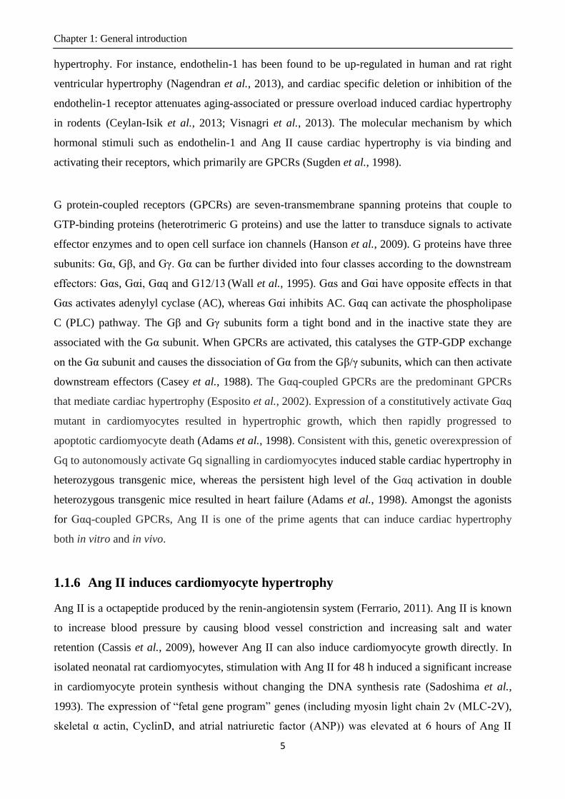
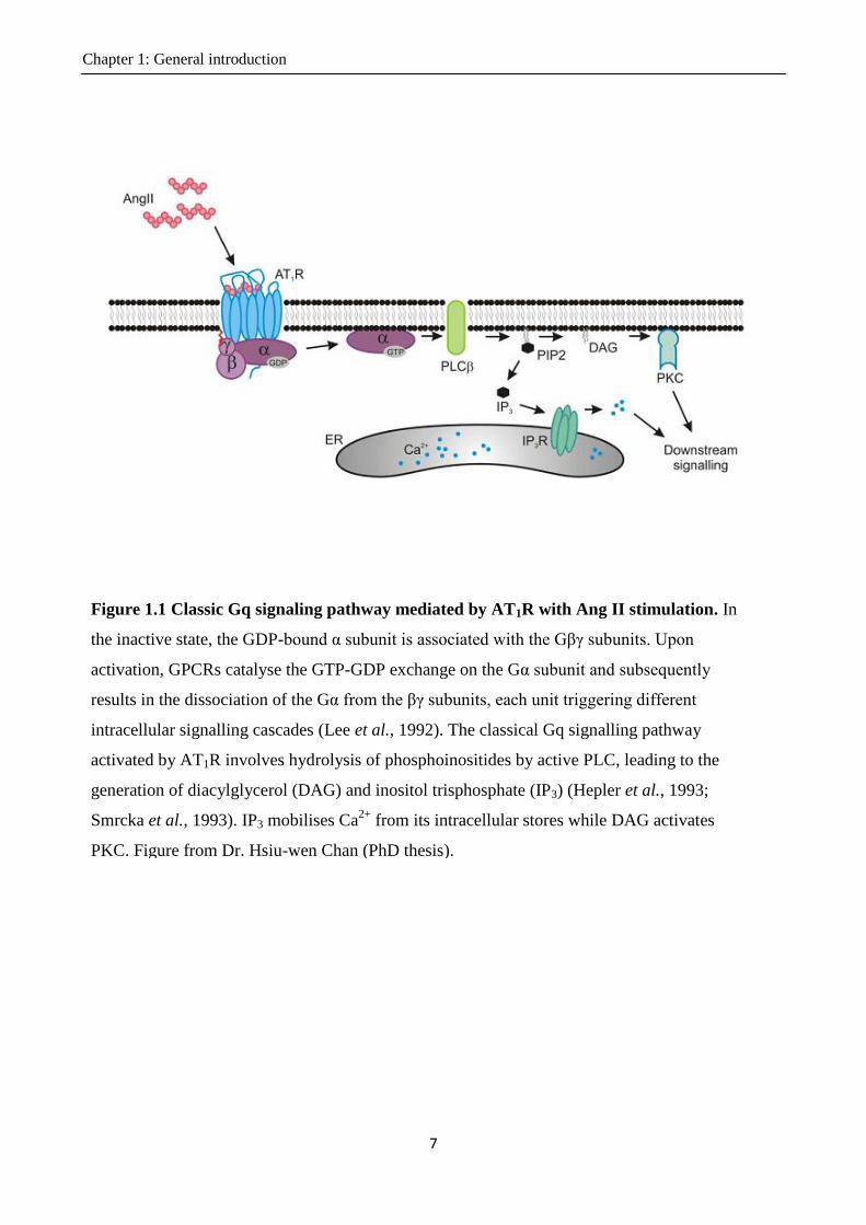
Ang II induces cardiomyocyte hypertrophy by binding to and activating the AT1R receptor. The
AT1R is the major receptor for Ang II in cardiomyocytes and is a member of the GPCR family with
a molecular mass of approximately 41 kDa (Mukoyama et al., 1993; Yasuda et al., 2008). Upon
binding Ang II, AT1R is activated and predominantly trigger Gq signalling pathways (Mehta et al.,
2007; Harris et al., 2009). The classical Gαq signalling pathway activated by AT1R is shown in
Figure 1.1 (Dr. Hsiu-Wei Chan, PhD thesis): AT1R interacts with the Gq protein leading to the
activation of protein kinase C (PKC) and mobilization of Ca2+
from intracellular stores. The
activated PKC and mobilized Ca2+
trigger individual downstream signalling pathways (Ohtsu et al.,
2008). PKC is a critical kinase that regulates multiple cellular activities such as cell proliferation,
survival and apoptosis. In cardiomyocytes, it is involved in the regulation of cardiomyocyte
contraction and hypertrophic growth (Mackay et al., 2001; Braz et al., 2002). PKC has multiple
subtypes, and by now at least 12 isoforms have been identified (Steinberg, 2012). In
cardiomyocytes, the most abundant isoform is PKCα (Pucéat et al., 1994). The activation of PKCα
promotes the hypertrophic growth of cardiomyocytes (Sabri et al., 2003) and PKC inhibition
attenuates agonist-induced hypertrophy (Zou et al., 1996). Although some of these signalling
pathways have been discovered, the detailed molecular mechanisms of how AT1R activation leads
to hypertrophic growth have yet to be fully identified. There is evidence showing that activation of
epidermal growth factor receptor (EGFR), a member of the ErbB family, is involved in, and critical
for, the cardiomyocyte hypertrophy caused by Ang II.
Chapter 1: General introduction
7
Figure 1.1 Classic Gq signaling pathway mediated by AT1R with Ang II stimulation. In
the inactive state, the GDP-bound α subunit is associated with the Gβγ subunits. Upon
activation, GPCRs catalyse the GTP-GDP exchange on the Gα subunit and subsequently
results in the dissociation of the Gα from the βγ subunits, each unit triggering different
intracellular signalling cascades (Lee et al., 1992). The classical Gq signalling pathway
activated by AT1R involves hydrolysis of phosphoinositides by active PLC, leading to the
generation of diacylglycerol (DAG) and inositol trisphosphate (IP3) (Hepler et al., 1993;
Smrcka et al., 1993). IP3 mobilises Ca2+
from its intracellular stores while DAG activates
PKC. Figure from Dr. Hsiu-wen Chan (PhD thesis).

Chapter 1: General introduction
8
1.2 The ErbB receptors
1.2.1 Overview of ErbB receptors
ErbB receptors are a subfamily of receptor tyrosine kinases that regulate cell proliferation, survival
and differentiation (Burgess, 2008). There are currently four known ErbB receptor subtypes
encoded by distinct genes - ErbB1, ErbB2, ErbB3 and ErbB4 (Scaltriti et al., 2006). The sequence
identity of ErbB receptors ranges from 37% - 49% with a molecular mass of approximately 180
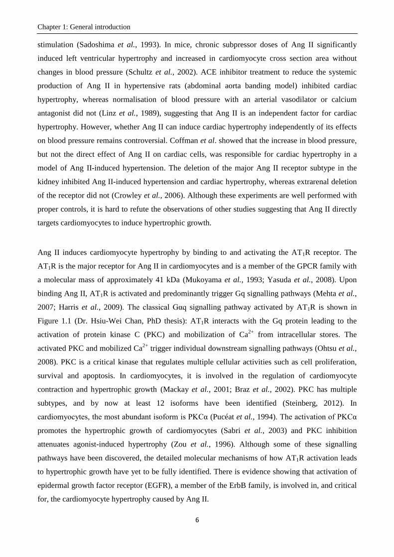
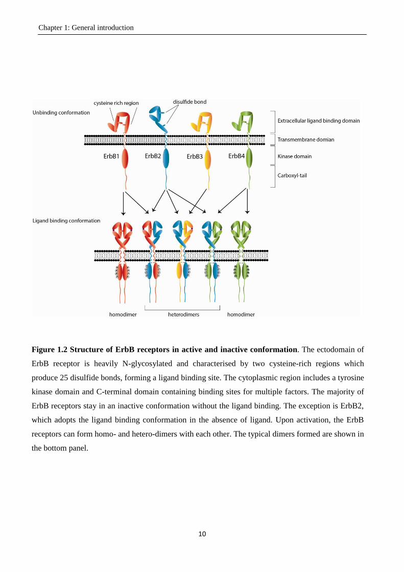
kDa (Jorissen et al., 2003). The typical ErbB receptor comprises a large extracellular ligand-binding
domain (ECD), a single transmembrane domain, an intracellular tyrosine kinase domain and a
carboxyl-tail (Figure 1.2). Most ErbB receptors are in an inactive conformation in the absence of
ligands. Ligand binding results in a conformational change in the ECD and tyrosine kinase domain,
exposing previously embedded surfaces. The surface exposure in the ECD promotes dimer
formation between ErbB receptors (Weiss et al., 1998) and the critical sites exposed in the tyrosine
kinase domain results in the activation of the kinase (Zhang et al., 2006). The receptor dimerisation
brings the intracellular kinase domains in each receptor into proximity to cross-phosphorylate the
tyrosine residues in the cytoplasmic tail of its neighbour receptor in the dimeric complex (Hubbard
et al., 1998) (Figure 1.2). The phosphotyrosine residues in the cytoplasmic tail provide binding sites
for multiple factors containing Src-homology 2 (SH2) domain or phosphotyrosine binding (PTB)
domains (Sweeney et al., 2000), which recruit downstream effectors to trigger signalling (Olayioye
et al., 2000; Yarden et al., 2001; Mendelsohn et al., 2003). The recruitment specificity is
determined by the amino acids surrounding the phosphotyrosine residues of the receptor. Thus, each
ErbB receptor can trigger a unique complex of intracellular signalling pathways based on this
specificity (Figure 1.3). ErbB receptors can form both homodimers and heterodimers (Figure 1.2).
The diversity of the heterodimerisation formed by different ErbB receptors adds more variety to the
ErbB signalling pathways (Burgess, 2008). ErbB2 differs from other subtypes as its extracellular
region is unable to bind any identified ligand, and thus it mainly functions as the preferred ErbB
dimerisation partner (Timolati et al., 2006). ErbB2 adopts an active conformation and can enhance
the dimerisation and the downstream signalling. ErbB3 can bind ligands and provides multiple
docking sites for downstream effectors in its cytoplasmic tail, however, due to an abrogated kinase
activity, it has to trigger downstream signalling with the assistance of dimerising partners
(Rohrbach et al., 2005). All ErbB receptors are detectable in the embryonic heart across different
species. In postnatal cardiomyocytes, the expression level of ErbB receptors declines and the
expressed subtypes include ErbB1, ErbB2 and ErbB4 (Zhao et al., 1998; Rohrbach et al., 1999),

Chapter 1: General introduction
9
with ErbB2 and ErbB4 being the most abundant subtypes. ErbB3 is not expressed at high levels in
postnatal cardiomyocytes (Zhao et al., 1998; Rohrbach et al., 1999).
Chapter 1: General introduction
10
Figure 1.2 Structure of ErbB receptors in active and inactive conformation. The ectodomain of
ErbB receptor is heavily N-glycosylated and characterised by two cysteine-rich regions which
produce 25 disulfide bonds, forming a ligand binding site. The cytoplasmic region includes a tyrosine
kinase domain and C-terminal domain containing binding sites for multiple factors. The majority of
ErbB receptors stay in an inactive conformation without the ligand binding. The exception is ErbB2,
which adopts the ligand binding conformation in the absence of ligand. Upon activation, the ErbB
receptors can form homo- and hetero-dimers with each other. The typical dimers formed are shown in
the bottom panel.
Chapter 1: General introduction
11
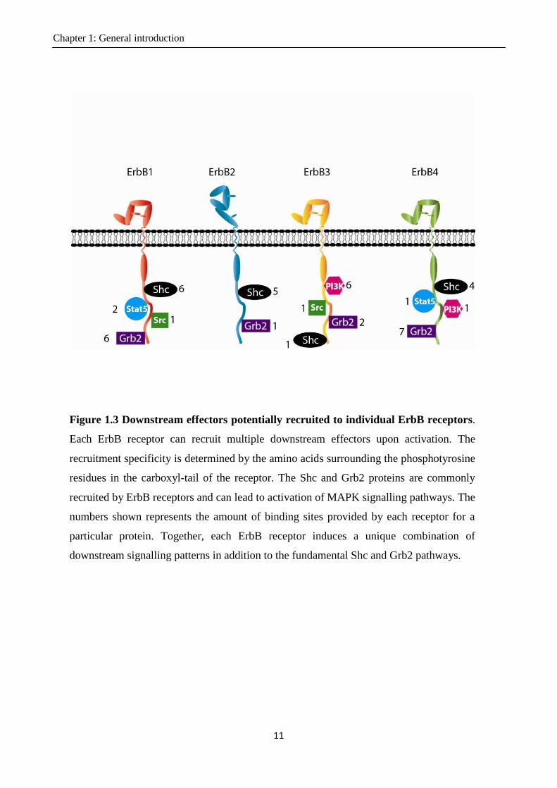
Figure 1.3 Downstream effectors potentially recruited to individual ErbB receptors.
Each ErbB receptor can recruit multiple downstream effectors upon activation. The
recruitment specificity is determined by the amino acids surrounding the phosphotyrosine
residues in the carboxyl-tail of the receptor. The Shc and Grb2 proteins are commonly
recruited by ErbB receptors and can lead to activation of MAPK signalling pathways. The
numbers shown represents the amount of binding sites provided by each receptor for a
particular protein. Together, each ErbB receptor induces a unique combination of
downstream signalling patterns in addition to the fundamental Shc and Grb2 pathways.

Chapter 1: General introduction
12
1.2.2 The function of ErbB receptors in heart
The importance of ErbB signalling in the heart is highlighted by observations from transgenic mice.
Mutations in ErbB2 and ErbB4 lead to a lethal phenotype at embryonic day (E) 10.5 due to defects
in ventricular trabeculae formation (Gassmann et al., 1995; Lee et al., 1995; Sibilia et al., 1995).
ErbB3 null mice have normal heart trabeculation but have disrupted endocardial cushion formation
and die at E13.5 (Erickson et al., 1997; Riethmacher et al., 1997). ErbB1 null mice have severe
defects, and die before birth or within 2-3 weeks postnatally depending on the genetic background
(Miettinen et al., 1995; Threadgill et al., 1995). Besides their essential function in heart
development, ErbB receptors also play important roles in regulating cardiac growth, survival and
function in the postnatal heart. Cardiomyocyte-specific expression of a dominant-negative ErbB1 in
adult mice blocked both ErbB1 and ErbB2 phosphorylation, resulting in cardiac dilation and
decreased cardiac contraction (fractional shortening) (Rajagopalan et al., 2008). The phenotype
caused by cardiac specific deletion of ErbB2 in adult heart has not yet been reported. However,
administration of an ErbB2 blocking antibody in humans increases the risk of heart failure
(Seidman et al., 2002). Consistent with this, inhibition of ErbB2 activity causes myofibrillilary
structural damage and associated inhibition of excitation-contraction coupling (Pentassuglia et al.,
2009b), suggesting that ErbB2 receptors are required for adult heart homeostasis. Currently, the
phenotype of ErbB4 deletion in adult heart has not been reported, although accumulating evidence
indicates that ErbB4 signalling may be important in regulating adult cardiomyocyte function (refer
to section 1.6.7).
In addition to its critical function in regulating cardiac homeostasis, activation of ErbB receptors
mediates cardiac hypertrophy both in vivo and in vitro. Activation of ErbB1 has been reported to
mediate cardiomyocyte hypertrophy to Ang II (Asakura et al., 2002). In cell culture, ErbB4 agonists
induced potent cardiomyocyte hypertrophy (Zhao et al., 1998). More detail on the cardiomyocyte
hypertrophy induced by direct administration of ErbB agonists is discussed in section 1.2.4.
1.2.3 Signalling pathways of ErbB receptors
As mentioned above, there are highly diversified pathways activated by ErbB receptors due to
heterodimerisation of these receptors. Among them are two fundamental and critical pathways
mediating cardiac hypertrophy, the MAPK and PI3K-Akt pathways. Upon activation, ErbB
receptors activate Ras GTPases (Yarden et al., 2001). Ras GTPases are a family of mostly
membrane resident proteins that shuttle between an inactive GDP-bound and active GTP bound

Chapter 1: General introduction
13
conformation (Scheffzek et al., 1997; Coleman et al., 2004). In its activation conformation,
Ras·GTP binds to a number of effector molecules, including the serine/threonine kinase Raf and
phosphoinositide 3-kinase and recruits them to the membrane compartment for activation and
signalling (Kumar et al., 2005).
The Ras-Raf-MEK-ERK (MAPK) pathway
The MAPK pathway is a primary mechanism by which extracellular mediators, such as growth
factors, can regulate cellular activities such as cell growth, survival, apoptosis and metabolism.
After recruitment to the cell membrane by Ras·GTP, Raf activates upstream kinases to stimulate the
extracellular signal-regulated kinases1/2 (ERK1/2). There are at least 5 members of the ERK family
(ERK1 to 5), which are activated and regulated by different upstream kinases (Nishimoto et al.,
2006). ERK1/2 proteins are phosphorylated on threonine and adjacent tyrosine residues in the Thr-
Glu-Tyr motif. ERK1/2 are the most intensively studied and abundantly expressed ERK family
members (Nishimoto et al., 2006). In cardiomyocytes, ERK1/2 activation plays an important role in
mediating cardiomyocyte hypertrophy. Cardiac-specific activation of ERK1/2 by overexpression of
activated MEK1 (the immediate upstream activator of ERK1/2) in 9 independent MEK1 transgenic
mouse lines led to stable concentric hypertrophy in the majority of these mice lines. Most activated
MEK1 transgenic lines demonstrated a 25-30% increase in heart to body weight ratio with increased
cardiac function and no signs of cardiomyopathy until 12 months of age (Bueno et al., 2000). The
mechanism by which ERK1/2 activation causes cardiomyocyte hypertrophy is complicated. It has
been proposed that ERK1/2 regulates protein synthesis during cardiomyocyte hypertrophy via an
association with p70 S6 kinase (Wang et al., 2001). ERK1/2 is also associated with phosphorylation
of transcriptional factors such as GATA4 that regulate ribosomal RNA transcription in
cardiomyocytes (Morimoto et al., 2000; Liang et al., 2001). GATA4 is a cardiac-enriched
transcriptional factor critical in the regulation of most cardiac-expressed structural genes such as α-
myosin heavy chain, myosin light chain 1/3, and cardiac troponin C and I (Molkentin, 2000);(Liang
et al., 2002; Akazawa et al., 2003), as well as hypertrophy responsive genes such as BNP
(Hasegawa et al., 1997).
PI3K-Akt pathway
Another important downstream pathway activated by ErbBs in cardiac hypertrophy is the
phosphoinositide 3-kinase (PI3K) pathway. PI3Ks belong to a family of lipid kinases which convert
phosphatidylinositol (4, 5)-bisphosphate (PIP2) into phosphatidylinositol (3, 4, 5)-trisphosphate
(PIP3). PIP3, in turn, causes the phosphorylation of the kinase Akt. Akt can activate multiple
Chapter 1: General introduction
14
transcriptional factors such as GATA4, β-catenin, c-Myc and NFAT to promote cardiac
hypertrophy (Heineke et al., 2006). Akt also promotes cardiac hypertrophy by enhancing protein
synthesis via activation of mTOR pathways (Proud, 2004). PI3K consists of two subunits. One is
the regulatory subunit that contains p85, p55α, p50α, p85β and p55γ, and is responsible for
anchorage to the docking site of other proteins, such as ErbB receptors. The other is the p110
subunit, containing p110α, p110β and p110δ, which are associated with recruitment of Akt to the
cellular membrane and activation of downstream signalling (Vanhaesebroeck et al., 2010). The p85
and p110α subunits are the most studied. Expression of the constitutively active p110α in the heart
leads to physiological cardiac hypertrophy with preserved cardiac function (Shioi et al., 2000).
Myocardial expression of a dominant negative form of p110α inhibits the physiological hypertrophy
during postnatal cardiac developmental growth or in response to exercise (McMullen et al., 2003).
Similarly, genetic mutations in the p85 subunit decrease heart size at baseline as well as
physiological hypertrophy after exercise training (Luo et al., 2005). These studies suggested an
important role of PI3K in mediating physiological cardiac hypertrophy.
1.2.4 EGF-like factors
EGF-like factors are the agonists for ErbB receptors (Harris et al., 2009). All EGF-like ligands are
made as type I transmembrane proteins that are inserted into the plasma membrane and are cleaved
by cell surface proteases to release mature growth factors (Schneider et al., 2009). The selectivity of
EGF-like factors in binding the ErbB receptors is shown in Table 1.1 (Linggi et al., 2006; Edwards
et al., 2008).
Matrix metalloproteases (MMPs) and a disintegrin and metalloproteases (ADAMs) are part of the
metalloprotease family (Page-McCaw et al., 2007; Reiss et al., 2009). They are anchored in the
membrane and are responsible for the ectodomain shedding of EGF-like ligand precursors to
produce the mature soluble EGF-like ligands (Nagase et al., 2006; Klein et al., 2011). Among 38
identified subtypes, ADAM 17 and ADAM 10 seem to be the principal sheddases for EGF-like
ligands, and the current understanding of ADAM-mediated shedding is largely based on studies of
ADAM17 and ADAM10 (Mochizuki et al., 2007). Among the many EGF-like ligands, only HB-
EGF is known to be a substrate for MMPs (Yu et al., 2002; Hao et al., 2004).
The EGF-like factors that have been identified in the cardiovascular system include EGF, TGF-α,
HB-EGF, amphiregulin (AREG), betacellulin (BTC), epigen, epiregulin (EREG), and neuregulin
(NRG)-1. Accumulated evidence from transgenic mice suggests a requirement of these factors in

Chapter 1: General introduction
15
regulating cardiac development and homeostasis, consistent with the observations for the ErbB
receptors. In addition, administration of growth factor peptides or over-expression of growth factors
induces cardiomyocyte hypertrophy both in vitro and in vivo (Zhao et al., 1998; Schneider et al.,
2005; Yoshioka et al., 2005). In our laboratory (Dr Hsiu-wen Chan, PhD thesis), the ability of
various EGF ligands to mediate hypertrophic growth (measured as protein:DNA ratio) in primary
isolated cardiomyocytes was compared. These EGF-like factors were categorized into three classes
according to their ability to promote cardiomyocyte hypertrophy. The most potent hypertrophic
agonists include BTC, NRG1β1 and NRG2β, which are primarily agonists for ErbB4. These
agonists increased the mass of cardiomyocytes (total protein content) by 33%-49% in absence of
changes in DNA content. Amphiregulin, TGFα, epiregulin and NRG2α caused a 16%-24% increase
in cell mass, and these ligands (except NRG2α) selectively activate ErbB1. EGF, HB-EGF, epigen
and NRG1α weakly induced hypertrophic growth (~10% increase), and are a mixture of ErbB1
agonists and ErbB4 agonists. Together, these studies suggested that the ErbB4 might have a
stronger ability to mediate cardiomyocyte hypertrophy than ErbB1.
Chapter 1: General introduction
16
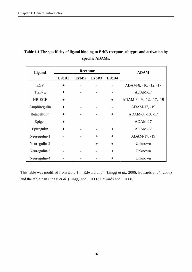
Table 1.1 The specificity of ligand binding to ErbB receptor subtypes and activation by
specific ADAMs.
Ligand Receptor
ADAM
ErbB1 ErbB2 ErbB3 ErbB4
EGF + - - - ADAM-8, -10, -12, -17
TGF- α + - - - ADAM-17
HB-EGF + - - + ADAM-8, -9, -12, -17, -19
Amphiregulin + - - - ADAM-17, -19
Betacellulin + - - + ADAM-8, -10, -17
Epigen + - - - ADAM-17
Epiregulin + - - + ADAM-17
Neuregulin-1 - - + + ADAM-17, -19
Neuregulin-2 - - + + Unknown
Neuregulin-3 - - - + Unknown
Neuregulin-4 - - - + Unknown
This table was modified from table 1 in Edward et.al. (Linggi et al., 2006; Edwards et al., 2008)
and the table 2 in Linggi et.al. (Linggi et al., 2006; Edwards et al., 2008).
Chapter 1: General introduction
17
1.3 Transactivation of ErbB receptors
1.3.1 Transactivation of ErbB by GPCRs
As discussed above, growth factors can cause cell growth via activating their receptors to trigger
downstream growth signalling pathways, such as the MAPK pathway. The growth induced by
activation of various GPCRs can also be mediated by activation of the MAPK pathway. However,
the mechanisms by which GPCRs activate growth signalling pathway is less clear compared to the
growth factor receptors. In 1996, for the first time, Daub et al. found that GPCRs could
transactivate the EGFR to trigger downstream growth signalling. They demonstrated that
endothelin-1, lysophosphatic acid (LPA) and thrombin treatment rapidly phosphorylated the EGFR
followed by activation of MAPK pathway in Rat-1 fibroblasts (an immortalized cell line). All of
these agonists activated GPCR receptors: endothelin-1 activated the endothelin receptor isoform
ETA, lysophosphatic acid activated the LPA receptor and thrombin activated proteinase-activated
receptors. Inhibition of EGFR (ErbB1) with tyrophostin AG1478 or a dominant-negative EGFR
inhibited the GPCR-activated MAPK signalling, confirming that the GPCR mitogenic signalling is
mediated by the transactivation of EGFRs (Daub et al., 1996).
The transactivation of ErbB1 by Ang II was first reported by Eguchi et al. in primary vascular
smooth muscle cells (Eguchi et al., 1998). Since then, increasing evidence demonstrates that the
transactivation of EGFR by Ang II occurs in various cells, including renal epithelial cells, hepatic
C9, cos-7, MCF-7 and importantly, cardiomyocytes (Asakura et al., 2002; Thomas et al., 2002;
Muscella et al., 2003; Shah et al., 2004; Chen et al., 2006). Infusion of antisense
oligodeoxynucleotides to ErbB1 in adult rats significantly attenuated the cardiac hypertrophy
induced by Ang II (Kagiyama et al., 2002). Transgenic mice expressing dominant negative ErbB1
are resistant to the Ang II induced cardiac hypertrophy (Zhai et al., 2006). Taken together, these
studies indicate that ErbB1 is required for Ang II induced cardiac hypertrophy in vivo.
1.3.2 Potential mechanisms for ErbB1 transactivation by Ang II: the TMPS
pathway
How does Ang II transactivate ErbB1? One proposed mechanism is the triple membrane-passing
signalling (TMPS) pathway. In this process, activation of the AT1R by Ang II leads to the activation
of metalloproteases that release ErbB1 ligands from the membrane. These ligands subsequently
bind to ErbB1 and trigger downstream signalling (Figure 1.4). The TMPS mechanism describes the
inside-outside-inside route of transactivation in three steps.

Chapter 1: General introduction
18
In the first step of this process, Ang II binding to the AT1R triggers Gq signalling (Ohtsu et al.,
2008), which in turn activates PKC and Ca2+
signalling. Both PKC and Ca2+
can potentially
contribute to triggering the next step in the TMPS pathway. In hepatic or breast cancer cell lines,
PKC mediates the release of EGF-like factors, contributing to transactivation (Shah et al., 2002;
Muscella et al., 2003). In cardiomyocytes and vascular smooth muscle cells, it has been shown that
Ca2+
but not PKC is essential for ErbB1 transactivation (Eguchi et al., 1998; Thomas et al., 2002;
Smith et al., 2011).
The second step involves cleavage of EGF-like factors. There is evidence to indicate that EGF-like
factor shedding (particularly of HB-EGF) might be required for the transactivation of ErbB1 by
Ang II. Inhibition of HB-EGF with a small molecule inhibitor or a blocking antibody reduces the
transactivation of EGFR by Ang II in various cell types, including Cos-7 cells, cardiomyocytes,
renal epithelial cells and hepatocytes (Asakura et al., 2002; Saito et al., 2002; Mifune et al., 2005;
Ohtsu et al., 2006b). HB-EGF shedding can be mediated by ADAM12, ADAM17 or MMP2/9
(Saito et al., 2002; Shah et al., 2004; Asakura et al., 2002). Dominant negative expression or
pharmacological inhibition of ADAM12, ADAM17 or MMP2/9 blocked HB-EGF shedding and
subsequent transactivation of EGFR by Ang II (Asakura et al., 2002; Saito et al., 2002; Ohtsu et al.,
2006b). Thus, ADAM 12, ADAM17 and MMP2/9 and HB-EGF are essential for the transactivation
of ErbB1, and ADAM/MMP activity may mediate transactivation by shedding HB-EGF.
The transactivation of ErbB1 by Ang II leads to dimerisation of ErbB receptors and the
phosphorylation of intracellular tyrosine residues (Scaltriti et al., 2006). The phosphorylated ErbB1
eventually recruits proteins and leads to phosphorylation of ERK1/2 (Shah et al., 2002).
Phosphorylated ERK1/2 proteins are imported into the nucleus where they phosphorylate specific
transcription factors involved in cell growth (Hill et al., 1995; Gaestel, 2006). To date, ErbB1 is the
only member of the ErbB family identified to be transactivated by Ang II to induce hypertrophy
(Asakura et al., 2002; Thomas et al., 2002; Muscella et al., 2003; Shah et al., 2004; Chen et al.,
2006).
The TMPS paradigm provides a potential scheme for transactivation mechanisms, however the
details of transactivation signalling pathways are more complicated and involve more kinases than
just those mentioned above. There is evidence showing that Src kinase links the activation of EGFR
and GPCRs (Dikic et al., 1996; Fischer et al., 2003). Pharmacological inhibition or RNAi-silencing
of Src abrogated the Ang II induced phosphorylation of EGFR (Bokemeyer et al., 2000; Yano et al.,
Chapter 1: General introduction
19
2007). The non-receptor tyrosine kinase Pyk2 has also been proposed to mediate the transactivation
of EGFR by GPCRs (Soltoff, 1998; Keely et al., 2000). In vascular smooth muscle cells, Pyk2
binds Src and this was associated with activation of EGFR by Ang II (Eguchi et al., 1999).
However, the roles of these factors are controversial. For instance, some studies have proposed
pathways for GPCR-mediated EGFR transactivation that are independent of Src activation:
expression of a kinase-mutant Pyk2 did not affect the EGFR activation induced by bradykinin
(which activates the bradykinin 2 GPCR) in PC-12 cells (Zwick et al., 1999). In addition, inhibition
of Pyk2 in cardiomyocytes did not affect the endothelin-1 induced activation of EGFR, suggesting
that there is no requirement for Pyk2 in the transactivation of EGFR by endothelin-1 (Kodama et
al., 2002). Despite this controversy, these studies suggested that it is highly possible that there are
more factors involved in transactivation that are yet to be discovered. Recently, a siRNA screen
identified multiple new candidates such as TRIO, BMX and CHKA that may mediate the
transactivation of EGFR by Ang II in a human mammary epithelial cell line (George et al., 2013).
Individual down regulation of TRIO, BMX or CHKA attenuated the activation of EGFR by Ang II,
but not by the EGFR agonist EGF, suggesting that these factors are located upstream of EGFR and
required for EGFR transactivation (George et al., 2013). These studies suggested that the pathway
for GPCR to transactivate EGFR is much more complicated than that previously proposed.

Chapter 1: General introduction
20
Figure 1.4 Triple membrane-passing signaling (TMPS) pathway. Activation of the AT1R
by Ang II leads to the activation of metalloproteases that release ErbB1 ligands from the
membrane. These ligands subsequently bind to ErbB1 and trigger downstream signaling.

Chapter 1: General introduction
21
1.4 Characteristics of the ErbB4 isoforms
1.4.1 Introduction to the ErbB4 isoforms
Among the ErbB family, ErbB4 is unique in having distinct isoforms as a result of alternative
splicing. The four isoforms of ErbB4 structurally differ in two domains: the extracellular
juxtamembrane (JM) domain (JM-a and JM-b) and intracellular CYT domain (CYT-1 and CYT-2)
(Figure 1.5). The four combinations of the JM and CYT domains form the four primary isoforms of
ErbB4. The JM-a isoform contains 23 amino acids in the JM domain that allow for ectodomain
shedding by ADAMs (Junttila et al., 2000). JM-b contains an alternative 13 amino acids that makes
it resistant to shedding (Junttila et al., 2000). Cleavage of the JM-a isoform occurs in 2 steps, and
produces an 80-kDa soluble fragment. First, the JM-a isoform can be subjected to ectodomain
cleavage by the metalloprotease ADAM17 to produce a 120-kDa ectodomain fragment and an 80-
kDa fragment (Vecchi et al., 1997; Rio et al., 2000; Zhou et al., 2000). The 120-kDa ectodomain
fragment is released into the extracellular medium and the 80-kDa fragment remains in the
membrane, containing the cytoplasmic and transmembrane domains and several ectodomain
residues. The 80-kDa fragment can be subsequently cleaved by γ-secretase within the lipid bilayer
to release the intracellular domain (Chang-Yuan et al., 2001).
The CYT-1 isoform has 16 amino acids inserted in the cytoplasmic tail when compared to the CYT-
2 isoform. It has been identified that the 16 amino acids of CYT-1 provide a docking site for PI3K,
WWOX containing proteins and other proteins, allowing the activation of specific signalling
pathways (Veikkolainen et al., 2011). The structural differences of the four ErbB4 isoforms could
lead to the functional differences between these isoforms.
The expression of each isoform is tissue specific. In human and mouse adult heart, the most
abundant isoform is the JM-b CYT-1, in kidney it is JM-a CYT-2, and in cerebellum it is JM-a
CYT-2 and JM-b CYT-2 (Elenius et al., 1997; Elenius et al., 1999). The tissue-specific expression
proposes a potentially distinct biological function of each isoform. In some tissues (e.g. mammary
cells, neurons) differences in biological function between isoforms has been shown (Naresh et al.,
2006; Feng et al., 2007; Law et al., 2007; Muraoka-Cook et al., 2009). For instance, during the
brain development only the cleavable JM-a isoform can regulate the time of astrogenesis in the
mouse (Sardi et al., 2006). However, whether there are functional differences between isoforms in
cardiomyocytes is unknown.
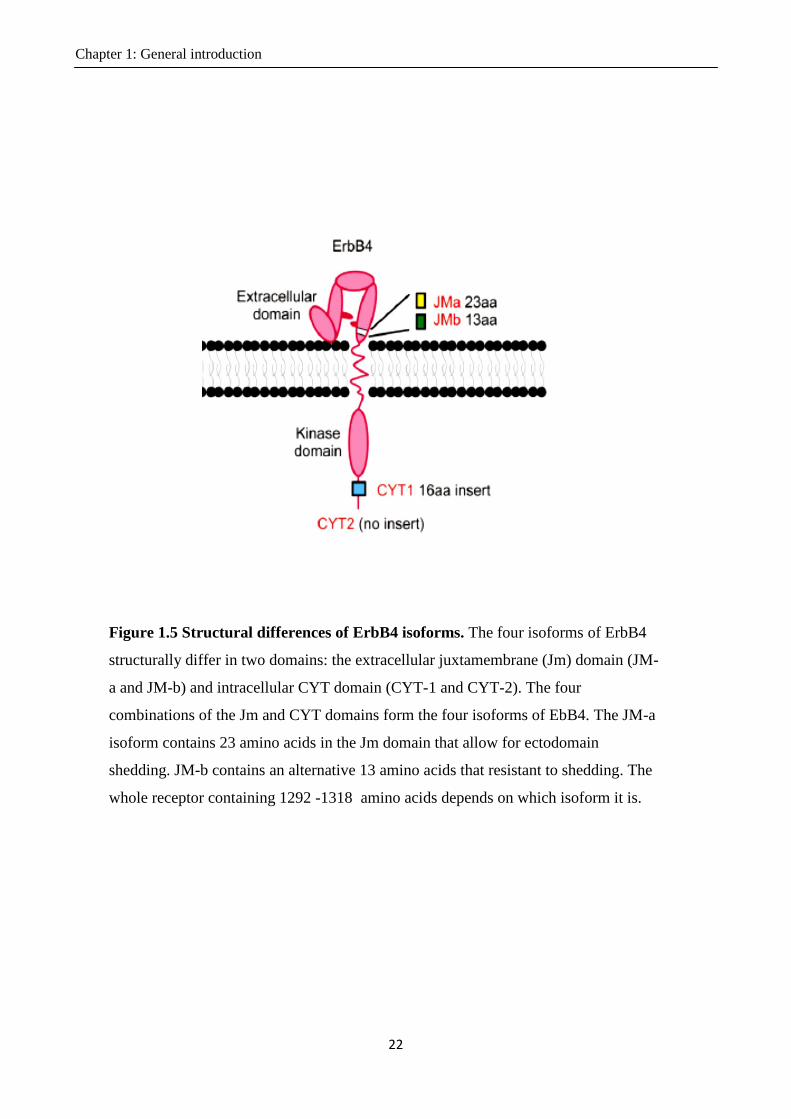
Chapter 1: General introduction
22
Figure 1.5 Structural differences of ErbB4 isoforms. The four isoforms of ErbB4
structurally differ in two domains: the extracellular juxtamembrane (Jm) domain (JM-
a and JM-b) and intracellular CYT domain (CYT-1 and CYT-2). The four
combinations of the Jm and CYT domains form the four isoforms of EbB4. The JM-a
isoform contains 23 amino acids in the Jm domain that allow for ectodomain
shedding. JM-b contains an alternative 13 amino acids that resistant to shedding. The
whole receptor containing 1292 -1318 amino acids depends on which isoform it is.
Chapter 1: General introduction
23
1.4.2 Cleavage in the JM-a domain
As mentioned above, the defining feature of JM-a-containing ErbB4 isoforms is the potential to be
cleaved. ADAM 17 is involved in the cleavage of ErbB4 (Rio et al., 2000). Thus, stimuli that
activate ADAM 17 such as PKC activators (e.g. the phorbol ester PMA), ErbB4 agonists (e.g.
NRG) and ligands that activate certain GPCRs (e.g. gonadotropin-releasing hormone) potentially
possess the ability to trigger the cleavage process. PMA was the first agonist found to trigger the
cleavage of the ErbB4, and can release the ICD of ErbB4 from the membrane in a wide range of
cell types, including breast cancer cells, ovarian cancer cells, lung cancer cells, and a glioma cell
line (Vecchi et al., 1996; Zhou et al., 2000; Määttä et al., 2006; Zeng et al., 2009). Cleavage of the
ErbB4 induced by NRG is also observed in multiple cell types such as T47D (breast cancer cell
line), OVCAR3, and OVCA432 (ovarian cell lines) (Vecchi et al., 1996; Zhou et al., 2000). It has
been shown recently that gonadotropin-releasing hormone (a GPCR agonist) caused ADAM 17-
dependent cleavage of the ErbB4 and production of 80 kDa fragment in immortalized neurons that
endogenously express the JM-a isoform (Higa‐Nakamine et al., 2012).
After being released from the membrane, the 80 kDa ICD can enter the nucleus. The residues 676-
684 of the ICD domain are essential for nuclear accumulation of the ICD. Mutation of these
residues destroyed the ICD localization to the nucleus without affecting production of the ICD
(Williams, Allison et al. 2004). Furthermore, it has been shown that the tyrosine kinase activity of
ErbB4 is essential for the nuclear localiztion of the ICD (Sundvall et al., 2007). The regulatory
mechanism for ICD nuclear localization is not clear. However, the CYT-2 ICD has been shown to
accumulate in the nucleus more than CYT-1 ICD due to enhanced tyrosine kinase activity in the
intracellular domain of the CYT-2 ICD (Sundvall et al., 2007). Both CYT-2 ICD and CYT-1 ICD
possess phosphorylation sites in the intracellular domain, but none of these are specifically located
in the extra 16 amino acid sequence possessed by CYT-1. The reason why CYT-2 ICD possesses
stronger tyrosine kinase activity than CYT-1 is still unknown.
1.4.3 Biological function of the ICD
By homing to the nucleus and interacting with transcriptional regulatory proteins, the 80 kDa ICD
may regulate cell proliferation, apoptosis, differentiation, and DNA damage responses. The
transcriptional regulatory proteins identified as interacting with the ErbB4 ICD include AP-2, Yes-
associated protein (YAP), signal transducer and activator of transcription 5A (STAT5A), estrogen
receptor α (ER α) and Krab-associated protein 1 (Kap1) (Omerovic et al., 2004; Williams et al.,

Chapter 1: General introduction
24
2004; Zhu et al., 2006a; Gilmore-Hebert et al., 2010; Gough, 2010). In a mouse fibroblast cell line,
AP-2 was found to interact with the ICD of JM-a CYT-2 to positively regulate the platelet-derived
growth factor receptor-alpha (PDGFA) promoter leading to cell proliferation under normal
conditions and survival during serum starvation (Gough, 2010). The interaction between YAP and
ICD interferes with p73-stimulated proapoptotic signalling (Komuro, Nagai et al. 2003; Omerovic,
Puggioni et al. 2004), whereas interaction of STAT5A with the ICD leads to nuclear localization of
STAT5A and activation of the milk genes, such as β-casein and whey acidic protein, thus
contributing to the differentiation of the mammary cells (Long et al., 2003; Williams et al., 2004).
In addition, interaction of ICD with ERα promotes cell proliferation in the T47D breast cancer cell
line (Zhu et al., 2006b); and the ErbB4 ICD interaction with Kap1 mediates the response to DNA
damage (Gilmore-Hebert et al., 2010). Clearly, the release and trafficking of the ICD into the
nucleus provide a new paradigm for ErbB4 signalling.
In the above section, I described how the ICD interacts with regulatory proteins to regulate cell
activity. However, it should be noted that there are two types of ICD. The CYT-1 ICD is different
from the CYT-2 ICD in possessing 16 extra amino acids. Compared to the CYT-2 ICD, the 16
amino acid insertion in CYT-1 ICD contains a unique PI3K binding site (“YTPM”) and extra PPXY
domain that binds WW-containing proteins, such as WWOX and YAP (Junttila et al., 2000;
Aqeilan et al., 2005). It has been proposed that this difference in structure may lead to differences in
regulatory ability.
Both in vitro and in vivo experiments have shown that CYT-1 ICD and CYT-2 ICD may have
different functions in promoting the growth, death and differentiation of cells. Mammary cell lines
expressing exogenous CYT-2 ICD grow more rapidly than the same cell line expressing the CYT-1
ICD (Muraoka-Cook et al., 2009). The CYT-1 ICD probably decreases mammary epithelial cell
proliferation by reducing of the rate of cell cycle progression, whereas CYT-2 ICD promotes cell
proliferation by increasing cyclinD via the wnt-β-catenin signalling pathway (Sartor et al., 2001;
Muraoka-Cook et al., 2006b; Strunk et al., 2007). CYT-1 ICD expression promotes cell apoptosis
during serum starvation, whereas the CYT-2 ICD prevented this effect (Naresh et al., 2006; Feng et
al., 2007; Muraoka-Cook et al., 2009). A mammary cell line exogenously expressing CYT1-ICD
formed more acinar structures in 3D culture when compared with the cells expressing CYT2-ICD
(Muraoka-Cook et al., 2006a), indicating that CY1-ICD promotes differentiation of these cells. In
vivo, transgenic mice expressing CYT-1 ICD have distended mammary ducts filled with secretory
material and ductal epithelium that consists of multiple cell layers. Furthermore, fewer terminal end

Chapter 1: General introduction
25
buds were observed in the mammary glands of mice expressing CYT-1 ICD compared to those of
mice expressing CYT-2 ICD (Muraoka-Cook, Sandahl et al. 2009). These studies suggest that
CYT-1 ICD and CYT-2 ICD have different roles in the regulation of mammary development.
Taken together, this work shows that the CYT-1 ICD and CYT-2 ICD can exert divergent functions
in cells.
In addition to studies where the CYT-1 ICD and/or CYT-2 ICD are expressed in cells, others have
noted profound differences in the function of full length ErbB4 receptors in vivo. One seminal study
directly compared the functions of the intact JM isoforms by overexpressing full-length JM-a and
JM-b in mouse embryonic astrocytes in an ErbB4-null background. The findings demonstrated that
only the cleavable JM-a isoform is capable of suppressing astrocyte differentiation (Sardi et al.,
2006). This supports the idea that the intact isoforms of ErbB4 may also have distinct functions in
vivo. In the heart, there have been no studies examining the distinct function of each ErbB4 isoform.

Chapter 1: General introduction
26
1.5 Cardiomyocyte proliferation
Amphibia and fish can repair their injured hearts by cardiac regeneration. Initially, this regeneration
was attributed to stem cells inherent in the heart (Lepilina et al., 2006), but the contemporary view
challenges this idea. Recently, with the assistance of genetic fate mapping technology, researchers
have been able to follow the fate of newly generated cells and this has been applied to trace the
source of newly generated cardiomyocytes (Jopling et al., 2010; Kikuchi et al., 2010). In a genetic
fate mapping model, zebrafish with the cardiac-specific cmlc2a (cardiac myosin light chain 2a)
promoter driving the expression of tamoxifen-inducible Cre recombinase were crossed with animals
possessing a GFP gene with its stop codon flanked by loxP sites. In the offspring, the pre-existing
cardiomyocytes can be induced to express GFP by tamoxifen treatment. If the newly generated
cardiomyocytes also expressed GFP, this suggests that the new cells are generated from pre-existing
cardiomyocytes; if not, it is potentially from other source, such as stem cells. With this method, two
groups independently demonstrated that heart regeneration in zebrafish occurs principally from the
proliferation of pre-existing cardiomyocytes (Jopling et al., 2010; Kikuchi et al., 2010).
In mammals, it is believed that cardiomyocyte proliferation only occurs during the embryonic stage,
and that shortly after birth the majority of cardiomyocytes exit the cell cycle and lose the ability to
proliferate. A study showed that in neonatal mice retain the capacity to regenerate the intact heart
after the left ventricle apex is resected (Porrello et al., 2011). However, this regenerative capacity is
only observed in the mice up to seven days. Older mice completely lose the ability for cardiac
regeneration, suggesting the proliferative potential in neonatal cardiomyocytes is reduced 1-2 weeks
after birth (Porrello et al., 2011). Genetic fate mapping in this study indicated that the majority of
new cardiomyocytes originated from preexisting cardiomyocytes (Porrello et al., 2011). This
suggested that the capacity to regenerate the heart is similar between fish and mammals at or around
birth. It is generally believed that the adult mammalian heart is not capable of fully regenerating the
heart following damage (e.g. a myocardial infarction), however the degree to which adult
mammalian cardiomyocytes can proliferate remains highly controversial.
Cardiomyocyte proliferation can be measured by immunofluorescence staining for Ki67 or
phosphorylated histone H3 (pH3), or BrdU incorporation. Ki67 and pH3 are proliferation markers
that label the cardiomyocytes in the transient mitosis stage. Due to the transient nature of the
mitosis stage and low proliferation levels of adult cardiomyocytes, pH3 and Ki67 will usually stain
only the small number of cells that are currently in mitosis. BrdU is a synthetic analog of thymidine.
Chapter 1: General introduction
27
It can be incorporated in the genomic DNA when cells undergo DNA synthesis, and is subsequently
detected via immunofluorescence. Thus, BrdU incorporation can reflect the total number of
proliferating cardiomyocytes over a longer time period. However, this method can induce a false
positive stain in the presence of DNA repair and thus potentially exaggerate the results. Despite the
limitations of these methods, there is an accumulation of evidence to propose that cardiomyocytes
can proliferate in postnatal mice in response to injury or various stimuli. Beltrami et al. showed that
in the infarcted heart, the fraction of myocytes undergoing mitosis (labeled by Ki67) is 4 percent in
the region adjacent to the infarct and 1 percent in those in regions distant from the infarct (Beltrami
et al., 2001). Treatment with periostin to induce the reentry of differentiated cardiomyocytes into
the cell cycle contributed to a reduction in infarct size and improvement of cardiac function in a
rodent infarction model (Kühn et al., 2007). Similarly, infusion of the ErbB4 ligand (NRG1) in
adult mice significantly increased the proliferation of mononucleated cardiomyocytes (measured by
BrdU incorporation) (Bersell et al., 2009). Interestingly, both the NRG1 and periostin-induced
cardiomyocyte proliferation is PI3K-dependent. Inhibition of ERK1/2 with compound PD985059
(1µM) did not inhibit the periostin-induced cell-cycle reentry, whereas inhibition of the PI3K
pathway by LY294002 (10 µM) or its downstream signalling Akt pathway by SH-6 (10 µM) fully
inhibited periostin-induced cardiomyocyte cell-cycle reentry (Kühn et al., 2007). Using functional
inhibition with PTEN (a PI3K inhibitor), Bersell et al. demonstrate that the NRG1-induced
proliferation of adult cardiomyocytes also required the PI3K pathway (Bersell et al., 2009).
Although these observations strongly suggest that cardiomyocytes in the adult mouse can proliferate
in response to various stimuli, there is a study that argues against this. With genetic fate mapping in
mice, Hsieh et al. showed that the percentage of GFP positive cardiomyocytes was significantly
reduced after infarction, indicating dilution of the pre-existing cardiomyocytes by the proliferation
of stem or progenitor cells and their subsequent differentiation into new cardiomyocytes. This
suggested that in the adult mouse heart, injury-induced cardiac repair depends more on the
proliferation of cardiomyocyte progenitor cells or stem cells rather than fully differentiated
cardiomyocytes (Hsieh et al., 2007). Irrespective of the source of the regenerated cardiomyocytes,
these studies describe above demonstrated that adult mammalian cardiomyocytes can be
regenerated to at least some degree.
The proliferation of cardiac cells discussed above has all been investigated in animal models.
Measuring the degree of cardiomyocyte regeneration in humans is more challenging. Two recent
studies have directly measured cardiomyocyte proliferation rates in the human heart. Bergmann et
Chapter 1: General introduction
28
al. established the age of cardiomyocytes in human heart by taking advantage of the integration into
DNA of carbon-14 generated by nuclear bomb testing during the cold war (Bergmann et al., 2009).
With mathematical modelling, they revealed that the cardiomyocyte turnover rate gradually
decreased from 1% per year at the age of 25 to 0.45% at the age of 75. In contrast, the non-
cardiomyocyte turnover rate is up to 18% per year depending on the cell type. All together, less
than half the cardiomyocytes are refreshed during a normal life span, and 55% of cardiomyocytes
persist from birth. In another study, cardiomyocyte proliferation in human heart was examined by
analysing the incorporation of iododeoxyuridine (Idu, a thymidine analog) in postmortem samples
obtained from cancer patients who were treated with this drug (Kajstura et al., 2010). The portion of
cardiomyocytes labeled with Idu was 2.5-46%. The large variation in the Idu incorporation rate is
attributed to the great differences in frequency, period and interval of Idu treatment given to
patients. Mathematical modelling of this data suggested that the rate of cardiomyocyte renewal is
around 22%. There is a large variation in the cardiomyocyte renewal rate reported in these two
studies, from 1% to nearly 22% per year in humans. It is hard to determine which is more accurate
by direct comparison of the studies given the totally different methods employed. Compared with
other observations, the annual 22% turnover rate of adult cardiomyocytes seems to be the highest
report, and thus needs to be carefully considered. Although these studies confirm that there is the
potential for cardiomyocytes to renew during human lifespan, the functional regeneration of a
damaged heart appears not to occur. While the innate rate of cardiomyocyte replacement seems
insufficient to permit complete repair of a damaged heart, the mere existence of a demonstrable
regenerative response (and its potential to be augmented by various stimuli) is encouraging in
moving towards directed therapies aimed at improving cardiac repair and function.

Chapter 1: General introduction
29
1.6 Importance of the NRG1-ErbB4 signalling axis
1.6.1 An introduction to NRG1
NRG1 (Neuregulin1) is encoded by a large gene (around 1400kb), less than 0.3% of which encodes
protein (Stefansson et al., 2002). Due to splicing, at least 15 different NRG1 isoforms are generated
(Buonanno et al., 2001; Falls, 2003). The motif common to all NRG isoforms is an epidermal
growth factor-like (EGF) domain that is responsible for binding and activation of receptors. NRG1
can be synthesized as either a secreted protein or membrane precursor. Based on differences in the
N-terminal sequence, the membrane-inserted NRG1 can be further divided into 3 types, I - III. Both
type I and type II isoforms are single-transmembrane spanning proteins. Cleavage of these isoforms
by ADAMs releases bioactive fragments into the circulation, where they then participate in
paracrine regulation. Type III isoforms are two-transmembrane spanning and will generate a
membrane anchor fragment containing the EGF-like receptor binding domain after cleavage by
ADAMs, and thus participate in juxtacrine regulation (Buonanno et al., 2001). Alternative splicing
at the C-terminus of the EGF-like domain leads to the generation of NRG1α and NRG1β variants.
NRG1α and NRG1β isoforms have different affinities for the ErbB receptors. The NRG1β isoform
possess 10-100 times greater bioactivity and a higher affinity for ErbB3 and ErbB4, whereas the
predominant isoform expressed in cardiovascular system is NRG1α (Cote et al., 2005). Thus,
although NRG1α is the predominant isoform in heart, the NRG1β isoforms may be equally
important. Combined with the structural differences in other domains, at least 3 different NRG1α
isoforms and 8 NRG1β isoforms are expressed in the adult heart (Pentassuglia et al., 2009a).
NRG1 is mainly distributed in the endocardial endothelium and the cardiac microvascular
endothelium (Kuramochi et al., 2004; Lemmens et al., 2006), which share a common embryonic
lineage (Misfeldt, 2008). Whether it is expressed in larger veins is controversial - some studies have
reported NRG1 expression in human coronary artery endothelial cells (Hedhli et al., 2011), whereas
others have failed to detect it in coronary arteries or aortic endothelium (Lemmens et al., 2006). The
released NRG1 can target cardiomyocytes and regulate their growth (Zhao et al., 1998; Giraud et al.,
2005), survival (Liu et al., 2006), contraction (Brero et al., 2010) and proliferation (Bersell et al.,
2009).
1.6.2 NRG1 signalling and cardiomyocyte hypertrophy
NRG1 potently induces cardiomyocyte hypertrophy in vitro. Treatment of neonatal cardiomyocytes
with 20-30 ng/ml NRG1 increased L-leucine uptake (a marker of protein synthesis), expression of

Chapter 1: General introduction
30
the hypertrophic genes ANP and α-skeletal actin, and promoted sarcomeric reorganization (Zhao et
al., 1998; Giraud et al., 2005). Similar results were observed in primary isolated adult
cardiomyocytes (Zhao et al., 1998; Giraud et al., 2005). NRG1-induced cardiomyocyte hypertrophy
is thought to be mediated by ErbB2/ErbB4-ERK1/2 signalling. Treatment with 20 ng/ml NRG1for
5 min induced phosphorylation of ErbB4 and its preferential dimerisation partner ErbB2 in both
neonatal and adult cardiomyocytes (Zhao et al., 1998). Intravenous administration of 10μg/kg
recombinant human NRG1 (rhNRG1) in mice for 10 min induced phosphorylation of ERK in the
heart, which reached a 7-fold increase after 90 min treatment (Liu et al., 2006). The rhNRG1-
enhanced sarcomeric organization was blocked by the ERK pathway inhibitor PD98059, but not the
PI3K kinase inhibitor Wortmannin, suggesting that the rhNRG1-enhanced sarcomere organization
is via ERK, but not PI3K, signalling pathways (Liu et al., 2006). Activation of ERK1/2 promotes
protein synthesis and sarcomere reorganization during NRG1 induced cardiomyocyte hypertrophy.
Activation of ErbB2 by treating cardiomyocytes with an ErbB2 activating antibody induced
phosphorylation of ERK1/2, demonstrating that ErbB2 phosphorylation is sufficient for activation
of ERK1/2. In contrast, ErbB2 inhibition reduces basal phosphorylation of ERK1/2 (Fukazawa et
al., 2003).
1.6.3 NRG1 signalling in cardiac protection
NRG1 can be released from cardiac microvascular endothelial cells (CMEC) under conditions of
oxidative stress. H2O2 increased the NRG1 level in culture medium of CMECs (Kuramochi et al.,
2004) in vitro, whilst in vivo ischaemia-reperfusion injury increased the NRG1 level in the perfused
coronary effluent (Kuramochi et al., 2004). The released NRG1 appears to have a cardioprotective
effect. Liu et al. showed that NRG1 improved both cardiac performance and survival in various
models of LV failure, including the LAD ligation infarct model, doxorubicin-induced
cardiomyopathy model, Coxsackie virus B3-induced myocarditis model, and a chronic rapid pacing
model (Liu et al., 2006). The cardioprotective effect of NRG1 is attributed, at least in part, to its
ability to promote cardiomyocyte survival and enhance myofibril organization.
Primary neonatal cardiomyocytes deprived of serum in culture exhibit a gradual loss in cell viability
(Zhao et al., 1998). Addition of NRG1 into the culture medium increases cell viability by 30%
compared to the control (as measured by the activity of NAD(P)H-dependent cellular
oxidoreductase enzymes using the colorimetric assay MTT) (Zhao et al., 1998). In addition, NRG1
treatment also significantly decreases apoptosis (measured by TUNEL) of primary cardiomyocytes
in serum-free medium (Zhao et al., 1998). A microarray study showed that NRG1 increased the

Chapter 1: General introduction
31
expression of oxidative stress defense related factors (e.g. thioredoxin) and reduced expression of
the apoptosis factors, harakiri (HRK) and programmed cell death 2 (PDCD2) in cardiomyocytes,
which may also contribute to cardiomyocyte survival (Giraud et al., 2005).
During cardiomyocyte damage, myofibrillar disarray and myofibril loss are often observed. NRG1
treatment significantly attenuates the myofibrillar disarray and myofibril loss induced by
doxorubicin. This attenuation contributes to the preservation of cardiac function in these animals
(Bian et al., 2009). Troponin is a complex of three regulatory proteins (troponin C, troponin I, and
troponin T) that is integral to cardiac muscle contraction. The degradation of myofibrils releases the
troponin subunits into the blood, where they are important markers for detection of myocardial
injury (Katus et al., 1991; Adams et al., 1993). Doxorubicin treatment significantly decreases levels
of cardiac troponin in mice, an effect that is attenuated by administration of NRG1 (Bian et al.,
2009). In agreement with this, another study showed that NRG1 reduces both troponin I release into
serum (which occurs during cardiomyocyte damage) and histological alterations associated with
myocarditis (Muraoka-Cook et al., 2006a).
The ErbB-PI3K-Akt pathway was proposed to be the central downstream signalling pathway that
mediates the ability of NRG to modulate the survival and function of cardiomyocytes (Brero et al.,
2010; Fang et al., 2010; Jie et al., 2012; An et al., 2013). For instance, treatment of cardiomyocytes
with the PI3-kinase inhibitor wortmannin or overexpression of a dominant-negative Akt abolished
the protective effect of NRG1 on daunorubicin-induced caspase-3 activation and apoptosis
(Fukazawa et al., 2003). Pretreating cardiomyocytes with anti-ErbB2 antibodies abolished the
protective actions of cardiac microvascular endothelium (a source for NRG1) against
cardiomyocyte apoptosis (Lemmens et al., 2006). AG879 (an ErbB2 inhibitor) treatment abolished
the ability of NRG1 to attenuate the doxorubicin- induced decrease in cardiac troponin I and
troponin T (Bian et al., 2009), suggesting that the NRG1 induced cardioprotective effect is ErbB2-
dependent.
1.6.4 NRG1 signalling in cardiomyocyte contraction/relaxation
Calcium is a critical factor for regulating cardiomyocyte contraction and relaxation. Disorders of
Ca2+
homeostasis contribute to the development of heart failure. Myocyte contraction is regulated
by Ca2+
release from the sarcoplasmic reticulum (SR) into the cytoplasm, and relaxation by uptake
of Ca2+
into the SR. This Ca2+
uptake is mediated by SR Ca2+
ATPase (SERCA2a). NRG1 can
enhance cardiomyocyte relaxation by regulating the Ca2+
uptake. NRG1 induced activation of

Chapter 1: General introduction
32
PI3K/Akt promotes the activation of PKG (Mery et al., 1991; Brero et al., 2010), which enhances
the relaxation of cardiomyocytes by regulating major components of excitation-contraction
coupling, such as L-type Ca2+
channel (LTCC) , phospholamban (PLB) and troponin I (Blumenthal
et al., 1978). PLB activation promoted the SERCA2a activation (Macdougall et al., 1991;
Verboomen et al., 1992) and caused Ca2+
uptake into the SR (Brero et al., 2010) to enhance
cardiomyocyte relaxation.
1.6.5 NRG1 signalling in cardiomyocyte proliferation
In addition to the functions mentioned above, a role for NRG1 in promoting cardiomyocyte
proliferation has also been proposed. Zhao et al. demonstrated that treatment with NRG1 for 30h
caused an approximately 2 fold increase in DNA synthesis in embryonic cardiomyocytes as
measured by [3H]-thymidine uptake. NRG1 also significantly increased the [
3H]-thymidine uptake
in neonatal cardiomyocytes cultured in serum-free medium (Zhao et al., 1998). In addition to
promoting DNA synthesis, NRG1can also stimulate cell division, including both karyokinesis and
cytokinesis. Bersell et al. detected cytokinesis in cardiomyocytes by using immunofluorescence to
stain the marker protein aurora B kinase, which is required for contractile ring formation in
cytokinesis. They found the number of positively stained cells was significantly increased by NRG1
treatment. A similar result was observed for karyokinesis measured with video microscopy (Bersell
et al., 2009). Interestingly, the effect of NRG1 on cardiomyocyte proliferation may be dictated by
the degree of stimulation. Zhou et al. showed that the maximum proliferative effect of NRG1
occurs at 0.1 nM (Zhou et al., patent application published as WO00/37095). Similar to this, Bersell
et al. also showed an increased DNA synthesis in response to NRG1, which was maximal between
0.1-1nM (Bersell et al., 2009). At higher concentrations, NRG1 may inhibit DNA synthesis due to
the persistent activation of the MAPK signalling pathway, which inhibits CyclinD to arrest the cell
at G1 phase (Zhou et al., patent application published as WO00/37095). Direct evidence that NRG-
induced cardiomyocyte proliferation is mediated by ErbB4 is also provided by Bersell et.al. (2009).
Administration of NRG1 induced 14.3% of mononucleated cardiomyocytes to proliferate (as
indicated by BrdU incorporation) in the adult mice heart, and this proliferation was attenuated in
transgenic mice with cardiac-specific deletion of ErbB4. In cultured adult cardiomyocytes, blocking
ErbB4’s preferential dimerisation partner ErbB2 with an inhibitory antibody also decreased DNA
synthesis (Bersell et al., 2009), suggesting an critical role of the ErbB2/ErbB4 dimer in mediating
NRG1-induced proliferation.

Chapter 1: General introduction
33
1.6.6 Interactions between NRG1 signalling and the endocrine system
As a critical paracrine factor that is potentially beneficial to the heart, it is important that the
synthesis and release of NRG1 is adaptive to cardiac activity. Indeed, the expression and release of
NRG1 is controlled, at least partially, by endocrine factors or stimuli such as pressure overload. As
mentioned earlier, NRG1 (detected in perfused coronary effluent) can be released from the
microvascular endothelium in response to ischaemia-reperfusion (Kuramochi et al., 2004). In
addition to that, stretch induces the expression of NRG1 in cardiac microvascular endothelium.
Correspondingly, transverse aortic constriction (TAC) induces the up-regulation of NRG1 in the
heart (Lemmens et al., 2006). A study showed that NRG1, in primary isolated microvascular
endothelial cells, is increased by endothelin-1 treatment (Zhao et al., 1998). In contrast, Ang II and
phenylephrine decreased the expression of NRG1 in cardiac microvascular endothelium (Lemmens
et al., 2006). Due to the beneficial effects of NRG1 on cardiac tissues (as discussed above), the up-
regulated NRG1 might serve as a mechanism to counterbalance the effect of pathological stimuli
and thus protect the cardiomyocytes to some degree in these pathological conditions. In contrast, in
other conditions, such as activation of RAS system, the NRG1 down-regulation might facilitate the
progression of the cardiac disease. NRG1 also provides a negative feedback on these stimuli.
Treatment with NRG1 in LAD ligation mice significantly reduced the increased concentrations of
renin, AngI, Ang II and aldosterone in the circulation, suggesting that the rhNRG1 attenuated
activation of the renin-angiotensin-aldosterone axis (Muraoka-Cook et al., 2006a). Besides, NRG1
can interact with adrenergic signalling to regulate cardiomyocyte contractility, where it has been
demonstrated that NRG1 reduces the contractile response of cardiomyocytes to α-adrenergic
stimulation (Lemmens et al., 2004). Cardiomyocytes lacking effective neuregulin signalling (NRG-
gene mutant animals) are unable to counterbalance the adrenergic effect (Okoshi et al., 2004).
1.6.7 Function of ErbB2 and ErbB4 in the heart
The importance of NRG1 signalling highlights the potential role of its receptor, ErbB4 and its
preferred dimerisation partner (ErbB2) in adult heart function. ErbB3 can also bind with NRG1,
however, ErbB3 expression is not detectable in the adult heart (Campreciós et al., 2011). ErbB2 is
overexpressed in breast cancer (Houston et al., 1999; Sørlie et al., 2001), and monoclonal
antibodies targeting ErbB2 have been used in treatment of this disease. The monoclonal antibody
for ErbB2, trastuzumab, has been trialled for breast cancer therapy, but its administration increases
the risk for cardiac dysfunction: the incident rate was 27% in patients receiving trastuzumab and
anthracycline-containing chemotherapy, compared with 8% incidence among patients who received
chemotherapy alone (Seidman et al., 2002). Despite the fact that anthracycline is also cardiac toxic

Chapter 1: General introduction
34
and trastuzumab-induced cardiac dysfunction is reversible (79% patients improved after receiving
standard treatment for congestive heart failure), this clinical observation led to the idea that ErbB2
signalling is critical in the adaptive response of adult heart to stress (Seidman et al., 2002). A
further study using ErbB2 conditional knockout animal models confirmed the importance of ErbB2
in cardiac function and structural maintenance (Özcelik et al., 2002). In this study, the MLC-2V
promoter was used to drive expression of Cre recombinase to specifically induce deletion of ErbB2
from 50-60% cardiomyocytes in heterozygous ErbB2flox/+
animals. There were no functional or
morphological differences between the wild type and mutant animals at birth, but the mice with
ErbB2 deletion developed severe cardiomyopathy in adulthood. This cardiomyopathy was
characterised by cardiac dysfunction (i.e., a decreased fractional shortening measured by
echocardiography), dilatation of both ventricles (i.e., increased chamber size with thinned walls)
and cardiac hypertrophy (Özcelik et al., 2002). In support of this, cultured adult myocytes treated
with a non-activating ErbB2 specific antibody display myofilament disarray (Pentassuglia et al.,
2009b), which was significantly increased following co-treatment with chemotherapeutic agents
like doxorubicin.
Given ErbB2 does not bind EGF ligands and is instead activated by forming dimers with other
ligand-stimulated ErbB receptors (particularly ErbB4 in heart), the above observations lead to the
hypothesis that the NRG1-ErbB4 interaction likely plays an important role in adult heart.
Accordingly, global deletion of ErbB4 leads to death during mid-embryogenesis due to the
defective development of myocardial trabeculae in the ventricles (Gassmann et al., 1995). The heart
defects in ErbB4 mutant mice could be rescued by cardiac-specific expression of ErbB4 (driven by
a myosin promoter). These rescued mice reached adulthood and were fertile, suggesting that ErbB4
is critical for heart development (Tidcombe et al., 2003). In contrast to global deletion, cardiac-
specific deletion of ErB4 using Cre (under the MLC-2V promoter) did not cause embryonic
lethality, nor did it alter cardiac morphology or function in neonatal mice. It is important to note
that the MLC-2v promoter becomes active during the differentiation of ventricular cardiomyocytes,
which might be later than the initiation of the heart generation (Klug et al., 1996; Franco et al.,
1999). However, when mice with cardiomyocyte-specific deletion of ErbB4 reached adulthood,
they developed dilated cardiomyopathy that was characterised by thinning of the ventricular walls,
eccentric hypertrophy, reduced contractility and conduction delays (Garcia-Rivello et al., 2005).
Interestingly, conditional knockout of ErbB4 also caused alterations in the morphology of
intercalated disks and an abnormal distribution of ErbB2 on the plasma membrane, indicating that

Chapter 1: General introduction
35
ErbB4 may play an important role in maintaining cell-cell contact and ErbB2 localisation (Garcia-
Rivello et al., 2005).
Chapter 1: General introduction
36
1.7 The rationale and aims for this project
In the past 10 years, the transactivation of ErbB1 by Ang II has been shown in various cell types,
including cardiomyocytes, renal epithelial cells, and cell lines such as hepatic C9, cos-7 and MCF-7
cells (Eguchi et al., 1998; Thomas et al., 2002; Muscella et al., 2003; Shah et al., 2004; Chen et al.,
2006). However, previous experiments in our laboratory (Hsiu-wen Chan, PhD thesis) showed that
ErbB4 agonists selectively induce cardiomyocyte hypertrophy (as measured by the increase in
protein/DNA ratio and reorganisation of the actin cytoskeleton), whereas the ErbB1 selective
agonist, EGF, did not. Thus, it seems that in cardiomyocytes, ErbB4 receptor activation might be a
stronger stimulus for hypertrophy than ErbB1 activation, and opens up the untested possibility that
Ang II/AT1R may transactivate ErbB4 in this setting. As a consequence, based on the studies
above, the first aim of my PhD project is to investigate the role of ErbB4 receptors in Ang II-
mediated cardiomyocyte hypertrophy.
As discussed above, ErbB4 is spliced into four isoforms (JM-a CYT-1, JM-a CYT-2, JM-b CYT-1
and JM-b CYT-2) that have unique characteristics. Whilst preliminary observations indicate that
these four ErbB4 isoforms are present in rat cardiomyocytes (Hsiu-wen Chan, PhD thesis), this
finding needs to be confirmed and quantified. There is also no information as to whether the
processing of the ErbB4 receptor (to cleave and release the extracellular and intracellular domains)
occurs in cardiomyocytes and if this can be stimulated by GPCRs like the AT1R. Furthermore,
whether ErbB4 isoforms and/or their processing contribute equally to cardiomyocyte hypertrophy
has not been examined. Thus, the second major aim of my study will be to investigate the role
of ErbB4 isoforms in mediating cardiomyocyte hypertrophy.
Finally, NRG1-ErbB4 signalling is critical for heart development, anti-apoptosis and hypertrophy
(refer to section 1.6). Despite the power of the elegant transgenic approaches (detailed above) to
reveal potential functions for ErbB4 in heart, it is critical to appreciate that these models all
modulate deletion of ErbB2 or ErbB4 in the embryo. Thus, the likelihood that the phenotypes
observed in adulthood are caused by defects in cardiac development cannot be completely excluded.
In order to unambiguously link the ErbB4 receptor to cardiac function in the adult, my final aim is
to develop a cardiomyocyte-specific conditional deletion of ErbB4 in mice and investigate the
cardiac phenotype of these mice.

CHAPTER 2
GENERAL METHODS

Chapter 2: General methods
38
2. General Methods
2.1 Animal Ethics statement
1-2 day old neonatal rats (Sprague Dawley) were used for cardiomyocyte isolation and handled in
accordance with the Australian code of practice for care and use of animals for scientific purposes
under ethics approval number SBMS/237/09/NHMRC/NHF “Regulation of cardiac hypertrophy”
from the University of Queensland Ethics Committee.
All experiments in Chapter 5 were done under the ethics approval number SNMS/253/12/NHMRC
“Growth factor receptors in cardiac hypertrophy”. The animals were bred under the ethics approval
numbers SBMS/295/11/NHMRC/BREED “Cardiac-specific gene deletion-floxed ErbB4 mice” and
SBMS/294/11/NHMRC/BREED “Cardiac-specific gene deletion-creER mice”.
2.2 Chemicals and reagents
All chemicals and reagents were analytical or cell culture grade and purchased from Sigma-Aldrich
(New South Wales, Australia), Invitrogen (Victoria, Australia), Applied Biosystem (Victoria,
Australia), Thermo Fisher Scientific (Victoria, Australia), Qiagen (Victoria, Australia), Gibco
(Victoria, Australia) and New England Biolabs (Massachusetts, USA). Plasticware such as Falcon
tube, Eppendorf tube, pipette tips, cell culture flasks/plates were purchased from Corning Life
Science (New South Wales, Australia), Biopointe Scientific (California, USA) and Eppendorf South
Pacific (New South Wales, Australia). Suppliers for each chemical or reagent will be listed in each
research chapter where they were used.
2.3 Vector constructs
The ErbB1 shRNA and ErbB2 shRNA expressing vectors (shErbB1 and shErbB2) were
constructed by James Goonan and the ErbB4 shRNA expressing vector (shErbB4) by Dr Hsiu-wen
Chan. The shRNA knockdown-resistant isoform of ErbB4 (kdr-JM-a CYT1; kdr-JM-a CYT2; kdr-
JM-b CYT1; kdr-JM-b CYT2) were constructed and tested by Dr Hsiu-wen Chan. The luciferase
reporter plasmids driven by hypertrophic gene promoters (MLC-2V, ANP-328, and CyclinD) were
gifts from Dr Mona Nemer (Canada). The GFP tagged isoforms of ErbB4 (pEGFP-N1-JM-a CYT-

Chapter 2: General methods
39
1, pEGFP-N1-JM-a CYT-2, pEGFP-N1-JM-b CYT-1 or pEGFP-N1-JM-b CYT-2) were
constructed by myself as described in Chapter 4 method 4.2.5.
2.4 Cell culture
2.4.1 Cardiomyocyte culture
Neonatal ventricular cardiomyocytes preparations were performed by myself, Dr. Brooke Purdue,
Dr. Simon Foster, Dr. Tamara Paravicini and Mr. Gregory Quaife-Ryan. 1-2 day old Sprague
Dawley rat pups were sacrificed by decapitation. Hearts were harvested and atria were carefully
removed. The ventricles were cut into multiple pieces and subject to 7 enzyme digestions (0.03%
collagenase and 0.08% pancreatin). Each digestion was processed at 37°C for 20 min using a water
jacketed spinner flask. The first collection rich in blood cells was discarded. The other collections
were kept on ice and combined with newborn calf serum to neutralize the enzymes. The cells were
centrifuged at 1000×g for 6 min at 4°C and then resuspended with ADS buffer (refer to the
Appendix A). The cardiomyocytes were isolated from non-myocardial cells using discontinuous
percoll gradient centrifugation. The percoll gradient was prepared by adding 4 ml 40% percoll
(diluted in ADS) on top of 3 ml 59 % percoll in 15 ml sterile falcon tube. Around 2 ml of cell
suspension was overlaid on top of the percoll gradient before centrifugation (3000 rpm for 30 min).
After centrifugation, there were two cell layers: the upper layer is rich in fibroblasts, whilst the
lower layer is rich is cardiomyocytes. The purified cardiomyocytes were collected and washed in
ADS buffer twice before being seeded in 0.1% gelatin-coated cell culture plates with MEM culture
medium (supplemented with 10% newborn calf serum, 5- bromo-2’-deoxyuridine (BrdU) and
antibiotics). The next day, cardiomyocytes were washed twice with PBS and maintained in DMEM
CCT SS2 (refer to Appendix A). DMEM CCT SS2 is the serum free media (using vitamins, amino
acids, insulin etc. to replace FBS) used for ongoing cardiomyocyte culture, with BrdU to prevent
fibroblast proliferation and 50 mM KCl to stop spontaneous contraction.
2.4.2 Maintenance of CHO and HEK293-T cell lines
HEK293-T cell and CHO cells were maintained in DMEM supplemented with 10% FBS and
antibiotics. All cells were incubated at 37°C in a humidified 5% CO2 incubator. When confluent,
cells were passaged by washing once with PBS and then incubating with 0.25% Trypsin-EDTA
(Invitrogen Australia) at 37°C for 1-3 min. Culture media was added to stop the digestion followed
by centrifugation (3000 rpm for 3min). The supernatant was removed and the cells were
resuspended in culture medium before plating.

Chapter 2: General methods
40
2.4.3 Drug treatment
Cells were incubated in serum free media for a day prior to drug treatment. Cells were treated with
various agonists (Ang II and NRG1β1) and inhibitors (AG1478) at certain concentrations for the
indicated time at 37°C. Treatment was terminated by washing cells with ice-cold PBS and
incubating cells on ice.
2.5 DNA/siRNA Transfection
2.5.1 DNA Transfection
DNA were transfected into cells using lipofectamine 2000TM
(Invitrogen) according to the
manufacturer’s instruction. The protocols used for transfection of cardiomyocytes and cell lines are
slightly different and will be briefly described here.
For luciferase assay, cardiomyocytes in 12 well plates were transfected in 360μl Opti-MEM
(Invitrogen) containing 0.8 μl lipofectamine 2000TM
and total of 500ng DNA for luciferase assays.
The lipofectamine 2000TM
and DNA were individually diluted in 45µl Opti-MEM and incubated
separately for 5 min at room temperature before mixed together for another 30 min incubation.
Then the DNA and lipofectamine 2000TM
mixture was added to individual cell culture wells
containing 270 μl Opti-MEM. After 6 hours or overnight, the transfection medium was changed
into the normal culture medium for cardiomyocytes (DMEM CCT SS2).
HEK293-T cells or CHO cells were seeded onto 6 well plates and transfected in 2ml Opti-MEM
containing 500-600 ng DNA and three times the amount lipofectamine 2000TM
relative to the DNA
amount (1.5-1.8 µl). The lipofectamine 2000TM
and DNA were individually diluted in 100 µl Opti-
MEM and incubated separately for 5 min at room temperature before mixed together for another 30
min incubation. The DNA and lipofectamine 2000TM
mixture was then added to individual cell
culture wells containing 1.8 ml Opti-MEM. The cells were incubated with the transfection reagents
for 6 hour before the Opti-MEM was replaced with normal culture media (DMEM supplemented
with 10% FBS). At 24-48 hours after transfection, cells were harvested and subjected to Western
blot analysis.
Chapter 2: General methods
41
2.5.2 siRNA transfection
The cardiomyocytes can be transfected with siRNA via forward or reverse methods. Both methods
can induce efficient knockdown and were developed separately by myself (reverse method) and Ms.
Choon Boon Sim (forward). For forward transfection, 5 µl Lipofectamine® RNAiMAX
(Invitrogen) and 2 µl siRNA (20 µM) was individually suspended in 250 µl DMEM CCTSS2
(antibiotic free) and incubated at room temperature for 5-10 min. siRNA and lipid were mixed
together and subject to 30 min incubation before being applied to one well of a 6 well-plate, which
already contained 1.5 ml DMEM CCTSS2. For reverse transfection, the same amount of
Lipofectamine® RNAiMAX and siRNA was suspended in 250 µl DMEM (antibiotic and serum
free). After incubation, the DNA and lipid mixture was mixed with 1.5 ml cell suspension (1.8×10⁶
cells in antibiotic free DMEM supplied with 13% serum) and then added to one well of a 6 well-
plate. For both reverse and forward methods, the transfection medium was changed into normal
cardiomyocyte culture medium (DMEM CCTSS2) 24 h later. For detection of phosphorylated
ERK1/2 in cardiomyocytes (Figure 3.6), the reverse transfection method was used. For the
hypertrophy assay, the forward transfection (Figure 3.7) was used. The forward transfection
efficiency is shown in Figure 3.5, and the reverse transfection had a similar efficiency with the same
siRNA (data not shown). The reason I switched from reverse transfection to forward transfection is
because the latter is easier to handle. The forward transfection method was developed by Ms.
Choon Boon Sim in our lab after I finished the experiments shown in Figure 3.6.
2.6 Luciferase assays
To detect the activation of the hypertrophic genes (ANP, CyclinD and MLC-2V), hypertrophic gene
promoter driven luciferase reporters were constructed by fusing the core sequence of each promoter
to the firefly luciferase reporter. The reporter constructs for ANP, CyclinD and MLC-2V have been
described previously (Henderson et al., 1989; Knowlton et al., 1991; Suzuki-Yagawa et al., 1997).
Cardiomyocytes seeded in 12 well plates were transfected with 300 ng of luciferase reporters along
with other DNA constructs (100 ng AT1R and 100 ng shRNA). 24-48 h after transfection,
cardiomyocytes were stimulated with agonists for 48 h. Then cells were washed with ice-cold PBS
twice and lysed with 150 μl of Luciferase Cell Culture Lysis Reagent (Promega). To ensure
sufficient lysis, cells were subjected to a cycle of freezing and thawing at -80°C and room
temperature and scraped off from the plates. Cell lysates were collected into 1.5 ml Eppendorf tube
before centrifuged at 12,000 x g for 2 min at 4°C. 20 μl aliquots of supernatant were transferred to a
96-well white flat-bottomed assay plates. Luminometry was performed with an Optima microplate

Chapter 2: General methods
42
Reader (BMG LABTECH Company) and 100 μl Luciferase Assay Mix (Promega) was injected into
each well containing cell lysate.
2.7 Protein extraction and Western analysis
2.7.1 Protein extraction and BCA assay
To detect ErbB receptors, total and phosphorylated ERK1/2 proteins were extracted from
cardiomyocytes or cell lines (HEK-293 or CHO). Cells were washed with PBS and harvested with
RIPA lysis buffer (a strong denaturing buffer). For cell lines cultured on 6 well plates, 200 µl RIPA
lysis buffer was applied per well. For cardiomyocytes, 70-80 µl RIPA lysis buffer was applied to
each well. Cells were scraped off on ice and the lysate collected into 1.5 ml Eppendorf tube and
vortexed before centrifugation at 12000 x g for 15 min at 4°C. The supernatant was transferred into
new Eppendorf tubes (pre-chilled). The concentration of total protein was estimated with the BCA
assay according to the manufacturing instruction (Thermo Scientific Pierce BCA Assay Kit).
Briefly, 25 μl cell lysates were added into 96 well plates (in triplicate) and mixed with 200 μl BCA
working reagent (Thermo Scientific Pierce BCA Assay Kit). Standard samples were prepared using
Bovine Serum Albumin (Sigma) dissolved in the same lysis buffer. After 30 min of incubation at
37°C (with plates covered by membrane), samples were analysed for absorbance at 560nm with an
Optima microplate reader (BMG Labtech).
2.7.2 SDS-PAGE and Western blot analysis
To separate proteins according to their size, total proteins were resolved by sodium dodecyl
sulphate polyacrylamide gel electrophoresis (SDS-PAGE) using the Mini-PROTEAN gel system
(Bio-Rad) (for samples ≤ 50 µl) or SE600 gel system (Hoefer) (for samples ≥ 50 µl). Protein
samples were mixed with SDS loading buffer and boiled for 5 minutes at 95°C. Protein samples and
a molecular weight standard (Precision Plus Protein™ Dual Color standards, BIO-RAD) were
electrophoresed at 100V for approximately 2 h (Mini-PROTEAN gel) or at 25 mA for 5 h (SE600
gel) using PowerPacTM
Basic Power Supply (Bio-Rad). Then proteins were transferred onto
polyvinylidene fluoride (PVDF) membrane using Mini Trans-Blot Cell system (for small gels) at 60
volts for 1.5 h (for small size proteins such as ERK1/2) or Trans-Blot Cell system (for large gels)
(Bio-Rad) at 400 mA for 5-6 h (for larger proteins such as ErbB receptors). The membrane was
incubated with blocking buffer (Odyssey) at RT for 1h and then with primary antibody (1:1000
dilution in blocking buffer) at 4°C overnight. The membrane was washed with washing buffer (PBS
with 0.1% Tween20) for 4 times for 5 min each and incubated with the secondary antibody for 1 h

Chapter 2: General methods
43
at RT (1:10000 dilution in blocking buffer). After further washing, membranes were scanned using
an Odyssey Licor Scanner (LI-COR Biosciences).
2.8 RT-qPCR
2.8.1 RNA extraction
To analyse mRNA for gene expression, RNA was extracted from animal tissues or cultured cells
using TRIzol reagent (Invitrogen) according to the company’s instruction. Briefly, for cells cultured
in 6 well plates, media was removed and cells washed with PBS before adding 1 mL of TRIzol per
well. For animal tissues (heart and skeletal muscle), ~20 mg of tissue was homogenised in a 2 ml
Eppendorf tube containing 500 µl TRIzol reagent using a Polytron tissue homogeniser
(Kinematica). To remove the fat and extracellular connective tissues, tissue samples were
centrifuged at 12000×g for 10 min at 4°C following homogenization, The supernatant containing
RNA was collected into a new tube and the pellet containing fat and extracellular connective tissues
were discarded. Then chloroform (1/5 volume relative to the amount of TRIzol) was added to the
cell lysate or homogenised tissue sample. After vigorously mixing by hand, the samples were
incubated at room temperature for 2-3 min and then centrifuged at 12000×g for 15 min at 4°C.
Three separate layers were observed after centrifugation. The top aqueous phase was carefully
transferred into a new tube without drawing any of the interphase or organic layer in the bottom.
Then ½ volume isopropanol (relative to the TRIzol reagent amount) was added to the aqueous
phase followed by 10 min incubation at RT. Samples were centrifuged at 12000×g for 10 min at
4°C and the supernatant removed. The pellet was washed with 1 volume of 75% ethanol (relative to
the TRIzol reagent amount). The wash was discarded after centrifuge at 7500 g for 5 min at 4°C.
The pellet was air dried for 5-10 min and resuspended in nuclease-free water at 55°C for 10 min.
2.8.2 DNase treatment
DNase I is a nuclease which causes the degradation of almost all forms of DNA (single-strand or
double strand DNA, DNA-RNA hybrids and chromatin). DNase I treatment is a routine method to
remove the potentially contaminating genomic DNA from RNA preparation. For this, the isolated
RNA was treated with using TURBO DNA free kit (Applied Biosystem) according to company’s
instructions. Briefly, the RNA was treated with DNase I in a 25 µl reaction volume (containing 2 µg
RNA, 1 µl TURBO DNase, and 1×TURBO DNase buffer) at 37°C for 30 min. The reaction was
stopped by adding 5 µl DNase inactivation reagent (supplied in TURBO DNA free kit). After 5 min

Chapter 2: General methods
44
incubation at room temperature, the mixture was centrifuged at 10000 g for 90s. The supernatant
was carefully collected using a 20 µl pipette without touching the bottom pellet.
2.8.3 cDNA synthesis
The mRNA was reverse transcribed into cDNA using the SuperScriptTM
III Reverse Transcriptase
Kit (Invitrogen). Briefly, 250 ng of random primers, 500 ng RNA and 10 nM dNTPs were mixed
and topped up to 13 µl reaction volume with nuclease-free water. After incubation at 65°C for 5
min, the samples were left on ice for 1 min and supplemented with 200 units SuperScript™ III RT,
40 units RNaseOUTTM
Recombinant RNase Inhibitor, 100 nM DTT, and 1× First-Strand Buffer
(final concentration). The mixture was collected to the bottom of the PCR tube by brief
centrifugation and then mixed thoroughly by pipetting. The reverse transcription was performed
using a PCR machine with the following conditions: 25°C for 5 min (incubation), 50°C for 60 min
(reverse transcription) and then 70°C for 15 min (inactivation).
2.8.4 Real-time PCR
Real-time PCR is a technology that detects the amount of PCR product formed following each cycle
of the reaction. It uses fluorescent chemicals to detect the generated DNA and combines the
amplification and detection into one step. Currently, two common types of fluorescent systems are
used: the SYBR Green reagent and TaqMan probe. SYBR Green dye binds with double-stranded
DNA formed during PCR and emits much stronger fluorescence signal when bound than when free
in solution. Thus, the fluorescence signal is in proportion to the amount of total DNA product
formed. Because the SYBR Green dye binds to all double-stranded DNA, the strength of the
fluorescence signal does not always reflect the amount of PCR product from the target gene,
especially if there is non-specific amplification. It is important that the primers are designed to
specifically amplify the targeted sequence. For some genes, this is very hard due to the shortness
and/or homology of gene sequences. Thus, a more specific method is often used, such as TaqMan.
The TaqMan method employs a TaqMan probe and a pair of normal PCR primers. A TaqMan probe
consists of 18-30 oligonucleotide annealed to the target gene. The primer pair is designed to amplify
the region the probe anneals to. TaqMan probe is labelled with a reporter dye on the 5’ end and a
quencher dye on the 3’ end. The quencher reduces the reporter fluorescence by FRET when the
probe is intact. During the extension step of the PCR, the probe annealed to the DNA template will
be degraded by the 5’ exonuclease activity of Taq polymerase. The reporter dye then emits the
fluorescence signal. As the fluorescence signal is only generated if the probe is annealed to
complementary template, the TaqMan method has higher specificity than SYBR green PCR. The

Chapter 2: General methods
45
primers or probes and detection method for the targeted genes are listed in Appendix B. The SYBR
Green PCR assay contained 3 µM of primers, 1× Advanced FAST SYBR Green PCR reagent
(Applied Biosystems) and 5-10 ng cDNA or DNA template in a 10 µl reaction volume. Thermal
cycling was performed with StepOnePlus Real-Time PCR system (Applied Biosystem) as: 95°C for
20 s, followed by 40 cycles of 3s at 95°C and 30s at 60°C. The TaqMan PCR was performed in a 10
µl reaction volume containing 300 nM of primers (Sigma), 200 nM of probe, 1×Taqman advanced
fast master mix (Applied Biosystems) and 5-10 ng cDNA templates. Cycling was initiated 20s at
95°C, followed by 40 cycles of 1s at 95°C and 20s at 60°C.
2.8.5 Relative quantification of the real-time PCR
Data from real-time PCR can be analysed using absolute quantification or relative quantification.
Absolute quantification requires a standard curve, which is used to determine the initial copy
number of the transcript interest (refer to section 4.2.3). Relative quantification describes the
expression of target gene in treated group(s) relative to a control. To perform relative quantification,
the expression of the gene of interest needs to be normalized to an internal control. The internal
control is usually a house-keeping gene and expressed abundant in majority cell types. In my
studies, 18S was consistently used as an internal control. Except for the ErbB4 isoforms in Chapter
4 (described in section 4.2.3), the gene expression data presented in this thesis were all analysed by
relative quantification using 2(-ΔΔCT)
method as described (Livak et al., 2001).
2.9 Data presentation and statistical analysis
Results are presented as mean ± standard error of the mean (SEM) throughout. Statistical analysis
was performed using Graphpad Prism 6 for Windows. For comparison between two groups,
Student’s t-tests were utilised for data analysis. For multiple comparisons, One-way or Two-way
ANOVA with Bonferroni or Dunnett’s post-hoc comparisons were used. The adopted statistical
analysis method is indicated in the figures or the method section in each chapter
.


CHAPTER 3
THE ROLE OF ErbB RECEPTORS IN
Ang II-INDUCED
CARDIOMYOCYTE
HYPERTROPHY

Chapter 3: The role of ErbB receptors in Ang II-induced cardiomyocyte hypertrophy
48
3. The role of ErbB receptors in Ang II-induced
cardiomyocyte hypertrophy
3.1 Background
Cardiac hypertrophy is characterised by cardiomyocyte growth and extracellular matrix
accumulation. It is an independent risk factor for heart failure, myocardial infarction, arrhythmias
and other cardiac morbidity and mortality (Frey et al., 2003; Adabag et al., 2010). Cardiac
hypertrophy can be induced by physiological stimuli (e.g. pregnancy or exercise training) or
pathological stimuli (e.g. the pressure/volume overload associated with hypertension, or increased
production of circulating or paracrine pro-hypertrophic factors) (Solomon et al., 2009; Iwata et al.,
2011). Angiotensin II (Ang II) is a key component of the renin-angiotensin system that is a major
regulator of blood pressure and cellular growth. Whilst Ang II can cause cardiac hypertrophy
subsequent to hypertension, it can also directly induce cardiomyocyte hypertrophy (Paradis et al.,
2000; Schultz et al., 2002). We have previously shown that Ang II can directly promote
cardiomyocyte hypertrophy via activation of the AT1AR and Gq protein coupling (Thomas et al.,
2002; Smith et al., 2011). At the molecular level, Ang II-induced cardiomyocyte hypertrophy is
characterized by the re-induction of the ‘fetal gene program’ (increased expression of ANP,
CyclinD and MLC-2V), along with reorganization of the cardiomyocyte sarcomere (Sadoshima et
al., 1993; Aoki et al., 1998). However, the exact molecular mechanisms responsible for the
hypertrophic growth of cardiomyocytes are yet to be fully elucidated.
One paradigm for Ang II-induced cardiomyocyte hypertrophy is the transactivation of receptor
tyrosine kinases, such as the epidermal growth factor receptors (ErbBs) (Asakura et al., 2002;
Thomas et al., 2002). ErbB receptors are a subfamily of receptor tyrosine kinases that regulate cell
proliferation, survival and differentiation (Burgess, 2008). There are four known ErbB receptor
subtypes (ErbB1-4), three of which (ErbB1, ErbB2 and ErbB4) (Scaltriti et al., 2006) are expressed
in postnatal rat cardiomyocytes (Zhao et al., 1998). The ErbB receptors can be selectively activated
by EGF family ligands, such as EGF (an agonist for ErbB1) and NRG1β1 (the agonist for ErbB4).
ErbB2 is not directly activated by ligand binding, but is the preferred dimerisation partner for the
other isoforms. Upon activation by agonists, the receptors form hetero- or homodimers (Burgess et
al., 2003), phosphorylate intracellular tyrosine residues and thus activate downstream signalling
pathways (Olayioye et al., 2000; Yarden et al., 2001; Mendelsohn et al., 2003). There is evidence

Chapter 3: The role of ErbB receptors in Ang II-induced cardiomyocyte hypertrophy
49
for a role of ErbB1, 2 and 4 in cardiac development and function (Gassmann et al., 1995; Wadugu
et al., 2012). Mutations in any of ErbB1, ErbB2 and ErbB4 lead to embryonic death or postnatal
death due to defects in cardiac development (Gassmann et al., 1995; Lee et al., 1995; Miettinen et
al., 1995; Sibilia et al., 1995; Threadgill et al., 1995). Cardiomyocyte-specific expression of the
dominant-negative ErbB1 in adult mice results in dilated cardiomyopathy (Rajagopalan et al.,
2008), and administration of an ErbB2 blocking antibody in humans increased the risk of heart
failure (Seidman et al., 2002).
We and others have shown that one mechanism for Ang II-induced hypertrophy is via the
‘hijacking’ or transactivation of EGFR (ErbB1) (Thomas et al., 2002; Ohtsu et al., 2006b; Smith et
al., 2011). Given previous studies showing important roles for ErbB2 and ErbB4 in cardiac
physiology (Zhao et al., 1998; Özcelik et al., 2002) we speculated that these receptors may also be
involved in AT1R-ErbB transactivation and cardiomyocyte hypertrophy. Thus, in this study we have
compared and contrasted the contribution of different ErbB receptor isoforms in AT1R
transactivation using both pharmacological and RNAi-based approaches.
Chapter 3: The role of ErbB receptors in Ang II-induced cardiomyocyte hypertrophy
50
3.2 Methods
3.2.1 Animal ethics
All experiments were conducted in accordance with the Australian Code of Practice for Care and
Use of Animals for Scientific Purposes and approved by the institutional ethics committees of The
University of Queensland.
3.2.2 Vector constructs
The ErbB1 shRNA and ErbB2 shRNA expressing vectors were constructed by James Goonan and
the ErbB4 shRNA expressing vector by Dr Hsiu-wen Chan. The shRNA knockdown-resistant
ErbB4 constructs were constructed and tested by Dr Hsiu-wen Chan. The luciferase reporter
plasmids driven by hypertrophic gene promoter (MLC-2V, ANP-328, and CyclinD) were gifts from
Dr Mona Nemer (Canada).
3.2.3 Cardiomyocyte culture
Cardiomyocytes were isolated from ventricles of 1-2 day old Sprague Dawley rats and purified via
gradient centrifugation as previously described (Thomas et al., 2002). To mimic the up regulation
of AT1R during hypertrophy, a modest level of AT1R was delivered into cardiomyocytes using
DNA transfection or adenoviral vectors as required and indicated in the figures (Thomas et al.,
2002).
3.2.4 Drug treatment
Cardiomyocytes were stimulated with the following hypertrophic agents: Ang II (100 nM),
NRG1β1 (10 nM), EGF (10 nM). Drugs to inhibit ErbB receptors (AG1478, 500 nM or 5 μM) or
Gq (YM254890, 100nM) were added to cells 30 min before stimulation. AG1478 was purchased
from Sigma-Aldrich Corporation (New South Wales, Australia). Human recombinant EGF and
NRG1β1 were purchased from R&D Systems (Minnesota, USA). Human Angiotensin II was
sourced from Auspep Pty Ltd (Victoria, Australia). YM254890 was a gift from Astellas
Incorporation, Japan.

Chapter 3: The role of ErbB receptors in Ang II-induced cardiomyocyte hypertrophy
51
3.2.5 DNA transfection and luciferase reporter assay
Reporter assays were used to measure the re-activation of the prototypic ‘hypertrophic genes’ ANP,
cyclin D and MLC2v. Cardiomyocytes were transiently transfected with equal amounts (total 500
ng) of DNA constructs (promoter driven luciferase reporters, AT1R and shRNAs targeting specific
ErbBs) using LipofectamineTM
2000 (Life Technologies). Twenty four hours after transfection,
hypertrophic signalling was induced by treatment with Ang II, NRG1β1 or EGF, and another forty-
eight hours later, the cells were harvested and assayed with a luciferase assay kit (Promega).
3.2.6 siRNA transfection
The siRNAs specific for ErbB1, ErbB2 and ErbB4 (ON-TARGETplus, Dharmacon) were
transfected (Lipofectamine®
RNAiMAX, Life Technologies) into cells 24 hours after isolation.
Non-targeting siRNA was used as a control.
3.2.7 Real-time PCR
RNA was isolated from cardiomyocytes using TRIzol® reagent (Life Technologies) and DNase-
treated before reverse transcription. The cDNA was amplified with Taq polymerase (FAST SYBR
Green PCR Master Mix, Life Technologies). The primer sequence used were: for ErbB1 forward
5’-TCCCTTTGGAGAACCTGCAG-3’ Reverse 5’-TCGCACAGCACCGATCAGAA-
3’(Mizobuchi et al., 2013); for 18S forward 5’-TCGAGGCCCTGTAATTGGAA-3’ Reverse 5’-
CCCTCCAATGGATCCTCGTT-3’ (Advani et al., 2009).
3.2.8 Western blot
Western Blotting was performed as described in detail previously (Carabatsos et al., 2000). Western
blots of cellular protein lysates were probed with primary antibodies to ErbB4 (sc-283, Santa Cruz),
ErbB2 (Ab-3, Calbiochem), ErbB1 (1005, Santa Cruz), p-Erk1/2 (#9106, Cell Signalling), Erk1/2
(#4695, Cell Signalling) and β-actin (AC-15, Sigma). IRDye-conjugated secondary antibodies and
the LI-COR odyssey infrared imaging system (Millennium Science) were used to quantify protein
expression.
3.2.9 Hypertrophy assay
Cells were lysed in TE buffer containing 0.06% SDS for 4 hours at room temperature. Lysates were
then assayed for both protein (BCA assay, Thermo Scientific) and DNA content (Picogreen dsDNA

Chapter 3: The role of ErbB receptors in Ang II-induced cardiomyocyte hypertrophy
52
assay kit, Life Technologies). Before the DNA assay, cell lysates were treated with 0.2g/L
proteinase K (Life Technologies) overnight. Hypertrophy was defined by the increase in the total
protein in the absence of change of DNA levels.
3.2.10 Phalloidin stain
Cardiomyocytes were fixed with 4% PFA followed by staining with Alexa594-labelled phalloidin
as per the manufacturer’s protocol (Invitrogen). The cardiomyocytes were then subjected to
confocal microscopy to examine the F-actin reorganization into the sarcomere. For each treatment,
images of cardiomyocytes were taken randomly under 400 × magnification.
3.2.11 Statistical analysis
Data are shown as mean ± SEM. Data were analysed by Student’s t-test, or One-way or Two-way
ANOVA with Bonferroni post-hoc comparisons, as indicated in the figures. P< 0.05 was
considered statistically significant.
Chapter 3: The role of ErbB receptors in Ang II-induced cardiomyocyte hypertrophy
53
3.3 Results
3.3.1 Different ErbB ligands produce different effects on hypertrophy and
remodelling
Ang II produces cardiomyocyte hypertrophy (Thomas et al., 2002) and cytoskeletal remodelling
(Figure 3.1) as has been previously described. EGF (an ErbB1 selective agonist) does not increase
protein:DNA ratio or induce sacromeric reorganization in cardiomyocytes across a range of
concentrations, suggesting that ErbB1 receptor activation is not tightly coupled to the hypertrophic
response (Figure 3.1). In contrast, NRG1β1 (an ErbB4 selective agonist) produces robust
cardiomyocyte hypertrophy, increasing protein:DNA ratio by up to 35% with a corresponding
alteration in cell morphology and F-actin reorganization (Figure 3.1).

Chapter 3: The role of ErbB receptors in Ang II-induced cardiomyocyte hypertrophy
54
Figure 3.1 Different ErbB ligands produce different effects on hypertrophy and
remodeling A. CM were subjected to hypertrophic assay following treatment with increasing
concentrations of NRG1β1 or EGF for 60-72h. B.CM were fixed and stained with phalloidin
after treated with 100 nM Ang, 10 nM NRG1β1 or 10nM EGF for 72h. The white bar in the first
panel is a 100 μm indicator. These images are representative images of three independent
experiments. Data shown as mean ± SEM (n=3).
Chapter 3: The role of ErbB receptors in Ang II-induced cardiomyocyte hypertrophy
55
3.3.2 Ang II-induced activation of hypertrophic gene promoter activity does not
require ErbB receptors
We designed shRNA plasmid constructs specifically targeting ErbB1, ErbB2 and ErbB4 receptors,
and confirmed that these constructs could down-regulate receptor expression (Figure 3.2). These
shRNA constructs were then used to selectively silence individual ErbB receptors in
cardiomyocytes, and subsequently examine the role of these receptors in hypertrophic signalling
responses to Ang II and NRG1β1. Ang II and NRG1β1 both increased promoter activity for MLC-
2v (Figure 3.3 A), ANP (Figure 3.3 B) and cyclin D (Figure 3.3 C), indicating re-activation of the
hypertrophic gene program. Knockdown of ErbB4, but not ErbB1 or ErbB2, significantly reduced
NRG1β1-induced activation of MLC-2v promoter activity by 62 % (Figure 3.3 A). NRG1β1-
induced activation of ANP and cyclin D were also inhibited by ErbB4 shRNA (Figure 3.3 B and C).
In contrast, knockdown of ErbB1, 2 or 4 did not reduce Ang II-induced activation of MLC-2v
(Figure 3.3 A), nor did knockdown of ErbB4 affect Ang II-induced activation of ANP or cyclin D
(Figure 3.3 B and C).
To complement our observation that ErbB receptors are not essential for Ang II-induced activation
of the hypertrophic gene program, we also used AG1478 to pharmacologically inhibit ErbB
activity, and used phosphorylation of ERK1/2 as a measure of ErbB1 or ErbB4 activation in
cardiomyocytes (Figure 3.4 A and B). At higher concentrations of AG1478 (5 μM), the ERK1/2
phosphorylation induced by both EGF (an ErbB1 agonist) and NRG1β1 (an ErbB4 agonist) was
blocked. However, at lower concentrations of AG1478 (0.5 μM) only the EGF-induced ERK1/2
phosphorylation was abolished (Figure 3.4 A). This suggests that at 0.5 μM AG1478 selectively
inhibits ErbB1 receptors, whereas at 5 μM AG1478 inhibits both ErbB1 and ErbB4 receptors.
Similarly, NRG1β1-induced ANP promoter activity was inhibited by 5 μM of AG1478 but not by
0.5 μM AG1478 (Figure 3.4 C), confirming that at higher concentrations, AG1478 has inhibitory
effects on hypertrophic signalling via ErbB4 receptors. In contrast, neither concentration of
AG1478 inhibited Ang II-stimulated ANP promoter activity (Figure 3.4 C), suggesting that neither
ErbB4 nor ErbB1 is involved in Ang II-induced activation of hypertrophic signalling. These results
are consistent with those obtained from the shRNA experiments described above. The Gq inhibitor
YM254890 (0.1 μM) significantly inhibited Ang II-stimulated activation of ANP, confirming that
the Gq signalling pathway is essential for this process. NRG1β1-induced activation of ANP
promoter activity was unaffected by YM254890 (Figure 3.4 C). Taken together, these data suggest
that in isolated cardiomyocytes the activation of hypertrophic signalling pathways by Ang II is

Chapter 3: The role of ErbB receptors in Ang II-induced cardiomyocyte hypertrophy
56
independent of ErbB receptor activation, whereas ErbB4 receptors are important for NRG1β1-
induced hypertrophic signalling.

Chapter 3: The role of ErbB receptors in Ang II-induced cardiomyocyte hypertrophy
57
Figure 3.2 ErbB silencing by shRNA in a cell line. CHO cells were transfected with 500 ng of
plasmids expressing either ErbB1, ErbB2 or ErbB4, and with 500 ng of shControl (shCont) or
shErbB1, shErbB2 or shErbB4 (shB1, shB2 and shB4). Controls were left untransfected.
Expression of ErbB receptors was measured by Western blot with β-actin as control.

Chapter 3: The role of ErbB receptors in Ang II-induced cardiomyocyte hypertrophy
58
Figure 3.3 A role for ErbB receptors in hypertrophic gene promoter activity. CMs were
transfected with 100 ng AT1R, 100 ng ErbB shRNA or non-silencing shRNA (shControl), and
300 ng of either MLC-2V (A), ANP (B) or CyclinD (C) promoter-driven luciferase reporter
constructs. 24 h after transfection cells were stimulated with NRG1β1 (10 nM) or Ang II (100
nM) for 48 h. Data are presented as mean ± SEM (A, n=5; B, n=4; C, n=3) of relative light units
(RLU) expressed as a percentage of the shControl. Data were analysed by Two-way ANOVA
with Bonferroni post-test (**P<0.01 vs shControl with NRG1β1 stimulated; ####P<0.0001,
###P<0.001, ##P<0.01, #P<0.05 vs unstimulated in each shRNA treated group).

Chapter 3: The role of ErbB receptors in Ang II-induced cardiomyocyte hypertrophy
59
Figure 3.4 Divergent effects of the ErbB inhibitor AG1478 and Gq inhibitor YM254890 on
ERK1/2 phosphorylation and ANP promoter activity in response to NRG1β1 and Ang II
stimulation. CMs were infected with AT1R adenovirus for 48h. Cells were then treated with
AG1478 (0.5 or 5 μM) or YM254890 (0.1 μM) for 30 min before stimulating with (A) EGF (10
nM) or (B) NRG1β1 (10 nM) for 5 min. Levels of total and phosphorylated ERK1/2 were
measured by Western blot; images are representative of two independent experiments. In (C)
CMs were transfected with 100 ng AT1R, 100 ng shControl and 300 ng ANP promoter-driven
luciferase reporter constructs. 24 h after transfection cells were treated with either AG1478 or
YM254890 and stimulated with NRG1β1 or Ang II, as described above. Data are presented as
mean ± SEM (n=3) of RLU expressed as a percentage of the unstimulated control and were
analysed by Two-way ANOVA with Bonferroni post-test (***P<0.001 vs control treated with
Ang II, **P<0.01 vs control treated with NRG1β1; ; ####P<0.0001, ###P<0.001, ##P<0.01 vs
unstimulated in each shRNA treated group).

Chapter 3: The role of ErbB receptors in Ang II-induced cardiomyocyte hypertrophy
60
3.3.3 Ang II-induced MAPK signalling does not require ErbB receptors
Given the potential controversy of the above findings, we were concerned that the reporter
constructs used above are limited by only being transfected into a very small proportion (1-2%) of
cardiomyocytes. Thus, we wanted to expand our studies to measure indices of hypertrophic
signalling in the wider, more representative population of cardiomyocytes. To do so, we used
siRNA oligonucleotides that can be transfected into cells at much higher efficiencies than plasmid
constructs to silence individual ErbB receptors. Western blots confirmed successful siRNA-
mediated knockdown of ErbB1, ErbB2 and ErbB4 for up to 96 hours after transfection (Figure 3.5
A-C). This knockdown was selective for the targeted isoform, as knockdown of one ErbB subtype
did not affect expression of other ErbB receptors (Figure 3.5).
Both NRG1β1 and Ang II increase ERK1/2 phosphorylation (Figure 3.6 A). Stimulation with
NRG1β1 increased the activation of ERK1/2 by 21-fold relative to the unstimulated group. siRNA-
mediated knockdown of ErbB4 reduced this activation by approximately 40% relative to the
siControl (Figure 3.6 B), confirming that ErbB4 is required for the NRG1β1-induced ERK1/2
signalling. However, similar to the results for hypertrophic gene promoter activity, Ang II-
stimulated ERK1/2 activation was unaffected by knockdown of ErbB receptors (Figure 3.6 C).
Together, these data further indicate that whilst ErbB4 is required for NRG1β1-induced activation
of hypertrophic signalling pathways, ErbB receptors are not involved in Ang II-induced
hypertrophic signalling in cardiomyocytes.

Chapter 3: The role of ErbB receptors in Ang II-induced cardiomyocyte hypertrophy
61
Figure 3.5 Selective knockdown of ErbB receptor expression with siRNA. CMs were
transfected with 20 nM siRNA targeting individual ErbB receptors. Expression of ErbB receptors
was determined via Western blotting or qPCR 48 - 96 h after transfection. A. Left panel shows a
representative image of two independent Western blot experiments probing ErbB1. The right is
quantification of the ErbB2 and ErbB4 expression by qPCR 48 h later after transfection (using
18S as internal control and expressed as a percentage of the siControl). Data shown as mean ±
SEM (n=3) and analysed by Student’s t-test. B and C. The left images are representative images
of three independent Western blots probing for ErbB2 or ErbB4. The right shows quantification
of ErbB2 and ErbB4 expression using Image Studio Lite software (version 3.1). Data shown as
mean ± SEM (n=3-4) and analysed by two way ANOVA (***P <0.001 vs. siControl).

Chapter 3: The role of ErbB receptors in Ang II-induced cardiomyocyte hypertrophy
62
Figure 3.6 Selective knockdown of ErbB receptors differentially affects activation of
ERK1/2 by NRG1β1 and Ang II. CMs were transfected with 20 nM of siErbB1, siErbB2,
siErbB4 or siControl 24h prior to infection with AT1R adenovirus. 48 h later, the cells were
stimulated with either NRG1β1 (10 nM) or Ang II (100 nM) for 5 min. Activation of ERK1/2
was determined by Western blotting (A). The data were quantified and presented as mean ± SEM
(n=4) for NRG1β1 (B) and Ang II (C). Data were analysed by Two-way ANOVA with
Bonferroni post-test, ***P <0.001 vs siControl; ####P<0.0001, ###P<0.001, ##P<0.01 vs
unstimulated in each siRNA treated group).
Chapter 3: The role of ErbB receptors in Ang II-induced cardiomyocyte hypertrophy
63
3.3.4 NRG1β1-induced hypertrophic growth requires ErbB4
Finally, we sought to confirm that NRG1β1 acts via the ErB4 receptor to cause hypertrophic growth
as well as activation of hypertrophic signalling pathways. Stimulation with NRG1β1 increased the
protein:DNA ratio by 18% in cardiomyocytes compared to unstimulated controls (Figure 3.7 A).
This NRG1β1-induced hypertrophic growth was significantly reduced (~ 40 % reduction compared
to siControl) by down-regulation of ErbB4. Knockdown of ErbB1 and ErbB2 caused a small
reduction in NRG1β1-induced hypertrophy (16% and 14% increases in protein:DNA ratios
respectively compared to unstimulated controls) however this was not statistically significant
(Figure 3.7 A). Consistent with this, NRG1β1 induced sarcomere reorganization was affected by
knockdown of ErbB4, but not ErbB1. The down regulation of ErbB2 seems to attenuate the
NRG1β1-induced sarcomere reorganization but this effect is less pronounced compared to the
siErbB4 treated cells (Figure 3.7 B).
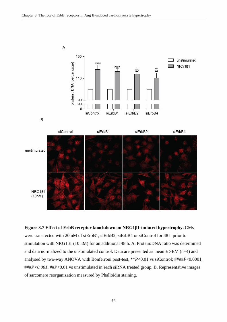
Chapter 3: The role of ErbB receptors in Ang II-induced cardiomyocyte hypertrophy
64
Figure 3.7 Effect of ErbB receptor knockdown on NRG1β1-induced hypertrophy. CMs
were transfected with 20 nM of siErbB1, siErbB2, siErbB4 or siControl for 48 h prior to
stimulation with NRG1β1 (10 nM) for an additional 48 h. A. Protein:DNA ratio was determined
and data normalized to the unstimulated control. Data are presented as mean ± SEM (n=4) and
analysed by two-way ANOVA with Bonferroni post-test, **P<0.01 vs siControl; ####P<0.0001,
###P<0.001, ##P<0.01 vs unstimulated in each siRNA treated group. B. Representative images
of sarcomere reorganization measured by Phalloidin staining.
Chapter 3: The role of ErbB receptors in Ang II-induced cardiomyocyte hypertrophy
65
3.4 Discussion
The mechanism of AT1R-EGFR transactivation has generated substantial interest due to the role of
this pathway in cardiac hypertrophy (Kagiyama et al., 2002; Thomas et al., 2002), renal growth and
dysfunction (Lautrette et al., 2005) and vascular remodelling (Ohtsu et al., 2006b). The central
paradigm for transactivation has involved the AT1R-mediated activation of metalloproteases that
cleave cell surface EGF ligand precursors that then activate EGFR (ErbB1). The number and
diversity of both metalloproteases (MMPs and ADAMs) (Edwards et al., 2008; Hadler-Olsen et al.,
2011), EGF ligands and ErbB receptors means that it has been difficult to identify the specific
players involved in any individual transactivation process. Based on the prevailing literature, we
were surprised to discover in this study that ErbB1 appears to play little or no role in cardiomyocyte
hypertrophy. In contrast, we have confirmed a significant role for ErbB4 in this process. Based on
this, we then hypothesized that the pro-hypertrophic effects of Ang II may in fact be mediated by
ErbB4; however, this was not the case. Using a variety of approaches (RNA interference,
pharmacological blockade) we were unable to demonstrate any link between AT1R activation and
ErbB4. In total, our data raise questions concerning the role of individual ErbB receptors in Ang II-
mediated cardiomyocyte hypertrophy.
However, a lack of involvement of ErbB receptors in Ang II-mediated cardiomyocyte growth does
not discount these receptors from playing a major role in the heart. Indeed, a major finding of our
study is that the ErbB4 receptor is critical for NRG1β1-induced cardiomyocyte hypertrophy. This
supports previous studies which found that NRG1β1 induced hypertrophy in both neonatal and
adult ventricular myocytes accompanied by up-regulation of hypertrophic genes (Zhao et al., 1998).
Indeed, NRG1β1-ErbB4 signalling is important in cardiac development: deletion of either gene
causes embryonic lethality due to heart malformations (Gassmann et al., 1995; Meyer et al., 1995).
NRG1β1-ErbB4 signalling also induces cardiomyocyte proliferation both in vitro and in vivo
(Bersell et al., 2009). We now show that ErbB4 activation by NRG1β1 can also stimulate ERK1/2
activity and hypertrophy in cardiomyocytes, further illustrating the important role of this
mechanism in cardiac physiology.
We were surprised to observe that ErbB1 activation does not cause cardiomyocyte hypertrophy. In a
variety of cells, we and others have previously demonstrated that AT1R activation leads to
phosphorylation of the ErbB1 receptor, downstream signalling and subsequent changes in cell
physiology (Thomas et al., 2002; Ohtsu et al., 2006b). Moreover, deletion of ErbB1 in vascular

Chapter 3: The role of ErbB receptors in Ang II-induced cardiomyocyte hypertrophy
66
smooth muscle cells reduces GPCR-mediated signalling and has marked effects on vascular and
cardiac remodelling (Schreier et al., 2011; Schreier et al., 2013).
One possible explanation for the discrepancy between this work and earlier studies relates to the
method of inhibiting ErbB receptors. AG1478 is regarded as a selective inhibitor for ErbB1,
however as an ATP-competitive antagonist it may also inhibit other receptor tyrosine kinases when
used at higher concentrations (Levitzki et al., 1995; Anastassiadis et al., 2011). In this study, we
used two different concentrations of AG1478 (0.5 and 5 μM), and found that the lower
concentration selectively inhibited ErbB1, whereas the higher concentration inhibited both ErbB1
and ErbB4, consistent with previous reports (Anastassiadis et al., 2011). Thus, it is possible that
previous studies using AG1478 at 5 μM may have attributed actions to ErbB1 activation that are in
fact being mediated by ErbB4 or other tyrosine kinases. Indeed, we have previously shown that 5
μM AG1478 is required to abolish Ang II-induced ERK1/2 activation and cardiomyocyte
hypertrophy (Thomas et al., 2002; Smith et al., 2011), which suggests that multiple ErbB receptor
subtypes may involved in this process.
We then sought to further validate these data using a third method of inhibiting ErbB receptors,
siRNA. siRNAs to ErbB1, 2 and 4 all showed significant knockdown of their targeted receptor, and
we found no evidence of compensatory up-regulation of the other receptor isoforms (Figure 3.5).
Knockdown of individual ErbB receptors did not inhibit Ang II-induced ERK1/2 activation. Whilst
it could be argued that perhaps the magnitude of ErbB receptor knockdown was insufficient to
inhibit Ang II signalling, our results with NRG1β1 do not support this explanation. siRNA targeting
of the ErbB4 receptor reduces both NRG1β1-induced ERK1/2 activation and cardiomyocyte
hypertrophy, thus we would argue that siRNA-mediated down-regulation of ErbB4 was sufficient to
cause a functional reduction in expression, but that Ang II does not appear to act via this receptor.
Taken together, we have used three different methods to inhibit ErbB receptors (shRNA, siRNA
and pharmacological inhibition) and found no evidence for the role of any individual ErbB receptor
isoforms in Ang II-mediated hypertrophic signalling.
By raising questions about the mechanism of AT1R-ErbB receptor transactivation in
cardiomyocytes, this study highlights the potential for investigating signalling pathway networks
using an unbiased, rather than candidate-driven, approach. Indeed, our group has recently used
functional genomics to perform a siRNA screen of AT1R-ErbB1 transactivation in a mammary
epithelial cell line, and have identified a number of novel molecules involved in this process

Chapter 3: The role of ErbB receptors in Ang II-induced cardiomyocyte hypertrophy
67
(George et al., 2013). It is also important to recognize the difficulties in characterizing systems that
involve multiple molecular participants, and thus may have a high degree of redundancy. In the case
of AT1R-ErbB transactivation, this is typified by the number of potential MMP/EGF ligand/ErbB
receptor combinations.
Transactivation of GPCRs and receptor tyrosine kinases is a broad phenomenon that is important in
both normal physiology and numerous disease processes. We confirm an important role for
NRG1β1-ErbB4 signalling in cardiomyocyte growth, yet our data calls into question the existence
of a straightforward mechanism for the transactivation of ErbB receptors by the AT1R.
Accordingly, I will focus on further examining the role of ErbB4 in cardiomyocytes in the
following studies described in this thesis.

CHAPTER 4
INVESTIGATION INTO THE ROLES
OF ErbB4 ISOFORMS IN
CARDIOMYOCYTE
HYPERTROPHY
Chapter 4: Investigation into the roles of ErbB4 isoforms in cardiomyocyte hypertrophy
70
4. Investigation into the roles of ErbB4 isoforms in
cardiomyocyte hypertrophy
4.1 Background
ErbB4 has four isoforms resulting from alternative splicing: JM-a CYT-1, JM-a CYT-2, JM-b
CYT-1 and JM-b CYT-2 (as discussed in section 1.4). The four isoforms are structurally different in
two domains: the extracellular juxtamembrane JM domain (JM-a and JM-b) and intracellular CYT
domain (CYT-1 and CYT-2). Compared to the CYT-2 domain, the CYT-1 contains a 16 amino acid
insertion that acts as a docking site for multiple factors including STAT5, Yap and PI3K (Elenius et
al., 1999; Omerovic et al., 2004; Williams et al., 2004). The JM-a isoform contains 23 amino acids
in the JM domain that allow for ectodomain shedding (Junttila et al., 2000). JM-b contains an
alternative 13 amino acids that resistant to shedding (Junttila et al., 2000). Cleavage of the JM-a
isoform occurs in 2 steps, and produces an 80-kDa soluble fragment. First, the JM-a isoform can be
subjected to ectodomain cleavage by the metalloprotease ADAM17 to produce a 120-kDa
ectodomain fragment released into the extracellular medium and an 80-kDa fragment anchored in
the membrane (Vecchi et al., 1997; Rio et al., 2000; Zhou et al., 2000). The 80-kDa fragment can
subsequently be subjected to further cleavage by γ-secretase within the lipid bilayer to release the
intracellular domain (Chang-Yuan et al., 2001). The cleavage can be induced by various
stimulations such as PMA, NRG1-β1 or GPCR agonists, which potentially activate the ADAMs
(Chang-Yuan et al., 2001). The cleavage of JM-a isoform is cell type dependent. In breast cancer
cells, ovarian cancer cells, lung cancer cells, and a glioma cell line (Vecchi et al., 1996; Zhou et al.,
2000; Määttä et al., 2006; Zeng et al., 2009), the JM-a isoform of ErbB4 is cleaved to produce an
80 kDa intracellular domain (ICD). The ICD can enter the nucleus and interact with transcriptional
factors to regulate transcriptional activity (Tidcombe et al., 2003; Zhu et al., 2006b; Gilmore-
Hebert et al., 2010). Depending on whether they can be cleaved to release the ICD, or possess the
extra 16 amino acids in the intracellular domain, each ErbB4 isoform may therefore play a different
role in regulating growth and differentiation (Muraoka-Cook et al., 2009). For instance, the
cleavable JM-a isoform is capable of regulating the astrogenesis timing by suppressing astrocyte
differentiation, whereas the non-cleavable isoform JM-b cannot (Sardi et al., 2006) (refer to the
Chapter 1 section 1.4.3 for more detail); the CYT-2 ICD promotes mammary cell proliferation,
whereas CYT-1 ICD promotes differentiation (more acinar structures in 3D culture) (Muraoka-
Cook et al., 2009). We have previously shown that all ErbB4 isoforms are detectable in rat neonatal

Chapter 4: Investigation into the roles of ErbB4 isoforms in cardiomyocyte hypertrophy
71
primary cardiomyocytes (Dr. Hsiu-wen Chan, PhD thesis). However, the regulation of these
isoforms in cardiac development and pathological conditions has not been investigated. In addition,
whether the JM-a-containing isoforms can be cleaved to release the ICD and enable trafficking of
the ICD to the nucleus of cardiomyocytes is not known. Finally, the ability of each individual
isoform to mediate cardiomyocyte hypertrophy has not been previously examined. The following
studies aim to answer these questions.

Chapter 4: Investigation into the roles of ErbB4 isoforms in cardiomyocyte hypertrophy
72
4.2 Methods
4.2.1 Animals
To investigate the regulation of the ErbB4 isoforms in cardiac development and cardiac pathology
models (ischaemia-reperfusion model and pathological cardiac hypertrophy model), we
collaborated with other groups who have isolated RNA samples from these models. RNA isolated
from embryonic CD-1 mouse hearts (E10.5, E13.5, E16.5, and E18.5) was a gift from Dr. David
Pennisi (University of Queensland). The RNA from CD-1 mouse hearts at postnatal days 1, 6, 14
and 28 was kindly provided by Dr. Enzo Porrello (University of Queensland). The hearts from an
ischaemia-reperfusion model and a pathological cardiac hypertrophy model were from Prof. Eric N.
Olson’s group (University of Texas) and RNA was extracted by Dr. Enzo Porrello. For the
ischaemia model, 8-12-week old mice (under isoflurane anaesthesia) underwent 45 min of
ischaemia induced by temporary ligation of the left anterior descending coronary artery (LAD)
followed by reperfusion. Control animals underwent a sham operation without occlusion of the
LAD. After 24 hours or 7 days recovery, cardiac tissues were collected from both groups (Aurora et
al., 2012) and RNA extracted. For pathological cardiac hypertrophy model, the calcium-dependent
phosphatase calcineurin driven by cardiac-specific gene promoter αMHC was overexpressed in
mice. The mutant mice develop cardiac hypertrophy soon after birth and reached maximal
hypertrophy at 8 weeks of age. Then they progressed to dilated cardiomyopathy and heart failure
(Molkentin et al., 1998). RNA was extracted from hearts collected from transgenic and wild type
animals at 8 weeks of age.
4.2.2 Relative quantitation real-time PCR with TaqMan probes
Real-time PCR quantification of ErbB4 isoforms was performed with specific primers and probes
described previously in the literature (Junttila et al., 2003; Veikkolainen et al., 2012). Probes for
JM-a, JM-b, CYT-1 and CYT-2 annealed to exons 16, 15, 26 or the junction between exons 25 and
27, respectively (Figure 4.1 A). The primers were synthesized based on the exon sequences flanking
the site recognized by the probe. Amplification of ribosomal 18S was used as an internal control.
PCR was performed in a solution containing 300 nM of each primer (Sigma), 200 nM of probe,
1×TaqMan advanced fast master mix (Applied Biosystems) and 5-10 ng cDNA template in a final
volume of 10 µl. Thermal cycling was performed with StepOnePlus Real-Time PCR system
(Applied Biosystems). Cycling was initiated 20 s at 95°C, followed by 40 cycles of 1 s at 95°C and
20 s at 60°C. Expression levels of all isoforms were normalized to 18S expression using the 2(-ΔΔCT)
method as described (Livak et al., 2001).
Chapter 4: Investigation into the roles of ErbB4 isoforms in cardiomyocyte hypertrophy
73
4.2.3 Absolute quantification real-time PCR
Absolute quantification uses serially diluted standards of known concentrations to generate a
standard curve. The standard curve produces a linear relationship between the initial amounts of
DNA template (N0) and the threshold cycle (CT) and thus can be used to quantify the unknown
samples. This method assumes all standards and samples have equal amplification efficiency. Thus,
standards and samples have to be amplified in the same plate under same PCR conditions (same
probe, primers, reagents, etc). The relationship between the log(N0) and log(CT) is described in the
linear function: Log(N0)= -Log(E+1)·CT + Log(NT) as previously described (Rutledge et al., 2003).
E is amplification efficiency; NT is the number of amplicon molecules at fluorescent threshold; NT
and E are constants (Rutledge et al., 2003). In my study, serially diluted (1:5) standards for each
ErbB4 isoform (see section 4.2.4 for standards generation) and the unknown samples were
amplified in the same plates using the primers and probe described in section 4.2.2. Standard curves
for each isoform of ErbB4 were generated by analyzing the initial amounts of standards and
threshold cycle (Figure 4.2). The initial template amounts (N0) of unknowns can be calculated by
putting its CT value into the corresponding function.
4.2.4 Generation of standards for absolute quantification real-time PCR
Standards should be made from a high quality template because any degradation will affect the
results. PCR products can slightly degrade during storage and thus are not a suitable standards
resource, whilst the cloned target sequence is more stable (Dhanasekaran et al., 2010). To get a
clean and consistent source for the standards, the PCR products in section 4.2.2 were cloned into
TA vectors. Briefly, the PCR products were subjected to DNA purification (DNA extraction kit,
Qiagen) following agarose gel electrophoresis. Then the purified PCR products were ligated into
linearized T-overhang vectors using TOPO® TA Cloning
® Kit (Invitrogen). The ligation was
transformed into competent E. coli cells (New England Biolabs) and then subjected to kanamycin
selection on LB agar plates. Positive clones were confirmed with plasmid extraction followed by
sequencing. The positive clone was preserved at -80°C supplemented with 40 % glycerine.
The recombinant T vector cannot be used directly as a standard because it is circular DNA, whereas
the samples to be tested are linear cDNAs. Circular DNA templates are amplified with a different
efficiency from linear DNA templates even under the same PCR condition (Lin et al., 2011). Thus,
before performing the real-time PCR, the concentrated standard is released from the recombinant T
vector by double enzyme digestion with Hind III and EcoV (New England Biolabs). These two
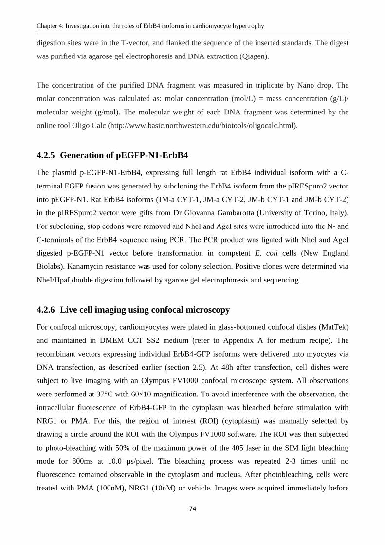
Chapter 4: Investigation into the roles of ErbB4 isoforms in cardiomyocyte hypertrophy
74
digestion sites were in the T-vector, and flanked the sequence of the inserted standards. The digest
was purified via agarose gel electrophoresis and DNA extraction (Qiagen).
The concentration of the purified DNA fragment was measured in triplicate by Nano drop. The
molar concentration was calculated as: molar concentration (mol/L) = mass concentration (g/L)/
molecular weight (g/mol). The molecular weight of each DNA fragment was determined by the
online tool Oligo Calc (http://www.basic.northwestern.edu/biotools/oligocalc.html).
4.2.5 Generation of pEGFP-N1-ErbB4
The plasmid p-EGFP-N1-ErbB4, expressing full length rat ErbB4 individual isoform with a C-
terminal EGFP fusion was generated by subcloning the ErbB4 isoform from the pIRESpuro2 vector
into pEGFP-N1. Rat ErbB4 isoforms (JM-a CYT-1, JM-a CYT-2, JM-b CYT-1 and JM-b CYT-2)
in the pIRESpuro2 vector were gifts from Dr Giovanna Gambarotta (University of Torino, Italy).
For subcloning, stop codons were removed and NheΙ and AgeΙ sites were introduced into the N- and
C-terminals of the ErbB4 sequence using PCR. The PCR product was ligated with NheΙ and AgeΙ
digested p-EGFP-N1 vector before transformation in competent E. coli cells (New England
Biolabs). Kanamycin resistance was used for colony selection. Positive clones were determined via
NheI/HpaI double digestion followed by agarose gel electrophoresis and sequencing.
4.2.6 Live cell imaging using confocal microscopy
For confocal microscopy, cardiomyocytes were plated in glass-bottomed confocal dishes (MatTek)
and maintained in DMEM CCT SS2 medium (refer to Appendix A for medium recipe). The
recombinant vectors expressing individual ErbB4-GFP isoforms were delivered into myocytes via
DNA transfection, as described earlier (section 2.5). At 48h after transfection, cell dishes were
subject to live imaging with an Olympus FV1000 confocal microscope system. All observations
were performed at 37°C with 60×10 magnification. To avoid interference with the observation, the
intracellular fluorescence of ErbB4-GFP in the cytoplasm was bleached before stimulation with
NRG1 or PMA. For this, the region of interest (ROI) (cytoplasm) was manually selected by
drawing a circle around the ROI with the Olympus FV1000 software. The ROI was then subjected
to photo-bleaching with 50% of the maximum power of the 405 laser in the SIM light bleaching
mode for 800ms at 10.0 µs/pixel. The bleaching process was repeated 2-3 times until no
fluorescence remained observable in the cytoplasm and nucleus. After photobleaching, cells were
treated with PMA (100nM), NRG1 (10nM) or vehicle. Images were acquired immediately before

Chapter 4: Investigation into the roles of ErbB4 isoforms in cardiomyocyte hypertrophy
75
and 1 min, 5 min and 15 min after stimulation. The 405 and 473 lasers were sequentially used for
fluorescence excitation with 5% of maximum laser power.

Chapter 4: Investigation into the roles of ErbB4 isoforms in cardiomyocyte hypertrophy
76
4.3 Results
4.3.1 Expression profiles of ErbB4 isoforms throughout development using
relative quantification
The expression of ErbB4 isoforms (JM-a CYT-1, JM-a CYT-2, JM-b CYT-1 and JM-b CYT-2)
exhibits distinct tissue specific patterns (Elenius et al., 1997; Elenius et al., 1999). Moreover, some
studies showed that the expression of ErbB4 isoform is developmentally regulated by physiological
stage: the JM-a isoform is reduced in neuronal precursor cells at late stages of embryonic
development (Fox et al., 2005; Sardi et al., 2006). In the adult mouse and human heart, JM-b CYT-
1 is predominant isoform (Elenius et al., 1997; Elenius et al., 1999), however there is limited
information on how ErbB4 isoform expression is regulated during heart development. To address
this, the developmental expression profile of each isoform was assessed. Mouse hearts at embryonic
days (E) 13.5, 16.5, 18.5 and postnatal days (P) 1, 7, 14 and 28 were collected and RNA extracted.
Quantitative PCR (TaqMan, relative expression) was used to detect each isoform with 18S as an
internal control. Throughout development from E13.5 to P28 expression of all four isoforms
decreased: JM-a isoform at P28 is approximately 300-fold lower than E13.5, JM-b is 17-fold lower,
CYT-1 isoform is 73-fold lower, and CYT-2 is 40 fold lower (Figure 4.1 B). This pattern is similar
for all four isoforms, suggesting that they are regulated in a similar manner during heart
development.

Chapter 4: Investigation into the roles of ErbB4 isoforms in cardiomyocyte hypertrophy
77
Figure 4.1 The developmental expression profile of ErbB4 isoforms. A. Design of primers
and probes to specifically detect individual isoforms using qPCR. Probes for JM-a, JM-b,
CYT-1 and CYT-2 were designed to anneal to exons 17, 16, 27 or the junction between exons
26 and 28, respectively. The primers were complementary to the exon sequences flanking the
site recognized by the probe. The same pair of primer was used for detection of either JM-a
and JM-b isoforms or CYT-1 and CYT-2 isoforms. B. Quantitative qPCR (TaqMan) was used
to detect JM-a, JM-b, CYT-1 and CYT-2 isoforms at in mouse hearts embryonic days 13.5,
16.5, 18.5 and postnatal days 1, 7, 14 and 28. Ribosomal 18S was used as an internal control.
Data was expressed as ratio of expression at P28 and presented as mean ± SEM, n=3.

Chapter 4: Investigation into the roles of ErbB4 isoforms in cardiomyocyte hypertrophy
78
4.3.2 Expression profiles of ErbB4 isoforms throughout development using
absolute quantification
The results described above use different primer/probe sets to amplify the individual ErbB4
isoforms, and therefore the efficiency of PCR amplification may differ for each isoform. Thus, we
cannot directly compare the expression levels of individual isoforms to each other from the relative
quantification PCR data. Instead, we used absolute real-time PCR to identify the predominant
isoforms expressed at different stages of cardiac development. Generation of the standard curves
(Figure 4.2) is described in section 4.2.3 - 4.2.4. All four isoforms were detectable in mice heart
throughout development (Figure 4.3). From the embryo stage (E10.5) into young adulthood (P28),
the expression profile of all four isoforms was consistent with that identified using relative
quantification (Figure 4.1). The JM-b:JM-a ratio ranged from 3:1 at E10.5 to 40:1 at P28,
suggesting that during embryogenesis expression of the two isoforms is similar, but the JM-b
isoform dominates as the heart matures. This is consistent with the literature (Elenius et al., 1997;
Elenius et al., 1999) that JM-b is the primary isoform in the adult mice heart. In contrast, the CYT-
2:CYT-1 ratio did not differ notably and only shifted from 1:1 at E10.5 to 1.7:1 at P28, suggesting
that there are similar levels of CYT-1 and CYT-2 in the heart throughout development. The sum of
the CYT-1 and CYT-2 copy numbers is approximately equal to the sum of the JM-a and JM-b copy
numbers at each individual developmental stage, indicating this absolute quantification is accurate.
Together, these suggest that the non-cleavable JM-b isoforms predominate in the adult heart, and
may therefore be the isoforms most relevant for function.

Chapter 4: Investigation into the roles of ErbB4 isoforms in cardiomyocyte hypertrophy
79
Figure 4.2 Standard curves for individual ErbB4 isoforms. DNA template dilution series
(1:5) was used to generate standard curves by qPCR using the primers and probes specific
for the JM-a, JM-b, CYT-1 or CYT-2 isoforms. The template for standard curve was
generated by digestion of recombinant vectors containing JM or CYT domain of ErbB4
isoforms. N0 is the copy number of templates for each dilution. CT is the threshold cycle
value.

Chapter 4: Investigation into the roles of ErbB4 isoforms in cardiomyocyte hypertrophy
80
Figure 4.3 Absolute expression of ErbB4 isoforms in the heart at different
developmental stages. Absolute quantitative qPCR (TaqMan) was used to measure
expression of JM-a, JM-b, CYT-1 and CYT-2 ErbB4 isoforms at in mouse heart at E10.5,
E18.5, P1 and P28. Ribosomal 18S was used as an internal control. Data was expressed as
absolute copy number of each isoform per ng RNA, presented as mean ± SEM, n=3 for
each time point. Data was analysed by one-way ANOVA with Bonferroni post-test,
****P<0.0001, ***P<0.001, vs E10.5.

Chapter 4: Investigation into the roles of ErbB4 isoforms in cardiomyocyte hypertrophy
81
4.3.3 Regulation of ErbB4 isoform expression in models of cardiac pathology
The regulation of ErbB4 isoform expression by cardiac stressors such as pathological cardiac
hypertrophy or myocardial infarction was also assessed. First, we examined the regulation of the
four isoforms in an ischaemia-reperfusion (IR) model (section 4.2.1). JM-a isoform expression was
significantly increased at 24 h after reperfusion, whereas JM-b was not, suggesting that the
cleavable JM-a isoform was up-regulated by the ischaemia-reperfusion injury (Figure 4.4). The
mRNA level of the CYT-2 isoform was significantly reduced (by ~ 40 %) at 24 h after IR while
CYT-1 was not changed (Figure 4.4). The JM-b:JM-a ratio is reduced by 50% at 24 h and 35% at 7
days after reperfusion compared to the sham-operated controls (Figure 4.4), suggesting that these
two isoforms may play different roles in regulating cardiac biological activity after IR. In contrast,
the ratio of CYT-2/CYT-1 was not changed indicating no potential functional differences related to
the CYT-2 and CYT-1 isoforms in the heart after IR (Figure 4.4).
A transgenic model of cardiac hypertrophy caused by calcineurin overexpression was used to
investigate the regulation of ErbB4 isoforms in pathological hypertrophy (section 4.2.1).
Surprisingly, expression of all of four isoforms was significantly reduced in calcineurin-
overexpressing animals compared to the controls (Figure 4.5). The JM-b:JM-a and CYT-2:CYT-1
ratios were not changed, suggesting that the reduction of all isoforms may be due to the inhibition
of whole gene expression, but not specific regulation of an individual isoform. The down-regulation
of ErbB4 expression in the pathologically hypertrophic heart suggests that ErbB4 may not mediate
this pathological hypertrophy, and that calcineurin-induced signalling may negatively regulate
ErbB4 signalling.

Chapter 4: Investigation into the roles of ErbB4 isoforms in cardiomyocyte hypertrophy
82
Figure 4.4 Regulation of ErbB4 isoform expression in an ischaemia-reperfusion
model. Mouse hearts were collected following 24h (IR24h) or 7 days (IR7d) of
reperfusion after 45 min of ischaemia, or from non-ischemic sham controls. Absolute
quantitation qPCR (TaqMan) was used to measure the copy number of JM-a, JM-b,
CYT-1 and CYT-2 isoforms per ng of total RNA. The right hand panels show the ratios
(JM-b:JM-a and CYT-2:CYT-1) of the copy numbers. Data is presented as mean ± SEM
and analysed with One-way ANOVA with Bonferroni’s multiple comparisons; n=3 for
each treatment, *P<0.05, **P<0.01 vs. control.

Chapter 4: Investigation into the roles of ErbB4 isoforms in cardiomyocyte hypertrophy
83
Figure 4.5 Regulation of ErbB4 isoform expression in a transgenic model of cardiac
hypertrophy. Hearts were collected from calcineurin-overexpressing and wild type mice 8
weeks after birth and absolute quantitiation qPCR (TaqMan) used to measure the copy
number of JM-a, JM-b, CYT-1 and CYT-2 isoforms per ng of total RNA. The right hand
panels show the ratios (JM-b:JM-a and CYT-2:CYT-1) of the copy numbers. Data is
presented as mean ± SEM and analysed with Student’s unpaired t-test; n=3, **P<0.01 vs.
control.

Chapter 4: Investigation into the roles of ErbB4 isoforms in cardiomyocyte hypertrophy
84
4.3.4 The cleavage of ErbB4 isoforms in isolated cardiomyocytes
As discussed in section 4.3.3, the JM-a isoform is significantly up-regulated and the JM-b:JM-a
ratio reduced after ischaemia-reperfusion injury. It can be inferred that the JM-a isoform has a
unique role in heart after IR. There are many biological processes occurring after IR, such as
cardiomyocytes apoptosis, fibroblast proliferation, and activation of immune system. In addition, a
few studies showed that a small proportion of cardiomyocytes can proliferate as part of regeneration
after IR (Beltrami et al., 2001; Malliaras et al., 2013). Given the ErbB4 receptors are mainly
expressed in cardiomyocytes and primarily function as growth regulators, I proposed that the up-
regulated JM-a isoform might play a role in cardiomyocyte regeneration, and this function might be
contributed by the JM-a domain. The JM-a isoform has a protease cleavage site in the
juxtamembrane domain, which allows the receptor to be cleaved, releasing an intracellular domain
(ICD) that can enter into cytoplasm or nucleus (Vecchi et al., 1997; Rio et al., 2000; Zhou et al.,
2000). It has been suggested that the ICD plays a role in regulating cell apoptosis and proliferation
in many cell types (Zhu et al., 2006b; Gilmore-Hebert et al., 2010). However, shedding of the ICD
has not yet been demonstrated in primary cardiomyocytes. Thus, to investigate the function of the
ICD in cardiomyocytes, we studied whether the JM-a isoform is cleaved after agonist stimulation.
Phorbol 12-myristate 13-acetate (PMA) causes cleavage of the JM-a isoform and release of the ICD
in many cell lines (Vecchi et al., 1996; Zhou et al., 2000; Määttä et al., 2006; Zeng et al., 2009).
NRG1 also induces the cleavage of JM-a isoforms in fibroblast and ovarian carcinoma cell lines
(Vecchi et al., 1996; Zhou et al., 2000). Thus, we stimulated isolated rat cardiomyocytes with PMA
(10 nM - 5 µM) for 30 min and probed for the ICD and full-length intact ErbB4 using Western
blotting. As shown in Figure 4.6 A, we detected no ICD in cardiomyocytes treated with increasing
concentrations of PMA for 30 min. We then examined different time points for PMA (100 nM)
stimulation. As shown in Figure 4.6 B treatment with PMA for 5 to 60 min did not produce any
detectable ICD in cardiomyocytes. However, it should be noted that the endogenous JM-a isoform
has very limited expression in primary cardiomyocytes, where expression of the ErbB4 JM-b
isoforms is predominant (Figure 4.3). In addition, PMA only cleaves a portion of JM-a isoforms to
generate the ICD in other cell types (Vecchi et al., 1996), which could make it difficult to detect.

Chapter 4: Investigation into the roles of ErbB4 isoforms in cardiomyocyte hypertrophy
85
Figure 4.6 No evidence for cleavage of endogenous ErbB4 in primary cardiomyocytes
following PMA stimulation. A. Cardiomyocytes were treated with increasing
concentrations (10 nM – 5 µM) PMA or a vehicle control for 30 min, harvested with lysis
buffer and subjected to Western blotting. B. Cardiomyocytes were treated with 100 nM
PMA for 0 – 60 min before harvesting and Western blotting. A primary antibody (sc-283,
Santa Cruz) recognizing the C-terminal of ErbB4 was used to probe for both the ICD and
intact ErbB4, and β-actin was used as a loading control.

Chapter 4: Investigation into the roles of ErbB4 isoforms in cardiomyocyte hypertrophy
86
To further examine the cleavage of ErbB4 isoforms in cardiomyocytes, we generated four vectors
expressing the individual isoforms tagged with GFP at the C-terminus. The vectors were delivered
into cardiomyocytes via transfection, and the cleavage and trafficking of the ICD following
stimulation with PMA or NRG1 was observed by live-cell imaging using confocal microscopy. To
confirm that the GFP-tagged ErbB4 isoforms can be successfully expressed, the recombinant
expressing vector was delivered into HEK-293 cells and Western blot used to detect the
exogenously expressed ErbB4-GFP. We also confirmed that the GFP-tag did not affect the response
of ErbB4 to PMA and the ability of cleavable isoform to release the ICD by stimulating the HEK-
293 cells with PMA (100 nM) after transfection. Full-length ErbB4 and the ICD were detected by
Western blot using an antibody to the C-terminus of ErbB4. As shown in Figure 4.7 A, a 210 kDa
band was detected in all samples, representing the intact GFP-tagged JM-a CYT-1 isoform. An 110
kDa band representing the ICD-GFP was detected after 3 min of stimulation with PMA. The
intensity of this 110 kDa band increased with prolonged PMA treatment, suggesting that cleavage
of JM-a CYT-1-GFP (a1-GFP) and release of the ICD-GFP is PMA dependent and that the
minimum time required for cleavage is 3 min. Cells transfected with JM-a CYT-2-GFP (a2-GFP)
showed low levels of ICD-GFP in the absence of stimulation (Figure 4.7 B), suggesting some auto-
cleavage of JM-a CYT-2 under basal conditions. The ICD-GFP band generated from JM-a CYT-2-
GFP has more intensity than the band from JM-a CYT-1-GFP under the same conditions,
suggesting that the intracellular CYT-2 domain promotes cleavage of the JM-a isoform. This is
consistent with previous work showing that cleavage of the JM-a isoform and generation of the ICD
is kinase dependent, and the CYT-2 isoform possesses stronger kinase activity (Sundvall et al.,
2007). There were no ICD-GFP bands detected in HEK-293 cells transfected with the non-cleavable
isoforms JM-b CYT-1-GFP (b1-GFP, Figure 4.7 C) and JM-b CYT-2-GFP (b2-GFP, Figure 4.7 D).

Chapter 4: Investigation into the roles of ErbB4 isoforms in cardiomyocyte hypertrophy
87
Figure 4.7 Cleavage of exogenous ErbB4-GFP in PMA-stimulated HEK-293 cells.
HEK-293 cells were transfected with 600 ng of either JM-a CYT-1-GFP (a1-GFP, A),
JM-a CYT-2-GFP (a2-GFP, B), JM-b CYT-1-GFP (b1-GFP, C) or JM-b CYT-2-GFP
(b2-GFP, D). Following transfection, cells were cultured in serum-free DMEM for 24 h
before stimulation with 100 nM PMA for 1 to 30 min. Cells were then harvested for
Western blot analysis. The primary antibody (sc-283, Santa Cruz) recognizing the C-
terminal of ErbB4 was used to detect both the ICD and the full-length ErbB4.

Chapter 4: Investigation into the roles of ErbB4 isoforms in cardiomyocyte hypertrophy
88
After confirming that the GFP tag did not affect the cleavage of ErbB4, we introduced these
recombinant vectors to cardiomyocytes via transfection. Confocal microscopy was used to record
the trafficking of ErbB4-GFP in primary cardiomyocytes following stimulation with PMA or NRG1
(section 4.2.6). Before stimulation, the majority of fluorescence representing the ErbB4-GFP was
located on the cell membrane, as expected (Figure 4.8). However, a small amount of intracellular
fluorescence was observed, potentially due to activities such as ErbB4 protein synthesis, recycling
and trafficking. The intracellular fluorescence might interfere with observing the location of the
ICD-GFP stimulation. To avoid this, I photo-bleached the intracellular area without affecting
membrane before stimulation (section 4.2.6). As shown in Figure 4.8, after bleaching the
fluorescence was only located in the cell membrane. After PMA and NRG1 stimulation, a small
amount of fluorescence in a1-GFP transfected cells accumulated in the cytosol (Figure 4.8 A).
However, little fluorescence was observed in the nucleus after stimulation. Vehicle (PBS) treatment
did not induce trafficking of the green fluorescence from the membrane into the cytosol. Similarly,
a2-GFP transfected live cardiomyocytes displayed PMA- and NRG1- but not vehicle- induced
accumulation of the green fluorescence in cytosol (Figure 4.8 B). However, unexpectedly, the non-
cleavable isoforms b1-GFP and b2-GFP also displayed intracellular fluorescence accumulation in
cardiomyocytes (Figure 4.8 C and D). Thus, the fluorescence observed in the cytoplasm may
represent the trafficking of intact receptor and not the released ICD. The mechanism by which the
intact ErbB4 receptor trafficks into the cytosol is unclear. It is maybe due to receptor endocytosis,
or movement of the receptor in cytoplasm following stimulation. When taken together with the
Western blot data described above, this suggests that PMA or NRG1 do not cleave the JM-a
isoform to release the ICD in cardiomyocytes.

Chapter 4: Investigation into the roles of ErbB4 isoforms in cardiomyocyte hypertrophy
89
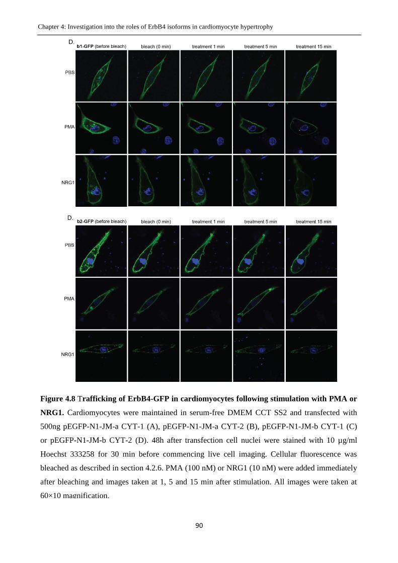
Chapter 4: Investigation into the roles of ErbB4 isoforms in cardiomyocyte hypertrophy
90
Figure 4.8 Trafficking of ErbB4-GFP in cardiomyocytes following stimulation with PMA or
NRG1. Cardiomyocytes were maintained in serum-free DMEM CCT SS2 and transfected with
500ng pEGFP-N1-JM-a CYT-1 (A), pEGFP-N1-JM-a CYT-2 (B), pEGFP-N1-JM-b CYT-1 (C)
or pEGFP-N1-JM-b CYT-2 (D). 48h after transfection cell nuclei were stained with 10 µg/ml
Hoechst 333258 for 30 min before commencing live cell imaging. Cellular fluorescence was
bleached as described in section 4.2.6. PMA (100 nM) or NRG1 (10 nM) were added immediately
after bleaching and images taken at 1, 5 and 15 min after stimulation. All images were taken at
60×10 magnification.

Chapter 4: Investigation into the roles of ErbB4 isoforms in cardiomyocyte hypertrophy
91
4.3.5 The role of individual ErbB4 isoforms in mediating NRG1-induced
cardiomyocyte hypertrophy
The results in section 4.3.4 suggest that the JM-a isoform cannot be cleaved in isolated
cardiomyocytes in vitro, although it is endogenously expressed in neonatal cardiomyocytes (Dr.
Hsiu-wen Chan, PhD thesis). This leads to the question of whether there is a functional difference
between the JM-a and JM-b isoforms if the JM-a isoform cannot be cleaved in cardiomyocytes. In
addition, whether there are any functional differences between the CYT-1 and CYT-2 isoforms in
cardiomyocytes is unknown. The results in chapter 3 of this thesis show that NRG1-induced
cardiomyocyte hypertrophy requires ErbB4. To investigate which isoform of ErbB4 contributes to
this hypertrophic signalling, we designed a rescue system to generate cardiomyocytes in which only
one ErbB4 isoform is expressed. In this system, the shRNA used to knockdown endogenous ErbB4
was co-transfected with a vector expressing a knockdown resistant variant of one ErbB4 isoform.
NRG1 was used to induce hypertrophic signalling, and activation of ANP, CyclinD and MLC-2V
measured with promoter-reporter assays as previously described (section 2.6). Knockdown of
ErbB4 reduced the NRG1-induced MLC-2V (Figure 4.9 A) ANP (Figure 4.9 B), and Cyclin D
(Figure 4.9 C) promoter activity. Interestingly, all isoforms of ErbB4 could rescue the hypertrophic
response, suggesting that they can all mediate hypertrophic signalling. This may indicate that
ErbB4-mediated activation of hypertrophic gene expression is dependent on a function common to
all four isoforms - i.e. the tyrosine kinase activity. The hypertrophic signalling mediated by the
individual isoforms follows a similar pattern for all hypertrophic genes: the CYT-2 isoform seems
to have a greater ability to promote the signalling than CYT-1 isoform, although this difference is
not statistically significant. In contrast, the JM-a and JM-b isoforms both promote hypertrophic
signalling to a similar extent. Promoter activity for each hypertrophic gene was elevated in cells
expressing individual ErbB4 isoforms (compared to the knockdown group) even without
stimulation, indicating a low level of auto-activation of ErbB4. This activation is probably caused
by the over-expression of the delivered isoform(s) of ErbB4.

Chapter 4: Investigation into the roles of ErbB4 isoforms in cardiomyocyte hypertrophy
92
Figure 4.9: Hypertrophic gene promoter activity in NRG1-stimulated cardiomyocytes
expressing only a single isoform of ErbB4. All cardiomyocytes were transfected with three
separate constructs. The first construct (100 ng) encoded a knockdown resistant (kdr) isoform of
ErbB4 (kdr-JM-a CYT1, a1; kdr-JM-a CYT2, a2; kdr-JM-b CYT1, b1; kdr-JM-b CYT2, b2) or
an empty vector control. The second construct (100 ng) encoded either shErbB4 or shControl
(shCon), whilst the third was a hypertrophic gene promoter driven luciferase reporter construct
(300 ng) using the promoter sequence of either ANP (A), MLC-2V (B) or CyclinD (C). Cells
were then stimulated with NRG1β1 for 48 h or left unstimulated. The promoter activity of each
hypertrophic gene was measured using luciferase assays. Data are normalized to the
unstimulated shControl group and presented as mean ± SEM. For statistical analysis, the
original data was normalized by log transformation to achieve equal variance distribution for
each group. Normalised data was then analysed by Two-way ANOVA with Dunnett’s multiple
comparison tests. ****P<0.0001, ***P<0.001 and **P<0.01 vs. NRG1-stimulated knockdown
(vector); ###P<0.001 and ####P<0.0001 vs. unstimulated knockdown (ANP, n=5; MLC-2V,
n=4; CyclinD, n=5).

Chapter 4: Investigation into the roles of ErbB4 isoforms in cardiomyocyte hypertrophy
93
4.4 Discussion
In this chapter, I investigated the expression and cleavage of ErbB4 isoforms, and their ability to
promote hypertrophy in cardiomyocytes. The main discoveries of these studies are: (1) cardiac
ErbB4 expression is highest in the embryonic state and declines postnatally. The expression of all
four isoforms of ErbB4 gradually decreased from embryogenesis to the adult stage. The
predominant isoform in the adult heart is the JM-b isoform. There is no obvious isoform alteration
in IR injury or pathological models of cardiac hypertrophy. (2) JM-a cleavage cannot be observed in
purified cultures of primary cardiomyocytes following stimulation with NRG1 or PMA. However,
cleavage of the JM-a isoform could be demonstrated in HEK-293 cells. (3) All four ErbB4 isoforms
could mediate NRG1-induced cardiomyocyte hypertrophy.
In the literature, the JM-b isoform is proposed to be the only isoform in the adult mice heart
(Elenius et al., 1997; Elenius et al., 1999). My study showed that, whilst the JM-b isoform is
predominant, both the JM-a and JM-b isoforms are detectable in the adult mice heart. The reason
these previous studies did not detect JM-a may be because they used traditional end-point PCR and
agar gel electrophoresis, which is less sensitive than the TaqMan method used in my study. Despite
this disagreement on the expression of JM-a in postnatal heart, my finding that the JM-b isoform is
the major isoform in adult heart is in agreement with previous work.
One interesting result in my study is that ErbB4 is expressed at a much higher level in the
embryonic heart than in the adult. ErbB4 is essential for cardiac growth: deletion of ErbB4 inhibited
normal cardiac growth in both the embryonic (Gassmann et al., 1995) and postnatal stages (Garcia-
Rivello et al., 2005). The high level of ErbB4 in embryonic heart is consistent with the strong
requirement for cardiac growth during development. However, determining which isoforms of
ErbB4 are predominant in embryonic heart has not been investigated previously. Our results
showed that in the earlier embryonic stage (E10.5) the JM-b:JM-a ratio is 3:1, whilst in the adult
this increases to 40:1. We were unable to detect the expression of four isoforms any earlier than E9
due to the tiny size of the heart tube and inefficient RNA extraction from the heart tube at this stage.
While alternative methods to detect RNA expression (i.e., in situ hybridisation) were not performed,
based on the expression pattern throughout the cardiac development (Figure 4.1), it may be inferred
that at earlier embryo stage E9, the JM-b and JM-a might be expressed at a similar level. Because
ErbB4 is primarily expressed in cardiomyocytes (which comprise two thirds of the volume of the
whole heart), and not detectable in fibroblasts (which comprise one third of the volume) (Zhao et

Chapter 4: Investigation into the roles of ErbB4 isoforms in cardiomyocyte hypertrophy
94
al., 1998), I would propose that my study reflects the developmental regulation of ErbB4 expression
in cardiomyocytes instead of other cells, although further experiments can be employed to confirm
this. The decrease in the JM-a:JM-b ratio is coincident with loss of the ability of cardiomyocytes to
proliferate. In the embryonic heart, cardiomyocytes can both proliferate and enlarge to contribute to
the cardiac growth. However, shortly after birth, the cardiomyocytes exit the cell cycle and heart
growth becomes dependent on cardiomyocyte enlargement (cardiomyocyte hypertrophy)
(Markwald et al., 2010).Whilst the ability of ErbB4 to promote cardiac development is well-
established in literature (section 1.6.7), it is not yet known whether the different isoforms of ErbB4
can mediate different functional effects. In many cell lines, ErbB4 isoforms have different growth
promoting abilities. These differences are primarily related to the JM and CYT domains. By
overexpressing full-length JM-a and JM-b in mouse embryonic astrocytes in an ErbB4-null
background, Sardi et al. found only the cleavable JM-a isoform is capable of suppressing astrocyte
precursor differentiation and maintaining their neurogenic potential (Sardi et al., 2006). In human
breast cancer cells, the JM-a CYT-2 isoform was found to promote the phosphorylation of itself and
proliferation of the cancer cells, which was proposed to be related to cleavage of the receptor
(Määttä et al., 2006; Zhu et al., 2006b). Mammary cell lines expressing exogenous CYT-2 ICD
grow more rapidly than the same cell line expressing CYT-1 ICD (Muraoka-Cook et al., 2009). In a
mouse fibroblast cell line, the ICD of JM-a CYT-2 was found to interact with AP-2 to positively
regulate the platelet-derived growth factor receptor-alpha (PDGFA) promoter leading to cell
proliferation (Sundvall et al., 2010). Taken together with these observations, our data suggests that
cardiomyocytes require different isoforms to proliferate or hypertrophy. Embryonic
cardiomyocytes, for example, might require the JM-a isoform, but not the JM-b isoform, to
proliferate.
Although it has been confirmed in the literature (Elenius et al., 1997; Elenius et al., 1999) as well as
in our laboratory that primary isoforms of ErbB4 in the adult heart are JM-b isoforms, there was the
possibility that this could be altered in pathological conditions. We therefore characterized the
cardiac expression of ErbB4 isoforms using in vivo models of ischaemia-reperfusion injury and
pathological hypertrophy. Only the JM-a and CYT-2 isoforms are regulated after ischaemia while
the expression levels of the JM-b and CYT-1 isoforms were not significantly altered. Although the
increase of JM-a isoforms at 24h after ischaemia reperfusion is up to 2 fold, the absolute amount of
JM-a is still diminutive compared to the JM-b isoforms (approximately 1:20). This suggests there is
no alteration in the primary isoform of ErbB4 expressed in cardiac tissue, and that expression of the
JM-b isoforms dominates in the heart, even in pathological conditions. If JM-a does relate to the

Chapter 4: Investigation into the roles of ErbB4 isoforms in cardiomyocyte hypertrophy
95
proliferation of the cardiomyocytes as discussed above, the low expression levels of JM-a in IR
model described here is consistent with the limited regenerative capacity of the injured heart
(Laflamme et al., 2011). It is believed that shortly after birth the majority of cardiomyocytes in
heart exit the cell cycle and lose the ability to proliferate. Thus, the injured adult heart cannot be
repaired via generation of new cardiomyocytes.
In the pathological hypertrophy model, expression of all four isoforms was significantly reduced.
However, since this affects all isoforms equally this change may reflect reduced transcription of the
entire gene and not isoform specific regulation. In this model, the pathological hypertrophy is
induced by genetically overexpressed calcineurin, a calcium-dependent phosphatase. It causes
cardiac hypertrophy via dephosphorylating the transcription factor NF-AT3 and enabling its
translocation to the nucleus where NF-AT3 can interact with GATA4 to activate cardiac
transcription and hypertrophic growth (Molkentin et al., 1998). The decrease of ErbB4 in
pathological hypertrophy suggests that ErbB4 is not required by the calcineurin signalling to
mediate cardiac hypertrophy. NRG1 induces cardiomyocyte hypertrophy via activation of ERK
(Baliga et al., 1999), which can also activate the GATA4 to contribute to the hypertrophic growth
(Yanazume et al., 2002; Akazawa et al., 2003). However, the activation of calcineurin signalling
and NRG1-ErbB4 signalling in vivo leads to different results in the heart. The former eventually
leads to heart failure following cardiac hypertrophy (Molkentin et al., 1998) whereas the latter
enhances cardiac function via regulating contractile and sarcomere proteins in cardiomyocytes
(Jiang et al., 2010) and reducing the cardiomyocyte death even in the presence of pathological
stress, such as infarction, cardiomyopathy and myocarditis (Liu et al., 2006).
In addition to investigating the regulation of ErbB4 isoform expression in various models, I also
studied the cleavage of JM-a isoforms. While specific cleavage of JM-a ErbB4 isoforms was
confirmed in transfected HEK294 cells, no cleavage was observed in neonatal rat cardiomyocytes
stimulated with either PMA or NRG1. This result was first demonstrated by Western blot and
confirmed by observing the trafficking of ErbB4 with confocal microscopy live cell imaging. If the
JM-a isoform cannot be cleaved in cardiomyocytes, there may be no functional differences between
the JM-a and JM-b isoforms. Our lab has previously detected expression of both isoforms in
purified cultures of primary neonatal rat cardiomyocytes (Dr. Hsiu-wen Chan, PhD thesis) and
expression of the JM-b isoform is much higher than the JM-a isoform (Mr. Hengbo Shi,
unpublished data). If there is no functional difference, the reasons underlying the marked difference
in the expression levels of JM-a and JM-b isoforms in postnatal cardiomyocytes remain unclear.
Chapter 4: Investigation into the roles of ErbB4 isoforms in cardiomyocyte hypertrophy
96
The reason for the lack of cleavage of JM-a in cardiomyocytes has not been identified. One
possibility is that cardiomyocytes lose some components required for the cleavage during the
isolation and purification of the cells, and therefore cannot cleave ErbB4 in vitro. For instance, the
functional loss of protein, such as the expression of AT1R, during cardiomyocyte isolation has been
proposed previously (Thomas et al., 2002). Another possibility is that cardiomyocytes require the
surrounding cells, such as fibroblasts or endothelial cells, in order to undertake cleavage. As
discussed in section 1.4.2, the mechanism for JM-a isoform cleavage (in other cell types) involves
the activation of ADAM17 or MMP to cleave the JM-a isoform in the juxtamembrane region and
remove the ectodomian from the receptor (Rio et al., 2000; Chang-Yuan et al., 2001). The
remaining intracellular part of the receptor anchored in the cell membrane is then subjected to
additional cleavage by γ-secretase to generate the 80 kD ICD (Rio et al., 2000; Chang-Yuan et al.,
2001). However, the mechanism is still not fully resolved and additional proteins may be involved
in this process. Whether cardiomyocytes are subject to the full range of ErbB4 regulation observed
in other cell types is unknown. Perhaps some unidentified components located on the membrane of
neighbouring cells can cause cleavage of a substrate located on the membrane of adjacent
cardiomyocytes - if so, this would explain why we did not observe JM-a cleavage in purified
cardiomyocytes. Moreover, we only examined ErbB4 receptor trafficking after stimulation with
factors that are known to induce cleavage of the JM-a isoform in other cell types. It is possible that
there are additional factors that can induce cleavage of the JM-a isoform which have not yet been
identified, and that these may initiate cleavage of the JM-a isoform in cardiomyocytes.
Finally, my study found that all four isoforms of ErbB4 can mediate NRG1-induced cardiomyocyte
hypertrophy. This suggests that the pro-hypertrophic ability of the four isoforms depends on a
common feature among them (e.g. the tyrosine kinase activity), but does not relate to possession of
the cleavable domain or intracellular binding domain. However, the magnitude of hypertrophy
seems to be influenced by the CYT domain although this was not statistically significant due to
inherent variability in the data set. One reason why the CYT2 isoform can promote hypertrophic
signalling more than CYT1 may be due to the intracellular kinase domain, which is more easily
phosphorylated in CYT2 isoforms (Sundvall et al., 2007). The mechanism for this is still not clear,
however it is proposed that a “loop structure” exists in the intracellular domain possessing CYT1,
and that this may inhibit tyrosine kinase activity (Sundvall et al., 2007). Our data suggests that the
JM-a and JM-b isoforms have comparable abilities to promote hypertrophic signalling. This is
consistent with earlier studies in this chapter suggesting the JM-a isoform cannot be cleaved in
purified cardiomyocytes. The only known difference between the JM-a and JM-b isoforms is that
Chapter 4: Investigation into the roles of ErbB4 isoforms in cardiomyocyte hypertrophy
97
the JM-a isoform can be cleaved while the JM-b cannot. If cardiomyocytes lack the JM-a cleavage
mechanism, I would not predict any functional difference between the JM-a and JM-b isoforms.
This could explain the absence of functional differences between JM-a and JM-b isoforms in our
hypertrophic signalling experiments. It should be noted that the hypertrophic gene signal in all the
‘rescued’ groups is greater than that of the controls, suggesting that the expression level of the
delivered ErbB4 isoforms is higher than endogenous expression levels. This high expression might
mask the potential differences between the four isoforms in their ability to mediate hypertrophy.
There is the possibility that when expressed at a lower level (closer to that of endogenous
expression), the capacity of individual ErbB4 isoforms to differentially regulate hypertrophic gene
expression may be exposed. Taken together, in my study, no cleavage of the JM-a isoforms has
been found in cardiomyocytes, and the ability of ErbB4 to promote hypertrophic seems to be kinase
activity dependent and independent of isoform cleavage.


CHAPTER 5
THE FUNCTION OF ErbB4 IN THE
ADULT HEART

Chapter 5: Function of ErbB4 in adult heart
100
5. Function of ErbB4 in the adult heart
5.1 Background
ErbB4 is critical for heart development, anti-apoptosis and hypertrophy. Global deletion of ErbB4 is
lethal by embryonic day 11 in mice due to defective development of the heart (Gassmann et al.,
1995). The heart defect in the ErbB4 null mice could be rescued by expressing ErbB4 under a heart
specific myosin promoter. These mice reached adulthood and are fertile (Tidcombe et al., 2003),
indicating that ErbB4 plays a significant role in heart development. In addition to cardiac
development, ErbB4 signalling also prevents cardiac injury in adults. The ErbB4 agonist
neuregulin1 (NRG1) rescues adult ventricular myocytes from hypoxia-reoxygenation-induced
apoptosis, whereas the endothelium-selective deletion of NRG1 decreases the ability of the
myocytes to tolerate an ischemic insult (Hedhli et al., 2011). Exogenous NRG1 also decreased the
number of apoptotic cells and improved heart function in a rat model of diabetic cardiomyopathy
with systolic and diastolic dysfunction (Li et al., 2011), and in cultured myocytes NRG1 inhibits the
apoptosis induced by serum starvation (Zhao et al., 1998). Taken together, these studies indicate
that NRG-ErbB4 signalling is important in cardiac protection. Finally, ErbB4 also mediates cardiac
hypertrophy. NRG1 promotes hypertrophy in both adult and neonatal rat primary cardiomyocytes,
which is accompanied by enhanced expression of the hypertrophic genes ANP and skeletal α-actin
(Zhao et al., 1998), indicating that ErbB4 might play an important role in adult cardiac function and
structure maintenance. To date, few studies have examined the role of ErbB4 in the adult heart. A
study using a targeted knockout model demonstrated that cardiac specific deletion of ErbB4 led to
severe dilated cardiomyopathy in adult mice, characterized by thinner ventricular walls with
reduced contractility and conduction delays (Garcia-Rivello et al., 2005). However, in this study
deletion of the ErbB4 occurred before birth making it hard to exclude the possibility that the
cardiomyopathy observed in the adult stage was due to a defect during cardiac development. To
overcome the requirement of ErbB4 for cardiac development and investigate the function of ErbB4
in the adult heart, we used a conditional knockout model, the αMHC-MerCreMer/loxP system
(Sohal et al., 2001), to specifically delete ErbB4 from adult cardiomyocytes.
The Cre-loxP system is a commonly used tool to provide targeted gene deletion. Cre recombinase is
found in P1 bacteriophage and is a 38kD site-specific recombinase that catalyzes the recombination
between two DNA recognition sites, termed loxP (Hamilton and Abremski, 1984). The loxP site is
a 34 bp consensus sequence, consisting of a core spacer sequence of 8bp and two 13bp palindromic

Chapter 5: Function of ErbB4 in adult heart
101
flanking sequences. The asymmetric core sequence defines the orientation of the loxP site. The Cre
recombinase can excise a DNA segment from the genome when the segment is flanked by loxP
sites in the same orientation (Nagy, 2000). To delete genes in a tissue specific manner, Cre
recombinase can be driven by a tissue specific gene promoter. To give temporal control of gene
deletion, Cre recombinase can be fused to a mutated estrogen receptor ligand binding domain (ER-
LBD) (MerCreMer) (Nagy, 2000; Chien, 2001). This mutant ER selectively binds the synthetic ER
antagonist tamoxifen (rather than endogenous estrogen), and upon tamoxifen treatment, the
activated ER-LBD causes translocation of Cre recombinase to the nucleus, where it causes
recombination of the gene flanked by loxP (Sohal et al., 2001). Thus, in this system, the targeted
gene deletion is not only tissue specific but also temporally controlled by tamoxifen injection. The
first application of this system to gene deletion from cardiomyocytes was achieved by Sohal et al.
in 2001 (Sohal et al., 2001). They linked the cardiac-specific α-MHC (alpha-myosin heavy chain)
promoter to the Cre/ER-LBD gene (αMHC-MerCreMer transgene). Transgenic mice with Cre-
dependent, loxP-inactivated lacZ were crossed with transgenic mice containing the αMHC-
MerCreMer gene to generate the αMHC-MerCreMer/lacZ mice. Using lacZ as a reporter, they
demonstrated that tamoxifen-induced Cre activity caused 80% recombination of targeted gene in the
embryonic, neonatal, or adult heart, but not other tissues. Taking advantage of this model, we will
delete ErbB4 from adult mice heart to investigate its function in this stage.
5.2 Methods
5.2.1 Animals
The homozygous ErbB4fl/fl
mouse line (with exon2 of ErbB4 gene flanked by loxP sites) was
crossed with the homozygous αMHC-MerCreMer mouse line (Cre+/+
) to generate double
heterozygous αMHC-MerCreMer and ErbB4-floxed mice (Cre+/-
/ErbB4fl/wt
). These mice were then
backcrossed with the ErbB4fl/fl
line to generate heterozygous αMHC-MerCreMer and homozygous
ErbB4-floxed animals (Cre+/-
/ErbB4fl/fl
), and homozygous Cre-/-
ErbB4fl/fl
mice in the offspring
(Figure 5.1). To generate the Cre+/-
/ErbB4wt/wt
animals, Cre+/-
/ErbB4fl/wt
mice were backcrossed with
Cre+/+
mice (Figure 5.4). The original homozygous ErbB4fl/fl
and homozygous α-MHC-MerCreMer
mice lines (Cre+/+
) were obtained from Dr Kent Lloyd, University of California (Mutant Mouse
Regional Resource Centres) and Jackson Laboratories, respectively. Both of the strains are on a
B6/129 mixed background.

Chapter 5: Function of ErbB4 in adult heart
102
Animals were housed under standard conditions at 22°C with a 12-h light/12-h dark cycle and free
access to water and commercial mouse chow. All experiments were conducted in accordance with
the Australian Code of Practise for Care and Use of Animals for Scientific Purposes and approved
by the institutional ethics committees of The University of Queensland.
5.2.2 Genotyping
Mice were toe-clipped at 7-10 days of age to provide tissue samples for genotyping. The toes were
digested in DNA extraction buffer (100 mM Tris-HCl (pH8.5), 5 mM EDTA (pH8.0), 200 mM
NaCl, 0.2% SDS and 0.48mg/ml proteinase K) at 55°C overnight. The genomic DNA was
precipitated with ammonium acetate and ethanol before PCR amplification using primers specific
for Cre, or ErbB4 wild type and ErbB4 floxed DNA sequences (Figure 5.2 A). The former reaction
will yield a 440bp band (Cre), and the latter will yield 350bp and 400bp products for ErbB4 wild
type and ErbB4 floxed alleles respectively. The primer sequences are listed in Appendix B. PCR
reactions (20μl total volume) contained 20 ng genomic DNA, 0.5 µM of each primer, 0.2 mM each
of dNTPs, 5% DMSO, 0.375 U of Phire Hot Start II DNA Polymerase (Finnzymes) and 1× Phire
reaction buffer (final concentration). The PCR cycling conditions started with an initial denaturation
at 98°C for 30s, followed by 35 cycles of denaturing at 98°C for 5 s, annealing at 58°C for 5 s (for
ErbB4 floxed and wild type) or 55°C for 5 s (for Cre), extension at 72°C for 10s, and a final
incubation at 72°C for 1 min. PCR products were separated using 1.5-2% agarose gel
electrophoresis and images were captured with the Quantity One system. To detect the Cre copy
number, qPCR (SYBR) was employed to quantify the genomic Cre level and genomic 18S level
was used as an internal control. The qPCR condition followed the standard amplification protocol
(refer to section 2.8). The sequence of primers used is listed in Appendix B.
5.2.3 Tamoxifen injection
To induce Cre-mediated recombination, all animals (9-10 week of age) were treated with tamoxifen
(Sigma) by intraperitoneal injection for 10 consecutive days at a dosage of 20 mg/kg/day.
Tamoxifen was dissolved in peanut oil at a concentration of 4 mg/ml at 37°C for 2 hours and made
up fresh each day. The success of deletion of ErbB4 in the Cre+/-
ErbB4fl/fl
mouse heart was
determined by qPCR in an initial cohort of animals at 10 days after tamoxifen injection. Separate
cohorts of animals were then phenotypically examined at 3-4 months or 7-8 months after tamoxifen
injection.
Chapter 5: Function of ErbB4 in adult heart
103
5.2.4 Echocardiography
Echocardiography was conducted on all animals using a Philips HD15 ultrasound system equipped
with a 15-MHz transducer. Mice were anesthetized with 2% isoflurane in O2 by inhalation and
normal body temperature was maintained using an external heat source. Two-dimensional mid-
papillary level short axis views of the ventricle were captured. M-mode images were obtained by
positioning a cursor in the 2-dimensional view from the anterior to the posterior wall of the left
ventricle and adjacent to the two papillary muscles. M-mode images were used to obtain the
following measurements during both systole and diastole: interventricular septal thickness (IVS),
left ventricular posterior wall thickness (LVPW), left ventricular internal diameter (LVID). The
fractional shortening (FS) was calculated using the equation (FS= (LVID (diastolic)-LVID
(systolic))/LVID (diastolic)). The measurements were repeated 5 times over 5 cardiac cycles for
each parameter and the average of these used for statistical analysis. Heart rate was monitored by
electrocardiography during the procedure. The experimenter was blinded to the genotype of the
animals for all echocardiographic assessments and decoded post-analysis.
5.2.5 Measurement of heart/kidney/body weight and tibial length
Animals were euthansed and the heart and kidneys was carefully removed and weighed. Tibial
length was measured after the skeletal muscle was completely removed from the bone. The left and
right tibias were measured three times each with a digital calliper, and the average of these 6
measurements was used as the final tibial length for each mouse.
5.2.6 Histology
Mid-ventricular sections were fixed in 4% PFA for 48 hours and then embedded in paraffin.
Sections (5 μm thick) cut from paraffin-embedded specimens was deparaffinised in xylene followed
by hydration through an ethanol gradient to PBS or distilled water before being used for wheat germ
agglutinin (WGA) staining, Masson's trichrome staining, TUNEL staining or immunofluoresence.
5.2.7 WGA staining
Sections were incubated with 50 μg/ml WGA conjugated with Alexa Fluor®594 (Invitrogen) for 60
min, washed 3 times with PBS and mounted for fluorescence microscopy. 5 images were taken for
each section under 400 × magnification. The images were taken randomly from two fields next to
the endocardium, two fields next to the epicardium and one field in the papillary muscle area. In
each field, the cross sectional area of 40 - 130 cells was measured. For the cell size quantification,

Chapter 5: Function of ErbB4 in adult heart
104
images were opened with Image J. To perform an unbiased measurement, a grid with 3×4
(horizontal × vertical) test lines was generated with Image J and applied to every image. Only cells
intersected with the grid lines were measured and the experimenter was blinded for all
measurements.
5.2.8 Masson’s trichrome staining
Masson’s trichrome staining was performed according to the manufacturer’s protocol
(Polysciences). Briefly, sections were merged in Bouin’s solution at 60°C for 1 hour, washed in
running tap water to for 5 minutes, followed by incubation in Weigert’s iron hematoxylin working
solution for 10 minutes. After washing in running tap water, sections were placed in Biebrich
Scarlet-Acid Fuchsin Solution for 5 minutes and rinsed in distilled water. Sections were
differentiated with phosphotungstic/phosphomolybdic acid for 10 minutes and transferred directly
into Aniline blue for 5 minutes for counterstaining. Sections were rinsed in distilled water before
differentiating in 1% Acetic acid for 1 minute and performing a final rinse in distilled water. This
stains collagen fibres in blue, nuclei in black and the cytoplasm in red. Five images were captured
for each section and the area of cardiac fibrosis (excluding the perivascular area) was measured
using Image J.
5.2.9 Immunofluorescence
For phospho-histone3 (pH3) staining and Troponin-T co-staining, the slides were pretreated with
antigen retrieval buffer (refer to Appendix A for composition) at 110°C for 20 min in a Decloaker
apparatus before gradually cooling to room temperature by addition of PBS. Following this, the
slides were permeabilized in 0.3% Triton for 5 min before being washed with PBS and blocked
with 10% goat serum at room temperature for 20 min. Sections were then incubated with anti-
phosphohistone H3 (rabbit, Millipore) and anti-Troponin-T (mouse, Thermoscientific) primary
antibodies (1:100 dilution with 2% goat serum in PBS) at 4°C overnight. After washing 3 times
with PBS, the sections were incubated with Alexa Fluor 488-conjugated (goat anti-rabbit,
Invitrogen) and Alexa Fluor 555-conjugated secondary antibodies (goat anti-mouse, Invitrogen) at
RT for 1h. Hoechst (10µg/ml, Sigma) stain was used to visualize nuclei before slides were washed
3 times in PBS and mounted with Fluoromount G mounting medium (Southern Biotech).

Chapter 5: Function of ErbB4 in adult heart
105
5.2.10 Statistical analysis
Values are expressed as mean ± SEM, unless indicated otherwise. Comparisons between wild-type
and ErbB4 conditional knockout animals were analysed by unpaired Student’s t-test using
Graphpad Prism 6. P values ≤ 0.05 were considered significant.
Chapter 5: Function of ErbB4 in adult heart
106
5.3 Result
5.3.1 Generation of Cre+/-
ErbB4fl/fl
mice
To investigate the function of ErbB4 in adult mice heart, I adopted the αMHC-MerCreMer/loxP
system, which allowed me to delete the ErbB4 receptor from cardiomyocytes in adult mice. Two
original breeding lines of mice (homozygous αMHC-MerCreMer mice (Cre+/+
) and homozygous
ErbB4fl/fl
) were used to generate Cre+/-
/ErbB4fl/fl
, and Cre-/-
/ErbB4fl/fl
mice (section 5.2.1 and Figure
5.1). Following tamoxifen injection, the ErbB4 can be deleted from cardiomyocytes in Cre+/-
/ErbB4fl/fl
animals since they contain both Cre and loxP flanked-ErbB4 genes, but not in the Cre-/-
/ErbB4fl/fl
animals, which lack the Cre gene. Thus Cre+/-
/ErbB4fl/fl
animals were used as the
knockout group and ErbB4fl/fl
animals were used as the control group. In total, 9 breeding pairs of
Cre+/-
/ErbB4fl/wt
and ErbB4fl/fl
produced 75 offspring.

Chapter 5: Function of ErbB4 in adult heart
107
Figure 5.1 Breeding scheme for transgenic animal generation. The original homozygous
ErbB4fl/fl
breeders were bred with homozygous Cre+/+
breeders to generate double heterozygous
Cre+/-
/ErbB4fl/wt
mice. These were then backcrossed with the ErbB4fl/fl
mice to generate Cre+/-
/ErbB4fl/fl
mice (conditional knockout group) and the Cre-/-
ErbB4fl/fl
mice (control group).

Chapter 5: Function of ErbB4 in adult heart
108
5.3.2 Genotyping
All offspring were genotyped using separate PCR reactions for the Cre and Floxed ErbB4
transgenes. To detect the inserted Cre gene, PCR was used to amplify a 440 bp Cre gene fragment
from whole DNA genome (Figure 5.2 A). A single band in 440bp in electrophoresis indicates the
Cre positive genotype and otherwise indicates the Cre negative genotype (Figure 5.2 B). To
differentiate the ErbB4fl/fl
animals from the ErbB4fl/wt
animals, PCR was used to amplify the wild
type ErbB4 gene fragment and the mutated fragment (flanked by loxP site) with three primer sets in
one reaction. One is a common sense primer annealing the 5’ sequence in exon2 of ErbB4, and the
others are two reverse primers annealing to the 3’ sequence in exon2 and the sequence in the loxP
site, separately (Figure 5.2 A). A single band in 400 bp in electrophoresis indicates the ErbB4fl/fl
genotype, and two bands in a 350 bp and 400 bp, separately indicated the ErbB4fl/wt
genotype
(Figure 5.2 B) (Jackson-Fisher et al., 2006). Among the 75 offspring produced, 20 mice were the
Cre+/-
/ErbB4fl/fl
genotype and 20 mice were the Cre-/-
ErbB4fl/fl
.

Chapter 5: Function of ErbB4 in adult heart
109
Figure 5.2 Genotyping of the Cre recombinase and floxed ErbB4 transgenes. A. The
presence or absence of the Cre recombinase gene was determined by amplifying a 440 bp
region of the Cre gene. In separate PCR reactions, we used a common sense primer binding
within exon 2 of ErbB4, and two anti-sense primers which bound either the downstream
region of exon2 or within the loxP site. This led to amplification of both the wild type (350 bp
PCR product) and floxed ErbB4 (400 bp product) genes, respectively. Reaction products were
separated by agarose gel electrophoresis (1.5% and 2% for Cre recombinase and ErbB4
respectively) B. Representative image of agarose gel electrophoresis for genotyping. Lane 1
represents the Cre-/-
ErbB4fl/wt
genotype, lane 2 and 3 represent Cre+/-
/ErbB4fl/fl
, lane 4, 6, and 7
represent Cre-/-
ErbB4fl/fl
and lane 5 represents the Cre+/-
ErbB4fl/wt
genotype.

Chapter 5: Function of ErbB4 in adult heart
110
5.3.3 Optimization of tamoxifen administration
In this conditional knockout animal model, the deletion of ErbB4 is triggered by administration of
tamoxifen. An insufficient dose of tamoxifen may cause failure of the deletion, but too high a dose
of tamoxifen is toxic and may lead to the death of the mouse. Previous work in our lab used 3
different doses of tamoxifen: 20, 30 and 40 mg/kg/day for four days of consecutive ip injection. At
the highest dose (40 mg/kg/day), tamoxifen induced an efficient deletion of ErbB4, but with a high
mortality rate. The intermediate dose of tamoxifen (30 mg/kg/day) induced less deletion of ErbB4,
and greatly reduced the mortality rate. The lowest dose of tamoxifen (20 mg/kg/day) failed to
induce the deletion of ErbB4 (Schlegel et al., unpublished data). To avoid the toxicity associated
with high dose tamoxifen yet still achieve significant deletion of ErbB4, we tested the low dose of
tamoxifen over an extended injection period (Hsieh et al., 2007). We injected Cre+/-
/ErbB4fl/fl
and
Cre-/-
/ErbB4fl/fl
animals with 20 mg/kg/day tamoxifen for 10 consecutive days. Animals were
allowed 10 days recovery after the end of the tamoxifen treatment before being euthanased and
tissues examined for ErbB4 expression by qPCR. This treatment regime had no obvious adverse
effects on the health of the animals. The RT-qPCR revealed that the mRNA level of ErbB4 in heart
was reduced by ~ 90% compared to the control, but the mRNA level of ErbB4 in skeletal muscle
was not changed (Figure 5.3), thus confirming specific deletion of ErbB4 in the heart.
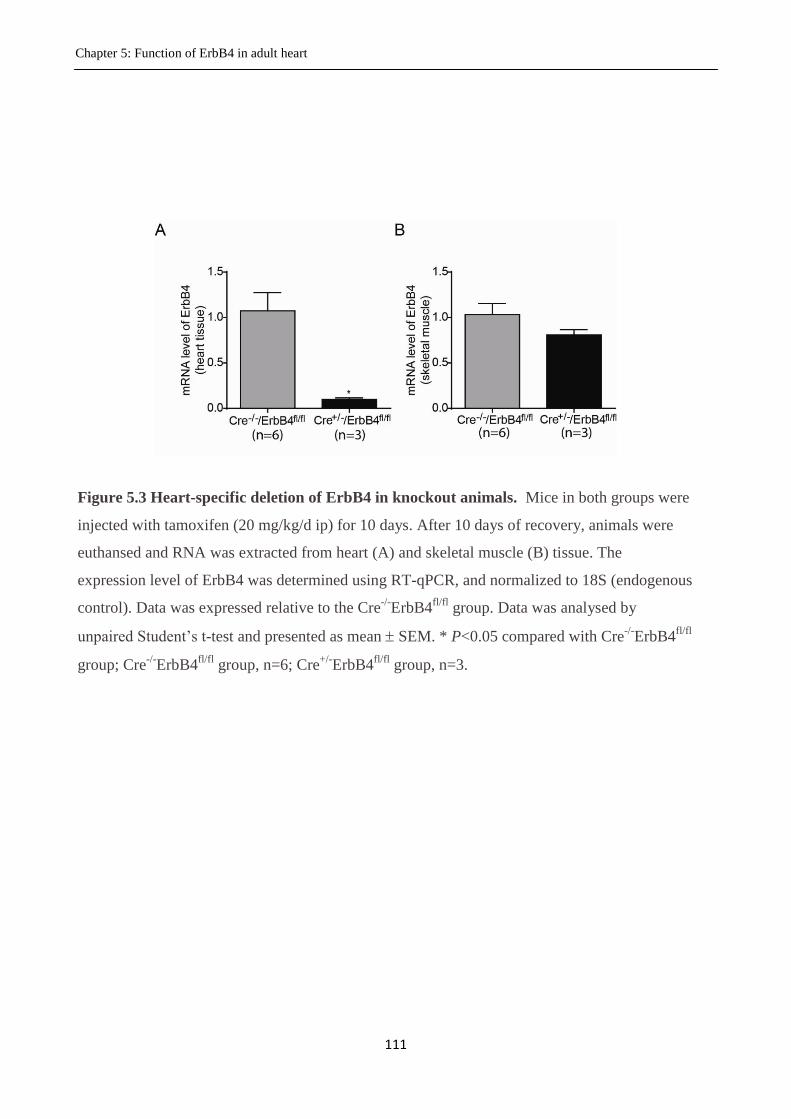
Chapter 5: Function of ErbB4 in adult heart
111
Figure 5.3 Heart-specific deletion of ErbB4 in knockout animals. Mice in both groups were
injected with tamoxifen (20 mg/kg/d ip) for 10 days. After 10 days of recovery, animals were
euthansed and RNA was extracted from heart (A) and skeletal muscle (B) tissue. The
expression level of ErbB4 was determined using RT-qPCR, and normalized to 18S (endogenous
control). Data was expressed relative to the Cre-/-
ErbB4fl/fl
group. Data was analysed by
unpaired Student’s t-test and presented as mean SEM. * P<0.05 compared with Cre-/-
ErbB4fl/fl
group; Cre-/-
ErbB4fl/fl
group, n=6; Cre+/-
ErbB4fl/fl
group, n=3.

Chapter 5: Function of ErbB4 in adult heart
112
5.3.4 Generation of the Cre+/-
/ErbB4wt/wt
animals
In the study above, Cre+/-
/ErbB4fl/fl
animals were used as the ErbB4 conditional knockout group
(ErbB4-cKO) and Cre
-/-/ErbB4
fl/fl animals as the control group. Because all animals received
tamoxifen treatment, the effect of the drug itself potentially affecting the result can be excluded in
this model. Another advantage of this model is that ErbB4-cKO and control animals can be
generated from the same breeding and thus the cost for breeding was reduced and the workload for
genotyping was minimized. Using these animals, we answered the question whether the ErbB4 can
be deleted from the heart with tamoxifen injection and screened the optimal dose and treatment
period of tamoxifen. However, the obvious possible confounder in this model is that the ErbB4-
cKO group has increased Cre activity whereas the control group did not. Thus, enhanced Cre
activity is another factor induced in the ErbB4-cKO group, besides the ErbB4 deletion.
Accumulated studies have proposed that the Cre recombinase itself might be cardiotoxic
(Koitabashi et al., 2009; Hall et al., 2011). Accordingly, whether any phenotype of the cardiac
ErbB4 deleted mice (Cre+/-
/ErbB4fl/fl
) is due to the loss of ErbB4, or due to the induction of Cre
activity might be hard to determine. To evaluate the effect of Cre recombinase activity, I compared
cardiac parameters in Cre positive animals (Cre+/-
/ErbB4wt/wt
) and Cre negative animals (Cre-/-
/ErbB4fl/fl
) at 3-4 and 7-8 months after tamoxifen injection. The generation of the Cre+/-
/ErbB4wt/wt
animals was described in 5.2.1. There are four potential genotypes in the offspring (Figure 5.4). To
distinguish between offspring that were homozygous or heterozygous for Cre recombinase, we used
qPCR to determine the Cre recombinase copy number. Two pairs of primers, pair Cre1 (pcre1) and
pair Cre2 (pcre2) (refer to Appendix B for sequence), were designed to amplify two different region
of Cre gene (Figure 5.5 A). Animals of a known genotype were used to show that we could
successfully identify animals with any pair of primers that were heterozygous, homozygous or
lacking the Cre gene (Figure 5.5 B). This method was then applied to detect the copy number of Cre
in animals with unknown genotype (Figure 5.5 C). Among 107 offspring generated, 22 mice were
Cre+/-
ErbB4wt/wt
animals.
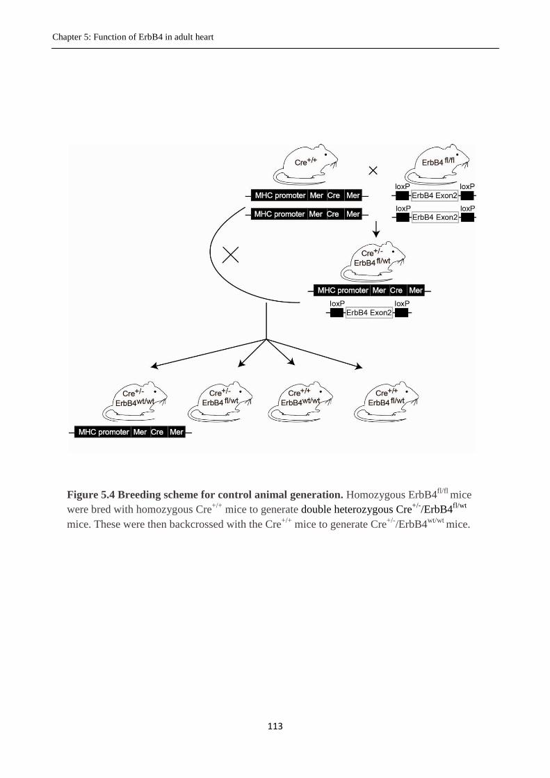
Chapter 5: Function of ErbB4 in adult heart
113
Figure 5.4 Breeding scheme for control animal generation. Homozygous ErbB4fl/fl
mice
were bred with homozygous Cre+/+
mice to generate double heterozygous Cre+/-
/ErbB4fl/wt
mice. These were then backcrossed with the Cre+/+
mice to generate Cre+/-
/ErbB4wt/wt
mice.

Chapter 5: Function of ErbB4 in adult heart
114
Figure 5.5 Cre copy number detected with qPCR. A. Two pairs of primers (termed as pcre1
and pcre2, refer to the Appendix B for the primer sequence) were designed to amplify the
different regions of the Cre gene. B. Homozygous, heterozygous and wild type Cre animals can
be differentiated by qPCR with both pairs of primers (validation using animals of known
genotype). C. Representative graph for detection of Cre copy number in animals with unknown
genotypes.

Chapter 5: Function of ErbB4 in adult heart
115
5.3.5 Determining the effects of Cre recombinase on the heart
To detect whether Cre activity itself induces a cardiac phenotype in adult mice without deletion of
the floxed gene, we examined cardiac functional parameters on Cre+/-
/ErbB4wt/wt
(Cre positive) and
Cre-/-
/ErbB4fl/fl
(Cre negative) animals at 3-4 or 7-8 months after the tamoxifen treatment.
Echocardiography revealed that the left ventricular internal dimensions diastole (LVIDd) and
systole (LVIDs) and fractional shortening (FS) (calculated as (LVIDd-LVIDs/LVIDd) was not
significantly different between the two groups at 3-4 months post-tamoxifen, suggesting that the
contractile ability of the heart was not affected by the induction of Cre (Figure 5.6 A and B).
Similarly, the left ventricular wall thickness (reflected by the intraventricular septal wall thickness
(IVS) and left ventricle posterior wall thickness (LVPW)) was not changed at 3-4 months post-
tamoxifen (Figure 5.6 C). There was a modest decrease in fractional shortening in Cre positive
animals at 7-8 months after tamoxifen injection, but this was not statistically significant (Figure 5.7
A). However, at 7-8 months post-tamoxifen both the IVS (systolic) and LVPW (both systolic and
diastolic) were significantly decreased in Cre positive animals (Figure 5.7 B and C). This data
suggests that that even short-term increases in Cre activity can have long-term effects on cardiac
structure. This result was confirmed by the data from post-mortem studies, which showed that the
heart weight:tibia length ratio of the Cre positive animals was significantly decreased at both 3-4
and 7-8 months post-tamoxifen (Figures 5.8 and 5.9). This was specific to the heart, as kidney
weight was not changed during this period. This indicates that the cardiac-specific induction of Cre
activity might cause the loss of cardiac mass. Then we investigated the expression of hypertrophic
(α-MHC,β-MHC and BNP) and fibrosis genes (COL1A1 and PAI-1) in Cre positive and negative
animals. RT-qPCR revealed that the hypertrophic genes and fibrosis genes are expressed at similar
levels in both groups (Figure 5.10 A and B). In addition, expression of the ErbB receptors (ErbB1
and ErbB4) was also not affected by Cre activity (Figure 5.11). Together, these data indicate that
induction of Cre activity can have a long term effect on the heart, although the mechanism of this is
not clear. However, this does suggest that the Cre-/-
/ErbB4fl/fl
genotype is not an appropriate control
for our study. To circumvent the negative effect of Cre activity on cardiac tissues, I used the Cre-
only transgenic mice (Cre+/-
/ErbB4wt/wt
) as the control in our following study examining the effects
of cardiac ErbB4 deletion (Molkentin et al., 2009). Both Cre+/-
/ErbB4fl/fl
(ErbB4 conditional
knockout group, ErbB4-cKO) and Cre+/-
/ErbB4wt/wt
(control group) animals received tamoxifen
treatment. Around 3-4 months or 7-8 months later, the cardiac phenotype of ErbB4 deletion was
determined.

Chapter 5: Function of ErbB4 in adult heart
116
Figure 5.6 Effect of Cre recombinase induction on cardiac function at 3-4 months post-
tamoxifen. A. Example of a typical echocardiogram in 2D (top) and M-mode (bottom) obtained in
short axis view for the Cre negative (Cre-/-
/ErbB4fl/fl
, left) and Cre positive (Cre+/-
/ErbB4wt/wt
, right)
animals. B. Left ventricular internal dimensions during diastole (left) and systole (middle) and
fractional shortening (right) in Cre negative (Cre Neg.) and Cre positive (Cre Pos.) animals. C.
Intraventricular septal wall thickness of diastolic (IVSd) and systolic (IVSs) dimension and left
ventricle posterior wall thickness of diastolic (LVPWd) and systolic (IVPWs) dimension in Cre
negative and Cre positive animals. Data was analysed by unpaired Student’s t-test and presented as
mean SEM.

Chapter 5: Function of ErbB4 in adult heart
117
Figure 5.7 Effect of Cre recombinase induction on cardiac function at 7-8 months post-
tamoxifen. A. Left ventricular dimensions during diastole (left) and systole (middle) and fractional
shortening (right) in Cre negative (Cre-/-
/ErbB4fl/fl
, Cre Neg.) and Cre positive (Cre
+/-/ErbB4
wt/wt, Cre
Pos.) animals. B. Intraventricular septal wall thickness of diastolic (IVSd) and systolic (IVSs)
dimension and left ventricle posterior wall thickness of diastolic (LVPWd) and systolic (LVPWs)
dimension in Cre negative and Cre positive animals. Data was analysed by unpaired Student’s t-test
and presented as mean SEM. * P<0.05 compared with Cre Neg.; ** P<0.01 compared with Cre
Neg.; Cre Neg., n=9; Cre Pos., n=7.

Chapter 5: Function of ErbB4 in adult heart
118
Figure 5.8 Effect of Cre recombinase induction on heart weight at 3-4 months post-
tamoxifen. The heart weight normalized to tibial length (HW:TL) (A), kidney weight to tibial
length (KW:TL) (B), the tibial length (TL) (C) and body weight (BW) (D) was measured in
Cre positive and Cre negative animals at 3-4 months after induction of Cre activity. Data was
analysed using unpaired Student’s t-test and presented as mean SEM. * P<0.05 compared
with Cre Neg.; Cre Neg., n=9; Cre Pos., n=8.

Chapter 5: Function of ErbB4 in adult heart
119
Figure 5.9 Effect of Cre recombinase induction on heart weight at 7-8 months post-
tamoxifen. The heart weight (normalized to tibial length) (A), kidney weight to tibial length
(B), the tibial length (C) and body weight (D) was measured in Cre positive and Cre negative
animals at 7 months after induction of Cre activity. Data was analysed using unpaired
Student’s t-test and presented as mean SEM. ** P<0.01 compared with Cre Neg.; Cre Neg.,
n=9; Cre Pos., n=8.

Chapter 5: Function of ErbB4 in adult heart
120
Figure 5.10 The regulation of hypertrophic genes and fibrosis genes at 8 months following Cre
induction. Expression of the hypertrophic genes BNP, α-MHC, β-MHC and fibrosis genes
COL1A1 and PAI-1 were detected by RT-qPCR, normalized to 18S (endogenous control) and
expressed relative to the Cre negative group. Data was analysed using unpaired Student’s t-test and
presented as mean SEM. Cre Neg., n=9; Cre Pos., n=8.

Chapter 5: Function of ErbB4 in adult heart
121
Figure 5.11 The expression of ErbBs was not affected by the induction of Cre activity.
Expression of ErbB1 and ErbB4 was detected with RT-qPCR, normalized to18S (endogenous
control) and expressed relative to the Cre negative group. Data was analysed using unpaired
Student’s t-test and presented as mean SEM. Cre Neg., n=9; Cre Pos., n=8.
Chapter 5: Function of ErbB4 in adult heart
122
5.3.6 Effect of cardiac-specific deletion of ErbB4 on cardiac function
As shown in Figure 5.3, tamoxifen treatment of ErbB4-cKO (Cre+/-
ErbB4fl/fl
) mice deleted
approximate 90% of cardiac ErbB4 at 10 days after the tamoxifen treatment (measured by RT-
qPCR). At 3-4 months after the tamoxifen injection, we performed echocardiography on the mice to
examine their cardiac function. Key echocardiography parameters were measured in both systolic
and diastolic phase, including left ventricular internal dimension (LVID), intraventricular septum
thickness (IVS) and left ventricle posterior wall thickness (LVPW). Statistical analysis revealed a
marked increase in the diastolic LVID, suggesting the ErbB4-cKO animals might undergo heart
enlargement. No significant difference in systolic LVID was observed between the ErbB4-cKO and
control (Cre-/-
ErbB4fl/fl
) animals (Figure 5.12 A). In addition, there was no significant change in the
fractional shortening in ErbB4-cKO animals, indicating that the contractile ability of the heart was
not affected after 3-4 months of ErbB4 deletion. The systolic IVS in ErbB4-cKO animals was
significantly increased and the diastolic IVS tended to increase, but was not statistically significant
(Figure 5.12 B).

Chapter 5: Function of ErbB4 in adult heart
123
Figure 5.12 Echocardiographic assessment of cardiac function at 3-4 months after ErbB4
deletion. A. The diastolic (left) and systolic (middle) left ventricular internal dimension and
fractional shortening (right) were measured in ErbB4-cKO and control animals. B. The diastolic
and systolic IVS and left ventricle posterior wall thickness in diastolic and systolic dimension were
measured in ErbB4-cKO (n=5) and control animals (n=7). Data was analysed using unpaired
Student’s t-test and presented as mean SEM. * P<0.05 compared with control; control, n=7;
ErbB4-cKO, n=5.
Chapter 5: Function of ErbB4 in adult heart
124
5.3.7 Effect of cardiac-specific deletion of ErbB4 on heart weight
After the echocardiography measurement, both ErbB4-cKO animals and control animals were
euthanased and the heart weight:tibia length ratio was determined. The ratio of heart weight to tibia
length in ErbB4-cKO animals was significantly increased compared to the controls (Figure 5.13 A).
In contrast, the kidney weight:tibia length was not changed (Figure 5.13 A). This suggested that the
knockout of ErbB4 in cardiomyocytes leads to cardiac hypertrophy. RT-qPCR was used to examine
whether this increase in heart weight was associated with altered expression of pathological
hypertrophy marker genes. We found that the mRNA levels of α-MHC, β-MHC and BNP in ErbB4-
cKO animals were not changed relative to the controls (Figure 5.13 B). Together, this suggested
that the ErbB4-cKO animals undergo physiological, not pathological, hypertrophy.
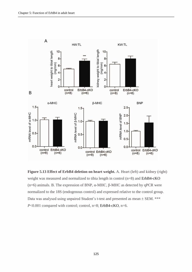
Chapter 5: Function of ErbB4 in adult heart
125
Figure 5.13 Effect of ErbB4 deletion on heart weight. A. Heart (left) and kidney (right)
weight was measured and normalized to tibia length in control (n=8) and ErbB4-cKO
(n=6) animals. B. The expression of BNP, α-MHC, β-MHC as detected by qPCR were
normalized to the 18S (endogenous control) and expressed relative to the control group.
Data was analysed using unpaired Student’s t-test and presented as mean SEM. ***
P<0.001 compared with control; control, n=8; ErbB4-cKO, n=6.
Chapter 5: Function of ErbB4 in adult heart
126
Following the study above, we investigated the mechanisms underlying the cardiac hypertrophy
observed in ErbB4-cKO animals. We first investigated whether the cardiomyocytes undergo
hypertrophy, which is defined as an increase in cell mass without proliferation. As we have not
established a system in our lab to accurately determine the mass of individual cardiomyocytes, we
used the measurement of cross sectional area to reflect cardiomyocyte hypertrophy. Statistical
analysis showed that there was no significant change in the cross sectional area of cardiomyocytes
in the ErbB4-cKO group compared to the control (Figure 5.14 A and B). To further confirm this
result, the frequency distribution of cardiomyocyte area was analysed for both ErbB4-cKO and
control groups. If cardiomyocytes undergo hypertrophic growth, the frequency of cardiomyocytes
with a larger cell area will increase, thus shifting the frequency distribution curve to the right and
increasing the area range in the X axis. However, our data showed no shift, and the frequency
distribution curves for ErbB4-cKO and control groups were similar (Figure 5.14 C). Together, these
studies suggested that the cardiac hypertrophy caused by cardiac ErbB4 deletion in vivo is not via
hypertrophic growth of the cardiomyocytes, but occurs by some other mechanism.
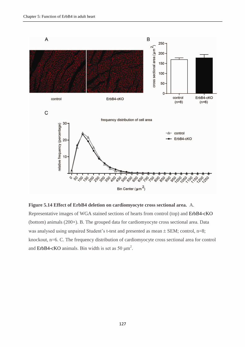
Chapter 5: Function of ErbB4 in adult heart
127
Figure 5.14 Effect of ErbB4 deletion on cardiomyocyte cross sectional area. A.
Representative images of WGA stained sections of hearts from control (top) and ErbB4-cKO
(bottom) animals (200×). B. The grouped data for cardiomyocyte cross sectional area. Data
was analysed using unpaired Student’s t-test and presented as mean SEM; control, n=8;
knockout, n=6. C. The frequency distribution of cardiomyocyte cross sectional area for control
and ErbB4-cKO animals. Bin width is set as 50 μm2.

Chapter 5: Function of ErbB4 in adult heart
128
5.3.8 Effect of cardiac-specific deletion of ErbB4 on cardiac fibrosis.
Fibroblasts are the second major component of heart tissue besides cardiomyocytes (Brown et al.,
2005). Cardiac fibrosis (where lost cardiomyocytes are replaced by proliferating fibroblasts and the
secreted collagen) can be observed in the late stages of pathological cardiac hypertrophy (Manabe et
al., 2002). Cardiac fibrosis can be measured by histological staining for collagen or by qPCR for the
expression of fibrosis molecular marker genes such as alpha-1 type I collagen (COL1A1), alpha-1
type III collagen (COL3A1) and plasminogen activator inhibitor type 1 (PAI-1). In our study, RT-
qPCR revealed that the mRNA levels of COL1A1, COL3A1 and PAI-1 didn’t change in the ErbB4-
cKO animals compared to the controls (Figure 5.15 A). To further confirm these results, we
examined collagen deposition in heart sections using Masson’s Trichrome. Masson’s Trichrome
stains the collagen in blue, the cytoplasm in red and the nucleus in black. The normal heart should
have little collagen accumulated around cardiomyocytes, but some should be present in the
perivascular area. Here, we observed blue collagen staining around the vascular area in heart
sections from both the ErbB4-cKO and control animals. However, the myocardial area of both
groups showed minimal collagen staining (Figure 5.15 B), suggesting that knockout of ErbB4 did
not lead to cardiac fibrosis
.

Chapter 5: Function of ErbB4 in adult heart
129
Figure 5.15 Effect of ErbB4 deletion on cardiac fibrosis. A. The expression levels of the
fibrosis marker genes, COL1A1, COL3A1, and PAI-1 were detected by RT-qPCR and
normalized to 18S. Data was expressed relative to the control group. Data was analysed using
unpaired Student’s t-test and presented as mean SEM; control, n=8; ErbB4-cKO, n=6. B.
Representative sections of Masson’s Trichrome staining on heart tissues from control (left) and
ErbB4-cKO animals (right).

Chapter 5: Function of ErbB4 in adult heart
130
5.3.9 Expression of the ErbB receptor family following ErbB4 deletion
Only a mild phenotype was observed in the ErbB4-cKO animals, which were characterized by
physiological cardiac hypertrophy. No severe cardiac dysfunction or other structural abnormalities
were found. This result was surprising because accumulated evidence in literature indicates that
ErbB4 is critical for cardiac function and structural maintenance (Gassmann et al., 1995; Tidcombe
et al., 2003; Garcia-Rivello et al., 2005). Thus in the following study, we investigated whether the
deletion of ErbB4 was compensated for by other ErbB receptor systems. There are four subtypes of
ErbB receptor (ErbB1, ErbB2, ErbB3, and ErbB4) detectable in the heart. We examined whether
the deletion of ErbB4 may trigger a compensatory up-regulation of other ErbB receptors. At 3-4
months after ErbB4 deletion, there was a small increase in ErbB3 mRNA levels and a small
decrease in both ErbB1 and ErbB2, however these effects did not reach statistical significance
(Figure 5.16). Together, this suggests that the other members of the ErbB family were not
significantly up-regulated to compensate for the deletion of the ErbB4.

Chapter 5: Function of ErbB4 in adult heart
131
Figure 5.16 Expression of ErbB1, ErbB2 and ErbB3 following ErbB4 deletion. The
expression level of ErbB1, ErbB2 and ErbB3 was detected by RT-qPCR and normalized
to the 18S. Data was expressed relative to the control group. Data was analysed using
unpaired Student’s t-test and presented as mean SEM; control, n=8; ErbB4-cKO, n=6.

Chapter 5: Function of ErbB4 in adult heart
132
5.3.10 Expression of the EGF family following ErbB4 deletion.
Although expression of other ErbB receptors does not change after ErbB4 deletion, the activity of
ErbBs can also be influenced by up-regulation of endogenous agonists, and this may serve as
compensation for the lost ErbB4. There are multiple agonists (EGF-like factors) for each ErbB
receptor, and thus we investigated whether the expression of these agonists was altered by cardiac
ErbB4 deletion. We first studied the ErbB4 selective agonists, the NRG family (NRG1, NRG2,
NRG3 and NRG4). NRG1 has two isoforms, NRG1α and NRG1β that are generated by alternative
splicing. Using RT-qPCR, the expression of both NRG1α and NRG1β is dramatically up-regulated
in ErbB4-cKO animals compared to controls (Figure 5.17 A). In contrast, the expression of NRG2,
NRG3 and NRG4 (Figure 5.17 B) didn’t change, suggesting that the up-regulation of NRG1 is a
specific response to the lost ErbB4. We also examined the regulation of the endogenous agonists for
other ErbB receptors. TGFα and amphiregulin (AREG) are selective agonists for ErbB1. HB-EGF,
epiregulin (EREG) and betacellulin (BTC) are agonists for both ErbB1 and ErbB4. ErbB2 has no
identified ligands. However, none of these factors were regulated at the mRNA level in ErbB4-cKO
animals (Figure 5.18). Together, this suggests that NRG1 is the only member of the EGF family
that is specifically up-regulated after deletion of ErbB4. NRG1 is a cardioprotective factor (see
section 1.6.3) thus the up-regulation of NRG1 might serve as compensatory mechanism to for the
deletion of ErbB4, and may explain why we did not see any cardiac dysfunction at 3-4 months after
ErbB4 deletion.

Chapter 5: Function of ErbB4 in adult heart
133
Figure 5.17 Expression of NRG family following ErbB4 deletion. The expression
levels of NRG1α, NRG1β, NRG2, NRG3 and NRG4 was detected by RT-qPCR and
normalized to 18S. Data was expressed relative to the control group. Data was analysed
using unpaired Student’s t-test and presented as mean SEM. ** P<0.01 compared with
control; control, n=8; ErbB4-cKO, n=6.

Chapter 5: Function of ErbB4 in adult heart
134
Figure 5.18 Expression of other EGF-like factors following deletion of ErbB4. The
expression level of TGF-alpha, amphiregulin (AREG), HB-EGF, epiregulin (EREG) and
betacelulin (BTC) was detected by the RT-qPCR and normalized to 18S. Data was
expressed relative to the control group. Data was analysed using unpaired Student’s t-test
and presented as mean SEM; control, n=8; ErbB4-cKO, n=6.

Chapter 5: Function of ErbB4 in adult heart
135
5.3.11 The proliferation of cardiomyocytes following ErbB4 deletion
One question raised from the experiments described in 5.3.7 is how the ErbB4-cKO mice developed
cardiac hypertrophy when there was no evidence of cardiomyocyte hypertrophy or fibrosis. The
classic theory considers that in adult mammals the majority of cardiomyocytes are fully
differentiated, and therefore have a very low rate of proliferation. However, a recent study
suggested that NRG1 might promote the proliferation of adult cardiomyocytes (Bersell et al., 2009).
To investigate whether the up-regulation of NRG1 in ErbB4-cKO animals causes cardiomyocyte
proliferation, we used immunofluorescence to examine expression of the proliferation marker
phosphorylated histone H3 (pH3) in heart sections (Figure 5.19). Statistical analysis shows that the
total number of pH3 positive cells in ErbB4-cKO animals was significantly increased, although the
absolute number of proliferating cells remains low.

Chapter 5: Function of ErbB4 in adult heart
136
Figure 5.19 The proliferation of cardiac cells at 3-4 months following ErbB4 deletion.
Heart sections in control and ErbB4-cKO animals were stained for pH3 by
immunofluorescence. Data was analysed using unpaired Student’s t-test and presented as mean
SEM. * P<0.05 compared with control; control, n=8; ErbB4-cKO, n=6.

Chapter 5: Function of ErbB4 in adult heart
137
5.3.12 Expression of ErbB4 at 3-4 months after tamoxifen treatment
Following the investigation into the phenotype of ErbB4-cKO animals, we confirmed that the
ErbB4 deletion is still valid 3-4 months after the tamoxifen injection. The RT-qPCR data revealed
that the mRNA level of ErbB4 in the ErbB4-cKO group was reduced by ~67% compared to the
control group (Figure 5.20).

Chapter 5: Function of ErbB4 in adult heart
138
Figure 5.20 Expression of ErbB4 at 3-4 months after tamoxifen injection. At 3-4 months after
tamoxifen treatment, the expression level of the ErbB4 was measured by RT-qPCR and
normalized to the 18S. Data was expressed relative to the control group. Data was analysed using
unpaired Student’s t-test and presented as mean SEM. * P<0.05 compared with control; control,
n=8; ErbB4-cKO, n=6.
Chapter 5: Function of ErbB4 in adult heart
139
5.3.13 Cardiac function at 7-8 months after ErbB4 deletion
All of the phenotypic experiments described above were conducted at 3-4 months after cardiac
ErbB4 deletion was induced with tamoxifen. However, 3-4 months may be not long enough for the
conditional ErbB4-cKO mice to develop a severe phenotype. To determine whether in the longer
term the heart will develop dysfunction, we performed similar measurements on a separate cohort
of ErbB4-cKO and control animals at 7-8 months after the deletion of ErbB4.
At 7-8 months after tamoxifen injection, we measured left ventricular internal dimension (LVID),
interventricular septum thickness (IVS) and left ventricle posterior wall thickness (LVPW) in both
the systolic and diastolic phase using echocardiography. Remarkably, in contrast to 3-4 months,
statistical analysis revealed there were no marked differences between ErbB4-cKO and control
animals at 7-8 months. The LVPW in ErbB4-cKO animals tended to increase but was not
statistically significant compared to the controls (Figure 5.21).

Chapter 5: Function of ErbB4 in adult heart
140
Figure 5.21 Echocardiographic assessment of cardiac function at 7-8 months following
ErbB4 deletion. A. The diastolic (left) and systolic (middle) left ventricular internal
dimension and fractional shortening (right) were measured in control and knockout animals. B.
The diastolic and systolic IVS and LVPW dimension were measured in control and ErbB4-
cKO animals. Data was analysed using unpaired Student’s t-test and presented as mean
SEM. * P<0.05 compared with control; control, n=7; knockout, n=7.

Chapter 5: Function of ErbB4 in adult heart
141
5.3.14 Cardiac hypertrophy at 7-8 months after ErbB4 deletion
Animals were euthanased following echocardiography, and indices of cardiac hypertrophy
measured. The heart weight:tibia length ratio (HW:TL) of the ErbB4-cKO group was significantly
increased (~ 19 %) compared to the control group (Figure 5.22). This suggests that ErbB4-cKO
animals undergo mild hypertrophy. However, the magnitude of this increase is lower than in
previous experiments when HW:TL was measured at 3-4 months after tamoxifen injection (Figure
5.13). We tested the expression of hypertrophic marker genes with RT-qPCR and found no
significant changes in expression of the MHC isoforms or BNP (Figure 5.23 A). However, the ratio
of α-MHC to β-MHC expression was reduced in the ErbB4-cKO, which may indicate a switch from
α-MHC (which is predominant in adult heart) to β-MHC isoform (which is predominant in fetal
heart) (Figure 5.23 A). This aligns with our data from the earlier set of experiments, which suggests
that there may be increased generation of new cardiomyocytes. The fibrosis genes in both groups
were expressed at similar levels (Figure 5.23 B), indicating there is no overt cardiac fibrosis in the
ErbB4-cKO animals. Together, this suggests that the cardiac phenotype is milder at 7-8 months
post-ErbB4 deletion compared to at 3-4 months after deletion, and that the phenotype observed at 3-
4 months seems to undergo recovery by 7-8 months.

Chapter 5: Function of ErbB4 in adult heart
142
Figure 5.22 Effect of ErbB4 deletion on heart weight at 7-8 months after deletion. Heart
(left) and kidney (right) weight was measured and normalized to tibia length in control (n=8) and
knockout (n=9) animals. Data was analysed using unpaired Student’s t-test and presented as
mean SEM. * P<0.05 compared with control; control, n=8; ErbB4-cKO, n=9.
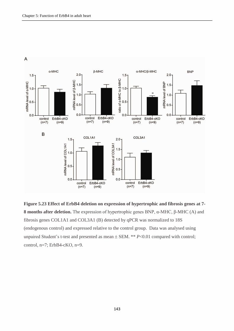
Chapter 5: Function of ErbB4 in adult heart
143
Figure 5.23 Effect of ErbB4 deletion on expression of hypertrophic and fibrosis genes at 7-
8 months after deletion. The expression of hypertrophic genes BNP, α-MHC, β-MHC (A) and
fibrosis genes COL1A1 and COL3A1 (B) detected by qPCR was normalized to 18S
(endogenous control) and expressed relative to the control group. Data was analysed using
unpaired Student’s t-test and presented as mean SEM. ** P<0.01 compared with control;
control, n=7; ErbB4-cKO, n=9.
Chapter 5: Function of ErbB4 in adult heart
144
5.3.15 Expression of ErbB4 at 7-8 months after tamoxifen treatment
After investigating the function of the heart after long-term deletion of ErbB4, we wanted to
confirm that the ErbB4 is still absent in ErbB4-cKO animals at 7-8 months after ErbB4 gene
deletion. We tested the expression of ErbB4 in heart tissues with RT-qPCR after 7-8 months of
tamoxifen injection. Surprisingly, the ErbB4 expression in ErbB4-cKO animals was not
significantly reduced compared to the controls (Figure 5.24). This indicated that either the gene was
not initially deleted (unlikely), or that ErbB4 gene expression had somehow recovered by 7-8
months following tamoxifen treatment.

Chapter 5: Function of ErbB4 in adult heart
145
Figure 5.24 Expression of ErbB4 at 7-8 months after tamoxifen injection. At 7-8
months after tamoxifen treatment, the expression level of ErbB4 was measured by RT-
qPCR, normalized to 18S and expressed relative to the control group. Data was analysed
using unpaired Student’s t-test and presented as mean SEM; control, n=7; ErbB4-cKO,
n=9.

Chapter 5: Function of ErbB4 in adult heart
146
5.4 Discussion
The importance of NRG1 signalling in the cardiovascular system and its protective effect on the
postnatal heart have been previously established (Zhao et al., 1998; Kuramochi et al., 2004; Liu et
al., 2006). As NRG1’s primary receptor, ErbB4 is essential in heart (Gassmann et al., 1995;
Tidcombe et al., 2003; Garcia-Rivello et al., 2005). Based on these studies, we hypothesised that
ErbB4 would be essential for adult heart maintenance, and predicted that ErbB4 deletion from the
adult heart would lead to severe cardiomyopathy or heart failure. In this chapter, I used the αMHC-
MerCreMer/loxP system to specifically delete ErbB4 from the adult heart, and investigated the
cardiac phenotype for up to 7-8 months following deletion. The main findings in this study are: 1)
An optimised tamoxifen treatment can specifically reduce ErbB4 mRNA levels by greater than 90%
in the heart using the αMHC-MerCreMer/loxP system; 2) Transient Cre activity has long term
effects on cardiac physiology; 3) Surprisingly, cardiac-specific deletion of ErbB4 did not lead to
any obvious cardiac dysfunction or structural abnormalities except for a mild physiological
hypertrophy; 4) Unique among EGF-like ligands, NRG1 was found to be selectively unregulated
after ErbB4 deletion; and 5) There is increased proliferation of cardiac cells after ErbB4 deletion.
Tamoxifen is a drug has been used to treat breast cancer due to its anti-estrogen effects, and it is
also used in the laboratory to induce gene deletion in MerCreMer/loxP conditional knockout mouse
models. The described treatment regimes (dose, method of administration, duration of treatment)
for tamoxifen induced gene deletion are highly variable. However, a high dose of tamoxifen can be
toxic. In a previous study in our lab, tamoxifen treatment at 40mg/kg/day ip for 4 consecutive days
led to a high mortality rate in adult mice. However, lower doses are sometimes not sufficient to
cause efficient deletion of the target gene. To conquer this, we adopted the low dose but long term
treatment method (Hsieh et al., 2007). This idea is based on the fact that as long as the gene is
deleted from genome, it cannot be recovered in the same cell. Although the efficiency of gene
deletion is very low with a single tamoxifen injection at low dose, a prolonged accumulating
treatment can achieve an excellent outcome. With this method, we achieved a more than 90
percentage deletion of the ErbB4 gene in adult mouse heart with minimal toxicity. There is no
obvious or marked difference in the physiology or behaviour between the tamoxifen treated and
untreated animals.
Although we were using the optimised dose tamoxifen which is not lethal, the potential side effect
of this drug is hard to be excluded. To minimize the potential interference by tamoxifen itself, we

Chapter 5: Function of ErbB4 in adult heart
147
decided to treat both experimental and control groups with tamoxifen. The genotype of the
conditional knockout animals is Cre+/-
/ ErbB4fl/fl
. Thus, the control animals have to lack either Cre
(Cre-/-
/ ErbB4fl/fl
) or loxP sites (Cre+/-
/ ErbB4wt/wt
) to avoid deletion of ErbB4 when they received
tamoxifen. If Cre-/-
/ ErbB4fl/fl
animals are used as controls, tamoxifen injection will induce Cre
activity in the ErbB4-cKO animals (Cre+/-
/ ErbB4fl/fl
) but not the controls (Cre-/-
/ ErbB4fl/fl
). Thus,
Cre activity is another possible confounding factor induced in the ErbB4-cKO group besides the
ErbB4 deletion. When Sohal et al. established the MHC-MerCreMer-loxP system in 2001, it was
claimed that expression of the MerCreMer fusion protein in adult heart did not affect cardiac
performance or cellular architecture. However, subsequent studies have indicated that the Cre
recombinase is cardiotoxic. Hall et al. showed that the induction of Cre recombinase activity leads
to decreased systolic function (SF) and alterations in proteins associated with cardiac oxidative
phosphorylation. The decreased SF returned to baseline 10 days after tamoxifen injection (Hall et
al., 2011). Another study showed that cardiac Cre activity induced a transient dilated
cardiomyopathy marked by cardiac dysfunction and reduction in the expression of genes related to
metabolism and calcium handling. Some of these defects had disappeared by 1 month after the Cre
activity was induced (Koitabashi et al., 2009). Given these studies, supporting the idea that Cre
activity might be cardiotoxic, it was important for me to evaluate the effect of Cre in our animals.
To determine the Cre effect, we compared the cardiac function and structure of Cre positive animals
to Cre negative animals. We found that the transient Cre activity decreased the heart weight (up to 8
months), while the cardiac function is not altered at either 3-4 months or 7-8 months after tamoxifen
treatment. To circumvent the negative effect of Cre activity on cardiac tissues, I decided to use the
Cre-only transgenic mice (Cre+/-
/ErbB4wt/wt
) as the control for my studies.
Against our expectations, we observed that the deletion of ErbB4 in adult mice did not lead to
cardiac dysfunction. The important role of ErbB4 in cardiac development is well established
(Gassmann et al., 1995; Tidcombe et al., 2003; Garcia-Rivello et al., 2005), and the essentiality of
ErbB4 in postnatal heart was confirmed by Garcia-Rivello et al. using a MLC-2V-Cre/loxP system
to demonstrate that specific deletion of ErbB4 in cardiomyocytes leads to severe dilated
cardiomyopathy in adulthood. Interestingly, no obvious cardiac abnormality (ventricle wall
thickness, cardiac cell number) was observed at birth, and the phenotype they observed in adulthood
primarily resulted from lack of ErbB4 in the postnatal stage (juvenile or adult) (Garcia-Rivello et
al., 2005). The results of my experiments in this chapter indicate that ErbB4 is not critical for
maintenance and function of the adult heart, and remarkably its deletion did not result in
cardiomyopathy. One interpretation could be that ErbB4 might be essential for the cardiac

Chapter 5: Function of ErbB4 in adult heart
148
development in the juvenile, but not in the older adult. Supporting this, Bersell et al. (Bersell et al.,
2009) showed that genetic deletion of ErbB4 at postnatal days 16-18 blocked the DNA synthesis of
cardiomyocytes at baseline (measured by BrdU incorporation). Taken together, these studies and
ours might suggest the ErbB4 is required for the cardiac development both in embryonic and early
postnatal stages. However, once the cardiac development is completed, the subsequent loss of
ErbB4 does not lead to severe cardiac dysfunction or structural abnormalities.
One clinical study has indirectly indicated that ErbB4 might be important for adult heart
maintenance: trastuzumab (an ErbB2 blocking antibody) has been used for breast cancer therapy
and when combined together with other chemotherapy agents the rate of heart failure was
significantly increased (27% incidence) (Seidman et al., 2002). Based on this observation, it has
been proposed that ErbB2 is important in the adult heart. ErbB2 lacks any identified agonists, and
preferentially dimerizes with other ErbB receptors to trigger downstream signalling (Timolati et al.,
2006). The inhibition of ErbB2 might affect the signalling of other ErbB receptors, especially its
preferential partner ErbB4. Our study suggested ErbB4 deletion did not caused cardiac dysfunction.
Thus, the cardiac dysfunction induced by trastuzumab treatment probably reflects effects on the
signalling of other ErbB receptors. As mentioned earlier (section 1.2.1), the ErbB2 receptor can
adopt an active formation without binding any agonists (Garrett et al., 2003). It is therefore possible
that the active ErbB2 signalling itself is essential for the maintenance of heart function.
ErbB4 has been suggested to play a role in the transition from hypertrophy to heart failure. In
patients with heart failure, the cardiac expression of ErbB2 and ErbB4 are depressed (Rohrbach et
al., 2005). A study in rats also showed that ErbB2 and ErbB4 are significantly reduced in the heart
failure period, but not the preceding hypertrophic period in an aortic stenosis model (Rohrbach et
al., 1999). However, in our study, the ErbB4-cKO mice developed significant hypertrophy but did
not develop into heart failure in the longer term. This suggested that the loss of ErbB4 in the earlier
studies might be the result of heart failure but not the reason.
Another interesting result in my study is that ErbB4 deletion caused (somewhat paradoxically)
cardiac hypertrophy. Cardiomyocyte hypertrophy and cardiac fibrosis were absent in the ErbB4-
cKO animals, and so we proposed that there might be more cardiomyocytes generated. Two
potential sources could possibly regenerate new cardiomyocytes: one is stem cells or progenitor
cells and another is existing cardiomyocytes. Detecting cardiac stem cells is currently not available
in our lab, so I attempted to test whether cardiomyocytes were proliferating in our ErbB4-cKO

Chapter 5: Function of ErbB4 in adult heart
149
animals via co-staining of proliferation maker pH3 (located in nucleus) and the cardiomyocyte
marker Troponin by immunofluorescence. However, the cell boundary could not be clearly resolved
and there were many very small cells, such as endothelial cells, fibroblast or potential cardiac stem
cells located among the cardiomyocytes. Thus, it was difficult to tell whether the positively stained
nucleus for pH3 with positive Troponin staining around it was in a cardiomyocyte, or in a small cell
among cardiomyocytes. This difficulty has also been described in the literature (Ang et al., 2010).
Thus, the positively stained cell number was presented as total cardiac cell number instead of
“cardiomyocyte number”. Although we cannot tell exactly the contribution of each cell type in this
cardiac cell proliferation, we believe that new cardiomyocytes were generated by cardiomyocytes or
stem cells, and contribute to the cardiac hypertrophy. It is important to note that pH3 only indicates
cells undergoing the mitogenic phase, which is known to be transient in the whole cell cycle. The
heart is one of the least regenerative organs in the body, so even if there is a regenerative response,
it usually happens in a very small proportion of cells. Thus, the pH3 stained only a few cells in each
section. It is still hard to predict how much of the heart can be regenerated with these proliferated
cells. However, we believe with the accumulation effect over time, many new cardiomyocytes were
generated in our ErbB4-cKO model and these might have contributed to the cardiac hypertrophy.
Another important discovery of our study is that we observed the up-regulation of NRG1 in our
animals after ErbB4 deletion. To our knowledge, this is a new observation in ErbB4 deletion
studies, but is concordant with the observation made by Robrbach et al. in heart failure where the
ErbB4 expression was decreased and NRG1 increased (Rohrbach et al., 1999; Rohrbach et al.,
2005). The link between the loss of ErbB4 and the compensatory up-regulation of its agonist NRG1
is indicative, but the mechanism is unknown. However, given that NRG1 is predominantly released
by microvascular endothelial cells (Kuramochi et al., 2004; Lemmens et al., 2006) and targets the
cardiomyocytes, we predict that there is a crosstalk between the cardiomyocytes and endothelial
cells to regulate the expression of NRG1 after the ErbB4 deletion. The up-regulated NRG1 may
function to compensate for the ErbB4 loss. NRG1 is a cardioprotective factor that enhances cardiac
contraction and reduces the cardiomyocyte apoptosis caused by stress (Liu et al., 2006; Bian et al.,
2009). I therefore propose that this NRG1 up-regulation explains why we haven’t observed a severe
phenotype following ErbB4 deletion. If this assumption is true, the prevailing question will be how
NRG1 can mediate a cardioprotective effect in the absence of its cognate receptor ErbB4. One
possibility is that NRG1 is acting via a different receptor, possibly ErbB3.

Chapter 5: Function of ErbB4 in adult heart
150
Currently, it is unclear whether cardiac stem cells can respond to NRG1 stimulation to proliferate.
However, NRG1 can promote cardiomyocyte proliferation in the adult heart (Zhao et al., 1998;
Bersell et al., 2009). Another question from the present study is how NRG1 induced cardiac cell
proliferation in the absence of its receptor ErbB4. One possibility is that ErbB4 was not completely
deleted from 100% of cardiomyocytes, and that the proliferation happened in ErbB4-replete cells.
They would then pass their intact genome to their progeny cells, which thus possess the unaffected
ErbB4 gene and could in turn respond to NRG1. When the newly generated cardiac cells
accumulate, the proliferation process is accelerated. However, NRG1 only induces a small
proportion of cardiomyocytes to proliferate in wild type animals (Bersell et al., 2009). Accordingly,
it can be inferred that in ErbB4 deleted animals, this cell population is even smaller and so it is very
hard to imagine how a few cells proliferating at the initial stage (after deleting ErbB4 from the
majority of cardiomyocytes) could promote such a robust increase in heart size in 3 months given
the poor regenerative capacity of the adult heart. It is possible that the deletion of ErbB4 happens in
cells expressing MHC but not in progenitor cells that do not express MHC. In this case, the
proliferation rate of cardiac cells induced by NRG1 will be similar for wild type and ErbB4 deleted
animals. Hence, when the NRG1 is up-regulated in ErbB4-cKO animals (but not wild type
animals), the ErbB4-cKO animals will have more cardiomyocytes generated. A final possibility is
that ErbB3 also plays a role in adult heart function and structural maintenance. ErbB3 is a receptor
for NRG1 with a similar structure to ErbB4 but possessing additional PI3K binding domains, which
contribute to cardiomyocyte survival. However, this possibility does not explain the recovery of the
ErbB4 expression in the long-term after deletion. Whatever the merit of these speculations, we need
to perform additional studies such as lineage tracing with immunofluorescence markers to tell if the
proliferating cells are stem cells, cardiomyocytes with ErbB3 or ErbB4, or other cell types.
A final surprising discovery from this work was that the expression of ErbB4 in the ErbB4-cKO
group gradually recovered to the level of the control group at 7 months after ErbB4 deletion. One
trivial possibility is we didn’t inject the tamoxifen properly. We cannot fully exclude this possibility
because each set of animals was injected with tamoxifen independently, and the successful deletion
of ErbB4 in one cohort does not mean the definite success of deletion in a later set of animals.
However, the injections of tamoxifen were performed by the same person following the same
procedures, and for ten consecutive days, so we consider that the possibility of technical failure is
low. The only reasonable explanation is that ErbB4 was successfully deleted from the genome
immediately after tamoxifen treatment, but that the expression of ErbB4 gradually recovered over
7-8 months via a process that likely involves the generation of new cardiomyocytes to replace the

Chapter 5: Function of ErbB4 in adult heart
151
cardiomyocytes with genomic deletion of ErbB4. The replacement of cardiomyocytes would
presumably require apoptosis of the old cells in addition to new cell generation. We tried to detect
cardiomyocyte apoptosis with TUNEL at 3 months after ErbB4 deletion, however, the results was
not definitive due to the high levels of background staining in all groups. In summary, the cardiac
specific deletion of ErbB4 in adult mice did not result in physiological hypertrophy without obvious
cardiac dysfunction. The compensatory up-regulation of NRG1 in ErbB4-cKO mice heart might
contribute to the cardiac function preservation and lead to cardiac hypertrophy via causing
cardiomyocyte proliferation.


CHAPTER 6
GENERAL DISCUSSION

Chapter 6: General discussion
154
6. General Discussion
This thesis has explored the role of the epidermal growth factor receptors (particularly ErbB4) in
mediating cardiomyocyte hypertrophy, the potential signalling differences among the ErbB4
isoforms, and the physiological function of ErbB4 in the adult mouse heart. While ErbB4 did not
contribute to a classic model of cardiomyocyte hypertrophy (Ang II-induced hypertrophic
signalling), this receptor was clearly involved in the hypertrophy caused by NRG1. ErbB4 has four
isoforms due to alternative splicing, and the non-cleavable isoform JM-b was found to be
predominant under both physiological and pathophysiological conditions. Interestingly, there was
no significant difference in the ability of the four isoforms to mediate NRG1-induced hypertrophy.
When ErbB4 was specifically deleted from the adult mouse heart using Cre-loxP technology, the
mice developed physiological cardiac hypertrophy with up-regulation of NRG1 and no alterations
in cardiac function and structure. Surprisingly, an increased proliferation of cardiac cells was
observed following cardiac ErbB4 deletion, and I propose that the NRG1 up-regulation contributes
to this cell proliferation, although the mechanisms remain unclear.
Crosstalk between ErbB receptors and GPCRs in cardiomyocyte
hypertrophy
Cardiomyocytes are continuously exposed to diverse stimuli, such as soluble endocrine and
paracrine factors from blood or neighboring cells. It is important that these signals are integrated to
achieve an appropriate cellular response. Tyrosine kinase receptors (for factors including the EGF
ligands, insulin-like growth factor-1, insulin, etc.) and GPCRs (for ligands such as angiotensin II,
endothelin 1, adrenaline, etc.) play pivotal roles in interpreting such signals (Sugden et al., 1998;
Clerk et al., 1999; Heineke et al., 2006). These receptor systems do not work in isolation and there
is irrefutable evidence for crosstalk between GPCRs and the growth factor receptors. An
accumulating literature supports the idea that the type 1 angiotensin receptor (AT1R) is able to
transactivate the ErbB receptors (principally ErbB1) and thereby mediate cellular growth in a
variety of tissues and organs (Eguchi et al., 1998; Shah et al., 2003b; Chen et al., 2006). In this
setting, and based on previous work from my host laboratory that showed the ErbB4 selective
agonist NRG1 was a more potent inducer of cardiomyocyte hypertrophy than ErbB1 ligands, my
first aim was to investigate whether the classic AT1R-ErbB1 transactivation by extended to other
ErbB receptors, explicitly ErbB4. To accomplish this aim, I used an approach that involved

Chapter 6: General discussion
155
knockdown of ErbB receptors using RNAi as well as pharmacological inhibition with the small
molecule EGFR kinase antagonist, AG1478, which has been widely used in the field to discern
EGFR function (Eguchi et al., 1998; Asakura et al., 2002; Thomas et al., 2002; Shah et al., 2003b;
Chen et al., 2006). I was able to unambiguously demonstrate knockdown of each ErbB receptor,
and tested the effect of this knockdown on the capacity of Ang II (and NRG1) to promote
cardiomyocyte hypertrophic signalling. As expected, knockdown of ErbB4, but not ErbB1 or
ErbB2, was able to prevent NRG1 mediated cardiomyocyte hypertrophy. However, despite careful
experimentation, I found no evidence that Ang II-mediated cardiomyocyte hypertrophic signalling
was dependent on ErbB4. Much to my initial disappointment, I was also unable to prevent AT1R-
mediated cardiomyocyte hypertrophy with RNAi targeting ErbB1 or ErbB2. Based on extensive,
compelling, previous literature in the heart, but also in many other cellular systems (Shah et al.,
2003a; Mendez et al., 2005; Liebmann, 2011), our observation was unsettling but repeatable.
Given the importance (and potential controversy) of my data questioning the role of ErbB receptors
in AT1R hypertrophy, I undertook with my supervisors a systemic review of my approach and
observations. We retested the specificity of my RNAi constructs - they were shown to be effective
and specific - notably, the knockdown of individual ErbB receptors was confirmed by Western
blotting and qPCR, showing there is up to 90% reduction of ErbB1or ErbB4 by RNAi. In addition
to my initial data, using luciferase-based hypertrophic gene promoter reporter assays, I went on to
confirm that there was also minimal effect of ErbB knockdown on the activation of AT1R-mediated
MAPK signalling and hypertrophic growth (measured as protein:DNA ratios and sacromeric
reorganisation). We performed power calculations for luciferase assay data and determined that my
experimental repeats (n = 7-8) were more than sufficient to allow valid conclusions. My grouped
data for all experiments clearly showed no differences between knockdown and control groups with
Ang II treatment. In contrast, under parallel experimental conditions (i.e., same cells, same days,
same experimenter, same analyses), I was able to consistently and unequivocally observe that
ErbB4 RNAi (but not ErbB1/2 RNAi) reduced cardiomyocyte hypertrophic signalling in response
to the ErbB4 selective agonist, NRG1. The only interpretation for these results is that NRG1
requires the ErbB4 to mediate hypertrophy, whereas ErbB1, ErbB2 and ErbB4 are not individually
required for AT1R mediated cardiomyocyte hypertrophy.
So, how do I explain these results? It is difficult to answer this, as the experiments included
appropriate controls, yet yielded different results from previous studies, where the majority of the
literature indicates that GPCRs, like the AT1R, use EGFRs to mediate growth signalling (Porcile et

Chapter 6: General discussion
156
al., 2005; Ohtsu et al., 2006a; Ohtsu et al., 2006b; Luppi et al., 2007; Schlange et al., 2007). A
number of possibilities might be considered: 1) the most trivial of explanations could be that despite
overt knockdown of the ErbB mRNA/protein, the residual receptors were sufficient to mediate
growth pathway signalling. This idea is consistent with the pharmacological concept of “spare
receptors” (where in many cases only a portion of cell surface receptors are required for full
efficacy) (Strickland et al., 1981). Obviously, my control experiment, detailing the loss of NRG1
efficacy with ErbB4 RNAi knockdown, refutes such reasoning; 2) Primary cultures of neonatal
cardiomyocytes vary tremendously in terms of cellular composition (i.e., % contamination by
fibroblasts, endothelial cells etc) and sensitivity to hypertrophic stimuli, between laboratories and
even within laboratories over time. This might reflect subtle changes in protocols and purity (or
otherwise) of the cultures; 3) Many laboratories have evoked the contribution of EGFR
transactivation based, almost exclusively, on experiments using pharmacological blockade with the
small molecule inhibitor, AG1478. Certainly, the use of AG1478 alone (without confirmation with
another technique like RNAi) can be problematic and is open to misinterpretation (detailed below);
and 4) My RNAi experiments tested the hypothesis that individual ErbB receptors were involved in
cardiomyocyte hypertrophy and it remains possible that some redundancy exists between the ErbB
receptors (given that multiple receptors can bind multiple ligands) such that knockdown of a single
receptor might be compensated by another. So, whatever the merits of these possibilities, it
certainly raises some uncertainties and potentially some reevaluation of the some of the literature in
this broad field.
Indeed, as mentioned above, the reliance on data using AG1478 needs careful reexamination. There
is no doubt that in controlled assays, under ideal conditions, AG1478 at nanomolar concentrations
is a selective and specific inhibitor of the EGFR kinase (Anastassiadis et al., 2011). However, in
cell-based assays, where often conditions can widely vary and concentrations of the inhibitor range
up to tens of micromolar, that specificity/selectivity is not assured. In regard to the EGFRs, the
current consensus is that AG1478 is reasonably selective for ErbB1 (the EGFR) at concentrations
<0.5 µM, but can inhibit the other ErbBs (and probably other tyrosine kinases) at higher
concentrations (>5 µM). My data on MAPK signalling in cardiomyocytes (where EGF activation of
ErbB1 was blocked by 0.5 μM, but where NRG1 mediated MAPK via ErbB4 was only inhibited by
5 μM AG1478) are entirely consistent with this consensus view. Previous studies, including some
from my own laboratory, have used high μM levels of AG1478 and therefore cannot exclude the
contribution of tyrosine kinases other than EGFR kinase. Indeed, other receptor tyrosine kinases, as
well as soluble tyrosine kinases, have been associated with Ang II/AT1R signalling and growth (Yin
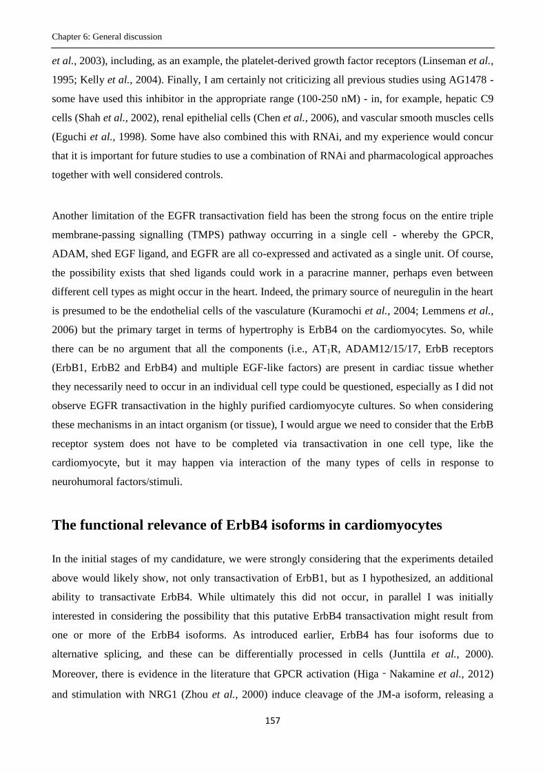
Chapter 6: General discussion
157
et al., 2003), including, as an example, the platelet-derived growth factor receptors (Linseman et al.,
1995; Kelly et al., 2004). Finally, I am certainly not criticizing all previous studies using AG1478 -
some have used this inhibitor in the appropriate range (100-250 nM) - in, for example, hepatic C9
cells (Shah et al., 2002), renal epithelial cells (Chen et al., 2006), and vascular smooth muscles cells
(Eguchi et al., 1998). Some have also combined this with RNAi, and my experience would concur
that it is important for future studies to use a combination of RNAi and pharmacological approaches
together with well considered controls.
Another limitation of the EGFR transactivation field has been the strong focus on the entire triple
membrane-passing signalling (TMPS) pathway occurring in a single cell - whereby the GPCR,
ADAM, shed EGF ligand, and EGFR are all co-expressed and activated as a single unit. Of course,
the possibility exists that shed ligands could work in a paracrine manner, perhaps even between
different cell types as might occur in the heart. Indeed, the primary source of neuregulin in the heart
is presumed to be the endothelial cells of the vasculature (Kuramochi et al., 2004; Lemmens et al.,
2006) but the primary target in terms of hypertrophy is ErbB4 on the cardiomyocytes. So, while
there can be no argument that all the components (i.e., AT1R, ADAM12/15/17, ErbB receptors
(ErbB1, ErbB2 and ErbB4) and multiple EGF-like factors) are present in cardiac tissue whether
they necessarily need to occur in an individual cell type could be questioned, especially as I did not
observe EGFR transactivation in the highly purified cardiomyocyte cultures. So when considering
these mechanisms in an intact organism (or tissue), I would argue we need to consider that the ErbB
receptor system does not have to be completed via transactivation in one cell type, like the
cardiomyocyte, but it may happen via interaction of the many types of cells in response to
neurohumoral factors/stimuli.
The functional relevance of ErbB4 isoforms in cardiomyocytes
In the initial stages of my candidature, we were strongly considering that the experiments detailed
above would likely show, not only transactivation of ErbB1, but as I hypothesized, an additional
ability to transactivate ErbB4. While ultimately this did not occur, in parallel I was initially
interested in considering the possibility that this putative ErbB4 transactivation might result from
one or more of the ErbB4 isoforms. As introduced earlier, ErbB4 has four isoforms due to
alternative splicing, and these can be differentially processed in cells (Junttila et al., 2000).
Moreover, there is evidence in the literature that GPCR activation (Higa‐Nakamine et al., 2012)
and stimulation with NRG1 (Zhou et al., 2000) induce cleavage of the JM-a isoform, releasing a

Chapter 6: General discussion
158
functional intracellular domain (ICD) into the nucleus to regulate cell activity as first shown by
Graham Carpenter’s group (Vecchi et al., 1996). So, even though I did not observe transactivation
of ErbB4 by Ang II in purified cardiomyocytes, I was still interested in investigating whether the
specific ErbB4 isoforms could be cleaved to release the ICD in cardiomyocytes and whether this
might contribute to the NRG1-induced cardiomyocyte hypertrophic signalling. Because previous,
unpublished work in our laboratory had detected all of the four ErbB4 isoforms in primary neonatal
rat cardiomyocytes, my prediction was that the cleavable JM-a isoform would be present and that I
would be able to demonstrate that it could be processed to release the intracellular domain (80 kD
ICD), which might interact with transcriptional factors to regulate cellular activity. Secondly, based
on a study in the brain, where a specific ErbB4 isoform was cleaved and shown to regulate the time
of astrogenesis in the mouse (Sardi et al., 2006), I was intrigued by the idea that the four isoforms
might make differing contributions to cardiomyocyte hypertrophy.
As part of investigating the cleavage of ErbB4 and any isoform-specific differences, I first sought to
establish whether these isoforms are indeed expressed in whole mouse heart. In addition, I
wondered whether the isoforms were expressed differently in various stages of cardiac
development, or during pathology. There is strong evidence that ErbB4 is required for normal
mouse cardiac development (Gassmann et al., 1995; Tidcombe et al., 2003) but the relevance of a
specific ErbB4 isoform in normal growth or disease has not been reported. I performed an
expression profile in the heart of all four isoforms from embryonic day 10.5 to postnatal day 28. All
isoforms declined throughout cardiac development - the expression of total ErbB4 at the earlier
embryonic stage (E10.5) is 250 times higher than at postnatal day 28. Interestingly, the expression
levels of JM-a and JM-b isoforms are similar in the earlier embryonic stages. However, by the
postnatal stage in mice, JM-b is the overwhelmingly predominant isoform. Why this might be is
currently unclear, but it could relate to the proliferative (i.e., embryonic) or hypertrophic (i.e., post-
natal) growth that occurs in the heart. It is tempting to speculate that perhaps JM-a is required for
cardiac proliferation in the embryo and JM-b is required for hypertrophy in the adult. It would be
interesting to isolate the embryonic cardiomyocytes and investigate whether the JM-a isoform
mediates proliferation, especially given that JM-a is capable of regulating the timing of astrogenesis
by suppressing astrocyte progenitor cell differentiation (Sardi et al., 2006). Moreover, the release of
the ICD (from JM-a) allows interaction with YAP to facilitate the translocation of the ICD into the
nucleus (Komuro, Nagai et al. 2003; Omerovic, Puggioni et al. 2004). Given that YAP activation is
sufficient to stimulate both embronic and postnatal cardiomyocyte proliferation (von Gise et al.,
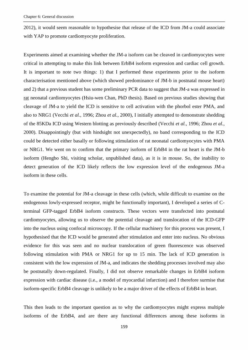
Chapter 6: General discussion
159
2012), it would seem reasonable to hypothesise that release of the ICD from JM-a could associate
with YAP to promote cardiomyocyte proliferation.
Experiments aimed at examining whether the JM-a isoform can be cleaved in cardiomyocytes were
critical in attempting to make this link between ErbB4 isoform expression and cardiac cell growth.
It is important to note two things: 1) that I performed these experiments prior to the isoform
characterisation mentioned above (which showed predominance of JM-b in postnatal mouse heart)
and 2) that a previous student has some preliminary PCR data to suggest that JM-a was expressed in
rat neonatal cardiomyocytes (Hsiu-wen Chan, PhD thesis). Based on previous studies showing that
cleavage of JM-a to yield the ICD is sensitive to cell activation with the phorbol ester PMA, and
also to NRG1 (Vecchi et al., 1996; Zhou et al., 2000), I initially attempted to demonstrate shedding
of the 85KDa ICD using Western blotting as previously described (Vecchi et al., 1996; Zhou et al.,
2000). Disappointingly (but with hindsight not unexpectedly), no band corresponding to the ICD
could be detected either basally or following stimulation of rat neonatal cardiomyocytes with PMA
or NRG1. We went on to confirm that the primary isoform of ErbB4 in the rat heart is the JM-b
isoform (Hengbo Shi, visiting scholar, unpublished data), as it is in mouse. So, the inability to
detect generation of the ICD likely reflects the low expression level of the endogenous JM-a
isoform in these cells.
To examine the potential for JM-a cleavage in these cells (which, while difficult to examine on the
endogenous lowly-expressed receptor, might be functionally important), I developed a series of C-
terminal GFP-tagged ErbB4 isoform constructs. These vectors were transfected into postnatal
cardiomyocytes, allowing us to observe the potential cleavage and translocation of the ICD-GFP
into the nucleus using confocal microscopy. If the cellular machinery for this process was present, I
hypothesised that the ICD would be generated after stimulation and enter into nucleus. No obvious
evidence for this was seen and no nuclear translocation of green fluorescence was observed
following stimulation with PMA or NRG1 for up to 15 min. The lack of ICD generation is
consistent with the low expression of JM-a, and indicates the shedding processes involved may also
be postnatally down-regulated. Finally, I did not observe remarkable changes in ErbB4 isoform
expression with cardiac disease (i.e., a model of myocardial infarction) and I therefore surmise that
isoform-specific ErbB4 cleavage is unlikely to be a major driver of the effects of ErbB4 in heart.
This then leads to the important question as to why the cardiomyocytes might express multiple
isoforms of the ErbB4, and are there any functional differences among these isoforms in

Chapter 6: General discussion
160
cardiomyocytes? An initial focus of my thesis was the role of ErbB4 in NRG1-mediated
cardiomyocyte hypertrophy, so I examined whether the four ErbB4 isoforms could all equally
mediate this effect. For this purpose, I knocked down the endogenous ErbB4 receptor and then
replaced it with individual knockdown-resistant version of the ErbB4 isoforms. Hypertrophy was
induced with the ErbB4 agonist NRG1 and hypertrophic signalling measured with luciferase
reporter assays. I observed that all four isoforms can rescue NRG1-induced hypertrophy, indicating
that the most likely scenario is that the hypertrophic signalling is reliant on the common tyrosine
kinase activity of the ErbB4 receptor isoforms, and does not seem to involve either shedding to
release ICD or a particular docking event mediated by CYT1/2.
The function of ErbB4 in the adult mice heart
My thesis then moved away from the focus on transactivation to a broader investigation of the role
of ErbB4 in adult heart in vivo. For this purpose, I adopted the tamoxifen inducible MHC-
MerCreMer/loxP system (Garcia-Rivello et al., 2005) to specifically delete the ErbB4 from
cardiomyocytes in adult mice. As described in Chapter 5, tamoxifen treatment specifically
decreased ErbB4 expression in the adult heart by ~ 90%. My prediction was that the mice would
develop cardiac dysfunction or structural abnormalities in the weeks/months following ErbB4
deletion. To my surprise, the knockout mice were active and healthy following ErbB4 deletion and
no obvious pathology was observed, which suggested a potential lack of a cardiac phenotype in
these mice. At 3 months after ErbB4 deletion, I measured cardiac contractility and heart weight, and
analysed cardiac structure, including the cardiomyocyte size and cardiac fibrosis. Surprisingly, the
cardiac function was not affected in the ErbB4 deleted mice. There were no structural changes
(cardiomyocyte hypertrophy or cardiac fibrosis) observed in knockout animals, although these mice
did develop cardiac hypertrophy (increased heart weight:tibia length ratio). This result is opposite to
my original hypothesis that ErbB4 is essential in adult heart. This suggests a few possibilities.
Firstly, there might be up-regulation of the EGFR family or their ligands to compensate for the
reduced ErbB4 function. Secondly, maybe 3-4 months is not long enough to observe a phenotype.
In addition to these questions, the mechanism by which the knockout mice developed cardiac
hypertrophy without cardiomyocyte hypertrophy needs to be elucidated.
To answer the first question, I investigated the expression of the other members of the ErbB family
(ErbB1, ErbB2 and ErbB3) and their selective agonists (the EGF-like factors) after ErbB4 deletion,
and found that only the ErbB4 selective agonist NRG1 was significantly up-regulated (4-5 fold

Chapter 6: General discussion
161
increase). Accumulated evidence suggests that NRG1 is a beneficial factor that can preserve cardiac
function in various cardiac pathological models (Liu et al., 2006). NRG1 could protect the
cardiomyocytes from apoptosis (Zhao et al., 1998) and regulate the contraction of the
cardiomyocytes by regulating the sarcomere reorganization (Muraoka-Cook et al., 2006a; Bian et
al., 2009) and Ca2+
intake (Mery et al., 1991; Brero et al., 2010). In our model, it is possible that
ErbB4 deletion causes a cardiac phenotype, but this is concealed by the protective effect of NRG1.
How NRG1 functions in the absence of ErbB4 remains a question. It has been accepted in the field
in the past 15 years that ErbB4 is the only receptor for NRG1 in cardiomyocytes, as the other NRG1
receptor, ErbB3, is not expressed. However, a recent publication found that the ErbB3 is also
detected in cardiomyocytes, where it can be activated by NRG1 (Campreciós et al., 2011). Whether
the ErbB3 is activated by up-regulated NRG1 in our ErbB4-cKO mice to compensate for the lost
ErbB4 function is unknown and needs further investigation.
There is evidence in the literature that NRG1 induces cardiomyocyte proliferation in adult mice
(Bersell et al., 2009). Thus, I investigated whether there is proliferation of cardiac cells in our
ErbB4-cKO mice. For this purpose, I used the mitosis marker pH3 to label the proliferating cells by
immunofluorescence, and found the number of pH3 positive cardiac cells was significantly
increased in ErbB4-cKO heart. Whilst we did not perform lineage-tracing studies in this thesis, I
hypothesize that these new cells were generated from existing cardiomyocytes or a subpopulation of
stem/progenitor cells, which contributes to the cardiac hypertrophy in ErbB4-cKO mice in the
absence of cardiomyocyte hypertrophy.
If the existing cardiomyocytes do proliferate in ErbB4-cKO mice to contribute to cardiac
hypertrophy, the results will be very interesting. In chapter 3, I have shown that NRG1 induced
cardiomyocyte hypertrophy in vitro. However, in chapter 5 the up-regulation of NRG did not result
in cardiomyocyte hypertrophy in vivo but may have caused myocyte proliferation. Since 1998, the
ability of NRG1 to promote the hypertrophic growth and proliferation of isolated cardiomyocytes
has been documented (Zhao et al., 1998). However, few studies have reported NRG1 induced
cardiomyocyte hypertrophy in vivo. One recent study showed that NRG1 infusion in adult mice
induced the proliferation, but not hypertrophy of cardiomyocytes (Bersell et al., 2009). The reasons
underlying the different observations in vitro and in vivo are unknown. However, differences in the
local cardiomyocyte environment may provide one explanation. In vitro, the cardiomyocytes are
purified and maintained at a subconfluent density. In contrast, in vivo cardiomyocytes are connected

Chapter 6: General discussion
162
with each other via junctions, and surrounded by many other cells (e.g. fibroblasts, endothelial
cells) that may potentially influence cardiomyocyte function.
In addition to the proliferation of existing cardiomyocytes, another potential resource for new
cardiomyocytes is cardiac progenitor cells or stem cells. This has been discussed in Chapter 5. If
this does happen in our ErbB4-cKO mice, it remains to be seen whether these new cells have the
same predominant isoforms of ErbB4 as the mature cardiomyocytes. In chapter 4, I found that
expression of the JM-a isoform is much higher in embryonic hearts than adult hearts. It is possible
that the JM-a expressing cells are the stem cells or cardiac progenitor cells. Currently, there have
been no reports on the ErbB4 isoform expression in these cells. If the cardiac stem cells express the
JM-a isoforms, it will be very important to know whether the JM-a isoforms can be cleaved in these
cells to release the ICD. It is known that the coactivator of the ErbB4 ICD (Komuro et al., 2003),
YAP, is essential for the proliferation of stem cells (Zhao et al., 2011). It will be interesting to know
whether the cleavage of the JM-a isoform and release of the ICD are involved in the YAP
controlled proliferation.
To investigate whether ErbB4 deletion leads to cardiac dysfunction or structural changes in the
longer term, I measured the cardiac phenotype at 7-8 months post deletion in a separate cohort of
animals. Again, cardiac function was preserved in knockout animals. Interestingly, these animals
have milder cardiac hypertrophy than that seen at 3 months after ErbB4 deletion. Similar to the
results from the study at 3 months, we observed neither cardiomyocyte hypertrophy nor cardiac
fibrosis. Astonishingly, at 8 months the expression of ErbB4 in knockout animals was no longer
different from the control animals, whereas it was reduced by ~67% at 3-4 months post-tamoxifen
and ~90% at 10 days after tamoxifen treatment. To my knowledge, there have been no reports of
this type of deleted gene recovery. Cardiomyocytes (consisting of the two thirds volume of the
whole heart) are the main source of ErbB4 in cardiac tissues whilst other cells either don’t express
the ErbB4 (fibroblasts, taking one third volume of the heart), or express smaller amounts
(endothelial cells). The only explanation I could propose is that new cardiomyocytes with an intact
ErbB4 gene (that are thus able to express ErbB4) have been generated and replaced the
cardiomyocytes lacking ErbB4. If this hypothesis is true, two processes would be required. First,
new cardiomyocytes with an intact ErbB4 gene need to be generated. Second, the death of
cardiomyocytes lacking ErbB4 would be needed for ErbB4 regeneration. I tried to detect
cardiomyocyte apoptosis using TUNEL staining 3 months after ErbB4 deletion. However, we
observed extensive positive staining in sections from both knockout and control animals. We

Chapter 6: General discussion
163
suspect that this staining may be an artifact of the fixation procedure (immersion in 4% PFA for 48
hours). To conquer this, in a future set of experiments we will perfusion fix the hearts using a
Langendorff setup to immediately fix the cardiomyocytes after heart collection.
When taking my in vivo study along with those in the literature, it seems that the phenotype
observed is closely related to the developmental stage where ErbB4 is deleted. Deletion of ErbB4 in
the germline leads to embryonic death due to impaired cardiac development (Gassmann et al.,
1995). Targeted knockout of ErbB4 from cardiomyocytes using the MLC-2V promoter does not
cause embryonic lethality, but does lead to dilated cardiomyopathy in the adult (Garcia-Rivello et
al., 2005). The deletion of ErbB4 from the adult mouse heart in my study led to the development of
physiological cardiac hypertrophy without cardiac dysfunction or structure abnormalities. This
might indicate the primary function of ErbB4 under physiological conditions is to promote
cardiomyocyte growth. As long as the cardiac development has been completed, the reduction in
ErbB4 does not affect the heart function. This hypothesis is consistent with data in Chapter 4
showing the relatively higher expression of ErbB4 in the embryonic stage (which probably reflects
high demand for cardiomyocyte growth) compared to the adult (where there is a low requirement
for cardiac growth in physiological conditions). However, it has been suggested that NRG1 is a
beneficial cardiac factor that may protect the cardiomyocytes in various pathological conditions.
Given that ErbB4 is the primary receptor for NRG1 in cardiomyocytes, these observations might
suggest that ErbB4 may also be cardioprotective in pathological conditions. Thus, in a future study
we plan to use our cardiac ErbB4 deletion model to investigate the function of ErbB4 in a model of
pathological cardiac hypertrophy.
Limitations and future directions
In this thesis, the transactivation of ErbB receptors has been carefully studied with RNAi. Down-
regulation of individual ErbB receptors did not affect the Ang II signalling in cardiomyocytes, and
we have excluded the possibility that other ErbB subtypes were up-regulation after knockdown.
However, although the expression level was not changed, these ErbBs may have been activated via
alternative mechanisms to compensate for the deleted subtypes. To exclude this possibility, in the
future down regulation of all of the ErbB receptors should be employed. In addition, all of the
transactivation studies in my thesis were performed in purified cardiomyocytes. However, the
transactivation might require the involvement of multiple cardiac cell types such as fibroblasts,
endothelial cells and cardiomyocytes. It is important to know how these cells interact in any
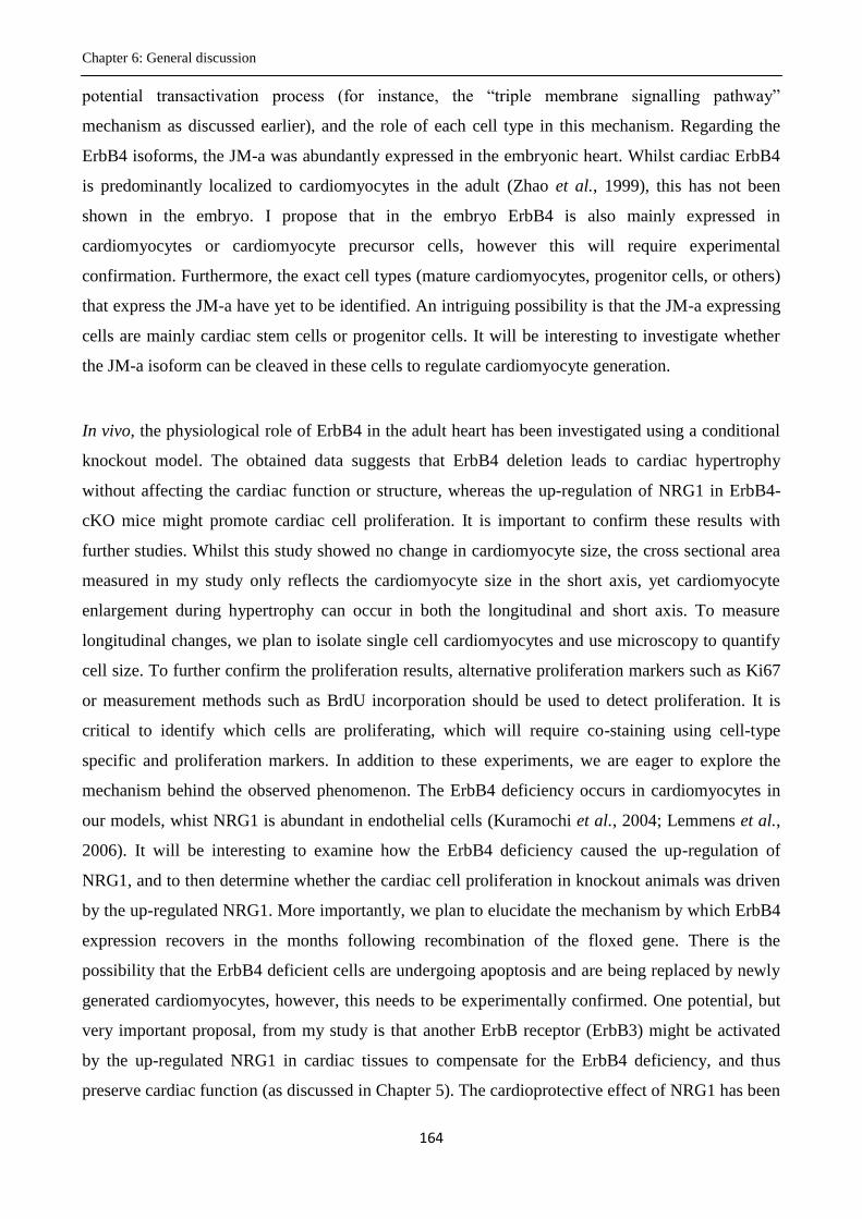
Chapter 6: General discussion
164
potential transactivation process (for instance, the “triple membrane signalling pathway”
mechanism as discussed earlier), and the role of each cell type in this mechanism. Regarding the
ErbB4 isoforms, the JM-a was abundantly expressed in the embryonic heart. Whilst cardiac ErbB4
is predominantly localized to cardiomyocytes in the adult (Zhao et al., 1999), this has not been
shown in the embryo. I propose that in the embryo ErbB4 is also mainly expressed in
cardiomyocytes or cardiomyocyte precursor cells, however this will require experimental
confirmation. Furthermore, the exact cell types (mature cardiomyocytes, progenitor cells, or others)
that express the JM-a have yet to be identified. An intriguing possibility is that the JM-a expressing
cells are mainly cardiac stem cells or progenitor cells. It will be interesting to investigate whether
the JM-a isoform can be cleaved in these cells to regulate cardiomyocyte generation.
In vivo, the physiological role of ErbB4 in the adult heart has been investigated using a conditional
knockout model. The obtained data suggests that ErbB4 deletion leads to cardiac hypertrophy
without affecting the cardiac function or structure, whereas the up-regulation of NRG1 in ErbB4-
cKO mice might promote cardiac cell proliferation. It is important to confirm these results with
further studies. Whilst this study showed no change in cardiomyocyte size, the cross sectional area
measured in my study only reflects the cardiomyocyte size in the short axis, yet cardiomyocyte
enlargement during hypertrophy can occur in both the longitudinal and short axis. To measure
longitudinal changes, we plan to isolate single cell cardiomyocytes and use microscopy to quantify
cell size. To further confirm the proliferation results, alternative proliferation markers such as Ki67
or measurement methods such as BrdU incorporation should be used to detect proliferation. It is
critical to identify which cells are proliferating, which will require co-staining using cell-type
specific and proliferation markers. In addition to these experiments, we are eager to explore the
mechanism behind the observed phenomenon. The ErbB4 deficiency occurs in cardiomyocytes in
our models, whist NRG1 is abundant in endothelial cells (Kuramochi et al., 2004; Lemmens et al.,
2006). It will be interesting to examine how the ErbB4 deficiency caused the up-regulation of
NRG1, and to then determine whether the cardiac cell proliferation in knockout animals was driven
by the up-regulated NRG1. More importantly, we plan to elucidate the mechanism by which ErbB4
expression recovers in the months following recombination of the floxed gene. There is the
possibility that the ErbB4 deficient cells are undergoing apoptosis and are being replaced by newly
generated cardiomyocytes, however, this needs to be experimentally confirmed. One potential, but
very important proposal, from my study is that another ErbB receptor (ErbB3) might be activated
by the up-regulated NRG1 in cardiac tissues to compensate for the ErbB4 deficiency, and thus
preserve cardiac function (as discussed in Chapter 5). The cardioprotective effect of NRG1 has been

Chapter 6: General discussion
165
demonstrated in various pathological animal models whilst phase II clinical trials demonstrated that
NRG1 could improve the cardiac function of patients with chronic heart failure (Gao et al., 2010) .
Currently, ErbB4 is considered to be the only receptor for NRG1 in cardiomyocytes. If ErbB3 can
be activated by NRG1 in the heart, the proposed molecular mechanism (where NRG1’s beneficial
effect occurs via ErbB4) needs to be re-evaluated and this new potential pathway via ErbB3
requires further exploration. Indeed, whether ErbB4 deficiency will affect the ability of NRG1 to
protect the heart is yet to be determined. More fundamentally, the phenotype of the ErbB4-cKO
mice under pathological conditions has not been elucidated. For this purpose, as a next step we are
going to investigate how cardiac deletion of ErbB4 affects the response of the heart to a
hypertrophic stimulus using an Ang II infusion model of hypertension and hypertrophy. This work
will provide important information regarding the role of ErbB4 in cardiac function and structure
under pathological conditions.
Conclusion
During my candidature, I have fully investigated the transactivation of ErbB family by Ang II in
cardiomyocytes and provided evidence that the Ang II signalling in cardiomyocyte hypertrophy is
independent of ErbB receptors. This is very important for elucidating the molecular pathway of the
pathological cardiac hypertrophy related to the activation of local RAS system. My studies
confirmed the requirement for ErbB4 in NRG1 induced hypertrophic signalling. The discovery that
all four ErbB4 isoforms could mediate NRG1 signalling in postnatal cardiomyocytes suggested that
NRG1 induced hypertrophic signalling is dependent on a fundamental common feature of the four
isoforms. The decline in expression of the cleavable isoforms (JM-a) throughout development, and
the lack of a cleavage mechanism in postnatal cardiomyocytes suggests that the postnatal heart does
not require the ICD function. In contrast, the relative higher level of JM-a in embryonic stages
suggests that the ICD might be released and required by the embryonic heart. This study indicates
that further investigation on the biological function of JM-a isoform in the embryonic heart is
required, particularly with respect to cardiomyocyte generation. The in vivo study established a new
conditional knockout model in our lab, where the gene deletion is both tissue specific and
temporally controlled. Using this model, we investigated the function of ErbB4 in adult mice heart.
The cardiac specific deletion of ErbB4 in adult mice did not result in a severe cardiac phenotype
(cardiac dysfunction and structural abnormality) but a series of compensatory responses (up-
regulation of NRG1 and physiological cardiac hypertrophy). This proposes that, in contrast to the
embryonic heart, the intact adult heart adopts alternative mechanisms to satisfy cardiac
physiological requirements when ErbB4 is absent. More importantly, the lack of an obvious

Chapter 6: General discussion
166
phenotype may indicate that the primary function of the ErbB4 in the adult heart is different from
the embryonic heart. The former might be mediating the NRG1 signalling to preserve cardiac
function and structure under pathological conditions, whereas the latter promotes growth. Thus,
deletion of ErbB4 in embryonic heart leads to pre-birth death due to the cessation of cardiac growth.
In contrast, the phenotype of ErbB4 deletion in adult heart may only be observed under pathological
conditions. In a future study we plan to use our conditional knockout model to investigate the
function of ErbB4 in a model of pathological cardiac hypertrophy.

REFERENCES

Referemce
168
Adabag AS, Luepker RV, Roger VL, Gersh BJ (2010). Sudden cardiac death: epidemiology and
risk factors. Nature Reviews Cardiology 7: 216-225.
Adams Jr, Bodor GS, Davila-Roman VG, Delmez J, Apple F, Ladenson J, et al. (1993). Cardiac
troponin I. A marker with high specificity for cardiac injury. Circulation 88: 101-106.
Adams JW, Sakata Y, Davis MG, Sah VP, Wang Y, Liggett SB, et al. (1998). Enhanced Gαq
signaling: a common pathway mediates cardiac hypertrophy and apoptotic heart failure.
Proceedings of the National Academy of Sciences 95: 10140-10145.
Adler C, Ringlage W, Böhm N (1981). DNA content and cell number in heart and liver of children.
Comparable biochemical, cytophotometric and histological investigations (author's transl).
Pathology, research and practice 172: 25.
Advani A, Kelly DJ, Cox AJ, White KE, Advani SL, Thai K, et al. (2009). The (Pro) Renin
Receptor Site-Specific and Functional Linkage to the Vacuolar H+-ATPase in the Kidney.
Hypertension 54: 261-269.
Akazawa H, Komuro I (2003). Roles of Cardiac Transcription Factors in Cardiac Hypertrophy.
Circulation Research 92: 1079-1088.
An T, Zhang Y, Huang Y, Zhang R, Yin S, Guo X, et al. (2013). Neuregulin-1 protects against
doxorubicin-induced apoptosis in cardiomyocytes through an Akt-dependent pathway. Physiol Res
Acad Sci Bohemoslov 62: 379-385.
Anastassiadis T, Deacon SW, Devarajan K, Ma H, Peterson JR (2011). Comprehensive assay of
kinase catalytic activity reveals features of kinase inhibitor selectivity. Nature biotechnology 29:
1039-1045.
Ang K-L, Shenje LT, Reuter S, Soonpaa MH, Rubart M, Field LJ, et al. (2010). Limitations of
conventional approaches to identify myocyte nuclei in histologic sections of the heart. American
Journal of Physiology-Cell Physiology 298: C1603-C1609.

Referemce
169
Aoki H, Izumo S, Sadoshima J (1998). Angiotensin II Activates RhoA in Cardiac Myocytes: A
Critical Role of RhoA in Angiotensin II–Induced Premyofibril Formation. Circulation Research 82:
666-676.
Aqeilan RI, Donati V, Palamarchuk A, Trapasso F, Kaou M, Pekarsky Y, et al. (2005). WW
Domain–Containing Proteins, WWOX and YAP, Compete for Interaction with ErbB-4 and
Modulate Its Transcriptional Function. Cancer Research 65: 6764-6772.
Artham SM, Lavie CJ, Milani RV, Patel DA, Verma A, Ventura HO (2009). Clinical Impact of Left
Ventricular Hypertrophy and Implications for Regression. Progress in Cardiovascular Diseases 52:
153-167.
Asakura M, Kitakaze M, Liao Y, Ishikura F, Yoshinaka T, Ohmoto H, et al. (2002). Cardiac
hypertrophy is inhibited by antagonism of ADAM12 processing of HB-EGF: Metalloproteinase
inhibitors as a new therapy. Nature Medicine 8: 35-40.
Aurora AB, Mahmoud AI, Luo X, Johnson BA, van Rooij E, Matsuzaki S, et al. (2012).
MicroRNA-214 protects the mouse heart from ischemic injury by controlling Ca2+ overload and
cell death. The Journal of clinical investigation 122: 1222-1232.
Baliga RR, Pimental DR, Zhao Y-Y, Simmons WW, Marchionni MA, Sawyer DB, et al. (1999).
NRG-1-induced cardiomyocyte hypertrophy. Role of PI-3-kinase, p70S6K, and MEK-MAPK-RSK.
edn, vol. 277.
Barry SP, Davidson SM, Townsend PA (2008). Molecular regulation of cardiac hypertrophy. The
international journal of biochemistry & cell biology 40: 2023-2039.
Beltrami AP, Urbanek K, Kajstura J, Yan S-M, Finato N, Bussani R, et al. (2001). Evidence that
human cardiac myocytes divide after myocardial infarction. New England Journal of Medicine 344:
1750-1757.
Bergmann O, Bhardwaj RD, Bernard S, Zdunek S, Barnabé-Heider F, Walsh S, et al. (2009).
Evidence for cardiomyocyte renewal in humans. Science 324: 98-102.

Referemce
170
Bersell K, Arab S, Haring B, Kühn B (2009). Neuregulin1/ErbB4 signaling induces cardiomyocyte
proliferation and repair of heart injury. Cell 138: 257-270.
Bian Y, Sun M, Silver M, Ho KKL, Marchionni MA, Caggiano AO, et al. (2009). Neuregulin-1
attenuated doxorubicin-induced decrease in cardiac troponins. edn, vol. 297.
Blumenthal DK, Stull JT, Gill GN (1978). Phosphorylation of cardiac troponin by guanosine 3': 5'-
monophosphate-dependent protein kinase. Journal of Biological Chemistry 253: 334-336.
Bokemeyer D, Schmitz U, Kramer HJ (2000). Angiotensin II-induced growth of vascular smooth
muscle cells requires an Src-dependent activation of the epidermal growth factor receptor. Kidney
international 58: 549-558.
Bombelli M, Facchetti R, Carugo S, Madotto F, Arenare F, Quarti-Trevano F, et al. (2009). Left
ventricular hypertrophy increases cardiovascular risk independently of in-office and out-of-office
blood pressure values. J Hypertens 27: 2458-2464.
Braz JC, Bueno OF, De Windt LJ, Molkentin JD (2002). PKCα regulates the hypertrophic growth
of cardiomyocytes through extracellular signal–regulated kinase1/2 (ERK1/2). The Journal of cell
biology 156: 905-919.
Brero A, Ramella R, Fitou A, Dati C, Alloatti G, Gallo MP, et al. (2010). Neuregulin-1β1 rapidly
modulates nitric oxide synthesis and calcium handling in rat cardiomyocytes. Cardiovascular
research 88: 443-452.
Brown RD, Ambler SK, Mitchell MD, Long CS (2005). The cardiac fibroblast: therapeutic target in
myocardial remodeling and failure. Annu. Rev. Pharmacol. Toxicol. 45: 657-687.
Bueno OF, De Windt LJ, Tymitz KM, Witt SA, Kimball TR, Klevitsky R, et al. (2000). The
MEK1–ERK1/2 signaling pathway promotes compensated cardiac hypertrophy in transgenic mice.
The EMBO journal 19: 6341-6350.
Buonanno A, Fischbach GD (2001). Neuregulin and ErbB receptor signaling pathways in the
nervous system. Current opinion in neurobiology 11: 287-296.

Referemce
171
Burgess AW (2008). EGFR family: structure physiology signalling and therapeutic targets. Growth
Factors 26: 263-274.
Burgess AW, Cho H-S, Eigenbrot C, Ferguson KM, Garrett TPJ, Leahy DJ, et al. (2003). An Open-
and-Shut Case? Recent Insights into the Activation of EGF/ErbB Receptors. Molecular Cell 12:
541-552.
Cabrera-Bueno F, García-Pinilla JM, Gómez-Doblas JJ, Montiel-Trujillo A, Rodríguez-Bailón I, de
Teresa-Galván E (2007). Beta-blocker therapy for dynamic left ventricular outflow tract obstruction
induced by exercise. International journal of cardiology 117: 222-226.
Campreciós G, Lorita J, Pardina E, Peinado‐Onsurbe J, Soley M, Ramírez I (2011). Expression,
localization, and regulation of the neuregulin receptor ErbB3 in mouse heart. Journal of cellular
physiology 226: 450-455.
Carabatsos MJ, Combelles CM, Messinger SM, Albertini DF (2000). Sorting and reorganization of
centrosomes during oocyte maturation in the mouse. Microscopy research and technique 49: 435-
444.
Carabello BA (2002). Concentric versus eccentric remodeling. Journal of cardiac failure 8: S258-
S263.
Casey P, Gilman A (1988). G protein involvement in receptor-effector coupling. The Journal of
biological chemistry 263: 2577-2580.
Cassis LA, Gupte M, Thayer S, Zhang X, Charnigo R, Howatt DA, et al. (2009). ANG II infusion
promotes abdominal aortic aneurysms independent of increased blood pressure in
hypercholesterolemic mice. Am J Physiol Heart Circ Physiol 296: H1660-1665.
Ceylan-Isik AF, Dong M, Zhang Y, Dong F, Turdi S, Nair S, et al. (2013). Cardiomyocyte-specific
deletion of endothelin receptor A rescues aging-associated cardiac hypertrophy and contractile
dysfunction: role of autophagy. Basic research in cardiology 108: 1-19.
Chang-Yuan N, Murphy NP, Golde TE, Carpenter G (2001). Gamma-secretase cleavage and
nuclear localization of ErbB-4 receptor tyrosine kinase. Science 294: 2179-2181.

Referemce
172
Chen J, Chen J-K, Neilson EG, Harris RC (2006). Role of EGF receptor activation in angiotensin
II-induced renal epithelial cell hypertrophy. Journal of the American Society of Nephrology : JASN
17: 1615-1623.
Chien KR (2001). To Cre or Not To Cre The Next Generation of Mouse Models of Human Cardiac
Diseases. Circulation research 88: 546-549.
Clerk A, Sugden PH (1999). Activation of protein kinase cascades in the heart by hypertrophic G
protein–coupled receptor agonists. The American journal of cardiology 83: 64-69.
Coleman ML, Marshall CJ, Olson MF (2004). RAS and RHO GTPases in G1-phase cell-cycle
regulation. Nature Reviews Molecular Cell Biology 5: 355-366.
Cote GM, Miller TA, LeBrasseur NK, Kuramochi Y, Sawyer DB (2005). Neuregulin-1α and β
isoform expression in cardiac microvascular endothelial cells and function in cardiac myocytes in
vitro. Experimental cell research 311: 135-146.
Crowley SD, Gurley SB, Herrera MJ, Ruiz P, Griffiths R, Kumar AP, et al. (2006). Angiotensin II
causes hypertension and cardiac hypertrophy through its receptors in the kidney. Proceedings of the
National Academy of Sciences 103: 17985-17990.
Dahlöf B, Devereux RB, Kjeldsen SE, Julius S, Beevers G, de Faire U, et al. (2002).
Cardiovascular morbidity and mortality in the Losartan Intervention For Endpoint reduction in
hypertension study (LIFE): a randomised trial against atenolol. The Lancet 359: 995-1003.
Dahlöf B, Gosse P, Guéret P, Dubourg O, De Simone G, Schmieder R, et al. (2005).
Perindopril/indapamide combination more effective than enalapril in reducing blood pressure and
left ventricular mass: the PICXEL study. Journal of hypertension 23: 2063-2070.
Daub H, Weiss FU, Wallasch C, Ullrich A (1996). Role of transactivation of the EGF receptor in
signalling by G-protein-coupled receptors. Nature 379: 557-560.
Devereux RB, Palmieri V, Sharpe N, De Quattro V, Bella JN, de Simone G, et al. (2001). Effects of
Once-Daily Angiotensin-Converting Enzyme Inhibition and Calcium Channel Blockade-Based

Referemce
173
Antihypertensive Treatment Regimens on Left Ventricular Hypertrophy and Diastolic Filling in
Hypertension: The Prospective Randomized Enalapril Study Evaluating Regression of Ventricular
Enlargement (PRESERVE) Trial. Circulation 104: 1248-1254.
Dhanasekaran S, Doherty TM, Kenneth J (2010). Comparison of different standards for real-time
PCR-based absolute quantification. Journal of immunological methods 354: 34-39.
Dikic I, Tokiwa G, Lev S, Courtneidge SA, Schlessinger J (1996). A role for Pyk2 and Src in
linking G-protein-coupled receptors with MAP kinase activation.
Dorn GW (2007). The Fuzzy Logic of Physiological Cardiac Hypertrophy. Hypertension 49: 962-
970.
Drazner MH (2005). The Transition From Hypertrophy to Failure. Circulation 112: 936-938.
Edwards DR, Handsley MM, Pennington CJ (2008). The ADAM metalloproteinases. Molecular
aspects of medicine 29: 258-289.
Eghbali M, Deva R, Alioua A, Minosyan TY, Ruan H, Wang Y, et al. (2005). Molecular and
functional signature of heart hypertrophy during pregnancy. Circulation research 96: 1208-1216.
Eguchi S, Iwasaki H, Ueno H, Frank GD, Motley ED, Eguchi K, et al. (1999). Intracellular
signaling of angiotensin II-induced p70 S6 kinase phosphorylation at Ser(411) in vascular smooth
muscle cells. Possible requirement of epidermal growth factor receptor, Ras, extracellular signal-
regulated kinase, and Akt. The Journal of biological chemistry 274: 36843-36851.
Eguchi S, Numaguchi K, Iwasaki H, Matsumoto T, Yamakawa T, Utsunomiya H, et al. (1998).
Calcium-dependent Epidermal Growth Factor Receptor Transactivation Mediates the Angiotensin
II-induced Mitogen-activated Protein Kinase Activation in Vascular Smooth Muscle Cells. Journal
of Biological Chemistry 273: 8890-8896.
Elenius K, Choi CJ, Paul S, Santiestevan E, Nishi E, Klagsbrun M (1999). Characterization of a
naturally occurring ErbB4 isoform that does not bind or activate phosphatidyl inositol 3-kinase.
Oncogene 18: 2607-2615.

Referemce
174
Elenius K, Corfas G, Paul S, Choi CJ, Rio C, Plowman GD, et al. (1997). A Novel Juxtamembrane
Domain Isoform of HER4/ErbB4 ISOFORM-SPECIFIC TISSUE DISTRIBUTION AND
DIFFERENTIAL PROCESSING IN RESPONSE TO PHORBOL ESTER. Journal of Biological
Chemistry 272: 26761-26768.
Ellison GM, Waring CD, Vicinanza C, Torella D (2011). Physiological cardiac remodelling in
response to endurance exercise training: cellular and molecular mechanisms. Heart: heartjnl-2011-
300639.
Erickson SL, O'Shea KS, Ghaboosi N, Loverro L, Frantz G, Bauer M, et al. (1997). ErbB3 is
required for normal cerebellar and cardiac development: a comparison with ErbB2-and heregulin-
deficient mice. Development 124: 4999-5011.
Esposito G, Rapacciuolo A, Naga Prasad SV, Rockman HA (2002). Cardiac hypertrophy: role of G
protein-coupled receptors. Journal of cardiac failure 8: S409-S414.
Falls DL (2003). Neuregulins: functions, forms, and signaling strategies. Experimental cell research
284: 14-30.
Fang S-j, Wu X-s, Han Z-h, Zhang X-x, Wang C-m, Li X-y, et al. (2010). Neuregulin-1
preconditioning protects the heart against ischemia/reperfusion injury through a PI3K/Akt-
dependent mechanism. Chinese Medical Journal (English Edition) 123: 3597.
Feng S-M, Sartor CI, Hunter D, Zhou H, Yang X, Caskey LS, et al. (2007). The HER4 Cytoplasmic
Domain, But Not Its C Terminus, Inhibits Mammary Cell Proliferation. Molecular Endocrinology
21: 1861-1876.
Ferrario CM (2011). ACE2: more of Ang-(1–7) or less Ang II? Current Opinion in Nephrology and
Hypertension 20: 1-6 10.1097/MNH.1090b1013e3283406f3283457.
Fischer O, Hart S, Gschwind A, Ullrich A (2003). EGFR signal transactivation in cancer cells.
Biochemical Society Transactions 31: 1203-1208.
Fox IJ, Kornblum HI (2005). Developmental profile of ErbB receptors in murine central nervous
system: Implications for functional interactions. Journal of Neuroscience Research 79: 584-597.

Referemce
175
Franco D, Markman MM, Wagenaar G, Ya J, Lamers WH, Moorman AF (1999). Myosin light
chain 2a and 2v identifies the embryonic outflow tract myocardium in the developing rodent heart.
The Anatomical Record 254: 135-146.
Frey N, Olson E (2003). Cardiac hypertrophy: the good, the bad, and the ugly. Annual review of
physiology 65: 45-79.
Frey N, Katus HA, Olson EN, Hill JA (2004). Hypertrophy of the Heart. Circulation 109: 1580-
1589.
Fukazawa R, Miller TA, Kuramochi Y, Frantz S, Kim YD, Marchionni MA, et al. (2003).
Neuregulin-1 protects ventricular myocytes from anthracycline-induced apoptosis via erbB4-
dependent activation of PI3-kinase/Akt. J Mol Cell Cardiol 35: 1473-1479.
Gaestel M (2006). MAPKAP kinases — MKs — two's company, three's a crowd. Nature Reviews
Molecular Cell Biology 7: 120-130.
Gao R, Zhang J, Cheng L, Wu X, Dong W, Yang X, et al. (2010). A Phase II, Randomized,
Double-Blind, Multicenter, Based on Standard Therapy, Placebo-Controlled Study of the Efficacy
and Safety of Recombinant Human Neuregulin-1 in Patients With Chronic Heart Failure. Journal of
the American College of Cardiology 55: 1907-1914.
Garcia-Rivello H, Taranda J, Said M, Cabeza-Meckert P, Vila-Petroff M, Scaglione J, et al. (2005).
Dilated cardiomyopathy in Erb-b4-deficient ventricular muscle. Am J Physiol Heart Circ Physiol
289: H1153-1160.
Garrett TP, McKern NM, Lou M, Elleman TC, Adams TE, Lovrecz GO, et al. (2003). The crystal
structure of a truncated ErbB2 ectodomain reveals an active conformation, poised to interact with
other ErbB receptors. Molecular cell 11: 495-505.
Gassmann M, Casagranda F, Orioli D, Simon H, Lai C, Klein R, et al. (1995). Aberrant neural and
cardiac development in mice lacking the ErbB4 neuregulin receptor.
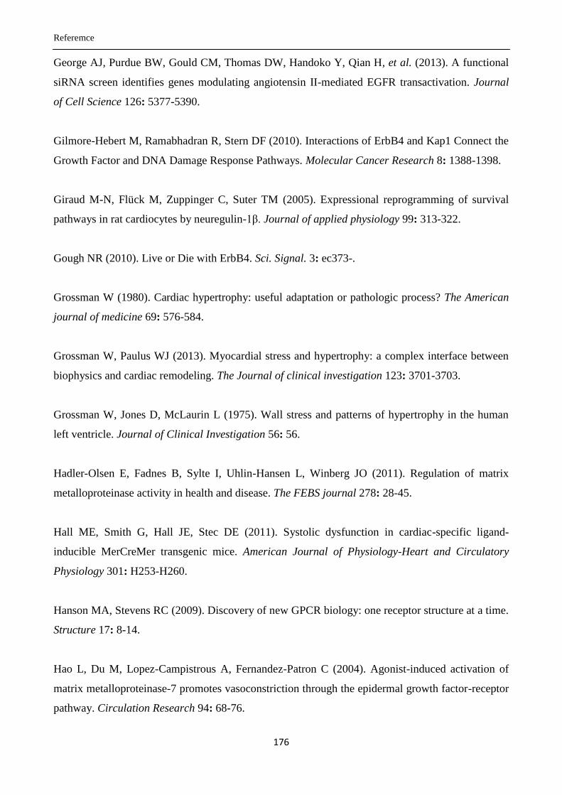
Referemce
176
George AJ, Purdue BW, Gould CM, Thomas DW, Handoko Y, Qian H, et al. (2013). A functional
siRNA screen identifies genes modulating angiotensin II-mediated EGFR transactivation. Journal
of Cell Science 126: 5377-5390.
Gilmore-Hebert M, Ramabhadran R, Stern DF (2010). Interactions of ErbB4 and Kap1 Connect the
Growth Factor and DNA Damage Response Pathways. Molecular Cancer Research 8: 1388-1398.
Giraud M-N, Flück M, Zuppinger C, Suter TM (2005). Expressional reprogramming of survival
pathways in rat cardiocytes by neuregulin-1β. Journal of applied physiology 99: 313-322.
Gough NR (2010). Live or Die with ErbB4. Sci. Signal. 3: ec373-.
Grossman W (1980). Cardiac hypertrophy: useful adaptation or pathologic process? The American
journal of medicine 69: 576-584.
Grossman W, Paulus WJ (2013). Myocardial stress and hypertrophy: a complex interface between
biophysics and cardiac remodeling. The Journal of clinical investigation 123: 3701-3703.
Grossman W, Jones D, McLaurin L (1975). Wall stress and patterns of hypertrophy in the human
left ventricle. Journal of Clinical Investigation 56: 56.
Hadler-Olsen E, Fadnes B, Sylte I, Uhlin-Hansen L, Winberg JO (2011). Regulation of matrix
metalloproteinase activity in health and disease. The FEBS journal 278: 28-45.
Hall ME, Smith G, Hall JE, Stec DE (2011). Systolic dysfunction in cardiac-specific ligand-
inducible MerCreMer transgenic mice. American Journal of Physiology-Heart and Circulatory
Physiology 301: H253-H260.
Hanson MA, Stevens RC (2009). Discovery of new GPCR biology: one receptor structure at a time.
Structure 17: 8-14.
Hao L, Du M, Lopez-Campistrous A, Fernandez-Patron C (2004). Agonist-induced activation of
matrix metalloproteinase-7 promotes vasoconstriction through the epidermal growth factor-receptor
pathway. Circulation Research 94: 68-76.

Referemce
177
Harris DM, Chen X, Pesant S, Cohn HI, MacDonnell SM, Boucher M, et al. (2009). Inhibition of
angiotensin II Gq signaling augments beta-adrenergic receptor mediated effects in a renal artery
stenosis model of high blood pressure. J Mol Cell Cardiol 46: 100-107.
Hasegawa K, Lee SJ, Jobe SM, Markham BE, Kitsis RN (1997). cis-Acting sequences that mediate
induction of β-myosin heavy chain gene expression during left ventricular hypertrophy due to aortic
constriction. Circulation 96: 3943-3953.
Hedhli N, Huang Q, Kalinowski A, Palmeri M, Hu X, Russell RR, et al. (2011). Endothelium-
Derived Neuregulin Protects the Heart Against Ischemic Injury / Clinical Perspective. Circulation
123: 2254-2262.
Heineke J, Molkentin JD (2006). Regulation of cardiac hypertrophy by intracellular signalling
pathways. Nature Reviews Molecular Cell Biology 7: 589-600.
Henderson SA, Spencer M, Sen A, Kumar C, Siddiqui MA, Chien KR (1989). Structure,
organization, and expression of the rat cardiac myosin light chain-2 gene. Identification of a 250-
base pair fragment which confers cardiac-specific expression. Journal of Biological Chemistry 264:
18142-18148.
Hepler JR, Kozasa T, Smrcka AV, Simon MI, Rhee SG, Sternweis PC, et al. (1993). Purification
from Sf9 cells and characterization of recombinant Gq alpha and G11 alpha. Activation of purified
phospholipase C isozymes by G alpha subunits. The Journal of biological chemistry 268: 14367-
14375.
Higa‐Nakamine S, Maeda N, Toku S, Yamamoto T, Yingyuenyong M, Kawahara M, et al. (2012).
Selective cleavage of ErbB4 by G‐protein‐coupled Gonadotropin‐Releasing Hormone Receptor in
Cultured Hypothalamic Neurons. Journal of cellular physiology 227: 2492-2501.
Hill CS, Treisman R (1995). Transcriptional Regulation by Extracellular signals: Mechanisms and
Specificity. Cell 80: 199-211.
Houston S, Plunkett T, Barnes D, Smith P, Rubens R, Miles D (1999). Overexpression of c-erbB2
is an independent marker of resistance to endocrine therapy in advanced breast cancer. British
journal of cancer 79: 1220.

Referemce
178
Hsieh PCH, Segers VFM, Davis ME, MacGillivray C, Gannon J, Molkentin JD, et al. (2007).
Evidence from a genetic fate-mapping study that stem cells refresh adult mammalian
cardiomyocytes after injury. Nat Med 13: 970-974.
Hubbard SR, Mohammadi M, Schlessinger J (1998). Autoregulatory mechanisms in protein-
tyrosine kinases. The Journal of biological chemistry 273: 11987-11990.
Iemitsu M, Miyauchi T, Maeda S, Sakai S, Kobayashi T, Fujii N, et al. (2001). Physiological and
pathological cardiac hypertrophy induce different molecular phenotypes in the rat. American
Journal of Physiology-Regulatory, Integrative and Comparative Physiology 281: R2029-R2036.
Iwata H, Goto H, Tanaka H, Sakaguchi Y, Kimura K, Kuwayama T, et al. (2011). Effect of
maternal age on mitochondrial DNA copy number, ATP content and IVF outcome of bovine
oocytes. Reproduction, fertility, and development 23: 424-432.
Jiang Z, Zhou M (2010). Neuregulin signaling and heart failure. Current heart failure reports 7: 42-
47.
Jie B, Zhang X, Wu X, Xin Y, Liu Y, Guo Y (2012). Neuregulin-1 suppresses cardiomyocyte
apoptosis by activating PI3K/Akt and inhibiting mitochondrial permeability transition pore.
Molecular and cellular biochemistry 370: 35-43.
Jopling C, Sleep E, Raya M, Martí M, Raya A, Belmonte JCI (2010). Zebrafish heart regeneration
occurs by cardiomyocyte dedifferentiation and proliferation. Nature 464: 606-609.
Jorissen RN, Walker F, Pouliot N, Garrett TPJ, Ward CW, Burgess AW (2003). Epidermal growth
factor receptor: mechanisms of activation and signalling. Experimental Cell Research 284: 31-53.
Junttila TT, Sundvall M, Määttä JA, Elenius K (2000). ErbB4 and Its Isoforms: Selective
Regulation of Growth Factor Responses by Naturally Occurring Receptor Variants. Trends in
Cardiovascular Medicine 10: 304-310.
Junttila TT, Laato M, Vahlberg T, Söderström K-O, Visakorpi T, Isola J, et al. (2003).
Identification of patients with transitional cell carcinoma of the bladder overexpressing ErbB2,

Referemce
179
ErbB3, or specific ErbB4 isoforms real-time reverse transcription-PCR analysis in estimation of
ErbB receptor status from cancer patients. Clinical cancer research 9: 5346-5357.
Kagiyama S, Eguchi S, Frank GD, Inagami T, Zhang YC, Phillips MI (2002). Angiotensin II-
induced cardiac hypertrophy and hypertension are attenuated by epidermal growth factor receptor
antisense. Circulation 106: 909-912.
Kajstura J, Urbanek K, Perl S, Hosoda T, Zheng H, Ogórek B, et al. (2010). Cardiomyogenesis in
the adult human heart. Circulation research 107: 305-315.
Katholi RE, Couri DM (2011). Left Ventricular Hypertrophy: Major Risk Factor in Patients with
Hypertension: Update and Practical Clinical Applications. International Journal of Hypertension
2011.
Katus HA, Remppis A, Neumann FJ, Scheffold T, Diederich KW, Vinar G, et al. (1991).
Diagnostic efficiency of troponin T measurements in acute myocardial infarction. Circulation 83:
902-912.
Keely SJ, Calandrella SO, Barrett KE (2000). Carbachol-stimulated transactivation of epidermal
growth factor receptor and mitogen-activated protein kinase in T(84) cells is mediated by
intracellular ca(2+), PYK-2, and p60(src). The Journal of biological chemistry 275: 12619-12625.
Kelly DJ, Cox AJ, Gow RM, Zhang Y, Kemp BE, Gilbert RE (2004). Platelet-derived growth factor
receptor transactivation mediates the trophic effects of angiotensin II in vivo. Hypertension 44: 195-
202.
Kikuchi K, Holdway JE, Werdich AA, Anderson RM, Fang Y, Egnaczyk GF, et al. (2010). Primary
contribution to zebrafish heart regeneration by gata4+ cardiomyocytes. Nature 464: 601-605.
Kjeldsen SE, Dahlöf B, Devereux RB, Julius S, Aurup P, Edelman J, et al. (2002). Effects of
losartan on cardiovascular morbidity and mortality in patients with isolated systolic hypertension
and left ventricular hypertrophy: a Losartan Intervention for Endpoint Reduction (LIFE) substudy.
Jama 288: 1491-1498.

Referemce
180
Klein T, Bischoff R (2011). Active metalloproteases of the A Disintegrin and Metalloprotease
(ADAM) family: biological function and structure. J Proteome Res 10: 17-33.
Klingbeil AU, Schneider M, Martus P, Messerli FH, Schmieder RE (2003). A meta-analysis of the
effects of treatment on left ventricular mass in essential hypertension. The American journal of
medicine 115: 41-46.
Klug MG, Soonpaa MH, Koh GY, Field LJ (1996). Genetically selected cardiomyocytes from
differentiating embronic stem cells form stable intracardiac grafts. Journal of Clinical Investigation
98: 216.
Knowlton KU, Baracchini E, Ross R, Harris A, Henderson S, Evans S, et al. (1991). Co-regulation
of the atrial natriuretic factor and cardiac myosin light chain-2 genes during alpha-adrenergic
stimulation of neonatal rat ventricular cells. Identification of cis sequences within an embryonic and
a constitutive contractile protein gene which mediate inducible expression. Journal of Biological
Chemistry 266: 7759-7768.
Kodama H, Fukuda K, Takahashi T, Sano M, Kato T, Tahara S, et al. (2002). Role of EGF
Receptor and Pyk2 in endothelin-1-induced ERK activation in rat cardiomyocytes. Journal of
molecular and cellular cardiology 34: 139-150.
Koitabashi N, Bedja D, Zaiman AL, Pinto YM, Zhang M, Gabrielson KL, et al. (2009). Avoidance
of transient cardiomyopathy in cardiomyocyte-targeted tamoxifen-induced MerCreMer gene
deletion models. Circulation research 105: 12-15.
Komuro A, Nagai M, Navin NE, Sudol M (2003). WW domain-containing protein YAP associates
with ErbB-4 and acts as a co-transcriptional activator for the carboxyl-terminal fragment of ErbB-4
that translocates to the nucleus. Journal of Biological Chemistry 278: 33334-33341.
Kühn B, del Monte F, Hajjar RJ, Chang Y-S, Lebeche D, Arab S, et al. (2007). Periostin induces
proliferation of differentiated cardiomyocytes and promotes cardiac repair. Nature medicine 13:
962-969.

Referemce
181
Kumar V, Zhang M-X, Swank MW, Kunz J, Wu G-Y (2005). Regulation of dendritic
morphogenesis by Ras–PI3K–Akt–mTOR and Ras–MAPK signaling pathways. The Journal of
neuroscience 25: 11288-11299.
Kuramochi Y, Cote GM, Guo X, Lebrasseur NK, Cui L, Liao R, et al. (2004). Cardiac endothelial
cells regulate reactive oxygen species-induced cardiomyocyte apoptosis through neuregulin-
1β/erbB4 signaling. Journal of Biological Chemistry 279: 51141-51147.
Laflamme MA, Murry CE (2011). Heart regeneration. Nature 473: 326-335.
Lautrette A, Li S, Alili R, Sunnarborg SW, Burtin M, Lee DC, et al. (2005). Angiotensin II and
EGF receptor cross-talk in chronic kidney diseases: a new therapeutic approach. Nature Medicine
11: 867-874.
Law AJ, Kleinman JE, Weinberger DR, Weickert CS (2007). Disease-associated intronic variants in
the ErbB4 gene are related to altered ErbB4 splice-variant expression in the brain in schizophrenia.
Human Molecular Genetics 16: 129-141.
Lee E, Taussig R, Gilman AG (1992). The G226A mutant of Gs alpha highlights the requirement
for dissociation of G protein subunits. Journal of Biological Chemistry 267: 1212-1218.
Lee K-F, Simon H, Chen H, Bates B, Hung M-C, Hauser C (1995). Requirement for neuregulin
receptor erbB2 in neural and cardiac development.
Lemmens K, Segers VF, Demolder M, De Keulenaer GW (2006). Role of neuregulin-1/ErbB2
signaling in endothelium-cardiomyocyte cross-talk. Journal of Biological Chemistry 281: 19469-
19477.
Lemmens K, Fransen P, Sys SU, Brutsaert DL, De Keulenaer GW (2004). Neuregulin-1 Induces a
Negative Inotropic Effect in Cardiac Muscle Role of Nitric Oxide Synthase. Circulation 109: 324-
326.
Lepilina A, Coon AN, Kikuchi K, Holdway JE, Roberts RW, Burns CG, et al. (2006). A dynamic
epicardial injury response supports progenitor cell activity during zebrafish heart regeneration. Cell
127: 607-619.

Referemce
182
Levitzki A, Gazit A (1995). Tyrosine kinase inhibition: an approach to drug development. Science
267: 1782-1788.
Li B, Zheng Z, Wei Y, Wang M, Peng J, Kang T, et al. (2011). Therapeutic effects of neuregulin-1
in diabetic cardiomyopathy rats. Cardiovascular Diabetology 10: 69.
Liang Q, Molkentin JD (2002). Divergent signaling pathways converge on GATA4 to regulate
cardiac hypertrophic gene expression. Journal of molecular and cellular cardiology 34: 611-616.
Liang Q, Wiese RJ, Bueno OF, Dai Y-S, Markham BE, Molkentin JD (2001). The Transcription
Factor GATA4 Is Activated by Extracellular Signal-Regulated Kinase 1- and 2-Mediated
Phosphorylation of Serine 105 in Cardiomyocytes. Molecular and Cellular Biology 21: 7460-7469.
Liebmann C (2011). EGF receptor activation by GPCRs: an universal pathway reveals different
versions. Molecular and cellular endocrinology 331: 222-231.
Lin C-H, Chen Y-C, Pan T-M (2011). Quantification bias caused by plasmid DNA conformation in
quantitative real-time PCR assay. PloS one 6: e29101.
Linggi B, Carpenter G (2006). ErbB receptors: new insights on mechanisms and biology. Trends in
Cell Biology 16: 649-656.
Linseman DA, Benjamin CW, Jones DA (1995). Convergence of Angiotensin II and Platelet-
derived Growth Factor Receptor Signaling Cascades in Vascular Smooth Muscle Cells. Journal of
Biological Chemistry 270: 12563-12568.
Linz W, Schölkens BA, Ganten D (1989). Converting Enzyme Inhibition Specifically Prevents the
Development and Induces Regression of Cardiac Hypertrophy in Rats. Clinical and Experimental
Hypertension a11: 1325-1350.
Liu X, Gu X, Li Z, Li X, Li H, Chang J, et al. (2006). Neuregulin-1/erbB-activation improves
cardiac function and survival in models of ischemic, dilated, and viral cardiomyopathy. Journal of
the American College of Cardiology 48: 1438-1447.

Referemce
183
Livak KJ, Schmittgen TD (2001). Analysis of Relative Gene Expression Data Using Real-Time
Quantitative PCR and the 2< sup>− ΔΔCT</sup> Method. methods 25: 402-408.
Long W, Wagner K-U, Lloyd KCK, Binart N, Shillingford JM, Hennighausen L, et al. (2003).
Impaired differentiation and lactational failure of Erbb4-deficient mammary glands identify ERBB4
as an obligate mediator of STAT5. Development 130: 5257-5268.
Luo J, McMullen JR, Sobkiw CL, Zhang L, Dorfman AL, Sherwood MC, et al. (2005). Class IA
phosphoinositide 3-kinase regulates heart size and physiological cardiac hypertrophy. Molecular
and cellular biology 25: 9491-9502.
Luppi F, Longo A, De Boer W, Rabe K, Hiemstra P (2007). Interleukin-8 stimulates cell
proliferation in non-small cell lung cancer through epidermal growth factor receptor transactivation.
Lung Cancer 56: 25-33.
Määttä JA, Sundvall M, Junttila TT, Peri L, Laine VJO, Isola J, et al. (2006). Proteolytic Cleavage
and Phosphorylation of a Tumor-associated ErbB4 Isoform Promote Ligand-independent Survival
and Cancer Cell Growth. Molecular Biology of the Cell 17: 67-79.
Macdougall LK, JONES LR, COHEN P (1991). Identification of the major protein phosphatases in
mammalian cardiac muscle which dephosphorylate phospholamban. European journal of
biochemistry 196: 725-734.
Mackay K, Mochly-Rosen D (2001). Localization, anchoring, and functions of protein kinase C
isozymes in the heart. Journal of molecular and cellular cardiology 33: 1301-1307.
Malliaras K, Zhang Y, Seinfeld J, Galang G, Tseliou E, Cheng K, et al. (2013). Cardiomyocyte
proliferation and progenitor cell recruitment underlie therapeutic regeneration after myocardial
infarction in the adult mouse heart. EMBO molecular medicine 5: 191-209.
Manabe I, Shindo T, Nagai R (2002). Gene expression in fibroblasts and fibrosis involvement in
cardiac hypertrophy. Circulation research 91: 1103-1113.
Markwald RR, Norris RA, Moreno-Rodriguez R, Levine RA (2010). Developmental basis of adult
cardiovascular diseases. Annals of the New York Academy of Sciences 1188: 177-183.
Referemce
184
McMullen JR, Jennings GL (2007). Differences between pathological and physiological cardiac
hypertrophy: novel therapeutic strategies to treat heart failure. Clinical and Experimental
Pharmacology and Physiology 34: 255-262.
McMullen JR, Shioi T, Zhang L, Tarnavski O, Sherwood MC, Kang PM, et al. (2003).
Phosphoinositide 3-kinase (p110α) plays a critical role for the induction of physiological, but not
pathological, cardiac hypertrophy. Proceedings of the National Academy of Sciences 100: 12355-
12360.
Mehta PK, Griendling KK (2007). Angiotensin II cell signaling: physiological and pathological
effects in the cardiovascular system. Am J Physiol Cell Physiol 292: C82-97.
Mendelsohn J, Baselga J (2003). Status of epidermal growth factor receptor antagonists in the
biology and treatment of cancer. Journal of clinical oncology : official journal of the American
Society of Clinical Oncology 21: 2787-2799.
Mendez M, LaPointe MC (2005). PGE2-induced hypertrophy of cardiac myocytes involves EP4
receptor-dependent activation of p42/44 MAPK and EGFR transactivation. American Journal of
Physiology-Heart and Circulatory Physiology 288: H2111-H2117.
Mery P-F, Lohmann SM, Walter U, Fischmeister R (1991). Ca2+ current is regulated by cyclic
GMP-dependent protein kinase in mammalian cardiac myocytes. Proceedings of the National
Academy of Sciences 88: 1197-1201.
Meyer D, Birchmeier C (1995). Multiple essential functions of neuregulin in development.
Miettinen PJ, Berger JE, Meneses J, Phung Y, Pedersen RA, Werb Z, et al. (1995). Epithelial
immaturity and multiorgan failure in mice lacking epidermal growth factor receptor. Nature 376:
337-341.
Mifune M, Ohtsu H, Suzuki H, Nakashima H, Brailoiu E, Dun NJ, et al. (2005). G Protein Coupling
and Second Messenger Generation Are Indispensable for Metalloprotease-dependent, Heparin-
binding Epidermal Growth Factor Shedding through Angiotensin II Type-1 Receptor. Journal of
Biological Chemistry 280: 26592-26599.

Referemce
185
Misfeldt AM Endocardial cells are a distinct endothelial lineage derived from multipotent
cardiovascular progenitors, Vanderbilt University, 2008.
Mizobuchi S, Kanzaki H, Omiya H, Matsuoka Y, Obata N, Kaku R, et al. (2013). Spinal nerve
injury causes upregulation of ErbB2 and ErbB3 receptors in rat dorsal root ganglia. Journal of pain
research 6: 87-94.
Mochizuki S, Okada Y (2007). ADAMs in cancer cell proliferation and progression. Cancer
Science 98: 621-628.
Molkentin JD (2000). The zinc finger-containing transcription factors GATA-4,-5, and-6
ubiquitously expressed regulators of tissue-specific gene expression. Journal of Biological
Chemistry 275: 38949-38952.
Molkentin JD, Robbins J (2009). With great power comes great responsibility: using mouse
genetics to study cardiac hypertrophy and failure. Journal of molecular and cellular cardiology 46:
130-136.
Molkentin JD, Lu J-R, Antos CL, Markham B, Richardson J, Robbins J, et al. (1998). A
calcineurin-dependent transcriptional pathway for cardiac hypertrophy. Cell 93: 215-228.
Mone SM, Sanders SP, Colan SD (1996). Control mechanisms for physiological hypertrophy of
pregnancy. Circulation 94: 667-672.
Morimoto T, Hasegawa K, Kaburagi S, Kakita T, Wada H, Yanazume T, et al. (2000).
Phosphorylation of GATA-4 Is Involved in α1-Adrenergic Agonist-responsive Transcription of the
Endothelin-1 Gene in Cardiac Myocytes. Journal of Biological Chemistry 275: 13721-13726.
Mukoyama M, Nakajima M, Horiuchi M, Sasamura H, Pratt RE, Dzau VJ (1993). Expression
cloning of type 2 angiotensin II receptor reveals a unique class of seven-transmembrane receptors.
Journal of Biological Chemistry 268: 24539-24542.
Müller P, Kazakov A, Semenov A, Jagoda P, Friedrich EB, Böhm M, et al. (2012). Ramipril and
Telmisartan Exhibit Differential Effects in Cardiac Pressure Overload-Induced Hypertrophy

Referemce
186
Without an Additional Benefit of the Combination of Both Drugs. Journal of Cardiovascular
Pharmacology and Therapeutics.
Muraoka-Cook RS, Sandahl M, Husted C, Hunter D, Miraglia L, Feng S-m, et al. (2006a). The
Intracellular Domain of ErbB4 Induces Differentiation of Mammary Epithelial Cells. Molecular
Biology of the Cell 17: 4118-4129.
Muraoka-Cook RS, Sandahl MA, Strunk KE, Miraglia LC, Husted C, Hunter DM, et al. (2009).
ErbB4 Splice Variants Cyt1 and Cyt2 Differ by 16 Amino Acids and Exert Opposing Effects on the
Mammary Epithelium In Vivo. Molecular and cellular biology 29: 4935-4948.
Muraoka-Cook RS, Caskey LS, Sandahl MA, Hunter DM, Husted C, Strunk KE, et al. (2006b).
Heregulin-Dependent Delay in Mitotic Progression Requires HER4 and BRCA1. Molecular and
cellular biology 26: 6412-6424.
Muscella A, Greco S, Elia MG, Storelli C, Marsigliante S (2003). PKC-? is required for angiotensin
II-induced activation of ERK and synthesis of C-FOS in MCF-7 cells. Journal of cellular
physiology 197: 61-68.
Nagase H, Visse R, Murphy G (2006). Structure and function of matrix metalloproteinases and
TIMPs. Cardiovascular Research 69: 562-573.
Nagendran J, Sutendra G, Paterson I, Champion HC, Webster L, Chiu B, et al. (2013). Endothelin
Axis Is Upregulated in Human and Rat Right Ventricular Hypertrophy. Circulation Research 112:
347-354.
Nagy A (2000). Cre recombinase: the universal reagent for genome tailoring. genesis 26: 99-109.
Naresh A, Long W, Vidal GA, Wimley WC, Marrero L, Sartor CI, et al. (2006). The ERBB4/HER4
Intracellular Domain 4ICD Is a BH3-Only Protein Promoting Apoptosis of Breast Cancer Cells.
Cancer Research 66: 6412-6420.
Nishimoto S, Nishida E (2006). MAPK signalling: ERK5 versus ERK1/2. EMBO reports 7: 782-
786.

Referemce
187
Ohtsu H, Dempsey PJ, Eguchi S (2006a). ADAMs as mediators of EGF receptor transactivation by
G protein-coupled receptors. American Journal of Physiology-Cell Physiology 291: C1-C10.
Ohtsu H, Dempsey PJ, Frank GD, Brailoiu E, Higuchi S, Suzuki H, et al. (2006b). ADAM17
mediates epidermal growth factor receptor transactivation and vascular smooth muscle cell
hypertrophy induced by angiotensin II. Arteriosclerosis, thrombosis, and vascular biology 26:
e133-E137.
Ohtsu H, Higuchi S, Shirai H, Eguchi K, Suzuki H, Hinoki A, et al. (2008). Central Role of Gq in
the Hypertrophic Signal Transduction of Angiotensin II in Vascular Smooth Muscle Cells.
Endocrinology 149: 3569-3575.
Okin PM, Devereux RB, Jern S, Kjeldsen SE, Julius S, Nieminen MS, et al. (2003). Regression of
Electrocardiographic Left Ventricular Hypertrophy by Losartan Versus Atenolol The Losartan
Intervention For Endpoint Reduction in Hypertension (LIFE) Study. Circulation 108: 684-690.
Okoshi K, Nakayama M, Yan X, Okoshi MP, Schuldt AJ, Marchionni MA, et al. (2004).
Neuregulins Regulate Cardiac Parasympathetic Activity Muscarinic Modulation of β-Adrenergic
Activity in Myocytes From Mice With Neuregulin-1 Gene Deletion. Circulation 110: 713-717.
Olayioye MA, Neve RM, Lane HA, Hynes NE (2000). The ErbB signaling network: receptor
heterodimerization in development and cancer. The EMBO journal 19: 3159-3167.
Omerovic J, Puggioni EMR, Napoletano S, Visco V, Fraioli R, Frati L, et al. (2004). Ligand-
regulated association of ErbB-4 to the transcriptional co-activator YAP65 controls transcription at
the nuclear level. Experimental Cell Research 294: 469-479.
Özcelik C, Erdmann B, Pilz B, Wettschureck N, Britsch S, Hübner N, et al. (2002). Conditional
mutation of the ErbB2 (HER2) receptor in cardiomyocytes leads to dilated cardiomyopathy.
Proceedings of the National Academy of Sciences 99: 8880-8885.
Page-McCaw A, Ewald AJ, Werb Z (2007). Matrix metalloproteinases and the regulation of tissue
remodelling. Nat Rev Mol Cell Biol 8: 221-233.

Referemce
188
Paradis P, Dali-Youcef N, Paradis FW, Thibault G, Nemer M (2000). Overexpression of
angiotensin II type I receptor in cardiomyocytes induces cardiac hypertrophy and remodeling.
Proceedings of the National Academy of Sciences of the United States of America 97: 931-936.
Parker TG, Packer SE, Schneider MD (1990). Peptide growth factors can provoke "fetal" contractile
protein gene expression in rat cardiac myocytes. The Journal of clinical investigation 85: 507-514.
Pentassuglia L, Sawyer DB (2009a). The role of Neuregulin-1β/ErbB signaling in the heart.
Experimental cell research 315: 627-637.
Pentassuglia L, Graf M, Lane H, Kuramochi Y, Cote G, Timolati F, et al. (2009b). Inhibition of
ErbB2 by receptor tyrosine kinase inhibitors causes myofibrillar structural damage without cell
death in adult rat cardiomyocytes. Experimental cell research 315: 1302-1312.
Pitt B, Reichek N, Willenbrock R, Zannad F, Phillips RA, Roniker B, et al. (2003). Effects of
Eplerenone, Enalapril, and Eplerenone/Enalapril in Patients With Essential Hypertension and Left
Ventricular Hypertrophy The 4E–Left Ventricular Hypertrophy Study. Circulation 108: 1831-1838.
Porcile C, Bajetto A, Barbieri F, Barbero S, Bonavia R, Biglieri M, et al. (2005). Stromal cell-
derived factor-1α (SDF-1α/CXCL12) stimulates ovarian cancer cell growth through the EGF
receptor transactivation. Experimental cell research 308: 241-253.
Porrello ER, Mahmoud AI, Simpson E, Hill JA, Richardson JA, Olson EN, et al. (2011). Transient
regenerative potential of the neonatal mouse heart. Science 331: 1078-1080.
Proud CG (2004). Ras, PI3-kinase and mTOR signaling in cardiac hypertrophy. Cardiovascular
research 63: 403-413.
Pucéat M, Hilal-Dandan R, Strulovici B, Brunton LL, Brown JH (1994). Differential regulation of
protein kinase C isoforms in isolated neonatal and adult rat cardiomyocytes. Journal of Biological
Chemistry 269: 16938-16944.
Rajagopalan V, Zucker IH, Jones JA, Carlson M, Ma YJ (2008). Cardiac ErbB-1/ErbB-2 mutant
expression in young adult mice leads to cardiac dysfunction. American Journal of Physiology-Heart
and Circulatory Physiology 295: H543-H554.

Referemce
189
Razeghi P, Young ME, Alcorn JL, Moravec CS, Frazier OH, Taegtmeyer H (2001). Metabolic
Gene Expression in Fetal and Failing Human Heart. Circulation 104: 2923-2931.
Reiss K, Saftig P (2009). The “A Disintegrin And Metalloprotease” (ADAM) family of sheddases:
Physiological and cellular functions. Seminars in Cell & Developmental Biology 20: 126-137.
Riethmacher D, Sonnenberg-Riethmacher E, Brinkmann V, Yamaai T, Lewin GR, Birchmeier C
(1997). Severe neuropathies in mice with targeted mutations in the ErbB3 receptor. Nature 389:
725-730.
Rio C, Buxbaum JD, Peschon JJ, Corfas G (2000). Tumor Necrosis Factor-α-converting Enzyme Is
Required for Cleavage of erbB4/HER4. Journal of Biological Chemistry 275: 10379-10387.
Rohrbach S, Niemann B, Silber RE, Holtz J (2005). Neuregulin receptors erbB2 and erbB4 in
failing human myocardium. Basic Research in Cardiology 100: 240-249.
Rohrbach S, Yan X, Weinberg EO, Hasan F, Bartunek J, Marchionni MA, et al. (1999). Neuregulin
in Cardiac Hypertrophy in Rats With Aortic Stenosis : Differential Expression of erbB2 and erbB4
Receptors. Circulation 100: 407-412.
Roman MJ, Okin PM, Kizer JR, Lee ET, Howard BV, Devereux RB (2010). Relations of central
and brachial blood pressure to left ventricular hypertrophy and geometry: the Strong Heart Study.
Journal of Hypertension 28: 384-388 310.1097/HJH.1090b1013e328333d328228.
Ruilope LM, Schmieder RE (2008). Left ventricular hypertrophy and clinical outcomes in
hypertensive patients. American journal of hypertension 21: 500-508.
Rutledge R, Cote C (2003). Mathematics of quantitative kinetic PCR and the application of standard
curves. Nucleic Acids Research 31: e93-e93.
Sabri A, Steinberg SF (2003). Protein kinase C isoform-selective signals that lead to cardiac
hypertrophy and the progression of heart failure. In. Biochemistry of Hypertrophy and Heart
Failure, edn: Springer. p^pp 97-101.
Referemce
190
Sadoshima J, Izumo S (1993). Molecular characterization of angiotensin II--induced hypertrophy of
cardiac myocytes and hyperplasia of cardiac fibroblasts. Critical role of the AT1 receptor subtype.
Circulation Research 73: 413-423.
Saito S, Frank GD, Motley ED, Dempsey PJ, Utsunomiya H, Inagami T, et al. (2002).
Metalloprotease inhibitor blocks angiotensin II-induced migration through inhibition of epidermal
growth factor receptor transactivation. Biochemical and Biophysical Research Communications
294: 1023-1029.
Sardi SP, Murtie J, Koirala S, Patten BA, Corfas G (2006). Presenilin-Dependent ErbB4 Nuclear
Signaling Regulates the Timing of Astrogenesis in the Developing Brain. Cell 127: 185-197.
Sartor CI, Zhou H, Kozlowska E, Guttridge K, Kawata E, Caskey L, et al. (2001). HER4 Mediates
Ligand-Dependent Antiproliferative and Differentiation Responses in Human Breast Cancer Cells.
Molecular and cellular biology 21: 4265-4275.
Scaltriti M, Baselga J (2006). The Epidermal Growth Factor Receptor Pathway: A Model for
Targeted Therapy. Clinical Cancer Research 12: 5268-5272.
Scheffzek K, Ahmadian MR, Kabsch W, Wiesmüller L, Lautwein A, Schmitz F, et al. (1997). The
Ras-RasGAP complex: structural basis for GTPase activation and its loss in oncogenic Ras mutants.
Science 277: 333-339.
Schlange T, Matsuda Y, Lienhard S, Huber A, Hynes NE (2007). Autocrine WNT signaling
contributes to breast cancer cell proliferation via the canonical WNT pathway and EGFR
transactivation. Breast Cancer Res 9: R63.
Schneider MR, Wolf E (2009). The epidermal growth factor receptor ligands at a glance. Journal of
cellular physiology 218: 460-466.
Schneider MR, Dahlhoff M, Herbach N, Renner-Mueller I, Dalke C, Puk O, et al. (2005).
Betacellulin overexpression in transgenic mice causes disproportionate growth, pulmonary
hemorrhage syndrome, and complex eye pathology. Endocrinology 146: 5237-5246.

Referemce
191
Schreier B, Dohler M, Rabe S, Schneider B, Schwerdt G, Ruhs S, et al. (2011). Consequences of
epidermal growth factor receptor (ErbB1) loss for vascular smooth muscle cells from mice with
targeted deletion of ErbB1. Arterioscler Thromb Vasc Biol 31: 1643-1652.
Schreier B, Rabe S, Schneider B, Bretschneider M, Rupp S, Ruhs S, et al. (2013). Loss of
epidermal growth factor receptor in vascular smooth muscle cells and cardiomyocytes causes
arterial hypotension and cardiac hypertrophy. Hypertension 61: 333-340.
Schultz JEJ, Witt SA, Glascock BJ, Nieman ML, Reiser PJ, Nix SL, et al. (2002). TGF-β1 mediates
the hypertrophic cardiomyocyte growth induced by angiotensin II. The Journal of clinical
investigation 109: 787-796.
Seidman A, Hudis C, Pierri MK, Shak S, Paton V, Ashby M, et al. (2002). Cardiac Dysfunction in
the Trastuzumab Clinical Trials Experience. Journal of Clinical Oncology 20: 1215-1221.
Shah BH, Catt KJ (2002). Calcium-Independent Activation of Extracellularly Regulated Kinases 1
and 2 by Angiotensin II in Hepatic C9 Cells: Roles of Protein Kinase Cδ, Src/Proline-Rich Tyrosine
Kinase 2, and Epidermal Growth Factor Receptor trans-Activation. Molecular Pharmacology 61:
343-351.
Shah BH, Catt KJ (2003a). A central role of EGF receptor transactivation in angiotensin II-induced
cardiac hypertrophy. Trends in pharmacological sciences 24: 239-244.
Shah BH, Farshori MP, Jambusaria A, Catt KJ (2003b). Roles of Src and epidermal growth factor
receptor transactivation in transient and sustained ERK1/2 responses to gonadotropin-releasing
hormone receptor activation. The Journal of biological chemistry 278: 19118-19126.
Shah BH, Yesilkaya A, Olivares-Reyes JA, Chen H-D, Hunyady L, Catt KJ (2004). Differential
pathways of angiotensin II-induced extracellularly regulated kinase 1/2 phosphorylation in specific
cell types: role of heparin-binding epidermal growth factor. Molecular endocrinology (Baltimore,
Md.) 18: 2035-2048.
Shioi T, Kang PM, Douglas PS, Hampe J, Yballe CM, Lawitts J, et al. (2000). The conserved
phosphoinositide 3‐kinase pathway determines heart size in mice. The EMBO journal 19: 2537-
2548.
Referemce
192
Shiojima I, Sato K, Izumiya Y, Schiekofer S, Ito M, Liao R, et al. (2005). Disruption of coordinated
cardiac hypertrophy and angiogenesis contributes to the transition to heart failure. The Journal of
clinical investigation 115: 2108-2118.
Sibilia M, Wagner EF (1995). Strain-dependent epithelial defects in mice lacking the EGF receptor.
Science 269: 234-238.
Smith NJ, Chan HW, Qian H, Bourne AM, Hannan KM, Warner FJ, et al. (2011). Determination of
the exact molecular requirements for type 1 angiotensin receptor epidermal growth factor receptor
transactivation and cardiomyocyte hypertrophy. Hypertension 57: 973-980.
Smrcka AV, Sternweis PC (1993). Regulation of purified subtypes of phosphatidylinositol-specific
phospholipase C beta by G protein alpha and beta gamma subunits. The Journal of biological
chemistry 268: 9667.
Sohal DS, Nghiem M, Crackower MA, Witt SA, Kimball TR, Tymitz KM, et al. (2001).
Temporally regulated and tissue-specific gene manipulations in the adult and embryonic heart using
a tamoxifen-inducible Cre protein. Circulation research 89: 20-25.
Solomon SD, Appelbaum E, Manning WJ, Verma A, Berglund T, Lukashevich V, et al. (2009).
Effect of the Direct Renin Inhibitor Aliskiren, the Angiotensin Receptor Blocker Losartan, or Both
on Left Ventricular Mass in Patients With Hypertension and Left Ventricular Hypertrophy.
Circulation 119: 530-537.
Solomon SD, Hee Shin S, Shah A, Skali H, Desai A, Kober L, et al. (2011). Effect of the direct
renin inhibitor aliskiren on left ventricular remodelling following myocardial infarction with
systolic dysfunction. European Heart Journal 32: 1227-1234.
Soltoff SP (1998). Related adhesion focal tyrosine kinase and the epidermal growth factor receptor
mediate the stimulation of mitogen-activated protein kinase by the G-protein-coupled P2Y2
receptor. Phorbol ester or [Ca2+]i elevation can substitute for receptor activation. The Journal of
biological chemistry 273: 23110-23117.

Referemce
193
Søraas CL, Wachtell K, Okin PM, Dahlöf B, Devereux RB, Tønnessen T, et al. (2010). Lack of
regression of left ventricular hypertrophy is associated with higher incidence of revascularization in
hypertension: The LIFE Study. Blood Pressure 19: 145-151.
Sørlie T, Perou CM, Tibshirani R, Aas T, Geisler S, Johnsen H, et al. (2001). Gene expression
patterns of breast carcinomas distinguish tumor subclasses with clinical implications. Proceedings
of the National Academy of Sciences 98: 10869-10874.
Stefansson H, Sigurdsson E, Steinthorsdottir V, Bjornsdottir S, Sigmundsson T, Ghosh S, et al.
(2002). Neuregulin 1 and susceptibility to schizophrenia. American journal of human genetics 71:
877-892.
Steinberg SF (2012). Cardiac actions of protein kinase C isoforms. Physiology 27: 130-139.
Strickland S, Loeb JN (1981). Obligatory separation of hormone binding and biological response
curves in systems dependent upon secondary mediators of hormone action. Proceedings of the
National Academy of Sciences 78: 1366-1370.
Strunk KE, Husted C, Miraglia LC, Sandahl M, Rearick WA, Hunter DM, et al. (2007). HER4 D-
Box Sequences Regulate Mitotic Progression and Degradation of the Nuclear HER4 Cleavage
Product s80HER4. Cancer Research 67: 6582-6590.
Sugden PH, Clerk A (1998). Cellular mechanisms of cardiac hypertrophy. Journal of molecular
medicine 76: 725-746.
Sundvall M, Veikkolainen V, Kurppa K, Salah Z, Tvorogov D, van Zoelen EJ, et al. (2010). Cell
Death or Survival Promoted by Alternative Isoforms of ErbB4. Molecular Biology of the Cell 21:
4275-4286.
Sundvall M, Peri L, Määttä JA, Tvorogov D, Paatero I, Savisalo M, et al. (2007). Differential
nuclear localization and kinase activity of alternative ErbB4 intracellular domains. Oncogene 26:
6905-6914.

Referemce
194
Suzuki-Yagawa Y, Guermah M, Roeder RG (1997). The ts13 mutation in the TAF (II) 250 subunit
(CCG1) of TFIID directly affects transcription of D-type cyclin genes in cells arrested in G1 at the
nonpermissive temperature. Molecular and cellular biology 17: 3284-3294.
Sweeney C, Carraway 3rd K (2000). Ligand discrimination by ErbB receptors: differential signaling
through differential phosphorylation site usage. Oncogene 19: 5568-5573.
Taegtmeyer H, Sen S, Vela D (2010). Return to the fetal gene program. Annals of the New York
Academy of Sciences 1188: 191-198.
Telley I, Denoth J (2007). Sarcomere dynamics during muscular contraction and their implications
to muscle function. Journal of Muscle Research and Cell Motility 28: 89-104.
Thomas WG, Brandenburger Y, Autelitano DJ, Pham T, Qian H, Hannan RD (2002). Adenoviral-
directed expression of the type 1A angiotensin receptor promotes cardiomyocyte hypertrophy via
transactivation of the epidermal growth factor receptor. Circulation Research 90: 135-142.
Threadgill DW, Dlugosz AA, Hansen LA, Tennenbaum T, Lichti U, Yee D, et al. (1995). Targeted
disruption of mouse EGF receptor: effect of genetic background on mutant phenotype. Science 269:
230-234.
Tidcombe H, Jackson-Fisher A, Mathers K, Stern DF, Gassmann M, Golding JP (2003). Neural and
mammary gland defects in ErbB4 knockout mice genetically rescued from embryonic lethality.
Proceedings of the National Academy of Sciences 100: 8281-8286.
Timolati F, Ott D, Pentassuglia L, Giraud M-N, Perriard J-C, Suter TM, et al. (2006). Neuregulin-1
beta attenuates doxorubicin-induced alterations of excitation–contraction coupling and reduces
oxidative stress in adult rat cardiomyocytes. Journal of Molecular and Cellular Cardiology 41:
845-854.
Vanhaesebroeck B, Guillermet-Guibert J, Graupera M, Bilanges B (2010). The emerging
mechanisms of isoform-specific PI3K signalling. Nature reviews Molecular cell biology 11: 329-
341.

Referemce
195
Vecchi M, Carpenter G (1997). Constitutive Proteolysis of the ErbB-4 Receptor Tyrosine Kinase by
a Unique, Sequential Mechanism. The Journal of Cell Biology 139: 995-1003.
Vecchi M, Baulida J, Carpenter G (1996). Selective Cleavage of the Heregulin Receptor ErbB-4 by
Protein Kinase C Activation. Journal of Biological Chemistry 271: 18989-18995.
Veikkolainen V, Vaparanta K, Halkilahti K, Iljin K, Sundvall M, Elenius K (2011). Function of
ERBB4 is determined by alternative splicing. Cell Cycle 10: 2647-2657.
Veikkolainen V, Naillat F, Railo A, Chi L, Manninen A, Hohenstein P, et al. (2012). ErbB4
modulates tubular cell polarity and lumen diameter during kidney development. Journal of the
American Society of Nephrology 23: 112-122.
Verboomen H, Wuytack F, De Smedt H, Himpens B, Casteels R (1992). Functional difference
between SERCA2a and SERCA2b Ca2+ pumps and their modulation by phospholamban. Biochem.
J 286: 591-595.
Visnagri A, Kandhare AD, Ghosh P, Bodhankar SL (2013). Endothelin receptor blocker bosentan
inhibits hypertensive cardiac fibrosis in pressure overload-induced cardiac hypertrophy in rats.
Cardiovascular Endocrinology 2: 85-97 10.1097/XCE.0000000000000010.
Vliegen H, Van der Laarse A, Cornelisse C, Eulderink F (1991). Myocardial changes in pressure
overload-induced left ventricular hypertrophy A study on tissue composition, polyploidization and
multinucleation. European heart journal 12: 488-494.
von Gise A, Lin Z, Schlegelmilch K, Honor LB, Pan GM, Buck JN, et al. (2012). YAP1, the
nuclear target of Hippo signaling, stimulates heart growth through cardiomyocyte proliferation but
not hypertrophy. Proceedings of the National Academy of Sciences 109: 2394-2399.
Wachtell K, Okin PM, Olsen MH, Dahlöf B, Devereux RB, Ibsen H, et al. (2007). Regression of
Electrocardiographic Left Ventricular Hypertrophy During Antihypertensive Therapy and
Reduction in Sudden Cardiac Death. Circulation 116: 700-705.

Referemce
196
Wadugu B, Kühn B (2012). The role of neuregulin/ErbB2/ErbB4 signaling in the heart with special
focus on effects on cardiomyocyte proliferation. American Journal of Physiology-Heart and
Circulatory Physiology 302: H2139-H2147.
Wall MA, Coleman DE, Lee E, Iñiguez-Lluhi JA, Posner BA, Gilman AG, et al. (1995). The
structure of the G protein heterotrimer G< sub> iα1</sub>< i> β</i>< sub> 1</sub>< i> γ</i><
sub> 2</sub>. Cell 83: 1047-1058.
Wang L, Gout I, Proud CG (2001). Cross-talk between the ERK and p70 S6 Kinase (S6K)
Signaling Pathways: MEK-DEPENDENT ACTIVATION OF S6K2 IN CARDIOMYOCYTES.
Journal of Biological Chemistry 276: 32670-32677.
Weiss A, Schlessinger J (1998). Switching signals on or off by receptor dimerization. Cell 94: 277-
280.
Williams CC, Allison JG, Vidal GA, Burow ME, Beckman BS, Marrero L, et al. (2004). The
ERBB4/HER4 receptor tyrosine kinase regulates gene expression by functioning as a STAT5A
nuclear chaperone. The Journal of Cell Biology 167: 469-478.
Yamazaki T, Komuro I, Kudoh S, Zou Y, Shiojima I, Hiroi Y, et al. (1996). Endothelin-1 is
involved in mechanical stress-induced cardiomyocyte hypertrophy. Journal of Biological Chemistry
271: 3221-3228.
Yanazume T, Hasegawa K, Wada H, Morimoto T, Abe M, Kawamura T, et al. (2002). Rho/ROCK
pathway contributes to the activation of extracellular signal-regulated kinase/GATA-4 during
myocardial cell hypertrophy. Journal of Biological Chemistry 277: 8618-8625.
Yano N, Suzuki D, Endoh M, Zhao TC, Padbury JF, Tseng Y-T (2007). A novel phosphoinositide
3-kinase-dependent pathway for angiotensin II/AT-1 receptor-mediated induction of collagen
synthesis in MES-13 mesangial cells. Journal of Biological Chemistry 282: 18819-18830.
Yarden Y, Sliwkowski MX (2001). Untangling the ErbB signalling network. Nature reviews.
Molecular cell biology 2: 127-137.

Referemce
197
Yasuda N, Miura S, Akazawa H, Tanaka T, Qin Y, Kiya Y, et al. (2008). Conformational switch of
angiotensin II type 1 receptor underlying mechanical stress-induced activation. EMBO Rep 9: 179-
186.
Yin G, Yan C, Berk BC (2003). Angiotensin II signaling pathways mediated by tyrosine kinases.
The International Journal of Biochemistry & Cell Biology 35: 780-783.
Yoshioka J, Prince RN, Huang H, Perkins SB, Cruz FU, MacGillivray C, et al. (2005).
Cardiomyocyte hypertrophy and degradation of connexin43 through spatially restricted
autocrine/paracrine heparin-binding EGF. Proceedings of the National Academy of Sciences of the
United States of America 102: 10622-10627.
Yu W-H, Woessner JJF, McNeish JD, Stamenkovic I (2002). CD44 anchors the assembly of
matrilysin/MMP-7 with heparin-binding epidermal growth factor precursor and ErbB4 and
regulates female reproductive organ remodeling. Genes & development 16: 307-323.
Zeng F, Xu J, Harris RC (2009). Nedd4 mediates ErbB4 JM-a/CYT-1 ICD ubiquitination and
degradation in MDCK II cells. The FASEB Journal 23: 1935-1945.
Zhai P, Galeotti J, Liu J, Holle E, Yu X, Wagner T, et al. (2006). An angiotensin II type 1 receptor
mutant lacking epidermal growth factor receptor transactivation does not induce angiotensin II-
mediated cardiac hypertrophy. Circulation Research 99: 528-536.
Zhang X, Gureasko J, Shen K, Cole PA, Kuriyan J (2006). An Allosteric Mechanism for Activation
of the Kinase Domain of Epidermal Growth Factor Receptor. Cell 125: 1137-1149.
Zhao B, Tumaneng K, Guan K-L (2011). The Hippo pathway in organ size control, tissue
regeneration and stem cell self-renewal. Nat Cell Biol 13: 877-883.
Zhao Y-Y, Feron O, Dessy C, Han X, Marchionni MA, Kelly RA (1999). Neuregulin Signaling in
the Heart Dynamic Targeting of erbB4 to Caveolar Microdomains in Cardiac Myocytes.
Circulation research 84: 1380-1387.

Referemce
198
Zhao Y-y, Sawyer DR, Baliga RR, Opel DJ, Han X, Marchionni MA, et al. (1998). Neuregulins
Promote Survival and Growth of Cardiac Myocytes. Journal of Biological Chemistry 273: 10261-
10269.
Zhou W, Carpenter G (2000). Heregulin-dependent Trafficking and Cleavage of ErbB-4. Journal of
Biological Chemistry 275: 34737-34743.
Zhu M, Feng J, Lucchinetti E, Fischer G, Xu L, Pedrazzini T, et al. (2006a). Ischemic
postconditioning protects remodeled myocardium via the PI3K–PKB/Akt reperfusion injury salvage
kinase pathway. Cardiovascular research 72: 152-162.
Zhu Y, Sullivan LL, Nair SS, Williams CC, Pandey AK, Marrero L, et al. (2006b). Coregulation of
Estrogen Receptor by ERBB4/HER4 Establishes a Growth-Promoting Autocrine Signal in Breast
Tumor Cells. Cancer Research 66: 7991-7998.
Zou Y, Komuro I, Yamazaki T, Aikawa R, Kudoh S, Shiojima I, et al. (1996). Protein kinase C, but
not tyrosine kinases or Ras, plays a critical role in angiotensin II-induced activation of Raf-1 kinase
and extracellular signal-regulated protein kinases in cardiac myocytes. Journal of Biological
Chemistry 271: 33592-33597.
Zwick E, Wallasch C, Daub H, Ullrich A (1999). Distinct calcium-dependent pathways of
epidermal growth factor receptor transactivation and PYK2 tyrosine phosphorylation in PC12 cells.
Journal of Biological Chemistry 274: 20989-20996.

APPENDICES
Appendix
200
Appendix A: Solutions and buffers
DMEM + 10%FBS: DMEM powder (#12100-038, Invitrogen), NaHCO3 (3.75 g/L), penicillin
(1×10⁵
U/L), streptomycin (100 mg/L), FBS (10%, v/v), pH7.2.
DMEM CCTSS2: DMEM powder (#12100-038, Invitrogen), NaHCO3 (3.75g/L), 1×vitamins
solution (#11120-052, Invitrogen), 1×essential amino acid solution (#11130-051, Invitrogen),
1×non-essential amino acids solution (#11140-050, Invitrogen), sodium pyruvate (1 mM), insulin (5
mg/L), BrdU(0.1 mM), antibiotic antimyocotic (#15240-062, Invitrogen), potassium chloride
(50mM), pH7.2.
MEM + 10% NBCS: MEM powder (#11900-016, Invitrogen), NaHCO3 (3.75 g/L), 1×vitamins
solution (#11120-052, Invitrogen), 1×essential amino acid solution (#11130-051, Invitrogen),
1×non-essential amino acids solution (#11140-050, Invitrogen), antibiotic antimyocotic (#15240-
062, Invitrogen), NBCS (10%, v/v), pH7.2.
ADS buffer: NaCl (6.8 g/L), HEPES (4.76 g/L), NaH2PO4 (0.12 g/L), glucose (1 g/L), KCl (0.4
g/L), MgSO4 (0.1 g/L), pH7.3-7.4.
Myocyte enzyme solution: Collagenase II (1.15×10⁵ U/L, Worthington Biochemical Corporation),
Pancreatin (0.8 g/L, Gibco) in ADS buffer.
Cell line enzyme solution: 0.5% trypsin in PBS
PBS: NaCl (80 g/L), KCl (2 g/L), Na2HPO4·12H2O (15.4 g/L), KH2PO4 (2 g/L), pH7.2.
LB agar: tryptone (10 mg/ml), yeast extract (5 mg/ml), agar (15 mg/ml), NaCl (10 mg/ml), pH7.3.
LB media: tryptone (10 mg/ml), yeast extract (5 mg/ml), NaCl (10 mg/ml), pH7.3.
RIPA lysis buffer: Tris (50 mM, pH 7.4), NaCl (100 mM), EDTA (2 mM), sodium fluoride (50
mM), TritonX-100 (1%, v/v), sodium deoxycholate (0.5%, w/v), sodium dodecyl sulfate (SDS)
(0.1%, w/v), sodium pyrophosphate (10 mM), aprotinin (1 μg/ml), leupeptin (5 μg/ml), pepstatin
(1μg/ml).
5× SDS loading buffer: Tris-HCl (250 mM, pH 6.8), SDS (10%, w/v), bromophenol blue (0.5%,
w/v), β-mercaptoethanol (20%, v/v), and glycerol (50%, v/v).
Western blot running Buffer: Tris (25 mM), glycine (192 mM), SDS (0.1%, w/v).

Appendix
201
Western blot wash Buffer: NaCl (80 g/L), KCl (2 g/L), Na2HPO4·12H2O (15.4 g/L), KH2PO4 (2
g/L), Tween-20 (0.1%, v/v), pH7.3.
Western blot transfer Buffer: Tris (48 mM), glycine (39 mM), methanol (0.2%, v/v).
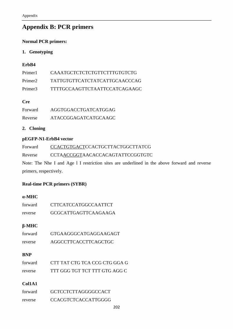
Appendix
202
Appendix B: PCR primers
Normal PCR primers:
1. Genotyping
ErbB4
Primer1 CAAATGCTCTCTCTGTTCTTTGTGTCTG
Primer2 TATTGTGTTCATCTATCATTGCAACCCAG
Primer3 TTTTGCCAAGTTCTAATTCCATCAGAAGC
Cre
Forward AGGTGGACCTGATCATGGAG
Reverse ATACCGGAGATCATGCAAGC
2. Cloning
pEGFP-N1-ErbB4 vector
Forward CCACTGTGACTCCACTGCTTACTGGCTTATCG
Reverse CCTAACCGGTAACACCACAGTATTCCGGTGTC
Note: The Nhe I and Age l I restriction sites are underlined in the above forward and reverse
primers, respectively.
Real-time PCR primers (SYBR)
α-MHC
forward CTTCATCCATGGCCAATTCT
reverse GCGCATTGAGTTCAAGAAGA
β-MHC
forward GTGAAGGGCATGAGGAAGAGT
reverse AGGCCTTCACCTTCAGCTGC
BNP
forward CTT TAT CTG TCA CCG CTG GGA G
reverse TTT GGG TGT TCT TTT GTG AGG C
Col1A1
forward GCTCCTCTTAGGGGCCACT
reverse CCACGTCTCACCATTGGGG

Appendix
203
Col3A1
forward CACCCTTCTTCATCCCACTC
reverse TGGTTCTGGCTTCCAGACAT
PAL
forward TTCAGCCCTTGCTTGCCTC
reverse ACACTTTTACTCCGAAGTCGGT
ErbB1
Forward CAGATGGATGTCAACCCTGAAG
Reverse TGGAGAGTGTGTCTTTAAATTCACC
ErbB2
Forward GCAAGCACTGTCTGCCATGC
Reverse GGGCACAAGCCTCACACTGG
ErbB3
Forward CGAGATGGGCAACTCTCAGGC
Reverse AGGTTACCCATGACCACCTCACAC
HB-EGF
Forward CGGGGAGTGCAGATACCTG
Reverse TTCTCCACTGGTAGAGTCAGC
TGFα
Forward GCCCAGATTCCCACACTCAG
Reverse CACGGCACCACTCACAGTG
EREG
Forward CTGCCTCTTGGGTCTTGACG
Reverse GCGGTACAGTTATCCTCGGATTC
BTC
Forward AATTCTCCACTGTGTGGTAGCA
Reverse GGTTTTCACTTTCTGTCTAGGGG
NRG3
Forward CCAGCCTATCAAGCACCACA
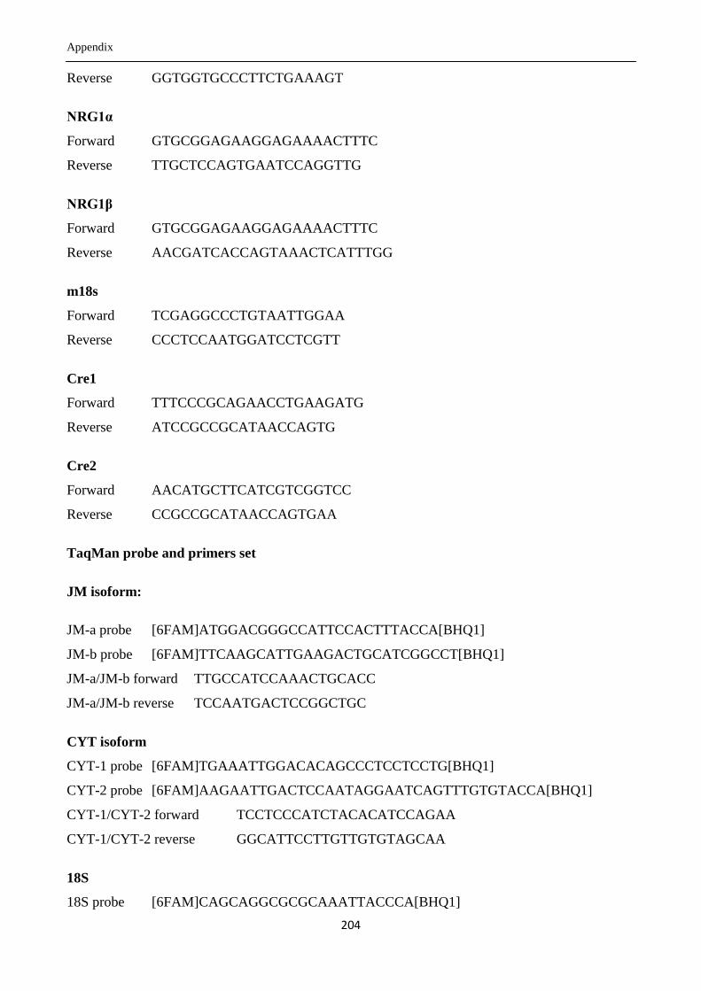
Appendix
204
Reverse GGTGGTGCCCTTCTGAAAGT
NRG1α
Forward GTGCGGAGAAGGAGAAAACTTTC
Reverse TTGCTCCAGTGAATCCAGGTTG
NRG1β
Forward GTGCGGAGAAGGAGAAAACTTTC
Reverse AACGATCACCAGTAAACTCATTTGG
m18s
Forward TCGAGGCCCTGTAATTGGAA
Reverse CCCTCCAATGGATCCTCGTT
Cre1
Forward TTTCCCGCAGAACCTGAAGATG
Reverse ATCCGCCGCATAACCAGTG
Cre2
Forward AACATGCTTCATCGTCGGTCC
Reverse CCGCCGCATAACCAGTGAA
TaqMan probe and primers set
JM isoform:
JM-a probe [6FAM]ATGGACGGGCCATTCCACTTTACCA[BHQ1]
JM-b probe [6FAM]TTCAAGCATTGAAGACTGCATCGGCCT[BHQ1]
JM-a/JM-b forward TTGCCATCCAAACTGCACC
JM-a/JM-b reverse TCCAATGACTCCGGCTGC
CYT isoform
CYT-1 probe [6FAM]TGAAATTGGACACAGCCCTCCTCCTG[BHQ1]
CYT-2 probe [6FAM]AAGAATTGACTCCAATAGGAATCAGTTTGTGTACCA[BHQ1]
CYT-1/CYT-2 forward TCCTCCCATCTACACATCCAGAA
CYT-1/CYT-2 reverse GGCATTCCTTGTTGTGTAGCAA
18S
18S probe [6FAM]CAGCAGGCGCGCAAATTACCCA[BHQ1]

Appendix
205
18S forward CGGCTACCACATCCAAGGAA
18S reverse GGGCCTCGAAAGAGTCCTGT
The TaqMan probe and primer set used for amplifying following genes are predesigned and
premixed by company:
NRG2: Mm01158087_m1 (Applied Biosystem)
NRG4: Mm00446254_m1 (Applied Biosystem)
AGEG: Mm00437583_m1 (Applied Biosystem)
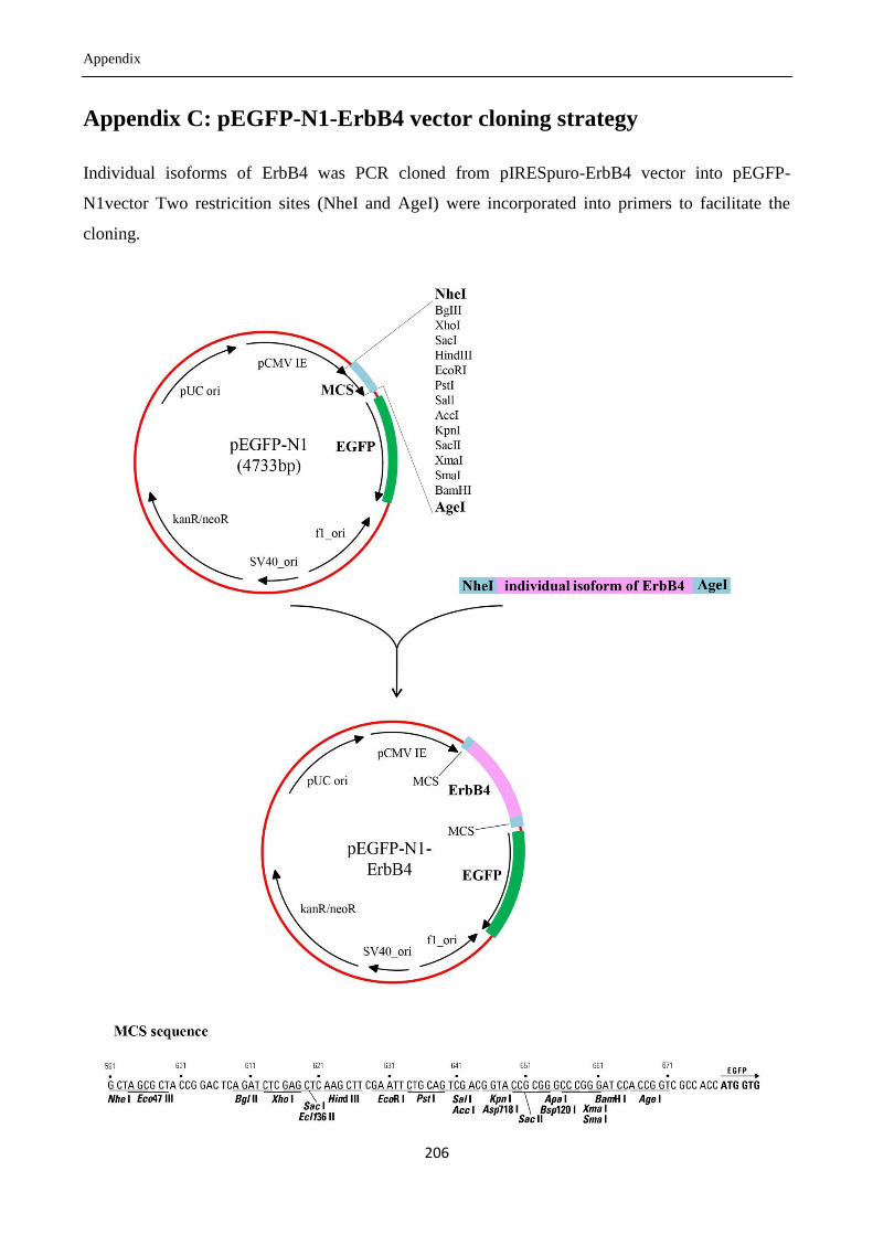
Appendix
206
Appendix C: pEGFP-N1-ErbB4 vector cloning strategy
Individual isoforms of ErbB4 was PCR cloned from pIRESpuro-ErbB4 vector into pEGFP-
N1vector Two restricition sites (NheI and AgeI) were incorporated into primers to facilitate the
cloning.

Appendix
207
Appendix D: pCR®-TOPO-ErbB4-JM/CYT vector cloning
strategy
JM or CYT domains of ErbB4 were PCR cloned from mouse cDNA into pCR®-TOPO vector
using the overhanging T-A sites.